
Consistent Characterization of Color Degradation Due to Artificial Aging Procedures at Popular Pigments of Byzantine Iconography


Abstract
:1. Introduction
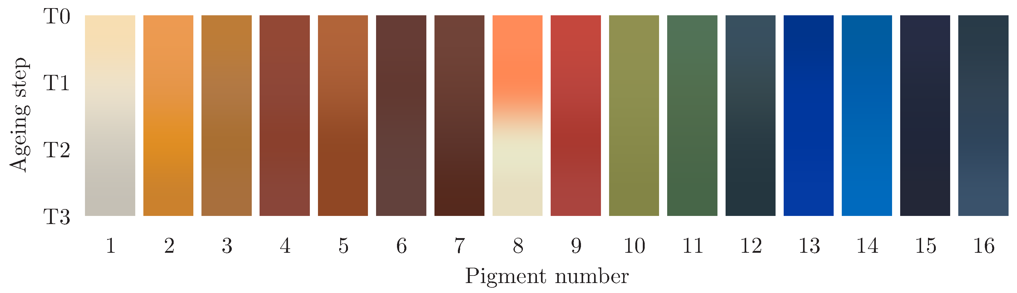
1.1. Examined Historical Pigments
- Massicot, a type of yellow lead monoxide (PbO) that, according to ancient sources, such as Pliny, is known since antiquity. It is produced by slight firing lead white, which eliminates CO and water, leaving behind a soft yellow powder with a sulfur color. Generally, it is not affected by light but, with prolonged exposure to moisture, can be reversed in white lead.
- The warm yellow of Naples is the oldest artificially produced pigment, used from ancient times. Today, yellow of Naples is known as lead antimoniate, and it is produced artificially by the firing of lead oxide with antimony oxide or the salts of the two metals. The color ranges from light yellow to orange-yellow, depending on the ratio of the two components and the temperature of the preparation.
- 3.
- The yellow ochre color owes its color to the presence of hydrated iron oxide, mainly in the goethite mineral. Apart from iron oxides, it may also contain impurities of plaster and magnesium carbonate.
- 4.
- On the other hand, red ochre consists of iron oxide in a substantially anhydrous form with aluminosilicate contaminants. The complexion varies according to the degree of hydration and the origin of minerals. Similar to every iron oxide, the red pigment is very stable and insensitive to light. Generally, the pigment tends to dry quickly.
- 5.
- Finally, warm ochre is a variant of yellow ochre with a warmer hue.
- 6.
- The mineral hematite is quite hard and compact and is almost composed of pure anhydrous iron oxide FeO. The pigment is characterized by a rich dark color with purple-red hue. Microscopically, the granules differ from other ochres as they present a branched form of bright elongated alder.
- 7.
- The raw sienna is a special form of yellow ochre that got its name from the Tuscan region where the particularly beautiful shade of its color comes from. The rich warm maroon color of burnt Sienna is a result of heating under oxidizing conditions (calcining) the raw Sienna. By calcination, the hydrated iron oxide is converted to anhydrous iron oxide.
- 8.
- Although the mineral minium occurs quite frequently in nature, its use as a pigment in antiquity is questioned. It was often found as an illumination on cinnabar or as a thin decoration to create a glowing grid that resembles lace. Chemically, minium is a lead tetroxide (PbO). According to some ancient recipes, the pigment is produced when metallic lead or its minerals are heated. Pliny calls it “the color of the flame” and, indeed, the bright intense red color of minium is mostly characterized as orange rather than red.
- 9.
- The cinnabar is a compact heavy red mineral (HgS), which is the main mineral of mercury. The pigment is characterized by its rich cherry color, often located in the folds of clothing and dress of the Virgin as well as at the pink color of flesh. The cinnabar is considered sufficiently stable to light but sometimes tends to blacken, especially in egg-tempera.
- 10.
- The term green earth is applied to the earthy grey-green aluminosilicate minerals found in abundance all over the planet, and it is mainly composed of two related minerals: glauconite and celadonite. The color of the green earth minerals varies from grey-green to oily brown. Green earths are often described as the most permanent of all pigments, as they are not affected by atmospheric conditions or sunlight and as they do not react with solvent or other pigments.
- 11.
- The malachite mineral results from weathering copper ores, and it is found on the upper levels of copper ore deposits. In nature, malachite is associated with the rarer azurite, containing less chemically bound water. A particular feature of the malachite is that the performance of the color depends on the size of the particles. The richest intense green color is obtained from the coarse grains, while the fine species produces a pale green color.
- 12.
- The blue basic copper carbonate, azurite, is a natural pigment much more popular than the green counterpart, malachite. It is considered the most important blue pigment in European painting throughout the Middle Ages. Similar to malachite, its color depends on the grain size and it is, generally, unaffected by light. However, it can be darkened when heated or degrade to a green tint, while the pigment gradually losses its color over time, according to the literature.
- 13.
- No other dye was held in the highest regard in the Middle Ages other than the beautiful blue ultramarine. This rare and precious dye came from the natural mineral lapis lazuli, which was mainly, if not exclusively, found in the quarries of Badakhshan Province in Afghanistan. Ultramarine composition, essentially, includes Sodium (Na), aluminium (Al), sulfur (S), and silicon dioxide (SiO). In general, the strong and bright blue color is preserved when ultramarine is used with egg-tempera as a binder.
- 14.
- The cobalt blue in its pure form is a modern synthetic pigment discovered in 1802. It stands out in the blue color palette due to its strong cyan complexion and brightness compared to other blue pigments that tend to be darker and approach black when used in heavy layers, while it is stable in light.
- 15.
- Prussian blue is the first modern pigment that was introduced early in the 18th century. It is characterized by its strong coloring power using small concentrations of the pigment with any kind of binder. Characterized by the dark blue color with a slightly greenish glint, it tends to resemble black. The pigment has been described as both stable and unstable due to the variety of additives, methods of preparation, and choice of binder.
- 16.
- Another very important blue pigment used in the Middle Ages is the natural organic pigment indigo prepared from herbs of the Papilionaceae (Indigofera tinctoria L.) family, native to India. In Byzantine art, a special combination included an underpainting of indigo with thin coats of azurite as a lazure. As a pigment, it is sufficiently stable in light and air, and insoluble in water, alcohol, and ether but tends to discolor when exposed to ozone and atmospheric nitrogen dioxide.
2. Materials and Methods
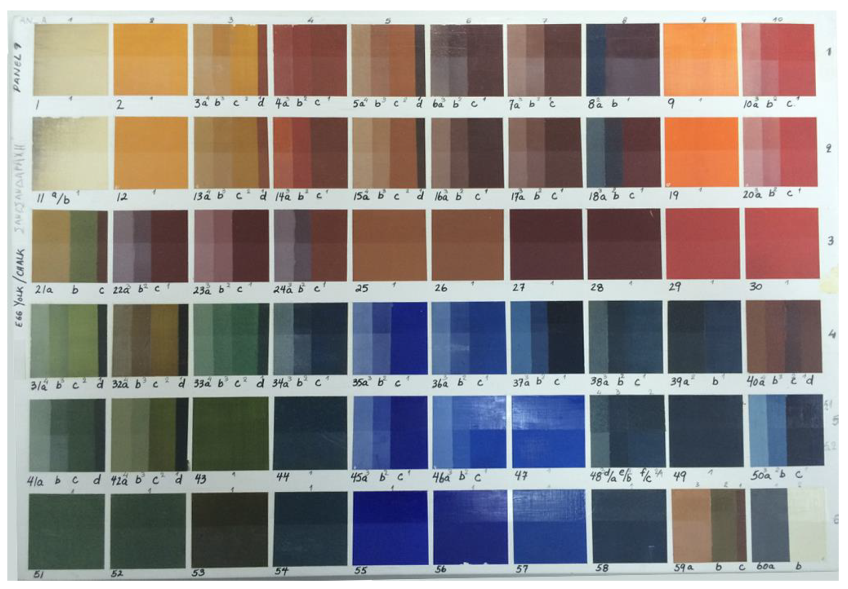
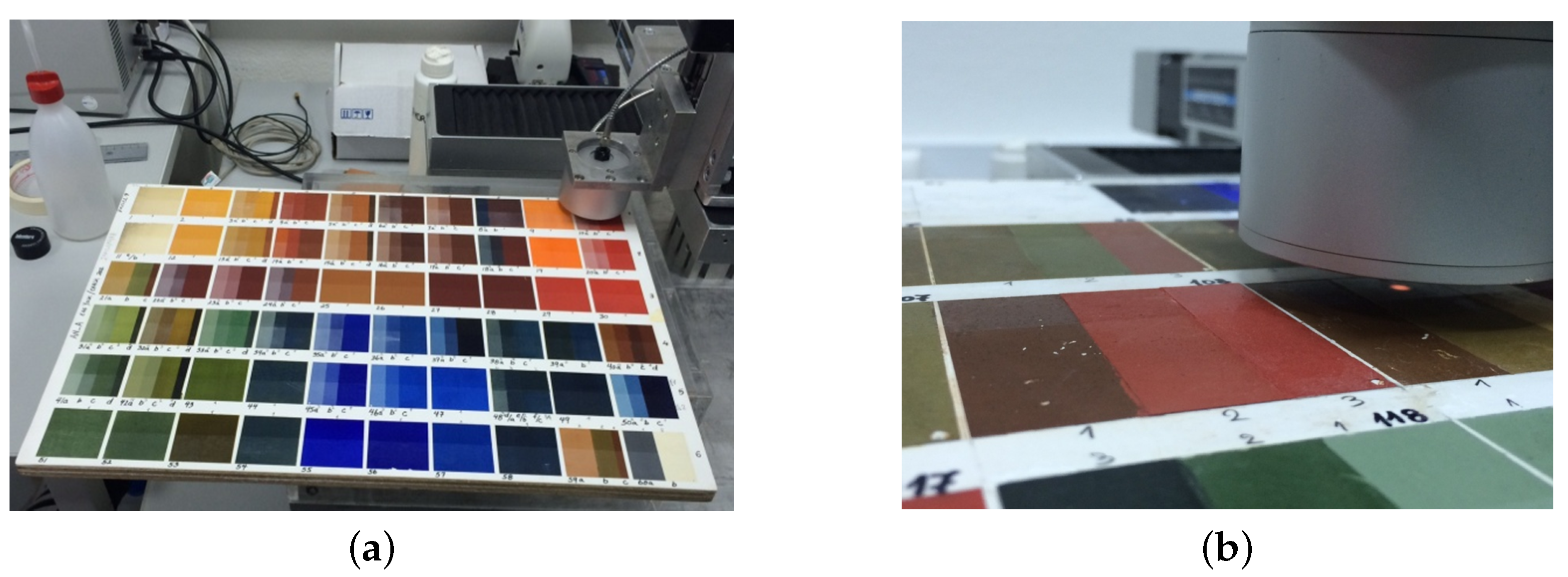
2.1. Multi-Material Palette Details
2.2. Artificial Aging Procedure
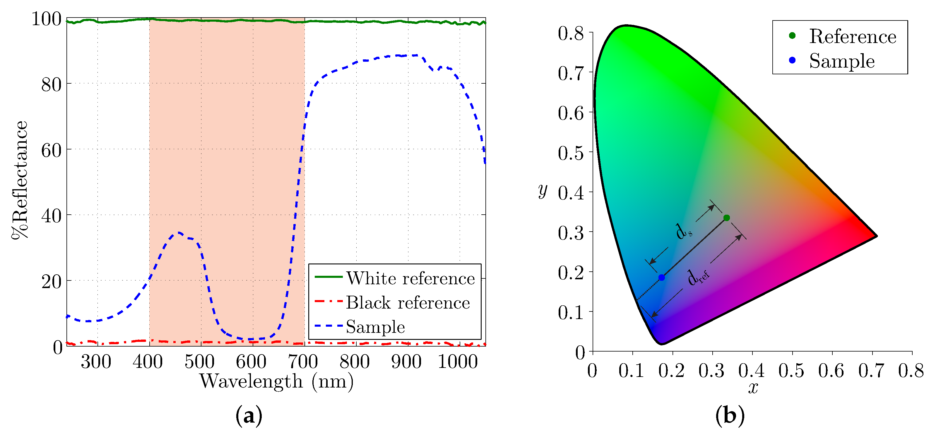
2.3. Measurements at the Visible Spectrum and Quantification
2.3.1. Lightness Evaluation from Visible Spectrum
2.3.2. Saturation Calculation via Tristimulus Values
2.3.3. Conventional CIELAB Color Space Representation
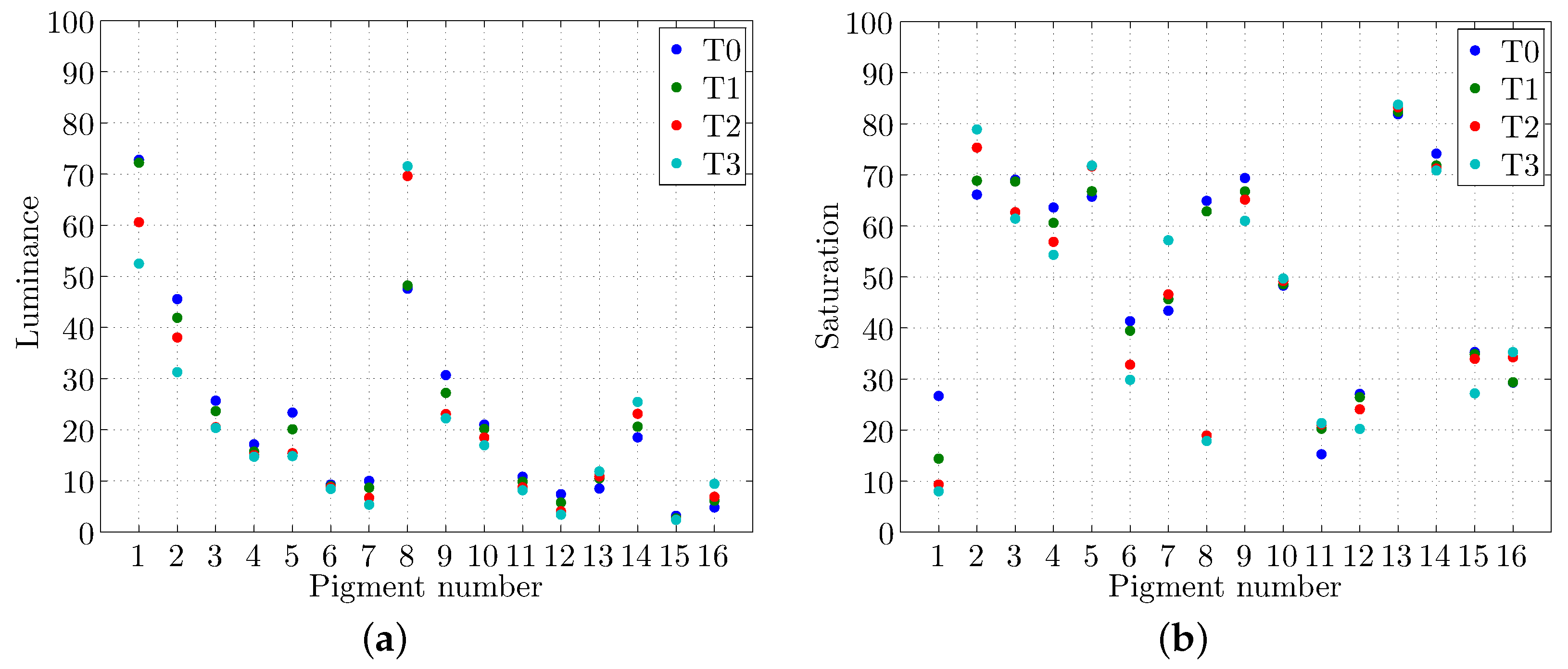
3. Results and Discussion
- the overall color stability,
- whether the color degradation is observed at early exposure instances or later, and
- the type of color change in terms of lightness and saturation.
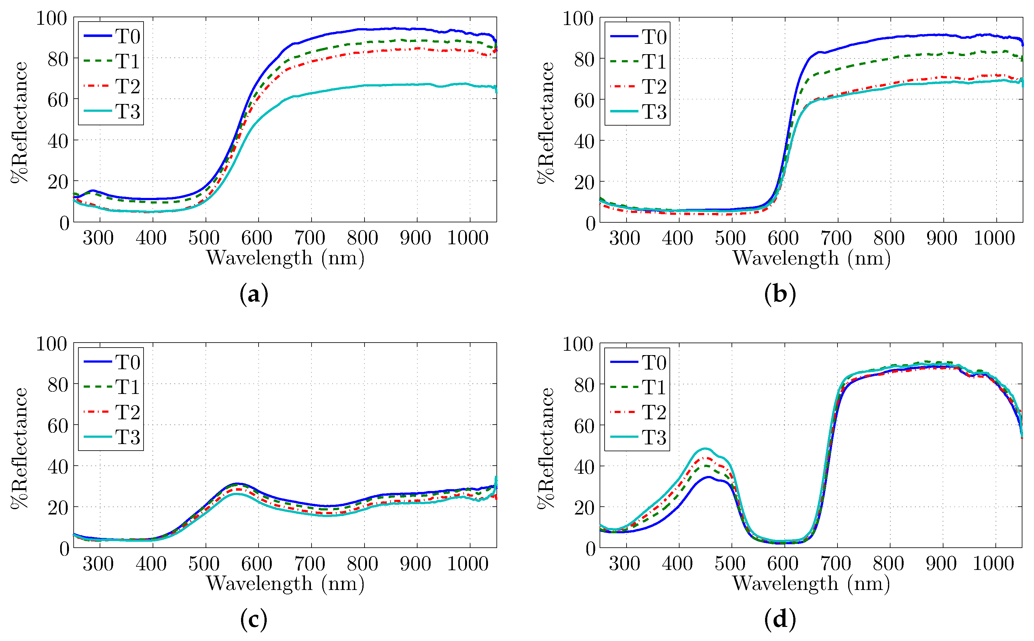
- Massicot is one of the two pigments showing a severe degradation in Figure 5, and it is confirmed by the quantification diagrams. Specifically, the lightness is almost not affected after the first aging step but a significant reduction is observed at the second and third ones. Although the saturation is decreased considerably at every step, the rate is limited until T3 instance. These results are validated by the color difference investigation since the alteration is severe at T1, but it tends to a smoother behavior as the aging progresses.
- The lightness of Naples yellow is fairly reduced especially at the last aging step, while the saturation is always at high levels and it is increasing at every step with a noticeable change at T2. The color difference proves that, at the first aging step, there is a slight alteration, while a significant difference is observed at T2, due to saturation change, and at T3, due to lightness variation.
- Yellow ochre is generally darker than Naples yellow, and its lightness reduction is observed at the first two aging steps since at the third the alteration is only slight. The saturation is maintained at T1, but it is significantly decreased as the aging progresses, especially at T2. The color difference is observable at every time-step but mainly in the first aging step.
- The color stability of red ochre is remarkable regarding its lightness since a slight reduction is observed that tends to stabilize at the last aging steps. Nevertheless, the reduction of saturation is clear almost equally for every time-step. The color difference proves that the red ochre is quite stable since the alteration is noticeable mainly at T3.
- Another pigment that darkens is warm ochre since its already low lightness value is further decreased as the aging progress, especially at T2. On the contrary, its saturation is maintained in relatively high values and it is increased, mainly after the second step, while the color seems stabilized at T3. This observation is validated through the color difference since a noticeable alteration is observed at T2 in contrast to the insignificant one at T3.
- Hematite is a pigment of very low lightness that is almost not affected by aging since its overall alteration is the lowest regarding the investigated pigments. However, the saturation presents a decreasing tendency, while this effect is severe mainly after the second time step; thus, a more grayish tone is observed. This moderate alteration is observed in color difference, but the effect is more obvious in T1 step.
- The lightness of burnt Sienna is very low at the initial condition, and it is slightly dropped at every aging step. A completely different behavior is noted for the saturation that is fairly rising at T1; it seems unaffected at T2, and a significant increment is observed at T3. The color difference throughout aging procedure indicates that the alteration is augmented as the exposure is continued.
- The other pigment that presents severe degradation is minium. Specifically, although the first time-step seems to influence the pigment’s color only slightly, a complete differentiation occurs on the second step. The initial moderate lightness is significantly increased at this step, while the relatively high saturation degrades to low values, therefore, tending to white. At the final step, the color is retained both in lightness and saturation. This behavior is confirmed by color difference since, at the second aging step, the most significant color transformation is noted.
- The intense color of cinnabar is degraded via aging since both lightness and saturation are decreased. Specifically, the lightness is influenced mainly at the first aging steps, while saturation drops significantly at T3. Consequently, a darker and less intense red is observed after the final aging procedure. This color alteration is observed in color difference values, since there is a change at every time-step, especially at T3 due to the saturation reduction.
- It is already stated that green earth is considered the most stable of pigments, and this is clarified via its saturation observation that is almost equivalent for any of the time-steps. Moreover, the lightness is reduced only by a slight factor, particularly after the second aging step. The color stability of green earth is clarified also via color difference values since they are less than the observable limit () even though the alteration is slightly increased after T2 due to lightness reduction.
- Malachite is another very stable pigment in terms of its color since the lightness is nearly unaffected by aging. Specifically, malachite is less luminous by a negligible factor, especially at T2. However, saturation is somewhat increased after the first time-step but is retained as the aging procedure continues. Color difference validates our results considering that the alteration is observable only at T1.
- The counterpart to malachite is azurite that is generally darker, and its lightness is further decreased at the first aging steps while it is almost unaffected at the final step. Additionally, the saturation is stable at T1 but it is reduced as the aging progresses in contrast to malachite. The color difference indicates that the alteration is more evident at the initial stages of aging.
- The ultramarine blue is a really intense color, as confirmed by the very high saturation values that are influenced only slightly by aging since a negligible increment is observed. Moreover, it is relatively dark; it gets somewhat brighter at T1, while its lightness is further increased at T2 and T3 by a very small factor. The color stability of ultramarine blue is verified by its color difference since the only observable change is occurred at T1.
- An additional pigment of intense color is cobalt blue, presenting high saturation values of a fair reduction after the first aging step and insignificant counterparts for the remaining steps. However, the pigment becomes even brighter since its lightness is increased to moderate values at almost equivalent increments during aging. The color difference indicates that the aforementioned saturation change results in a moderate color alteration at T1, while the differentiation is decreased as aging progresses and the pigment tends to stabilize in terms of its color.
- The darkest pigment of the panel, namely Prussian blue, presents the lowest lightness that it is affected negligibly by the aging process and particularly at first two aging steps. At these stages, the saturation is almost retained, while a considerable reduction is observed at the last time-step leading the pigment color to black. Finally, the color difference values indicate a relatively stable pigment that its alteration is augmented through time.
- Indigo is an additional dark pigment but the aging procedure increases both its lightness and saturation; thus, a brighter color is perceived. Specifically, the lightness changes mainly at T1 and T3 steps, while a significant increment in saturation takes place in T2 and this value is further increased towards T3 via a small factor. The color difference clarifies the previous results since the alteration is always observable and it is boosted as the aging progresses.
Comparison to State-of-the-Art Results
4. Conclusions
Author Contributions
Funding
Institutional Review Board Statement
Informed Consent Statement
Data Availability Statement
Conflicts of Interest
References
- Sotiropoulou, S.; Karagiannis, G.; Chryssoulakis, Y. Colour correction and storage of digitized image captures representing art paintings. In Proceedings of the 1st International Conference on Color in Graphics and Image Processing, Saint-Etienne, France, 1–4 October 2000. [Google Scholar]
- Creagh, D. The characterization of artefacts of cultural heritage significance using physical techniques. Radiat. Phys. Chem. 2005, 74, 426–442. [Google Scholar] [CrossRef]
- Polikreti, K.; Othonos, A.; Christofides, C. Optical characterization of varnish films by spectroscopic ellipsometry for application in artwork conservation. Appl. Spectrosc. 2005, 59, 94–99. [Google Scholar] [CrossRef]
- Whitney, A.V.; Casadio, F.; Van Duyne, R.P. Identification and characterization of artists’ red dyes and their mixtures by surface-enhanced Raman spectroscopy. Appl. Spectrosc. 2007, 61, 994–1000. [Google Scholar] [CrossRef]
- Karagiannis, G.; Vavliakis, K.; Sotiropoulou, S.; Damtsios, A.; Alexiadis, D.; Salpistis, C.; Daniilia, S. Using signal processing and semantic web technologies to analyze byzantine iconography. IEEE Intell. Syst. 2009, 24, 73–81. [Google Scholar] [CrossRef]
- Romani, A.; Clementi, C.; Miliani, C.; Favaro, G. Fluorescence spectroscopy: A powerful technique for the noninvasive characterization of artwork. Accounts Chem. Res. 2010, 43, 837–846. [Google Scholar] [CrossRef] [PubMed]
- Miliani, C.; Rosi, F.; Brunetti, B.G.; Sgamellotti, A. In situ noninvasive study of artworks: The MOLAB multitechnique approach. Accounts Chem. Res. 2010, 43, 728–738. [Google Scholar] [CrossRef] [PubMed]
- Rarakou, A.; Gkouvas, D.; Karagiannis, G.; Maria, S. Spectroscopic analysis of nano- modified thermal insulated colours on mortar substrates. In Proceedings of the Nanotexnology NN14, Thessaloniki, Greece, 8–11 July 2014. [Google Scholar]
- Cosentino, A. Panoramic, macro and micro multispectral imaging: An affordable system for mapping pigments on artworks. J. Conserv. Mus. Stud. 2015, 13. [Google Scholar] [CrossRef] [Green Version]
- Karagiannis, G.; Alexiadis, D.; Sergiadis, G.; Salpistis, C. UV/VIS/nIR/mIR diffuse reflectance spectra and acoustic microscopy echo graphs for stratigraphy determination, using neural networks and wavelet transform. In Proceedings of the 2008 3rd International Conference on Information and Communication Technologies: From Theory to Applications, Damascus, Syria, 7–11 April 2008; pp. 1–7. [Google Scholar]
- Bisulca, C.; Picollo, M.; Bacci, M.; Kunzelman, D. UV-Vis-NIR reflectance spectroscopy of red lakes in paintings. In Proceedings of the 9th International Conference on NDT of Art, Jerusalem, Israel, 25–30 May 2008; pp. 25–30. [Google Scholar]
- Aceto, M.; Agostino, A.; Fenoglio, G.; Idone, A.; Gulmini, M.; Picollo, M.; Ricciardi, P.; Delaney, J.K. Characterisation of colourants on illuminated manuscripts by portable fibre optic UV-visible-NIR reflectance spectrophotometry. Anal. Methods 2014, 6, 1488–1500. [Google Scholar] [CrossRef]
- Rosi, F.; Grazia, C.; Gabrieli, F.; Romani, A.; Paolantoni, M.; Vivani, R.; Brunetti, B.G.; Colomban, P.; Miliani, C. UV–Vis-NIR and micro Raman spectroscopies for the non destructive identification of Cd1- xZnxS solid solutions in cadmium yellow pigments. Microchem. J. 2016, 124, 856–867. [Google Scholar] [CrossRef]
- Feller, R.L. Some factors to be considered in accelerated-aging tests. In Proceedings of the Preprints of Papers Presented at the Fifteenth Annual Meeting of the American Institute for Conservation of Historic and Artistic Works, Vancouver, BC, Canada, 20–24 May 1987; pp. 56–67. [Google Scholar]
- Kockott, D. Natural and artificial weathering of polymers. Polym. Degrad. Stab. 1989, 25, 181–208. [Google Scholar] [CrossRef]
- Brown, R. Survey of status of test methods for accelerated durability testing. Polym. Test. 1991, 10, 3–30. [Google Scholar] [CrossRef]
- Feller, R. Accelerating Aging—Photochemical and Thermal Aspects; Getty Conservation Institute: Marina del Rey, CA, USA, 1994. [Google Scholar]
- ISO 16474-1:2013. Paints and Varnishes—Methods of Exposure to Laboratory Light Sources—Part 1: General Guidance; Standard; International Organization for Standardization: Geneva, Switzerland, 2013. [Google Scholar]
- ISO 16474-2:2013. Paints and Varnishes—Methods of Exposure to Laboratory Light Sources—Part 2: Xenon-Arc Lamps; Standard; International Organization for Standardization: Geneva, Switzerland, 2013. [Google Scholar]
- Plater, M.J.; De Silva, B.; Gelbrich, T.; Hursthouse, M.B.; Higgitt, C.L.; Saunders, D.R. The characterisation of lead fatty acid soaps in ‘protrusions’ in aged traditional oil paint. Polyhedron 2003, 22, 3171–3179. [Google Scholar] [CrossRef]
- Boon, J.J.; Hoogland, F.; Keune, K.; Parkin, H.M. Chemical processes in aged oil paints affecting metal soap migration and aggregation. In Proceedings of the AIC Paintings Specialty Group Postprints, Providence, RI, USA, 16–19 June 2006; Volume 19, pp. 16–23. [Google Scholar]
- Cotte, M.; Checroun, E.; Susini, J.; Walter, P. Micro-analytical study of interactions between oil and lead compounds in paintings. Appl. Phys. A 2007, 89, 841–848. [Google Scholar] [CrossRef]
- Manzano, E.; García-Atero, J.; Dominguez-Vidal, A.; Ayora-Cañada, M.J.; Capitán-Vallvey, L.F.; Navas, N. Discrimination of aged mixtures of lipidic paint binders by Raman spectroscopy and chemometrics. J. Raman Spectrosc. 2012, 43, 781–786. [Google Scholar] [CrossRef]
- Pallipurath, A.; Skelton, J.; Bucklow, S.; Elliott, S. A chemometric study of ageing in lead-based paints. Talanta 2015, 144, 977–985. [Google Scholar] [CrossRef] [Green Version]
- Howatt-Krahn, A. Technology as Metaform, Cultural Conservation in the Global Environment. Ph.D. Thesis, National Library of Canada Bibliothèque Nationale du Canada, Ottawa, ON, Canada, 1999. [Google Scholar]
- Monico, L.; Janssens, K.; Miliani, C.; Van der Snickt, G.; Brunetti, B.G.; Cestelli Guidi, M.; Radepont, M.; Cotte, M. Degradation process of lead chromate in paintings by Vincent van Gogh studied by means of spectromicroscopic methods. 4. Artificial aging of model samples of co-precipitates of lead chromate and lead sulfate. Anal. Chem. 2013, 85, 860–867. [Google Scholar] [CrossRef] [PubMed]
- Ghelardi, E.; Degano, I.; Colombini, M.P.; Mazurek, J.; Schilling, M.; Khanjian, H.; Learner, T. A multi-analytical study on the photochemical degradation of synthetic organic pigments. Dyes Pigment. 2015, 123, 396–403. [Google Scholar] [CrossRef]
- Ecco, L.; Rossi, S.; Fedel, M.; Deflorian, F. Color variation of electrophoretic styrene-acrylic paints under field and accelerated ultraviolet exposure. Mater. Des. 2017, 116, 554–564. [Google Scholar] [CrossRef]
- Daniilia, S.; Bikiaris, D.; Burgio, L.; Gavala, P.; Clark, R.J.; Chryssoulakis, Y. An extensive non-destructive and micro-spectroscopic study of two post-Byzantine overpainted icons of the 16th century. J. Raman Spectrosc. 2002, 33, 807–814. [Google Scholar] [CrossRef]
- Westlake, P.; Siozos, P.; Philippidis, A.; Apostolaki, C.; Derham, B.; Terlixi, A.; Perdikatsis, V.; Jones, R.; Anglos, D. Studying pigments on painted plaster in Minoan, Roman and Early Byzantine Crete. A multi-analytical technique approach. Anal. Bioanal. Chem. 2012, 402, 1413–1432. [Google Scholar] [CrossRef]
- Rutherford, J.G.; George, L.S. Painting Materials: A Short Encyclopaedia; Dover Publications, Inc.: New York, NY, USA, 1966. [Google Scholar]
- Mayer, R. The Artist’s Handbook of Materials and Techniques; Viking Press: New York, NY, USA, 1991. [Google Scholar]
- FitzHugh, E.W. Artists’ Pigments: A Handbook of Their History and Characteristics; National Gallery of Art: Washington, DC, USA, 1997. [Google Scholar]
- Cennini, C.D. The Craftsman’s Handbook; de Thompson, D.V., Jr., Translator; Dover Publications Inc.: New York, NY, USA, 1960; Volume 2, p. 1. [Google Scholar]
- Kontoglu, P.; Cavarnos, C. Byzantine Sacred Art: Selected Writings of the Contemporary Greek Icon Painter Fotis Kontoglous on the Sacred Arts According to the Tradition of Eastern Orthodox Christianity; Institute for Byzantine and Modern Greek Studies: Belmont, MA, USA, 1985. [Google Scholar]
- Thompson, D.V. The Materials and Techniques of Medieval Painting; Courier Corporation: North Chelmsford, MA, USA, 1956; Volume 327. [Google Scholar]
- Thompson, D.V. The Practice of Tempera Painting; Courier Corporation: North Chelmsford, MA, USA, 1962. [Google Scholar]
- Hawthorne, J.G.; Smith, C.S. Theophilus on Divers Arts: The Foremost Medieval Treatise on Painting, Glassmaking and Metal-Work; Translated from the Latin with introduction and notes; Dover Publications, Inc.: New York, NY, USA, 1979. [Google Scholar]
- Doerner, M. The Materials of the Artist and Their Use in Painting, with Notes on the Techniques of the Old Masters; Houghton Mifflin Harcourt: Boston, MA, USA, 1984. [Google Scholar]
- Perkampus, H.H. UV-VIS Spectroscopy and Its Applications; Springer Science & Business Media: Berlin/Heidelberg, Germany, 2013. [Google Scholar]
- ASTM E903-20. Standard Test Method for Solar Absorptance, Reflectance, and Transmittance of Materials Using Integrating Spheres; Standard; ASTM International: West Conshohocken, PA, USA, 2020. [Google Scholar]
- ISO/CIE 11664-4:2019. Colorimetry—Part 4: CIE 1976 L*a*b* Colour Space; Standard; International Organization for Standardization: Geneva, Switzerland, 2019. [Google Scholar]
- Coccato, A.; Moens, L.; Vandenabeele, P. On the stability of mediaeval inorganic pigments: A literature review of the effect of climate, material selection, biological activity, analysis and conservation treatments. Herit. Sci. 2017, 5, 1–25. [Google Scholar] [CrossRef] [Green Version]
- Elert, K.; Cardell, C. Weathering behavior of cinnabar-based tempera paints upon natural and accelerated aging. Spectrochim. Acta Part A Mol. Biomol. Spectrosc. 2019, 216, 236–248. [Google Scholar] [CrossRef] [PubMed]
- Cardell, C.; Herrera, A.; Guerra, I.; Navas, N.; Simón, L.R.; Elert, K. Pigment-size effect on the physico-chemical behavior of azurite-tempera dosimeters upon natural and accelerated photo aging. Dyes Pigment. 2017, 141, 53–65. [Google Scholar] [CrossRef]
- Sotiropoulou, S.; Sciutto, G.; Tenorio, A.L.; Mazurek, J.; Bonaduce, I.; Prati, S.; Mazzeo, R.; Schilling, M.; Colombini, M.P. Advanced analytical investigation on degradation markers in wall paintings. Microchem. J. 2018, 139, 278–294. [Google Scholar] [CrossRef] [Green Version]
- Bacci, M.; Picollo, M.; Porcinai, S.; Radicati, B. Evaluation of the museum environmental risk by means of tempera-painted dosimeters. Thermochim. Acta 2000, 365, 25–34. [Google Scholar] [CrossRef]
- Szabó, F.; Kéri, R.; Csuti, P. Impact of LED-based lighting on selected historical pigments. In Proceedings of the 2016 IEEE Lighting Conference of the Visegrad Countries (Lumen V4), Karpacz, Poland, 13–16 September 2016; pp. 1–6. [Google Scholar]


| Color | UVC | UVC | UVA | Violet | Blue | Green | Yellow |
|---|---|---|---|---|---|---|---|
| Wavelength (nm) | 184.45 | 253.7 | 365.4 | 404.7 | 435.8 | 546.1 | 578.2 |
| Pigment | Lightness | ||||||
|---|---|---|---|---|---|---|---|
| T0 | T1 | T2 | T3 | ||||
| 1. Massicot | 72.78 | 72.21 | (−0.57) | 60.59 | (−11.62) | 52.49 | (−8.10) |
| 2. Naples yellow | 45.55 | 41.88 | (−3.67) | 38.03 | (−3.85) | 31.26 | (−6.77) |
| 3. Yellow ochre | 25.69 | 23.62 | (−2.07) | 20.61 | (−3.01) | 20.50 | (−0.11) |
| 4. Red ochre | 17.14 | 16.72 | (−1.42) | 15.72 | (−1.00) | 15.01 | (−0.71) |
| 5. Warm ochre | 23.34 | 20.09 | (−3.25) | 15.41 | (−4.68) | 14.84 | (−0.57) |
| 6. Hematite | 9.29 | 8.94 | (−0.35) | 8.74 | (−0.20) | 8.43 | (−0.31) |
| 7. Burnt Sienna | 9.99 | 8.67 | (−1.32) | 6.70 | (−1.97) | 5.34 | (−1.36) |
| 8. Minium | 47.58 | 48.21 | (+0.63) | 69.63 | (+21.42) | 71.51 | (+1.88) |
| 9. Cinnabar | 30.68 | 27.20 | (−3.48) | 23.06 | (−4.14) | 22.24 | (−0.82) |
| 10. Green earth | 20.98 | 20.18 | (−0.80) | 18.47 | (−1.71) | 16.96 | (−1.51) |
| 11. Malachite | 10.79 | 9.84 | (−0.95) | 8.74 | (−1.10) | 8.17 | (−0.57) |
| 12. Azurite | 7.40 | 5.76 | (−1.64) | 4.04 | (−1.72) | 3.38 | (−0.66) |
| 13. Ultramarine blue | 8.50 | 10.47 | (+1.97) | 10.87 | (+0.40) | 11.84 | (+0.97) |
| 14. Cobalt blue | 18.51 | 20.58 | (+2.07) | 23.11 | (+2.53) | 25.44 | (+2.33) |
| 15. Prussian blue | 3.18 | 2.65 | (−0.53) | 2.40 | (−0.25) | 2.38 | (−0.02) |
| 16. Indigo | 4.78 | 6.11 | (+1.33) | 6.88 | (+0.77) | 9.42 | (+2.54) |
| Pigment | Saturation | ||||||
|---|---|---|---|---|---|---|---|
| T0 | T1 | T2 | T3 | ||||
| 1. Massicot | 26.66 | 14.40 | (−12.26) | 9.32 | (−5.08) | 8.00 | (−1.32) |
| 2. Naples yellow | 66.11 | 68.85 | (+2.74) | 75.35 | (+6.50) | 78.91 | (+3.56) |
| 3. Yellow ochre | 69.05 | 68.68 | (−0.37) | 62.67 | (−6.01) | 61.43 | (−1.24) |
| 4. Red ochre | 63.60 | 60.59 | (−3.01) | 56.88 | (−3.71) | 54.33 | (−2.55) |
| 5. Warm ochre | 65.72 | 66.79 | (+1.07) | 71.60 | (+4.81) | 71.66 | (+0.06) |
| 6. Hematite | 41.34 | 39.46 | (−1.88) | 32.82 | (−6.64) | 29.85 | (−2.97) |
| 7. Burnt Sienna | 43.39 | 45.65 | (+2.26) | 46.59 | (+0.94) | 57.20 | (+10.61) |
| 8. Minium | 64.90 | 62.84 | (−2.06) | 18.93 | (−43.91) | 17.85 | (−1.08) |
| 9. Cinnabar | 69.39 | 66.78 | (−2.61) | 65.16 | (−1.62) | 61.00 | (−4.16) |
| 10. Green earth | 48.08 | 48.31 | (+0.23) | 49.22 | (+0.91) | 49.69 | (+0.47) |
| 11. Malachite | 15.26 | 20.27 | (+5.01) | 21.07 | (+0.80) | 21.11 | (+0.04) |
| 12. Azurite | 27.09 | 26.44 | (−0.65) | 24.07 | (−2.37) | 20.23 | (−3.84) |
| 13. Ultramarine blue | 81.88 | 82.38 | (+0.50) | 83.23 | (+0.85) | 83.75 | (+0.52) |
| 14. Cobalt blue | 74.16 | 71.82 | (−2.34) | 71.24 | (−0.58) | 70.85 | (−0.39) |
| 15. Prussian blue | 35.28 | 34.86 | (−0.42) | 33.95 | (−0.91) | 27.20 | (−6.75) |
| 16. Indigo | 29.30 | 29.35 | (+0.05) | 34.25 | (+4.90) | 35.27 | (+1.02) |
| Pigment | CIELAB 1976 | ||||||
|---|---|---|---|---|---|---|---|
| T0 | T1 | T2 | T3 | ||||
| , , | , , | , , | , , | ||||
| 1. Massicot | 90, 4, 24 | 90, 1, 13 | 11.3 | 83, 1, 8 | 8.4 | 78, 1, 6 | 4.9 |
| 2. Naples yellow | 72, 27, 52 | 70, 26, 54 | 2.8 | 68, 27, 64 | 10.8 | 62, 25, 56 | 10.1 |
| 3. Yellow ochre | 59, 22, 47 | 57, 20, 39 | 8.7 | 53, 20, 43 | 5.4 | 53, 20, 38 | 5.2 |
| 4. Red ochre | 42, 31, 28 | 40, 30, 25 | 3.8 | 39, 29, 23 | 2.3 | 38, 31, 28 | 5.8 |
| 5. Warm ochre | 52, 29, 38 | 48, 29, 37 | 3.9 | 41, 30, 36 | 6.9 | 41, 30, 36 | 1.0 |
| 6. Hematite | 32, 15, 10 | 31, 19, 13 | 5.3 | 31, 16, 11 | 3.8 | 30, 19, 14 | 4.3 |
| 7. Burnt Sienna | 34, 20, 16 | 32, 20, 16 | 2.6 | 28, 18, 15 | 4.4 | 24, 20, 18 | 4.9 |
| 8. Minium | 72, 43, 49 | 73, 40, 47 | 3.4 | 89, −1, 16 | 53.3 | 90, −1, 18 | 1.9 |
| 9. Cinnabar | 50, 50, 36 | 48, 48, 33 | 4.2 | 43, 46, 34 | 4.7 | 45, 43, 29 | 6.3 |
| 10. Green earth | 59, −8, 34 | 58, −9, 33 | 1.2 | 56, −9, 33 | 1.9 | 54, −9, 33 | 2.1 |
| 11. Malachite | 44, −19, 11 | 43, −17, 14 | 3.4 | 41, −17, 13 | 2.2 | 40, −18, 13 | 1.5 |
| 12. Azurite | 32, −6, −12 | 29, −7, −7 | 5.9 | 24, −6, −8 | 5.3 | 21, −6, −9 | 2.5 |
| 13. Ultramarine blue | 24, 20, −58 | 26, 22, −60 | 3.4 | 26, 23, −62 | 1.9 | 28, 22, −62 | 1.9 |
| 14. Cobalt blue | 36, −5, −47 | 37, −2, −52 | 6.4 | 40, −3, −51 | 3.4 | 42, −3, −53 | 2.7 |
| 15. Prussian blue | 18, 3, −16 | 17, 2, −14 | 2.7 | 16, 2, −11 | 3.1 | 15, 3, −14 | 3.3 |
| 16. Indigo | 23, −4, −12 | 27, −4, −13 | 3.7 | 28, −3, −17 | 4.0 | 33, −4, −18 | 5.2 |
Publisher’s Note: MDPI stays neutral with regard to jurisdictional claims in published maps and institutional affiliations. |
© 2021 by the authors. Licensee MDPI, Basel, Switzerland. This article is an open access article distributed under the terms and conditions of the Creative Commons Attribution (CC BY) license (https://creativecommons.org/licenses/by/4.0/).
Share and Cite
Amanatiadis, S.; Apostolidis, G.; Karagiannis, G. Consistent Characterization of Color Degradation Due to Artificial Aging Procedures at Popular Pigments of Byzantine Iconography. Minerals 2021, 11, 782. https://doi.org/10.3390/min11070782
Amanatiadis S, Apostolidis G, Karagiannis G. Consistent Characterization of Color Degradation Due to Artificial Aging Procedures at Popular Pigments of Byzantine Iconography. Minerals. 2021; 11(7):782. https://doi.org/10.3390/min11070782
Chicago/Turabian StyleAmanatiadis, Stamatios, Georgios Apostolidis, and Georgios Karagiannis. 2021. "Consistent Characterization of Color Degradation Due to Artificial Aging Procedures at Popular Pigments of Byzantine Iconography" Minerals 11, no. 7: 782. https://doi.org/10.3390/min11070782