
Revealing Artists’ Collaboration in a 14th Century Manuscript by Non-Invasive Analyses

 , , , , ,
Abstract
:1. Introduction
2. Materials and Methods
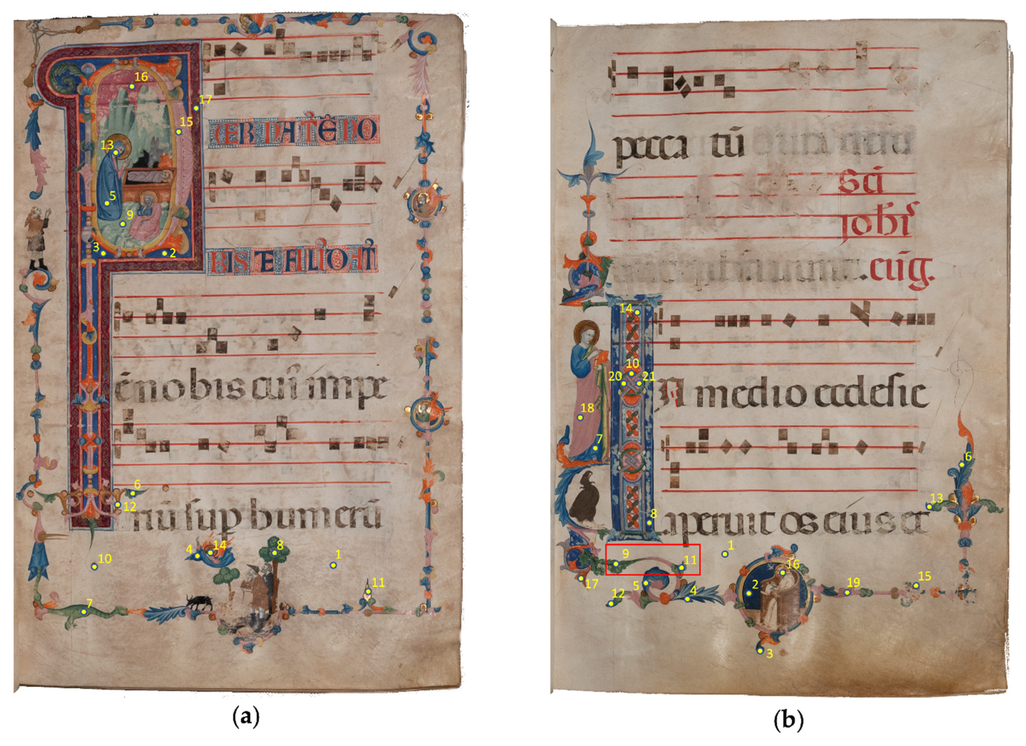
2.1. The Illuminated Manuscript Gradual “D”
2.2. Analytical Techniques
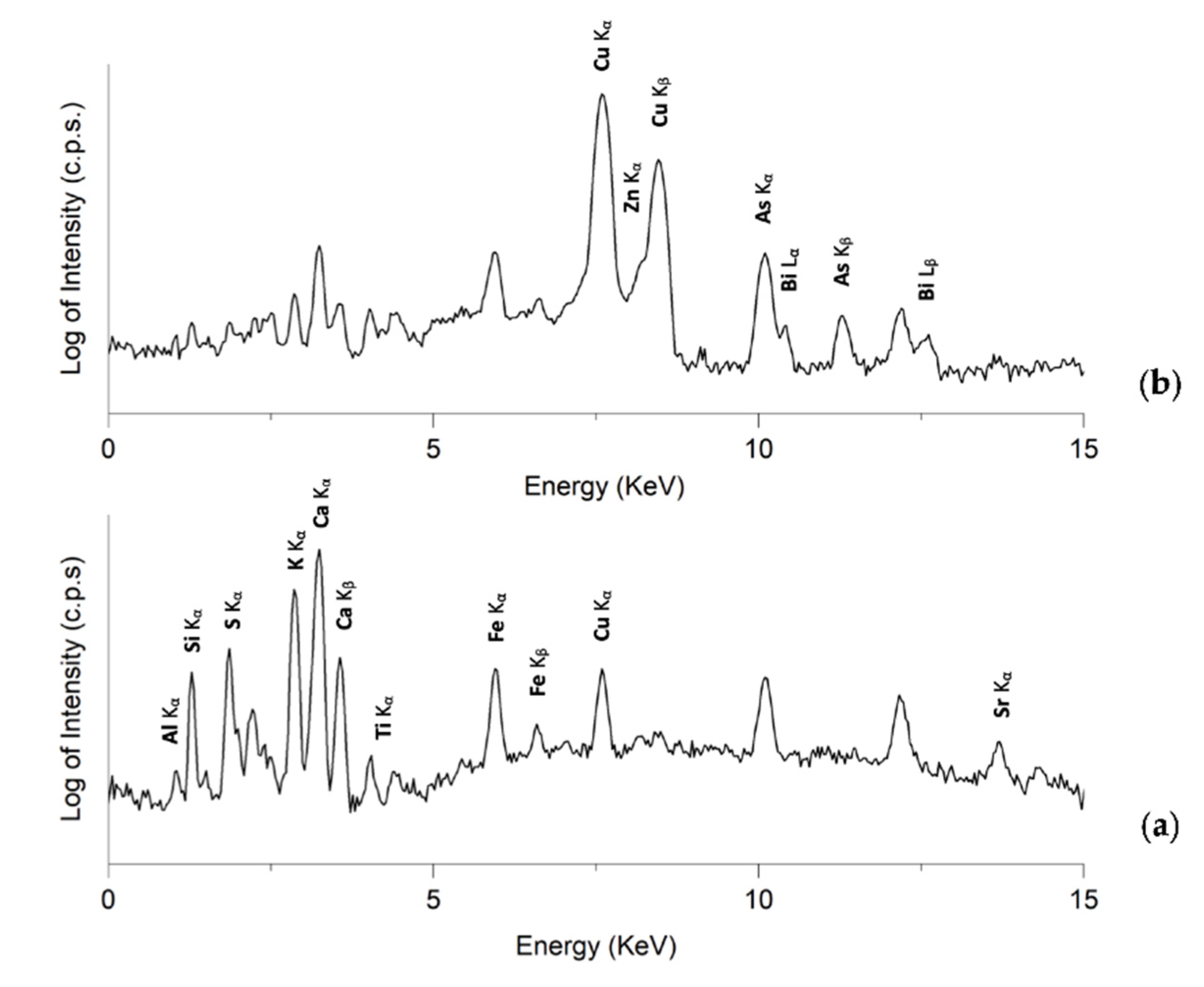
2.2.1. Energy Dispersive X-ray Fluorescence Spectrometry (ED-XRF)
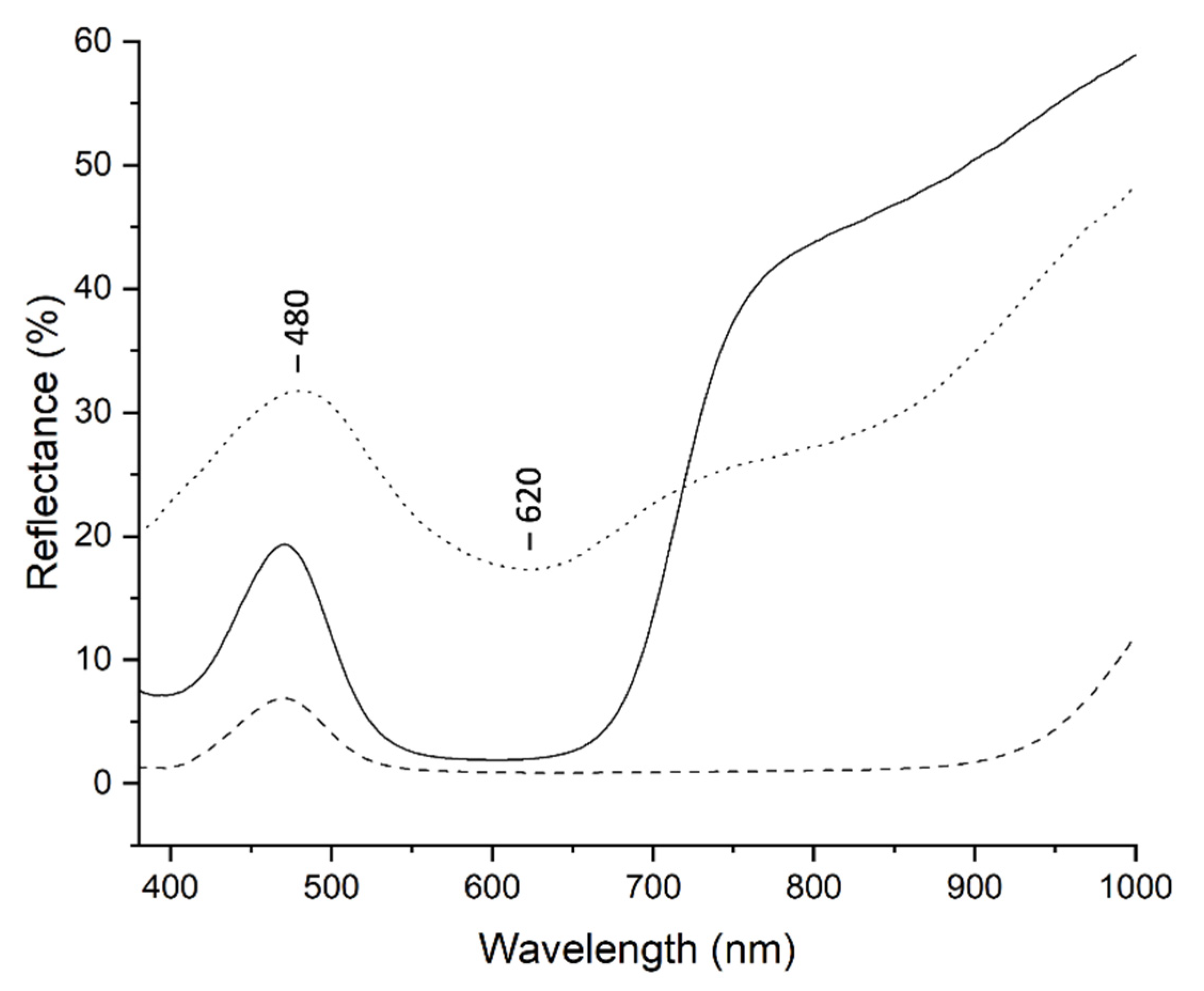
2.2.2. Fiber Optics Reflectance Spectroscopy (FORS)
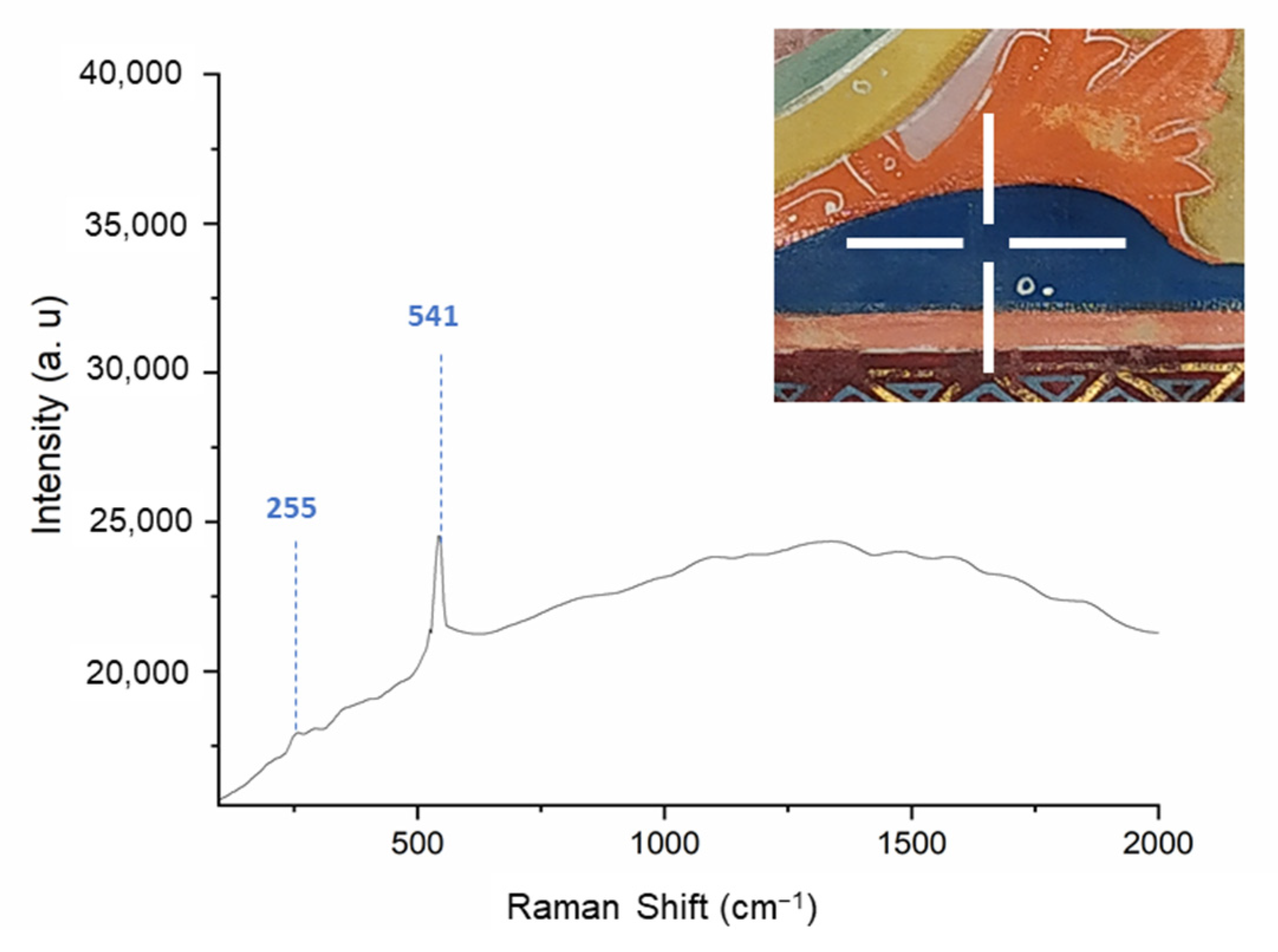
2.2.3. Raman spectroscopy
2.2.4. Microphotography
3. Results
3.1. Parchment
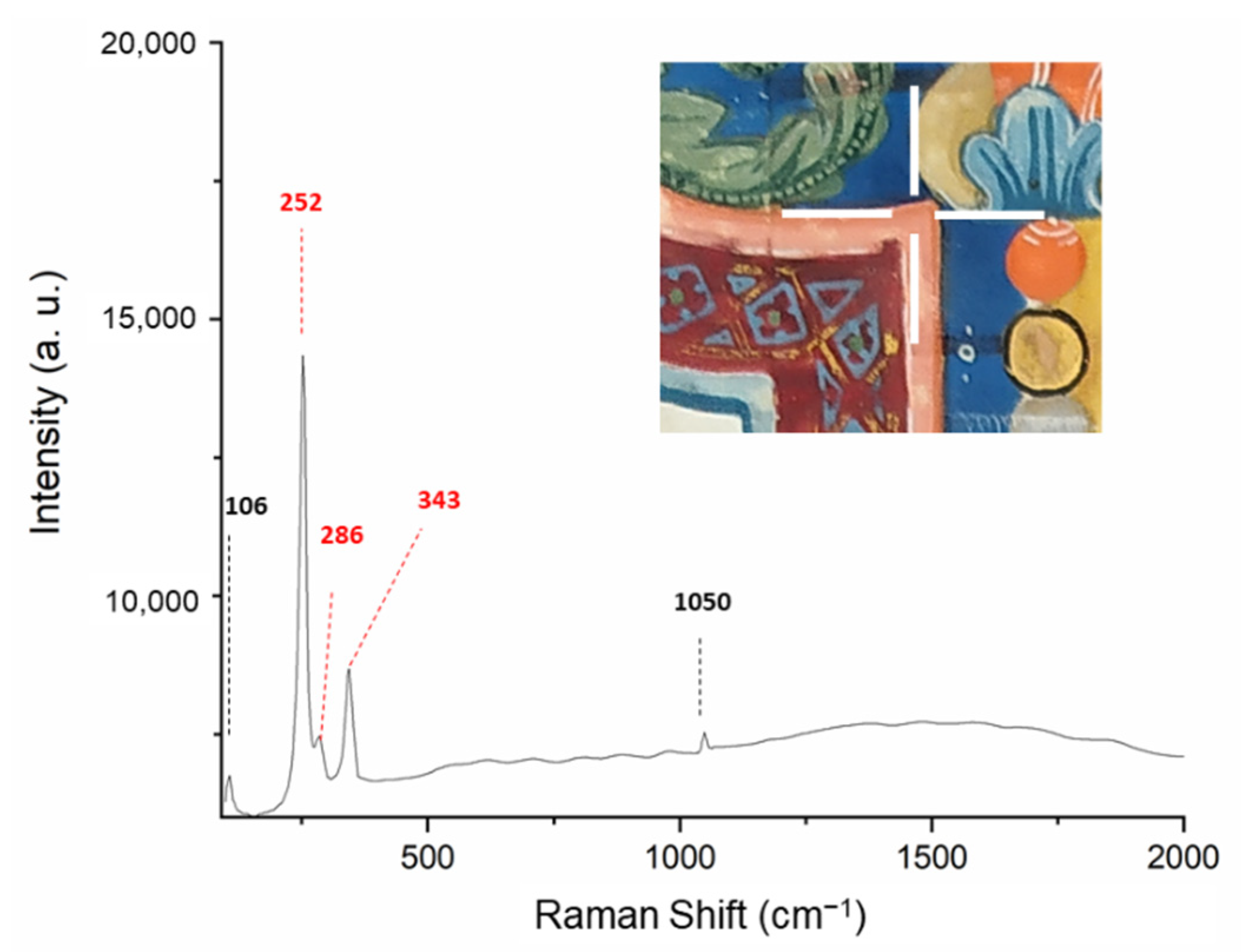
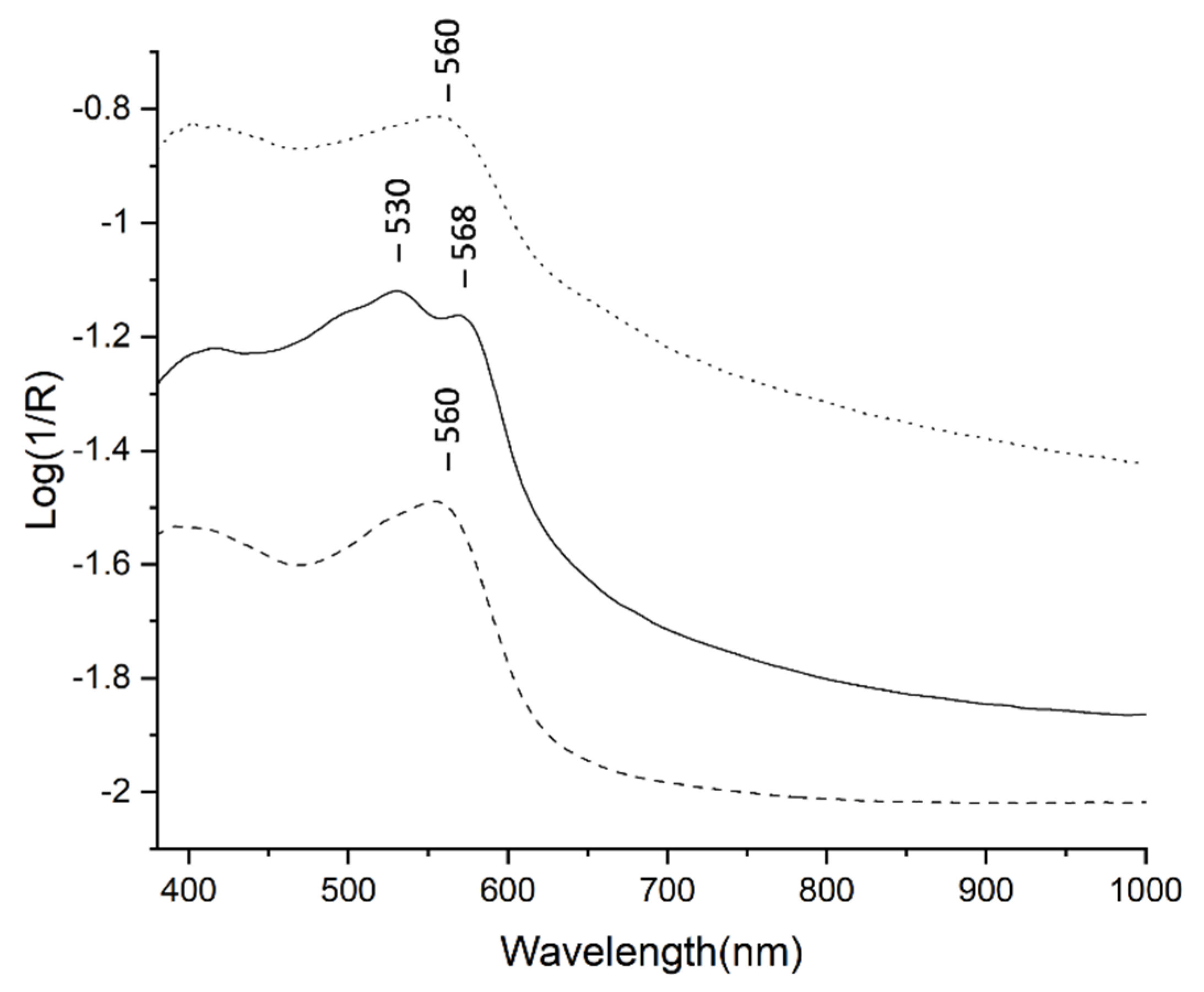
3.2. Ultramarine and Azurite
- ‒
- aluminium (Al), silicon (Si) and sulphur (S) that are expected in lazurite (Na3Ca(Si3Al3)O12S) [14];
- ‒
- potassium (K), likely contained in the ash lye solution of KOH used during the extraction process and/or in several mineral phases that can occur within the lapis lazuli stone [15];
- ‒
- iron (Fe) and calcium (Ca) contained into the minerals pyrite (FeS2) and diopside (CaMgSi2O6), the mostly commonly associated to the lapis lazuli [13].
3.3. Verdigris
3.4. Azurite and Yellow Mixture
3.5. Vergaut
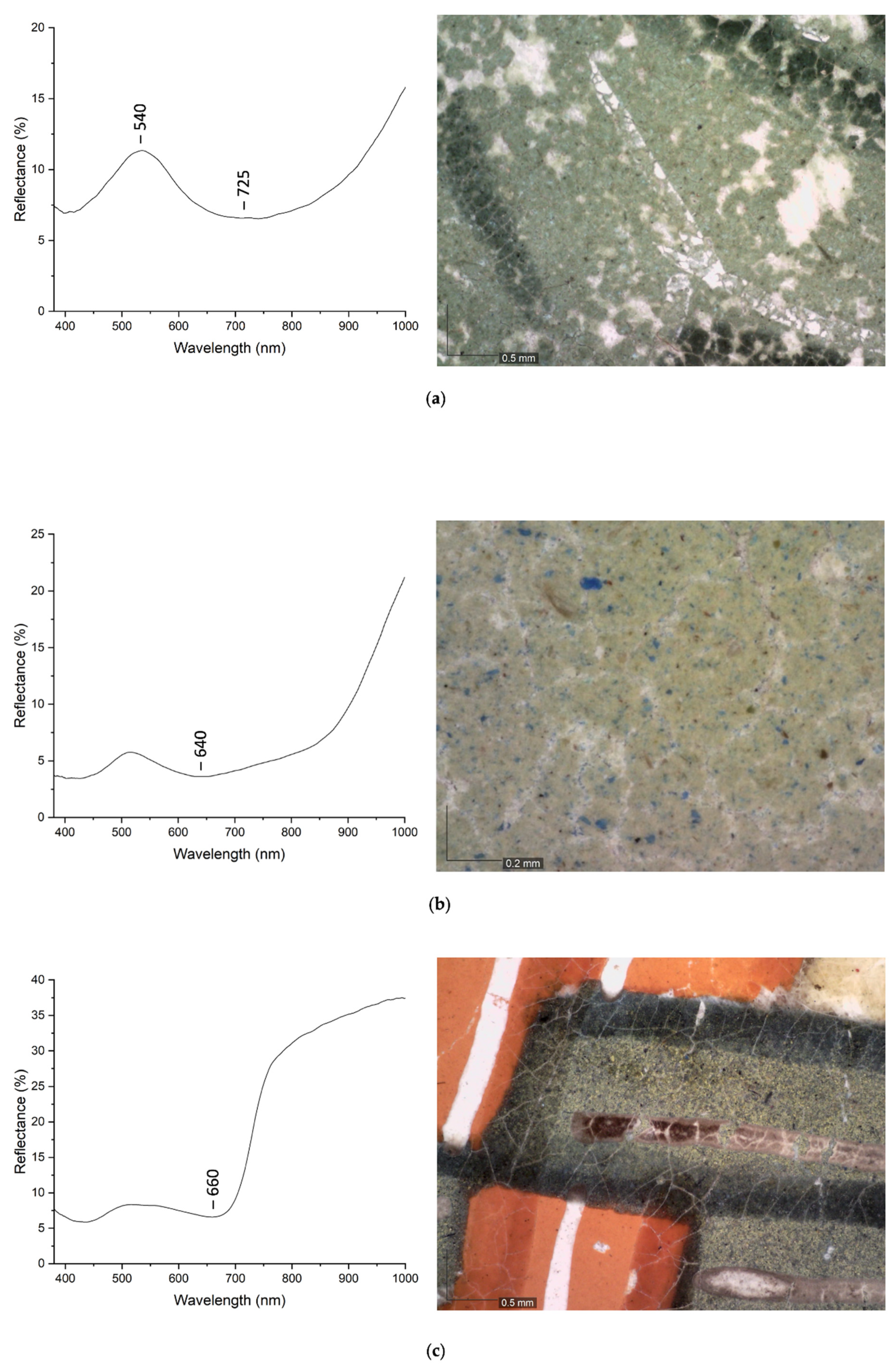
3.6. Earth Pigments
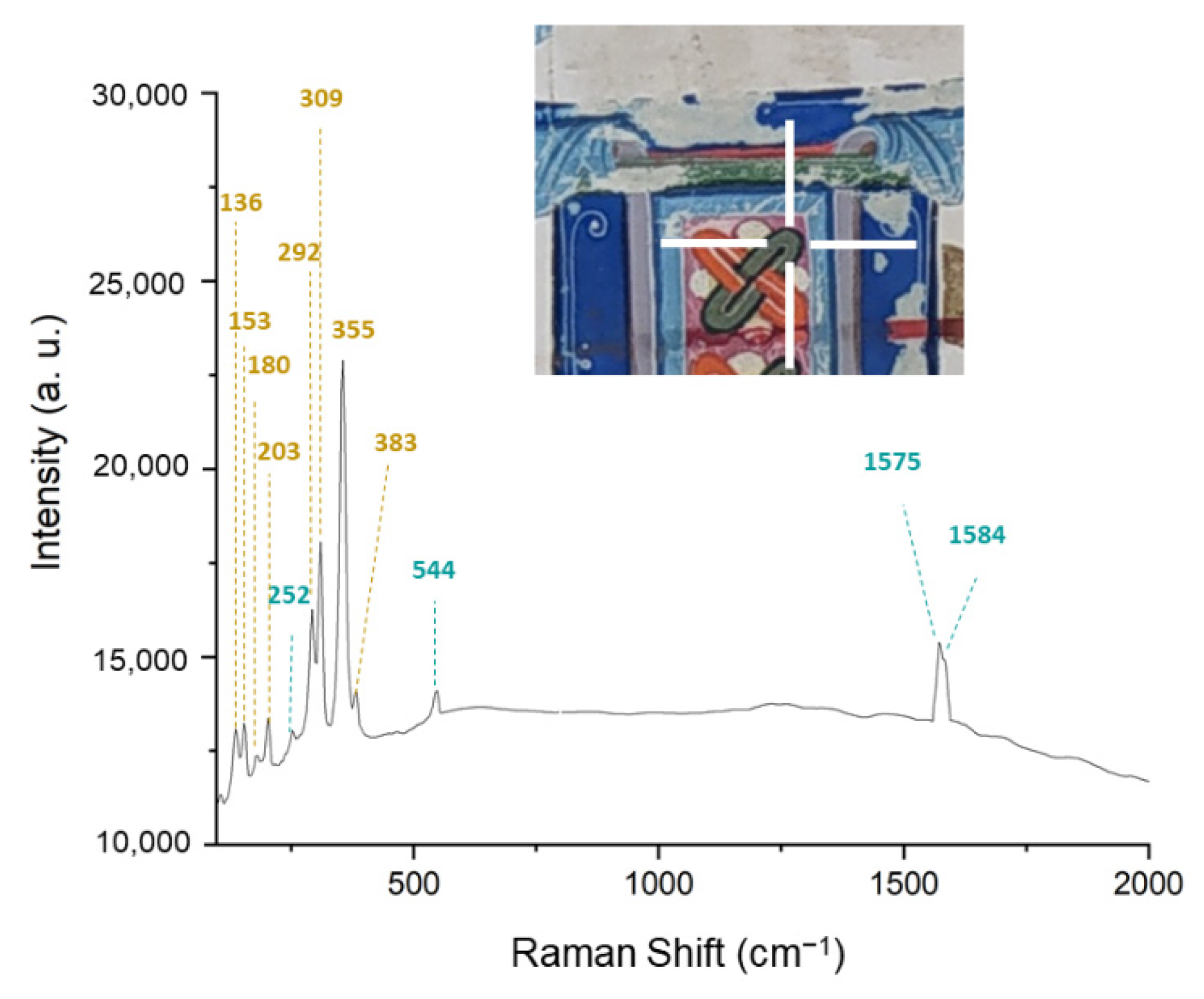
3.7. Vermillion
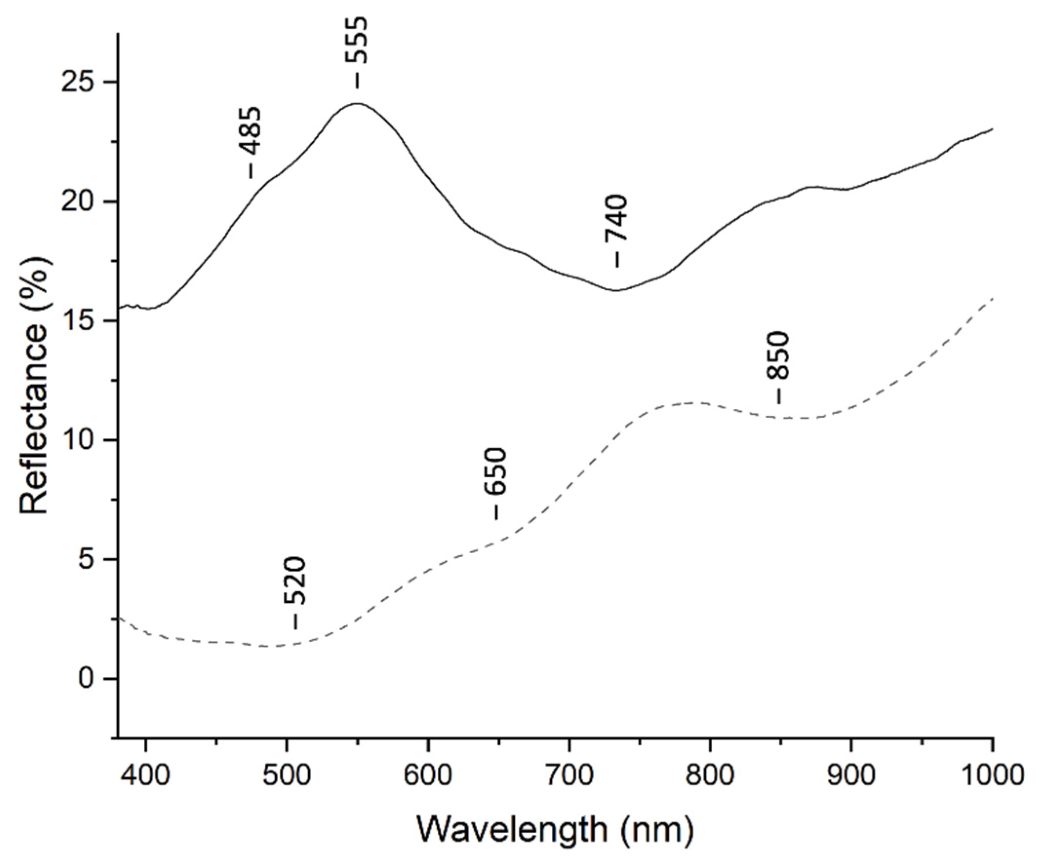
3.8. Organic Pink Pigments
4. Discussion
4.1. Identification of the Illuminators’ Palettes
4.2. The Workshop of Pacino di Bonaguida in Manuscripts
- ‒
- The use of a mixture of yellow orpiment and blue indigo (vergaut) appears both in the illumination of the Ascension from the Laudario and in the “I” of 18r from the Gradual “D”. Paterson et al. [43] attributed the use of the vergaut exclusively to the panel paintings of Pacino, so the vergaut found in the Ascension is considered as an unintentional inclusion, perhaps a contamination from the panel painter’s palette. The “Pacinesque” illuminator of the “I” seems to behave just like the painter Pacino. The vergaut and the blue paint, the latter flaked off perhaps due to the addition of egg glair, could be unusual materials deriving from a panel painter’s palette rather than a manuscript’s one. These findings possibly provide an evidence of the proximity of manuscript and panel painting activities in the work of the illuminator of the “I”.
- ‒
- The use of an insect dye not clearly specified in several leaves from the Laudario could be the same used by the illuminator of the “P”.
- ‒
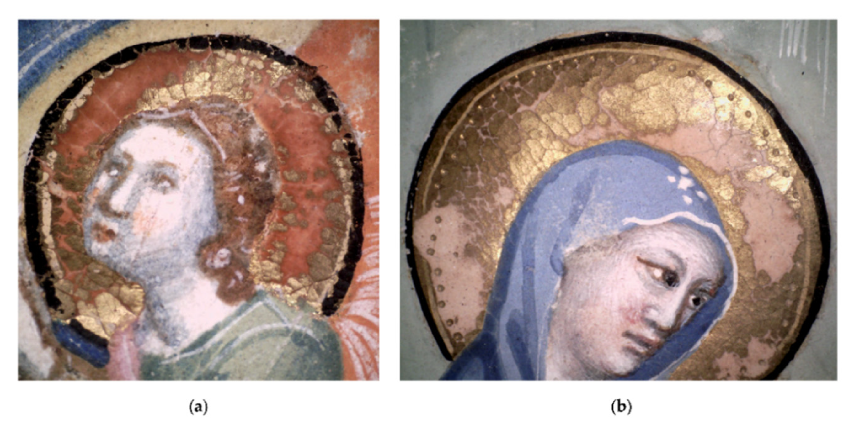
- The dotted halo shown in the image under magnification (Figure 8b) is identifiable as a characteristic of a “Pacinesque” style in the illuminations of Gradual “D” as well as in those from the Laudario.
- ‒
- When comparing the approaches to painting the flesh tones across Pacino’s works on panel paintings and illuminated manuscripts, it was highlighted that works dated or dateable to c. 1300–1320s do not include a green earth ground whereas those dated or datable to c. 1330 onwards do apply a green earth ground [54]. The absence of a ground of green earth in the flesh painting of “P” initial of the Gradual could support the dating of the illumination to c. 1300–1320 s.
5. Conclusions
Supplementary Materials
Author Contributions
Funding
Data Availability Statement
Acknowledgments
Conflicts of Interest
References
- Ricciardi, P. Manuscripts in the Making: Art and Science. Herit. Sci. 2019, 7, 60. [Google Scholar] [CrossRef] [Green Version]
- Panayatova, S. Introduction: The Integrated Analyses of Illuminated Manuscripts. In The Art & Science of Illuminated Manuscripts: A Handbook, 1st ed.; Panayatova, S., Ed.; Harvey Miller: London, UK; Brepols: Turnhout, Belgium, 2020; pp. 11–23. [Google Scholar]
- Bertelli, C. Un corale della Badia a Settimo scritto nel 1315. Paragone 1970, 21, 14–30. [Google Scholar]
- Guidotti, A. Precisazioni sul Maestro daddesco in alcuni codici miniati della Badia a Settimo. In La Miniatura Italiana in età Romanica e Gotica; Waldenburg, G.V.S., Leo, S., Eds.; Olschki: Firenze, Italy, 1979; pp. 419–441. [Google Scholar]
- Cheese, E. From Pelt to Painted Page. Western Medieval Manuscript Parchments. In The Art & Science of Illuminated Manuscripts: A Handbook, 1st ed.; Harvey Miller: London, UK; Brepols: Turnhout, Belgium, 2020; pp. 73–94. [Google Scholar]
- Franceschi, E.; Locardi, F. Strontium, a new marker of the origin of gypsum in cultural heritage? J. Cult. Herit. 2014, 15, 522–527. [Google Scholar] [CrossRef]
- Pronti, L.; Perino, M.; Cursi, M.; Santarelli, M.L.; Felici, A.C.; Bracciale, M.P. Characterization and Digital Restauration of XIV-XV Centuries Written Parchments by Means of Nondestructive Techniques: Three Case Studies. J. Spectrosc. 2018, 2018. [Google Scholar] [CrossRef] [Green Version]
- Piñar, G.; Sterflinger, K.; Pinzari, F. Unmasking the measles-like parchment discoloration: Molecular and microanalytical approach. Environ. Microbiol. 2015, 17, 427–443. [Google Scholar] [CrossRef]
- Plesters, J. Ultramarine Blue, Natural and Artificial. In Artists´ Pigments: A Handbook of Their History and Characteristics, 1st ed.; Roy, A., Ed.; Archetype Publications: London, UK, 1993; Volume 2, pp. 37–61. [Google Scholar]
- Vaggelli, G.; Sebar, L.S.; Borghi, A.; Cossio, R.; Re, A.; Fantino, F.; Lo Giudice, A. Improvements to the analytical protocol of lapis lazuli provenance: First study on Myanmar rock samples. Eur. Phys. J. Plus 2019, 134, 1–15. [Google Scholar] [CrossRef]
- Polo, M. Il Milione, 2nd ed.; Adelphi: Milano, Italy, 1975; p. 30. [Google Scholar]
- Brunello, F. De Arte Illuminandi e Altri Trattati Sulla Tecnica Della Miniatura Medievale, 2nd ed.; Neri Pozza: Vicenza, Italy, 1992; p. 206. [Google Scholar]
- Angelici, D.; Borghi, A.; Chiarelli, F.; Cossio, R.; Gariani, G.; Lo Giudice, A.; Re, A.; Pratesi, G.; Vaggelli, G. μ-XRF Analysis of Trace Elements in Lapis Lazuli-Forming Minerals for a Provenance Study. Microsc. Microanal. 2015, 21, 526–533. [Google Scholar] [CrossRef] [PubMed]
- The New IMA List of Minerals—A Work in Progress—Updated: May 2021. Available online: http://cnmnc.main.jp/ (accessed on 7 June 2021).
- Favaro, M.; Guastoni, A.; Marini, F.; Bianchin, S.; Gambirasi, A. Characterization of lapis lazuli and corresponding purified pigments for a provenance study of ultramarine pigments used in works of art. Anal. Bioanal. Chem. 2012, 402, 2195–2208. [Google Scholar] [CrossRef]
- Marucci, G.; Beeby, A.; Parker, A.W.; Nicholson, C.E. Raman spectroscopic library of medieval pigments collected with five different wavelengths for investigation of illuminated manuscripts. Anal. Methods 2018, 10, 1219–1236. [Google Scholar] [CrossRef] [Green Version]
- Panayatova, S. Colour: The Art and Science of Illuminated Manuscripts, 1st ed.; Harvey Miller: London, UK; Brepols: Turnhout, Belgium, 2016. [Google Scholar]
- Gettens, R.J.; Fitzhugh, W.E. Azurite and Blue Verditer. In Artists´ Pigments: A Handbook of Their History and Characteristics, 1st ed.; Roy, A., Ed.; Archetype Publications: London, UK, 1993; Volume 2, pp. 23–33. [Google Scholar]
- Pasqualetti, C. Il Libellus ad Faciendum Colores dell’Archivio di Stato dell’Aquila; SISMEL-Edizioni del Galluzzo: Firenze, Italy, 2011; p. 209. [Google Scholar]
- Delaney, J.K.; Ricciardi, P.; Glinsman, L.D.; Facini, M.; Thoury, M.; Palmer, M.; de la Rie, E.R. Use of imaging spectroscopy, fiber optic reflectance spectroscopy, and X-ray fluorescence to map and identify pigments in illuminated manuscripts. Stud. Conserv. 2014, 59, 91–101. [Google Scholar] [CrossRef] [Green Version]
- Berrie, B.H.; Leona, M.; McLaughlin, R. Unusual pigments found in a painting by Giotto (c. 1266–1337) reveal diversity of materials used by medieval artists. Herit. Sci. 2016, 4, 1–9. [Google Scholar] [CrossRef] [Green Version]
- Smieska, L.M.; Mullett, R.; Ferri, L.; Woll, A.R. Trace elements in natural azurite pigments found in illuminated manuscript leaves investigated by synchrotron x-ray fluorescence and diffraction mapping. Appl. Phys. A 2017, 123, 1–12. [Google Scholar] [CrossRef]
- Gabrieli, F.; Dooley, K.A.; Facini, M.; Delaney, J.K. Near-UV to mid-IR reflectance imaging spectroscopy of paintings on the macroscale. Sci. Adv. 2019, 5, eaaw7794. [Google Scholar] [CrossRef] [Green Version]
- Felici, A.C.; Perino, M. Caratterizzazione dei materiali impiegati nella miniatura dello Speculum Corsiniano mediante analisi non invasive. In Il Manoscritto 55.K.2 (Rossi 17): Le Storie di san Francesco nello Speculum Corsiniano, 1st ed.; Treccani: Roma, Italy, 2020; pp. 99–120. [Google Scholar]
- Aceto, M.; Agostino, A.; Fenoglio, G.; Idone, A.; Gulmini, M.; Picollo, M.; Ricciardi, P.; Delaney, J.K. Characterisation of colourants on illuminated manuscripts by portable fibre optic UV-visible-NIR reflectance spectrophotometry. Anal. Methods 2014, 5, 1488–1500. [Google Scholar] [CrossRef]
- Aceto, M.; Agostino, A.; Fenoglio, G.; Picollo, M. Non-invasive differentiation between natural and synthetic ultramarine blue pigments by means of 250–900 nm FORS analysis. Anal. Methods 2013, 5, 4184–4189. [Google Scholar] [CrossRef]
- Panayatova, S. Painting Materials and Techniques in Western Illuminated Manuscripts c. 600-c. 1600. In The Art & Science of Illuminated Manuscripts: A Handbook, 1st ed.; Panayatova, S., Ed.; Harvey Miller: London, UK; Brepols: Turnhout, Belgium, 2020; pp. 127–170. [Google Scholar]
- Kühn, H. Verdigris and Copper Resinate. In Artists´ Pigments: A Handbook of Their History and Characteristics, 1st ed.; Roy, A., Ed.; Archetype Publications: London, UK, 1993; Volume 1, pp. 131–139. [Google Scholar]
- de Viguerie, L.; Rochout, S.; Alfeld, M.; Walter, P.; Astier, S.; Gontero, V.; Boulc’h, F. XRF and reflectance hyperspectral imaging on a 15th century illuminated manuscript: Combining imaging and quantitative analysis to understand the artist’s technique. Herit. Sci. 2018, 6, 1–13. [Google Scholar] [CrossRef]
- Bonizzoni, L.; Gargano, M.; Ludwig, N.; Martini, M.; Galli, A. Looking for Common Fingerprints in Leonardo’s Pupils Using Nondestructive Pigment Characterization. Appl. Spectrosc. 2017, 7, 1915–1926. [Google Scholar] [CrossRef]
- Cavalieri, T.; Giovagnoli, A.; Nervo, M. Pigments and mixtures identification by Visible Reflectance Spectroscopy. Procedia Chem. 2013, 8, 45–54. [Google Scholar] [CrossRef] [Green Version]
- Coccato, A.; Moens, L.; Vandenabeele, P. On the stability of mediaeval inorganic pigments: A literature review of the effect of climate, material selection, biological activity, analysis and conservation treatments. Herit. Sci. 2017, 5, 1–25. [Google Scholar] [CrossRef] [Green Version]
- Tamburini, D.; Dyer, J. Fibre optic reflectance spectroscopy and multispectral imaging for the noninvasive investigation of Asian colourants in Chinese textiles from Dunhuang (7th–10th century AD). Dyes Pigm. 2019, 162, 494–511. [Google Scholar] [CrossRef]
- Ricciardi, P.; Pallipurath, P.; Rose, K. ‘It’s not easy being green’: A spectroscopic study of green pigments used in illuminated manuscripts. Anal. Methods 2013, 5, 3819–3824. [Google Scholar] [CrossRef] [Green Version]
- Fitzhugh, E.W. Orpiment and Realgar. In Artists´ Pigments: A Handbook of Their History and Characteristics, 1st ed.; Fitzhugh, E.W., Ed.; Archetype Publications: London, UK, 1993; Volume 3, pp. 47–66. [Google Scholar]
- Caggiani, M.C.; Cosentino, A.; Mangone, A. Pigments Checker version 3.0, a handy set for conservation scientists: A free online Raman spectra database. Microchem. J. 2016, 129, 123–132. [Google Scholar] [CrossRef]
- Brown, K.L.; Clark, J.H. The Lindisfarne Gospels and two other 8th century Anglo-Saxon/Insular manuscripts: Pigment identification by Raman microscopy. J. Raman Spectrosc. 2004, 35, 4–12. [Google Scholar] [CrossRef]
- Bioletti, S.; Leahy, R.; Fields, J.; Meehan, B.; Blau, W. The examination of the Book of Kells using micro-Raman spectroscopy. J. Raman Spectrosc. 2009, 40, 1043–1049. [Google Scholar] [CrossRef]
- Aceto, M.; Agostino, A.; Fenoglio, G.; Baraldi, P.; Zannini, P.; Hofmann, C.; Gamillscheg, E. First analytical evidences of precious colourants on Mediterranean illuminated manuscripts. Spectrochim. Acta A Mol. Biomol. Spectrosc. 2012, 95, 235–245. [Google Scholar] [CrossRef]
- Carter, E.A.; Perez, F.R.; Garcia, J.M.; Edwards, G.M. Raman spectroscopic analysis of an important Visigothic historiated manuscript. Phil. Trans. R. Soc. A 2016, 374, 20160041. [Google Scholar] [CrossRef]
- Illuminated: Manuscript in the Making. Available online: https://www.fitzmuseum.cam.ac.uk/illuminated/manuscript/discover/the-primer-of-claude-of-france (accessed on 9 June 2021).
- Illuminated: Manuscript in the Making. Available online: https://www.fitzmuseum.cam.ac.uk/illuminated/manuscript/discover/the-missal-of-cardinal-angelo-acciaiuoli (accessed on 9 June 2021).
- Paterson, C.S.; Phenix, A.; Trentelman, K. Scientific investigation of painting practices and materials in the work of Pacino di Bonaguida. In Florence at the Dawn of the Renaissance; Sciacca, C., Ed.; The J. Paul Getty Museum: Los Angeles, CA, USA, 2012; pp. 361–375. [Google Scholar]
- Hradil, D.; Grygar, T.; Hradilova, J.; Bezdička, P. Clay and iron oxide pigments in the history of painting. Appl. Clay Sci. 2003, 22, 223–236. [Google Scholar] [CrossRef]
- Hofmann, C.; Rabitsch, S.; Malissa, A.; Aceto, M.; Uhlir, K.; Griesser, M.; Calà, E.; Agostino, A.; Fenoglio, G. The miniatures of the Vienna Genesis: Colour identification and painters’ palettes. In The Vienna Genesis, 1st ed.; Hofmann, C., Ed.; Böhlau Verlag: Vienna, Austria, 2020. [Google Scholar] [CrossRef]
- Moretto, L.M.; Orsega, E.F.; Mazzochin, G.A. Spectroscopic methods for the analysis of celadonite and glauconite in Roman green wall paintings. J. Cult. Herit. 2011, 12, 384–391. [Google Scholar] [CrossRef]
- Cheilakou, E.; Troullinos, M.; Koui, M. Identification of pigments on Byzantine wall paintings from Crete (14th century AD) using non-invasive Fiber Optics Diffuse Reflectance Spectroscopy (FORS). J. Archaeol. Sci. 2014, 41, 541–555. [Google Scholar] [CrossRef]
- Calà, E.; Agostino, A.; Fenoglio, G.; Capra, V.; Porticelli, F.; Manzari, F.; Fiddyment, S.; Aceto, M. The Messale Rosselli: Scientific investigation on an outstanding 14th century illuminated manuscript from Avignon. J. Archaeol. Sci. Rep. 2019, 23, 721–730. [Google Scholar] [CrossRef] [Green Version]
- Fang, Q.; Hong, H.; Zhao, L.; Kukolich, S.; Yin, K.; Wanh, C. Visible and Near-Infrared Reflectance Spectroscopy for Investigating Soil Mineralogy: A Review. J. Spectrosc. 2018, 2018, 3168974. [Google Scholar] [CrossRef]
- Vitorino, T.; Melo, M.J.; Carlyle, L.; Otero, V. New insights into brazilwood lake pigments manufacture through the use of historically accurate reconstructions. Stud. Conserv. 2016, 61, 255–273. [Google Scholar] [CrossRef]
- Melo, M.J.; Nabais, P.; Araújo, R.; Vitorino, T. The conservation of medieval manuscript illuminations: A chemical perspective. Phys. Sci. Rev. 2019. [Google Scholar] [CrossRef]
- Vieira, M.; Nabais, P.; Angelin, E.M.; Araújo, R.; Lopes, J.A.; Martín, L.; Sameño, M.; Melo, M.J. Organic red colorants in Islamic manuscripts (12th–15th c.) produced in al-Andalus, part 1. Dyes Pigm. 2019, 166, 451–459. [Google Scholar] [CrossRef]
- Castro, R.; Pozzi, F.; Leona, M.; Melo, M.J. Combining SERS and microspectrofluorimetry with historically accurate reconstructions for the characterization of lac dye paints in medieval manuscript illuminations. J. Raman Spectrosc. 2014, 45, 1172–1179. [Google Scholar] [CrossRef]
- Keene, B.C. Pacino di Bonaguida (c. 1303–1347), Master of the Dominican Effiges and associates. Miniatures from the Laudario of Sant’Agnese. In The Art & Science of Illuminated Manuscripts: A Handbook, 1st ed.; Panayatova, S., Ed.; Harvey Miller: London, UK; Brepols: Turnhout, Belgium, 2020; pp. 327–336. [Google Scholar]


| MG | MD | PA 23r | PA 191r | Borders 8r, 18r | “P” Initial 8r | “I” Initial 18r | |
|---|---|---|---|---|---|---|---|
| Red lead (minium) |  |  |  |  |  |  |  |
| Vermillion |  |  | |||||
| Vermillion + lead white |  |  | |||||
| Brazilwood light |  |  |  |  |  |  |  |
| Brazilwood dark |  |  |  |  | |||
| Lac |  |  | |||||
| Yellow lake |  |  |  |  |  |  |  |
| Ultramarine |  |  |  |  | |||
| Azurite |  | ||||||
| Azurite + ultramarine |  |  |  |  | |||
| Azurite + yellow lake |  |  | |||||
| Ultramarine + yellow lake |  | ||||||
| Verdigris |  |  |  |  | |||
| Vergaut |  |  | |||||
| Green earth |  |  |  | ||||
| Brownish earth |  |  |  |  | |||
| Indigo |  |  |  |  |  |  |  |
| Carbon black |  |  |  |  |  |  |  |
| Lead white |  |  |  |  |  |  |  |
| Mosaic gold |  |  |  |  |
Publisher’s Note: MDPI stays neutral with regard to jurisdictional claims in published maps and institutional affiliations. |
© 2021 by the authors. Licensee MDPI, Basel, Switzerland. This article is an open access article distributed under the terms and conditions of the Creative Commons Attribution (CC BY) license (https://creativecommons.org/licenses/by/4.0/).
Share and Cite
Perino, M.; Pronti, L.; Di Forti, L.G.; Romani, M.; Taverna, C.; Massolo, L.; Manzari, F.; Cestelli-Guidi, M.; Nucara, A.; Felici, A.C. Revealing Artists’ Collaboration in a 14th Century Manuscript by Non-Invasive Analyses. Minerals 2021, 11, 771. https://doi.org/10.3390/min11070771
Perino M, Pronti L, Di Forti LG, Romani M, Taverna C, Massolo L, Manzari F, Cestelli-Guidi M, Nucara A, Felici AC. Revealing Artists’ Collaboration in a 14th Century Manuscript by Non-Invasive Analyses. Minerals. 2021; 11(7):771. https://doi.org/10.3390/min11070771
Chicago/Turabian StylePerino, Michela, Lucilla Pronti, Lucrezia Gaia Di Forti, Martina Romani, Cecilia Taverna, Lola Massolo, Francesca Manzari, Mariangela Cestelli-Guidi, Alessandro Nucara, and Anna Candida Felici. 2021. "Revealing Artists’ Collaboration in a 14th Century Manuscript by Non-Invasive Analyses" Minerals 11, no. 7: 771. https://doi.org/10.3390/min11070771