
Effect of Rhamnolipids and Lipopolysaccharides on the Bioleaching of Arsenic-Bearing Waste

Abstract
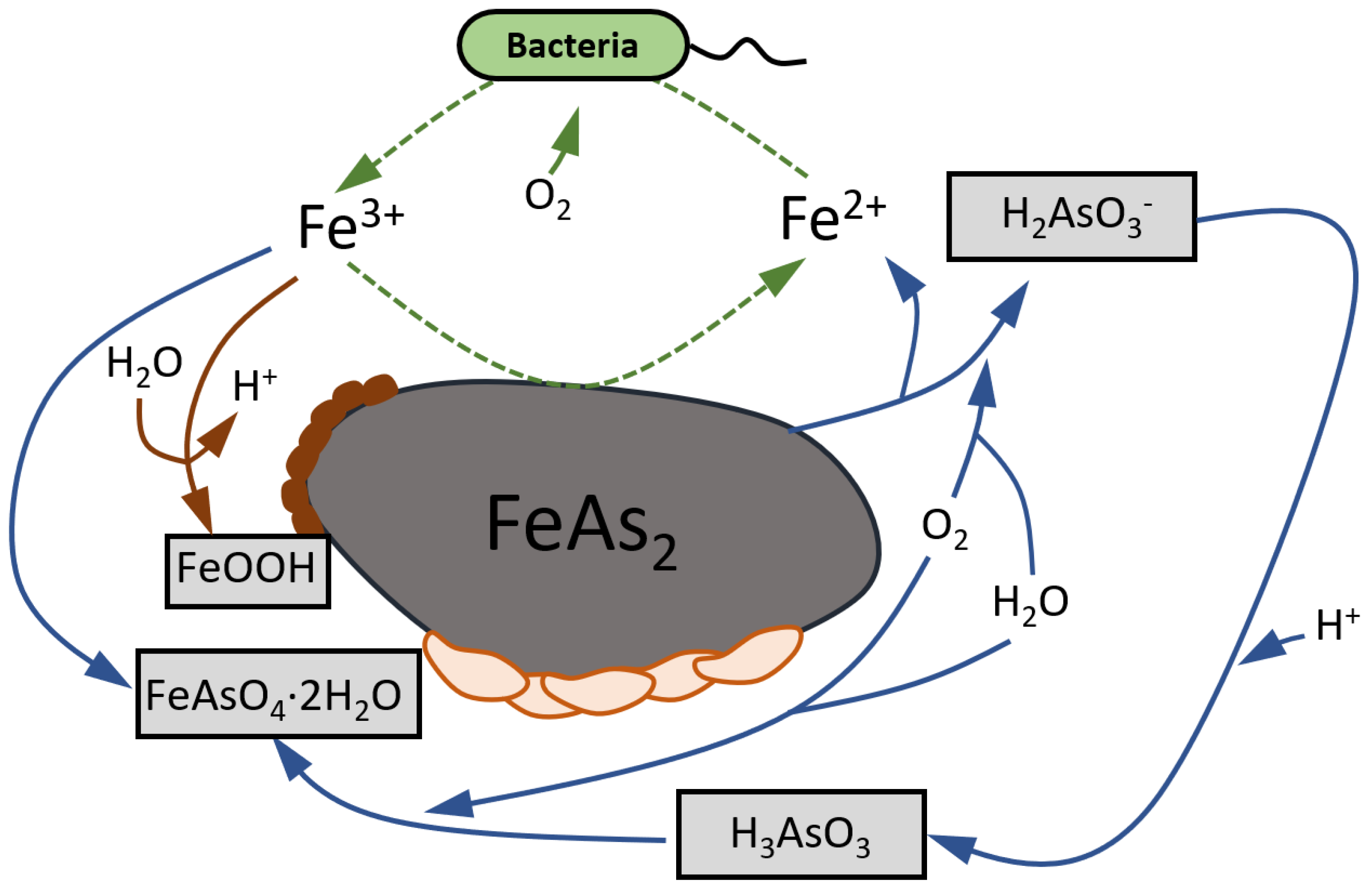
:1. Introduction
2. Materials and Methods
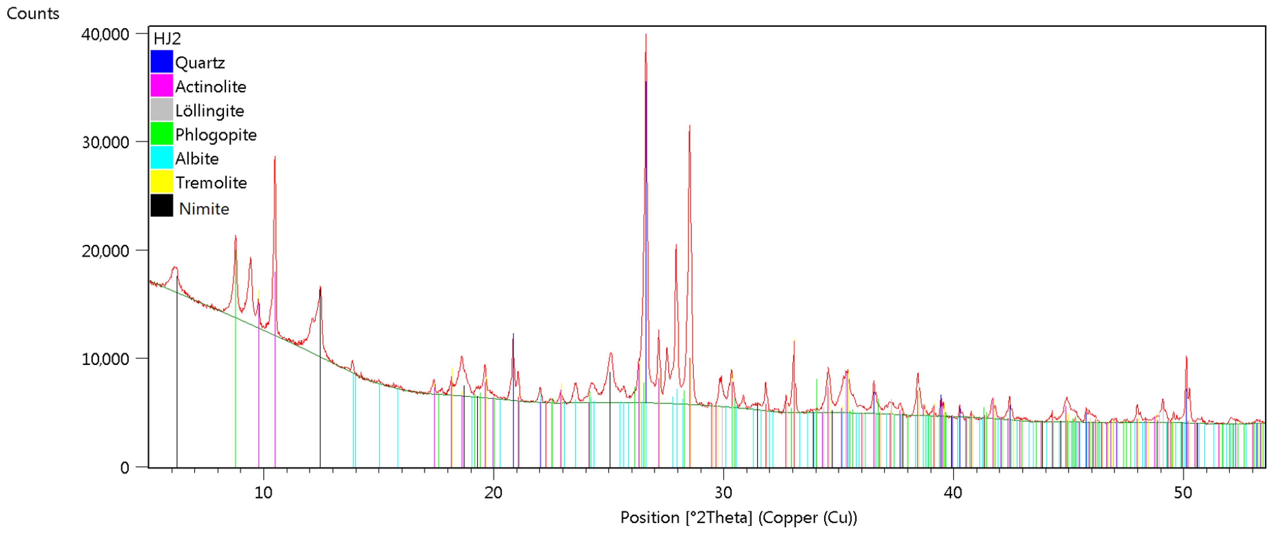
2.1. Solid Material
2.2. Adsorption of Rhamnolipids (Rh) and Lipopolysaccharides (LPS) onto Solids
2.3. Adhesion Experiments
2.4. Zeta Potential Measurements
2.5. Column Bioleaching
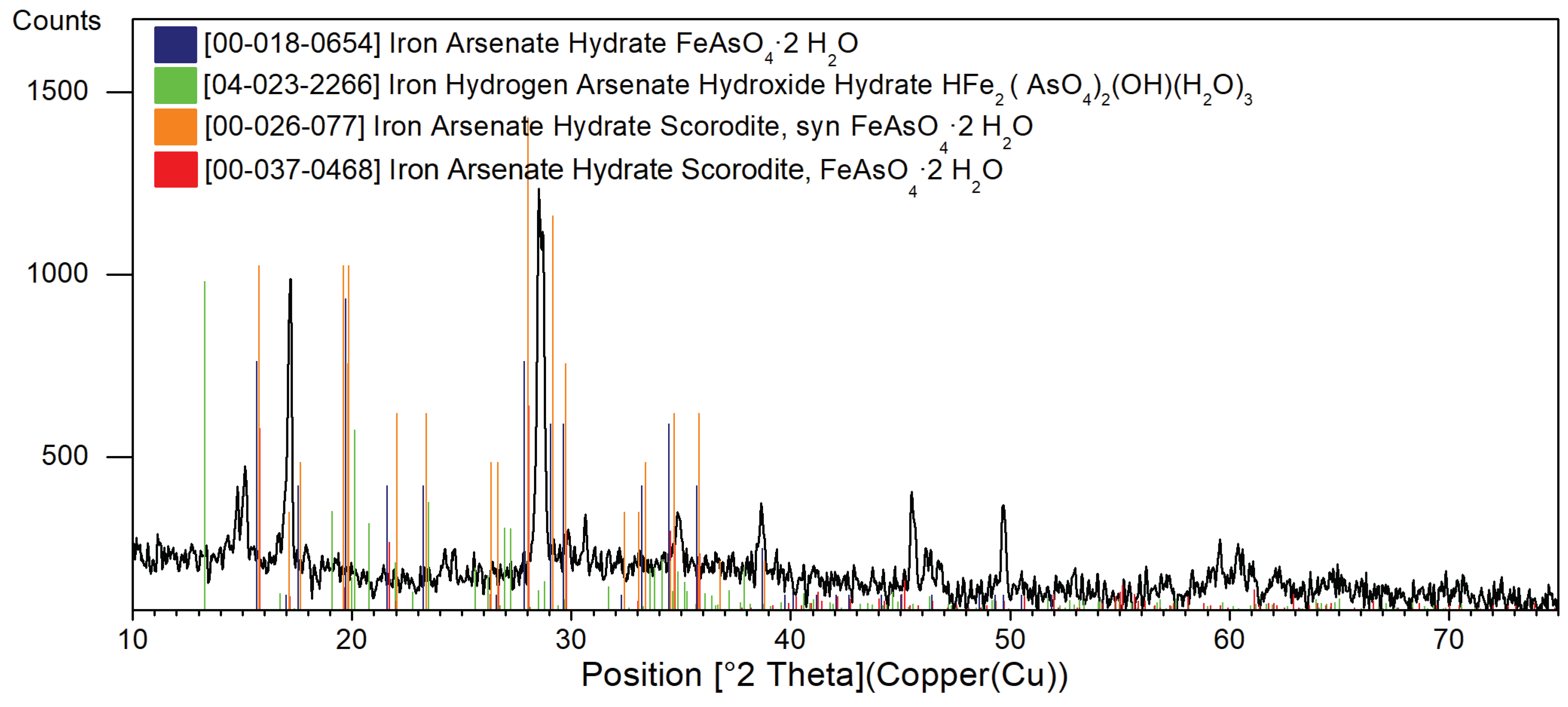
3. Results
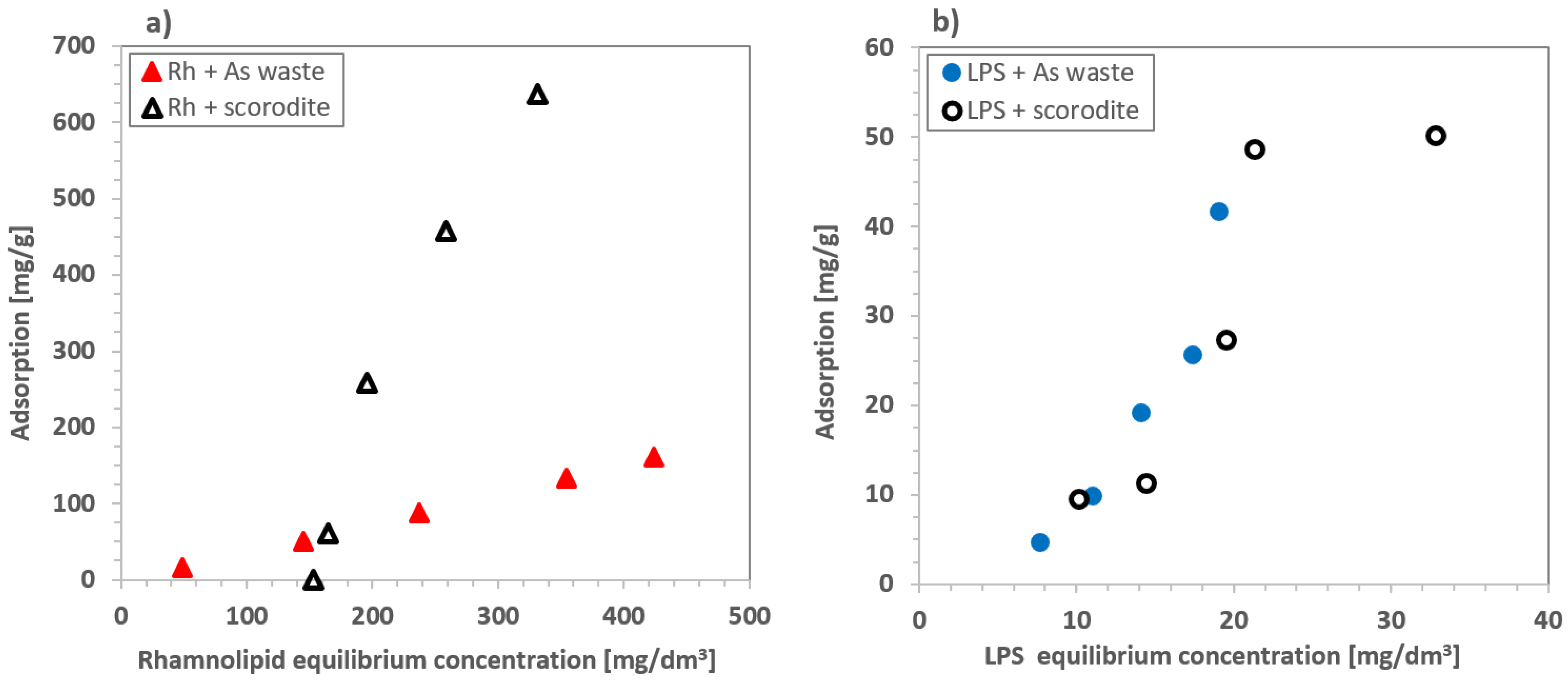
3.1. Adsorption of Rhamnolipids and Lipopolysaccharides on Arsenic Minerals
3.2. Adhesion of Scorodite to Arsenic-Bearing Mineral Bed
3.3. Electrokinetic Studies
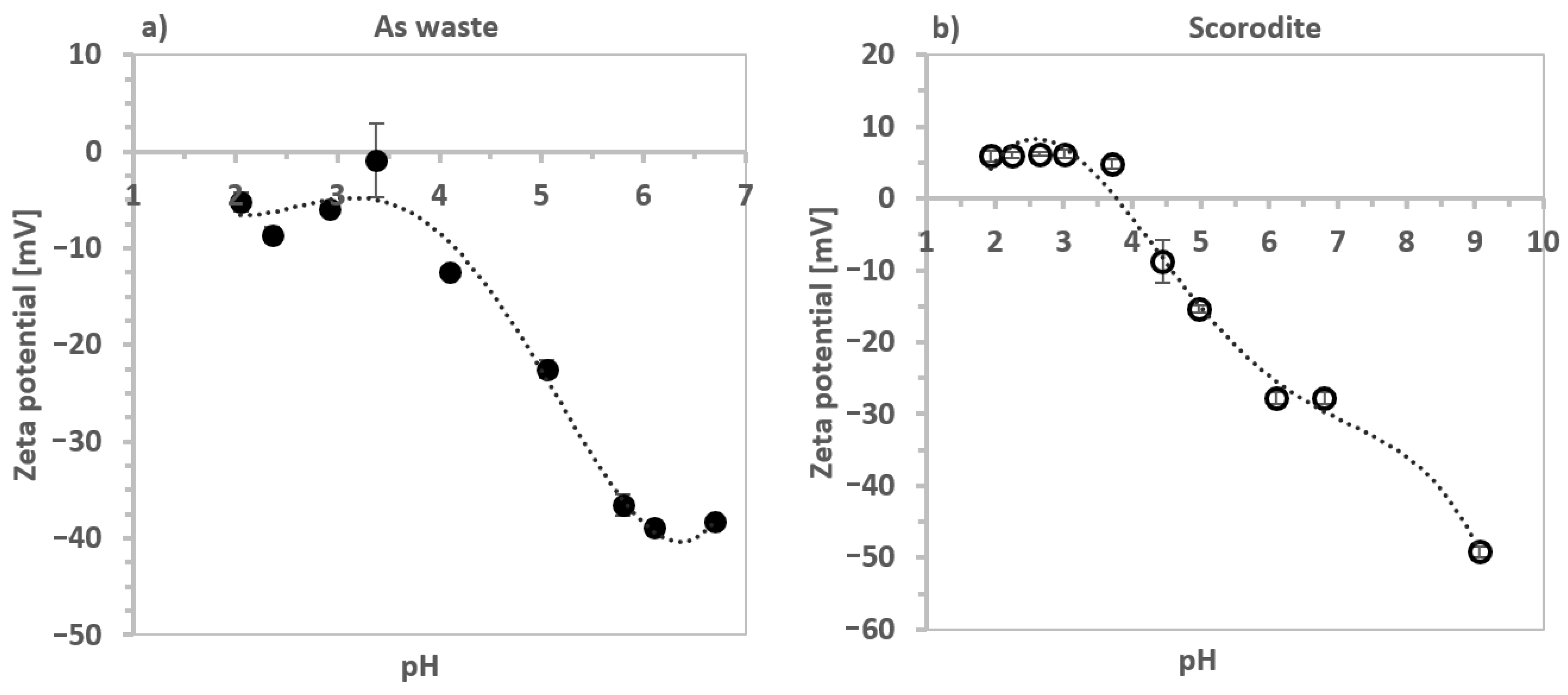
3.3.1. Influence of Arsenic Waste Suspension pH on the Zeta Potential
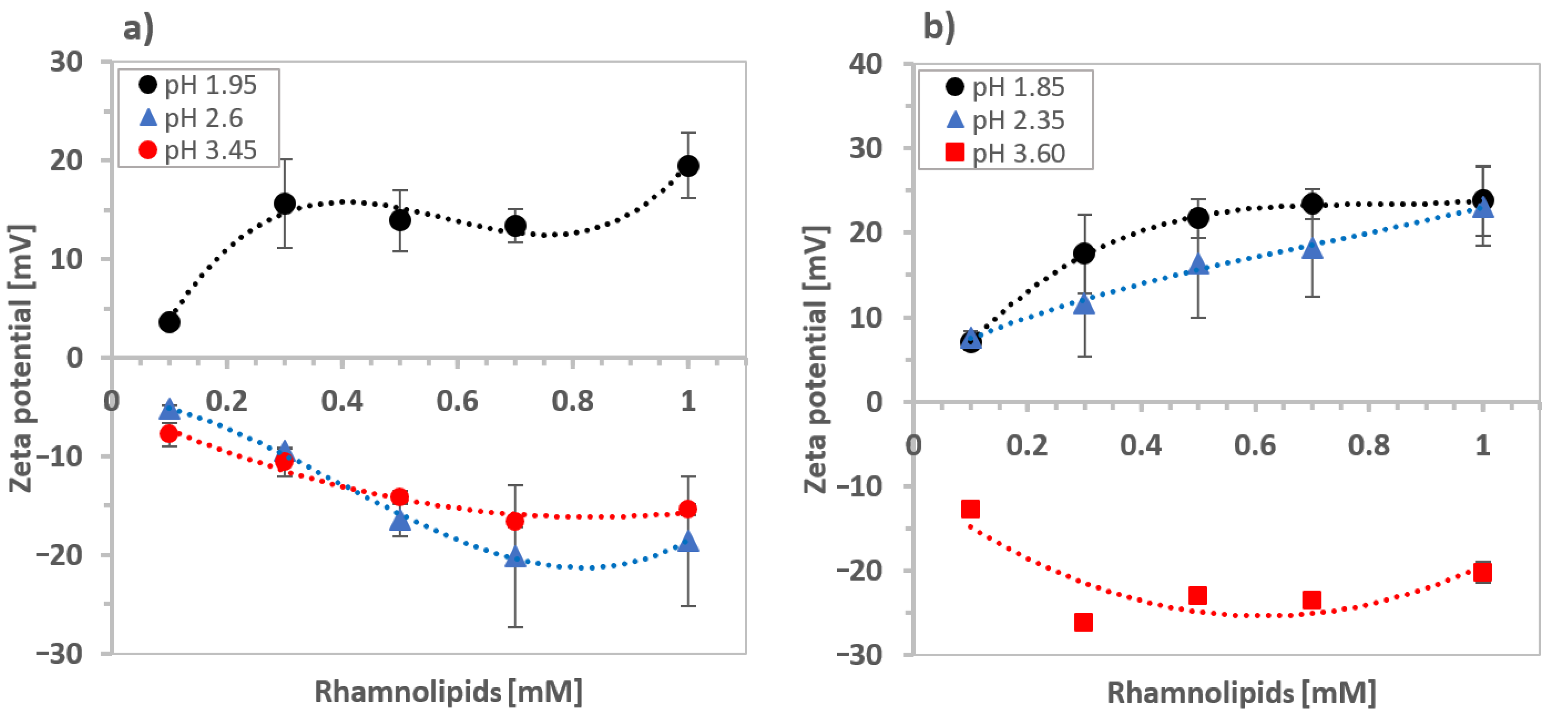
3.3.2. The Effect of Rhamnolipid Concentration on the Zeta Potential
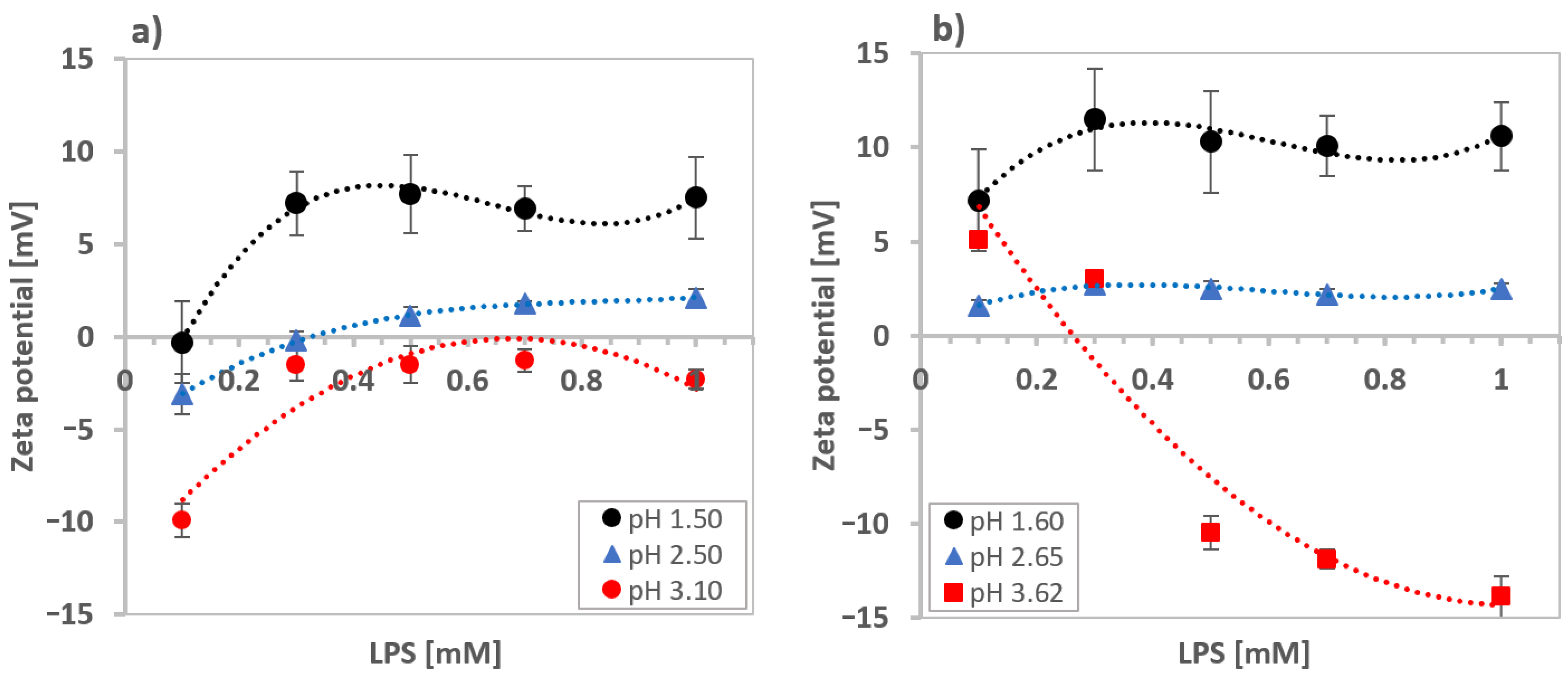
3.3.3. The Effect of Lipopolysaccharides on Zeta Potential
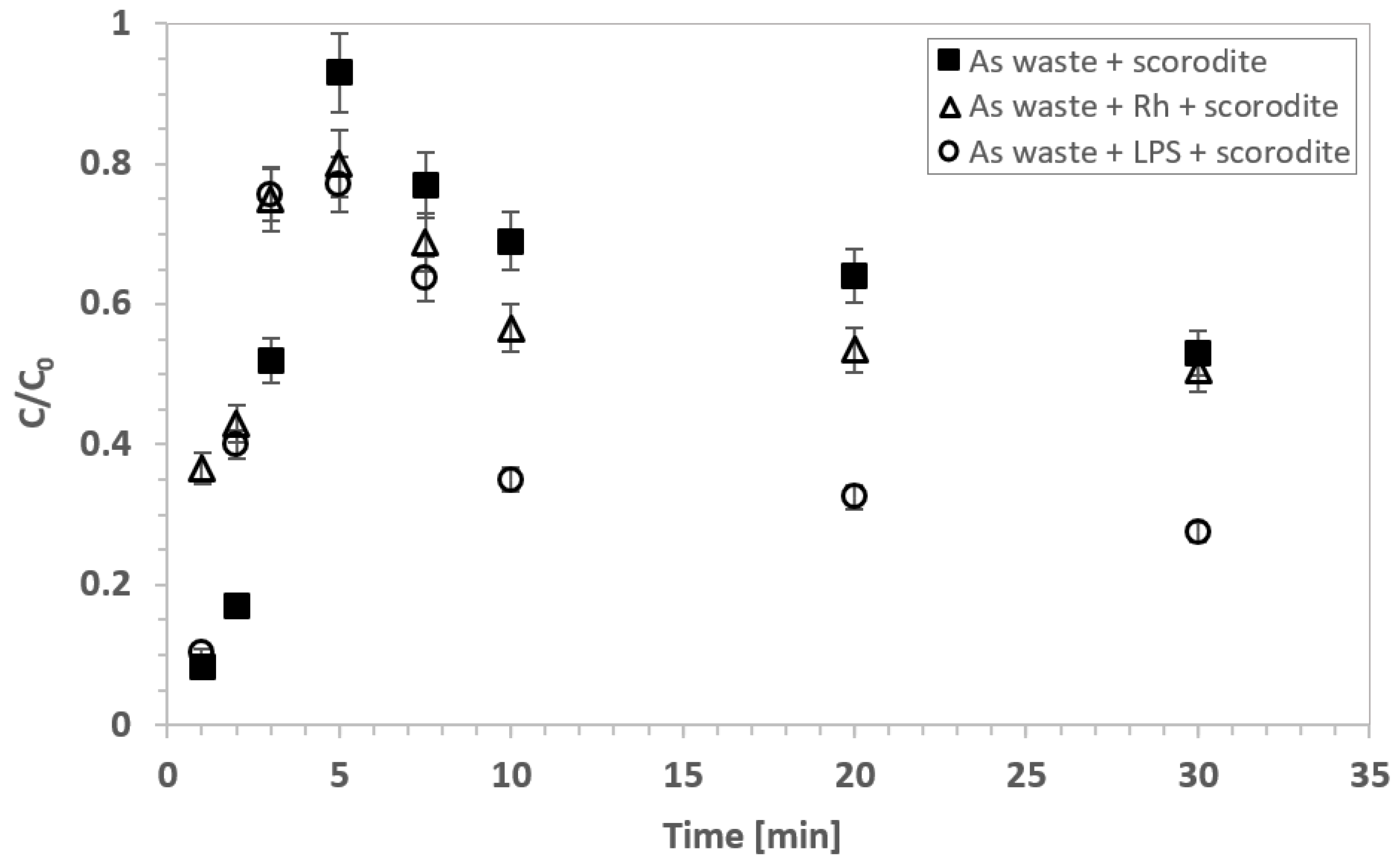
3.4. Bioleaching of Arsenic-Bearing Minerals
4. Discussion
5. Conclusions
Author Contributions
Funding
Data Availability Statement
Acknowledgments
Conflicts of Interest
References
- Mandal, B.; Suzuki, K. Arsenic round the world: A review. Talanta 2002, 58, 201–235. [Google Scholar] [CrossRef]
- Krysiak, A.; Karczewska, A. Arsenic extractability in soils in the areas of former arsenic mining and smelting, SW Poland. Sci. Total Environ. 2007, 379, 190–200. [Google Scholar] [CrossRef]
- Cłapa, T.; Narożna, R.; Siuda, R.; Borkowski, A.; Selwet, M.; Mądrzak, C.J.; Koźlecka, E. Bacterial Communities from the Arsenic Mine in Złoty Stok, Sudety Mountains, Poland. Pol. J. Microbiol. 2017, 66, 375–381. [Google Scholar] [CrossRef] [Green Version]
- Drewniak, L.; Skłodowska, A. Arsenic-transforming microbes and their role in biomining processes. Environ. Sci. Pollut. Res. 2013, 20, 7728–7739. [Google Scholar] [CrossRef] [Green Version]
- Sadowski, Z.; Jażdżyk, E.; Farbiszewska, T.; Farbiszewska-Bajer, J. Biooxidation of mining tailings from Zloty Stok. Physicochem. Probl. Miner. Process. 2000, 34, 47–56. [Google Scholar]
- Drewniak, L.; Matlakowska, R.; Rewerski, B.; Sklodowska, A. Arsenic release from gold mine rocks mediated by the activity of indigenous bacteria. Hydrometallurgy 2010, 104, 437–442. [Google Scholar] [CrossRef]
- Karczewska, A.; Krysiak, A.; Mokrzycka, D.; Jezierski, P.; Szopka, K. Arsenic distribution in soils of a former as mining area and processing. Pol. J. Environ. Stud. 2013, 22, 175–181. [Google Scholar]
- Paldyna, J.; Krasnodebska-Ostrega, B.; Kregielewska, K.; Kowalska, J.; Jedynak, Ł.; Golimowski, J.; Grobelski, T.; Farbiszewska-Kiczma, J.; Farbiszewska, T. The assessment of environmental pollution caused by mining and metallurgy wastes from highly polluted post-industrial regions in Southern Poland. Environ. Earth Sci. 2013, 68, 439–450. [Google Scholar] [CrossRef] [Green Version]
- Siuda, R.; Macioch, A. Secondary arsenic minerals from the Złoty Stok As-Au abandoned mine (SW Poland). Geol. Q. 2018, 62, 925. [Google Scholar] [CrossRef] [Green Version]
- Okibe, N.; Morishita, S.; Tanaka, M.; Sasaki, K.; Hirajima, T.; Hatano, K.; Ohata, A. Bioscorodite crystallization using Acidianus brierleyi: Effects caused by Cu(II) present in As(III)-bearing copper refinery wastewaters. Hydrometallurgy 2017, 168, 121–126. [Google Scholar] [CrossRef]
- Battistel, M.; Stolze, L.; Muniruzzaman, M.; Rolle, M. Arsenic release and transport during oxidative dissolution of spatially-distributed sulfide minerals. J. Hazard. Mater. 2021, 409, 124651. [Google Scholar] [CrossRef] [PubMed]
- Liu, X.; Li, Q.; Zhang, Y.; Yang, Y.; Xu, B.; Jiang, T. Formation Process of the Passivating Products from Arsenopyrite Bioleaching by Acidithiobacillus ferrooxidans in 9K Culture Medium. Metals 2019, 9, 1320. [Google Scholar] [CrossRef] [Green Version]
- Das, B. Theoretical Study of Formation of Secondary Arsenic Minerals: Scorodite and Pharmacosiderite. ACS Earth Space Chem. 2019, 3, 192–201. [Google Scholar] [CrossRef]
- Diao, M.; Taran, E.; Mahler, S.; Nguyen, A. A concise review of nanoscopic aspects of bioleaching bacteria–mineral interactions. Adv. Colloid Interface Sci. 2014, 212, 45–63. [Google Scholar] [CrossRef] [PubMed] [Green Version]
- Wang, H.; Sodagari, M.; Chen, Y.; He, X.; Newby, B.-M.Z.; Ju, L.-K. Initial bacterial attachment in slow flowing systems: Effects of cell and substrate surface properties. Colloids Surf. B Biointerfaces 2011, 87, 415–422. [Google Scholar] [CrossRef]
- Zhu, J.; Li, Q.; Jiao, W.; Jiang, H.; Sand, W.; Xia, J.; Liu, X.; Qin, W.; Qiu, G.; Hu, Y.; et al. Adhesion forces between cells of Acidithiobacillus ferrooxidans, Acidithiobacillus thiooxidans or Leptospirillum ferrooxidans and chalcopyrite. Colloids Surf. B Biointerfaces 2012, 94, 95–100. [Google Scholar] [CrossRef] [PubMed]
- Liu, G.; Zhong, H.; Yang, X.; Liu, Y.; Shao, B.; Liu, Z. Advances in applications of rhamnolipids biosurfactant in environmental remediation: A review. Biotechnol. Bioeng. 2018, 115, 796–814. [Google Scholar] [CrossRef] [PubMed]
- Heyd, M.; Kohnert, A.; Tan, T.-H.; Nusser, M.; Kirschhöfer, F.; Brenner-Weiss, G.; Franzreb, M.; Berensmeier, S. Development and trends of biosurfactant analysis and purification using rhamnolipids as an example. Anal. Bioanal. Chem. 2008, 391, 1579–1590. [Google Scholar] [CrossRef]
- Gunther, N.W.; Nuñez, A.; Fett, W.; Solaiman, D.K.Y.; Venugopalan, V.P.; Kuehn, M.; Hausner, M.; Springael, D.; Wilderer, P.A.; Wuertz, S. Production of Rhamnolipids by Pseudomonas chlororaphis, a Nonpathogenic Bacterium. Appl. Environ. Microbiol. 2005, 71, 2677–2686. [Google Scholar] [CrossRef] [Green Version]
- Tuleva, B.K.; Ivanov, G.R.; Christova, N.E. Biosurfactant Production by a New Pseudomonas Putida Strain. Z. Nat. Sect. C J. Biosci. 2002, 57, 356–360. [Google Scholar] [CrossRef]
- Costa, S.G.V.A.O.; De Souza, S.R.; Nitschke, M.; Franchetti, S.M.M.; Jafelicci, M.; Lovaglio, R.B.; Contiero, J. Wettability of Aqueous Rhamnolipids Solutions Produced by Pseudomonas aeruginosa LBI. J. Surfactants Deterg. 2008, 12, 125–130. [Google Scholar] [CrossRef]
- Al-Tahhan, R.A.; Sandrin, T.; Bodour, A.A.; Maier, R.M. Rhamnolipid-Induced Removal of Lipopolysaccharide from Pseudomonas aeruginosa: Effect on Cell Surface Properties and Interaction with Hydrophobic Substrates. Appl. Environ. Microbiol. 2000, 66, 3262–3268. [Google Scholar] [CrossRef] [Green Version]
- Genevaux, P.; Bauda, P.; Dubow, M.S.; Oudega, B. Identification of Tn 10 insertions in the rfaG, rfaP, and galU genes involved in lipopolysaccharide core biosynthesis that affect Escherichia coli adhesion. Arch. Microbiol. 1999, 172, 1–8. [Google Scholar] [CrossRef]
- Jacques, M. Role of lipo-oligosaccharides and lipopolysaccharides in bacterial adherence. Trends Microbiol. 1996, 4, 408–410. [Google Scholar] [CrossRef]
- Murphy, K.; Park, A.J.; Hao, Y.; Brewer, D.; Lam, J.S.; Khursigara, C.M. Influence of O Polysaccharides on Biofilm Development and Outer Membrane Vesicle Biogenesis in Pseudomonas aeruginosa PAO1. J. Bacteriol. 2014, 196, 1306–1317. [Google Scholar] [CrossRef] [Green Version]
- Orlova, N.V.; Proskurnin, M.A.; Samburova, V.A.; Tsvetkov, P.V. Determination of Polysaccharides and Lipopolysaccharides by Spectrophotometry and Thermal-Lens Spectrometry. J. Anal. Chem. 2003, 58, 149–151. [Google Scholar] [CrossRef]
- Escobar, B.; Huerta, G.; Rubio, J. Influence of lipopolysaccharides on the attachment of Thiobacillus ferrooxidans to minerals. World J. Microbiol. Biotechnol. 1997, 13, 593–594. [Google Scholar] [CrossRef]
- Arredondo, R.; García, A.; Jerez, C.A. Partial Removal of Lipopolysaccharide from Thiobacillus ferrooxidans Affects Its Adhesion to Solids. Appl. Environ. Microbiol. 1994, 60, 2846–2851. [Google Scholar] [CrossRef] [PubMed] [Green Version]
- Demopoulos, G.; Droppert, D.; Van Weert, G. Precipitation of crystalline scorodite (FeAsO4·2H2O) from chloride solutions. Hydrometallurgy 1995, 38, 245–261. [Google Scholar] [CrossRef]
- Fujita, T.; Taguchi, R.; Abumiya, M.; Matsumoto, M.; Shibata, E.; Nakamura, T. Effect of pH on atmospheric scorodite synthesis by oxidation of ferrous ions: Physical properties and stability of the scorodite. Hydrometallurgy 2009, 96, 189–198. [Google Scholar] [CrossRef]
- Gomez, M.; Becze, L.; Celikin, M.; Demopoulos, G.P. The effect of copper on the precipitation of scorodite (FeAsO4·2H2O) under hydrothermal conditions: Evidence for a hydrated copper containing ferric arsenate sulfate-short lived intermediate. J. Colloid Interface Sci. 2011, 360, 508–518. [Google Scholar] [CrossRef] [PubMed]
- Pawlowska, A.; Sadowski, Z. Effect of Schwertmannite Surface Modification by Surfactants on Adhesion of Acidophilic Bacteria. Microorganisms 2020, 8, 1725. [Google Scholar] [CrossRef]
- Galabova, D.; Sotirova, A.; Karpenko, E.; Karpenko, O. Role of Microbial Surface-Active Compounds in Environmental Protection. In The Role of Colloidal Systems in Environmental Protection; Elsevier: Amsterdam, The Netherlands, 2014. [Google Scholar] [CrossRef]
- Eltaboni, F.B.; Caseley, E.; Katsikogianni, M.; Swanson, L.; Swift, T.; Romero-González, M. Fluorescence spectroscopy analysis of the bacteria–mineral interface: Adsorption of lipopolysaccharides to silica and alumina. Langmuir 2020, 36, 1623–1632. [Google Scholar] [CrossRef]
- Bai, G.; Brusseau, M.L.; Miller, R.M. Influence of a Rhamnolipid Biosurfactant on the Transport of Bacteria through a Sandy Soil. Appl. Environ. Microbiol. 1997, 63, 1866–1873. [Google Scholar] [CrossRef] [PubMed] [Green Version]
- Lebrón-Paler, A.; Pemberton, J.E.; Becker, B.A.; Otto, W.H.; Larive, C.K.; Maier, R.M. Determination of the Acid Dissociation Constant of the Biosurfactant Monorhamnolipid in Aqueous Solution by Potentiometric and Spectroscopic Methods. Anal. Chem. 2006, 78, 7649–7658. [Google Scholar] [CrossRef] [PubMed]
- Kramer, G.; Somasundaran, P. Conformational Behavior of Polyelectrolyte Complexes at the Solid/Liquid Interface. Langmuir 2002, 18, 9357–9361. [Google Scholar] [CrossRef]
- Szewczuk-Karpisz, K.; Krasucka, P.; Boguta, P.; Skic, K.; Sokołowska, Z.; Fijałkowska, G.; Wiśniewska, M. Electrical double layer at the gibbsite/anionic polyacrylamide/supporting electrolyte interface—Adsorption, spectroscopy and electrokinetic studies. J. Mol. Liq. 2018, 261, 439–445. [Google Scholar] [CrossRef]
- Solari, J.A.; Huerta, G.; Escobar, B.; Vargas, T.; Badilla-Ohlbaum, R.; Rubio, J. Interfacial phenomena affecting the adhesion of Thiobacillus ferrooxidans to sulphide mineral surface. Colloids Surf. 1992, 69, 159–166. [Google Scholar] [CrossRef]
- Blake, R.C.; Shute, E.A.; Howard, G.T. Solubilization of Minerals by Bacteria: Electrophoretic Mobility of Thiobacillus ferrooxidans in the Presence of Iron, Pyrite, and Sulfur. Appl. Environ. Microbiol. 1994, 60, 3349–3357. [Google Scholar] [CrossRef] [Green Version]
- Vilinska, A.; Rao, K.H. Surface thermodynamics and extended DLVO theory of Leptospirillum ferrooxidans cells’ adhesion on sulfide minerals. Miner. Metall. Process. 2011, 28, 151–158. [Google Scholar] [CrossRef]


Publisher’s Note: MDPI stays neutral with regard to jurisdictional claims in published maps and institutional affiliations. |
© 2021 by the authors. Licensee MDPI, Basel, Switzerland. This article is an open access article distributed under the terms and conditions of the Creative Commons Attribution (CC BY) license (https://creativecommons.org/licenses/by/4.0/).
Share and Cite
Pawlowska, A.; Sadowski, Z.; Winiarska, K. Effect of Rhamnolipids and Lipopolysaccharides on the Bioleaching of Arsenic-Bearing Waste. Minerals 2021, 11, 1303. https://doi.org/10.3390/min11121303
Pawlowska A, Sadowski Z, Winiarska K. Effect of Rhamnolipids and Lipopolysaccharides on the Bioleaching of Arsenic-Bearing Waste. Minerals. 2021; 11(12):1303. https://doi.org/10.3390/min11121303
Chicago/Turabian StylePawlowska, Agnieszka, Zygmunt Sadowski, and Katarzyna Winiarska. 2021. "Effect of Rhamnolipids and Lipopolysaccharides on the Bioleaching of Arsenic-Bearing Waste" Minerals 11, no. 12: 1303. https://doi.org/10.3390/min11121303