
Mineralogical Characterization of Manganese Oxide Minerals of the Devonian Xialei Manganese Deposit

 ,
,
Abstract
:1. Introduction
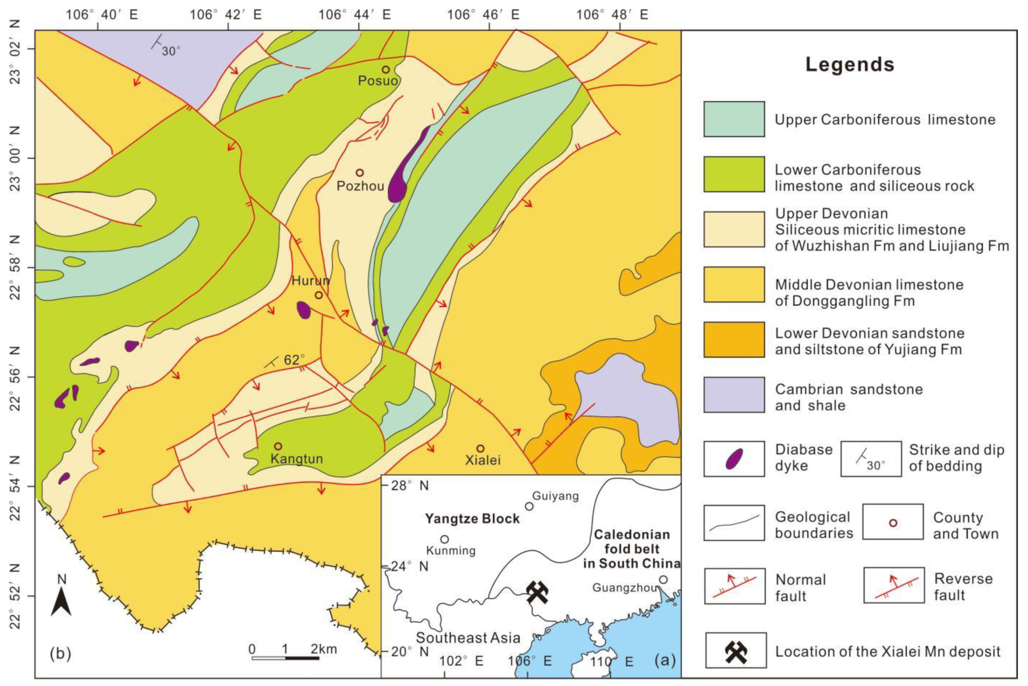
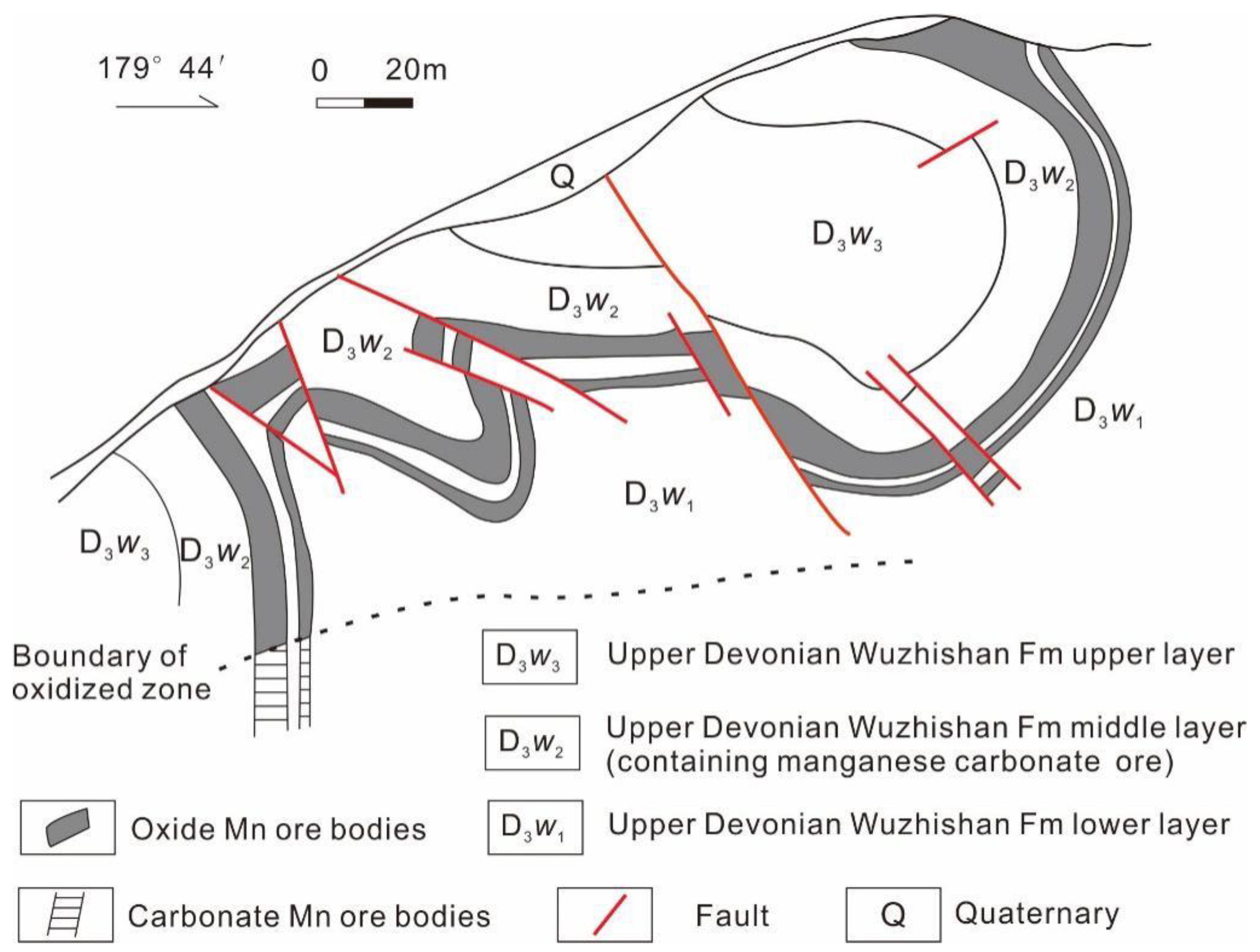
2. Geological Settings
3. Methodology
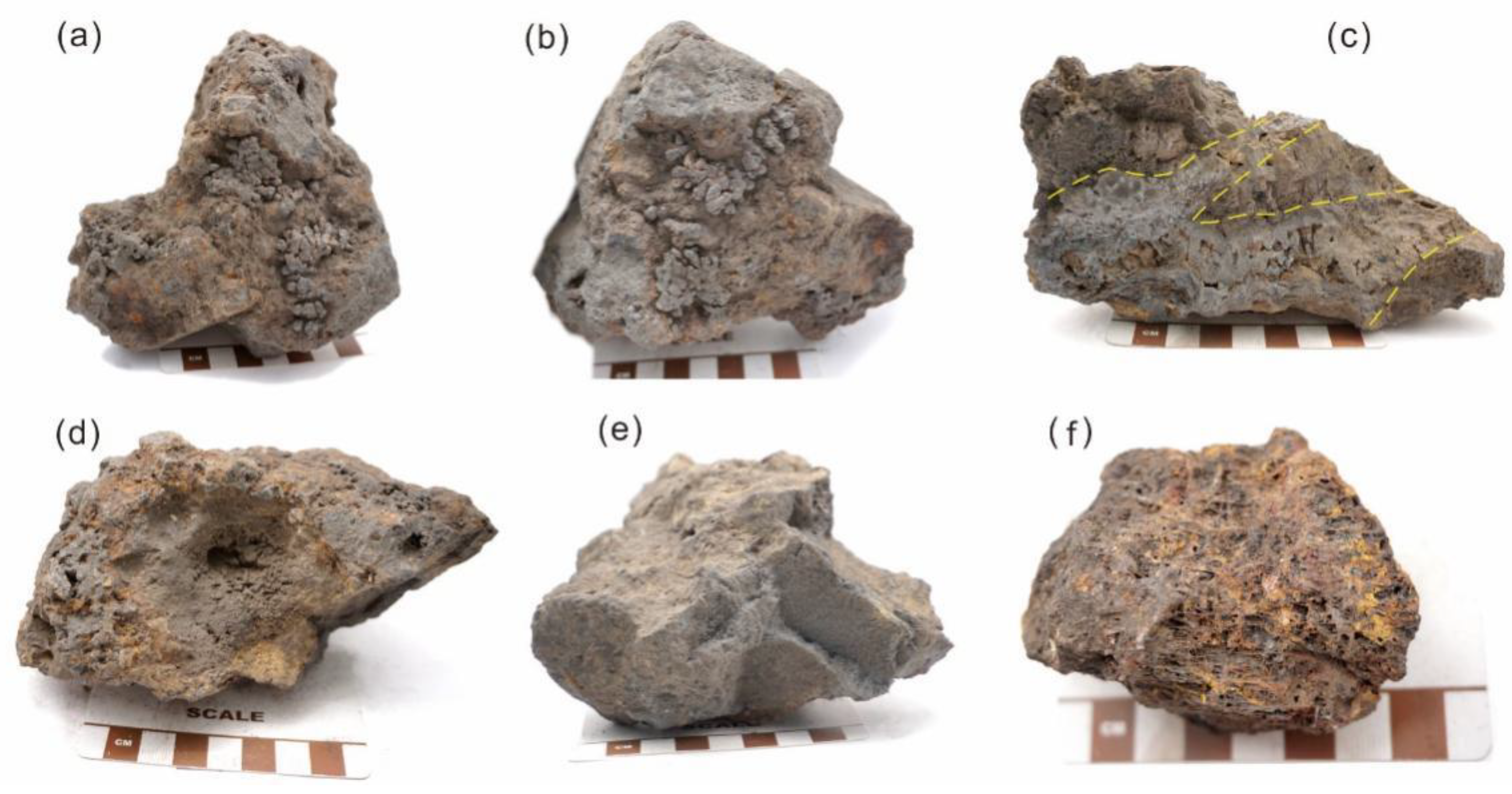
3.1. Sampling and Preparation
3.2. Experimental Methods
4. Results
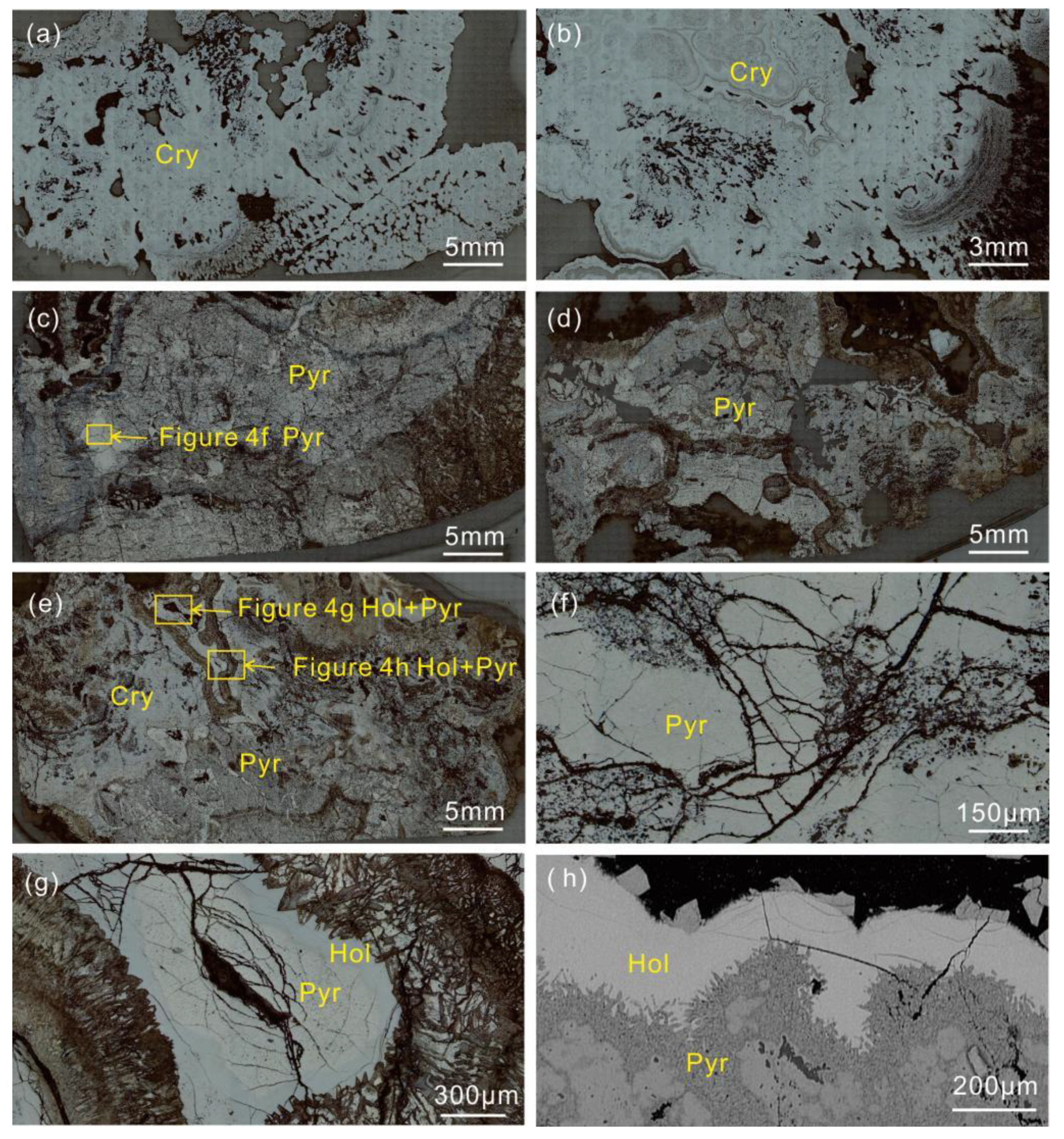
4.1. Optical Microscopy
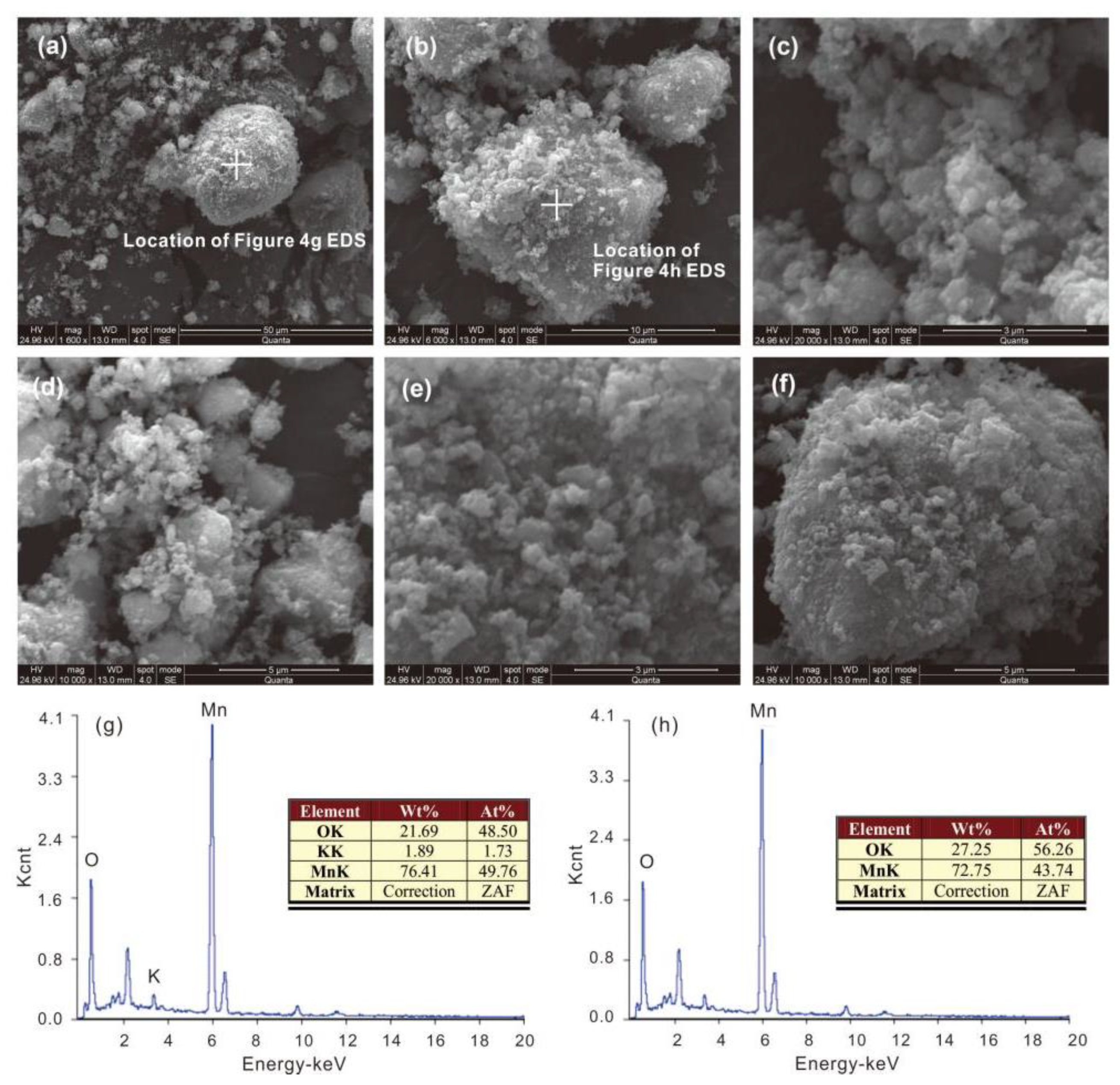
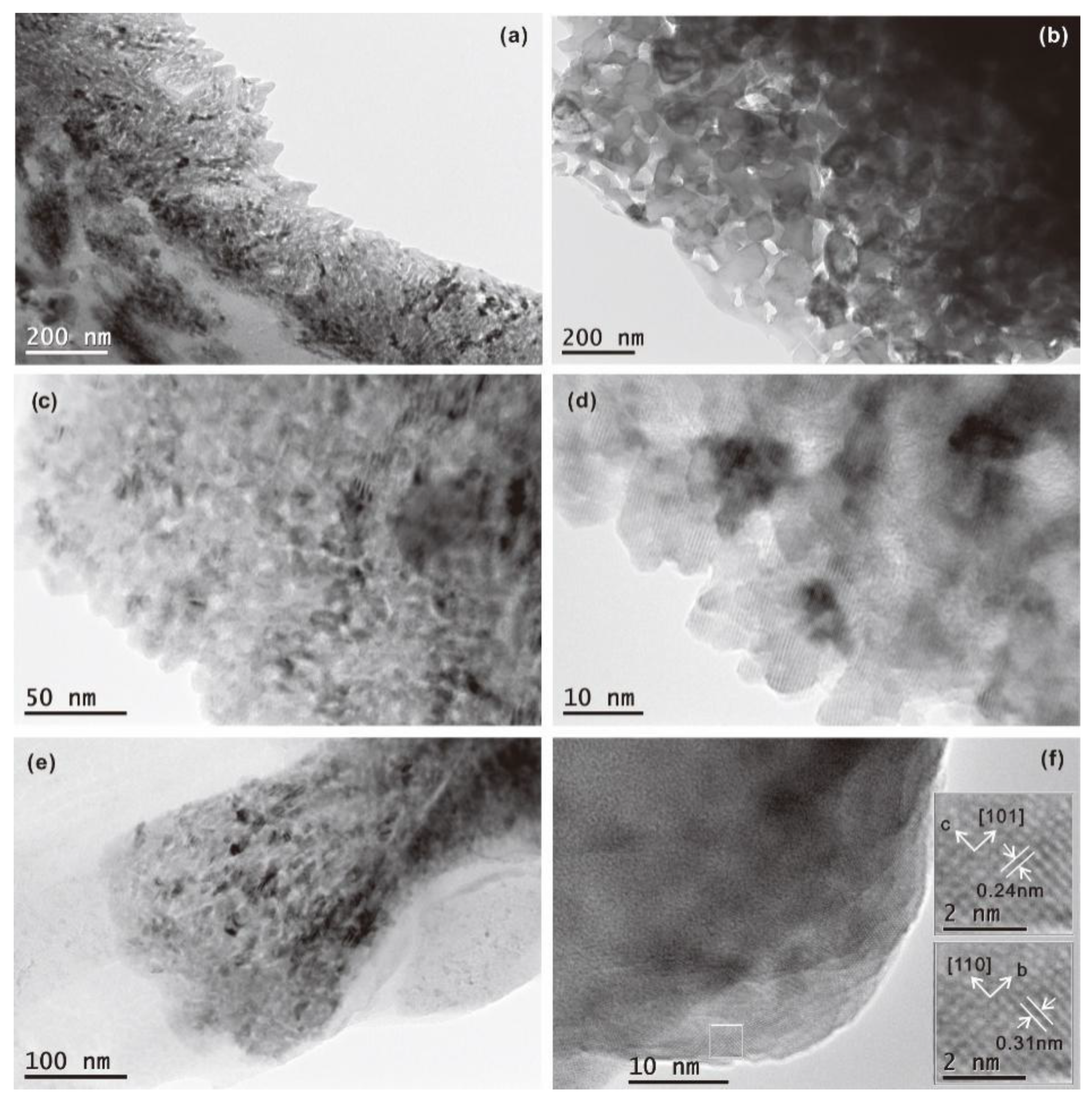
4.2. SEM and TEM
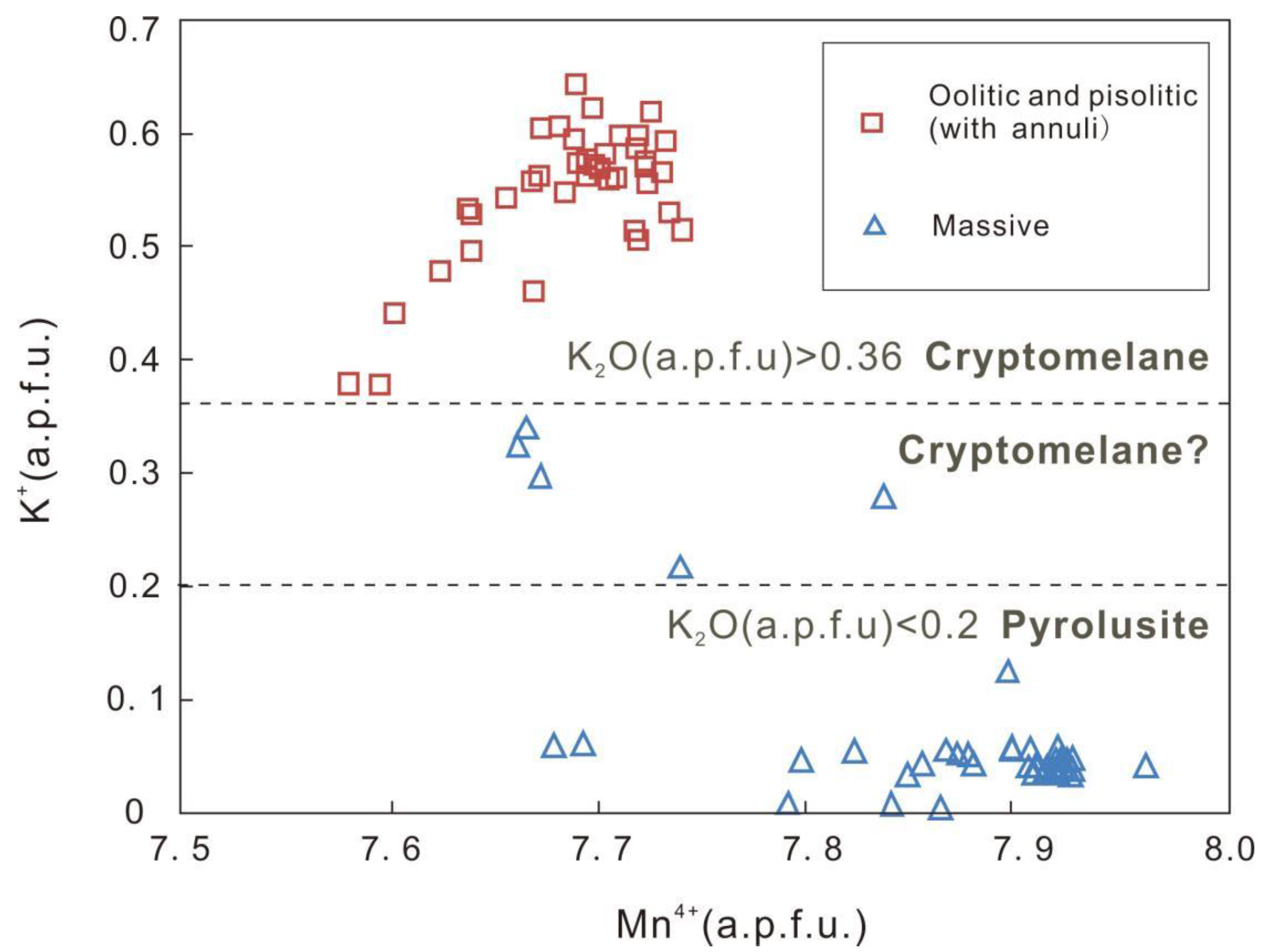
4.3. EPMA
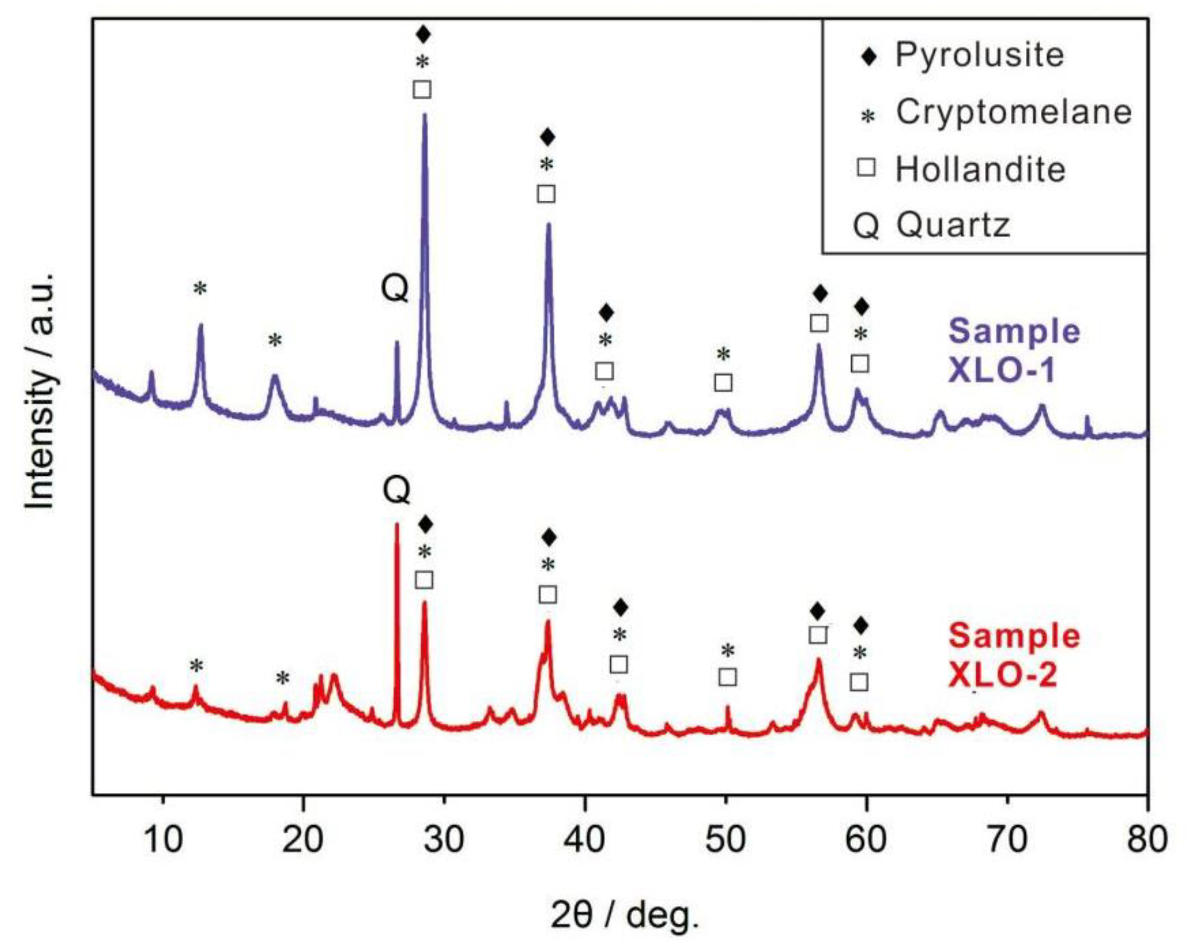
4.4. XRD
5. Summary and Discussion
5.1. Mn Oxide Phases, Chemical Composition, and Genesis
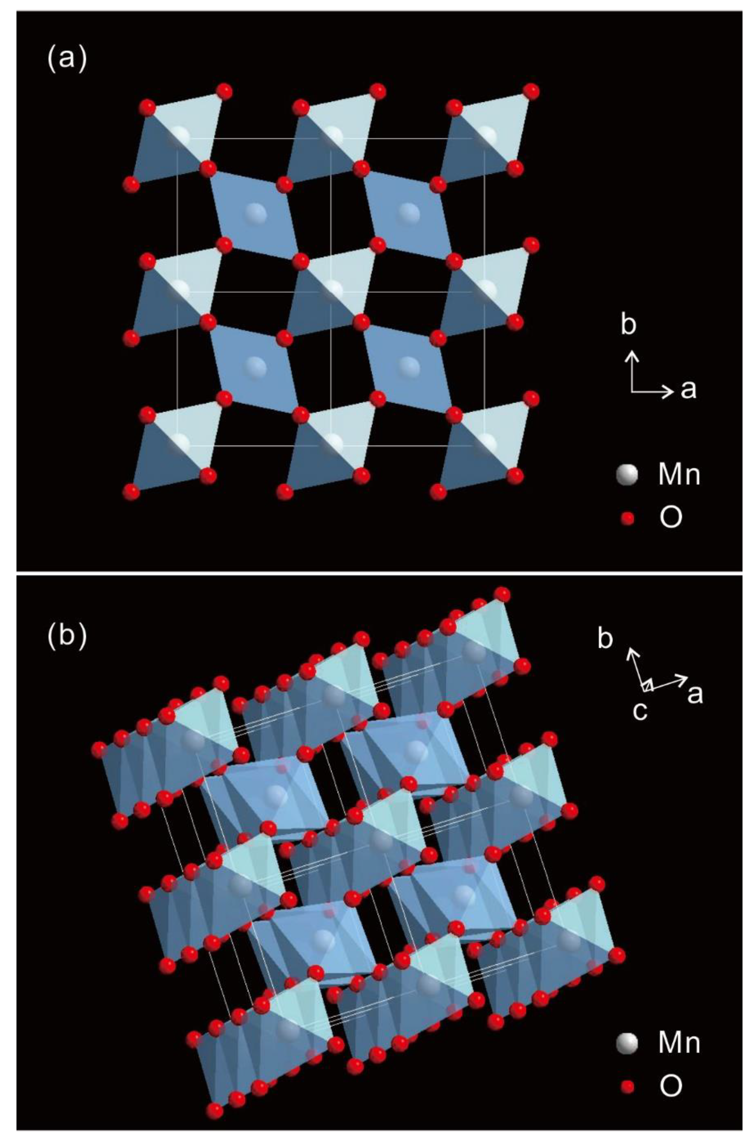
5.2. Crystal Structure of the Mn Oxide
6. Conclusions
Supplementary Materials
Author Contributions
Funding
Data Availability Statement
Acknowledgments
Conflicts of Interest
References
- Turekian, K.K.; Wedepohl, K.L. Distribution of the elements in some major units of the earth’s crust. Geol. Soc. Am. Bull. 1961, 72, 175–192. [Google Scholar] [CrossRef]
- Post, J.E. Manganese oxide minerals: Crystal structures and economic and environmental significance. Proc. Natl. Acad. Sci. USA 1999, 96, 3447–3454. [Google Scholar] [CrossRef] [PubMed] [Green Version]
- Volokhin, Y.G.; Mikhailik, P.E.; Mikhailik, E.V. Minerals in manganese deposits of Belyaevsky Volcano, the Sea of Japan. Russ. J. Pac. Geol. 2020, 14, 340–362. [Google Scholar] [CrossRef]
- Jin, S.S.; Zhang, L.M.; He, J.Z. Review of reactions of manganese oxides with organic compounds and applications of MnOx in environmental remediation. Acta Sci. Circumstantiae 2008, 28, 2394–2403. [Google Scholar]
- He, H.P.; Zhu, J.X.; Chen, M.; Tao, Q.; Tan, D.Y.; Liang, X.L.; Xian, H.Y. Progresses in researches on mineral structure and mineral physics (2011–2020). Bull. Mineral. Petrol. Geochem. 2020, 39, 697–713, (In Chinese with English Abstract). [Google Scholar]
- Lu, A.H.; Li, Y.; Ding, H.R.; Wang, C.Q. “Mineral membrane” of the surface: “New sphere” of the Earth. Acta Petrol. Sin. 2019, 35, 119–128, (In Chinese with English Abstract). [Google Scholar]
- Lu, A.H.; Li, Y.; Ding, H.R.; Wang, C.Q.; Xu, X.M.; Liu, F.F.; Liu, Y.W.; Zhu, Y.; Li, Y.Z. Natural mineral photoelectric effect: Non-classical mineral photosynthesis. Earth Sci. Front. 2020, 27, 179–194, (In Chinese with English Abstract). [Google Scholar]
- Lu, A.H.; Li, Y.; Liu, F.F.; Liu, Y.W.; Ye, H.; Zhuang, Z.Y.; Li, Y.Z.; Ding, H.R.; Wang, C.Q. The photogeochemical cycle of Mn oxides on Earth’s surface. Mineral. Mag. 2021, 85, 1–57. [Google Scholar] [CrossRef]
- Zhang, C.M.; Pang, X.; Wang, Y.Z. Controllable synthesis of one-dimensional cryptomelane-type manganese dioxide and its electrochemical performance. Acta Chim. Sin. 2018, 76, 133–137, (In Chinese with English Abstract). [Google Scholar] [CrossRef]
- Lu, A.H.; Gao, X.; Qin, S.; Wang, C.Q. Cryptomelane (KxMn8−xO16): Natural active octahedral molecular sieve (OMS-2). Chin. Sci. Bull. 2003, 6, 97–100, (In Chinese with English Abstract). [Google Scholar]
- Lu, A.H.; Li, Y. Reactivity of natural Mn oxide cryptomelane. In Advances in the Environmental Biogeochemistry of Manganese Oxides; ACS Symposium Series; ACS Publications: Washington, DC, USA, 2015; pp. 89–106. [Google Scholar]
- Zeng, Y.Y.; Liu, T.B. Characteristics of the Devonian Xialei manganese deposit, Guangxi Zhuang Autonomous Region, China. Ore Geol. Rev. 1999, 15, 153–163. [Google Scholar] [CrossRef]
- Zhu, J.D.; Zhu, K.J.; Zhou, S.G.; Tian, Y.M.; Zhang, Z.W.; Li, S.F.; Shi, C.H. Characteristics and genesis of deposits in the Xialei-Dongping Manganese Ore Belt of Guangxi Province. Geol. Explor. 2016, 52, 846–853, (In Chinese with English Abstract). [Google Scholar]
- Zhao, D.J.; Lu, A.H.; Wang, L.J.; Zheng, X.K.; Liu, R.; Guo, Y.J. Mineralogical characteristics study of natural cryptomelane in the Xialei Manganese Deposit, Guangxi. Acta Sci. Nat. Univ. Pekin. 2005, 41, 859–868, (In Chinese with English Abstract). [Google Scholar]
- Ismail, S.; Hushim, S.F.S.; Hussin, H.; Abdullah, N.S. Chemical and mineralogical characterization of Malaysian low-grade manganese ore. Period. Mineral. 2016, 85, 277–288. [Google Scholar]
- Wang, H.Z.; Shi, X.Y.; Wang, X.X. Research on Sequence Stratigraphy in China; Guangdong Science and Technology Press: Guangzhou, China, 2000; pp. 1–457. [Google Scholar]
- Zhao, L.Q.; Zhou, S.G.; Yi, H.S.; Dong, S.Y.; Li, L.M.; Zhang, M. Geochemical characteristics and sedimentary environment of the Xialei manganese deposit in southwest Guangxi. Geol. Explor. 2016, 52, 25–39, (In Chinese with English Abstract). [Google Scholar]
- Yan, D.R.; Li, J.W.; Hu, M.A.; Lang, Y.S. Characteristics and genesis of supergene manganese ores in Xialei, Guangxi. Geol. Sci. Technol. Inf. 2006, 25, 61–67, (In Chinese with English Abstract). [Google Scholar]
- Hao, R.X.; Guan, G.Y. The mineral assemblages and oxidizing mechanism of oxidized manganese deposits of Xialei-Hurun manganese mineralized zone. China’s Manganese Ind. 1995, 13, 3–7, (In Chinese with English Abstract). [Google Scholar]
- Richmond, W.E.; Fleischer, M. Cryptomelane, a new name for the commonest of the “psilomelane” minerals. Am. Mineral. 1942, 27, 607–610. [Google Scholar]
- Post, J.E.; Dreele, R.B.V.; Buseck, P.R. Symmetry and cation displacements in hollandites: Structure refinements of hollandite, cryptomelane and priderite. Acta Crystallogr. 1982, 38, 1056–1065. [Google Scholar] [CrossRef]
- Post, J.E.; Mckeown, D.A.; Heaney, P.J. Raman spectroscopy study of manganese oxides: Tunnel structures. Am. Mineral. 2020, 105, 1175–1190. [Google Scholar] [CrossRef]
- Gao, X.; Lu, A.H.; Qin, S.; Zheng, D.S.; Zheng, X.K.; Zheng, Z. A study of crystal structural characteristics and environmental properties of natural cryptomelane. Acta Petrol. Mineral. 2001, 20, 477–484. [Google Scholar]
- Vafeas, N.A.; Viljoen, K.S.; Blignaut, L.C. Characterization of fibrous cryptomelane from the todorokite-cryptomelane mineral assemblage at the Sebilo Mine, Northern Cape Province, South Africa. Can. Mineral. 2018, 56, 65–76. [Google Scholar] [CrossRef]
- Wang, P.; Pan, Z.L.; Weng, L.B. Systematic Mineralogy; Geological Publishing House: Beijing, China, 1987; pp. 1–654. (In Chinese) [Google Scholar]
- Bricker, O.P. Some stability relations in the system Mn-O2-H2O at 25 °C and one atmosphere total pressure. Amer. Minaral. 1965, 50, 1296–1354. [Google Scholar]
- Chen, T.; Dou, H.Y.; Li, X.L.; Tang, X.F.; Li, J.H.; Hao, J.M. Tunnel structure effect of manganese oxides in complete oxidation of formaldehyde. Microporous Mesoporous Mater. 2009, 122, 270–274. [Google Scholar] [CrossRef]
- Galezowski, L.; Recham, N.; Larcher, D.; Miot, J.; Skouri-Panet, F.; Guyot, F. Microbially induced mineralization of layered Mn oxides electroactive in Li batteries. Front. Microbiol. 2020, 11, 2031. [Google Scholar] [CrossRef]
- Poyraz, A.S.; Huang, J.P.; Wu, L.J.; Bock, D.C.; Zhu, Y.M.; Marschilok, A.C.; Takeuchi, K.J.; Takeuchi, E.S. Potassium-based alpha-manganese dioxide nanofiber binderfree self-supporting electrodes: A design strategy for high energy density batteries. Energy Technol. 2016, 4, 1–12. [Google Scholar] [CrossRef]
- Zhang, Q.; Didier, C.; Pang, W.K.; Liu, Y.J.; Wang, Z.J.; Li, S.; Peterson, V.K.; Mao, J.F.; Guo, Z.P. Structural insight into layer gliding and lattice distortion in layered manganese oxide electrodes for potassium-ion batteries. Adv. Energy Mater. 2019, 9, 1900568. [Google Scholar] [CrossRef]
- Li, G.Y.; Lu, A.H.; Gao, X.; Zhao, Y.Q.; Li, Q.R.; Yang, X. An investigation into the oxidation and degradation of phenol in water by natural cryptomelane. Acta Petrol. Mineral. 2003, 22, 162–166, (In Chinese with English Abstract). [Google Scholar]
- Li, G.Y.; Lu, A.H.; Rao, Z. Mechanism of oxidation and degradation of phenol in water by natural cryptomelane. Acta Petrol. Mineral. 2003, 22, 365–368, (In Chinese with English Abstract). [Google Scholar]
- Yang, X.; Lu, A.H.; Li, G.Y.; Li, Q.R.; Gao, X.; Ren, Z.P. Oxidative decolorization of printing and dyeing wastewater by natural cryptomelane. Acta Petrol. Mineral. 2003, 22, 370–373, (In Chinese with English Abstract). [Google Scholar]
- Zhao, L.Q.; Niu, S.D.; Li, L.; Niu, X.L.; Wu, H.Y.; Mo, L.C.; Chen, T.; Zhang, M. Mineralogy and crystal structure studies of the manganese oxide in Xiangtan manganese deposit, South China and its preliminary application on absorption and catalysis of formaldehyde. In Proceedings of the Goldschmidt Conference, Lyon, France, 4–9 July 2021. [Google Scholar]
- Jokic, A.; Frenkel, A.I.; Vairavamurthy, M.A.; Huang, P.M. Birnessite catalysis of the maillard reaction: Its significance in natural humification. Geophys. Res. Lett. 2001, 28, 3899–3902. [Google Scholar] [CrossRef]
- Selig, H.; Keinath, T.M.; Weber, W.J. Sorption and manganese-induced oxidative coupling of hydroxylated aromatic compounds by natural geosorbents. Environ. Sci. Technol. 2003, 37, 4122–4127. [Google Scholar] [CrossRef] [PubMed]


| Sample No. | Mn | Mn Error | Ti | Ti Error | Fe | Fe Error | K | K Error | Ba | Ba Error |
|---|---|---|---|---|---|---|---|---|---|---|
| XLO-1 | 20.4 | 0.855 | 0.1 | 0.019 | 6.1 | 0.283 | 1.1 | 0.213 | 0.0 | 0.012 |
| 41.0 | 1.914 | 0.2 | 0.025 | 10.2 | 0.521 | 1.1 | 0.236 | 0.7 | 0.063 | |
| 41.3 | 1.717 | 0.1 | 0.021 | 9.2 | 0.419 | 0.9 | 0.220 | 0.3 | 0.031 | |
| XLO-2 | 30.4 | 1.461 | 0.1 | 0.019 | 15.2 | 0.770 | 0.4 | 0.191 | 0.1 | 0.022 |
| 30.8 | 1.418 | 0.1 | 0.019 | 17.8 | 0.872 | 0.4 | 0.185 | 0.0 | 0.016 | |
| 42.5 | 2.579 | 0.1 | 0.020 | 2.5 | 0.228 | 0.3 | 0.198 | 0.1 | 0.025 | |
| XLO-1 average | 34.2 | 1.495 | 0.2 | 0.022 | 8.5 | 0.408 | 1.1 | 0.223 | 0.3 | 0.035 |
| XLO-2 average | 34.6 | 1.819 | 0.1 | 0.019 | 11.8 | 0.623 | 0.4 | 0.191 | 0.1 | 0.021 |
| 2-Theta | D-Spacing | Intensity | Width |
|---|---|---|---|
| Sample XlO-1 | |||
| 12.669 | 6.9816 | 692 | 0.233 |
| 17.935 | 4.9416 | 293 | 0.332 |
| 28.580 | 3.1207 | 2565 | 0.226 |
| 37.368 | 2.4045 | 1850 | 0.237 |
| 56.569 | 1.6256 | 771 | 0.264 |
| 72.419 | 1.3039 | 227 | 0.268 |
| 75.663 | 1.2559 | 101 | 0.189 |
| Sample XlO-2 | |||
| 12.339 | 7.1672 | 142 | 0.089 |
| 18.692 | 4.7432 | 137 | 0.094 |
| 26.630 | 3.3447 | 1808 | 0.068 |
| 28.579 | 3.1208 | 769 | 0.113 |
| 37.323 | 2.4073 | 475 | 0.112 |
| 42.759 | 2.1130 | 206 | 0.094 |
| 56.605 | 1.6246 | 360 | 0.119 |
| 59.957 | 1.5416 | 351 | 0.078 |
| 67.747 | 1.3820 | 215 | 0.078 |
| 68.142 | 1.3749 | 259 | 0.075 |
| 72.360 | 1.3048 | 151 | 0.127 |
Publisher’s Note: MDPI stays neutral with regard to jurisdictional claims in published maps and institutional affiliations. |
© 2021 by the authors. Licensee MDPI, Basel, Switzerland. This article is an open access article distributed under the terms and conditions of the Creative Commons Attribution (CC BY) license (https://creativecommons.org/licenses/by/4.0/).
Share and Cite
Niu, S.; Zhao, L.; Lin, X.; Chen, T.; Wang, Y.; Mo, L.; Niu, X.; Wu, H.; Zhang, M.; Huizenga, J.M.; et al. Mineralogical Characterization of Manganese Oxide Minerals of the Devonian Xialei Manganese Deposit. Minerals 2021, 11, 1243. https://doi.org/10.3390/min11111243
Niu S, Zhao L, Lin X, Chen T, Wang Y, Mo L, Niu X, Wu H, Zhang M, Huizenga JM, et al. Mineralogical Characterization of Manganese Oxide Minerals of the Devonian Xialei Manganese Deposit. Minerals. 2021; 11(11):1243. https://doi.org/10.3390/min11111243
Chicago/Turabian StyleNiu, Sida, Liqun Zhao, Xiaoju Lin, Tong Chen, Yingchao Wang, Lingchao Mo, Xianglong Niu, Huaying Wu, Min Zhang, Jan Marten Huizenga, and et al. 2021. "Mineralogical Characterization of Manganese Oxide Minerals of the Devonian Xialei Manganese Deposit" Minerals 11, no. 11: 1243. https://doi.org/10.3390/min11111243