Article

Anatomic Comparison of Contralateral Maxillary Second Molars Using High-Resolution Micro-CT

1
Department of Endodontics, Faculty of Dentistry, Near East University, Nicosia 99138, Cyprus
2
Department of Orthodontics, Faculty of Dentistry, Kocaeli University, Kocaeli 41190, Turkey
3
Department of Dentomaxillofacial Radiology, Faculty of Dentistry, Ankara University, Ankara 06170, Turkey
4
Medical Design Application and Research Center (MEDITAM), Ankara University, Ankara 06170, Turkey
*
Author to whom correspondence should be addressed.
Symmetry 2023, 15(2), 420; https://doi.org/10.3390/sym15020420
Submission received: 31 December 2022 / Revised: 27 January 2023 / Accepted: 29 January 2023 / Published: 4 February 2023
(This article belongs to the Special Issue Symmetry in Dentistry: From the Clinic to the Lab II)

Abstract

:
The present study aimed to measure and compare anatomic parameters in contralateral maxillary second molars. A total of 18 intact maxillary second molar pairs (n = 36) extracted from 18 patients were scanned with micro-computed tomography (micro-CT) and then reconstructed. Axial, sagittal, and cross-sectional slices were used to analyze the parameters (lengths, widths, and thicknesses) and evaluate the symmetry of the right and left sides. The number of root canals and their internal patterns were classified following Vertucci’s classification. The number of lateral canals and their locations were also noted. Contralateral second molar pairs demonstrated a high degree of similarity in terms of the linear measurements (lengths, widths, and thicknesses). The root canal anatomy configuration symmetry of mesiobuccal, distobuccal, and palatal roots according to Vertucci’s classification were 41.1%, 88.2%, and 94.4%, respectively. In total, 41.6% of mesiobuccal roots, 2.7% of distobuccal roots, and 30.6% of palatal roots had at least one lateral canal. The apical third (38.7%) was the most frequent location of lateral canals, followed by the middle third (32.3%) of the root. This study provides insight into the anatomy of the root canals of contralateral maxillary second molars, which is valuable for both practitioners and researchers.

1. Introduction

Endodontic therapy is primarily aimed at biomechanically shaping and cleaning all root canal systems thoroughly, as well as completely obturating them in three dimensions with an appropriate material. To accomplish such goals, a thorough understanding of the morphology, variety, and characteristics of the root canal system is required, which varies based on ethnicity and demographics [1]. Nevertheless, various researchers have noted that the failure rate of endodontic therapy varies between 10% and 15% [1,2,3,4]. Although there are several reasons for endodontic treatment failure, complex internal anatomy and missed canals are two of the most common [5,6].
In terms of endodontic treatment frequency, the group of teeth that are most commonly subject to treatment is the maxillary molars [7,8]. Typically, maxillary first and second molars are described as three-rooted teeth having three or four root canals; nevertheless, the internal anatomy of the respective groups of teeth can be quite different. Although the prevalence of mesiobuccal root canals was found to be increased in maxillary first molars (69.6%) compared to second molars (39.0%), the external root morphology in second molars has less predictability. The fusing or grouping together of maxillary second molars is also more common than in maxillary first molars [9,10,11].
Several studies have examined maxillary second molar root canal morphology with many different techniques, including diaphanization, staining, cross-sectioning, clinical operating microscope, dentin troughing under magnification, scanning electron microscope, radiographic examination, cone-beam computed tomography (CBCT), and more recently, high-resolution micro-computed tomography (micro-CT) [12,13]. However, there has been relatively little attention paid to the anatomic symmetry of teeth and root canal morphology between patients, i.e., the symmetry between the left and right sides. This has significant clinical relevance when it is necessary to deal with cases involving two opposite teeth for the same patient.
Since studies have revealed that maxillary second molars present a broad range of external and internal challenges, including C-shaped canal configuration and merged roots, the knowledge of the presence (or absence) of bilateral differences in these teeth may guide the clinical management of patients and improve the treatment outcomes. Previously published studies on the bilateral symmetry of teeth have relied heavily on CBCT imaging, where even utilizing the settings with the highest resolution may not allow the root canal morphology’s anatomy to be accurately depicted [13]. Micro-CT scans provide high-quality information with high-micrometer-resolution images for extra-fine anatomic details and reliable measurements [14,15]. However, only a few existing micro-CT studies exploring bilateral symmetry have been conducted on mandibular and maxillary premolars [16,17]. As far as the authors are aware, previous researchers have not employed micro-CT for the purpose of comparing anatomic features between opposing maxillary second molars of the same patient. Therefore, the aim of the current study was to explore the morphology and root canal configurations of contralateral maxillary second molars and to determine the extent to which they were bilaterally symmetrical in a group of orthodontic patients using high-resolution micro-CT imaging. The null hypothesis was that the right and left sides of the maxillary second molar teeth displayed no differences in the number of roots, root canal configuration, or the morphological characteristics.

2. Materials and Methods

2.1. Patient Enrolment

The Research Ethics Committee of the University provided the necessary ethical approval (Protocol No: 2021-90-1339). There have been no studies that compare the right and left sides of the same teeth using micro-CT. A limited number of studies have used CBCT for comparison. As a result, we did not conduct a power analysis. Instead, as many patients as possible were included who met the selection criteria within the study duration, following the principle of the “Law of Large Numbers”. Based on the orthodontic indications, the Department of Orthodontics referred the patients for extraction of maxillary second molar teeth, and informed consent was given. Inclusion criteria of the patients were the absence of conditions predisposing patients to dental developmental/congenital disorders and having contralateral maxillary second molar teeth that had no grave caries or coronal restorations, no root canal fillings, and periapical lesions. These criteria were evaluated by both clinical and radiological examinations of the teeth. Patients for whom the inclusion criteria were not satisfied were not included.

2.2. Micro-CT Analysis

A high-resolution micro-CT system was used for the purpose of scanning the maxillary second molar tooth pairs (Bruker Skyscan 1275, Belgium). The device settings were fixed at 210 mA, 32 kVp, with no filter, and 0.2 rotation steps with a pixel size of 10.1 lm. Calibration of the detector was performed prior to every imaging process for the purpose of reducing artifacts. Rotation of samples was performed for an entire circle during 5 min of integration time. On average, it took approximately 1 h to process the scans. Inputting of the optimal contrast limit and the settings for beam hardening correct were based on the user guidelines according to prior scans and reconstructing images of the respective samples.
CTAn was used for visualizing and quantitatively analyzing the obtained samples (version 1.19.11.1; SkyScan) and NRecon (version 1.6.10.5; SkyScan) software in addition to the aforementioned modified algorithm [18]. Two-dimensional (2D) axial images (1000 · 1000 pixels) were obtained. In terms of the parameters for reconstruction, ring artifact and smoothing were set at zero, while beam artifact correction was set at 40%. After being acquired with the scanner, images were reconstructed using NRecon software to allow 2D slices of the samples to be displayed. Skyscan CTVox (version 3.3.1; Skyscan) was used for further processing of the images after they were reconstructed for visualization purposes. All regions of interest were drawn such that the whole sample was included using CTAn software, and the sample was then analyzed after the image had been reconstructed. Serial sagittal, coronal, and axial sections were obtained to enable the teeth’s external and internal morphology to be evaluated. Two observers (observer 1 had 15 years of experience in three-dimensional imaging and software; observer 2 had 5 years of experience in three-dimensional imaging and software) evaluated 36 teeth twice over a period of four weeks to ensure reliability. One consultant radiologist attended the calibration session, where 50 micro-CT images were examined separately and analyzed before the actual examination.
The following parameters were evaluated:
1. Morphology of external root and root canal morphology based on Vertucci’s classification with modification [19].
2. Linear measurement of distances between each cusp (mesiobuccal, distobuccal, and mesiopalatal) to the cementoenamel junction (CEJ), corresponding root apex, and corresponding pulp horn.
3. Linear measurement of dentinal thicknesses in mesiodistal (M-D) and buccopalatal (B-P) directions in three different root levels of each root: 1.5 mm before apex (apical), halfway between the apex and cementoenamel junction (middle), and at the level of furcation (coronal).
4. Lateral canal presence, number, and location (any additional canals branching off from the main root canal to the root surface and located coronal to the apical 0.5 mm) [20].
5. The number of apical foramina in the root canal’s apical third the sum of main (the main apical opening of the root canal) and accessory (an orifice on the surface of a root communicating with a lateral or accessory canal) foramina) [21].
6. Volumetric measurements of tooth and pulp space.

2.3. Statistical Analysis

All statistical analyses were conducted using GraphPad Prism 5.0 (GraphPad Software, Inc., San Diego, CA, USA). The Kolmogorov–Smirnov test was employed to verify whether the data were normally distributed. Analysis of the normally distributed data was performed using one-way analysis of variance, while paired comparisons were assessed with Tukey’s test. Data that were not normally distributed were assessed with the Kruskal–Wallis test and then Dunn’s test. In all statistical analyses, the significance level was accepted as p < 0.05.

3. Results

A total of 36 contralateral maxillary second molars were collected from 18 volunteer patients (14 females, 4 males; mean age, 14.25 years (standard deviation ± 1.71 years) at the Dental Hospital of the Faculty of Dentistry in Near East University.)

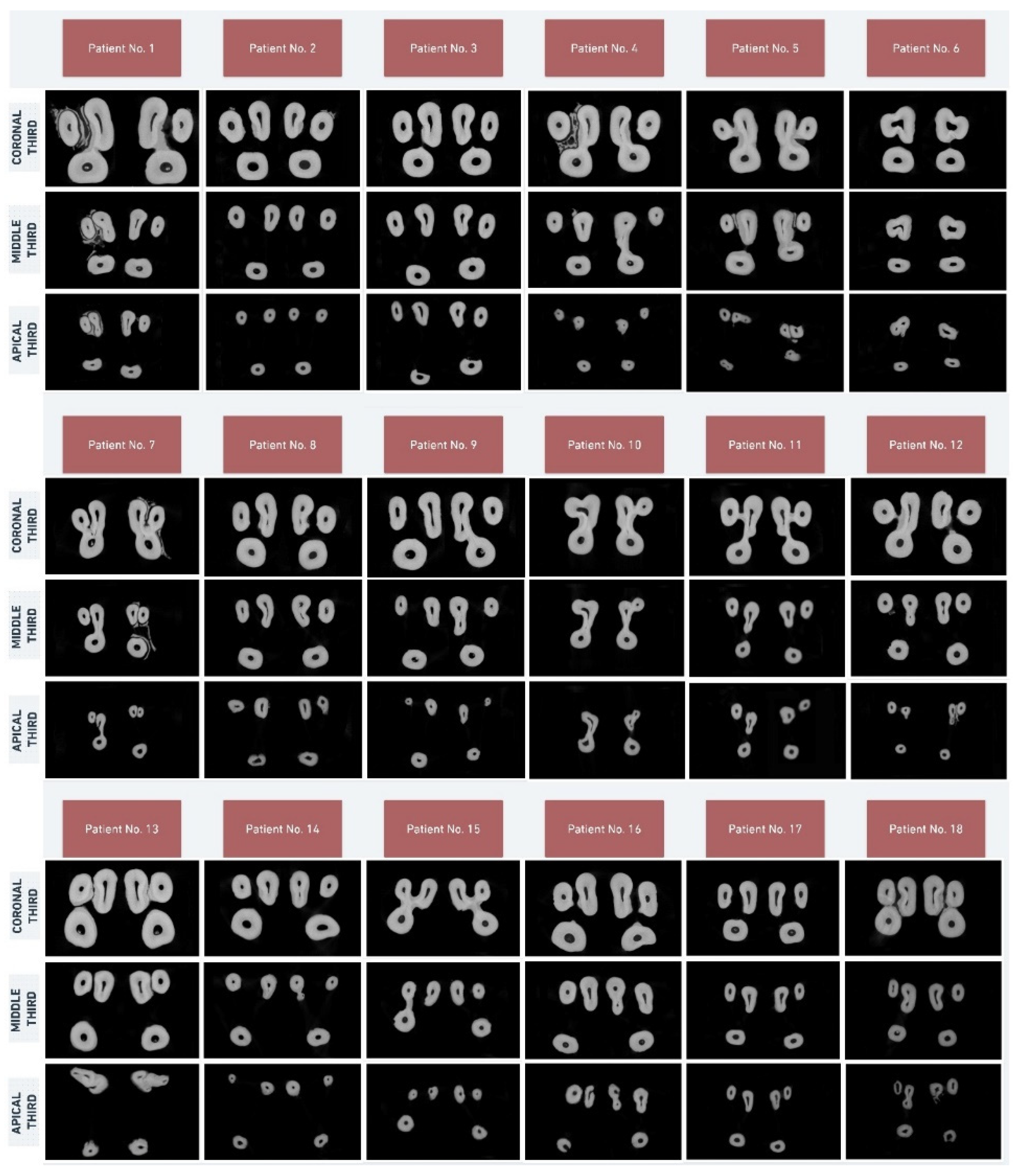
3.1. External Root and Root Canal Morphology

Figure 1 shows exemplary 3D models of contralateral pairs maxillary second molar teeth and the external morphology of roots. Figure 2 shows the axial sections of eighteen maxillary second molar teeth pairs. Table 1 shows the number of roots and configurations of the root canals in contralateral maxillary second molars. Of the thirty-six teeth, three had two roots, and the other thirty-three had three roots. Regarding the number of roots, symmetry of the maxillary second molars was detected in 94.4% of the 18 pairs. In the maxillary second molars, the configuration of the root canal of the mesiobuccal (MB) root was 57.1% type I, 18.2% type V, 12.1% type II, 6.1% type III, and 6.1% type IV. The configuration of the root canal of the distobuccal (DB) root was 97% type I and 3% type III, while the canal configuration of the palatal (P) root was 97.2% type I and 2.8% type III. Of the three buccal (B) roots in teeth with two roots, two had type V, and the configuration of the root canal in one was type I. Regarding the root canal configurations, MB, DB, and P root pairs were found to be symmetrical in 41.1%, 88.2%, and 94.4% of cases, respectively.

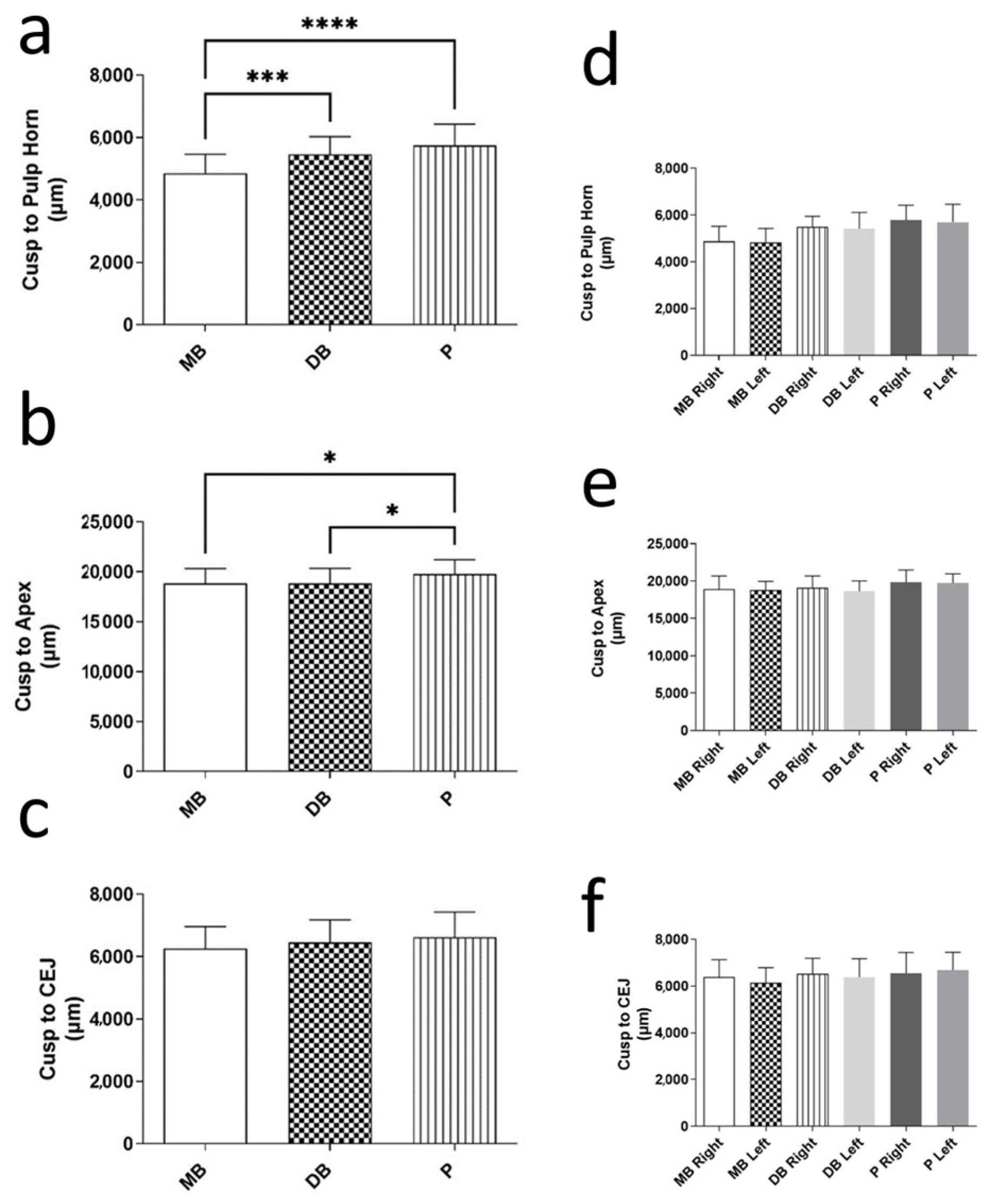
3.2. Cusp-to-Pulp Horn, Cusp-to-Apex, and Cusp-to-CEJ Distances

The mean distances from the MB, DB, and P cusps to the corresponding pulp horn, root apex, and CEJ are presented in Figure 3. The mean distance between the MB cusp and the pulp horn was significantly less than the DB cusp-to-pulp horn (p < 0.01) and P cusp-to-pulp horn distances (p < 0.01). Regarding the cusp-to-apex measurements, the P cusp-to-apex distance was considerably greater than the others (p < 0.05). With regard to the cusp-to-CEJ distances, significant differences were not detected (p > 0.05). The contralateral measurements of the same parameters for the right and left sides were not significantly different (p > 0.05).

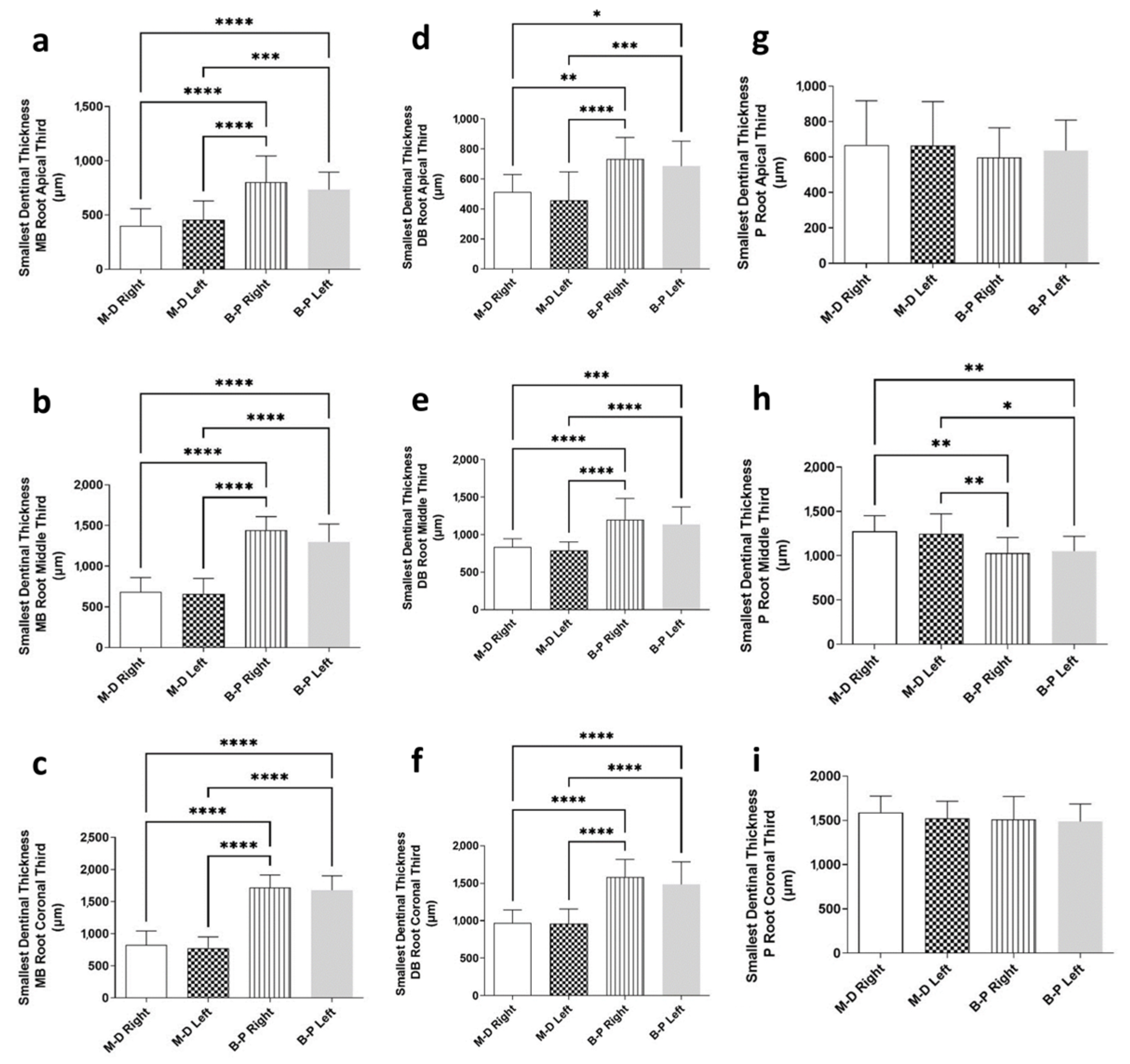
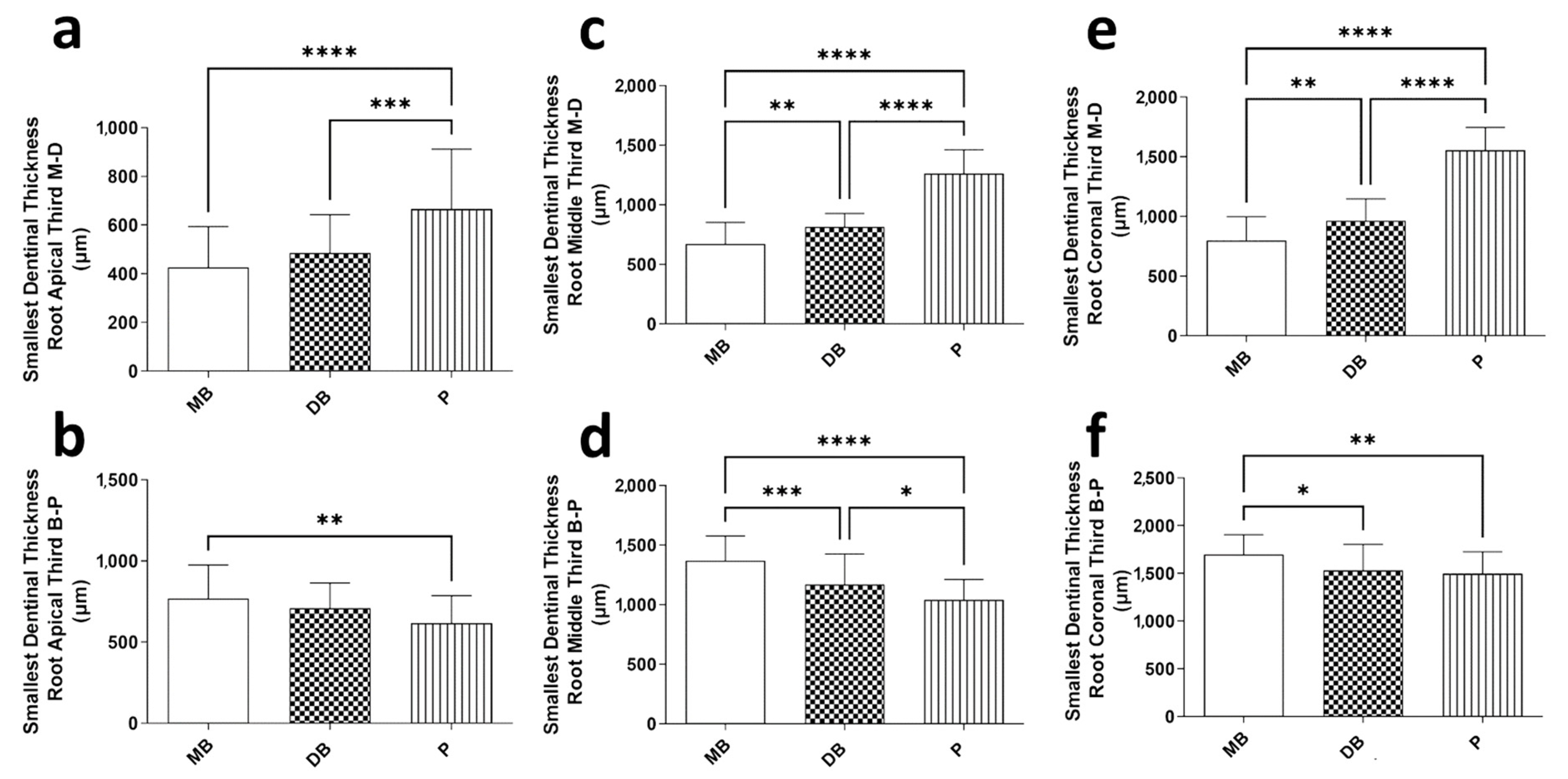
3.3. Smallest Dentinal Thickness in Apical, Middle, and Coronal Thirds

Figure 4 shows the smallest dentinal thickness values of MB, DB, and P roots in two different directions (M-D and B-P) and three root levels (apical, middle, and coronal) of each root. Concerning the contralateral measurements of all evaluated parameters for the smallest dentinal thickness, no significant differences between the right and left sides were found (p > 0.05). The B-P dimensions of the DB and MB roots were significantly higher compared to the M-D dimensions in all root levels (p < 0.01). For the P root, the smallest dentinal thickness values were similar between M-B and B-P measurements in the apical and coronal levels (p > 0.05). However, in the P root’s middle third, the M-D dimension was significantly higher than the B-P dimension for the smallest dentinal thickness (p < 0.05; p < 0.01). Figure 5 represents the differences between the MB, DB, and P roots for the smallest dentinal thickness values in the M-B and D-B directions. The P root showed the thickest dentinal tissue in the M-D direction for all root levels (p < 0.01), whereas in the B-P direction, the MB root showed the thickest dentinal tissue for all root levels (p < 0.01).

3.4. Number of Foramina and Lateral Canals and Locations of Lateral Canals

Table 2 and Table 3 demonstrate how the foramina and number of lateral canals were distributed for MB, DB, and P roots. Concerning the contralateral measurements for the number of foramina and lateral canals, the right and left sides of the same root did not exhibit any significant differences (p > 0.05). However, the number of lateral canals in the MB and P roots was significantly higher compared to that in the DB root (p < 0.01). In addition, the mean number of foramina in the MB root was significantly higher than in the DB and P roots (p > 0.01). Table 4 demonstrates how the lateral canals were distributed. With respect to the MB root, the highest number of lateral canals was in the middle third, whereas for the P root, the apical third had the highest number of lateral canals.

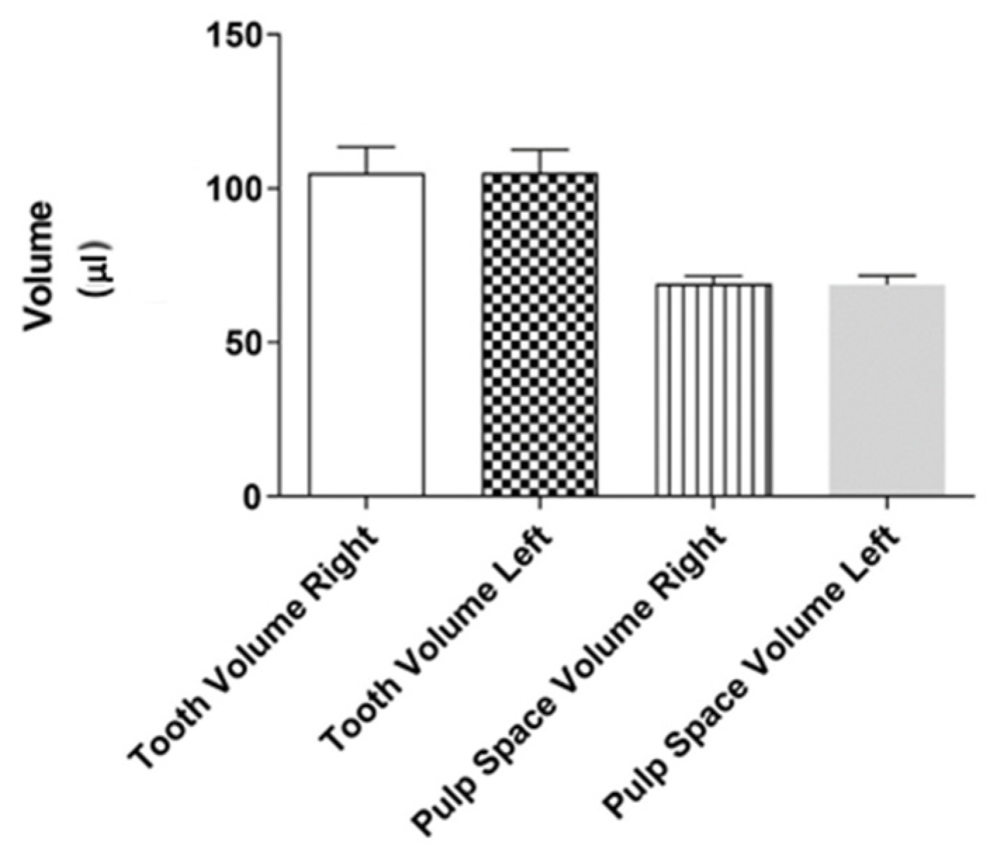
3.5. Tooth and Pulp Space Volume

Figure 6 shows the mean volumes of teeth and pulp spaces. In terms of volumetric measurements, the left and right sides of the same tooth did not exhibit any significant differences (p > 0.05).

4. Discussion

The use of advanced imaging technologies allows root canal systems to be examined more precisely as well as the teeth’s external and internal morphology to be analyzed in greater detail. For studying root canal systems and understanding their complex morphology, micro-CT technology is currently regarded as a research tool that offers optimal accuracy and reliability [12]. The present research utilized the nondestructive micro-CT imaging technique to precisely measure linear and volumetric parameters. Thus, it was possible to evaluate tooth morphology quantitatively and classify it at the same time without causing damage to the tooth structures [22].
The current study’s findings reveal that 94.4% of the patients had symmetrical second molars (17/18) concerning the number of roots. However, the root canal configuration was found to be symmetrical in only 38.8% of the patients (7/18). This outcome is quite contrary to that of Plotino et al. [13], who found that only 79.6% of patients studied had symmetrical maxillary second molars. In their study, the results also only showed the presence of the second mesiobuccal canal in 13.4% of the maxillary second molars, whereas many other researchers have reported an increased prevalence of second mesiobuccal canals, varying between 21% and 83% of teeth [23,24,25]. The latest meta-analysis on the second mesiobuccal root canal in maxillary molars reported that multiple root canals were present in the mesiobuccal root in 37.0% of the maxillary second molars [26]. This also accords with the present results, which show that 41.1% of the evaluated teeth had a second mesiobuccal canal. Demographics such as the age, sex, and location of the individual could be factors that influence the internal anatomy of teeth. It was hypothesized that Asians and Europeans have smaller teeth compared to Africans, which may explain the lower incidence of second mesiobuccal canals [27].
The present study’s results reveal a high incidence of asymmetry in the root canal configuration of maxillary second molars, which could have some implications for endodontic treatment planning and execution. It is important to keep in mind that there can be differences in the root canal anatomy of opposite molars in the same patient, with variations of up to 60%. This should be considered when treating both molars. This information is crucial from a clinical standpoint as failure to identify, clean, and fill an additional canal in molar teeth can result in poor long-term outcomes due to lingering infection [5]. Studies have shown that root canal treatment for maxillary molars has high failure rates, possibly because of their complex root and canal structure, and variations in additional root canals [28,29]. Thus, specialized instruments or techniques, such as an operating microscope or laser-assisted irrigation, may need to be used during treatment in order to navigate and clean the root canals more effectively.
According to the limited findings of this study, MB pulp horns are more prominent in maxillary second molars than in other pulp horns. Analogous findings were also recorded by Baltacioglu et al. [27] for MB pulp horns of maxillary first molars. Thus, caution is advised when providing restorative treatment to prevent accidental pulp exposure. The results of the current research also show that the distances on the left and right sides, from different pulp horns to cusps, were quite similar to each other. The same observation was made for the cusp-to-apex and cusp-to-CEJ distances. These results suggest that the right and left teeth are highly symmetrical in terms of these specific parameters. Similar morphometric measurements such as thickness, width, and length were also studied by Johnsen et al. [16] in maxillary premolars, and they reported that contralateral premolar teeth were highly bilaterally symmetrical.
Significant differences were shown for the smallest dentinal thickness values when comparing differences between palatal, distobuccal, and mesiobuccal roots. The dentine thickness of the MB canal in the mesiodistal dimension was significantly thinner than the others in all root levels. Several reports in the literature have investigated the thickness of dentine at the furcation level of maxillary molars, commonly known as the “danger zone” [30]. It is important to reduce the volume of dentin that is removed through mechanical means when endodontic treatment is applied, especially for mesiobuccal canals that are placed close to furcal concavities, in order to avoid the development of perforations [30]. This study confirms that the mesiobuccal root is particularly at risk for root perforations, which are challenging to treat, and so care should be taken to prevent them.
In the present study, mesiobuccal roots were found to have the highest number of lateral canals. Similar observations have been reported by others [29,30]. Vertucci [31] reported the rates of mesiobuccal, distobuccal, and palatal roots that have a lateral canal were 50%, 29%, and 42%, respectively. Wolf et al. [32] also reported that mesiobuccal roots had the highest rate of lateral canals at 27%, while distobuccal roots had a rate of 11.3% and palatal roots had a rate of 14.6%, respectively. In the present report, mesiobuccal (%44.1), distobuccal (2.9%), and palatal (%30.5) roots had at least one lateral canal. The distinct difference regarding findings on the distobuccal root between this study and others can be attributed to factors such as the size of the sample, the methods and design of the study, the ethnicity of the participants, and distinctions in the age and gender of the subjects. The complicated anatomy of the canal, which includes lateral canals and other potentially inaccessible areas, particularly on maxillary molars’ mesiobuccal roots, poses a threat to chemomechanical preparation. Therefore, the use of advanced disinfection techniques in the endodontic treatment of these teeth will increase the success rate.
The present research had several limitations, the most significant being the small sample size. Another limitation is that the impact of sexual dimorphism was not considered due to the small sample size. As a result, the findings may not be applicable to other demographics, such as age, sex, and location. In order to address these limitations, future studies should include larger samples and investigate possible differences related to age, gender, and population.

5. Conclusions

To date, no micro-CT studies have been conducted to examine the extent to which right and left maxillary second molars are anatomically symmetrical due to the difficulty of finding a patient with both types of teeth. The present data indicate that the contralateral maxillary second molars were highly bilaterally symmetrical according to the morphometric measurements. Root canal configuration varied between contralateral pairs, most notably in mesiobuccal roots. Therefore, the null hypothesis was partially accepted. Micro-CT can be effectively used to study and compare contralateral teeth morphology.

Author Contributions

Conceptualization, G.D., U.A., M.B.O. and K.O.; methodology, K.O. and U.A.; investigation, G.D. and U.A.; resources, M.B.O. and G.D.; data curation, G.D. and U.A.; writing—original draft preparation, G.D. and U.A; writing—review and editing, U.A., M.B.O. and K.O.; visualization, U.A.; supervision, K.O. All authors have read and agreed to the published version of the manuscript.

Funding

This research received no external funding.

Data Availability Statement

The data presented in this study are available on request from the corresponding author.

Acknowledgments

This paper was part of the PhD Thesis of Ghassan Dandache. The authors thank Ahmet Özer Sehirli for assistance in statistical analysis.

Conflicts of Interest

The authors declare no conflict of interest.

References

  1. Martins, J.N.R.; Nole, C.; Ounsi, H.F.; Parashos, P.; Plotino, G.; Ragnarsson, M.F.; Aguilar, R.R.; Santiago, F.; Seedat, H.C.; Vargas, W.; et al. Worldwide Assessment of the Mandibular First Molar Second Distal Root and Root Canal: A Cross-sectional Study with Meta-analysis. J. Endod. 2022, 48, 223–233. [Google Scholar] [CrossRef]
  2. Friedman, S.; Abitbol, S.; Lawrence, H.P. Treatment outcome in endodontics: The Toronto Study. Phase 1: Initial treatment. J. Endod. 2003, 29, 787–793. [Google Scholar] [CrossRef]
  3. Setzer, F.C.; Boyer, K.R.; Jeppson, J.R.; Karabucak, B.; Kim, S. Long-term prognosis of endodontically treated teeth: A retrospective analysis of preoperative factors in molars. J. Endod. 2011, 37, 21–25. [Google Scholar] [CrossRef] [PubMed]
  4. Ran, S.J.; Yang, X.; Sun, Z.; Zhang, Y.; Chen, J.X.; Wang, D.M.; Liu, B. Effect of length of apical root resection on the biomechanical response of a maxillary central incisor in various occlusal relationships. Int. Endod. J. 2020, 53, 111–121. [Google Scholar] [CrossRef] [PubMed]
  5. Baruwa, A.O.; Martins, J.N.R.; Meirinhos, J.; Pereira, B.; Gouveia, J.; Quaresma, S.A.; Monroe, A.; Ginjeira, A. The Influence of Missed Canals on the Prevalence of Periapical Lesions in Endodontically Treated Teeth: A Cross-sectional Study. J. Endod. 2020, 46, 34–39. [Google Scholar] [CrossRef] [PubMed]
  6. Pereira, B.; Martins, J.N.R.; Baruwa, A.O.; Meirinhos, J.; Gouveia, J.; Quaresma, S.A.; Monroe, A.; Ginjeira, A. Association between Endodontically Treated Maxillary and Mandibular Molars with Fused Roots and Periapical Lesions: A Cone-beam Computed Tomography Cross-sectional Study. J. Endod. 2020, 46, 771–777. [Google Scholar] [CrossRef]
  7. Markvart, M.; Tibbelin, N.; Pigg, M.; EndoReCo; Fransson, H. Frequency of additional treatments in relation to the number of root filled canals in molar teeth in the Swedish adult population. Int. Endod. J. 2021, 54, 826–833. [Google Scholar] [CrossRef]
  8. Fransson, H.; Dawson, V.S.; Frisk, F.; Bjørndal, L.; EndoReCo; Kvist, T. Survival of Root-filled Teeth in the Swedish Adult Population. J. Endod. 2016, 42, 216–220. [Google Scholar] [CrossRef]
  9. Martins, J.N.; Mata, A.; Marques, D.; Caramês, J. Prevalence of Root Fusions and Main Root Canal Merging in Human Upper and Lower Molars: A Cone-beam Computed Tomography In Vivo Study. J. Endod. 2016, 42, 900–908. [Google Scholar] [CrossRef] [PubMed]
  10. Mashyakhy, M.; Chourasia, H.R.; Jabali, A.; Almutairi, A.; Gambarini, G. Analysis of Fused Rooted Maxillary First and Second Molars with Merged and C-shaped Canal Configurations: Prevalence, Characteristics, and Correlations in a Saudi Arabian Population. J. Endod. 2019, 45, 1209–1218. [Google Scholar] [CrossRef]
  11. Zhang, Q.; Chen, H.; Fan, B.; Fan, W.; Gutmann, J.L. Root and root canal morphology in maxillary second molar with fused root from a native Chinese population. J. Endod. 2014, 40, 871–875. [Google Scholar] [CrossRef] [PubMed]
  12. Aksoy, U.; Küçük, M.; Versiani, M.A.; Orhan, K. Publication trends in micro-CT endodontic research: A bibliometric analysis over a 25-year period. Int. Endod. J. 2021, 54, 343–353. [Google Scholar] [CrossRef]
  13. Plotino, G.; Tocci, L.; Grande, N.M.; Testarelli, L.; Messineo, D.; Ciotti, M.; Glassman, G.; D’ambrosio, F.; Gambarini, G. Symmetry of root and root canal morphology of maxillary and mandibular molars in a white population: A cone-beam computed tomography study in vivo. J. Endod. 2013, 39, 1545–1548. [Google Scholar] [CrossRef]
  14. Kirmizi, D.; Aksoy, U.; Orhan, K. Efficacy of Laser-Activated Irrigation and Conventional Techniques in Calcium Hydroxide Removal from Simulated Internal Resorption Cavities: Micro-CT Study. Photobiomodul. Photomed. Laser Surg. 2021, 39, 674–681. [Google Scholar] [CrossRef] [PubMed]
  15. Ordinola-Zapata, R.; Bramante, C.M.; Versiani, M.A.; Moldauer, B.I.; Topham, G.; Gutmann, J.L.; Nuñez, A.; Duarte, M.A.; Abella, F. Comparative accuracy of the Clearing Technique, CBCT and Micro-CT methods in studying the mesial root canal configuration of mandibular first molars. Int. Endod. J. 2017, 50, 90–96. [Google Scholar] [CrossRef] [PubMed]
  16. Johnsen, G.F.; Dara, S.; Asjad, S.; Sunde, P.T.; Haugen, H.J. Anatomic Comparison of Contralateral Premolars. J. Endod. 2017, 43, 956–963. [Google Scholar] [CrossRef] [PubMed]
  17. Johnsen, G.F.; Sundnes, J.; Wengenroth, J.; Haugen, H.J. Methodology for Morphometric Analysis of Modern Human Contralateral Premolars. J. Comput. Assist. Tomogr. 2016, 40, 617–625. [Google Scholar] [CrossRef]
  18. Feldkamp, L.A.; Goldstein, S.A.; Parfitt, A.M.; Jesion, G.; Kleerekoper, M. The direct examination of three-dimensional bone architecture in vitro by computed tomography. J. Bone Miner Res. 1989, 4, 3–11. [Google Scholar] [CrossRef]
  19. Alavi, A.M.; Opasanon, A.; Ng, Y.L.; Gulabivala, K. Root and canal morphology of Thai maxillary molars. Int. Endod. J. 2002, 35, 478–485. [Google Scholar] [CrossRef]
  20. Divine, K.A.; McClanahan, S.B.; Fok, A. Anatomic Analysis of Palatal Roots of Maxillary Molars Using Micro-computed Tomography. J. Endod. 2019, 45, 724–728. [Google Scholar] [CrossRef]
  21. American Association of Endodontists. Glossary of Endodontic Terms, 10th ed.; American Association of Endodontists: Chicago, IL, USA, 2020; Available online: https://www.aae.org/specialty/clinical-resources/glossary-endodontic-terms/ (accessed on 22 January 2023).
  22. Aktuna Belgin, C.; Serindere, G.; Orhan, K. Accuracy and reliability of enamel and dentin thickness measurements on micro-computed tomography and digital periapical radiographs#. J. Foren. Rad. Imag. 2019, 18, 32–36. [Google Scholar]
  23. Fernandes, N.A.; Herbst, D.; Postma, T.C.; Bunn, B.K. The prevalence of second canals in the mesiobuccal root of maxillary molars: A cone beam computed tomography study. Aust. Endod. J. 2019, 45, 46–50. [Google Scholar] [CrossRef] [PubMed]
  24. Kalender, A.; Celikten, B.; Tufenkci, P.; Aksoy, U.; Basmacı, F.; Kelahmet, U.; Orhan, K. Cone beam computed tomography evaluation of maxillary molar root canal morphology in a Turkish Cypriot population. Biotech. Biotechn. Equip. 2016, 30, 145–150. [Google Scholar] [CrossRef]
  25. Reis, A.G.; Grazziotin-Soares, R.; Barletta, F.B.; Fontanella, V.R.; Mahl, C.R. Second canal in mesiobuccal root of maxillary molars is correlated with root third and patient age: A cone-beam computed tomographic study. J. Endod. 2013, 39, 588–592. [Google Scholar] [CrossRef]
  26. Martins, J.N.R.; Marques, D.; Silva, E.J.N.L.; Caramês, J.; Mata, A.; Versiani, M.A. Second mesiobuccal root canal in maxillary molars-A systematic review and meta-analysis of prevalence studies using cone beam computed tomography. Arch. Oral Biol. 2020, 113, 104589. [Google Scholar] [CrossRef]
  27. Baltacıoğlu, İ.H.; Demirel, G.; Kolsuz, M.E.; Orhan, K. In-vitro analysis of maxillary first molars morphology using three dimensional Micro-CT imaging: Considerations for restorative dentistry. Eur. Oral Res. 2018, 52, 75–81. [Google Scholar] [CrossRef]
  28. Carrion, S.J.; Coelho, M.S.; Soares, A.J.; Frozoni, M. Apical periodontitis in mesiobuccal roots of maxillary molars: Influence of anatomy and quality of root canal treatment, a CBCT study. Restor. Dent. Endod. 2022, 19, e37. [Google Scholar] [CrossRef] [PubMed]
  29. Kalender, A.; Orhan, K.; Aksoy, U.; Basmaci, F.; Er, F.; Alankus, A. Influence of the quality of endodontic treatment and coronal restorations on the prevalence of apical periodontitis in a Turkish Cypriot population. Med. Princ. Pract. 2013, 22, 173–177. [Google Scholar] [CrossRef]
  30. Ordinola-Zapata, R.; Martins, J.N.R.; Versiani, M.A.; Bramante, C.M. Micro-CT analysis of danger zone thickness in the mesiobuccal roots of maxillary first molars. Int. Endod. J. 2019, 52, 524–529. [Google Scholar] [CrossRef]
  31. Vertucci, F.J. Root canal anatomy of the human permanent teeth. Oral Surg. Oral Med. Oral Pathol. Oral Radiol. 1984, 58, 589–599. [Google Scholar] [CrossRef]
  32. Wolf, T.G.; Paqué, F.; Woop, A.C.; Willershausen, B.; Briseño-Marroquín, B. Root canal morphology and configuration of 123 maxillary second molars by means of micro-CT. Int. J. Oral Sci. 2017, 9, 33–37. [Google Scholar] [CrossRef] [PubMed] [Green Version]
Figure 1. Three-dimensional models of contralateral pairs of three-rooted (A) and two-rooted (B) maxillary second molar teeth. (C,D) show the root canal foramina of mesiobuccal (MB), distobuccal (DB), and palatal (P) roots. Red arrow indicates apically located lateral canal opening in the distal side of the mesiobuccal root.
Symmetry 15 00420 g001
Figure 2. The axial sections of eighteen maxillary second molar teeth pairs.
Symmetry 15 00420 g002
Figure 3. The mean distances from the mesiobuccal (MB), distobuccal (DB), and palatal (P) cusps to the corresponding pulp horn (a), root apex (b), and cementoenamel junction (CEJ) (c). The contralateral measurements of the same parameters for the right and left sides are presented in (df). * = p < 0.05, ** = p < 0.01, *** = p < 0.001, **** = p < 0.0001.
Symmetry 15 00420 g003
Figure 4. The smallest dentinal thickness values of mesiobuccal (ac), distobuccal (df), and palatal roots (gi) in two different directions (mesiodistal (M-D) and buccopalatal (B-P)) and three root levels (apical, middle, and coronal) of each root. * = p < 0.05, ** = p < 0.01, *** = p < 0.001, **** = p < 0.0001.
Symmetry 15 00420 g004
Figure 5. The differences between mesiobuccal (MB), distobuccal (DB), and palatal (P) roots for the smallest dentinal thickness values in mesiobuccal (M-B) and distobuccal (D-B) directions at the apical (a,b), middle (c,d), and coronal (e,f) root levels. * = p < 0.05, ** = p < 0.01, *** = p < 0.001, **** = p < 0.0001.
Symmetry 15 00420 g005
Figure 6. The mean volumes of teeth and pulp spaces.
Symmetry 15 00420 g006
Table 1. Number of roots and root canal configurations in contralateral maxillary second molars.
Patient No.RightLeftSymmetry in No. of RootsSymmetry in Root Canal Configurations
No. of RootsRootVertucci’s ClassificationNo. of RootsRootVertucci’s Classification
13MBType V3MBType IYesNo
DBType IDBType IYes
PType IPType IYes
23MBType I3MBType IYesYes
DBType IDBType IYes
PType IPType IYes
33MBType I3MBType IYesYes
DBType IDBType IYes
PType IPType IYes
43MBType I3MBType IIYesNo
DBType IDBType IYes
PType IPType IYes
53MBType V3MBType IIIYesNo
DBType IDBType IYes
PType IPType VNo
62BType I2BType VYesNo
PType IPType IYes
73MBType I3MBType IYesYes
DBType IDBType IYes
PType IPType IYes
83MBType II3MBType IIYesYes
DBType IDBType IYes
PType IPType IYes
93MBType I3MBType IIYesNo
DBType IDBType IYes
PType IPType IYes
102BType V3MBType INoNo
DBType INo
PType IPType IYes
113MBType I3MBType IYesYes
DBType IDBType IYes
PType IPType IYes
123MBType III3MBType VYesNo
DBType IDBType IYes
PType IPType IYes
133MBType I3MBType VYesNo
DBType IDBType IYes
PType IPType IYes
143MBType V3MBType IVYesNo
DBType IDBType IYes
PType IPType IYes
153MBType I3MBType IYesYes
DBType IDBType IYes
PType IPType IYes
163MBType I3MBType VYesNo
DBType IDBType IYes
PType IPType IYes
173MBType I3MBType IYesYes
DBType IDBType IYes
PType IPType IYes
183MBType IV3MBType IYesNo
DBType IIIDBType INo
PType IPType IYes
MB: mesiobuccal root; DB: distobuccal root; B: buccal root; P: palatal root.
Table 2. Number of foramina.
Patient
No.
MBDBBP
RightLeftRightLeftRightLeftRightLeft
12321 11
21112 11
31111 11
43211 11
52211 22
6 2411
72121 21
86411 11
91111 11
102 1 311
111111 11
123211 11
132111 11
142111 11
151111 11
162211 11
171411 11
184311 11
MB: mesiobuccal root; DB: distobuccal root; B: buccal root; P: palatal root.
Table 3. Number of lateral canals.
Patient
No.
MBDBBP
RightLeftRightLeftRightLeftRightLeft
11100 10
20000 00
30000 00
41000 00
50100 12
6 1001
71210 12
80000 00
90100 11
100 0 200
111200 00
121200 01
131000 00
140300 00
150000 00
162100 10
170000 01
180000 00
MB: mesiobuccal root; DB: distobuccal root; B: buccal root; P: palatal root.
Table 4. Locations of lateral canals.
Lateral Canal
Locations
MBDBBP
Coronal_Buccal1003
Coronal_Palatal2000
Coronal_Mesial1000
Coronal_Distal2000
Middle_Buccal2000
Middle_Palatal3010
Middle_Mesial3000
Middle_Distal3001
Apical_Buccal1006
Apical_Palatal2021
Apical_Mesial0100
Apical_Distal1002
MB: mesiobuccal root; DB: distobuccal root; B: buccal root; P: palatal root.
Disclaimer/Publisher’s Note: The statements, opinions and data contained in all publications are solely those of the individual author(s) and contributor(s) and not of MDPI and/or the editor(s). MDPI and/or the editor(s) disclaim responsibility for any injury to people or property resulting from any ideas, methods, instructions or products referred to in the content.

