Article

Relapse after Orthodontic-Surgical Treatment: A Retrospective Longitudinal Study

by
João Matos
1,
Raquel Travassos
1,2,3,4,
Francisco Caramelo
2,3,4,5,6,
Madalena Prata Ribeiro
1,
Filipa Marques
1,
Carlos Miguel Marto
2,3,4,6,7,8,*,
Anabela Baptista Paula
1,2,3,4,6,7,
Catarina Nunes
1,2,3,4,
Inês Francisco
1,2,3,4,6,* and
Francisco Vale
1,2,3,4,6
1
Institute of Orthodontics, Faculty of Medicine, University of Coimbra, 3000-075 Coimbra, Portugal
2
Coimbra Institute for Clinical and Biomedical Research (iCBR), Area of Environment Genetics and Oncobiology (CIMAGO), Faculty of Medicine, University of Coimbra, 3000-075 Coimbra, Portugal
3
Centre for Innovative Biomedicine and Biotechnology (CIBB), University of Coimbra, 3000-075 Coimbra, Portugal
4
Clinical Academic Center of Coimbra (CACC), 3030-370 Coimbra, Portugal
5
Laboratory of Biostatistics and Medical Informatics (LBIM), Faculty of Medicine, University of Coimbra, 3004-531 Coimbra, Portugal
6
Laboratory for Evidence-Based Sciences and Precision Dentistry, University of Coimbra, 3000-075 Coimbra, Portugal
7
Institute of Integrated Clinical Practice, Faculty of Medicine, University of Coimbra, 3004-531 Coimbra, Portugal
8
Institute of Experimental Pathology, Faculty of Medicine, University of Coimbra, 3004-531 Coimbra, Portugal
*
Authors to whom correspondence should be addressed.
Symmetry 2023, 15(5), 1083; https://doi.org/10.3390/sym15051083
Submission received: 1 May 2023 / Revised: 11 May 2023 / Accepted: 13 May 2023 / Published: 15 May 2023
(This article belongs to the Special Issue Symmetry in Dentistry: From the Clinic to the Lab II)

Abstract

:
Long-term stability is a crucial point in order to keep the patient’s aesthetic and functional balance. The aim of this study was to evaluate dental and skeletal relapse in patients who underwent orthodontic-surgical treatment. This retrospective study included 25 patients who corrected their dentofacial deformity through orthodontics and orthognathic surgery. The dental casts and lateral cephalograms were evaluated prior to orthodontic treatment (T0), final of orthodontic-surgical treatment (T1) and long-term retention phase (T2). The Wilcoxon test with p-value corrected by the Benjamini–Hochberg method was used to assess differences between the groups. The influence of retention duration was assessed using the Kruskal–Wallis method. The association of nominal variables and differences between quantitative variables were assessed using the Fisher and Mann–Whitney tests, respectively. No dental or skeletal variable presented statistically significant differences between the final orthodontic-surgical treatment and the long-term retention phase. Eight patients presented dental relapse (32–95% CI [12.4%; 51.7%]), but no skeletal relapse was observed in any of the 25 individuals. The type of malocclusion did not influence the relapse rate of orthodontic-surgical treatment (Fisher, p = 0.202). No differences were found between the different retention times, sex and age at the end of treatment. Orthodontic-surgical treatment showed long-term stability in the present study group.

1. Introduction

Dentofacial deformity (DFD) is characterized by a disharmony between the jaws or the structures of the skull base, resulting from interference during the individual’s growth and development process (Table S1 in Supplementary Materials) [1]. The literature refers to a prevalence of approximately 5% in the population of western countries [2].
The etiology of DFD can be congenital, acquired or as a consequence of trauma or tumor processes. Congenital DFD may or may not be associated with craniofacial syndromes, while acquired DFD results from neuromuscular and functional modifications. However, there are still situations in which the etiology of DFD remains unknown [2]. DFD usually develops a malocclusion which can result in aesthetic and functional consequences, affecting the patient’s quality of life [1].
In growing patients, the correction or reduction of the severity of DFD can be achieved by using dentofacial orthopedics. When growth is completed, there are two options that remain for DFD correction: orthodontic camouflage or orthodontic-surgical treatment. However, patients with severe anteroposterior skeletal discrepancies, skeletal transverse maxillary constriction, airway problems, inadequate facial aesthetics or facial asymmetry often require orthodontic-surgical treatment in order to obtain satisfactory functional and aesthetic results with long-term stability [3,4,5].
Despite maxillofacial asymmetry being present in all individuals, in cases of moderate to severe severity, there is a compromise in the patient’s quality of life at a psychosocial level. Individuals with maxillofacial asymmetry have an aesthetic impairment but also impairment in chewing and speech. Bimaxillary orthognathic surgery (bilateral sagittal ramus osteotomy combined with Le Fort I osteotomy) is a surgical technique more accepted by the scientific community in the treatment of patients with severe facial asymmetry. Orthognathic surgery allows patients with severe asymmetry to achieve a harmonious skeletal structure and subsequently improve their function and aesthetics, which would not be possible to achieve only with conventional orthodontic treatment [3,4,6,7].
The first surgical correction of malocclusion was performed by Hullihen in 1846 [8]. Since then, surgical procedures have been the target of several improvements, with the bilateral mandibular sagittal osteotomy (BSSO) and Le Fort I osteotomy being the most used surgical techniques today [4,8,9].
BSSO (Table S1 in Supplementary Materials) was initially described by Trauner and Obwegeser in 1957, and it involves the division of the mandible in its inferior branch, promoting the controlled repositioning of the proximal segment. This surgical technique has several advantages, including the possibility of manipulating the mandible in the simulation of forward and backward movements, as well as downward and forward movements; compatibility with the use of rigid internal fixation; favorable post-surgical stability due to the excellent bone–bone contact that minimizes problems arising from healing. In about 30% of patients undergoing BSSO, especially in advancement surgeries, it is recommended to add genioplasty to reposition the chin in relation to the mandibular body in the transverse plane [4].
The Le Fort I osteotomy, first described by Langebeck in 1861, is characterized by a horizontal fracture from the piriform aperture to the pterygoid process of the maxilla bilaterally. This procedure was considered a safe surgical approach based on bone microcirculation studies published by Bell in 1975. This intervention allows the maxilla to be moved in the vertical and sagittal planes. However, the maxillary setback is difficult due to the presence of bone interferences and anatomical structures that relate posteriorly, such as the pterygopalatine canal and the pterygoid process [4,10,11].
Orthodontic-surgical treatment is divided into three stages: pre-surgical orthodontics, orthognathic surgery and post-surgical orthodontics. The pre-surgical stage aims to decompensate the dental arches, while the post-surgical stage focuses on final adjustments in order to achieve a stable occlusion [3,4,12].
Several reports have studied factors that influence occlusal stability in orthodontic-surgical treatments to optimize the treatment plan. However, the etiology of skeletal and dental relapse remains unclear seen, as this condition is complex, and the etiology may be multifactorial [13,14].
Treatment relapse can be immediate (short-term relapse) or after a considerable amount of time of the surgical procedure (long-term relapse) [12]. Short-term relapse is described as a physiological adaptation that is related to post-surgical healing and orthodontic treatment and can be the result of an intraoperative error. The most common intraoperative errors occur due to imprecise planning in the direction of movements and applied surgical technique, failure of bone fixation or incorrect and imprecise osteosynthesis, and it can also be identified immediately after the surgical procedure [12]. Long-term relapse can be influenced by several factors, namely surgical, orthodontic and patient-related factors. The surgical factors include condylar adaptation and repositioning in the glenoid fossa, type of fixation, and surgeon experience [12,15]. Patient-related factors comprise masticatory function, occlusion, parafunctional habits (orofacial muscles and adaptive postural pressure of the tongue) and bone remodeling. Orthodontic factors are related to long-term occlusion stability [16,17,18,19].
According to the systematic review by Haas Junior OL et al., the most understandable description of stability in orthognathic surgery refers to two clinical studies carried out by Proffit et al., who report a hierarchical scale of stability, based on their clinical experience with a sample compiled over more than 30 years [16,20].
The orthognathic surgical procedure classified with greater stability is the maxillary impaction and, subsequently, the mandibular advancement. These are the main procedures for correcting severe skeletal class II malocclusions and are considered to be highly stable even without rigid fixation. Most patients with skeletal class III undergoing orthodontic-surgical treatment undergo maxillary advancement surgery or jaw advancement surgery in combination with a mandibular setback. The literature reports that bimaxillary surgery, with maxillary advancement and mandibular setback, is considered stable when rigid fixation is used. However, isolated mandibular setback and lower repositioning of the mandible can be unstable due to the extent of setback and clockwise rotation of the mandible, decreased overbite, and increased overjet [21,22].
According to Proffit et al., clinically relevant recurrence occurs when there is a deviation greater than 2°/2 mm in relation to postoperative baseline values [17,18,20].
According to the studies by Proffit et al., during the first year after surgery, patients with Class II malocclusion with long face present greater stability than Class III patients; one to five years after orthodontic-surgical treatment, patients with skeletal Class III are more stable than Class II patients with long face. In the long term, there are fewer patients with changes in dental occlusion than skeletal changes because dentition generally adapts to the skeletal change [20].
Several studies report that most clinical changes develop over the course of 1–5 years after orthognathic surgery, with skeletal relapse occurring primarily in the first year [20,23,24,25,26,27]. At the dental level, patients with skeletal class II have seen an increase in overjet and overbite in the long term [28,29,30]. In patients with skeletal class III, Bailey et al. reported clinically significant recurrence of the overjet when undergoing bimaxillary surgery, 2% of which with recurrence greater than 4 mm [9,18]. At the skeletal level, patients with skeletal class II had a greater recurrence rate with an increase in the mandibular plane angle and ANB compared to patients with skeletal class III [31].
However, these reports presented several confounding factors, such as different fixation methods, distinct treatment planning and different orthodontists and/or surgeons. Therefore, more information about long-term monitoring is crucial in order to obtain treatments with aesthetic and functional balance in the correction of severe dental and skeletal deformities [9].
The aim of this study was to assess dental and skeletal long-term relapse in patients who underwent orthodontic-surgical treatment and, secondarily, evaluate the influence of the type of malocclusion, retention time, sex and age at the end of treatment.

2. Materials and Methods

2.1. Study Design and Selection of Participants

In this retrospective longitudinal study, the data were collected from the Institute of Orthodontics, Faculty of Medicine, University of Coimbra, between November 2020 and February 2021. The study was conducted in accordance with the Declaration of Helsinki, and the protocol (CE-145/2020) was approved by the Ethics Committee of the Faculty of Medicine, University of Coimbra. All patients gave their informed consent before participating in the study. The subjects of the study were healthy adults that underwent orthodontic-surgical treatment with BSSO and/or Le Fort I with dental casts and lateral cephalograms before and after orthodontic-surgical treatment and at least one year after appliance removal during the retention stage. All orthodontic treatments were performed by the same orthodontist using the 0.018-slot Roth technique. Regarding surgeries, all were performed by the same (team of) surgeons, and semi-rigid fixation was used. Patients with syndromes or craniofacial anomalies, temporomandibular disorder, who underwent surgically assisted rapid maxillary expansion previously or with the loss of more than three teeth for non-orthodontic reasons were excluded from this study. For each patient, dental casts and lateral cephalograms were collected at three time points: prior to orthodontic treatment (T0), final of orthodontic-surgical treatment (T1) and long-term retainer phase (T2). Sex and age at the end of treatment were also recorded.
All measurements were evaluated by the same researcher (J.M.). In order to assess the intraobserver agreement, the researcher repeated the measurements one month after the first observation.

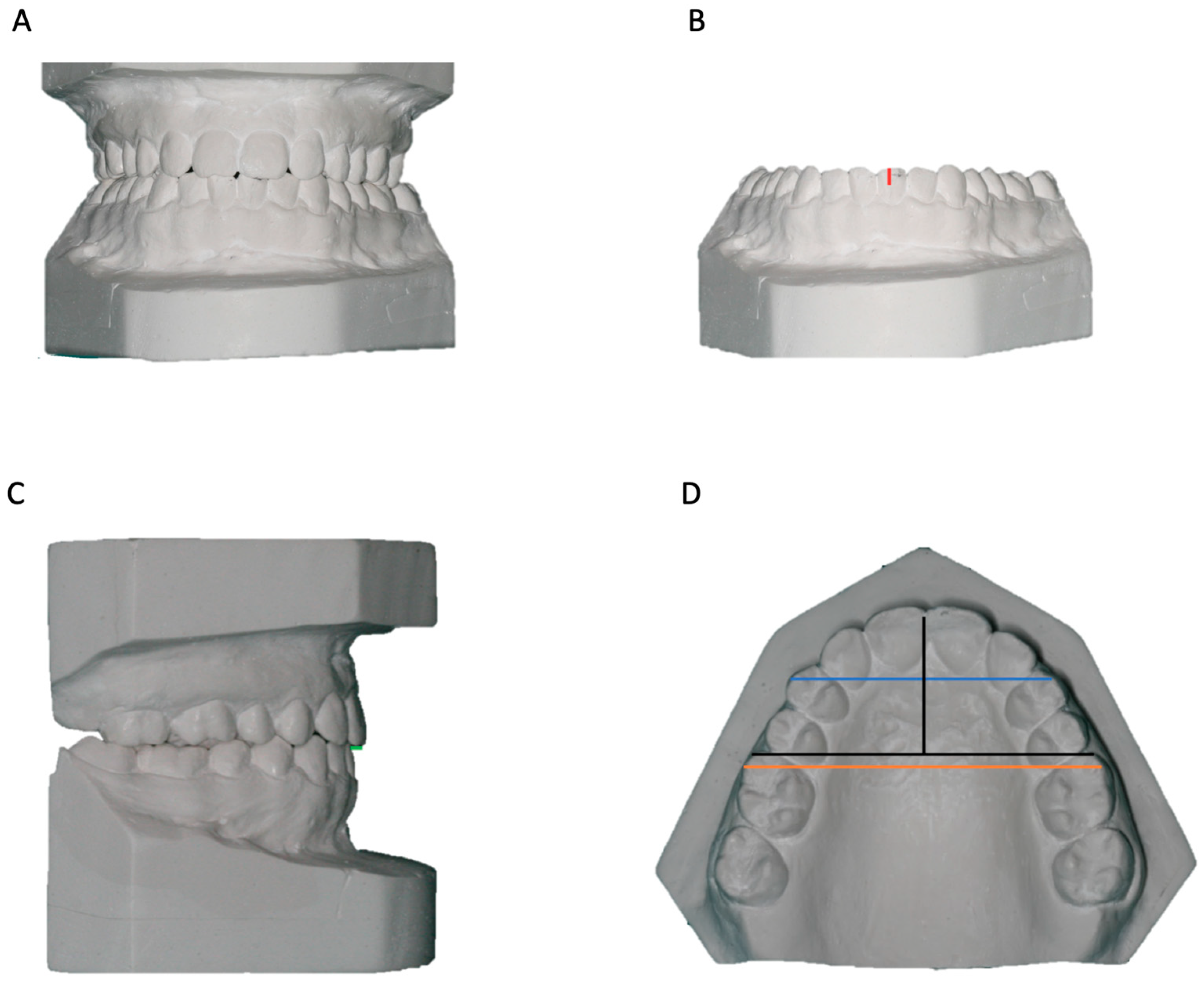
2.2. Evaluation of Dental Cast

The evaluation of dental casts included the measurement of the following variables: overjet, overbite, upper arch depth, inter-canine distance and inter-molar distance in both jaws. The measurements were performed with a caliper and a ruler with a 1 mm measurement scale. Overbite was determined as the vertical overlap of the upper incisor on the corresponding lower incisor in the occluded dental casts (Figure 1A,B). The overjet was measured from the buccal surface of the lower incisor to the incisal edge of the corresponding upper incisor in the occluded dental casts (Figure 1C). The highest overjet/overbite value is considered. The upper arch depth was measured as the perpendicular distance from a tangent mesial to the first molars to the contact point between the central incisors (Figure 1D). The inter-canine distance was established as the linear distance between the cusp tips of the contralateral canines or, in the case of dental wear, the distance between the centers of the worn surfaces (Figure 1D). In the case of upper lateral incisor agenesis and its replacement by the canine, the buccal cusp tip of the contralateral first premolar is considered. The inter-molar distance was defined as the linear distance between the mesiobuccal cusp tips of the contralateral first molars or, in the case of wear, the distance between the centers of the worn surfaces (Figure 1D).

2.3. Analysis of Lateral Cephalograms

Lateral cephalograms were performed by a professional calibrated for this exam using a Siemens® Othophos CD cephalostat with the following adjustments: potential difference from 73 to 84 Kv, exposure time from 13 to 15 mA, 0.6 mm focus, 150 cm focus-film distance, 10 cm distance between the film and the midsagittal plane of the head.
Cephalometric analysis was carried out using Dolphin Image software version 11.9 (Dolphin Image & Management Solutions®, Chatsworth, CA, USA). The variables studied are described in Table 1. All lateral cephalograms were calibrated before the tracing.

2.4. Statistical Analysis

Statistical analysis of the data was performed in the IBM® SPSS® v26 platform and in R v3.3.2 with a statistical significance level of 0.05. The dental and skeletal variables were described using the mean, standard deviation, minimum and maximum at each time point (T0, T1, T2) and for T2 − T1. The Wilcoxon test was used to evaluate the differences between T2 and T1 after verifying the violation of the assumption of normality using the Shapiro–Wilk test. The p-value had been corrected for multiple comparisons by the Benjamini–Hochberg method, with a false positive rate (false discovery rate) equal to 5%. Dental relapse was defined as a difference greater than 2 mm between T2 and T1 in the overjet or (isa-is)–(ii-iia) variables. A skeletal relapse was defined as an ANB difference greater than 2°. The influence of retention time on the relapse was evaluated using the Kruskal–Wallis test and represented by confidence interval graphs whose values were determined using bootstrapping with 1000 samples. The association between nominal variables was assessed using the Fisher test, and the Mann–Whitney test was applied to assess differences between quantitative variables. Intraobserver agreement was assessed using the iota coefficient [32].

3. Results

The sample was comprised of 25 patients (14 female and 11 male) who corrected dentofacial deformity with orthodontic-surgical treatment and were already in a retention stage between June 2005 and October 2021. The iota coefficient for the intra-operator agreement was 1.0, indicating an excellent agreement.

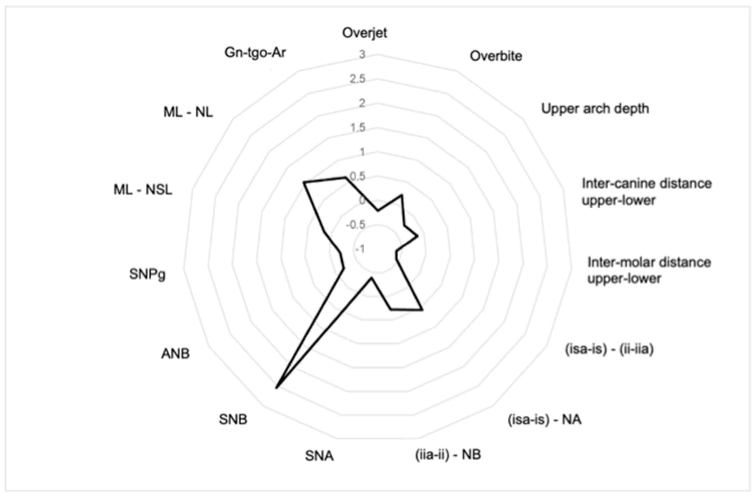
3.1. Dental and Skeletal Relapse

No dental or skeletal variable evaluated presented a statistically significant difference between the final orthodontic-surgical treatment and the long-term retention phase. The mean of the difference between T2 and T1 is illustrated in the radar chart (Figure 2). Table 2 shows the mean, standard deviation, minimum and maximum values for dental and skeletal variables in the three time points as well as the difference between T2 and T1. Eight patients presented dental relapse (32–95% CI [12.4%; 51.7%]), but none (0%) had a skeletal relapse.

3.2. Influence of Malocclusion Type

The sample was comprised of 14 skeletal Class II and 11 skeletal Class III patients. The differences in means of overjet, (isa-is)–(ii-iia) and ANB between T1 and T0 for patients without relapse were −2.45 mm (±6.16), 1.64° (±12.55) and 2.08° (±3.93), respectively. In patients with relapse, the differences in means were −1.14 mm (±4.90), 0.78° (±15.70) and −0.40° (±4.39), respectively. However, no statistically significant differences were found (MW, p = 0.798, MW, p = 0.842 MW, p = 0.0215, respectively).
Regarding the influence of the type of skeletal class, two cases of relapse in patients with skeletal Class III (16.7%) and six cases in patients with skeletal Class II (46.2%) were found without any statistically significant association (Fisher, p = 0.202).

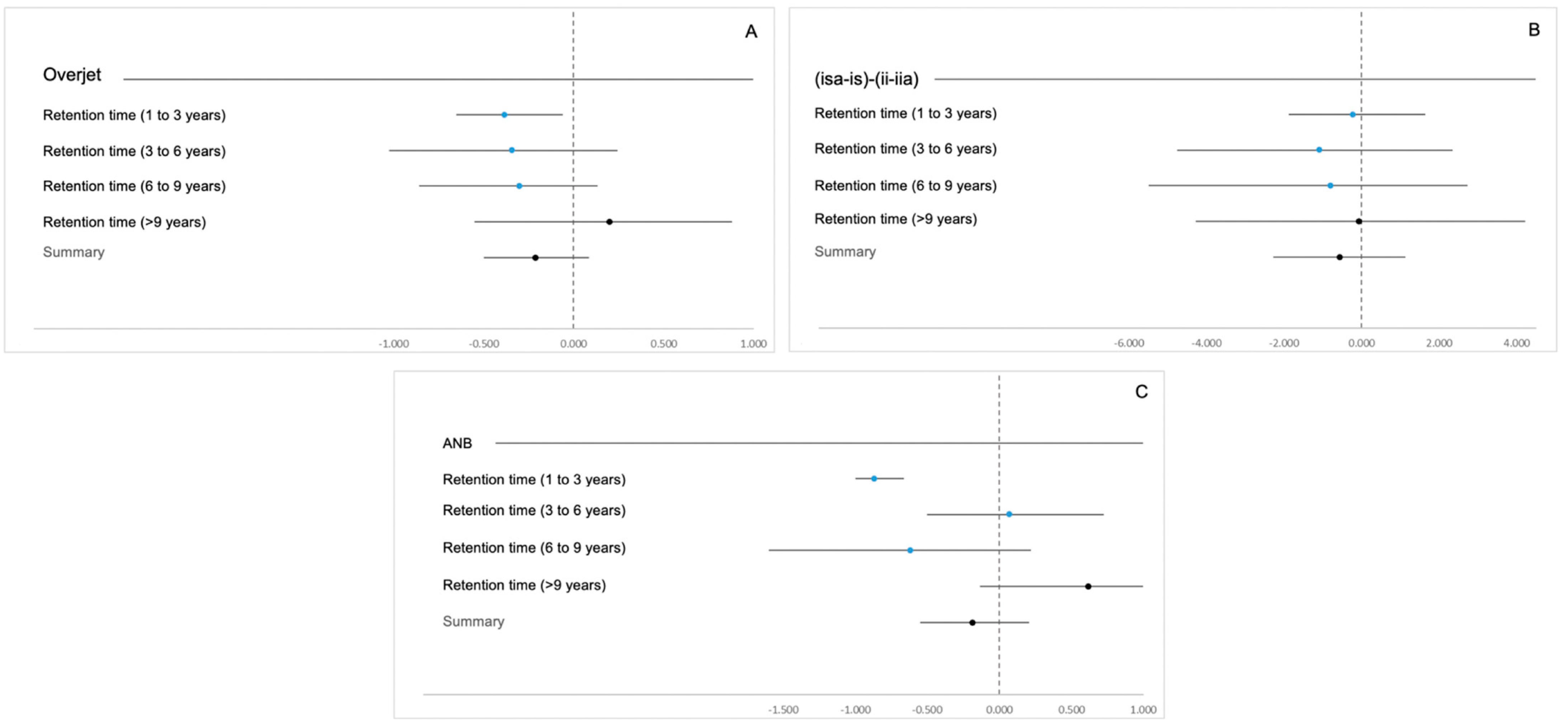
3.3. Influence of Retention Time

There were no statistically significant differences in the confidence intervals of the value of the difference of overjet, (isa-is)–(ii-iia) and ANB between T2 and T1 (KW, p = 0.821 KW, p = 0.988 and KW, p = 0.107, respectively) and the different retention times (Figure 3).

3.4. Influence of Sex and Age at the End of Treatment

Relapse was observed in four female patients (28.6%) and in four male patients (36.4%) with no statistically significant association (Fisher, p = 1000). The mean age at the end of orthodontic-surgical treatment in patients who had relapsed was 27.1 years (±5.06) and 27.1 years (±8.48) in patients with no relapse. There were no statistically significant differences (MW, p = 0.667).

4. Discussion

This study aimed to evaluate dental and skeletal relapse after orthodontic-surgical treatment as well as the influence of malocclusion type, retention time, sex and age at the end of treatment in the stability of the treatment. Monitoring treatment relapse avoids economic and resource expenditure while simultaneously maintaining the patient’s aesthetic and functional balance. The most reported variables to evaluate dental and skeletal relapse in the literature are the overjet and the ANB, SNB and ML–NL angles [9,33,34,35,36,37].
This study showed that orthodontic-surgical treatment seems to have long-term dental and skeletal stability since none of the variables showed statistically significant differences (p > 0.05). However, the variables that registered the greatest changes between T2 and T1 were skeletal (SNB, ML–NL, Gn-tgo-Ar) and dental (inter-molar distance upper-lower, (isa-is)–(ii-iia), (isa-is)–NA). Recently, a systematic review reported a significant relapse in the SNB angle variable, attributing this variation to the condylar displacement by rotation of the proximal segments of the mandible ramus in the long term [9]. Eggensperger et al. reported a significant skeletal long-term relapse, with an increase of the Gn-tgo-Ar and ML–NL angles (2.7° and 2.4°, respectively), justified by continuous condylar resorption that was more prone in patients with higher ML–NL [9,34]. Similarly, Kerstens et al. stated that counterclockwise rotation leads to compression of the most anterior part of the articular surface of the condyle, which can result in progressive condylar resorption [34,38]. According to Sahoo et al., the intraoperative change in the angle of the mandibular plane is positively correlated with the magnitude of recurrence [39].
The difference between these articles and the results of the present study can be explained by the fact that T1 corresponds to the end of orthodontic-surgical treatment and, according to current literature, skeletal relapse occurs, on average, one year after orthognathic surgery [4]. Above 1 year, changes result from some combination of compensatory bone remodeling, postural change, and late growth in the pattern of the original dentofacial deformity and dental changes [40]. Moreover, several factors can influence the relapse rate, explaining once more the discrepancies between studies, namely the type of fixation, the surgeon’s experience, post-surgical occlusal instability and the bone remodeling phenomenon [13,17,18]. Regarding long-term stability, the clinical effect of relapse between T2 and T1 was evaluated using dental (overjet and intercincisal angle (is-isa)–(ii-iia)) and skeletal (ANB) variables, establishing the reference value of 2 mm for linear measurements and 2° for angular measurements [20]. This study did not show any skeletal relapse, which can be explained by the time considered for T1 evaluation (end of orthodontic-surgical treatment) and by the mean of the retention phase, which was about 2 years. However, the retention interval included in this study proves the long-term stability of the treatment as patients had 1 to more than 9 years of retention.
Furthermore, the relapse rate did not increase with the difference between T1 and T0. These results are consistent with the study of Moen et al., which did not find a correlation between relapse and initial cephalometric variables [36]. However, in most studies published, T0 corresponds to the pre-surgical moment, and therefore, it does not consider the dental decompensation performed during a pre-surgical orthodontic phase, whereas, in this study, T0 corresponds to the initial phase of orthodontic-surgical treatment. Pre-surgical dental decompensation is described in the literature as the essential phase to improve the skeletal result obtained and, subsequently, ensure greater stability [41]. Nonetheless, the initial situation should be considered in the evaluation of relapse seen, as it can interfere with long-term stability. Will et al. and Eggensperger et al. reported that patients with skeletal Class II malocclusion showed a correlation between the magnitude of mandibular movement and skeletal relapse, which could be attributed to neuromuscular adaptations and subsequent stretching of the pterygoid, masseter and suprahyoid muscles [42]. Similarly, Chen et al. found that increased postoperative skeletal relapse was associated with a greater mandibular setback. Likewise, Gaitan-Romero et al. performed a systemic review and meta-analysis, which reported that the dentofacial variants of the pre-surgical facial morphology influence the changes in condilar structures [15].
Regarding the retention stage, the long follow-up periods chosen are in line with the findings of Sahoo et al., which indicate that relapse is a continuing process and should be evaluated in the long rather that the short term [39]. In this study, the clinical impact of relapse between T2 and T1 was similar in the different retention times. Nevertheless, the results of dental variables in this study contrast with the study by Eriksen et al., which reported a statistically significant relapse in the overjet (p = 0.012) and (isa-is)–(ii-iia) (p = 0.027) between 1 and 12.5 years after orthognathic surgery, attributing this variation to anterior skeletal relapse of the mandible [37]. Mulier et al. evaluated the long-term dental stability after orthognathic surgery and found that the overjet tends to increase in skeletal Class II patients and decrease in skeletal Class III patients regardless of the type of osteotomy or direction of surgical movement performed [18]. The Joss and Thüer studies found an increase of 2.41° in ANB 12.7 years after surgery (p 0.0025) in class II patients [35,37]. This relapse is higher than the one observed in the present study, seen as the ANB relapse showed a maximum value of 1.8. These differences can be explained by varying methodologies since most of these studies evaluated relapse immediately after orthognathic surgery, while the present study assessed relapse after the conclusion of orthodontic-surgical treatment. This study did not consider the relapse that occurred during the post-surgical orthodontic phase since there will still be dental compensation promoted by the fixed appliance.
In terms of sex and age, there were no statistically significant differences found at the end of treatment which is in agreement with the study by Joss and Thüer [35]. However, according to Moen et al., long-term relapse may be affected by factors with sex variance, such as condylar resorption, which is more frequent in females with an increased mandibular plane angle [36].
This study presents some limitations, such as the size of the sample and the inclusion of skeletal class II and III patients. The sample size in this study did not allow for subgroup analysis regarding the type of skeletal class, which may result in a bias since different surgical movements could be performed. The inclusion of different skeletal classes allows for a comprehensive assessment of relapse without considering the type of skeletal class. In spite of these limitations, this study presented several strengths that are worth mentioning, namely, the inclusion of different retention times and long follow-up periods (over 9 years), allowing the assessment of long-term stability with a sample evenly distributed by the retention intervals evaluated. Secondly, the design of this study is also a strength since T0 is considered the initial stage of orthodontic-surgical treatment rather than the pre-surgical stage, and T1 is the end of orthodontic-surgical treatment rather than the immediate post-surgical stage. This design allows for the appraisal of dental and skeletal changes over a longer follow-up time whilst also allowing for greater precision in the assessment of skeletal stability since dental angulation and interocclusal stability are characteristics that only become evident at the end of the orthodontic-surgical treatment.
Further studies regarding analysis of subgroups, type and magnitude of surgical movements as well as patient collaboration in the retention stage should be performed in the future. Additionally, the use of standardized three-dimensional assessment methodologies should be considered to overcome the projection and identification errors associated with two-dimensional images.

5. Conclusions

The treatment of some patients with dentofacial deformities requires orthodontic-surgical treatment to obtain satisfactory functional and aesthetic results with long-term stability.
This study showed the long-term stability of orthodontic-surgical treatment for the selected sample, regardless of the type of malocclusion, retention time, sex and age at the end of treatment.
Stability factors, together with the functional equilibrium of the stomatognathic system, need to be taken into consideration in order to prevent relapse of orthodontic-surgical treatment.

Supplementary Materials

The following supporting information can be downloaded at: https://www.mdpi.com/article/10.3390/sym15051083/s1, Table S1: List of abbreviations.

Author Contributions

Conceptualization, F.V. and I.F.; methodology, F.C. and R.T.; validation, M.P.R., F.M. and C.M.M.; formal analysis, F.C. and C.M.M.; investigation, J.M., R.T. and C.N.; data curation, I.F.; writing—original draft preparation, J.M. and R.T.; writing—review and editing, I.F., A.B.P. and M.P.R.; visualization, F.M. and C.N.; supervision, F.V.; project administration, F.V. and I.F. All authors have read and agreed to the published version of the manuscript.

Funding

This research received no external funding.

Institutional Review Board Statement

The study was conducted according to the guidelines of the Declaration of Helsinki and the Ethics Committee of the Faculty of Medicine, University of Coimbra approved the protocol (CE-145/2020). The informed consent was obtained from the patients.

Informed Consent Statement

Prior to participation in the study, each patient signed an informed consent form. The study was conducted in accordance with the Declaration of Helsinki.

Data Availability Statement

The data presented in this study are available on request from the corresponding author. The data are not publicly available due to ethical concerns.

Conflicts of Interest

The authors declare no conflict of interest.

References

  1. Cakirer, B.; Kucukkeles, N.; Nevzatoglu, S.; Koldas, T. Sagittal Airway Changes: Rapid Palatal Expansion versus Le Fort i Osteotomy during Maxillary Protraction. Eur. J. Orthod. 2012, 34, 381–389. [Google Scholar] [CrossRef]
  2. Yamada, T.; Sugiyama, G.; Mori, Y. Masticatory Muscle Function Affects the Pathological Conditions of Dentofacial Deformities. In Japanese Dental Science Review; Elsevier Ltd.: Amsterdam, The Netherlands, 2020; pp. 56–61. [Google Scholar] [CrossRef]
  3. Burgaz, M.A.; Eraydın, F.; Esener, S.D.; Ülkür, E. Patient with Severe Skeletal Class II Malocclusion: Double Jaw Surgery with Multipiece Le Fort I. Turk. J. Orthod. 2018, 31, 95–102. [Google Scholar] [CrossRef] [PubMed]
  4. Proffit, W.R.; Fields, H.W.; Sarver, D.M. Contemporary Orthodontics, 5th ed.; Mosby: St. Louis, MO, USA, 2013. [Google Scholar]
  5. Choi, J.W.; Park, H.; Kwon, S.M.; Lee, J.Y. Surgery-First Orthognathic Approach for the Correction of Facial Asymmetry. J. Cranio-Maxillofac. Surg. 2021, 49, 435–442. [Google Scholar] [CrossRef]
  6. Jo, H.; Ohe, J.Y.; Lee, J.W.; Lee, B.S.; Jung, J. Posterior Bending Osteotomy: An Effective Measure to Secure Facial Symmetry in Orthognathic Surgery. J. Cranio-Maxillofac. Surg. 2021, 49, 538–544. [Google Scholar] [CrossRef]
  7. Antonarakis, G.S.; Herzog, G.; Kiliaridis, S. Vertical Relapse after Orthodontic and Orthognathic Surgical Treatment in a Patient with Myotonic Dystrophy. Eur. J. Paediatr. Dent. 2019, 20, 53–58. [Google Scholar] [CrossRef] [PubMed]
  8. Halvorsen, E.T.; Beddari, I.Y.; Schilbred Eriksen, E.; Boe, O.E.; Wisth, P.J.; Loes, S.; Moen, K. Relapse and Stability after Mandibular Setback Surgery One Year Postoperatively: A Retrospective Study. J. Oral Maxillofac. Surg. 2014, 72, e1181. [Google Scholar] [CrossRef]
  9. Gaitán Romero, L.; Mulier, D.; Orhan, K.; Shujaat, S.; Shaheen, E.; Willems, G.; Politis, C.; Jacobs, R. Evaluation of Long-Term Hard Tissue Remodelling after Skeletal Class III Orthognathic Surgery: A Systematic Review. Int. J. Oral Maxillofac. Surg. 2020, 49, 51–61. [Google Scholar] [CrossRef]
  10. Depeursinge, A.; Racoceanu, D.; Iavindrasana, J.; Cohen, G.; Platon, A.; Poletti, P.-A.; Muller, H. Fusing Visual and Clinical Information for Lung Tissue Classification in HRCT Data. Artif. Intell. Med. 2010, 50, 13–21. [Google Scholar] [CrossRef]
  11. De Mol Van Otterloo, J.J.; Tuinzing, D.B.; Greebe, R.B.; Van Der Kwast, W.A.M. Intra-and Early Postoperative Complications of the Le Fort I Osteotomy A Retrospective Study on 410 Cases. J. Cranio-Maxillo-Facial Surg. 1991, 19, 217–222. [Google Scholar] [CrossRef]
  12. Bharti, D.; Mohindru, K.; Shetty, V.; Sunil Khanna, S.; Murugendrappa Gowdar, I.; Tiwari, R.V.; Tiwari, H. Evaluation Of Relapse In Orthognathic Surgery: An Original Research. Eur. J. Mol. Clin. Med. 2020, 7, 3210–3215. [Google Scholar]
  13. Joss, C.U.; Vassalli, I.M. Stability After Bilateral Sagittal Split Osteotomy Advancement Surgery With Rigid Internal Fixation: A Systematic Review. J. Oral Maxillofac. Surg. 2009, 67, 301–313. [Google Scholar] [CrossRef] [PubMed]
  14. Mobarak, K.A.; Espeland, L.; Krogstad, O.; Lyberg, T. Mandibular Advancement Surgery in High-Angle and Low-Angle Class II Patients: Different Long-Term Skeletal Responses. Am. J. Orthod. Dentofac. Orthop. 2001, 119, 368–381. [Google Scholar] [CrossRef] [PubMed]
  15. Gaitan Romero, L.; Shujaat, S.; Ma, H.; Orhan, K.; Shaheen, E.; Mulier, D.; Willems, G.; Politis, C.; Jacobs, R. Evaluation of long-term hard tissue relapse following surgical-orthodontic treatment in skeletal class II patients: A systematic review and meta-analysis. Int. J. Oral Maxillofac. Surg. 2021, 50, 477–486. [Google Scholar] [CrossRef] [PubMed]
  16. Haas Junior, O.L.; Guijarro-Martínez, R.; de Sousa Gil, A.P.; da Silva Meirelles, L.; de Oliveira, R.B.; Hernández-Alfaro, F. Stability and Surgical Complications in Segmental Le Fort I Osteotomy: A Systematic Review. Int. J. Oral Maxillofac. Surg. 2017, 46, 1071–1087. [Google Scholar] [CrossRef]
  17. de Haan, I.F.; Ciesielski, R.; Nitsche, T.; Koos, B. Evaluation of relapse after orthodontic therapy combined with orthognathic surgery in the treatment of skeletal class III. J. Orofac. Orthop. 2013, 74, 362–369. [Google Scholar] [CrossRef] [PubMed]
  18. Mulier, D.; Gaitán Romero, L.; Führer, A.; Martin, C.; Shujaat, S.; Shaheen, E.; Politis, C.; Jacobs, R. Long-Term Dental Stability after Orthognathic Surgery: A Systematic Review. Eur. J. Orthod. 2021, 43, 104–112. [Google Scholar] [CrossRef]
  19. Kaklamanos, E.G.; Makrygiannakis, M.A.; Athanasiou, A.E. Could Medications and Biologic Factors Affect Post-Orthodontic Tooth Movement Changes? A Systematic Review of Animal Studies. In Orthodontics and Craniofacial Research; Blackwell Publishing Ltd.: Oxford, UK, 2021; pp. 39–51. [Google Scholar] [CrossRef]
  20. Proffit, W.R.; Turvey, T.A.; Phillips, C. The Hierarchy of Stability and Predictability in Orthognathic Surgery with Rigid Fixation: An Update and Extension. Head. Face Med. 2007, 3, 21. [Google Scholar] [CrossRef]
  21. Al-Delayme, R.; Al-Khen, M.; Hamdoon, Z.; Jerjes, W. Skeletal and Dental Relapses after Skeletal Class III Deformity Correction Surgery: Single-Jaw versus Double-Jaw Procedures. Oral Surg. Oral Med. Oral Pathol. Oral Radiol. 2013, 115, 466–472. [Google Scholar] [CrossRef]
  22. Choi, T.H.; Kim, S.H.; Yun, P.Y.; Kim, Y.K.; Lee, N.K. Factors Related to Relapse after Mandibular Setback Surgery with Minimal Presurgical Orthodontics. J. Oral Maxillofac. Surg. 2019, 77, e1072. [Google Scholar] [CrossRef]
  23. Fahradyan, A.; Wolfswinkel, E.M.; Clarke, N.; Park, S.; Tsuha, M.; Urata, M.M.; Hammoudeh, J.A.; Yamashita, D.D.R. Impact of the Distance of Maxillary Advancement on Horizontal Relapse after Orthognathic Surgery. Cleft Palate-Craniofacial J. 2018, 55, 546–553. [Google Scholar] [CrossRef]
  24. Willmar, K. On Le Fort I Osteotomy. A Follow-up Study of 106 Operated Patients with Maxillo-Facial Deformity. Scand. J. Plast. Reconstr. Surg. 1974, 12, 1–68. [Google Scholar] [CrossRef] [PubMed]
  25. Posnick, J.C.; Ewing, M.P. Skeletal stability after Le Fort I maxillary advancement in patients with unilateral cleft lip and palate. Plast. Reconstr. Surg. 1990, 85, 706–710. [Google Scholar] [CrossRef] [PubMed]
  26. Eskenazi, L.B.; Schendel, S.A. An analysis of Le Fort I maxillary advancement in cleft lip and palate patients. Plast. Reconstr. Surg. 1992, 90, 779–786. [Google Scholar] [CrossRef] [PubMed]
  27. Dowling, P.A.; Espeland, L.; Sandvik, L.; Mobarak, K.A.; Hogevold, H.E. LeFort I Maxillary Advancement: 3-Year Stability and Risk Factors for Relapse. Am. J. Orthod. Dentofac. Orthop. 2005, 128, 560–567. [Google Scholar] [CrossRef]
  28. Dolce, C.; Hatch, J.P.; Van Sickels, J.E.; Rugh, J.D. Rigid versus Wire Fixation for Mandibular Advancement: Skeletal and Dental Changes after 5 Years. Am. J. Orthod. Dentofac. Orthop. 2002, 121, 610–619. [Google Scholar] [CrossRef] [PubMed]
  29. Brandtner, C.; Hachleitner, J.; Rippel, C.; Krenkel, C.; Gaggl, A. Long-Term Skeletal and Dental Stability after Orthognathic Surgery of the Maxillo-Mandibular Complex in Class II Patients with Transverse Discrepancies. J. Cranio-Maxillofac. Surg. 2015, 43, 1516–1521. [Google Scholar] [CrossRef]
  30. Proffit, W.R.; Phillips, C.; Turvey, T.A. Long-Term Stability of Adolescent versus Adult Surgery for Treatment of Mandibular Deficiency. Int. J. Oral Maxillofac. Surg. 2010, 39, 327–332. [Google Scholar] [CrossRef]
  31. De Lir, A.D.L.S.; De Moura, W.L.; Oliveira Ruellas, A.C.; Gomes Souza, M.M.; Nojima, L.I. Long-Term Skeletal and Profile Stability after Surgical-Orthodontic Treatment of Class II and Class III Malocclusion. J. Cranio-Maxillofac. Surg. 2013, 41, 296–302. [Google Scholar] [CrossRef]
  32. Janson, H.; Olsson, U. A Measure of Agreement for Interval or Nominal Multivariate Observations by Different Sets of Judges. Educ. Psychol. Meas. 2004, 64, 62–70. [Google Scholar] [CrossRef]
  33. Gallego-Romero, D.; José-Llamas-Carrera, M.; Torres-Lagares, D.; Paredes, V.; Espinar, E.; Guevara, E.; Gutiérrez-Pérez, J.L. Long-Term Stability of Surgical-Orthodontic Correction of Class III Malocclusions with Long-Face Syndrome. Med. Oral Patol. Oral Cir. Bucal 2012, 17. [Google Scholar] [CrossRef]
  34. Eggensperger, N.; Smolka, K.; Luder, J.; Iizuka, T. Short- and Long-Term Skeletal Relapse after Mandibular Advancement Surgery. Int. J. Oral Maxillofac. Surg. 2006, 35, 36–42. [Google Scholar] [CrossRef] [PubMed]
  35. Joss, C.U.; Thüer, U.W. Stability of the Hard and Soft Tissue Profile after Mandibular Advancement in Sagittal Split Osteotomies: A Longitudinal and Long-Term Follow-up Study. Eur. J. Orthod. 2008, 30, 16–23. [Google Scholar] [CrossRef]
  36. Moen, K.; Wisth, P.J.; Skaale, S.; Bøe, O.E.; Tornes, K. Dental or Skeletal Relapse after Sagittal Split Osteotomy Advancement Surgery? Long-Term Follow-Up. J. Oral Maxillofac. Surg. 2011, 69, e461–e468. [Google Scholar] [CrossRef] [PubMed]
  37. Schilbred Eriksen, E.; Wisth, P.J.; Løes, S.; Moen, K. Skeletal and Dental Stability after Intraoral Vertical Ramus Osteotomy: A Long-Term Follow-Up. Int. J. Oral Maxillofac. Surg. 2017, 46, 72–79. [Google Scholar] [CrossRef] [PubMed]
  38. Kerstens, H.C.J.; Tuinzing, D.B.; Golding, R.P.; Van Der Kwast, W.A.M. Condylar Atrophy and Osteoarthrosis after Bimaxillary Surgery. Oral Surg. Oral Med. Oral Pathol. 1990, 69, 274–280. [Google Scholar] [CrossRef] [PubMed]
  39. Sahoo, N.K.; Agarwal, S.S.; Datana, S.; Bhandari, S.K. Long-Term Study of Relapse After Mandibular Orthognathic Surgery: Advancement Versus Setback. J. Maxillofac. Oral Surg. 2022, 21, 469–480. [Google Scholar] [CrossRef]
  40. Eckmüller, S.; Paddenberg, E.; Hiller, K.A.; Proff, P.; Knüttel, H.; Kirschneck, C. Relapse in Class II Orthognathic Surgery: A Systematic Review. BMC Oral Health 2022, 22, 605. [Google Scholar] [CrossRef]
  41. Quast, A.; Santander, P.; Leding, J.; Klenke, D.; Moser, N.; Schliephake, H.; Meyer-Marcotty, P. Orthodontic Incisor Decompensation in Orthognathic Therapy—Success and Efficiency in Three Dimensions. Clin. Oral Investig. 2021, 25, 4001–4010. [Google Scholar] [CrossRef]
  42. Miao, M.Z.; Wang, B.; Wu, D.; Zhang, S.; Wong, S.; Shi, O.; Hu, A.; Mao, L.; Fang, B. Temporomandibular Joint Positional Change Accompanies Post-Surgical Mandibular Relapse—A Long-Term Retrospective Study among Patients Who Underwent Mandibular Advancement. Orthod. Craniofac Res. 2018, 21, 33–40. [Google Scholar] [CrossRef]
Figure 1. Evaluation of dental cast: (A,B)—overbite (red line), (C)—overjet (green line), (D)—upper arch depth (black lines), inter-canine distance (blue line) and the inter-molar distance (orange line).
Symmetry 15 01083 g001
Figure 2. Radar graph of the mean of the difference between T2 and T1.
Symmetry 15 01083 g002
Figure 3. The confidence interval of the difference of overjet (A), (isa-is)–(ii-iia) (B) and ANB (C) between T2 and T1 regarding the different retention times.
Symmetry 15 01083 g003
Table 1. Cephalometric variables.
Cephalometric VariablesDescription
Skeletal
Sagittal relationship
SNA
Maxilla position in relation to the anterior portion of the skull base
SNBMandible position in relation to the anterior portion of the skull base
ANB
SNPg
Intermaxillary basal relationship
Chin position in relation to the anterior portion of the skull base
Vertical relationship
ML–SNAngle of the mandibular plane in relation to the anterior portion of skull base
ML–NL
Gn-tgo-Ar
Intermaxillary angle
Gonial angle
Dental
(isa-is)–(ii-iia)
(isa-is)–NA
Inter-incisal angle
Inclination of the upper incisor in relation to NA
(iia-ii)–NBInclination of the lower incisor in relation to NB
Table 2. Descriptive statistics of dental and skeletal variables.
VariablesTOT1T2T2 − T1p padj 
Overjet (mm)3.71 (5.90)1.68 (0.79)1.46 (0.83)−0.21 (0.74)0.1750.525
−6.70/17.800.40/3.100.30/3.10−1.70/1.30
Overbite(mm)1.47 (4.48)1.77 (0.59)1.98 (0.67)0.21 (0.52)0.0700.390
−14.50/7.800.50/2.901.10/3.70−0.60/1.20
Upper arch depth (mm)27.04 (3.74)24.77 (4.06)24.50 (3.84)−0.27 (0.95)0.1180.443
19.80/33.5019.30/31.9019.50/32.40−2.60/2.10
Inter-canine distance upper-lower (mm)6.35 (2.68)7.83 (2.57)7.69 (2.74)−0.14 (1.77)0.9320.932
0.20/10.60−2.50/10.40−0.90/11.20−5.80/3.20
Inter-molar distance Upper-lower (mm)6.27 (8.99)7.16 (8.56)6.55 (9.25)−0.61 (1.68)0.0530.390
−3.90/45.80−1.80/44.90−7.00/45.90−7.10/2.20
(isa-is)–(ii-iia) (°)26.42 (14.68)127.78 (7.03)127.22 (6.40)−0.56 (4.40)0.8400.932
102.60/160.00115.70/142.70116.20/138.50−10.40/5.60
(isa-is)–NA (°)25.86 (9.12)22.71 (8.46)23.27 (8.72)0.56 (3.23)0.4930.740
5.70/42.807.70/39.609.40/37.60−6.10/7.60
(isa-is)–NB (°)26.42 (8.15)26.60 (6.37)26.87 (6.51)0.28 (3.11)0.7780.932
7.40/42.0014.70/36.9014.50/38.70−5.60/6.60
SNA (°)80.75 (4.81)83.06 (4.61)82.68 (4.27)−0.39 (1.13)0.0780.390
72.00/88.7074.60/94.8076.30/92.80−2.00/2.10
SNB (°)79.30 (8.07)77.55 (15.29)80.10 (5.50)2.54 (14.10)0.3990.665
63.20/95.109.20/96.6070.20/95.60−2.80/69.90
ANB (°)1.44 (6.57)2.72 (3.72)2.54 (3.23)−0.19 (1.00)0.3770.665
−8.00/12.40−3.00/9.80−2.80/9.80−2.80/1.80
SNPg (°)79.82 (8.18)81.36 (5.76)81.13 (5.78)−0.23 (1.26)0.2110.528
63.00/94.5070.50/97.6070.30/96.40−3.00/2.70
ML–NSL (°)38.29 (8.09)37.44 (6.31)37.59 (6.49)0.15 (1.79)0.8910.932
18.00/58.9027.30/55.8026.60/57.90−3.20/3.90
ML–NL (°)24.91 (8.34)23.35 (6.81)24.40 (6.48)1.05 (4.24)0.3190.665
10.00/46.809.10/34.1010.20/39.50−6.10/13.60
Gn-tgo-Ar (°)131.72 (6.53)130.68 (6.31)131.28 (6.69)0.61 (4.72)0.8400.932
115.00/145.00114.50/143.60121.00/153.90−5.20/14.80
Mean (standard deviation); minimum/maximum; —Wilcoxon test; —Benjamini–Hochberg adjustment; mm—millimeters; ° —degrees.
Disclaimer/Publisher’s Note: The statements, opinions and data contained in all publications are solely those of the individual author(s) and contributor(s) and not of MDPI and/or the editor(s). MDPI and/or the editor(s) disclaim responsibility for any injury to people or property resulting from any ideas, methods, instructions or products referred to in the content.

