Article

Differential Ageing of the Brain Hemispheres: Evidence from a Longitudinal Study of Hand Preferences in Common Marmosets

School of Science and Technology, University of New England, Armidale, NSW 2351, Australia
Symmetry 2021, 13(12), 2349; https://doi.org/10.3390/sym13122349
Submission received: 19 November 2021 / Revised: 29 November 2021 / Accepted: 30 November 2021 / Published: 7 December 2021
(This article belongs to the Special Issue Biology and Symmetry/Asymmetry:Feature Papers 2022)

Abstract

:
This paper is concerned with decreasing asymmetry of motor control in ageing. It discusses age-related changes in humans and reports a longitudinal study of hand preferences in common marmosets. An annual assessment of hand preference for holding food was recorded throughout the lifespan of 19 marmosets that lived for at least 9 years, and half of those lived for at least 11 years. Those with a left-hand preference showed a gradual reduction in the strength of their hand preference throughout adult life. No significant change in the strength of hand preference was found in right-handed marmosets. Hence, ageing has a specific effect on motor control by the right hemisphere.

Graphical Abstract

1. Introduction

Some studies have shown that, in humans, hand dominance attenuates with advancing age, such that there is a shift towards ambidexterity [1,2]. In other words, the strength of laterality of motor control declines with advancing age [1,3,4,5]. Although there may be several explanations for this decrease in motor dominance, it could depend on age-related changes in the corpus callosum and, hence, on the altered interhemispheric transfer of information [6]. In fact, interhemispheric inhibition of the motor cortex is known to decrease in elderly subjects [7], and this may be the cause of a reduced laterality of activation in the motor cortex [8]. Age-dependent changes in asymmetry are also more widespread in the cortex, and a number of studies in humans have reported a decline in asymmetry of processing of sensory inputs (e.g., of audition [9,10,11]; of proprioception [12]).
It is debated whether diminishing brain asymmetry with increasing age is due to changes in both hemispheres or to greater change in the right hemisphere compared to the left [13,14,15]. For example, in support of the right hemisphere being more susceptible to the effects of ageing, Jenkins et al. [16] reported a greater decline of right-hemispheric visuospatial processing compared to the decline of left-hemispheric verbal processing. However, other studies have shown equivalent declines in both these functions and, hence, support the hemispheric asymmetry reduction model of ageing, referred to as the HAROLD model [13,17,18,19].
Studies of hand preferences in primate species provide a useful comparison to the research on humans. However, there have been few studies on aged primates. Although the effects of age on hand preference have been examined frequently in juvenile versus adult primates [20,21,22], the hand preferences, or other behaviour, of middle-aged and old-aged primates have rarely been investigated. Typically, in common marmosets only small sample sizes have been tested for the possible effect of advanced age on hand preference, and these studies have done so by assessing the hand preferences of animals of different ages, each animal tested at only one age. Perhaps not surprisingly, no effect of age has been reported so far [23]. Longitudinal changes in behaviour across middle and old age are much needed and the common marmoset is an excellent species for such studies [24,25]. For example, a longitudinal study of middle-aged marmosets has shown decreasing fine motor control, particularly in males [26]. Without doubt, longitudinal studies of changing cognitive and motor behaviour in marmosets tested across the adult lifespan are recognised as the preferred method for revealing age-related changes.
Common marmosets aged 7 years are considered to be old [27], and the reported life-span of this species in captivity is 10 to 15 years [28,29], although it is much shorter in some colonies [30]. In fact, in many colonies the average lifespan of marmosets is only 4 to 6 years [31] or 5 to 7 years [30], meaning that the marmosets in these colonies die before they have reached very old age. This may partly explain the lack of research on older-aged marmosets.
Neurodegenerative changes have been reported in the brains of marmosets aged 7–10 years [30], and cognitive changes have also been reported at these ages [32]. So far, however, there have been no reports of longitudinal studies on hand preferences in common marmosets tested repeatedly throughout adulthood and extending up to 10 or more years of age.

2. Materials and Methods

2.1. Subjects and Housing

The subjects used were common marmosets, Callithrix jacchus, from a colony held at the University of New England. The criteria for selecting the subjects reported in this paper were: (1) they had lived at least 9 years and (2) their hand preferences had been measured at least once per year through adult life, except for year 8. A total of 19 marmosets (12 females, 7 males) met these criteria: 11 lived for more than 10 years and 9 lived for more than 11 years.
The marmosets were housed in same-sex groups of two to four animals in home-cages (2.3 × 1.0 × 2.0 m), with frequent access to runways leading to indoor rooms (4.0 × 4.0 × 3.5 m) and outdoor cages (1.7 × 1.7 × 2.6 m). All of these cages were furnished with branches, wooden perches, plastic tunnels, and rope for climbing. For further details on housing and feeding, see Gordon and Rogers [33] and Pines et al. [34]. This research entailed observation conducted only during the regular feeding time of the marmosets and was approved by the University of New England Animal Ethics Committee under licences, including AEC03/051, AEC08/044, AEC 08/064, AEC08/073, and AEC06/060.

2.2. Scoring Hand Preference
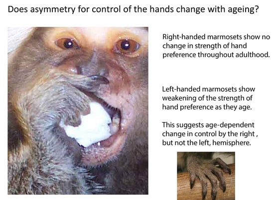
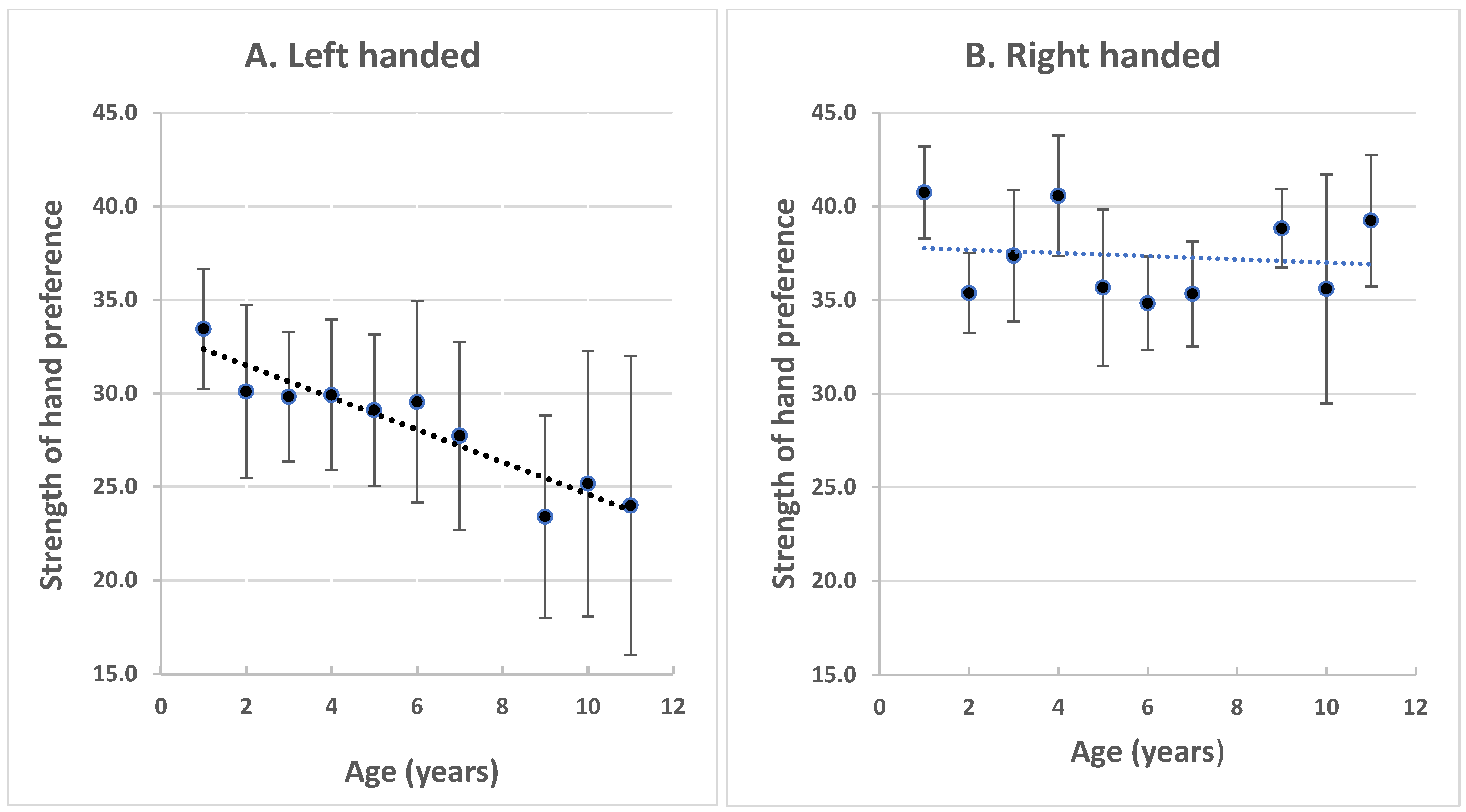
Hand preference was determined during unconstrained picking up of small pieces of food in one hand and raising the food to the mouth to eat it (Figure 1). This is referred to as simple reaching. A minimum of 100 scores were obtained over 8 to 10 days, and no more than 15 scores were recorded per day [35,36]. One score was given to each act of picking up food and bringing it to the mouth; the repeated taking of the same piece of food to the mouth while the marmoset maintained a stationary posture received one score only. The scores were always collected between 12.00 and 13.00 hours during the daily feeding time in the animals’ home cages.
From these scores, the percent left-hand use was determined and, since all of the marmosets had significant hand preferences that they retained throughout adulthood [21], they could be allocated to either a left- or right-hand group. In the left-hand group, there were 11 marmosets (9 females and 2 males). In the right-hand group, there were 8 marmosets (3 females and 5 males). The strength of hand preference in both these groups was calculated as the absolute difference of percent left-hand preference from 50% (no preference).

3. Results

The strength of hand preference across the adult life-span is plotted in Figure 2A,B.
Note the gradual decline in strength of hand preference in the left-hand group (Figure 2A), which occurs consistently across age with a slope of 0.995 (regression line, Y = −0.95545X + 33.476). At the age of 9 years, which included all of the subjects, the decline in the strength of the left-hand preference compared to that of year 1 was significant (mean ± standard error at year 1 was 33.45 ± 3.20 compared to year 9, when it was 23.40 ± 5.41: 1-tailed paired t-test, p = 0.0147, N = 11). The decreasing strength of left-hand preference continued across years 10 and 11, although there were fewer surviving marmosets at these ages (6 survivors at year 10 and 5 at year 11).
This result contrasts with the strength of hand preference of the right-handed marmosets (Figure 2B), which showed no significant decline with age (regression line, Y = −0.12891X + 38.068). A total of 5 of the 8 right-handed marmosets survived to year 10 of life and 4 survived to year 11.
A comparison of the strength of hand preference in year 9 of life in left-handed versus right-handed marmosets was significant (mean strength ± standard error of left-handed group was 23.40 ± 5.41 and of the right-handed group it was 38.83 ± 2.09; 1-tailed t-test, p = 0.025). Additionally, the strength of hand preference considered across adulthood was weaker in left-handed marmosets compared to right-handed marmosets (mean strength ± sem of the left-handed group was 28.82 ± 3.61; for the right-handed group, it was 37.58 ± 2.31; 1-tailed t-test comparison, p = 0.0386).

4. Discussion

In the marmosets with a preference to pick up food with their left hand, the strength of their hand preference declined in a regular trend throughout adulthood. From 1 year to 11 years of age, the strength of left-hand preference decreased by approximately 8%. By contrast, the marmosets with right-hand preference showed no significant change in the strength of their preference across adulthood and into old age.
This is one of very few longitudinal studies, throughout adulthood, on lateralized hand use by a non-human primate species. However, interpretation of the results is compromised by the small sample size used, particularly of the right-handed group. Additionally, the sample had almost twice as many females as males. As almost equal numbers of left- and right-handed marmosets, and no sex differences, were recorded in a larger colony of common marmosets (some of which were caught in the wild [37]) and the same was the case in early adulthood in our colony [21], the imbalance of the sexes in our long-living sample was probably due to females surviving longer than males. In fact, this seems to be particularly so for left-handed females. Because the left-handed group had only two males, the sample size of our study was insufficient to tease apart any effect of age versus sex. Nevertheless, a study of ageing in marmosets followed from five to seven years of age found that males showed a greater decline in fine motor skills than females did [26]. This indicates that, in the study reported here, the decreasing strength of hand preference with age found in the left-handed group (which had fewer males than females) and not in the right-handed group is likely to be a genuine effect of age rather than an interaction between age and sex. To emphasise the value of the results reported here, it is noted again that, in many if not most colonies, marmosets die well before they reach the advanced ages of our subjects.
In humans, age-related changes in hemispheric asymmetry have been shown to depend on sex and the task used to assess asymmetry. For example, Hausmann et al. [38] found that, in a word-matching task, there was greater decline with age of the right compared to the left hemisphere in both males and females; whereas, in a figural-comparison task, such a right hemisphere decline was found in males but the opposite was found in females. This highlights the need for further research investigating sex and age effects in primates.
If further studies of marmosets substantiate the declining strength of left-hand but not right-hand preference with age, this may show that the right hemisphere undergoes an age-dependent change in motor control, whereas the left hemisphere does not. If so, this would be an example of ageing having an earlier and greater effect on the right hemisphere. However, although the weakening strength of the left-hand preference is consistent, a significant left-hand preference is still present even in very old marmosets. In other words, the marmosets show an age-dependent weakening, but not loss, of right hemispheric control of motor function.
In humans, a reduced asymmetry of motor function has been reported to occur with ageing [1,2,3,5], although this depends on the task and related experience prior to testing. For an example of the latter, in a task assessing drawing ability, which is affected by practice, right-hand preference increases with ageing [39]. The reduced asymmetry of motor function with ageing seems to result from changes in both left and right hemispheric control dependent on age-related changes in interhemispheric communication [5,40,41] and supported by evidence of age-dependent structural changes in the corpus callosum [42,43]. Age-dependent change is also known to occur in the corpus callosum of marmosets; viz., a decline in the myelination of axonal fibres in the corpus callosum [44]. In a small sample of marmosets (N = 4), Phillips et al. [44] found that the age-dependent loss of myelin in one particular region of the corpus callosum was associated with a decline in performance in a detour-reach task. Since interhemispheric communication is known to have an important role in hemispheric dominance, as shown in pigeons [45], and that role is asymmetrical [46], an age-dependent reduction in interhemispheric communication could underlie reduced motor dominance and could have differential effects on the left and right hemispheres.
There is some evidence of a similar pattern of ageing occurring in other species. From research on humans, it has been found that the ageing of the right hemisphere alone causes decreased control of spatial attention [47] and, perhaps conversely, aged brains have increased right frontal activation when switching performance in a cognitive task [48]. A study of auditory responses made by rats of advanced age has shown a deterioration of the right hemisphere, whereas the left hemisphere improved [49]. Hence, overall, there is some convincing evidence of ageing having differential effects on the left and right hemispheres, as originally proposed by Goldstein and Shelly [50]. The results reported in this paper are possibly a manifestation of this pattern of ageing in motor behaviour.
Hand preferences in marmosets correlate with certain structural differences in the right hemisphere. The lateral sulcus in the right hemisphere is longer relative to brain weight in right-handed marmosets than it is in left-handed marmosets [51]. Additionally, in right-handed marmosets the secondary somatosensory cortex, which is involved in tactile discrimination, is thicker in the right hemisphere [51]. Although the study by Gorrie et al. [51] found no effect of age on these measures, the sample size examining the effect of age was small. Given the findings reported in this current paper, the differences in brain structure and tactile discrimination between left- and right-handed marmosets would be worth examining with respect to ageing in larger sample sizes. Relevant to this, a recent study has reported a reduced asymmetry of upper-limb proprioception in old compared to young humans [12]. It is, therefore, worth considering the possibility that the age-related reduction in left-hand preference found in the marmosets was associated with an age-dependent reduction in proprioception by the right hemisphere.
The gradual decline in the strength of left-hand preference with age could also be associated with broader behavioural differences between left-handed and right-handed marmosets. Left- and right-handed marmosets differ in social behaviour: right-handed marmosets display higher levels of social interaction [33,52]. In fact, long-term records of behaviour of marmosets in our colony revealed that right-handed marmosets were more aggressive than left-handed marmosets, with the latter being recipients of more aggression and, hence, suffering more stress [53]. It is known that there are cumulative effects of stress associated with lateralization, as discussed by Zach et al. [54], and, in studies on rats, it has been found that stress hormones, glucocorticoids, reduce the volume of the right hippocampal region of the cortex more than they do the left hippocampal region [55,56]. Depending on whether this effect is more widespread in the hemisphere and, since glucocorticoids are associated with ageing [57,58], a cumulative effect of stress with ageing is a potential explanation for declining strength of left-hand preference. Additionally, it has been found in marmosets that stress caused by unexpected and longer-term food deprivation effects the blood flow to, and activity of, the right hemisphere more than the left hemisphere [59], which is consistent with a greater effect of ageing on the right-hemisphere control of left-hand use.

5. Conclusions

The results reported in this study lend support to the model suggesting that the right hemisphere shows a greater decline with ageing than does the left hemisphere. It is recognised that any such result, provided that it is confirmed by subsequent investigations, pertains only to the asymmetry of hand use in one particular task. Hemispheric changes with ageing may be different in other brain regions and for other functions, even for performance in different motor tasks [60]. In fact, in marmosets there is no correspondence between eye preference and hand preference [61], meaning that each is a manifestation of a different laterality and may show different changes with age. Consistent with this, a recent longitudinal study of cortical thickness in humans from 20 to 80-plus years of age has found region-specific thinning in different areas of cortex in the left and right hemispheres [62], with an overall pattern of consistently decreasing asymmetry across the adult life-span [62].

Funding

This research was supported by an Australian Research Council grant number DP0556398.

Institutional Review Board Statement

This research entailed observation only conducted during the regular feeding time of the marmosets and was approved by the University of New England Animal Ethics Committee under licences, including AEC03/051, AEC08/044, AEC 08/064, AEC08/073, and AEC06/060.

Informed Consent Statement

This study did not involve human subjects.

Data Availability Statement

The data presented in this study are available on request from the corresponding author.

Conflicts of Interest

The author declares no conflict of interest. The funders had no role in the design of the study; in the collection, analysis, or interpretation of data; in the writing of the manuscript; or in the decision to publish the results.

References

  1. Kalisch, T.; Wilimzig, C.; Keibel, N.; Tegenthoff, M.; Dinse, H.R. Age-related attenuation of dominant hand superiority. PLoS ONE 2006, 1, e90. [Google Scholar] [CrossRef]
  2. Sebastjan, A.; Skrzek, A.; Ignasiak, Z.; Slawińska, T. Age-related changes in hand dominance and functional asymmetry in older adults. PLoS ONE 2017, 12, e0177845. [Google Scholar] [CrossRef] [Green Version]
  3. Przybyla, A.; Haaland, K.Y.; Bagesteiro, L.B.; Sainburg, R.L. Motor asymmetry reduction in older adults. Neurosci. Lett. 2011, 489, 99–104. [Google Scholar] [CrossRef] [Green Version]
  4. Raw, R.K.; Wilke, R.M.; Culmer, P.R.; Mon-Williams, M. Reduced motor asymmetry in older adults when manually tracing paths. Exp. Brain Res. 2011, 217, 35–41. [Google Scholar] [CrossRef] [PubMed] [Green Version]
  5. Wang, J.; Przybyla, A.; Wuebbenhorst, K.; Haaland, K.Y.; Sainburg, R.L. Aging reduces asymmetries in interlimb transfer of visuomotor adaptation. Exp. Brain Res. 2011, 210, 283–290. [Google Scholar] [CrossRef] [Green Version]
  6. Bernard, J.A.; Seidler, R. Hand dominance and age have interactive effects on motor cortical representations. PLoS ONE 2012, 7, e45443. [Google Scholar]
  7. Talelli, P.; Waddingham, W.; Ewas, A.; Rothwell, J.C.; Ward, N.S. The effect of age on task-related modulation of interhemispheric balance. Exp. Brain Res. 2008, 186, 59–66. [Google Scholar] [CrossRef] [Green Version]
  8. Talelli, P.; Ewas, A.; Waddingham, W.; Rothwell, J.C.; Ward, N.S. Neural correlates of age-related changes in cortical neurophysiology. NeuroImage 2008, 40, 1772–1781. [Google Scholar] [CrossRef] [PubMed] [Green Version]
  9. Bellis, T.J.; Nicol, T.; Kraus, N. Aging affects hemispheric asymmetry in the neural representation of speech sounds. J. Neurosci. 2000, 20, 791–797. [Google Scholar] [CrossRef]
  10. Chen, X.; Liang, Y.; Deng, Y.; Li, J.; Chen, S.; Wang, C.; Luo, P. Age-associated reduction of asymmetry in human central auditory function: A 1H-magnetic resonance spectroscopy study. Neural Plast. 2013, 2013, 735290. [Google Scholar] [CrossRef] [Green Version]
  11. Eddins, A.C.; Ozmeral, E.J.; Eddins, D.A. How aging impacts the encoding of binaural cues and the perception of auditory space. Hear. Res. 2018, 369, 79–89. [Google Scholar] [CrossRef]
  12. Acosta-Sojo, Y.; Martin, B.J. Age-related differences in proprioceptive asymmetries. Neurosci. Lett. 2021, 757, 135992. [Google Scholar] [CrossRef]
  13. Cabeza, R. Hemispheric asymmetry reduction in older adults: The HAROLD model. Psychol. Aging 2002, 17, 85–100. [Google Scholar] [CrossRef]
  14. Dolcos, F.; Rice, H.J.; Cabeza, R. Hemispheric asymmetry and aging: Right hemisphere decline or asymmetry reduction. Neurosci. Biobehav. Rev. 2002, 26, 819–825. [Google Scholar] [CrossRef]
  15. Hill, C.; Van Gemmert, A.W.A.; Fang, Q.; Hou, L.; Wang, J.; Pan, Z. Asymmetry in the aging brain: A narrative review of cortical activation patterns and implications for motor function. Laterality 2020, 25, 413–429. [Google Scholar] [CrossRef]
  16. Jenkins, L.; Myerson, J.; Joerding, J.A.; Hale, S. Converging evidence that visuospatial cognition is more age-sensitive than verbal cognition. Psychol. Ageing 2000, 15, 157–175. [Google Scholar] [CrossRef]
  17. Collins, K.; Mohr, C. Performance of younger and older adults in lateralised right and left hemisphere asymmetry tasks supports the HAROLD model. Laterality 2013, 18, 491–512. [Google Scholar] [CrossRef] [PubMed] [Green Version]
  18. Hatta, T.; Iwahara, A.; Hatta, T.; Ito, E.; Hatta, J.; Hotta, C.; Nagahara, N.; Fujiwara, K.; Hamajima, N. Developmental trajectories of verbal and visuospatial abilities in healthy older adults: Comparison of the hemisphere asymmetry reduction in older adults model and the right hemi-ageing model. Laterality 2015, 20, 69–81. [Google Scholar] [CrossRef]
  19. Sugiura, M. Functional neuroimaging of normal aging: Declining brain, adapting brain. Ageing Res. Rev. 2016, 30, 61–72. [Google Scholar] [CrossRef] [PubMed]
  20. Fu, W.-W.; Wang, X.-W.; Wang, C.-L.; Zhao, H.-T.; Ren, Y.; Li, B.-G. Effects of age, sex and manual task on hand preference in wild Rhinopithecus roxellana. Zool. Res. 2019, 40, 129–138. [Google Scholar] [PubMed]
  21. Hook, M.A.; Rogers, L.J. Development of hand preferences in marmosets (Callithrix jacchus) and effects of ageing. J. Comp. Psychol. 2000, 114, 263–271. [Google Scholar] [CrossRef] [PubMed]
  22. Laurence, A.; Wallez, C.; Blois-Heulin, C. Task complexity, posture, age, sex: Which is the main factor influencing manual laterality in captive Cercocebus torquatus torquatus? Laterality 2011, 16, 586–606. [Google Scholar] [CrossRef]
  23. Tomassetti, D.; Caracciolo, S.; Manciocco, A.; Chiarotti, F.; Vital, A.; De Filippis, B. Personality and lateralization in common marmosets (Callithrix jacchus). Behav. Processes 2019, 167, 103899. [Google Scholar] [CrossRef] [PubMed]
  24. Rothwell, E.S.; Freire-Cobo, C.; Varghese, M.; Edwards, M.; Janssen, W.G.M.; Hof, P.R.; Lacreuse, A. The marmoset as an important primate model for longitudinal studies of neurocognitive aging. Am. J. Primatol. 2021. [Google Scholar] [CrossRef] [PubMed]
  25. Rothwell, E.S.; Workman, K.P.; Wang, D.; Lacreuse, A. Sex differences in cognitive ageing: A 4-year longitudinal study on marmosets. Neurobiol. Aging 2021, 109, 88–99. [Google Scholar] [CrossRef] [PubMed]
  26. Workman, K.P.; Healey, B.; Carlotto, A.; Lacreuse, A. One-year change in cognitive flexibility and fine motor function in middle-aged male and female marmosets (Callithrix jacchus). Am. J. Primatol. 2019, 81, e22924. [Google Scholar] [CrossRef]
  27. Geula, C.; Nagykery, N.; Wu, C.K. Amyloid-B deposits in the cerebral cortex of the aged common marmoset (Callithrix jacchus): Incidence and chemical composition. Acta Neuropathol. 2002, 103, 48–58. [Google Scholar] [CrossRef]
  28. Clarke, J.M. The common marmoset (Callithrix jacchus). ANZCCAART News 1994, 7, 1–8. [Google Scholar]
  29. Nishijima, K.; Saitoh, R.; Tanaka, S.; Ohsato-Suzuki, M.; Ohno, T.; Kitajima, S. Life span of common marmoset (Callithrix jacchus) at CLEA Japan breeding colony. Biogerontology 2012, 13, 439–443. [Google Scholar] [CrossRef] [PubMed]
  30. Tardif, S.D.; Mansfield, K.G.; Ratnam, R.; Ross, C.N.; Ziegler, T.E. The marmoset as a model of aging and age-related diseases. ILAR J. 2011, 52, 54–65. [Google Scholar] [CrossRef] [PubMed]
  31. Ross, C.N.; Davis, K.; Dobek, G.; Tardif, S.D. Aging phenotypes of common marmosets (Callithrix jacchus). J. Aging Res. 2012, 2012, 567143. [Google Scholar] [CrossRef] [Green Version]
  32. Nephew, B.C.; Febo, M.; Cali, R.; Workman, K.P.; Payne, L.; Moore, C.M.; King, J.A.; Lacreuse, A. Robustness of sex differences in functional connectivity over time in middle-aged marmosets. Sci. Rep. 2020, 10, 16647. [Google Scholar] [CrossRef] [PubMed]
  33. Gordon, D.J.; Rogers, L.J. Differences in social and vocal behavior between left- and right-handed common marmosets. J. Comp. Psychol. 2010, 124, 402–411. [Google Scholar] [CrossRef]
  34. Pines, M.K.; Kaplan, G.; Rogers, L.J. Use of horizontal and vertical climbing structures by captive common marmosets (Callithrix jacchus). App. Anim. Behav. Sci. 2005, 91, 311–319. [Google Scholar] [CrossRef]
  35. Hook-Costigan, M.A.; Rogers, L.J. Lateralization of hand, mouth and eye use in the common marmoset (Callithrix jacchus). Folia Primatol. 1995, 64, 180–191. [Google Scholar] [CrossRef] [PubMed]
  36. Hook, M.A.; Rogers, L.J. Leading-limb preference in marmosets (Callithrix jacchus): Walking, leaping and landing? Laterality 2002, 7, 145–162. [Google Scholar] [CrossRef]
  37. Cordeiro de Sousa, M.B.; Xavier, N.S.; Alves da Silva, H.P.; Souza de Oliveira, M. Hand preference study in marmosets (Callithrix jacchus) using food reaching tests. Primates 2001, 42, 57–66. [Google Scholar] [CrossRef]
  38. Hausmann, M.; Güntürkün, O.; Corballis, M.C. Age-related changes in hemispheric asymmetry depend on sex. Laterality 2003, 8, 277–290. [Google Scholar] [CrossRef] [Green Version]
  39. Teixeira, L.A. Catergories of manual asymmetry and their variation with advancing age. Cortex 2008, 44, 707–716. [Google Scholar] [CrossRef]
  40. Davis, S.W.; Kragel, J.E.; Madden, D.J.; Cabeza, R. The architecture of cross-hemispheric communication in the aging brain: Linking behavior to functional and structural connectivity. Cereb. Cortex 2012, 22, 232–242. [Google Scholar] [CrossRef] [PubMed] [Green Version]
  41. Delvenne, J.-F.; Castronovo, J. Reduced interhemispheric interference in ageing: Evidence from a divided field Stroop paradigm. Brain Cogn. 2018, 122, 26–33. [Google Scholar] [CrossRef] [PubMed]
  42. Sullivan, E.V.; Pfefferbaum, A.; Adalsteinsson, E.; Swan, G.E.; Carmelli, D. Differential rates of regional brain change in callosal and ventricular size: A 4-year longitudinal MRI study of elderly men. Cereb. Cortex 2002, 12, 438–445. [Google Scholar] [CrossRef] [Green Version]
  43. Ota, M.; Obata, T.; Akine, Y.; Ito, H.; Ikehira, H.; Asada, T.; Suhara, T. Age-related degeneration of corpus callosum measured with diffusion tensor imaging. NeuroImage 2006, 31, 1445–1452. [Google Scholar] [CrossRef]
  44. Phillips, K.A.; Watson, C.; Bearman, A.; Knippenberg, A.R.; Adams, J.; Ross, C.; Tardif, S.D. Age-related changes in myelin of axons of the corpus callosum and cognitive decline in common marmosets. Am. J. Primatol. 2019, 81, e22949. [Google Scholar] [CrossRef]
  45. Manns, M.; Otto, T.; Salm, L. Pigeons show how meta-control enables decision-making in an ambiguous world. Sci. Rep. 2021, 11, 3838. [Google Scholar] [CrossRef] [PubMed]
  46. Xiao, Q.; Güntürkün, O. The commissura anterior compensates asymmetries of visual representation in pigeons. Laterality 2021, 26, 213–237. [Google Scholar] [CrossRef] [PubMed]
  47. Learmonth, G.; Benwell, C.S.Y.; Thut, G.; Harvey, M. Age-related reduction of hemispheric lateralisation for spatial attention: An EEG study. NeuroImage 2017, 153, 139–151. [Google Scholar] [CrossRef] [PubMed]
  48. Zhu, Z.; Hakun, J.G.; Johnson, N.F.; Gold, B.T. Age-related increases in right frontal activation during task switching are mediated by reaction time in white matter microstructure. Neurosci. 2014, 278, 51–61. [Google Scholar] [CrossRef] [Green Version]
  49. Bureš, Z.; Pysanenko, K.; Syka, J. Age-related changes in the temporal processing of acoustic signals in the auditory cortex of rats. Hear. Res. 2021, 402, 108025. [Google Scholar] [CrossRef]
  50. Goldstein, G.; Shelly, C. Does the right hemisphere age more rapidly than the left? J. Clin. Neuropsychol. 1981, 3, 65–78. [Google Scholar] [CrossRef] [PubMed]
  51. Gorrie, C.A.; Waite, P.M.E.; Rogers, L.J. Correlations between hand preference and cortical thickness in the secondary somatosensory (SII) cortex. Behav. Neurosci. 2008, 122, 1343–1351. [Google Scholar] [CrossRef] [PubMed] [Green Version]
  52. Vaughan, E.; Le, A.; Casey, M.; Workman, K.P.; Lacreuse, A. Baseline cortisol levels and social behavior differ as a function of handedness in marmosets (Callithrix jacchus). Am. J. Primatol. 2019, 81, e23057. [Google Scholar] [CrossRef]
  53. Gordon, D.J.; Rogers, L.J. Cognitive bias, hand preference and welfare in common marmosets. Behav. Brain Res. 2015, 287, 100–108. [Google Scholar] [CrossRef] [PubMed]
  54. Zach, P.; Vales, K.; Stuchlík, A.; Čermáková, P.; Mrzílková, J.; Koutella, A.; Kutová, M. Effect of stress on structural brain asymmetry. Neuroendocrinol. Lett. 2016, 37, 101–112. [Google Scholar]
  55. Zach, P.; Mrzílková, J.; Rezacova, L.; Stuchlík, A.; Vales, K. Delayed effects of elevated corticosterone level on volume of hippocampal formation in laboratory rat. Physiol. Res. 2010, 59, 985–996. [Google Scholar] [CrossRef] [PubMed]
  56. Frodl, T.; O’Keane, V. How does the brain deal with cumulative stress? A review with focus on developmental stress, HPA axis function and hippocampal structure in humans. Neurobiol. Dis. 2013, 52, 24–37. [Google Scholar] [CrossRef]
  57. Lupien, S.J.; McEwen, B.S.; Gunnar, M.R.; Heim, C. Effects of stress throughout the lifespan on the brain, behaviour and cognition. Nature 2009, 10, 434–445. [Google Scholar] [CrossRef]
  58. Landfield, P.W.; Blalock, E.M.; Chen, K.C.; Porter, N.M. A new glucocorticoid hypothesis of brain aging: Implications for Alzheimer’s disease. Curr. Alzheimer Res. 2007, 4, 205–212. [Google Scholar] [CrossRef]
  59. Pereira, L.C.; Maior, R.S.; Barros, M. Time-dependent changes in cortisol and tympanic temperature lateralization during food deprivation stress in marmoset monkeys. Front. Behav. Neurosci. 2020, 14, 123. [Google Scholar] [CrossRef]
  60. Sivagnanasunderam, M.; Gonzalez, D.A.; Bryden, P.J.; Young, G.; Forsyth, A.; Roy, E.A. Handedness throughout the lifespan: Cross-sectional view on sex differences as asymmetries change. Front. Psychol. 2015, 5, 1556. [Google Scholar] [CrossRef] [Green Version]
  61. Hook-Costigan, M.A.; Rogers, L.J. Eye preferences in common marmosets (Callithrix jacchus): Influence of age, stimulus, and hand preference. Laterality 1998, 3, 109–130. [Google Scholar] [CrossRef] [PubMed]
  62. Roe, J.M.; Vidal-Piñeiro, D.; Sørensen, Ø.; Brandmaier, A.M.; Düzel, S.; Gonzalez, H.A.; Kievit, R.A.; Knights, E.; Kühn, S.; Lindenberger, U.; et al. Asymmetric thinning of the cerebral cortex across the adult lifespan is accelerated in Alzheimer’s disease. Nat. Comm. 2021, 12, 721. [Google Scholar] [CrossRef] [PubMed]
Figure 1. A common marmoset using its right hand to hold a piece of food to its mouth. Excerpt from a photograph taken by D. Gordon.
Symmetry 13 02349 g001
Figure 2. Strength of hand preference was plotted as means with standard error scores against age in years. (A). Left-handed marmosets (N = 11). (B). Right-handed marmosets (N = 8). The strength was calculated as the absolute value of the difference of percent left-hand preference from the no-preference value of 50%.
Symmetry 13 02349 g002
Publisher’s Note: MDPI stays neutral with regard to jurisdictional claims in published maps and institutional affiliations.

