Review

Brain Asymmetry: Towards an Asymmetrical Neurovisceral Integration

by
Manuel Ramírez-Sánchez
1,*,
Isabel Prieto
1,
Ana Belén Segarra
1,
Inmaculada Banegas
1,
Magdalena Martínez-Cañamero
1,
Germán Domínguez-Vías
2 and
Marc de Gasparo
3
1
Department of Health Sciences, University of Jaén, 23071 Jaén, Spain
2
Department of Physiology, Faculty of Health Sciences, Ceuta, University of Granada, 18071 Granada, Spain
3
Cardiovascular & Metabolic Syndrome Adviser, Rue es Planches 5, 2842 Rossemaison, Switzerland
*
Author to whom correspondence should be addressed.
Symmetry 2021, 13(12), 2409; https://doi.org/10.3390/sym13122409
Submission received: 22 November 2021 / Revised: 7 December 2021 / Accepted: 8 December 2021 / Published: 13 December 2021
(This article belongs to the Special Issue Biology and Symmetry/Asymmetry:Feature Papers 2022)

Abstract

:
Despite the ancestral evidence of an asymmetry in motor predominance, going through the inspiring discoveries of Broca and Wernicke on the localization of language processing, continuing with the subsequent noise coinciding with the study of brain function in commissurotomized patients—and the subsequent avalanche of data on the asymmetric distribution of multiple types of neurotransmitters in physiological and pathological conditions—even today, the functional significance of brain asymmetry is still unknown. Currently, multiple evidence suggests that functional asymmetries must have a neurochemical substrate and that brain asymmetry is not a static concept but rather a dynamic one, with intra- and inter-hemispheric interactions between its various processes, and that it is modifiable depending on changing endogenous and environmental conditions. Furthermore, based on the concept of neurovisceral integration in the overall functioning of an organism, some evidence has emerged suggesting that this integration could be organized asymmetrically, using the autonomic nervous system as a bidirectional communication pathway, whose performance would also be asymmetric. However, the functional significance of this distribution, as well as the evolutionary advantage of an asymmetric nervous organization, is still unknown.

Graphical Abstract

1. The Early Development

The concept of neurovisceral integration comes from Claude Bernard [1], developed more recently by Thayer and Lane [2]. Initially, it has been built on the basis of the connection between brain and heart that Claude Bernard already masterfully anticipated [3]:
Le cœur et le cerveau se trouvent dès lors dans une solidarité d’actions réciproques des plus intimes, qui se multiplient et se resserrent d’autant plus que l’organisme devient plus développé et plus délicat. Ces rapports peuvent être constants ou passagers, varier avec le sexe et avec l’âge
However, without doubt, that reflection goes further and constitutes an integral concept that encompasses the entire organism [4,5]. Furthermore, there are data that suggest that this global organization is carried out in an asymmetric way [6,7,8,9].
Although no one yet knows for sure how life on earth acquired its asymmetric character or what the biological advantage of asymmetry over symmetry would be [10], some authors suggest that the asymmetry could have a molecular origin from which life would evolve [11,12,13]. If we go back to pure physical concepts, Joe Rosen suggests that symmetry underlies nature [14], but the same author indicated that symmetry implies asymmetry, and that the universe cannot have perfect symmetry [15]. Frank Close even argues that the Universe is asymmetric and that virtually all living species are essentially a function of cosmic asymmetry [16]. According to Michael Gazzaniga [17]:
The molecular aspects of life reflect a complex system laced with feedback loops and multiple interactions-nothing is linear and simple
If we briefly review the milestones in the knowledge of brain asymmetry, we could first highlight motor strength and ability. The fact that the neuronal groups that govern them are mostly located in the contralateral cortex to the body side which is considered to constitute the most evident case of an asymmetric brain function. There is evidence that such asymmetry also occurred in prehistoric times [18,19]. Some specific contributions that connect handedness with peripheral processes, such as ovarian function, are interesting. Thus, a connection between handedness and the appearance of sexual maturity in women has been described. In left-handed women, menarche appeared earlier than in right-handed women [20]. Furthermore, Jones and colleagues [21] had shown that the content of various neurotransmitters in the lizard brain predominated on the ipsilateral side to that of the ovary in which ovulation was occurring. The lizard, like humans and other primates, alternates ovulation between the left and right ovary. These results somehow take us beyond simple brain asymmetry, suggesting a neurovisceral integration, mediated, in part and in these cases, hormonally. Its functional meaning remains to be elucidated. In any case, these data suggest that handedness is integrated into a broader and more complex biological context, as we will see throughout this review.
In the second half of the 19th century, Paul Broca [22] and Carl Wernicke [23], almost simultaneously and independently, perfectly aware of the significance of their discoveries, demonstrated that the processes of expression and of understanding language were located in specific areas of the cortex in the left hemisphere and not in the right. Universal recognition of the importance of brain asymmetry came with the studies of Michael Gazzaniga [24] and Roger Sperry, who received the Nobel Prize in 1981 [25] for their discoveries regarding the functional specialization of the cerebral hemispheres. They carried out their study largely in commissurotomized patients, in which the corpus callosum—as the main bundle of fibers that connects both hemispheres—had been sectioned to alleviate the consequences of massive epileptic seizures. It was suggested that under physiological conditions, the two hemispheres were characterized by processing different functions. Thus, while the left hemisphere would be analytical, verbal, mathematical or sequential, the right one would be spatial, imaginative and synthetic [26,27]. The researchers observed that the right hemisphere is unable to carry out verbal functions after lesions of the left hemisphere (with intact corpus callosum) that compromise language function. However, after commissurotomy, the right hemisphere was capable of carrying out verbal functions. In the Nobel lecture [25] it was reported that with the intact commissure, the lesion that compromises a function of the left hemisphere will inhibit the expression of the same function, latent but suppressed, of the undamaged right hemisphere. This implies that, under physiological conditions, both hemispheres function as a unit, leading one or the other, depending on the studied function that will exist in both, but will be latent in one or the other. When a unilateral injury occurs, the function that is altered prevails over both hemispheres and the two continue acting as an integrated unit, although with that specific function altered. When an undamaged hemisphere is freed by commissurotomy from its integration with the other hemisphere and consequently from its inhibitory influence, its own latent function may become manifest [25]. In conclusion, from studies in split brains, Michael Gazzaniga speaks about two brains in one head, but connected to each other, informing and influencing each other, leading to integrated cognitive processing [24].

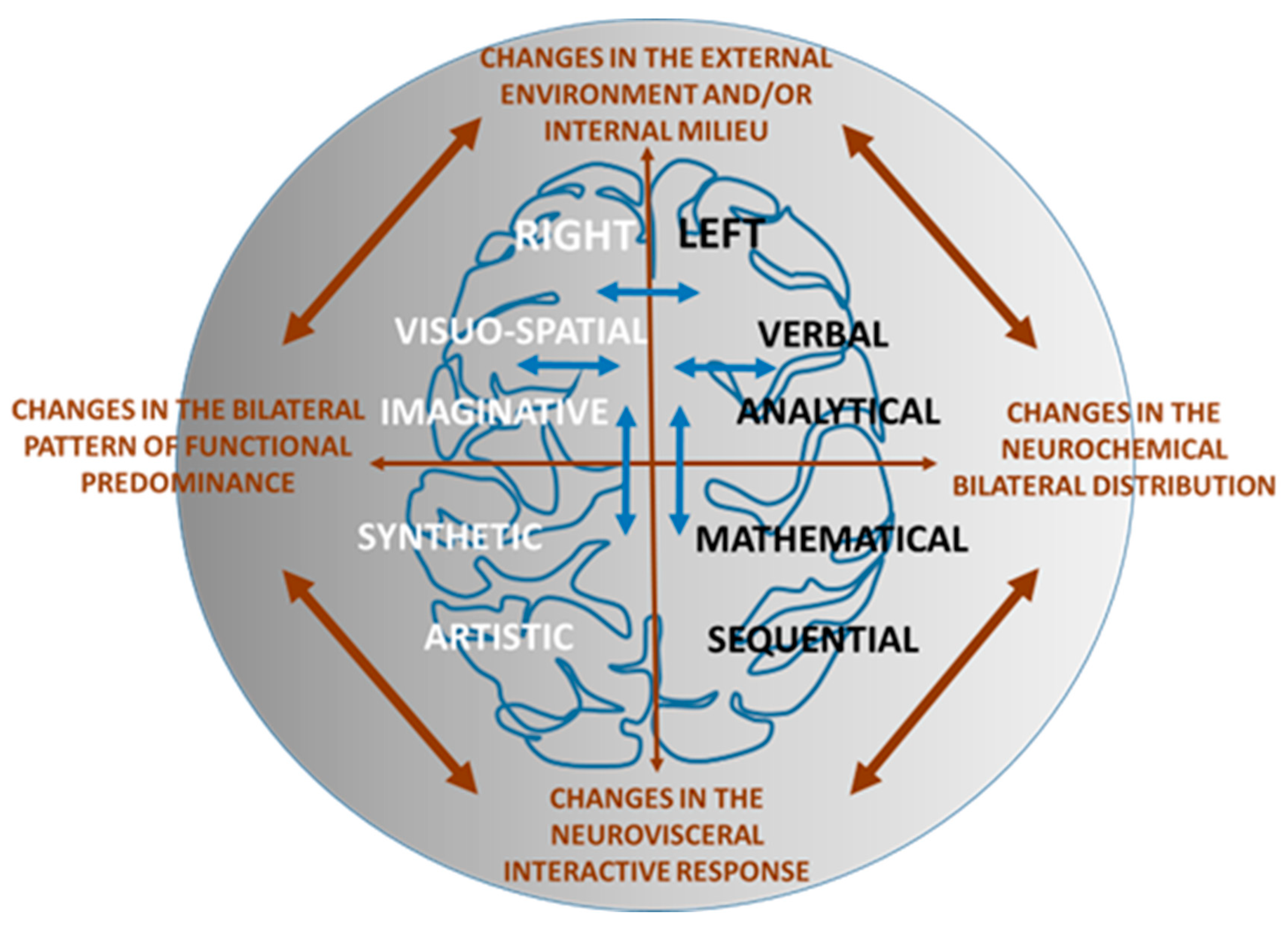
2. Neurochemical Substrate for Brain Asymmetry

In this context, on the basis that functional asymmetries must have neurochemical substrates that support them, the bilateral distribution of classical neurotransmitters was analyzed. As examples, the bilateral distribution of norepinephrine [28,29], serotonin [30] or dopamine [31] were studied with varying results of left or right predominance, depending on the brain region analyzed. The greatest attempts to relate a lateralized brain function, such as the circling behavior that rats spontaneously exhibit, with the bilateral distribution of a neurotransmitter, such as dopamine, were made by Shapiro et al. [32] and Nielsen et al. [33]. They observed that the animals rotated mainly toward the contralateral direction to the side that contains higher levels of dopamine or a higher number of activated postsynaptic dopamine receptors. However, simultaneously with such works, it is necessary to highlight the “revolution” [34] that led to the demonstration of the existence of receptors for opiates such as morphine in the brain [35]; the existence of endogenous opiate peptides, such as enkephalins, that bound to those receptors [36]; and the demonstration that other neuropeptides not only act as neurohormones [37,38] but also as neurotransmitters [39] that could coexist with the classic neurotransmitters [40].
The functional analysis of neuropeptides can also be carried out through the study of their processing through the action of proteolytic enzymes [41] known as neuropeptidases [42,43]. Therefore, the brain asymmetry of such neuropeptides could be reflected in the activity of the enzymes that metabolize them (reviewed in [44]). In addition to the demonstration of the existence of asymmetries in the activity of neuropeptidases in various brain regions under basal conditions—such as the predominance of leucine-aminopeptidase in the left frontal cortex and left hypothalamus of male rats [45], or the diversity in the left or right predominance of various aminopeptidases in other brain areas, such as amygdala, hippocampus or prefrontal cortex [46]—the asymmetry reflected by these enzymes showed a dynamic behavior, depending on the changing environmental conditions, such as light or darkness [44,47]. In this context, some specific contributions that relate brain asymmetry with seasonal changes, as well as with the month in which the birth took place [48] and even changes in the Earth’s magnetic field [49] can also be included.

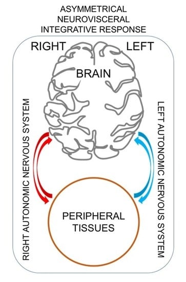
3. Asymmetric Neurovisceral Integration

The simultaneous study of neuropeptidase activities and other biochemical parameters in various brain regions, tissues and peripheral fluids, as well as cardiac or renal functions, revealed asymmetric interactions between all of them, which were modified under certain experimental conditions. Although correlation does not imply causality, it may be suggestive of some mutual interaction between different locations or functions, which allows us to speculate on a possible neurovisceral integration. In particular, while in control hypertensive animals a left predominance of correlations between neuropeptidases of the frontal cortex and left ventricular tissue was observed in male rats, the predominance changed radically to the right site after treatment of the animals with captopril [50]. However, in contrast, while plasma neuropeptidase activity was significantly correlated with the right frontal cortex of hypertensive control rats, the predominance shifted to the left in rats treated with captopril [51]. Spontaneously hypertensive rats dramatically increased their blood pressure following dopamine depletion of the left hemisphere, but not of the right [52]. In addition, under various vasoactive treatments, a predominance of correlations between neuropeptidase activities of the left frontal cortex and systolic and diastolic blood pressures was, in general, also observed [6]. In addition, left frontal cortex also correlated predominantly with water balance functions, such as water intake and diuresis [8]. In relation to an asymmetry in the gut–brain connection, it seems that the right vagus nerve predominates more than the left in the stimulation of intestinal endocrine cells [53]. Furthermore, the gut has been linked to the regulation of emotional state and central reward systems through the vagus nerve, specifically the right nerve, involving the substantia nigra, dopamine and cholecystokinin [54] (Figure 1).
It remains to be analyzed to what extent neurochemical changes in the bilateral brain pattern affect the level and prevalence of asymmetry of certain brain functions and consequently their neurovisceral response. For example, unilateral lesions of the nigrostriatal system, which at least unilaterally deplete dopamine levels, affect dopamine-dependent motor and presumably non-motor functions, such as the left or right direction of rotation after injury, and other neurochemical factors involved [55]. Specifically, the results varied depending on the side of the lesion and the strain studied (Wistar Kyoto or spontaneously hypertensive rat) and demonstrate the involvement, not only of dopamine but also of aminopeptidase A, a cholecystokinin regulatory neuropeptidase [55].
All these results could lead us to propose that, if neurochemical laterality is modified depending on changes in the external environment and physiological and/or pathological modifications of the internal environment, different functional lateralities would also be modified. If this is so, we could speculate that changes in the external and/or internal environment, which alters the bilateral neurochemical distribution, may imply changes in the level or patterns of predominance of certain functions, including modifications in the neurovisceral response. The interactions would be bidirectional, mediated by the autonomic nervous system in which the parasympathetic has been described to be regulated by the left hemisphere [56] and the sympathetic one by the right [57]. The bidirectional influence, through the autonomic nervous system, is supported by the description of anterograde and retrograde transport in the autonomic innervation [6,8,9,58,59]. In addition, intra- and inter-hemispheric neurochemical and functional interactions could also influence the final result of neurovisceral integration [7,60,61]. Particularly of interest is the fact that the classic motor and language dominances are mutually interconnected and that they influence and modify each other. In this sense, Knetchs et al. [62] show that there is a direct connection between handedness and the degree of left or right predominance of language: in right-handed subjects, the greater degree of lateralization of language implies left dominance for language, whereas in left-handed people, the greater degree of lateralization of the language implies right dominance for the language.

4. Neuropathologies and Brain Asymmetry

Lubben et al. [63] review the asymmetric nature of neurodegenerative diseases, such as Parkinson’s disease, Alzheimer’s disease, multiple sclerosis or amyotrophic lateral sclerosis, highlighting the greater prevalence of the left hemisphere at the origin of all of these diseases. The authors suggest that the brain already develops with innate differences between both hemispheres, which implies that one of them is more vulnerable, causing the onset of the pathology to precipitate earlier and more easily. There are also interesting studies that link brain asymmetry and cerebrovascular accidents. Ma et al. [64] describe that accidents located in the left hemisphere produce more severe sensory–motor and cognitive alterations than those produced in the right hemisphere. These accidents also involve asymmetric neurochemical alterations, such as in norepinephrine, dopamine or dynorphin, but the results are still not very consistent. However, the authors conclude that the incidence of cerebral ischemia is higher in the left hemisphere than in the right one, and therefore these patients should receive priority in their treatment [64]. It is worth highlighting the study by Zhang et al. [65] on the association between sex, the hemispheric location of the stroke and the subsequent appearance of depression as one of the common post-stroke sequelae. The authors concluded that the appearance of a post-stroke depression is more frequent in women who have suffered an accident in the left hemisphere than in the right. It is possible that gender differences in the consequences of unilateral accidents are partly due to inter-hemispheric differences in physiological endogenous or exogenous conditions, such as those that occur during the ovarian cycle [66,67] or those produced in both genders due to circadian rhythms [reviewed in 67]. On the other hand, it is necessary to take into account that, although there is a hemispheric dominance in brain functions, the left hemisphere for language and the right for visual–spatial functions, both functions are also elaborated, although to a lesser extent, in the contralateral hemisphere [68]. This side could therefore intervene in a hypothetical inter-hemispheric compensation of the deficit produced after a unilateral injury. However, the individual variability of the results does not yet provide a conclusive model of recovery, so more evidence is required to help assess and predict the degree of recovery after unilateral injuries [69].
In the study of brain asymmetry, we cannot forget the processing of pain, inevitably associated with emotional circuits. In this sense, Toutain et al. [70] have observed an increase in intra-hemispheric interactions in the left hemisphere in case of a painful sensation, this increase being greater in women. These authors also suggest that emotions can modulate the level of brain asymmetry under pain conditions. Regarding the relationship between pain threshold and brain asymmetry, an asymmetry in pain perception has been observed depending on the type of painful stimulus. The pain produced by pressure is predominant on the left body side (which implies the right hemisphere), but no body asymmetry was observed when the stimulus was thermal [71]. In this sense, Pauli et al. [72] already observed lower pain thresholds in the left hand than in the right and concluded that right frontal hyperactivity could be a marker of an increase in pain sensitivity associated with a negative emotional state, such as depression. As we have already mentioned in this review, these asymmetric functional behaviors must have a neurochemical correlate that has been partially analyzed in the relationship between the brain–heart connection and depression (reviewed in [7]). Analyzing the activity of various neuropeptidases responsible for the hydrolysis of neuropeptides involved in emotional processes, such as enkephalins or oxytocin in cortico-limbic regions, such as the prefrontal cortex (among others), a predominantly right lateralization in depressive states is suggested.
On the other hand, the socioeconomic implications of a deeper understanding of brain asymmetry and its neurovisceral integration could predict and, when appropriate, prevent neuropathologies or behavioral deviations. The greater or lesser incidence of left or right neuropathologies can derive from alterations in the general asymmetric processing of the organism, including the asymmetric bidirectional connection between the brain and the peripheral tissues [73,74,75,76,77,78].
Furthermore, the hemodynamic similarity between the brain and some previously discussed organs such as the kidney, should be taken into account regarding neurovisceral integration [79]. Both organs share anatomy–physiological hemodynamic properties, responding in parallel to injuries that can influence each other [79]. Unilateral lesions of either organ, brain or kidney, may thus influence each other asymmetrically.
Finally, we should emphasize that this asymmetric neurovisceral integration is self-regulating, trying to maintain homeostasis through compensatory mechanisms against unilateral physiological modifications but also in the front of unilateral lesions. This compensatory response is therefore an attempt to maintain homeostasis. This may partly explain the dynamic nature of brain asymmetry, which can be extended to the general concept of neurovisceral integration [80] (Figure 2).

5. Conclusions

From all the reported studies, we can clearly establish the existence of a cerebral asymmetry that extends to an asymmetric neurovisceral integration in which a neurochemical asymmetry underlies, but we can also conclude that this asymmetry changes neurochemically and functionally depending on the modification of multiple endogenous and exogenous factors.
In conclusion, brain asymmetry is a dynamic concept whose complexity increases progressively. There is no doubt that in addition to the functional asymmetries that we already know, there must be many more, without claiming that virtually all functions would be processed asymmetrically. Likewise, we could say that there are nuances of all of them, dependent on multiple factors that modulate them and in which a neurochemical substrate underlies, equally modulated by these factors.
Asymmetry involves the entire organism, so we can assume the concept of asymmetric neurovisceral integration that uses the autonomic nervous system, endocrine and neuroendocrine systems as communication channels. It is modulated by changes in the external and internal environment, the response of which depends, among other factors, on the brain site, sex or the type of function under consideration. It is a complex system that involves multiple functional and neurochemical processes that interact with each other.
From all these observations described above, it can also be deduced that, basically, our present knowledge about brain asymmetry is mostly descriptive. Therefore, to understand its biological significance, we need to continue advancing in this crucial research, without losing sight of the fact that it is a dynamic and global concept.

Author Contributions

Conceptualization, M.R.-S.; writing—original draft preparation, M.R.-S.; writing—review and editing, M.R.-S., I.P., A.B.S., I.B., M.M.-C., G.D.-V. and M.d.G.; supervision, M.d.G. and M.M.-C.; funding acquisition, M.R.-S., I.P., A.B.S. and I.B. All authors have read and agreed to the published version of the manuscript.

Funding

This review was supported by the group of the University of Jaén “Neuroendocrinology and Nutrition” BIO-221.

Conflicts of Interest

The authors declare no conflict of interest.

References

  1. Bernard, C. Étude sur la Physiologie du Cœur; Revue des Deux Mondes: Paris, France, 1878; pp. 316–366. [Google Scholar]
  2. Thayer, J.F.; Lane, R.D. Claude Bernard and the heart-brain connection: Further elaboration of a model of neurovisceral integration. Neurosci. Biobehav. Rev. 2009, 33, 81–88. [Google Scholar] [CrossRef]
  3. Bernard, C. Étude sur la Physiologie du Cœur, 2nd ed.; Revue des Deux Mondes: Paris, France, 1865; Volume 56, pp. 236–252. [Google Scholar]
  4. Kucmierz, J.; Frak, W.; Młynarska, E.; Franczyk, B.; Rysz, J. Molecular Interactions of Arterial Hypertension in Its Target Organs. Int. J. Mol. Sci. 2021, 22, 9669. [Google Scholar] [CrossRef]
  5. Segarra, A.B.; Prieto, I.; Banegas, I.; Martínez-Cañamero, M.; Villarejo, A.B.; Domínguez-Vías, G.; de Gasparo, M.; Ramírez-Sánchez, M. Interaction between Angiotensinase Activities in Pituitary and Adrenal Glands of Wistar-Kyoto and Spontaneously Hypertensive Rats under Hypotensive or Hypertensive Treatments. Int. J. Mol. Sci. 2021, 22, 7823. [Google Scholar] [CrossRef]
  6. Segarra, A.B.; Prieto, I.; Banegas, I.; Martinez-Cañamero, M.; de Gasparo, M.; Ramirez-Sanchez, M. Blood Pressure Correlates Asymmetrically with Neuropeptidase Activities of the Left and Right Frontal Cortices. Symmetry 2021, 13, 105. [Google Scholar] [CrossRef]
  7. Arias, J.A.; Williams, C.; Raghvani, R.; Aghajani, M.; Baez, S.; Belzung, C.; Booij, L.; Busatto, G.; Chiarella, J.; Fu, C.H.; et al. The neuroscience of sadness: A multidisciplinary synthesis and collaborative review. Neurosci. Biobehav. Rev. 2020, 111, 199–228. [Google Scholar] [CrossRef]
  8. Segarra, A.B.; Prieto-Gomez, I.; Banegas, I.; Martínez-Cañamero, M.; Luna, J.D.; de Gasparo, M.; Ramírez-Sánchez, M. Functional and neurometabolic asymmetry in SHR and WKY rats following vasoactive treatments. Sci. Rep. 2019, 9, 16098. [Google Scholar] [CrossRef] [PubMed]
  9. Balle, M.; Bornas, X.; Tortella-Feliu, M.; Llabrés, J.; Morillas-Romero, A.; Aguayo-Siquier, B.; Gelabert, J.M. Resting parietal EEG asymmetry and cardiac vagal tone predict attentional control. Biol. Psychol. 2013, 93, 257–261. [Google Scholar] [CrossRef] [PubMed]
  10. Gardner, M. The New Ambidextrous Universe; Dover Publications: New York, NY, USA, 2005; ISBN 0-486-44244-6. [Google Scholar]
  11. Corballis, M.C. Bilaterally Symmetrical: To Be or Not to Be? Symmetry 2020, 12, 326. [Google Scholar] [CrossRef] [Green Version]
  12. Corballis, M.C. How Asymmetries Evolved: Hearts, Brains, and Molecules. Symmetry 2021, 13, 914. [Google Scholar] [CrossRef]
  13. Hellige, J.B. Hemispheric Asymmetry. What’s Right and What’s Left; Harvard University Press: Cambridge, MA, USA, 1993; ISBN 0-674-38730-9. [Google Scholar]
  14. Rosen, J. Symmet.ry at the Foundation of Science and Nature. Symmetry 2009, 1, 3. [Google Scholar] [CrossRef] [Green Version]
  15. Rosen, J. Symmetry in Science. An introduction to the General Theory; Springer: New York, NY, USA, 1995; ISBN 0-387-94375-7. [Google Scholar]
  16. Close, F. Lucifer’s Legacy. The Meaning of Asymmetry; Oxford University Press: Oxford, UK, 2000; ISBN 0-19-850380-6. [Google Scholar]
  17. Gazzaniga, M.S. Tales from Both Sides of the Brain; HarperCollins Publishers: New York, NY, USA, 2015; ISBN 978-0-06-222880-2. [Google Scholar]
  18. Springer, S.P.; Deutchs, G. Left Brain, Right Brain; W.H. Freeman & Co Ltd.: New York, NY, USA, 1981; ISBN 9780716712695. [Google Scholar]
  19. McManus, C. Right Hand, Left Hand: The Origins of Asymmetry in Brains, Bodies, Atoms and Cultures; Harvard University Press: Cambridge, MA, USA, 2004; ISBN 978-0674016132. [Google Scholar]
  20. Nikolova, P.; Stoyanov, Z.; Negrev, N. Functional brain asymmetry, handedness and menarcheal age. Int. J. Psychophysiol. 1994, 18, 213–215. [Google Scholar] [CrossRef]
  21. Jones, R.E.; Desan, P.H.; Lopez, K.H.; Austin, H.B. Asymmetry in diencephalic monoamine metabolism is related to side of ovulation in a reptile. Brain Res. 1990, 506, 187–191. [Google Scholar] [CrossRef]
  22. Broca, P. Nouvelle observation d’aphémie par une lésion de la moitié postérieure des deuxième et troisième circonvolutions frontales. Bull. Soc. Anat. 1861, 6, 398–407. [Google Scholar]
  23. Wernicke, C. Der Aphasische Symptom Complex. Eine Psichologische Studie Ananatomischer Basis; Max Cohn & Wiegert: Breslau, Poland, 1874. [Google Scholar]
  24. Gazzaniga, M.S.; Bogen, J.E.; Sperry, R.W. Some functional effects of sectioning the cerebral commissures in man. Proc. Natl. Acad. Sci. USA 1962, 48, 1765–1769. [Google Scholar] [CrossRef] [PubMed] [Green Version]
  25. The Nobel Prize. Roger W. Sperry—Nobel Lecture. Available online: https://www.nobelprize.org/prizes/medicine/1981/sperry/25059-roger-w-sperry-nobel-lecture-1981 (accessed on 8 December 1981).
  26. Gazzaniga, M.S. One brain-two minds? Am. Sci. 1972, 60, 311–317. [Google Scholar]
  27. Corballis, M. Human Laterality; Academic Press: New York, NY, USA, 1983; ISBN 9780323158466. [Google Scholar]
  28. Oke, A.; Keller, R.; Mefford, I.; Adams, R.N. Lateralization of norepinephrine in human thalamus. Science 1978, 200, 1411–1413. [Google Scholar] [CrossRef]
  29. Oke, A.; Lewis, R.; Adams, R.N. Hemispheric asymmetry of norepinephrine distribution in rat thalamus. Brain Res. 1980, 188, 269–272. [Google Scholar] [CrossRef]
  30. Rosen, G.D.; Finklestein, S.; Stoll, A.L.; Yutzey, D.A.; Denenberg, V.H. Neurochemical asymmetries in the albino rat’s cortex, striatum, and nucleus accumbens. Life Sci. 1984, 34, 1143–1148. [Google Scholar] [CrossRef]
  31. Glick, S.D.; Ross, D.A.; Hough, L.B. Lateral asymmetry of neurotransmitters in human brain. Brain Res. 1982, 234, 53–63. [Google Scholar] [CrossRef]
  32. Shapiro, R.M.; Glick, S.D.; Hough, L.B. Striatal dopamine uptake asymmetries and rotational behavior in unlesioned rats: Revising the model? Psychopharmacology 1986, 89, 25–30. [Google Scholar] [CrossRef] [PubMed]
  33. Nielsen, D.M.; Crosley, K.J.; Keller, R.W.; Glick, S.D.; Carlson, J.N. Rotation, locomotor activity and individual differences in voluntary ethanol consumption. Brain Res. 1999, 823, 80–87. [Google Scholar] [CrossRef]
  34. Crapo, L.M. Hormones: The Messengers of Life; W H Freeman & Co.: New York, NY, USA, 1985; ISBN 0716717530, 9780716717539. [Google Scholar]
  35. Pert, C.B.; Snyder, S.H. Opiate receptor: Demonstration in nervous tissue. Science 1973, 179, 1011–1014. [Google Scholar] [CrossRef] [PubMed]
  36. Hughes, J.; Smith, T.W.; Kosterlitz, H.W.; Fothergill, L.A.; Morgan, B.A.; Morris, H.R. Identification of two related pentapeptides from the brain with potent opiate agonist activity. Nature 1975, 258, 577–580. [Google Scholar] [CrossRef] [PubMed]
  37. Boler, J.; Enzmann, F.; Folkers, K.; Bowers, C.Y.; Schally, A.V. The identity of chemical and hormonal properties of the thyrotropin releasing hormone and pyroglutamyl-histidyl-proline amide. Biochem. Biophys. Res. Commun. 1969, 37, 705–710. [Google Scholar] [CrossRef]
  38. Burgus, R.; Dunn, T.F.; Desiderio, D.; Ward, D.N.; Vale, W.; Guillemin, R. Characterization of ovine hypothalamic hypophysiotropic TSH-releasing factor. Nature 1970, 226, 321–325, Erratum in Nature 1970, 226, 479. [Google Scholar] [CrossRef]
  39. Schwartz, J.C.; Roques, B.P. Opioid peptides as intercellular messengers. Biomedicine 1980, 32, 169–175. [Google Scholar] [PubMed]
  40. Grobecker, H. Transmitter-peptide coexistence in the central nervous system. Eur. Neurol. 1983, 22, 38–46. [Google Scholar] [CrossRef]
  41. Loh, Y.P.; Brownstein, M.J.; Gainer, H. Proteolysis in neuropeptide processing and other neural functions. Annu. Rev. Neurosci. 1984, 7, 189–222. [Google Scholar] [CrossRef] [PubMed]
  42. Merighi, A. Neuropeptides: Methods and Protocols; Humana Press: Totowa, NJ, USA, 2011; ISSN 1064-3745. [Google Scholar]
  43. Vasilev, D.S.; Dubrovskaya, N.M.; Zhuravin, I.A.; Nalivaeva, N.N. Developmental Profile of Brain Neprilysin Expression Correlates with Olfactory Behaviour of Rats. J. Mol. Neurosci. 2021, 71, 1772–1785. [Google Scholar] [CrossRef]
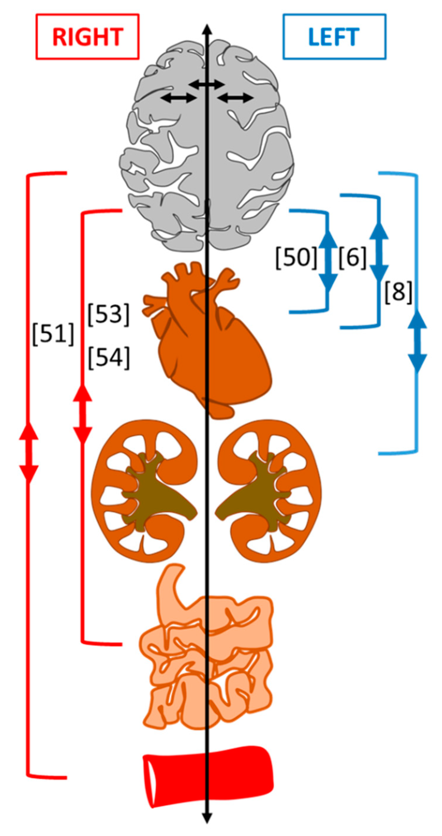
  44. Ramírez, M.; Prieto, I.; Vives, F.; de Gasparo, M.; Alba, F. Neuropeptides, neuropeptidases and brain asymmetry. Curr. Protein Pept. Sci. 2004, 5, 497–506. [Google Scholar] [CrossRef] [PubMed]
  45. Alba, F.; Ramirez, M.; Cantalejo, E.S.; Iribar, C. Aminopeptidase activity is asymmetrically distributed in selected zones of rat brain. Life Sci. 1988, 43, 935–939. [Google Scholar] [CrossRef]
  46. Banegas, I.; Prieto, I.; Alba, F.; Vives, F.; Araque, A.; Segarra, A.B.; Durán, R.; de Gasparo, M.; Ramírez, M. Angiotensinase activity is asymmetrically distributed in the amygdala, hippocampus and prefrontal cortex of the rat. Behav. Brain Res. 2005, 156, 321–326. [Google Scholar] [CrossRef] [PubMed]
  47. Domínguez-Vías, G.; Aretxaga, G.; Prieto, I.; Segarra, A.B.; Luna, J.D.; Martínez-Cañamero, M.; Ramírez-Sánchez, M. Asymmetrical influence of a standard light/dark cycle and constant light conditions on the alanyl-aminopeptidase activity of the left and right retinas in adult male rats. Exp. Eye Res. 2020, 198, 108149. [Google Scholar] [CrossRef] [PubMed]
  48. Volchek, O.D. Vliianie tsiklichnosti sredy na proiavleniia funktsional’noĭ asimmetrii mozga u cheloveka [Effect of environmental cyclicity on appearance of human brain functional asymmetry]. Biofizika 1995, 40, 1013–1019. [Google Scholar]
  49. Agadzhanian, N.A.; Makarova, I.I.; Golovko, M.; D’iachkova, L.; Kanonidi, K. Elektrofiziologicheskiĭ i neĭrokhimicheskiĭ analiz biologicheskikh éffektov vozmushcheniĭ magnitnogo polia Zemli [Electrophysiological and neurochemical analysis of the biological effects of disturbances of Earth’s magnetic field]. Aviakosm. Ekolog. Med. 2002, 36, 26–32. [Google Scholar]
  50. Segarra, A.B.; Prieto, I.; Banegas, I.; Villarejo, A.B.; Wangensteen, R.; de Gasparo, M.; Vives, F.; Ramírez-Sánchez, M. The brain-heart connection: Frontal cortex and left ventricle angiotensinase activities in control and captopril-treated hypertensive rats-a bilateral study. Int. J. Hypertens. 2013, 2013, 156179. [Google Scholar] [CrossRef]
  51. Segarra, A.B.; Prieto, I.; Banegas, I.; Villarejo, A.B.; Wangensteen, R.; de Gasparo, M.; Vives, F.; Ramírez-Sánchez, M. Asymmetrical effect of captopril on the angiotensinase activity in frontal cortex and plasma of the spontaneously hypertensive rats: Expanding the model of neuroendocrine integration. Behav. Brain Res. 2012, 230, 423–427. [Google Scholar] [CrossRef]
  52. Banegas, I.; Prieto, I.; Segarra, A.B.; Durán, R.; Vives, F.; Alba, F.; Luna, J.D.; de Gasparo, M.; Wangesteen, R.; Ruiz-Bailén, M.; et al. Blood pressure increased dramatically in hypertensive rats after left hemisphere lesions with 6-hydroxydopamine. Neurosci. Lett. 2011, 500, 148–150. [Google Scholar] [CrossRef] [PubMed]
  53. Qian, B.F.; el-Salhy, M.; Danielsson, A.; Shalaby, A.; Axelsson, H. Changes in intestinal endocrine cells in the mouse after unilateral cervical vagotomy. Histol. Histopathol. 1999, 14, 453–460. [Google Scholar] [PubMed]
  54. Han, W.; Tellez, L.A.; Perkins, M.H.; Perez, I.O.; Qu, T.; Ferreira, J.; Ferreira, T.L.; Quinn, D.; Liu, Z.W.; Gao, X.B.; et al. A Neural Circuit for Gut-Induced Reward. Cell 2018, 175, 665–678. [Google Scholar] [CrossRef] [Green Version]
  55. Banegas, I.; Segarra, A.B.; Prieto, I.; Vives, F.; de Gasparo, M.; Duran, R.; de Dios Luna, J.; Ramírez-Sánchez, M. Asymmetrical response of aminopeptidase A in the medial prefrontal cortex and striatum of 6-OHDA-unilaterally-lesioned Wistar Kyoto and spontaneously hypertensive rats. Pharmacol. Biochem. Behav. 2019, 182, 12–21. [Google Scholar] [CrossRef] [PubMed]
  56. Denenberg, V.H. Hemispheric laterality in animals and the effects of early experience. Behav. Brain Sci. 1981, 4, 1–49. [Google Scholar] [CrossRef] [Green Version]
  57. Rogers, L.J.; Vallortigara, G.; Andrew, R.J. Divided Brains. The Biology and Behaviour of Brain Asymmetries; Cambridge University Press: New York, NY, USA, 2013. [Google Scholar]
  58. Diz, D.I.; Ferrario, C.M. Bidirectional transport of angiotensin II binding sites in the vagus nerve. Hypertension 1988, 11, I139–I143. [Google Scholar] [CrossRef] [PubMed] [Green Version]
  59. Hung, C.O.; Coleman, M.P. KIF1A mediates axonal transport of BACE1 and identification of independently moving cargoes in living SCG neurons. Traffic 2016, 17, 1155–1167. [Google Scholar] [CrossRef]
  60. Hernández, J.; Prieto, I.; Segarra, A.B.; de Gasparo, M.; Wangensteen, R.; Villarejo, A.B.; Banegas, I.; Vives, F.; Cobo, J.; Ramírez-Sánchez, M. Interaction of neuropeptidase activities in cortico-limbic regions after acute restraint stress. Behav. Brain Res. 2015, 287, 42–48. [Google Scholar] [CrossRef]
  61. Segarra, A.B.; Hernández, J.; Prieto, I.; de Gasparo, M.; Ramírez-Sánchez, M. Neuropeptidase activities in plasma after acute restraint stress. Interaction with cortico-limbic areas. Acta Neuropsychiatr. 2016, 28, 239–243. [Google Scholar] [CrossRef]
  62. Knecht, S.; Dräger, B.; Deppe, M.; Bobe, L.; Lohmann, H.; Flöel, A.; Ringelstein, E.B.; Henningsen, H. Handedness and hemispheric language dominance in healthy humans. Brain 2000, 123, 2512–2518. [Google Scholar] [CrossRef] [Green Version]
  63. Lubben, N.; Ensink, E.; Coetzee, G.A.; Labrie, V. The enigma and implications of brain hemispheric asymmetry in neurodegenerative diseases. Brain Commun. 2021, 3, fcab211. [Google Scholar] [CrossRef]
  64. Ma, R.; Xie, Q.; Li, Y.; Chen, Z.; Ren, M.; Chen, H.; Li, H.; Li, J.; Wang, J. Animal models of cerebral ischemia: A review. Biomed. Pharmacother. 2020, 131, 110686. [Google Scholar] [CrossRef]
  65. Zhang, Y.; Zhao, H.; Fang, Y.; Wang, S.; Zhou, H. The association between lesion location, sex and poststroke depression: Meta-analysis. Brain Behav. 2017, 7, e00788. [Google Scholar] [CrossRef]
  66. Hodgetts, S.; Hausmann, M. Antipsychotic effects of sex hormones and atypical hemispheric asymmetries. Cortex 2020, 127, 313–332. [Google Scholar] [CrossRef]
  67. Hausmann, M. Why sex hormones matter for neuroscience: A very short review on sex, sex hormones, and functional brain asymmetries. J. Neurosci. Res. 2017, 95, 40–49. [Google Scholar] [CrossRef] [Green Version]
  68. Yarkoni, T.; Poldrack, R.A.; Nichols, T.E.; Van Essen, D.C.; Wager, T.D. Large-scale automated synthesis of human functional neuroimaging data. Nat. Methods. 2011, 8, 665–670. [Google Scholar] [CrossRef] [Green Version]
  69. Bartolomeo, P.; Thiebaut de Schotten, M. Let thy left brain know what thy right brain doeth: Inter-hemispheric compensation of functional deficits after brain damage. Neuropsychologia 2016, 93, 407–412. [Google Scholar] [CrossRef] [PubMed] [Green Version]
  70. Toutain, T.G.L.O.; Alba, G.; Miranda, J.G.V.; do Rosário, R.S.; Munõz, M.; de Sena, E.P. Brain Asymmetry in Pain Affective Modulation. Pain Med. 2021. [Google Scholar] [CrossRef] [PubMed]
  71. Spernal, J.; Krieg, J.C.; Lautenbacher, S. Pain thresholds as a putative functional test for cerebral laterality in major depressive disorder and panic disorder. Neuropsychobiology 2003, 48, 146–151. [Google Scholar] [CrossRef] [PubMed]
  72. Pauli, P.; Wiedemann, G.; Nickola, M. Pain sensitivity, cerebral laterality, and negative affect. Pain 1999, 80, 359–364. [Google Scholar] [CrossRef]
  73. Bolter, J.F.; Hannon, R. Lateralized cerebral dysfunction in early and late stage alcoholics. J. Stud. Alcohol 1986, 47, 213–218. [Google Scholar] [CrossRef]
  74. Nikolaeva, E.I.; Oteva, E.A.; Nikolaeva, A.A.; Shterental, I.S. Prognosis of myocardial infarction and brain functional asymmetry. Int. J. Cardiol. 1993, 42, 245–258. [Google Scholar] [CrossRef]
  75. Goldstein, J.M.; Seidman, L.J.; O’Brien, L.M.; Horton, N.J.; Kennedy, D.N.; Makris, N.; Caviness, V.S.; Faraone, S.V.; Tsuang, M.T. Impact of normal sexual dimorphisms on sex differences in structural brain abnormalities in schizophrenia assessed by magnetic resonance imaging. Arch. Gen. Psychiatry 2002, 59, 154–164. [Google Scholar] [CrossRef] [Green Version]
  76. Piras, F.; Cherubini, A.; Caltagirone, C.; Spalletta, G. Education mediates microstructural changes in bilateral hippocampus. Hum. Brain Mapp. 2011, 32, 282–289. [Google Scholar] [CrossRef] [PubMed]
  77. Visser, T.A.; Ohan, J.L.; Whittle, S.; Yücel, M.; Simmons, J.G.; Allen, N.B. Sex differences in structural brain asymmetry predict overt aggression in early adolescents. Soc. Cogn. Affect. Neurosci. 2014, 9, 553–560. [Google Scholar] [CrossRef] [PubMed] [Green Version]
  78. Gutman, B.A.; van Erp, T.G.; Alpert, K.; Ching, C.R.; Isaev, D.; Ragothaman, A.; Jahanshad, N.; Saremi, A.; Zavaliangos-Petropulu, A.; Glahn, D.C.; et al. A meta-analysis of deep brain structural shape and asymmetry abnormalities in 2,833 individuals with schizophrenia compared with 3,929 healthy volunteers via the ENIGMA Consortium. Hum. Brain Mapp. 2021. [Google Scholar] [CrossRef]
  79. Marini, S.; Georgakis, M.K.; Anderson, C.D. Interactions Between Kidney Function and Cerebrovascular Disease: Vessel Pathology That Fires Together Wires Together. Front. Neurol. 2021, 12, 785273. [Google Scholar] [CrossRef]
  80. Viruega, H.; Gaviria, M. Functional Weight of Somatic and Cognitive Networks and Asymmetry of Compensatory Mechanisms: Collaboration or Divergency among Hemispheres after Cerebrovascular Accident? Life 2021, 11, 495. [Google Scholar] [CrossRef] [PubMed]
Figure 1. Some examples of bidirectional neurovisceral interaction, between the brain and various peripheral tissues, mediated by neurochemical factors conveyed by the autonomic nervous system through anterograde and retrograde transport processes, in which the predominant side of the interaction is indicated. Intra-hemispheric left and right interactions, as well as inter-hemispheric interactions, may presumably influence the end result of neurovisceral integration. Some references are indicated in which such predominance in the interaction has been described: brain–ventricular tissue [50], brain–cardiac function [6], brain–kidney function [8], brain–intestine function [53,54] and brain–plasma function [51]. It is necessary to take into account that this pattern of prevalence will presumably be different depending on which neurochemical factors are considered or which central and peripheral functions are analyzed. In addition, it may be modified in degree and/or side depending on sex, age, species or other multiple endogenous or exogenous changing factors.
Symmetry 13 02409 g001
Figure 2. Hypothetical scheme of a global, dynamic and asymmetric model of neurovisceral integration. Changes in the external environment such as diurnal or seasonal, as well as differences in physiological or pathological changes in the internal environment, could lead to modifications in the level and/or bilateral profile of neurochemical and functional predominance. These changes could also condition an asymmetric visceral response and vice versa. Likewise, except for the external environment, all the previous factors would interact mutually in conditioning their responses. In short, the integral functioning of the organism could be carried out in a dynamic and asymmetric way with a neurochemical basis.
Symmetry 13 02409 g002
Publisher’s Note: MDPI stays neutral with regard to jurisdictional claims in published maps and institutional affiliations.

