
Antiphospholipid Antibodies Associated with Native Arteriovenous Fistula Complications in Hemodialysis Patients: A Comprehensive Review of the Literature
 , ,
, ,
Abstract
:1. Introduction
1.1. Hemodialysis and Vascular Access
1.2. Antiphospholipid Syndrome and Pathophysiology
1.3. Classification Criteria of Antiphospholipid Syndrome
1.4. Antiphospholipid Antibodies in Hemodialysis Patients
2. Methods
3. Interpretation and Limitation of aPL Positivity in HD Patients
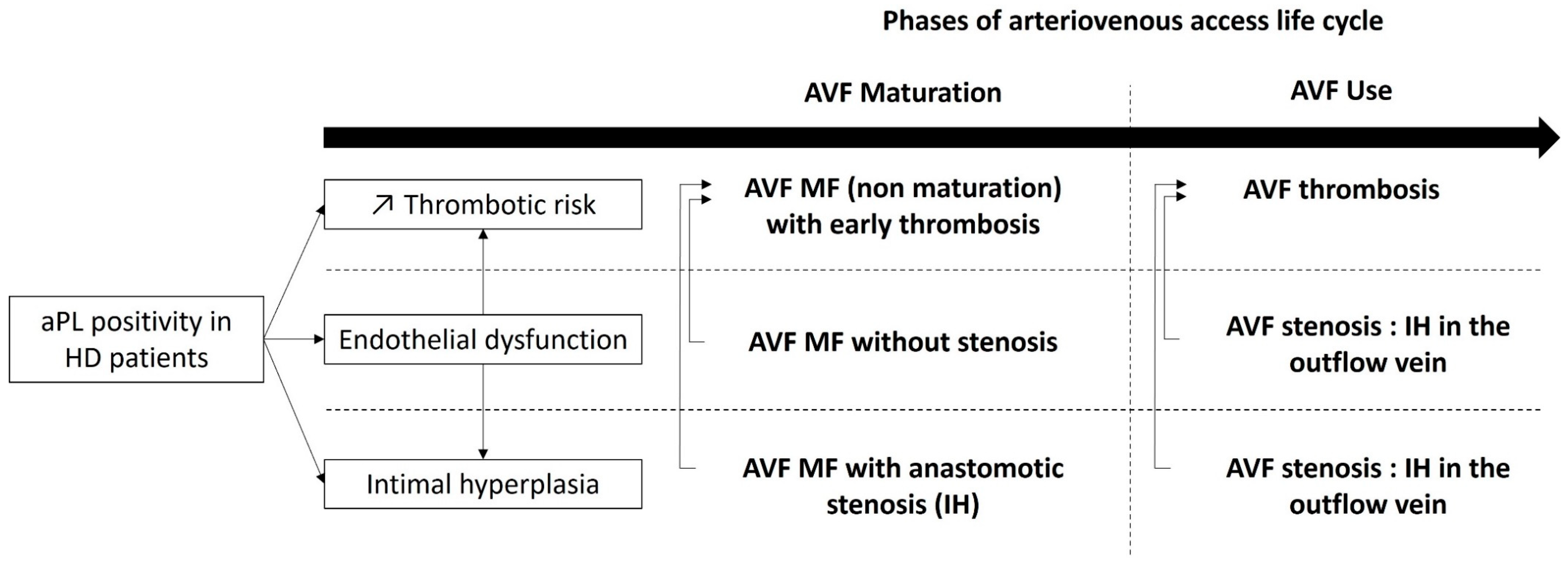
4. Antiphospholipid Mediated AVF Complications
4.1. AVF Maturation
4.2. AVF Thrombosis
4.3. Stenosis and Intimal Hyperplasia
4.4. Mortality
5. Antiphospholipid Antibody Testing before AVF Creation
6. Treatment Options
7. Conclusions
Author Contributions
Funding
Institutional Review Board Statement
Informed Consent Statement
Data Availability Statement
Conflicts of Interest
References
- Saran, R.; Robinson, B.; Abbott, K.C.; Bragg-Gresham, J.; Chen, X.; Gipson, D.; Gu, H.; Hirth, R.A.; Hutton, D.; Jin, Y.; et al. US Renal Data System 2019 Annual Data Report: Epidemiology of Kidney Disease in the United States. Am. J. Kidney Dis. 2020, 75 (Suppl. S1), A6–A7. [Google Scholar] [CrossRef] [PubMed]
- Casserly, L.F.; Dember, L.M. Thrombosis in end-stage renal disease. Semin. Dial. 2003, 16, 245–256. [Google Scholar] [CrossRef] [PubMed]
- Roy-Chaudhury, P.; Kelly, B.S.; Melhem, M.; Zhang, J.; Li, J.; Desai, P.; Munda, R.; Heffelfinger, S.C. Vascular access in hemodialysis: Issues, management, and emerging concepts. Cardiol. Clin. 2005, 23, 249–273. [Google Scholar] [CrossRef] [PubMed]
- Lawson, J.H.; Niklason, L.E.; Roy-Chaudhury, P. Challenges and novel therapies for vascular access in haemodialysis. Nat. Rev. Nephrol. 2020, 16, 586–602. [Google Scholar] [CrossRef] [PubMed]
- Knight, J.S.; Branch, D.W.; Ortel, T.L. Antiphospholipid syndrome: Advances in diagnosis, pathogenesis, and management. BMJ 2023, 380, e069717. [Google Scholar] [CrossRef] [PubMed]
- Knight, J.S.; Kanthi, Y. Mechanisms of immunothrombosis and vasculopathy in antiphospholipid syndrome. Semin. Immunopathol. 2022, 44, 347–362. [Google Scholar] [CrossRef]
- Velásquez, M.; Rojas, M.; Abrahams, V.M.; Escudero, C.; Cadavid, Á.P. Mechanisms of Endothelial Dysfunction in Antiphospholipid Syndrome: Association with Clinical Manifestations. Front. Physiol. 2018, 9, 1840. [Google Scholar] [CrossRef]
- Cugno, M.; Borghi, M.O.; Lonati, L.M.; Ghiadoni, L.; Gerosa, M.; Grossi, C.; De Angelis, V.; Magnaghi, G.; Tincani, A.; Mari, D.; et al. Patients with antiphospholipid syndrome display endothelial perturbation. J. Autoimmun. 2010, 34, 105–110. [Google Scholar] [CrossRef]
- Nochy, D.; Daugas, E.; Droz, D.; Beaufils, H.; Grünfeld, J.P.; Piette, J.C.; Bariety, J.; Hill, G. The intrarenal vascular lesions associated with primary antiphospholipid syndrome. J. Am. Soc. Nephrol. 1999, 10, 507–518. [Google Scholar] [CrossRef]
- Canaud, G.; Bienaimé, F.; Tabarin, F.; Bataillon, G.; Seilhean, D.; Noël, L.-H.; Dragon-Durey, M.-A.; Snanoudj, R.; Friedlander, G.; Halbwachs-Mecarelli, L.; et al. Inhibition of the mTORC pathway in the antiphospholipid syndrome. N. Engl. J. Med. 2014, 371, 303–312. [Google Scholar] [CrossRef]
- Miyakis, S.; Lockshin, M.D.; Atsumi, T.; Branch, D.W.; Brey, R.L.; Cervera, R.; Derksen, R.H.W.M.; DE Groot, P.G.; Koike, T.; Meroni, P.L.; et al. International consensus statement on an update of the classification criteria for definite antiphospholipid syndrome (APS). J. Thromb. Haemost. 2006, 4, 295–306. [Google Scholar] [CrossRef] [PubMed]
- Barbhaiya, M.; Zuily, S.; Naden, R.; Hendry, A.; Manneville, F.; Amigo, M.-C.; Amoura, Z.; Andrade, D.; Andreoli, L.; Artim-Esen, B.; et al. 2023 ACR/EULAR antiphospholipid syndrome classification criteria. Ann. Rheum. Dis. 2023, 82, 1258–1270. [Google Scholar] [CrossRef] [PubMed]
- Liu, X.; Zhu, L.; Liu, H.; Cai, Q.; Yun, Z.; Sun, F.; Jia, Y.; Guo, J.; Li, C. Non-criteria antiphospholipid antibodies in antiphospholipid syndrome: Diagnostic value added. Front. Immunol. 2022, 13, 972012. [Google Scholar] [CrossRef] [PubMed]
- Gracia-Tello, B.; Isenberg, D. Kidney disease in primary anti-phospholipid antibody syndrome. Rheumatology 2017, 56, 1069–1080. [Google Scholar] [CrossRef] [PubMed]
- Ames, P.R.J.; Merashli, M.; Bucci, T.; Pastori, D.; Pignatelli, P.; Violi, F.; Bellizzi, V.; Arcaro, A.; Gentile, F. Antiphospholipid antibodies in end-stage renal disease: A systematic review and meta-analysis. Hemodial. Int. 2020, 24, 383–396. [Google Scholar] [CrossRef] [PubMed]
- Chew, S.L.; Lins, R.L.; Daelemans, R.; Zachee, P.; De Clerck, L.S.; Vermylen, J. Are antiphospholipid antibodies clinically relevant in dialysis patients? Nephrol. Dial. Transplant. 1992, 7, 1194–1198. [Google Scholar] [CrossRef] [PubMed]
- Roozbeh, J.; Serati, A.R.; Malekhoseini, S.A. Arteriovenous fistula thrombosis in patients on regular hemodialysis: A report of 171 patients. Arch. Iran. Med. 2006, 9, 26–32. [Google Scholar]
- Ozmen, S.; Danis, R.; Akin, D.; Batun, S. Anticardiolipin antibodies in hemodialysis patients with hepatitis C and their role in fistula failure. Clin. Nephrol. 2009, 72, 193–198. [Google Scholar]
- Brunet, P.; Aillaud, M.F.; San Marco, M.; Philip-Joet, C.; Dussol, B.; Bernard, D.; Juhan-Vague, I.; Berland, Y. Antiphospholipids in hemodialysis patients: Relationship between lupus anticoagulant and thrombosis. Kidney Int. 1995, 48, 794–800. [Google Scholar] [CrossRef]
- Prakash, R.; Miller, C.C.; Suki, W.N. Anticardiolipin antibody in patients on maintenance hemodialysis and its association with recurrent arteriovenous graft thrombosis. Am. J. Kidney Dis. 1995, 26, 347–352. [Google Scholar] [CrossRef]
- Serrano, A.; García, F.; Serrano, M.; Ramírez, E.; Alfaro, F.J.; Lora, D.; de la Cámara, A.G.; Paz-Artal, E.; Praga, M.; Morales, J.M. IgA antibodies against β2 glycoprotein I in hemodialysis patients are an independent risk factor for mortality. Kidney Int. 2012, 81, 1239–1244. [Google Scholar] [CrossRef] [PubMed]
- Salmela, B.; Hartman, J.; Peltonen, S.; Albäck, A.; Lassila, R. Thrombophilia and arteriovenous fistula survival in ESRD. Clin. J. Am. Soc. Nephrol. 2013, 8, 962–968. [Google Scholar] [CrossRef] [PubMed]
- Ghisdal, L.; Broeders, N.; Wissing, K.M.; Mena, J.M.; Lemy, A.; Wijns, W.; Pradier, O.; Donckier, V.; Racapé, J.; Vereerstraeten, P.; et al. Thrombophilic factors in Stage V chronic kidney disease patients are largely corrected by renal transplantation. Nephrol. Dial. Transplant. 2011, 26, 2700–2705. [Google Scholar] [CrossRef] [PubMed]
- Dabit, J.Y.; Valenzuela-Almada, M.O.; Vallejo-Ramos, S.; Duarte-García, A. Epidemiology of Antiphospholipid Syndrome in the General Population. Curr. Rheumatol. Rep. 2022, 23, 85. [Google Scholar] [CrossRef]
- Wisløff, F.; Jacobsen, E.M.; Liestøl, S. Laboratory diagnosis of the antiphospholipid syndrome. Thromb. Res. 2002, 108, 263–271. [Google Scholar] [CrossRef]
- Blank, M.; Asherson, R.A.; Cervera, R.; Shoenfeld, Y. Antiphospholipid syndrome infectious origin. J. Clin. Immunol. 2004, 24, 12–23. [Google Scholar] [CrossRef]
- Chuang, F.R.; Chen, T.C.; Yang, C.C.; Cheng, Y.F.; Hsu, K.T.; Lee, C.H.; Lin, C.L.; Wang, I.K.; Chang, H.W.; Wang, P.H. IgM-anticardiolipin antibody and vascular access thrombosis in chronic hemodialysis patients. Ren. Fail. 2005, 27, 25–30. [Google Scholar] [CrossRef]
- Bataille, S.; Burtey, S.; Decourt, A.; Frère, C.; Henneuse, A.; Aillaud, M.-F.; Morange, P.; Bardin, N.; Duval, A.; Sallée, M.; et al. Antiphospholipids antibodies and hemodialysis: A frequent association linked to arteriovenous fistula thrombosis. Nephrol. Ther. 2015, 11, 27–33. [Google Scholar] [CrossRef]
- van Mourik, D.J.M.; Salet, D.M.; Middeldorp, S.; Nieuwdorp, M.; van Mens, T.E. The role of the intestinal microbiome in antiphospholipid syndrome. Front. Immunol. 2022, 13, 954764. [Google Scholar] [CrossRef]
- García-Martín, F.; De Arriba, G.; Carrascosa, T.; Moldenhauer, F.; Martin-Escobar, E.; Val, J.; Saiz, F. Anticardiolipin antibodies and lupus anticoagulant in end-stage renal disease. Nephrol. Dial. Transplant. 1991, 6, 543–547. [Google Scholar] [CrossRef]
- Haviv, Y.S. Association of anticardiolipin antibodies with vascular access occlusion in hemodialysis patients: Cause or effect? Nephron 2000, 86, 447–454. [Google Scholar] [CrossRef] [PubMed]
- Canaud, B.; Davenport, A.; Golper, T.A. On-line hemodiafiltration therapy for end-stage kidney disease patients: Promises for the future? What’s next? Semin. Dial. 2022, 35, 459–460. [Google Scholar] [CrossRef] [PubMed]
- Brandt, J.T.; Triplett, D.A.; Alving, B.; Scharrer, I. Criteria for the diagnosis of lupus anticoagulants: An update. On behalf of the Subcommittee on Lupus Anticoagulant/Antiphospholipid Antibody of the Scientific and Standardisation Committee of the ISTH. Thromb. Haemost. 1995, 74, 1185–1190. [Google Scholar] [CrossRef] [PubMed]
- George, J.; Aron, A.; Levy, Y.; Gilburd, B.; Ben-David, A.; Renaudineau, Y.; Zonana-Nachach, A.; Youinou, P.; Harats, D.; Shoenfeld, Y. Anti-cardiolipin, anti-endothelial-cell and anti-malondialdehyde-LDL antibodies in uremic patients undergoing hemodialysis: Relationship with vascular access thrombosis and thromboembolic events. Hum. Antibodies 1999, 9, 125–131. [Google Scholar] [CrossRef] [PubMed]
- Manns, B.J.; Burgess, E.D.; Parsons, H.G.; Schaefer, J.P.; Hyndman, M.E.; Scott-Douglas, N.W. Hyperhomocysteinemia, anticardiolipin antibody status, and risk for vascular access thrombosis in hemodialysis patients. Kidney Int. 1999, 55, 315–320. [Google Scholar] [CrossRef] [PubMed]
- Palomo, I.; Pereira, J.; Alarcón, M.; Vasquez, M.; Pierangeli, S. Vascular access thrombosis is not related to presence of antiphospholipid antibodies in patients on chronic hemodialysis. Nephron 2002, 92, 957–958. [Google Scholar] [CrossRef] [PubMed]
- Nampoory, M.R.N.; Das, K.C.; Johny, K.V.; Al-Hilali, N.; Abraham, M.; Easow, S.; Saed, T.; Al-Muzeirei, I.A.; Sugathan, T.N.; Al Mousawi, M. Hypercoagulability, a serious problem in patients with ESRD on maintenance hemodialysis, and its correction after kidney transplantation. Am. J. Kidney Dis. 2003, 42, 797–805. [Google Scholar] [CrossRef]
- Chuang, Y.C.; Chen, J.B.; Yang, L.C.; Kuo, C.Y. Significance of platelet activation in vascular access survival of haemodialysis patients. Nephrol. Dial. Transplant. 2003, 18, 947–954. [Google Scholar] [CrossRef]
- Molino, D.; De Santo, N.G.; Marotta, R.; Anastasio, P.; Mosavat, M.; De Lucia, D. Plasma levels of plasminogen activator inhibitor type 1, factor VIII, prothrombin activation fragment 1 + 2, anticardiolipin, and antiprothrombin antibodies are risk factors for thrombosis in hemodialysis patients. Semin. Nephrol. 2004, 24, 495–501. [Google Scholar] [CrossRef]
- Knoll, G.A.; Wells, P.S.; Young, D.; Perkins, S.L.; Pilkey, R.M.; Clinch, J.J.; Rodger, M.A. Thrombophilia and the risk for hemodialysis vascular access thrombosis. J. Am. Soc. Nephrol. 2005, 16, 1108–1114. [Google Scholar] [CrossRef]
- Gültekin, F.; Alagözlü, H.; Candan, F.; Nadir, I.; Bakici, M.Z.; Sezer, H. The relationship between anticardiolipin antibodies and vascular access occlusion in patients on hemodialysis. ASAIO J. 2005, 51, 162–164. [Google Scholar] [CrossRef] [PubMed]
- Hadhri, S.; Rejeb, M.B.; Belarbia, A.; Achour, A.; Skouri, H. Hemodialysis duration, human platelet antigen HPA-3 and IgA isotype of anti-β2glycoprotein I antibodies are associated with native arteriovenous fistula failure in Tunisian hemodialysis patients. Thromb. Res. 2013, 131, e202–e209. [Google Scholar] [CrossRef] [PubMed]
- Fadel, F.I.; Elshamaa, M.F.; Abdel-Rahman, S.M.; Thabet, E.H.; Kamel, S.; Kandil, D.; Ibrahim, M.H.; El-Ahmady, M. Coagulation, thrombophilia and patency of arteriovenous fistula in children undergoing haemodialysis compared with healthy volunteers: A prospective analysis. Blood Coagul. Fibrinolysis 2016, 27, 190–198. [Google Scholar] [CrossRef] [PubMed]
- Grupp, C.; Troche-Polzien, I.; Stock, J.; Bramlage, C.; Müller, G.A.; Koziolek, M. Thrombophilic risk factors in hemodialysis: Association with early vascular access occlusion and patient survival in long-term follow-up. PLoS ONE 2019, 14, e0222102. [Google Scholar] [CrossRef] [PubMed]
- Anapalli, S.R.; N, H.D.; Sarma, P.; Srikanth, L.; V, S.K. Thrombophilic risk factors and ABO blood group profile for arteriovenous access failure in end stage kidney disease patients: A single-center experience. Ren. Fail. 2022, 44, 34–42. [Google Scholar] [CrossRef] [PubMed]
- Gkrouzman, E.; Sevim, E.; Finik, J.; Andrade, D.; Pengo, V.; Sciascia, S.; Tektonidou, M.G.; Ugarte, A.; Chighizola, C.B.; Belmont, H.M.; et al. Antiphospholipid Antibody Profile Stability Over Time: Prospective Results from the APS ACTION Clinical Database and Repository. J. Rheumatol. 2021, 48, 541–547. [Google Scholar] [CrossRef] [PubMed]
- Radin, M.; Schreiber, K.; Sciascia, S.; Roccatello, D.; Cecchi, I.; Aguirre Zamorano, M.Á.; Cuadrado, M.J. Prevalence of Antiphospholipid Antibodies Negativisation in Patients with Antiphospholipid Syndrome: A Long-Term Follow-Up Multicentre Study. Thromb. Haemost. 2019, 119, 1920–1926. [Google Scholar] [CrossRef]
- Taghavi, M.; Demulder, A.; Mesquita, M.D.C.F.; Dernier, Y.; Nortier, J.; Collart, F.; Pozdzik, A. Underestimated Effect of Antiphospholipid Antibodies on Arteriovenous Fistula Maturation in Hemodialysis Patients, 09 May 2022, Preprint (Version 1). Research Square. Available online: https://www.researchsquare.com/article/rs-1606215/v1 (accessed on 9 May 2022).
- Zonnebeld, N.; Huberts, W.; van Loon, M.M.; Delhaas, T.; Tordoir, J.H.M. Natural Vascular Remodelling After Arteriovenous Fistula Creation in Dialysis Patients With and Without Previous Ipsilateral Vascular Access. Eur. J. Vasc. Endovasc. Surg. 2020, 59, 277–287. [Google Scholar] [CrossRef]
- Gameiro, J.; Ibeas, J. Factors affecting arteriovenous fistula dysfunction: A narrative review. J. Vasc. Access. 2020, 21, 134–147. [Google Scholar] [CrossRef]
- Lok, C.E.; Huber, T.S.; Lee, T.; Shenoy, S.; Yevzlin, A.S.; Abreo, K.; Allon, M.; Asif, A.; Astor, B.C.; Glickman, M.H.; et al. KDOQI Clinical Practice Guideline for Vascular Access: 2019 Update. Am. J. Kidney Dis. 2020, 75 (Suppl. S2), S1–S164. [Google Scholar] [CrossRef]
- Beathard, G.A.; Lok, C.E.; Glickman, M.H.; Al-Jaishi, A.A.; Bednarski, D.; Cull, D.L.; Lawson, J.H.; Lee, T.C.; Niyyar, V.D.; Syracuse, D.; et al. Definitions and End Points for Interventional Studies for Arteriovenous Dialysis Access. Clin. J. Am. Soc. Nephrol. 2018, 13, 501–512. [Google Scholar] [CrossRef] [PubMed]
- Huber, T.S.; Berceli, S.A.; Scali, S.T.; Neal, D.; Anderson, E.M.; Allon, M.; Cheung, A.K.; Dember, L.M.; Himmelfarb, J.; Roy-Chaudhury, P.; et al. Arteriovenous Fistula Maturation, Functional Patency, and Intervention Rates. JAMA Surg. 2021, 156, 1111–1118. [Google Scholar] [CrossRef] [PubMed]
- Ma, S.; Duan, S.; Liu, Y.; Wang, H. Intimal Hyperplasia of Arteriovenous Fistula. Ann. Vasc. Surg. 2022, 85, 444–453. [Google Scholar] [CrossRef] [PubMed]
- Siddiqui, M.A.; Ashraff, S.; Carline, T. Maturation of arteriovenous fistula: Analysis of key factors. Kidney Res. Clin. Pract. 2017, 36, 318–328. [Google Scholar] [CrossRef] [PubMed]
- Somarathna, M.; Hwang, P.T.; Millican, R.C.; Alexander, G.C.; Isayeva-Waldrop, T.; Sherwood, J.A.; Brott, B.C.; Falzon, I.; Northrup, H.; Shiu, Y.-T.; et al. Nitric oxide releasing nanomatrix gel treatment inhibits venous intimal hyperplasia and improves vascular remodeling in a rodent arteriovenous fistula. Biomaterials 2022, 280, 121254. [Google Scholar] [CrossRef] [PubMed]
- Singh, M.; Mahapatra, H.S.; Pursnani, L.; Muthukumar, B.; Neeraj Anant, I.; Kumar, A.; Kaur, N.; Singh, A.; Krishnan, C. on prediction of arterio-venous fistula maturation by flow mediated dilatation and AVF blood flow. J. Vasc. Access. 2023, 24, 443–451. [Google Scholar] [CrossRef]
- Bolleke, E.; Seferi, S.; Rroji, M.; Idrizi, A.; Barbullushi, M.; Thereska, N. Exhausting multiple hemodialysis access failures. Med. Arch. 2014, 68, 361–363. [Google Scholar] [CrossRef]
- Evangelatos, G.; Kravvariti, E.; Konstantonis, G.; Tentolouris, N.; Sfikakis, P.P.; Tektonidou, M.G. Atherosclerosis progression in antiphospholipid syndrome is comparable to diabetes mellitus: A 3 year prospective study. Rheumatology 2022, 61, 3408–3413. [Google Scholar] [CrossRef]
- Sorice, M.; Profumo, E.; Capozzi, A.; Recalchi, S.; Riitano, G.; Di Veroli, B.; Saso, L.; Buttari, B. Oxidative Stress as a Regulatory Checkpoint in the Production of Antiphospholipid Autoantibodies: The Protective Role of NRF2 Pathway. Biomolecules 2023, 13, 1221. [Google Scholar] [CrossRef]
- Barbhaiya, M.; Taghavi, M.; Zuily, S.; Domingues, V.; Chock, E.Y.; Tektonidou, M.G.; Erkan, D.; Seshan, S.V.; New APS Classification Criteria Steering Committee and APS ACTION Collaborators. Efforts to Better Characterize “Antiphospholipid Antibody Nephropathy” for the 2023 ACR/EULAR Antiphospholipid Syndrome Classification Criteria: Renal Pathology Subcommittee Report. J. Rheumatol. 2023, 50, jrheum.2022-1200. [Google Scholar] [CrossRef]
- Wattanakit, K.; Cushman, M.; Stehman-Breen, C.; Heckbert, S.R.; Folsom, A.R. Chronic kidney disease increases risk for venous thromboembolism. J. Am. Soc. Nephrol. 2008, 19, 135–140. [Google Scholar] [CrossRef] [PubMed]
- Go, A.S.; Bansal, N.; Chandra, M.; Lathon, P.V.; Fortmann, S.P.; Iribarren, C.; Hsu, C.-Y.; Hlatky, M.A.; ADVANCE Study Investigators. Chronic kidney disease and risk for presenting with acute myocardial infarction versus stable exertional angina in adults with coronary heart disease. J. Am. Coll. Cardiol. 2011, 58, 1600–1607. [Google Scholar] [CrossRef] [PubMed]
- Gondouin, B.; Cerini, C.; Dou, L.; Sallée, M.; Duval-Sabatier, A.; Pletinck, A.; Calaf, R.; Lacroix, R.; Jourde-Chiche, N.; Poitevin, S.; et al. Indolic uremic solutes increase tissue factor production in endothelial cells by the aryl hydrocarbon receptor pathway. Kidney Int. 2013, 84, 733–744. [Google Scholar] [CrossRef] [PubMed]
- Sosińska-Zawierucha, P.; Bręborowicz, A. Uremic serum induces prothrombotic changes in venous endothelial cells and inflammatory changes in aortic endothelial cells. Ren. Fail. 2021, 43, 401–405. [Google Scholar] [CrossRef] [PubMed]
- Montagnana, M.; Meschi, T.; Borghi, L.; Lippi, G. Thrombosis and occlusion of vascular access in hemodialyzed patients. Semin. Thromb. Hemost. 2011, 37, 946–954. [Google Scholar] [CrossRef]
- Vazquez-Padron, R.I.; Duque, J.C.; Tabbara, M.; Salman, L.H.; Martinez, L. Intimal Hyperplasia and Arteriovenous Fistula Failure: Looking Beyond Size Differences. Kidney360 2021, 2, 1360–1372. [Google Scholar] [CrossRef]
- Hu, H.; Patel, S.; Hanisch, J.J.; Santana, J.M.; Hashimoto, T.; Bai, H.; Kudze, T.; Foster, T.R.; Guo, J.; Yatsula, B.; et al. Future research directions to improve fistula maturation and reduce access failure. Semin. Vasc. Surg. 2016, 29, 153–171. [Google Scholar] [CrossRef]
- Martínez-Flores, J.A.; Serrano, M.; Morales, J.M.; Serrano, A. Antiphospholipid Syndrome and Kidney Involvement: New Insights. Antibodies 2016, 5, 17. [Google Scholar] [CrossRef]
- Turmel-Rodrigues, L.; Mouton, A.; Birmelé, B.; Billaux, L.; Ammar, N.; Grézard, O.; Hauss, S.; Pengloan, J. Salvage of immature forearm fistulas for haemodialysis by interventional radiology. Nephrol. Dial. Transplant. 2001, 16, 2365–2371. [Google Scholar] [CrossRef]
- Adler, S.; Szczech, L.; Qureshi, A.; Bollu, R.; Thomas-John, R. IgM anticardiolipin antibodies are associated with stenosis of vascular access in hemodialysis patients but do not predict thrombosis. Clin. Nephrol. 2001, 56, 428–434. [Google Scholar]
- Perl, L.; Netzer, A.; Rechavia, E.; Bental, T.; Assali, A.; Codner, P.; Mager, A.; Battler, A.; Kornowski, R.; Lev, E.I. Long-term outcome of patients with antiphospholipid syndrome who undergo percutaneous coronary intervention. Cardiology 2012, 122, 76–82. [Google Scholar] [CrossRef] [PubMed]
- Gallieni, M.; Hollenbeck, M.; Inston, N.; Kumwenda, M.; Powell, S.; Tordoir, J.; Al Shakarchi, J.; Berger, P.; Bolignano, D.; Cassidy, D.; et al. Clinical practice guideline on peri- and postoperative care of arteriovenous fistulas and grafts for haemodialysis in adults. Nephrol. Dial. Transplant. 2019, 34 (Suppl. S2), ii1–ii42. [Google Scholar] [CrossRef] [PubMed]
- Almajdi, A.; Almutairi, S.; Alharbi, M. Safety and efficacy of apixaban versus low-molecular weight heparin or vitamin-K antagonists for venous thromboembolism treatment in patients with severe renal failure: A systematic review and meta-analysis. Thromb. Res. 2023, 229, 77–85. [Google Scholar] [CrossRef] [PubMed]
- Zaragatski, E.; Grommes, J.; Schurgers, L.J.; Langer, S.; Kennes, L.; Tamm, M.; Koeppel, T.A.; Kranz, J.; Hackhofer, T.; Arakelyan, K.; et al. Vitamin K antagonism aggravates chronic kidney disease-induced neointimal hyperplasia and calcification in arterialized veins: Role of vitamin K treatment? Kidney Int. 2016, 89, 601–611. [Google Scholar] [CrossRef]
- Porta, S.V.; de Andrade, D.C.O.; Erkan, D.; Gómez-Puerta, J.A.; Jara, L.J.; Alba Moreyra, P.; Pons-Estel, G.J. Controversies in the Management of Antiphospholipid Syndrome. J. Clin. Rheumatol. 2023, 29, e107–e112. [Google Scholar] [CrossRef]


| 2006 Revised Sapporo | 2023 ACR/EULAR | |
|---|---|---|
| Classification | At least 1 clinical criterion AND 1 laboratory criterion | 3 points from clinical domains AND at least 3 points from laboratory domains |
| Clinical criteria | Entry criteria and scoring: count the highest weighted criterion towards the total score | |
| 2 clinical criteria 1. Vascular thrombosis: One or more clinical episodes of arterial, venous, or small vessel thrombosis, in any tissue or organ 2. Pregnancy morbidity | 6 clinical domains 1. Macrovascular-Venous Thromboembolism 2. Macrovascular-Arterial Thrombosis 3. Microvascular 4. Obstetric 5. Cardiac Valve 6. Hematology | |
| Considered as non-criteria-manifestations: | ||
| - Heart valve disease | Yes | No |
| - Livedo racemosa | Yes | No |
| - Thrombocytopenia | Yes | No |
| - Nephropathy, | Yes | No |
| - Neurological manifestations | Yes | Yes |
| - Pulmonary/Adrenal hemorrhage | Yes | No |
| Laboratory criteria | ||
| Persistent positivity (at 12 weeks) | Yes | Yes |
| Timeline of aPL positivity and clinical criteria | Less than 5 years of clinical criteria | Within 3 years of clinical criterion |
| Thresholds of aCL and/or aβ2GPI | aCL: >40 GPL or MPL, or >the 99th percentile aβ2GPI: >the 99th percentile | aCL or aβ2GPI: Moderate 40–79 units High >80 units |
| Antibodies for laboratory criteria: | ||
| - Positive LA | Yes | Yes |
| - IgG aCL or aβ2GPI | Yes | Yes |
| - IgM aCL and/or aβ2GPI | Yes | Yes. If isolated: are not sufficient (weight only 1 point) |
| Year | First Author (Reference) | Study Type, n | Follow Up (Months) | aPL | Cut-Off | aPL Confirmation | Proportion of Native AVF | AVF Thrombosis and Outcomes |
|---|---|---|---|---|---|---|---|---|
| 1991 | F. Garcia-Martin [30] | Retrospective n = 51 | NA | LA, IgG aCL | 12.5 GPL U/mL | NA | NA | IgG aCL: Higher incidence of early (6 to 96 h) thrombosis in IgG aCL (31%) versus control (17%). Concentrations of IgG aCL early AVF thrombosis were significantly greater than in patients without it (18.5 ± 7.4 GPL U/mL versus 7.4 ± 0.8, p < 0.001). |
| 1992 | S. L. Chew [16] | Prospective n = 60 | 12 | LA, IgG aCL | 10 GPL | Yes | 100% | LA, IgG aCL: no association with AVF thrombosis or death during the 12 months follow up. |
| 1995 | P. Brunet [19] | Cross-sectional n = 97 | 6 | LA, IgG aCL | 20 GPL | NA | 81.40% | LA: association with AVF thrombosis, IgG aCL: no association with AVF thrombosis |
| 1995 | R. Prakash [20] | Retrospective n = 17 | 30 | IgG aCL | 23 GPL | NA | 100% | No events of AVF thrombosis were encountered during the period of review. |
| 1999 | J. George [34] | case–control n = 81 | NA | aCL, aβ2GPI | NA | NA | 6.2% | aCL, aβ2-GPI: no association with AVF thrombosis |
| 1999 | B. J. Manns [35] | Cross-sectional n = 118 | 36 | IgG aCL | low, moderate, and highly positive as follows: 11 to 20 GPL, 21 to 80 GPL, and more than 80 GPL | NA | 75% | IgG aCL: not associated with AVF thrombosis |
| 2000 | Y.S. Haviv [31] | Retrospective n = 54 | NA | IgG and IgM aCL, IgG and IgM aβ2-GPI | 10 IU/mL | NA | 31.50% | IgG and IgM aCL: association with AVF occlusion (thrombosis or IH). IgG and IgM aβ2-GPI: not associated with occlusion |
| 2002 | I. Palomo [36] | Retrospective n = 208 | NA | aCL, aβ2-GPI and aPS | 3 SD above the average of the normal controls | NA | 100% | aPL: no association with AVF thrombosis. |
| 2003 | M.R.N. Nampoory [37] | Retrospective n = 82 | NA | LA, IgG and IgM aCL, IgG and IgM aPS | IgG aCL: ≥23 GPU, IgM aCL: ≥11 MPU, IgG aPS: ≥17 GPS, IgM aPS: ≥23 MPS | NA | 70.70% | LA: association with AVF thrombosis, IgG, IgM aCL and IgG, IgM aPS: no association with AVF thrombosis |
| 2003 | Y-C. Chuang [38] | Cross-sectional n = 48 | NA | IgG aCL | 12 GPL-Uuml | NA | 52.10% | No IgG aCL positivity in this cohort |
| 2004 | D.Molino [39] | Retrospective n = 40 | NA | LA, IgG and IgM aCL, Ig G and IgM aPT | NA | NA | 100.00% | Ig G, IgM aPT, IgG, IgM aCL: significantly associated with AVF thrombosis |
| 2005 | F-R. Chuang [27] | Cross-sectional n = 483 | NA | IgM aCL | IgM aCL: 6 GPL-U/mL | NA | 72.30% | IgM aCL: not associated with AVF thrombosis |
| 2005 | G. A. Knoll [40] | Case-control n = 419 | NA | LA, IgG aCL, IgM aCL | IgG, IgM aCL: medium titer of 30 GPL or MPL U/mL | No | 91.40% | aCL: not associated with AVF thrombosis |
| 2005 | F. Gültekin [41] | Retrospective n = 103 | NA | IgG, IgM aCL | NA | NA | 100% | IgG, IgM aCL: not associated with AVF thrombosis |
| 2006 | J. Roozbeh [17] | Prospective n = 171 | 14 | IgG aCL | Negative: <10 GPL. Low positive: 10 ≤ aCL < 20 GPL, Medium positive: 20 ≤ aCL < 40 GPL, and highly positive: ≥ 40 GPL units. | NA | 100% | IgG aCL: not associated with AVF thrombosis |
| 2009 | S. Ozmen [18] | Cross-sectional n = 103 | NA | IgG, IgM aCL | NA | NA | NA | Not associated with AVF thrombosis, and AVF survival |
| 2012 | A. Serrano [21] | Prospective n = 124 | 24 | IgG, IgM, IgA aCL, IgG, IgM, IgA aβ2-GPI | 20 U/mL | Yes | 100% | IgA aβ2-GPI: associated with AVF thrombosis, cardiovascular disease and mortality |
| 2013 | B. Salmela [22] | Retrospective n = 219 | NA | LA, IgG aCL, IgG aβ2-GPI | 15 U/mL | NA | 100% | aPL: not associated with AVF failure (thrombosis or stenosis) |
| 2013 | S. Hadhri [42] | Case-control n = 101 | NA | LA, IgG, IgM, IgA aCL, IgG, IgM, IgA aβ2-GPI | 95th percentile for healthy blood donors (7 MPL/mL, 10 GPL/mL and 10 APL/mL for IgM, IgG and IgA aCL, respectively, and 8 U/mL for IgM, IgG and IgA anti-β2-GPI) | NA | 100% | IgA aβ2-GPI: independent risk factors for AVF thrombosis (OR = 3.4; 95% CI, 1.21 to 9.55; p = 0.02) |
| 2014 | S. Bataille [28] | Retrospective n = 192 | NA | LA, IgG and IgM aCL, IgG and IgM aβ2-GPI | aCL and aβ2-GPI: 99e percentile | NA | 68% | aPL and LA: significantly associated with AVF thrombosis |
| 2016 | F. I. Fadel [43] | Prospective n = 50 | 48 | IgG aCL | NA | NA | 80% | IgG aCL: significantly associated with AVF thrombosis. |
| 2019 | C. Grupp [44] | Prospective n = 70 | 384 | LA, IgG, IgM, and IgA aCL | Respectively 12 GPLU/mL, 6 MPLU/mL, and 10 APL U/mL | No | 100% | LA, IgG, IgA, IgM aCL: significantly associated with AVF thrombosis. Patient survival tended to be shorter in patients aPL than in control group, but without statistical significance |
| 2022 | S. R. Anapalli [45] | Cross-sectional n = 100 | NA | IgG and IgM aCL | Respectively 10 and 15 MPL units | NA | 100% | IgG and IgM aCL: significantly associated with AVF thrombosis (p value < 0.001) |
| 2020 | P.R J. Ames [15] | systematic review and meta-analysis IgG aCL: n = 1554, LA: n = 511 | NA | LA, IgG aCL | NA | NA | NA | IgG aCL and LA associated with AVF thrombosis |
| Association with AVF Thrombosis | |||||||||
|---|---|---|---|---|---|---|---|---|---|
| Year | First Author (References) | Study Type | n | LA | IgG aCL | IgM aCL | IgG aβ2GPI | IgM aβ2GPI | Other |
| 1991 | F. Garcia-Martin [30] | retrospective | 51 | x | ✓ | ||||
| 1992 | S. L. Chew [16] | prospective | 60 | x | x | ||||
| 1995 | P. Brunet [19] | cross-sectional | 97 | ✓ | x | ||||
| 1995 | R. Prakash [20] | retrospective | 17 | ? | |||||
| 1999 | J. George [34] | case-control | 81 | x | x | ||||
| 1999 | B. J. Manns [35] | cross-sectional | 118 | x | |||||
| 2002 | Y.S. Haviv [31] | retrospective | 54 | ✓ | ✓ | x | x | ||
| 2002 | I. Palomo [36] | retrospective | 208 | x | x | aPS | |||
| 2003 | M. R.N. Nampoory [37] | retrospective | 82 | ✓ | x | x | aPS | ||
| 2003 | Y-C. Chuang [38] | cross-sectional | 48 | ? | ? | ||||
| 2004 | D. Molino [39] | retrospective | 40 | x | ✓ | ✓ | |||
| 2005 | F-R. Chuang [27] | cross-sectional | 483 | x | |||||
| 2005 | G. A. Knoll [40] | case-control | 419 | x | x | x | |||
| 2005 | F. Gültekin [41] | retrospective | 103 | x | x | ||||
| 2006 | J. Roozbeh [17] | prospective | 171 | x | |||||
| 2009 | S. Ozmen [18] | cross-sectional | 103 | x | x | ||||
| 2012 | A. Serrano [21] | prospective | 124 | x | x | x | x | IgA aβ2GPI | |
| 2013 | B. Salmela [22] | retrospective | 219 | x | x | x | |||
| 2013 | S. Hadhri [42] | case-control | 101 | IgA aβ2GPI | |||||
| 2014 | S. Bataille [28] | retrospective | 192 | ✓ | |||||
| 2016 | F. I. Fadel [43] | Prospective | 55 | ✓ | |||||
| 2019 | C. Grupp [44] | prospective | 70 | ✓ | ✓ | ✓ | IgA aCL | ||
| 2022 | S. R. Anapalli [45] | cross-sectional | 100 | ✓ | ✓ | ||||
| 2020 | P. R. J. Ames [15] | meta-analysis | 1554 | ✓ | |||||
| 511 | ✓ | ||||||||
Disclaimer/Publisher’s Note: The statements, opinions and data contained in all publications are solely those of the individual author(s) and contributor(s) and not of MDPI and/or the editor(s). MDPI and/or the editor(s) disclaim responsibility for any injury to people or property resulting from any ideas, methods, instructions or products referred to in the content. |
© 2024 by the authors. Licensee MDPI, Basel, Switzerland. This article is an open access article distributed under the terms and conditions of the Creative Commons Attribution (CC BY) license (https://creativecommons.org/licenses/by/4.0/).
Share and Cite
Taghavi, M.; Jabrane, A.; Jacobs, L.; Mesquita, M.D.C.F.; Demulder, A.; Nortier, J. Antiphospholipid Antibodies Associated with Native Arteriovenous Fistula Complications in Hemodialysis Patients: A Comprehensive Review of the Literature. Antibodies 2024, 13, 1. https://doi.org/10.3390/antib13010001
Taghavi M, Jabrane A, Jacobs L, Mesquita MDCF, Demulder A, Nortier J. Antiphospholipid Antibodies Associated with Native Arteriovenous Fistula Complications in Hemodialysis Patients: A Comprehensive Review of the Literature. Antibodies. 2024; 13(1):1. https://doi.org/10.3390/antib13010001
Chicago/Turabian StyleTaghavi, Maxime, Abla Jabrane, Lucas Jacobs, Maria Do Carmo Filomena Mesquita, Anne Demulder, and Joëlle Nortier. 2024. "Antiphospholipid Antibodies Associated with Native Arteriovenous Fistula Complications in Hemodialysis Patients: A Comprehensive Review of the Literature" Antibodies 13, no. 1: 1. https://doi.org/10.3390/antib13010001