Article

Hsp70 Gene Family in Sebastiscus marmoratus: The Genome-Wide Identification and Transcriptome Analysis under Thermal Stress

Fishery College, Zhejiang Ocean University, Zhoushan 316002, China
*
Author to whom correspondence should be addressed.
Genes 2023, 14(9), 1779; https://doi.org/10.3390/genes14091779
Submission received: 17 August 2023 / Revised: 2 September 2023 / Accepted: 7 September 2023 / Published: 9 September 2023
(This article belongs to the Special Issue Genetic Improvement of Aquatic Species)

Abstract

:
Heat shock protein 70 kDa (Hsp70) is a highly conserved heat stress protein that is important in biotic processes and responses to abiotic stress. Hsp70 genes may be important in Sebastiscus marmoratus, for it is a kind of nearshore reef fish, and habitat temperature change is more drastic during development. However, genome-wide identification and expression analysis in the Hsp70 gene family of S. marmoratus are still lacking. Here, a total of 15 Hsp70 genes in the genome of S. marmoratus are identified, and their expression patterns were investigated using transcriptomic data from thermal stress experiments. The expansion and gene duplication events of Hsp70 genes from the Hspa4, Hspa8, and Hspa12a subfamilies in S. marmoratus are revealed by phylogenetic analysis. qRT-PCR expression patterns demonstrated that seven Hsp70 genes were significantly up-regulated and none were significantly down-regulated after heat treatment. Only the hsp70 gene was significantly up-regulated after cold treatment. The selection test further showed a purifying selection on the duplicated gene pairs, suggesting that these genes underwent subfunctionalization. Our results add novel insight to aquaculture and biological research on S. marmoratus, providing important information on how Hsp70 genes are regulated in Scorpaeniformes under thermal stress.

1. Introduction

As a highly conserved protein, heat shock protein 70 kDa (Hsp70) could protect features in response to intense cellular stress by assisting in their refolding and stabilization [1,2]. By correcting protein folding, Hsps could minimize the aggregation of unnatural proteins and denatured proteins, which have a significant influence on resistance to adverse environmental stresses as a group of molecular chaperones [3,4,5,6]. Based on their function and weight, Hsps could be divided into several families, including Hsp110, Hsp90, Hsp70, Hsp60, and Hsp40, as well as some small heat shock proteins [7]. In those proteins, Hsp70 is one of the most extensively studied proteins because it is conserved in evolution. They widely exist in all organisms except some archaea [8,9,10].
S. marmoratus, commonly known as marbled rockfish, belongs to the family Scorpaenidae, order Scorpaeniformes, and is a kind of sedentary, warm-temperate, oviparous fish [11,12]. In the Western North Pacific, especially in China, Korea, Japan, and the Philippines, S. marmoratus is widely distributed and usually lives in the nearshore reef sea area without long migration [13,14]. Its meat is tender, has high nutritional value, and has potential value in aquaculture [15]. Due to its small size and ease of feeding, it has become the target of artificial breeding in China [16,17]. The suitable temperature of S. marmoratus is constantly changing during its growth period [18], ranging from 13 to 26 °C [19]. Additionally, the temperature of their habitat also varies dramatically. Consequently, strong adaptability to high and low temperatures means that the Hsp70 gene family may be very important in the temperature adaptation of S. marmoratus.
Global warming harms the growth and development of organisms [20]. Numerous studies have demonstrated that heat stress alters gene expression in plants and animals; for example, 14 StHsp20 genes in Solanum tuberosum were significantly up-regulated under heat stress [21]. The expansion or positive selection under thermal change of several HSP genes in crabs was detected based on full-length transcripts [22] and genome-wide analysis [23]. Therefore, studies on the identification and expression of Hsp gene families in organisms can provide insights into the protection of organisms under global warming.
The comprehensive description of the Hsp70 gene family has been studied in plants [24], fungi [25], insects [26], crustaceans [27], and mammals [28]. However, for the lack of fish genome resources and transcriptome sequences, genome-wide identification in fish has barely been carried out except for a few taxa, such as Ictalurus punctatus, Larimichthys crocea, and Boleophthalmus pectinirostris [8,29,30]. Specifically, 16 Hsp70 genes were identified in I. punctatus, showing significant up-regulation or down-regulation after bacterial infection [8]. In L. crocea, six Hsp70 genes out of 17 were significantly up-regulated or down-regulated under thermal stress [29]. Twenty Hsp70s were identified in B. pectinirostris, and eight genes were significantly involved in the high environmental ammonia (HEA) stress response [30]. However, genome-wide screening and transcriptome analysis of the Hsp70 gene family are still lacking in the Scorpaeniformes. We have completed the genome sequencing of S. marmoratus. These resources have made it possible to perform a comprehensive study of these gene families in the S. marmoratus genome.
Herein, genome-wide identification of Hsp70 genes in S. marmoratus was performed, and their gene expression levels under the stimulation of temperature were investigated. The findings will provide valuable resources for further studies on the evolutionary relationship of the Hsp70 gene in teleosts and the mechanism of Hsp70 gene regulation. The results could provide references to the molecular mechanism of Hsp70s in the adaptation of Scorpaeniformes under thermal stress and benefit offshore aquaculture for this high-economic species.

2. Results

2.1. The Hsp70 Genes in S. marmoratus Based on Genome Data Analysis

Twenty-seven candidate sequences were initially obtained from the HMM profiles of the HSP70 domain, Hsp12a, and Hsp12b (PF00012, PTHR14187: SF46, and PTHR14187: SF3). Some candidate sequences were deleted because of the absence of the Hsp70 domain. Finally, the presence of the HSP70 domain in 15 Hsp70 genes was confirmed, and we provided detailed information in the methods section. Fifteen Hsp70 gene members established robust nomenclature in the Zebrafish Nomenclature Guidelines. The CDS of these Hsp70 genes varied from 1320 to 3054 bp in length and encoded proteins from 439 to 1017 amino acids (aa). The predicted physicochemical properties of the amino acid sequences showed that hspa13 had the shortest conserved domain of 389 aa, and hspa4l had the longest conserved domain of 688 aa. The Hsp70 proteins had molecular weights of 48108.26–113767.84 kDa. Their predicted isoelectric points (pI) ranged from 4.98 (hspa5) to 8.73 (hspa12a). Fifteen Hsp70 genes were spread over eleven chromosomes. As shown in Table 1, hspa4, hyou1, and hspa8a were distributed on the same chromosome.

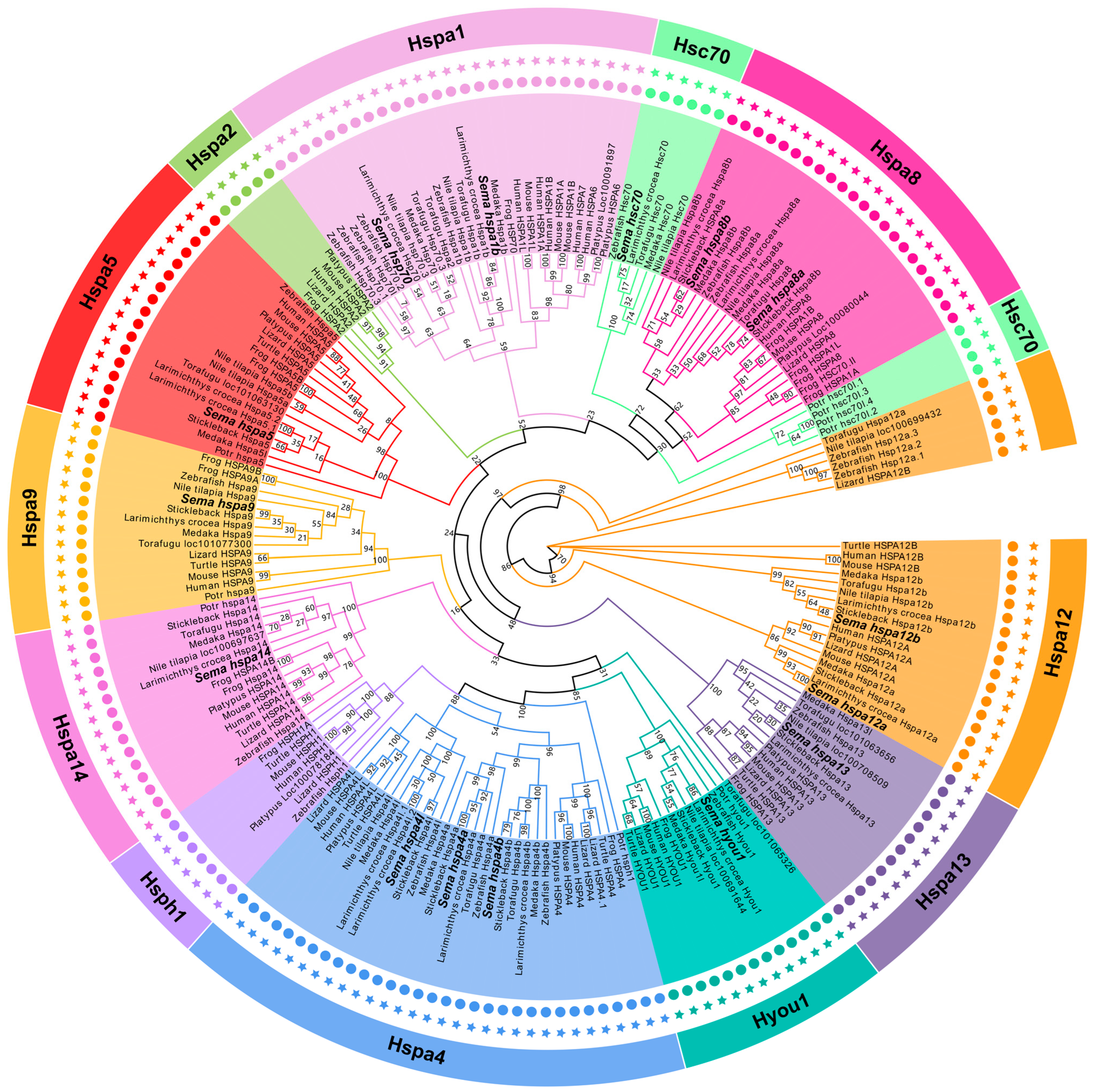
2.2. The Phylogeny of the Hsp70 Genes among Species

We constructed an unrooted phylogenetic tree of the Hsp70s from the amino acid sequence of S. marmoratus and the classic model species to analyze the evolutionary relationship and classification of this gene family (Figure 1). Two human-related genes, HSPA8 (named hspa8a and hspa8b according to the cluster), were detected in the genomes of six teleost fish. The names of the Hsp70 genes in S. marmoratus were standardized following the zebrafish and human orthologs. From the phylogenetic relationships, all the Hsp70 gene family members in S. marmoratus were located in different branches and clustered with the corresponding genes from other fish species at first with good bootstrap values, indicating that Hsp70 genes corresponding to different species are more closely related than the different Hsp70 genes of the same species.
We identified 12 homologous Hsp70 genes (present similarly in humans) in S. marmoratus, but hspa2, hspa6, and hspa7 were not identified. The hsph1 gene didn’t appear in any other teleosts except zebrafish. Most orthologous pairs among S. marmoratus and the other five fish species have been confirmed, which suggests that the same ancestor gene of the Hsp70 family may have appeared before the genetic differentiation of fish. Furthermore, three fish genes (hspa8a, hspa8b, and hsc70) might be closely related paralogs of the human HSPA8 gene (hsc70 vs. hspa8a and hspa8b).

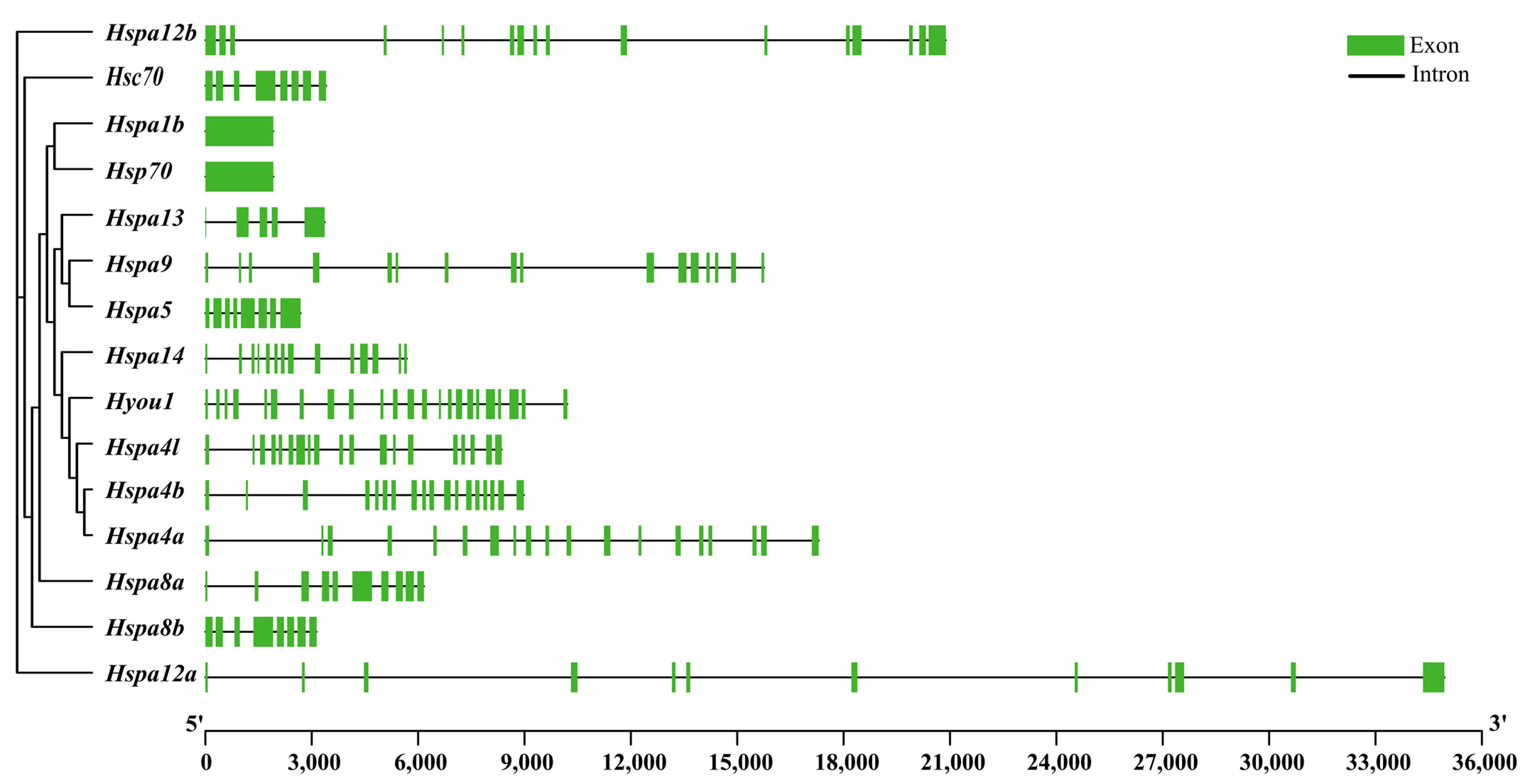
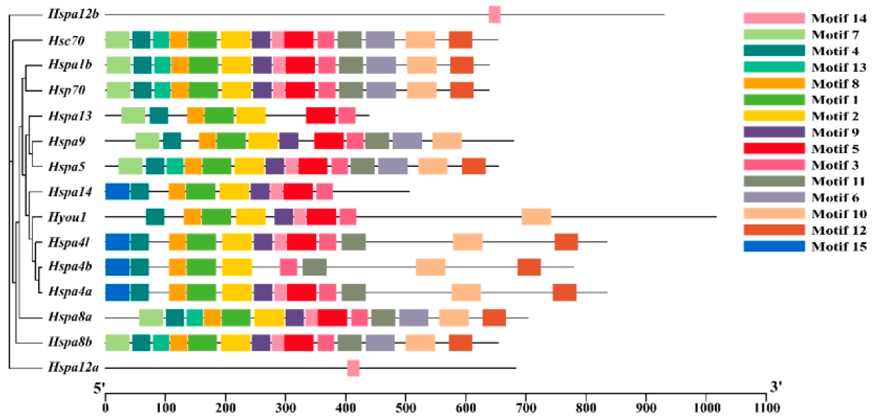
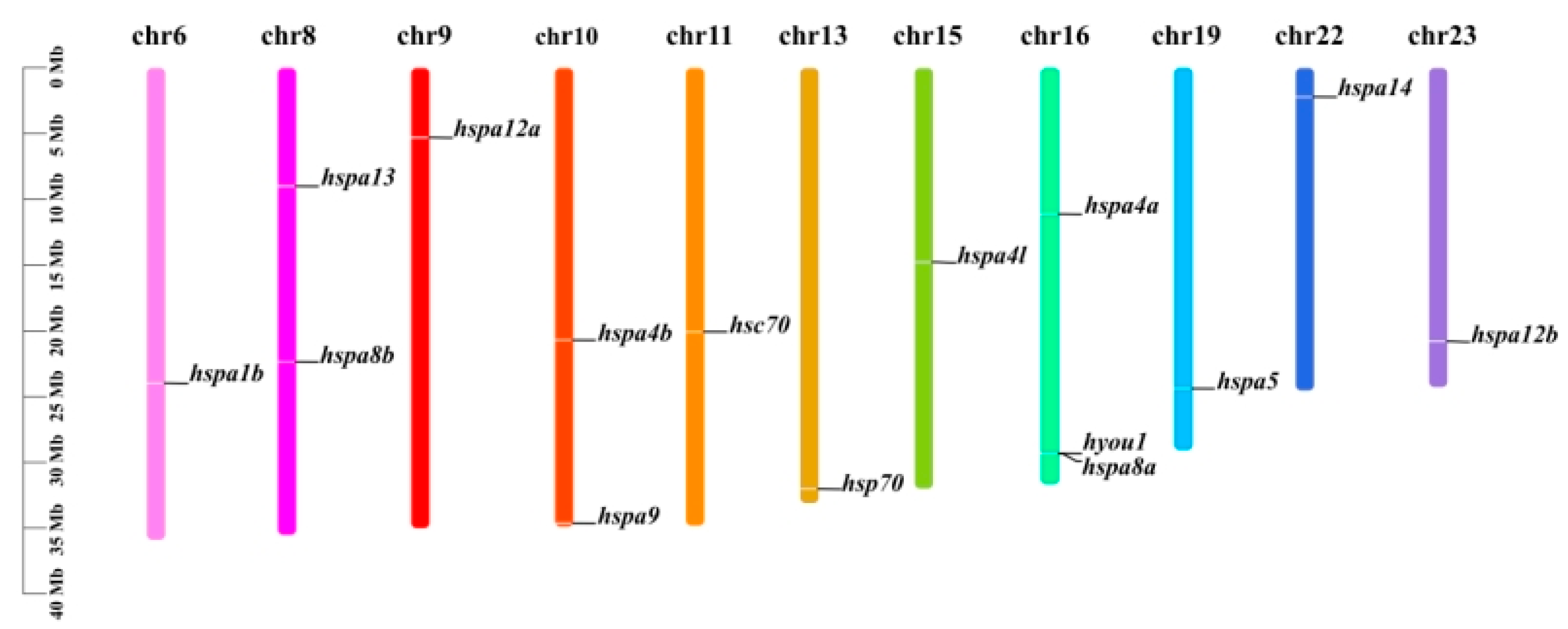
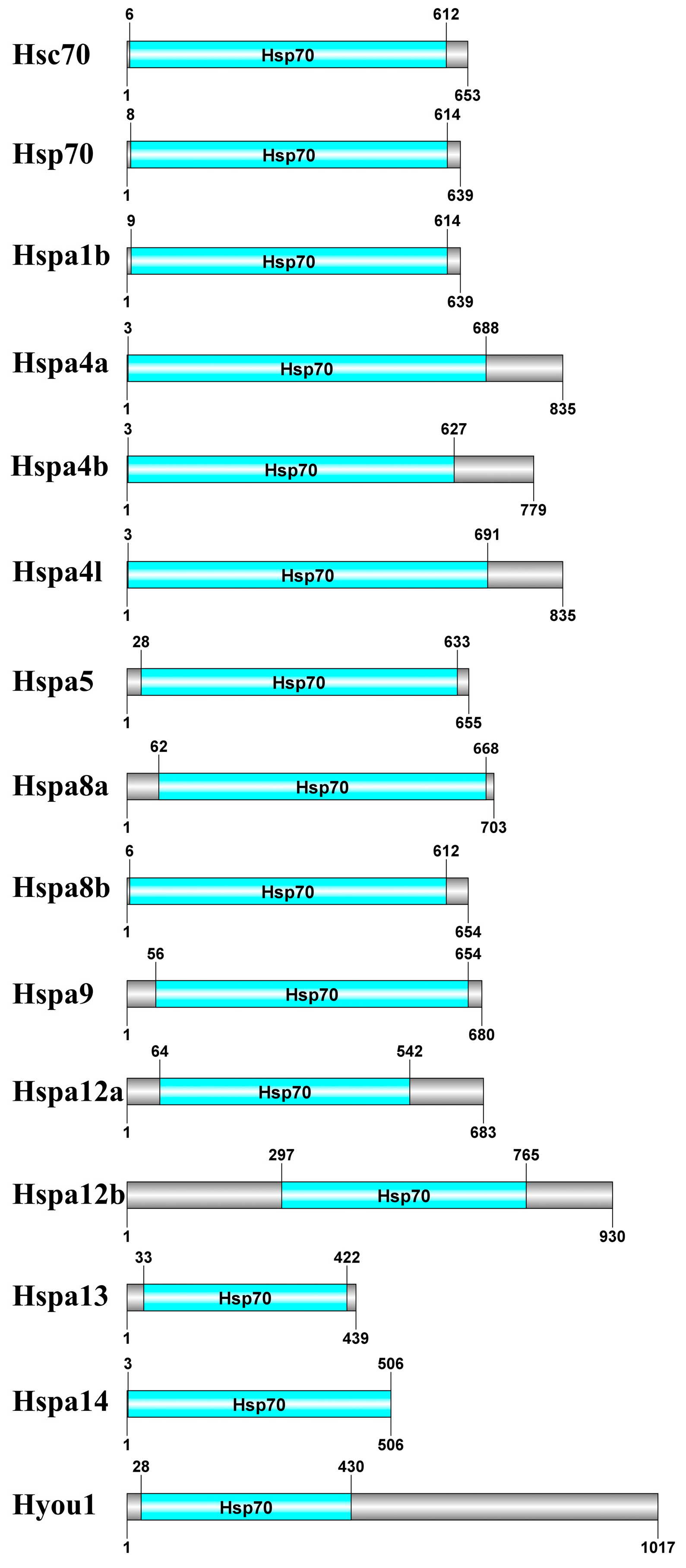
2.3. Gene Structure, Motif, and Chromosomal Location Analysis

The intron–exon structure diagram of every Hsp70 gene in S. marmoratus is shown in Figure 2. We performed the structure analysis based on a genome annotation file. Introns existed in all genes, ranging from 1 to 23. Among them, two members (hspa1b, hsp70) just had one exon; however, the number of exons in the remaining genes ranged from 4 to 23. The number of introns and exons varies greatly among different genes, indicating that these genes may have some differences in biological functions.
Conserved motif analysis was performed on the Hsp70s of S. marmoratus to understand its functional diversity. We searched for each Hsp70s with fifteen putative motifs (Figure 3). In addition, there was only one motif in hspa12a and hspa12b; motif 15 is present only in hspa4a, hspa4b, hspa4l, and hspa14; and motifs 1, 2, 3, 4, and 8 were present in the remaining 13 genes. Among them, some genes had similar or even common motif compositions that were closely related to the phylogenetic relationship, and they were often found in close evolutionary clusters. To some extent, the motif analysis results could support the phylogenetic relationship of the Hsp70 gene family.
The identified Hsp70 genes were sketched on chromosomes to investigate the hsp70 expansion events in S. marmoratus. As shown in Figure 4, 15 Hsp70 genes were distributed on 11 chromosomes. Of all Hsp70 genes, three were located on chromosome 16, which had the largest number of Hsp70 genes; two members were distributed on chromosomes 8 and 10, respectively; and the remaining chromosomes contained only one member. Our results suggest that the hsp70 expansion may occur after two rounds of whole genome duplication (WGD) in early vertebrate evolution, for the hspa12a and hspa12b (which existed in most vertebrates) are identified at different chromosomes [31].
As shown in Figure 5, the Hsp70s in S. marmoratus are highly conserved and further verified using the CDD and Pfam databases. All Hsp70 proteins contain a highly conserved domain between 3 and 765 aa at the N terminal, the presence of which confirms that they are Hsp70 proteins.

2.4. Protein Signal Peptides Predictive Analysis

Our analysis suggested that Hspa5 and Hyou1 were identified as secreted proteins by signal peptide prediction analysis. The highest values of the raw cleavage site score (CS) of two predicted proteins were 0.9823 and 0.5999, which appeared on the 16th alanine (A) and 26th valine (V), respectively. Our results suggested that Hyou1 and Hspa5 had signal peptides based on the SP value, and their lengths were approximately 15 and 25 amino acids, respectively. The results of the protein signal peptide predictive analysis can be found in Figure S1.

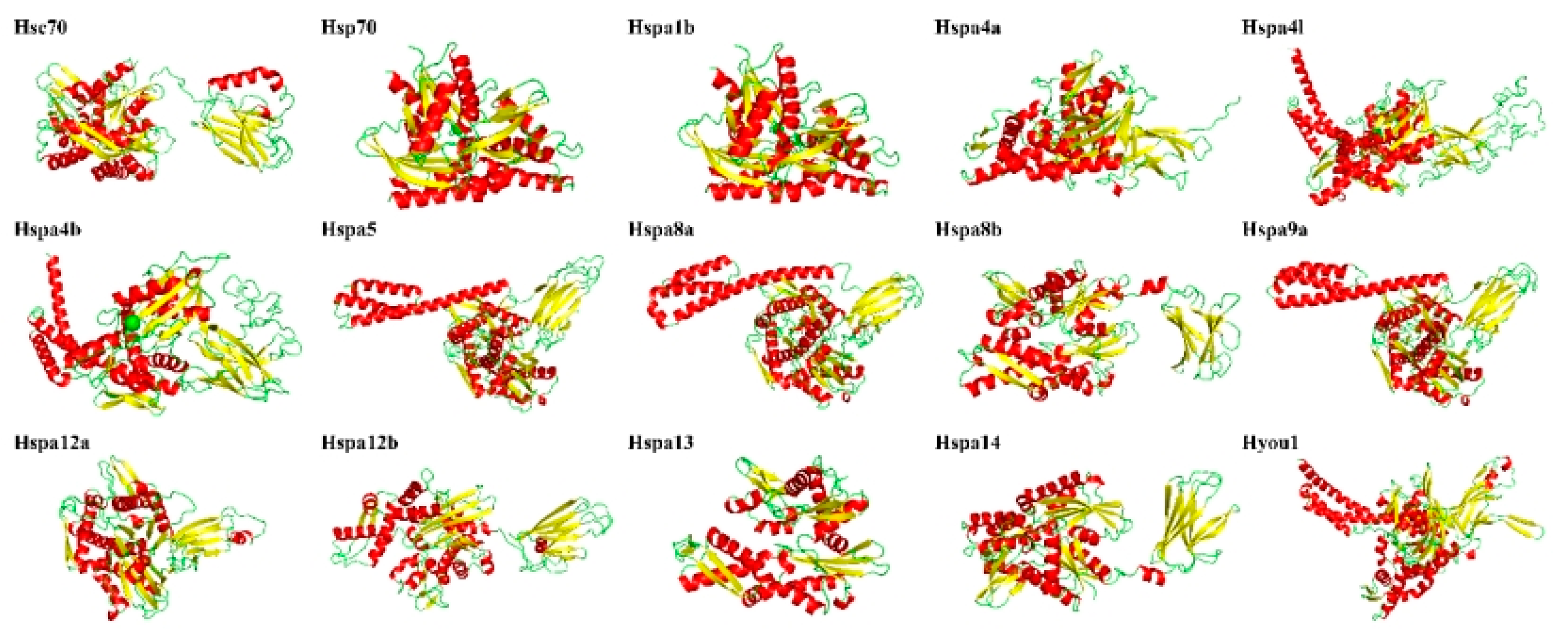
2.5. Secondary and Third Structure Prediction, Subcellular Localization of Hsp70 Proteins

Random coils and α helices were the main secondary structures of the 15 proteins encoded by the S. marmoratus Hsp70 genes. α helices accounted for 30.89% (Hspa12a)–47.89% (Hyou1), β turn accounted for 3.11% (Hspa4l)–7.74% (Hspa13), random coil accounted for 29.71% (Hspa9)–44.66% (Hspa12a), and extended strand accounted for 12.39% (Hyou1)–22.13% (Hspa14) (Table 2, Figure 6).
All Hsp70 gene members of S. marmoratus were expressed in the mitochondrion, plasma membrane, cytoplasm, endoplasmic reticulum, and nucleus. Hspa5 and Hyou1 proteins were expressed in the endoplasmic reticulum. Hspa9 and Hspa8a proteins were expressed in the mitochondrion. The Hspa12a protein was expressed in the nucleus. The rest of the proteins were all expressed in the cytoplasm (Table 2).

2.6. Selection Test on Duplicated Hsp70 Gene Pairs

By calculating the Ka, Ks, and Ka/Ks for five homologous Hsp70 gene pairs, we assess the evolutionary constraints and selection pressures on the Hsp70 genes. The synonymous substitution rate (Ks) represents the background base substitution rate, and we could use Ks values to evaluate the WGD times. Our results suggest that strong purifying selection may have occurred during evolution because the Ka/Ks values of the Hsp70 gene pairs were all less than 1.0 (Table 3).

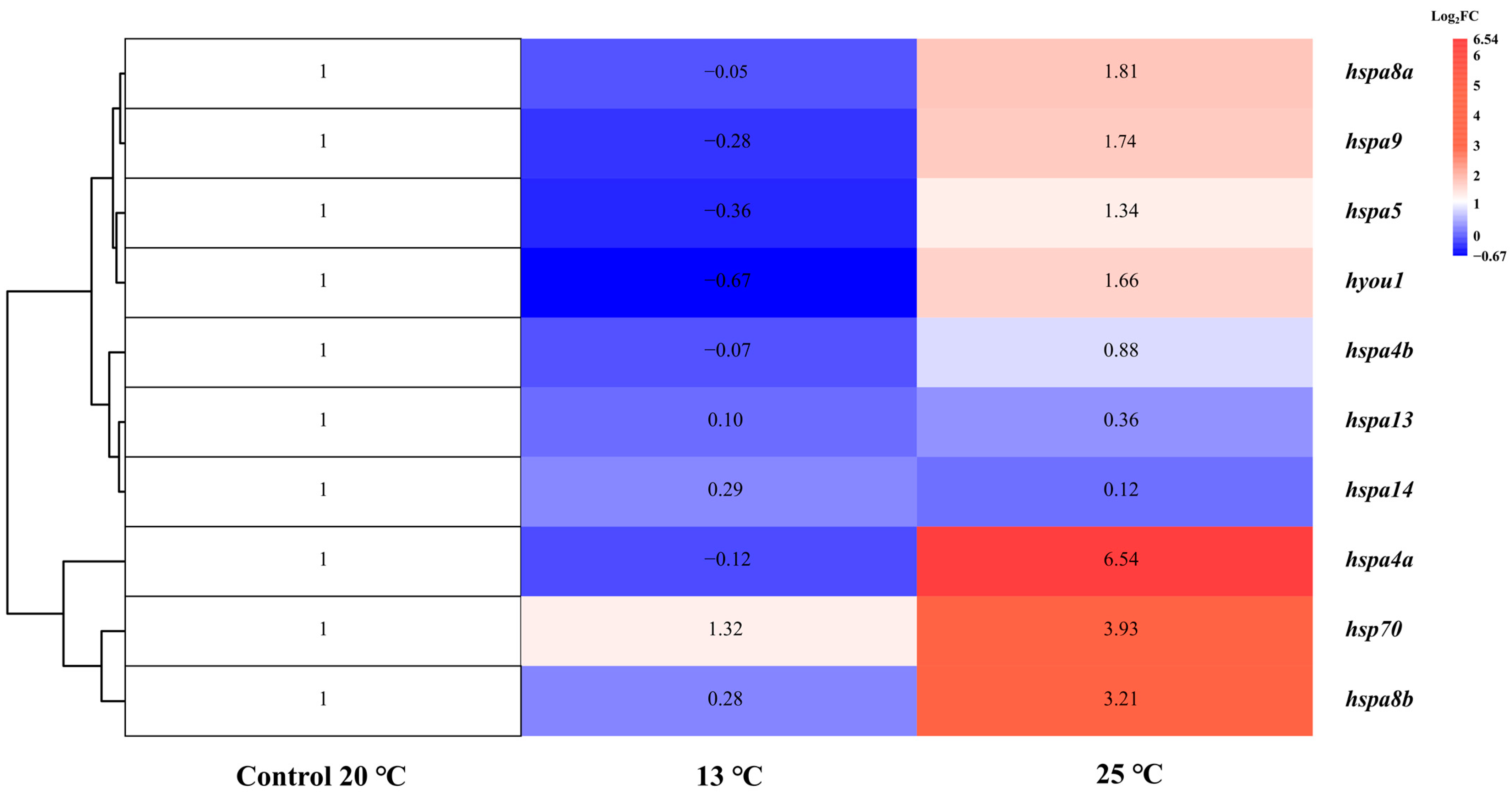
2.7. Expression Profiles of Hsp70 Genes under Thermal Stress Treatment

To understand the gene expression profiles of Hsp70s in S. marmoratus, high-temperature and low-temperature stress experiments were conducted, and corresponding transcriptome analysis was performed on its liver. Ten Hsp70 genes were expressed in liver tissue among the 15 Hsp70 genes (Table 4; Figure 7), which indicated a tissue expression pattern. Under high-temperature treatment, seven (hspa8a, hspa8b, hspa9, hsc70, hyou1, hspa4a, and hspa5) Hsp70 genes were significantly up-regulated (log2FC: 1.32–6.54), while no significantly down-regulated genes were found. Under low-temperature stress treatment at 13 °C, only hsp70 was significantly up-regulated. The expression levels of the remaining 4 Hsp70 genes (hspa4b, hspa5, hspa13, and hspa14) exhibited little change under cold or heat stress.

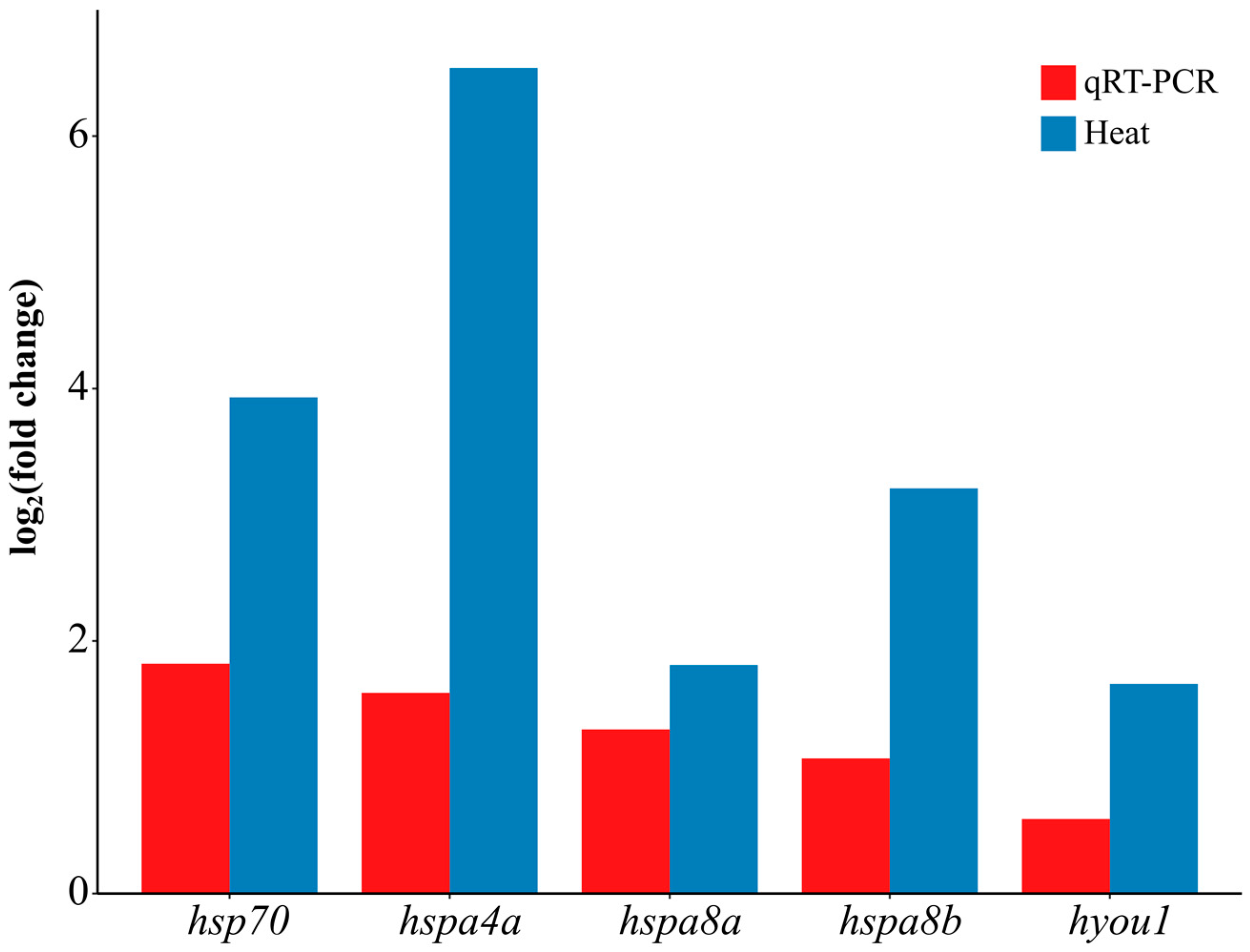
2.8. Validation of Transcriptomic Data via qRT-PCR

Five significant expressed genes in the high-temperature group were used for qRT-PCR analysis to verify the accuracy of all the transcriptome data (Figure 8). The qRT-PCR results showed a similar tendency to the RNA-Seq results, indicating credible transcriptome data despite slight value discrepancies.

3. Discussion

Hsp70s are essential in responding to biotic and environmental stress. In recent years, several studies have been conducted on the Hsp70s of fish; however, genome-wide identification and expression analysis have not yet been conducted in Scorpaeniformes. In the current study, the whole Hsp70 gene family of S. marmoratus under thermal stress was well investigated, including phylogeny, functional characterization, selection tests, and expression pattern analysis in the liver. These results provide a new way to study this physiological response and the regulatory mechanism of S. marmoratus against thermal stress and are a valuable theoretical basis for ovoviviparous fish aquaculture.
The literature often uses the member name of this gene family in confusion, resulting in a series of naming-related problems. Therefore, when the term Hsp70 is used, it is difficult to know which gene or protein in the family is described without further description. A total of 15 Hsp70 genes were identified and annotated in S. marmoratus and based on the evolutionary relationship, the names of Hsp70 proteins were well assigned (the identified Hsp70 genes can be found in File S1). We searched exhaustively in the genomic resources, but the hsph1 gene was still not found in S. marmoratus. It is unclear whether hsph1 is truly missing or whether it is due to incomplete sequencing or annotation of the S. marmoratus genome. The hsph1 gene was only found in Danio rerio and L. crocea and was absent in other fish species, including S. marmoratus, Oncorhynchus mykiss, B. pectinirostris, O. niloticus, and Takifugu rubripes. In six fish, the hspa6 and hspa7 genes were not found in any species. Compared to humans, most Hsp70 genes were found in S. marmoratus, except hspa2, hspa6, hspa7, and hsph1. Compared to L. crocea, although S. marmoratus had fewer copies of the hspa4l and hspa5 genes, most Hsp70 family genes of S. marmoratus and L. crocea were clustered together. They had high homology, as revealed by the phylogenetic tree. When compared to B. pectinirostris, the hspa4l, hspa8b, and hsc70 genes of S. marmoratus seemed to have been lost in B. pectinirostris [30]. Furthermore, B. pectinirostris has two more copies of hspa8a than S. marmoratus (hspa8a.1, hspa8a.2) [30], and the same result can also be found in I. punctatus [8]. The present study suggested that no tandem duplication of hsp70 was observed in S. marmoratus, whereas previous studies have shown that the tandem duplications of hsp70 (named hspa1a in B. pectinirostris) paralogs have been described in B. pectinirostris, I. punctatus, and D. rerio [8]. Since the number of tandem duplication genes differs among species, it may be closely related to the species adaptation to the environment.
Differences in the biological processes of subcellular compartments may be relevant to distinct functions in the Hsp70 gene structure [32]. Subcellular localization has shown that Hsp70 family members are located in the cytoplasm, mitochondria, plasma membrane, endoplasmic reticulum, and nucleus [33]. It has been demonstrated in past research that cytoplasmic Hsp70s play an important role under stressed and even non-stressed conditions [34,35]. Analysis of signal peptide prediction has shown that Hyou1 and Hspa5 were preliminary determined as secreted proteins. According to the chromosome location analysis, the Hsp70 gene members were located on different chromosomes. It is worth mentioning that when gene pairs are located on different chromosomes, the gene replication process can be considered fragmented [36]. Instead, tandem duplications refer to the copies of genes on the same chromosome [37]. Duplicated hsp70 genes (hspa4a, hspa4b, hspa4l, hspa8a, hspa8b, hspa12a, and hspa12b) were also found in S. marmoratus, analogous to their function by their gene structure and motifs.
Gene duplications occur in all kinds of life forms and provide the raw material for functional innovation [38]. Gene replication is the main mechanism for gene family amplification through WGD or single-gene duplication (SSD) in the process of evolution [39]. The phylogenetic tree in this study found that mammals tend to have fewer duplications than teleosts, which may be related to the teleost undergoing whole genome duplication. Compared with tetrapods (amphibians, reptiles, birds, and mammals), more paralogues of hspa8 and hspa4 were found in teleosts (referring to hspa8a and hspa8b, hspa4a and hspa4b, respectively). The teleost-specific genes indicated that hspa8a and hspa8b, hspa4a and hspa4b were present after the divergence between teleosts and tetrapods, which is consistent with the research results of Xu et al., 2018 [29].
According to the thermal stress experiment of S. marmoratus, 10 out of 15 Hsp70 genes were expressed in liver tissue. Based on these results, it can be inferred that they are involved in thermal adaptation and have different expression patterns under heat stress, which may be related to the different effects of cold and heat on physiology. Seven genes (hspa4a, hspa5, hyou1, hspa8a, hspa8b, hspa9, and hsp70) were significantly up-regulated and no significantly down-regulated genes were found under high-temperature treatment. In terms of high-temperature treatment, our results were the same as those from the temperature trials of L. crocea [29]. Severe cell damage can be caused by extreme temperatures, and different species of fish involve the rapid synthesis of Hsp70, which aims for cellular protection by stabilizing protein acquisition resistance and thermo-tolerance [40,41]. Only one gene (hsp70) was significantly up-regulated, and other genes were slightly changed under low-temperature treatment. In general, heat and cold stress are opposite to the regulation of enzyme reactions, diffusion, and membrane transport rates [42]. Under low-temperature treatment, one significantly up-regulated gene (hsp70) was found. Living organisms utilize various mechanisms to manage and repair damage caused by different types of cellular stressors [43]. So, at 13 °C, the hsp70 gene was highly expressed, protecting cells from the effects of low-temperature stress, including protein damage and misfolding. The number of damaged proteins increased under low-temperature stress (13 °C). The production of hsp70 gradually increased, presumably reflecting the need for more Hsp70 protein, which promoted the renaturation of abnormal proteins [1]. The hyou1 and hspa5 proteins have a signal peptide and a transmembrane domain and are found in the endoplasmic reticulum as secreted proteins. The hyou1 and hspa5 proteins, as companion proteins of the endoplasmic reticulum (ER), play a crucial role in protein folding and ensuring the quality control of protein synthesis in ER tubes. As the temperature decreases, protein synthesis in the ER is inhibited. Thus, the expression of the hspa5 and hyou1 genes is involved in the correct folding of proteins, and the degradation of misfolded proteins is limited by low-temperature stress. Expression of hspa5 and hyou1 under heat stress is temperature-induced. Similar results were found in O. mykiss [44].

4. Materials and Methods

4.1. Animal Materials and Experiment

In order to obtain the expression profile of Hsp70 genes under thermal stress, temperature stress experiments were conducted, and the RNA-seq reads were subsequently obtained. Briefly, several S. marmoratus individuals with similar morphology and physiology were collected in July 2019 and temporarily cultivated in the laboratory. The water temperature in the plastic pool was kept at 20 °C and the salinity was kept at 20‰. To reduce the unquantifiable influences on transcriptome sequencing, feeding was stopped and oxygen was continuously charged during the temporary-keeping period; the dissolved oxygen value remained at about 8mg/L. The suitable temperature of S. marmoratus is constantly changing during its growth period, ranging from 13 to 26 °C [18,19]. Three temperature groups, with ten fish in each group, were assigned as the low-temperature group (13 °C), the control group (20 °C), and the high-temperature group (25 °C), respectively. The trial lasted 6 h. Transcriptome sequencing was performed on the liver tissues of three fish randomly selected from each group. All fish were anesthetized with eugenol (Eugenol [CAS 97-53-0], Cat. No. CN00359) for dissection. These specimens were preserved in the RNA preservation solution and stored at −80 °C for later use. The total RNA was extracted using the TRIzol Reagent Kit (Invitrogen, Carlsbad, CA, USA). The RNA integrity number (RIN) of the RNA samples was 6.7, and the total RNA concentration was more than 195ng/ul. We used the NEBNext Ultra RNA Library Prep Kit for Illumina1 (NEB, Ipswich, MA, USA) to construct a sequencing library. The sequencing platform used was the Illumina Hiseq 2500. All of the above is completed in accordance with the manufacturer’s instructions.

4.2. Identification of Hsp70 Members

The genome and protein sequence of S. marmoratus were completed by the fishery ecology and biodiversity laboratory of Zhejiang Ocean University (https://doi.org/10.6084/m9.figshare.19161290) (accessed on 22 July 2023). The hidden Markov model (HMM) profile of the Hsp70s gene was obtained from the Pfam protein families database (http://pfam.Xfam.org/) (accessed on 10 August 2023) [45], and the HMM profiles of Hsp12a and Hsp12b (PTHR14187: SF46 and PTHR14187: SF39) were downloaded from the PANTHER classification system (http://www.pantherdb.org/) (accessed on 10 August 2023). HMMER 3.2.1 software was used to identify Hsp70 genes from the S. marmoratus genome [46]. The online program Pfam and the NCBI conserved domain database (http://www.ncbi.nlm.nih.gov/cdd/) (accessed on 11 August 2023) were used to survey the conserved domains of the candidate protein sequence [47]. Furthermore, the fully conserved domain sequences from the S. marmoratus genome were retained as queries to search against this species in RNA-seq datasets. The online software SOPMA was utilized to predict protein secondary structure (http://npsa-pbil.ibcp.fr/cgi-bin/npsa_automat.pl?page=npsa_sopma.html) (accessed on 11 August 2023) [48]. WoLF PSORT (https://wolfpsort.hgc.jp/) (accessed on 11 August 2023) software was used to predict subcellular localization by uploading protein sequences [49]. The ExPASy ProtParam tool (https://web.expasy.org/protparam/) (accessed on 11 August 2023) was used to calculate protein physicochemical parameters such as molecular weight (MW) and isoelectric point (pI) [50].

4.3. Phylogenetic Relationship Analysis

We used the protein sequences identified from S. marmoratus and 13 other species (including humans, fish, amphibians, and crabs) to construct phylogenetic trees. The protein sequences were retrieved from the NCBI (http://www.ncbi.nlm.nih.gov) (accessed on 22 July 2023), UniProt (http://www.uniprot.org) (accessed on 22 July 2023), and Ensemble (http://asia.ensembl.org/index.html) (accessed on 22 July 2023) databases (detailed information can be found in Table S1), as listed by Song et al., 2016 [8]. The Portunus trituberculatus sequences were downloaded from Jin et al., 2022 [27]. The ML analysis was employed to conduct phylogenetic and evolutionary analyses utilizing RAxML based on a PROTGAMMAIJTT matrix-based model with 1000 bootstrap replicates [51]. Muscle software was used to align multiple protein sequences.

4.4. Gene Structure, Motif, Chromosomal Location, and Conserved Domain Analysis

The structure, motif, and chromosome position of S. marmoratus Hsp70 genes were analyzed and visualized using TBtools v1.09861 [52] based on the genome annotation files. Conserved amino acid sequences of Hsp70 proteins in S. marmoratus were detected by MEME (Multiple Expectation Maximization for Motif Elicitation, version 5.4.1) [53] with the following parameters: site distribution was set at 0 or 1 occurrence per sequence, the maximum number of motifs to be found was 15, and the motif width was set to 10~50. The protein structure was visualized using DOG 2.0 with default parameters [54].

4.5. Three-Dimensional Structure Analysis of Hsp70 Proteins

Three-dimensional protein structures for various Hsp70 proteins were constructed through homology modeling using Swiss Model software (https://swissmodel.expasy.org/) (accessed on 12 August 2023) with default parameters, and the quality of the resulting models was assessed using SAVES v.6.0 (https://saves.mbi.ucla.edu/) (accessed on 12 August 2023) [55,56]. When the sequence identity of the module is less than 30%, then use Phyre2 online software (http://www.sbg.bio.ic.ac.uk/phyre2/html/page.cgi?id=index) (accessed on 12 August 2023) to generate 3D structure models with the highest confidence [57].

4.6. Predictive Analysis of Protein Signal Peptides and Selection Test of Hsp70s

Signal-peptide prediction analysis used the SignalP 6.0 online tool [58]. For the selection-pressure test, Hsp70 duplicated gene pairs from S. marmoratus were investigated through phylogenetic relationships. KaKs_Calculator2.0 software was used to calculate the rates of synonymous (Ks) and nonsynonymous (Ka) substitutions and their ratios [59].

4.7. Expression Pattern Analysis of Hsp70 Genes in S. marmoratus

The index of the transcript to the coding sequence map was created using Bowtie2 v2.5.0. Differential transcript expression levels were evaluated using RNA-Seq by Expectation Maximization (RSEM) [60]. We used the rsem-calculate-expression Perl script in RSEM 1.3.1 to quantify expression levels through Bowtie2 alignment of reads. The expression estimates of Hsp70 genes were normalized in the form of fragments per kilobase of transcript sequence per million fragments mapped (FPKM). The FPKM values were ≥5. Then, log2-based fold change (log2FC) values were calculated. Genes with t-test values (p < 0.05) and log2FC < −1.0 or log2FC > 1.0 were defined as differentially expressed genes.

4.8. Primer Design, RNA Isolation, and Quantitative Real-Time PCR (qRT-PCR) Validation

The total RNA of the samples was extracted using a standard Trizol Reagent Kit following the manufacturer’s protocol. RNA purity was checked using the NanoPhotometer® spectrophotometer (IMPLEN, Westlake Village, CA, USA). RNA integrity was assessed using the RNA Nano 6000 Assay Kit of the Agilent Bioanalyzer 2100 system (Agilent Technologies, Santa Clara, CA, USA). Quantitative reverse transcription PCR (qRT-PCR) was performed to validate the transcriptomic data. Based on the DEG analysis, five genes with significant expression were used in the qRT-PCR analysis, and primers were designed using Primer Premier 5.0 (Table S2). In addition, β-actin was selected as a reference gene for internal standardization. qRT-PCR was designed following the manufacturer’s instructions for TB Green® Premix Ex TaqTM (Tli RNase H Plus) RR420A. Using the ABI PRISM 7300 real-time PCR system (APPlied Biosystems, Carlsbad, CA, USA), the reactions were prepared in a total volume of 20 μL, containing 2 μL of diluted cDNA template, 0.4 μL of each primer (10 μM), 10 μL of TB Green Premix Ex Taq (Tli RNase H Plus) (2×), 6.8 μL of nuclease-free water and 0.4μL ROX Reference Dye (50×). The PCR program was 95 °C for 30 s, followed by 40 cycles of 95 °C for 5 s and 60 °C for 30 s. Three parallel experiments on each cDNA template were performed to strengthen the veracity of the result. Relative expression levels of genes were calculated using the 2-ΔΔCT (ΔCT = CTtarget gene − CTreference gene, ΔΔCT = ΔCTtreatment − ΔCTcontrol) method.

5. Conclusions

This study represented a comprehensive genomic and transcriptomic analysis of Hsp70 genes in S. marmoratus. Fifteen Hsp70 genes were identified for the first time in S. marmoratus. All members of the Hsp70 gene family were well classified, and the expansion of Hsp70 genes from the Hspa4, Hspa8, and Hspa12a subfamilies in S. marmoratus. Regulated expression of Hsp70s was observed; some inducible Hsp70s were significantly up-regulated after thermal stress exposure, and they were from the expanded Hspa4 and Hspa8 subfamilies. These findings provide a reference for further research on the biology of S. marmoratus and help to illuminate the regulatory mechanism of the Hsp70 gene family in response to thermal stress. Above all, our results promoted awareness of the temperature adaptation mechanism of marbled rockfish and presented valuable evidence for this species’ aquaculture. Nevertheless, the lack of knowledge of physiological biochemistry, further verifications are needed to obtain more intuitive and accurate results to predict the exact implications of these changes.

Supplementary Materials

The following supporting information can be downloaded at: https://www.mdpi.com/article/10.3390/genes14091779/s1, Table S1: The species accession numbers of Hsp70 cited in the present study; Table S2: Primers for qPCR; File S1: The identified Hsp70 genes of S. marmoratus; Figure S1: Signal-peptide prediction analysis of Hsp70 protein (hspa5, hyou1).

Author Contributions

Writing—original draft preparation and data analysis, X.H. and S.J.; experiment, C.S.; writing—review and editing, Z.H.; design of this study, Z.H.; All authors agreed on the final manuscript and the publication. All authors have read and agreed to the published version of the manuscript.

Funding

This work was supported by the Zhejinag Provincial Natural Science Foundation of China (LR21D060003).

Institutional Review Board Statement

The animal study was reviewed and approved by the Institutional Animal Care and Use Committee of Zhejiang Ocean University (No. 2022069).

Informed Consent Statement

Not applicable.

Data Availability Statement

The raw sequence data reported in this paper have been deposited in the Genome Sequence Archive in the National Genomics Data Center, China National Center for Bioinformation/Beijing Institute of Genomics, Chinese Academy of Sciences (GSA: CRA012447) that is publicly accessible at https://ngdc.cncb.ac.cn/gsa; The fifteen identified Hsp70 genes of S. marmoratus have been uploaded as fasta file in Supplementary File S1.

Acknowledgments

We thank Qi Liu (Wuhan Onemore-tech Co., Ltd., Wuhan, China) for the help of data analysis, and we thank Zhujun Li and Guangde Wu (Fishery College, Zhejiang Ocean University, Zhoushan, Zhejiang) for the help in sampling.

Conflicts of Interest

The authors declare no conflict of interest.

References

  1. Gestin, M.; Falato, L.; Ciccarelli, M.; Cerrato, C.P.; Andréasson, C.; Langel, Ü. Transfection of Heat Shock Protein 70 kDa (HSP70). Int. J. Pept. Res. Ther. 2022, 28, 105. [Google Scholar] [CrossRef]
  2. Grewal, H.S.; Yoshinaga, T.; Ehsan, H.; Yu, E.; Kaneko, G. A genome-wide screening of the 70 kDa heat shock protein (HSP70) genes in the rotifer Brachionus plicatilis sensu stricto with a characterization of two heat-inducible HSP70 genes. Cell Stress Chaperones 2022, 28, 593–594. [Google Scholar] [CrossRef] [PubMed]
  3. Song, L.; Zhang, J.; Li, C.; Yao, J.; Jiang, C.; Li, Y.; Liu, S.; Liu, Z. Genome-wide identification of hsp40 genes in channel catfish and their regulated expression after bacterial infection. PLoS ONE 2014, 9, e115752. [Google Scholar] [CrossRef] [PubMed]
  4. Feder, M.E.; Hofmann, G.E. Heat-shock proteins, molecular chaperones, and the stress response: Evolutionary and ecological physiology. Annu. Rev. Physiol. 1999, 61, 243–282. [Google Scholar] [CrossRef]
  5. Fan, K.P.; Hua, X.T.; Liu, Y.F.; Zhang, Z.Q.; Li, X.H.; Liu, Y.; Liu, P.F. HSP70 gene expression responses to the temperature stress in pufferfish (Takifugu rubripes). Biosci. Biotechnol. Biochem. 2021, 85, 1088–1096. [Google Scholar] [CrossRef]
  6. Chen, B.; Feder, M.E.; Kang, L. Evolution of heat-shock protein expression underlying adaptive responses to environmental stress. Mol. Ecol. 2018, 27, 3040–3054. [Google Scholar] [CrossRef]
  7. Wang, F.; Li, D.; Chen, Q.; Ma, L. Genome-wide survey and characterization of the small heat shock protein gene family in Bursaphelenchus xylophilus. Gene 2016, 579, 153–161. [Google Scholar] [CrossRef]
  8. Song, L.; Li, C.; Xie, Y.; Liu, S.; Zhang, J.; Yao, J.; Jiang, C.; Li, Y.; Liu, Z. Genome-wide identification of Hsp70 genes in channel catfish and their regulated expression after bacterial infection. Fish Shellfish Immunol. 2016, 49, 154–162. [Google Scholar] [CrossRef]
  9. Liu, J.; Wang, R.; Liu, W.; Zhang, H.; Guo, Y.; Wen, R. Genome-Wide Characterization of Heat-Shock Protein 70s from Chenopodium quinoa and Expression Analyses of Cqhsp70s in Response to Drought Stress. Genes 2018, 9, 35. [Google Scholar] [CrossRef]
  10. Karlin, S.; Brocchieri, L. Heat shock protein 70 family: Multiple sequence comparisons, function, and evolution. J. Mol. Evol. 1998, 47, 565–577. [Google Scholar] [CrossRef]
  11. Zhang, Y.; Wang, X.; Shi, H.; Song, N.; Han, F.; Chai, X.; Liu, Q.; Wang, Y.; Gao, T. Comparative transcriptomic analysis of the liver and spleen in marbled rockfish (Sebastiscus marmoratus) challenged with polyriboinosinic polyribocytidylic acid (poly(I:C)). Aquaculture 2022, 554, 738144. [Google Scholar] [CrossRef]
  12. Xu, S.-Y.; Sun, D.-R.; Song, N.; Gao, T.-X.; Han, Z.-Q.; Shui, B.-N. Local adaptation shapes pattern of mitochondrial population structure in Sebastiscus marmoratus. Environ. Biol. Fishes 2017, 100, 763–774. [Google Scholar] [CrossRef]
  13. Kita, J.; Tsuchida, S.; Setoguma, T. Temperature preference and tolerance, and oxygen consumption of the marbled rockfish, Sebastiscus marmoratus. Mar. Biol. 1996, 125, 467–471. [Google Scholar] [CrossRef]
  14. Yin, F.; Gong, Q.; Li, Y.; Dan, X.; Sun, P.; Gao, Q.; Shi, Z.; Peng, S.; Li, A. Effects of Cryptocaryon irritans infection on the survival, feeding, respiratory rate and ionic regulation of the marbled rockfish Sebastiscus marmoratus. Parasitology 2014, 141, 279–286. [Google Scholar] [CrossRef] [PubMed]
  15. Liu, L.; Zhang, X.; Li, C.; Zhang, H.; Yanagimoto, T.; Na, S.; Gao, T. Population genetic structure of Marbled Rockfish, Sebastiscus marmoratus (Cuvier, 1829), in the northwestern Pacific Ocean. ZooKeys 2019, 830, 127–144. [Google Scholar] [CrossRef] [PubMed]
  16. Yin, F.; Qian, D. Transcriptomic analysis reveals the key immune-related signalling pathways of Sebastiscus marmoratus in response to infection with the parasitic ciliate Cryptocaryon irritans. Parasites Vectors 2017, 10, 576. [Google Scholar] [CrossRef]
  17. Peng, S.; Yue, Y.; Gao, Q.; Shi, Z.; Yin, F.; Wang, J. Influence of dietary n-3 LC-PUFA on growth, nutritional composition and immune function in marine fish Sebastiscus marmoratus. Chin. J. Oceanol. Limnol. 2014, 32, 1000–1008. [Google Scholar] [CrossRef]
  18. Wu, C.J.O.Z.O.U. The Effect of Several Environmental Factors on the Survival Rate of Sebasticus marmoratus Larvae. J. Zhejiang Ocean. Univ. 2000, 19, 12–16. [Google Scholar]
  19. Xu, M. Preliminry Study on The Fishrdes Biology of Sebastiscus marmoratus. Mar. Tieheries 1999, 4, 159–162. [Google Scholar]
  20. Giorno, F.; Wolters-Arts, M.; Grillo, S.; Scharf, K.D.; Vriezen, W.H.; Mariani, C. Developmental and heat stress-regulated expression of HsfA2 and small heat shock proteins in tomato anthers. J. Exp. Bot. 2010, 61, 453–462. [Google Scholar] [CrossRef]
  21. Zhao, P.; Wang, D.; Wang, R.; Kong, N.; Zhang, C.; Yang, C.; Wu, W.; Ma, H.; Chen, Q. Genome-wide analysis of the potato Hsp20 gene family: Identification, genomic organization and expression profiles in response to heat stress. BMC Genom. 2018, 19, 61. [Google Scholar] [CrossRef]
  22. Lou, F.; Han, Z. Full-length transcripts facilitates Portunus trituberculatus genome structure annotation. J. Oceanol. Limnol. 2022, 40, 2042–2051. [Google Scholar] [CrossRef]
  23. Han, Z.Q.; Gao, T.X.; Lou, F.R.; Liu, Q. Charybdis japonica genome provides insights into desiccation adaptation and sex-determining region. Zool. Res. 2022, 43, 927–930. [Google Scholar] [CrossRef]
  24. Zhang, Y.; Wang, M.; Chen, J.; Rong, J.; Ding, M. Genome-wide analysis of HSP70 superfamily in Gossypium raimondii and the expression of orthologs in Gossypium hirsutum. Yi Chuan = Hereditas 2014, 36, 921–933. [Google Scholar]
  25. Wang, J.; Chen, J.; Hu, Y.; Ying, S.H.; Feng, M.G. Roles of six Hsp70 genes in virulence, cell wall integrity, antioxidant activity and multiple stress tolerance of Beauveria bassiana. Fungal Genet. Biol. FG B 2020, 144, 103437. [Google Scholar] [CrossRef] [PubMed]
  26. Xu, Y.; Shi, F.; Li, Y.; Zong, S.; Tao, J. Genome-wide identification and expression analysis of the Hsp gene superfamily in Asian long-horned beetle (Anoplophora glabripennis). Int. J. Biol. Macromol. 2022, 200, 583–592. [Google Scholar] [CrossRef] [PubMed]
  27. Jin, S.; Deng, Z.; Xu, S.; Zhang, H.; Han, Z. Genome-wide identification and low-salinity stress analysis of the Hsp70 gene family in swimming crab (Portunus trituberculatus). Int. J. Biol. Macromol. 2022, 208, 126–135. [Google Scholar] [CrossRef] [PubMed]
  28. Brocchieri, L.; Conway de Macario, E.; Macario, A.J. hsp70 genes in the human genome: Conservation and differentiation patterns predict a wide array of overlapping and specialized functions. BMC Evol. Biol. 2008, 8, 19. [Google Scholar] [CrossRef] [PubMed]
  29. Xu, K.D.; Xu, H.X.; Han, Z.Q. Genome-Wide Identification of Hsp70 Genes in the Large Yellow Croaker (Larimichthys crocea) and Their Regulated Expression Under Cold and Heat Stress. Genes 2018, 9, 590. [Google Scholar] [CrossRef]
  30. Deng, Z.; Sun, S.; Gao, T.; Han, Z. The Hsp70 Gene Family in Boleophthalmus pectinirostris: Genome-Wide Identification and Expression Analysis under High Ammonia Stress. Animals 2019, 9, 36. [Google Scholar] [CrossRef]
  31. Dehal, P.; Boore, J.L. Two rounds of whole genome duplication in the ancestral vertebrate. PLoS Biol. 2005, 3, e314. [Google Scholar] [CrossRef] [PubMed]
  32. Wang, X.; Komatsu, S. Plant subcellular proteomics: Application for exploring optimal cell function in soybean. J. Proteom. 2016, 143, 45–56. [Google Scholar] [CrossRef] [PubMed]
  33. Sung, D.Y.; Vierling, E.; Guy, C.L. Comprehensive expression profile analysis of the Arabidopsis Hsp70 gene family. Plant Physiol. 2001, 126, 789–800. [Google Scholar] [CrossRef] [PubMed]
  34. Jung, K.H.; Gho, H.J.; Nguyen, M.X.; Kim, S.R.; An, G. Genome-wide expression analysis of HSP70 family genes in rice and identification of a cytosolic HSP70 gene highly induced under heat stress. Funct. Integr. Genom. 2013, 13, 391–402. [Google Scholar] [CrossRef]
  35. Jungkunz, I.; Link, K.; Vogel, F.; Voll, L.M.; Sonnewald, S.; Sonnewald, U. AtHsp70-15-deficient Arabidopsis plants are characterized by reduced growth, a constitutive cytosolic protein response and enhanced resistance to TuMV. Plant J. 2011, 66, 983–995. [Google Scholar] [CrossRef]
  36. Freeling, M. Bias in plant gene content following different sorts of duplication: Tandem, whole-genome, segmental, or by transposition. Annu. Rev. Plant Biol. 2009, 60, 433–453. [Google Scholar] [CrossRef]
  37. Holland, P.W.; Marlétaz, F.; Maeso, I.; Dunwell, T.L.; Paps, J. New genes from old: Asymmetric divergence of gene duplicates and the evolution of development. Philos. Trans. R. Soc. Lond. B. Biol. Sci. 2017, 372, 20150480. [Google Scholar] [CrossRef]
  38. Conant, G.C.; Wolfe, K.H. Turning a hobby into a job: How duplicated genes find new functions. Nat. Rev. Genet. 2008, 9, 938–950. [Google Scholar] [CrossRef]
  39. Voldoire, E.; Brunet, F.; Naville, M.; Volff, J.N.; Galiana, D. Expansion by whole genome duplication and evolution of the sox gene family in teleost fish. PLoS ONE 2017, 12, e0180936. [Google Scholar] [CrossRef]
  40. Giri, S.S.; Sen, S.S.; Sukumaran, V. Role of HSP70 in cytoplasm protection against thermal stress in rohu, Labeo rohita. Fish Shellfish Immunol. 2014, 41, 294–299. [Google Scholar] [CrossRef]
  41. Das, T.; Pal, A.; Chakraborty, S.; Saydmohammed, M.; Chatterjee, N.; Apte, S. Metabolic Elasticity and Induction of Heat Shock Protein 70 in Labeo rohita Acclimated to Three Temperatures. Asian-Australas. J. Anim. Sci. 2006, 19, 1033–1039. [Google Scholar] [CrossRef]
  42. Sonna, L.A.; Fujita, J.; Gaffin, S.L.; Lilly, C.M. Invited review: Effects of heat and cold stress on mammalian gene expression. J. Appl. Physiol. 2002, 92, 1725–1742. [Google Scholar] [CrossRef] [PubMed]
  43. Valenzuela Castillo, A.; Sánchez-Paz, A.; Castro-Longoria, R.; López-Torres, M.; Grijalva-Chon, J.M. Hsp70 function and polymorphism, implications for mollusk aquaculture Hsp70 function and polymorphism, its implications for mollusk aquaculture: A review. Lat. Am. J. Aquat. Res. 2019, 47, 224–231. [Google Scholar] [CrossRef]
  44. Ma, F.; Luo, L. Genome-wide identification of Hsp70/110 genes in rainbow trout and their regulated expression in response to heat stress. PeerJ 2020, 8, e10022. [Google Scholar] [CrossRef] [PubMed]
  45. Eddy, S.R. Profile hidden Markov models. Bioinformatics 1998, 14, 755–763. [Google Scholar] [CrossRef] [PubMed]
  46. Finn, R.D.; Clements, J.; Eddy, S.R. HMMER web server: Interactive sequence similarity searching. Nucleic Acids Res. 2011, 39, W29–W37. [Google Scholar] [CrossRef] [PubMed]
  47. Lu, S.; Wang, J.; Chitsaz, F.; Derbyshire, M.K.; Geer, R.C.; Gonzales, N.R.; Gwadz, M.; Hurwitz, D.I.; Marchler, G.H.; Song, J.S.; et al. CDD/SPARCLE: The conserved domain database in 2020. Nucleic Acids Res. 2020, 48, D265–D268. [Google Scholar] [CrossRef]
  48. Geourjon, C.; Deléage, G. SOPMA: Significant improvements in protein secondary structure prediction by consensus prediction from multiple alignments. Comput. Appl. Biosci. 1995, 11, 681–684. [Google Scholar] [CrossRef]
  49. Horton, P.; Park, K.J.; Obayashi, T.; Fujita, N.; Harada, H.; Adams-Collier, C.J.; Nakai, K. WoLF PSORT: Protein localization predictor. Nucleic Acids Res. 2007, 35, W585–W587. [Google Scholar] [CrossRef]
  50. Gasteiger, E.; Gattiker, A.; Hoogland, C.; Ivanyi, I.; Appel, R.D.; Bairoch, A. ExPASy: The proteomics server for in-depth protein knowledge and analysis. Nucleic Acids Res. 2003, 31, 3784–3788. [Google Scholar] [CrossRef]
  51. Stamatakis, A. RAxML version 8: A tool for phylogenetic analysis and post-analysis of large phylogenies. Bioinformatics 2014, 30, 1312–1313. [Google Scholar] [CrossRef]
  52. Chen, C.; Chen, H.; Zhang, Y.; Thomas, H.R.; Frank, M.H.; He, Y.; Xia, R. TBtools: An Integrative Toolkit Developed for Interactive Analyses of Big Biological Data. Mol. Plant 2020, 13, 1194–1202. [Google Scholar] [CrossRef]
  53. Bailey, T.L.; Boden, M.; Buske, F.A.; Frith, M.; Grant, C.E.; Clementi, L.; Ren, J.; Li, W.W.; Noble, W.S. MEME SUITE: Tools for motif discovery and searching. Nucleic Acids Res. 2009, 37, W202–W208. [Google Scholar] [CrossRef]
  54. Ren, J.; Wen, L.; Gao, X.; Jin, C.; Xue, Y.; Yao, X. DOG 1.0: Illustrator of protein domain structures. Cell Res. 2009, 19, 271–273. [Google Scholar] [CrossRef]
  55. Waterhouse, A.; Bertoni, M.; Bienert, S.; Studer, G.; Tauriello, G.; Gumienny, R.; Heer, F.T.; de Beer, T.A.P.; Rempfer, C.; Bordoli, L.; et al. SWISS-MODEL: Homology modelling of protein structures and complexes. Nucleic Acids Res. 2018, 46, W296–W303. [Google Scholar] [CrossRef]
  56. Colovos, C.; Yeates, T.O. Verification of protein structures: Patterns of nonbonded atomic interactions. Protein. Sci. 1993, 2, 1511–1519. [Google Scholar] [CrossRef]
  57. Kelley, L.A.; Mezulis, S.; Yates, C.M.; Wass, M.N.; Sternberg, M.J. The Phyre2 web portal for protein modeling, prediction and analysis. Nat. Protoc. 2015, 10, 845–858. [Google Scholar] [CrossRef]
  58. Almagro Armenteros, J.J.; Tsirigos, K.D.; Sønderby, C.K.; Petersen, T.N.; Winther, O.; Brunak, S.; von Heijne, G.; Nielsen, H. SignalP 5.0 improves signal peptide predictions using deep neural networks. Nat. Biotechnol. 2019, 37, 420–423. [Google Scholar] [CrossRef]
  59. Wang, D.; Zhang, Y.; Zhang, Z.; Zhu, J.; Yu, J. KaKs_Calculator 2.0: A toolkit incorporating gamma-series methods and sliding window strategies. Genom. Proteom. Bioinform. 2010, 8, 77–80. [Google Scholar] [CrossRef]
  60. Li, B.; Dewey, C.N. RSEM: Accurate transcript quantification from RNA-Seq data with or without a reference genome. BMC Bioinform. 2011, 12, 323. [Google Scholar] [CrossRef]
Figure 1. The phylogenetic tree of Hsp70 protein sequences among all species. The multiple sequence alignment was conducted by Muscle, and RAxML (raxmlHPC-HYBRID-SSE3) was employed for maximum likelihood (ML) analysis with 1000 bootstrap replicates. Hsp70 genes from S. marmoratus are in bold.
Genes 14 01779 g001
Figure 2. Hsp70 gene family member structure analysis of S. marmoratus. Green boxes represent exons, and the black line represents introns. Visualized by TBtools.
Genes 14 01779 g002
Figure 3. Distribution of conserved motifs of Hsp70 proteins from S. marmoratus. Motif visualized by TBtools.
Genes 14 01779 g003
Figure 4. The location of Hsp70 genes in the chromosomes of S. marmoratus.
Genes 14 01779 g004
Figure 5. Conserved domain predictions for 15 Hsp70 proteins. Blue boxes and gray bars represent the conserved domains and the length of each protein sequence, respectively.
Genes 14 01779 g005
Figure 6. The 3-dimensional structures of Hsp70 proteins. The secondary structure elements include α helix (red), β turn (yellow), and random coil (green).
Genes 14 01779 g006
Figure 7. Expression profiles of S. marmoratus Hsp70 gene family in the liver. Heat map of hsp70 expression under thermal stress treatments based on the fold change (log2) in FPKM values.
Genes 14 01779 g007
Figure 8. Validation of five significantly differentially expressed genes in the high-temperature group by Quantitative Reverse Transcription PCR (qRT-PCR).
Genes 14 01779 g008
Table 1. Summary of 15 Hsp70 gene family members identified in S. marmoratus.
No.Gene NameGene IDCDS * Length
(bp)
Protein Length
(aa)
Hsp70
Domain
Location
(aa)
Molecular Weight
(kDa)
Theoretic al pI *ChromosomeLocation
1hsc70Seb01677019626536-61271,379.775.19chr1120077654:20081059
2hsp70Seb01932419206398-61470,157.355.41chr1332013287:32015206
3hspa1bSeb00122019206399-61470,308.535.44chr623970762:23972681
4hspa4aSeb02079425088353-68893,851.785.61chr1611119016:11136314
5hspa4bSeb00626923407793-62787,193.475.06chr1020703166:20712148
6hspa4lSeb00142525088353-69193,694.105.50chr1514780364:14788721
7hspa5Seb006799196865528-63372,285.764.98chr1924390720:24393407
8hspa8aSeb009609211270362-66876,830.315.47chr1629350778:29356943
9hspa8bSeb01399419656546-61271,622.945.24chr822354391:22357531
10hspa9Seb014614204368056-65473,556.676.24chr1034615309:34631069
11hspa12aSeb017726205268364-54276,345.168.73chr95309800:5344743
12hspa12bSeb0090632793930297-765103,325.026.86chr2320798860:20819742
13hspa13Seb000354132043933-42248,108.265.35chr89021496:9024862
14hspa14Seb01977315215063-50654,473.085.81chr222233197:2238880
15hyou1Seb0096053054101728-430113,767.845.38chr1629310839:29321048
* CDS: coding sequence. pl: isoelectric points.
Table 2. Secondary structure prediction and subcellular location prediction of Hsp70 proteins in S. marmoratus.
Proteinα Helixβ Turn Random CoilExtended StrandSubcellular Location Prediction
Hsc7041.65%7.04%33.38%17.92%Cytoplasm
Hsp7041.94%7.04%32.71%18.31%Cytoplasm
Hspa1b42.41%6.73%31.77%19.09%Cytoplasm
Hspa4a43.47%3.47%38.32%14.73%Cytoplasm
Hspa4b40.56%3.34%40.95%15.15%Cytoplasm
Hspa4l43.11%3.11%40.00%13.77%Cytoplasm
Hspa543.66%7.18%30.23%18.93%Endoplasmic reticulum
Hspa8a39.40%7.68%35.14%17.78%Mitochondrion
Hspa8b42.66%7.03%32.42%17.89%Cytoplasm
Hspa943.09%7.35%29.71%19.85%Mitochondrion
Hspa12a30.89%4.39%44.66%20.06%Nucleus
Hspa12b32.90%5.91%41.72%19.46%Cytoplasm
Hspa1340.32%7.74%31.66%20.27%Plasma membrane
Hspa1437.15%5.73%34.98%22.13%Cytoplasm
Hyou147.89%3.44%36.28%12.39%Endoplasmic reticulum
Table 3. Ka/Ks * values of homologous Hsp70 gene pairs.
Gene PairKaKsKa/Ks
hspa12a-hspa12b0.28703.94310.0728
hspa4a-hspa4b0.17884.20460.0425
hspa4a-hspa4l0.26493.93460.0673
hspa4b-hspa4l0.28613.74530.0764
hspa8a-hspa8b0.03221.81550.0177
* Ka: nonsynonymous substitution rate; Ks: synonymous substitution rate.
Table 4. Log2-based fold change (log2FC) of S. marmoratus Hsp70 gene expression in liver under thermal stress. The significant genes (p value < 0.05, FPKM number > 5, absolute log2FC > 1.0) and their fold changes are in bold.
Gene Namelog2FC
Control GroupLow-TemperatureHigh-Temperature
hsp7011.323.93
hspa4a1−0.126.54
hspa4b1−0.070.88
hspa51−0.361.34
hspa8a1−0.051.81
hspa8b10.283.21
hspa91−0.281.74
hspa1310.100.36
hspa1410.290.12
hyou11−0.671.66
Disclaimer/Publisher’s Note: The statements, opinions and data contained in all publications are solely those of the individual author(s) and contributor(s) and not of MDPI and/or the editor(s). MDPI and/or the editor(s) disclaim responsibility for any injury to people or property resulting from any ideas, methods, instructions or products referred to in the content.

