Article

Genome-Wide Integrative Transcriptional Profiling Identifies Age-Associated Signatures in Dogs

1
Division of Applied Life Science (BK21 Four), Gyeongsang National University, Jinju 52828, Republic of Korea
2
Institute of Agriculture and Life Sciences, Gyeongsang National University, Jinju 52828, Republic of Korea
*
Author to whom correspondence should be addressed.
Genes 2023, 14(6), 1131; https://doi.org/10.3390/genes14061131
Submission received: 26 April 2023 / Revised: 19 May 2023 / Accepted: 22 May 2023 / Published: 23 May 2023
(This article belongs to the Special Issue Genome-Wide Identifications: Recent Trends in Genomic Studies)

Abstract

:
Mammals experience similar stages of embryonic development, birth, infancy, youth, adolescence, maturity, and senescence. While embryonic developmental processes have been extensively researched, many molecular mechanisms regulating the different life stages after birth, such as aging, remain unresolved. We investigated the conserved and global molecular transitions in transcriptional remodeling with age in dogs of 15 breeds, which revealed that genes underlying hormone level regulation and developmental programs were differentially regulated during aging. Subsequently, we show that the candidate genes associated with tumorigenesis also exhibit age-dependent DNA methylation patterns, which might have contributed to the tumor state through inhibiting the plasticity of cell differentiation processes during aging, and ultimately suggesting the molecular events that link the processes of aging and cancer. These results highlight that the rate of age-related transcriptional remodeling is influenced not only by the lifespan, but also by the timing of critical physiological milestones.

1. Introduction

The dog’s significant role as a model species for comprehending genome evolution, population genetics, and the genes responsible for complex phenotypic characteristics can be attributed to its abundant genetic resources and advancement in genome sequencing [1]. Studies have shown that over 11 years of age, 28% of dogs have one or more diseases [2], and olfactory epithelial cells degenerate from 14 years of age or older [3]. Aging in dogs also leads to cognitive abnormalities and similar aging lesions and treatment methods as humans [4], which illustrates that dogs are an important model for studying aging and other related diseases [5]. The longevity of different dog breeds varies significantly, with larger breeds such as the Great Danes and Bouviers typically having a shorter lifespan of around 6 or 7 years, while smaller breeds such as the Chihuahuas and Toy Poodles can live up to 15 years. Dog breeds also show variation in age-associated diseases. For instance, Bernese Mountain Dogs exhibit a cancer mortality rate over 10 times higher than that of other dog breeds, even after adjusting for age and sex [6]. Further investigations specific to this breed have revealed variations in the CDKN2A/B gene region that are associated with histiocytic sarcoma, the most prevalent type of tumor observed in Bernese Mountain Dogs [7]. Dalmatians display an elevated risk of urolithiasis, characterized by the formation of urinary tract stones due to the increased excretion of uric acid [8]. Pedigree analyses of Dalmatians have confirmed the heritability of this trait [9], and linkage mapping has identified the SLC2A9 gene as the locus responsible for hyperuricemia in Dalmatians [10]. Similar to humans, as dogs age, their susceptibility to various diseases and mortality rates increases exponentially [11]. The observed variations in mortality risk and causes of death among these dog breeds highlight the valuable role of dogs as a robust model for investigating both the genetic and environmental factors that influence the aging process, as well as the underlying molecular mechanisms involved. These valuable insights have the potential to enhance our comprehension of aging, not only in canines but also in all mammals, including humans [12].
Biological aging should not be perceived solely as a gradual deterioration of organisms. Rather, organisms possess inherent mechanisms of self-repair that aim to counteract physical decline and maintain homeostasis [13]. However, for reasons that remain poorly elucidated, these self-repair mechanisms gradually diminish over time, rendering aged organisms susceptible to damage, deterioration, and mortality [14]. Aging is a complex process that involves the accumulation of harmful changes in cells and tissues [15]. The elderly are at an increased risk of developing severe illnesses such as cancer, cardiovascular disease, senile dementia, and type-II diabetes due to the gradual deterioration of several physiological processes that occur with aging [16]. In the last decade, a mounting body of literature has indicated that aging is associated with gradual alterations to epigenetic information in both dividing and nondividing cells [17]. These modifications manifest at different levels, including reductions in the bulk levels of the core histones, changes in the patterns of histone posttranslational modifications and DNA methylation, the substitution of canonical histones with histone variants, and modifications in noncoding RNA expression, throughout organismal aging, and replicative senescence [18]. Ultimately, the cumulative effects of these epigenetic changes during aging is a shift in the local accessibility to the genetic material, leading to dysregulated gene expression, reactivation of transposable elements, and genomic instability [19]. With the advent of the Next-Generation Sequencing (NGS) technique, the high-throughput study of DNA methylation and gene expression has become feasible, providing valuable insights into the biology of aging and associated diseases [20].
More specifically, although the DNA sequence is subject to the forces of natural selection over many generations, certain aspects of the genome can undergo changes within shorter timeframes, including within an individual’s lifespan. One notable type of change involves chemical modifications of the DNA sequence itself, specifically patterns of DNA methylation, which have been recognized as a characteristic feature of aging [21].
The initial investigations into the relationship between DNA methylation changes and aging were conducted on various organs and life stages of humpback salmon [22]. These studies revealed a significant decrease in levels of 5-methylcytosine (5mC) during ontogenesis, highlighting the evolutionary significance of DNA methylation. Subsequently, these findings were extended to mammals [23]. Notably, the highest levels of 5mC were observed in embryos, followed by a gradual decline. In rodents, it was demonstrated that the decrease in 5mC was inversely correlated with their lifespan [24]. Additional support for the association between global DNA hypomethylation and senescence was provided by experiments conducted on fibroblast cultures [25]. Furthermore, it was observed that the DNA methylation inhibitor 5-aza-2′-deoxycytidine substantially shortened the lifespan of cells [26], further strengthening the link between global DNA hypomethylation and the aging process. Through the application of epigenetic clocks, these DNA methylation patterns can accurately predict the chronological age and capture elements of the biological age that have implications for mortality risk beyond what chronological age alone can reveal [27]. In model organisms, these epigenetic clocks have demonstrated responsiveness to interventions aimed at extending lifespan, such as caloric restriction, making them valuable biomarkers for assessing how interventions, genotypes, and environmental exposures influence the rates of aging [28]. For example, Thompson et al. found that the age-related correlation between DNA methylation level and age remains conserved across syntenic regions in the genomes of these canid species, as well as in more distantly related mammalian genomes, including humans [29]. A study by Wang et al. further reported the enrichment of epigenetic alterations on extensively conserved modules of developmental genes, where methylation levels revealed a prevailing correlation with advancing age [30].
The transcriptome refers to the entirety of mRNA molecules produced by an individual cell or a group of cells [31]. It serves as a repository of information regarding cell populations, which is encoded in the genome. Consequently, profiling the transcriptome plays a crucial role in deciphering functional genomic elements [32]. Transcriptomics, or the examination of the transcriptome, aims to achieve three main objectives: (1) categorizing all transcripts, (2) analyzing gene expression variations, and (3) quantifying transcript levels across different conditions [33]. Subsequently, the functional characterization of these genes and the transcripts of interest derived from these transcriptome analyses thereby contribute to validating their biological significance [34]. The analysis of age-related alterations in gene expression has been conducted in various organisms, including yeast, flies, worms, rats, mice, and humans [35,36]. These studies have consistently demonstrated a decline in gene expression is significantly associated with the electron transport chain, protein translation, and growth signaling during aging. Conversely, there is an increase in the expression of genes related to innate immunity, inflammation, and DNA damage [37,38]. Nevertheless, it is important to acknowledge that changes in these pathways account for only a fraction of the numerous genes that exhibit altered expression with age, and most observed changes remain unexplained [39]. In human studies, the transcriptional profiles of peripheral blood samples obtained from large cohorts have been utilized to develop expression signatures that exhibit a high correlation with chronological age, regardless of ethnic backgrounds [35]. These expression signatures can serve as transcriptional clocks, effectively indicating an individual’s age [40]. It is worth noting that the construction of these signatures involved iterative correlations with chronological age, aiming to create molecular clocks, rather than unraveling the mechanisms by which gene expression influences the aging process [41]. Notably, different research groups have generated signatures that exhibit limited overlaps with each other and with epigenetic clocks, which are also derived from correlation analyses [42]. However, the strong correlation between these signatures and the chronological age suggests that transcriptomes contain age-related information that remains elusive to our current understanding [43].
In this work, our primary goal was to identify the molecular signatures of aging using transcriptomic profiles. Such signatures, which we define as distinguishing features of molecular changes with age, may be associated with, and play a biological role in the physiological decline that characterizes aging. Therefore, we collected publicly available RNA-sequencing data and investigated the differentially expressed genes involved in aging from various breeds of dogs to find the global genetic markers that could be applied across different dog breeds. Previous research by Wang et al. compared methylation status as an indicator of aging, but it is unclear whether methylation differences affect gene expression levels, and it may only apply to Labrador retrievers [30]. At present, the impact of age-related alterations in DNA methylation on the transcriptome or functionality of a given tissue or cell remains largely unknown due to the limited available data. Therefore, we also collected the whole-genome bisulfite sequencing data to navigate the genetic markers that could be used in combination with the methylation index genes. We contextualized our findings within the backdrop of established age-related physiological and biochemical alterations. Our results additionally expose an array of previously unidentified transcriptional changes in genes, processes, and functions that may serve as prospective targets for future investigations and provide enhanced insights into the relationship between transcriptional changes and the aging process at varying levels.

2. Materials and Methods

2.1. RNA-Sequencing Data and Analysis of Differentially Expressed Genes

RNA-sequencing data from 15 healthy dogs (15 different purebreds) and 25 healthy mice (C57BL/6J strain) with age information provided were sourced from the NCBI (National Center for Biotechnology Information) [44] and are presented in the Supplementary Tables S1 and S2. For both studies, RNA was extracted from whole blood following the manufacturer’s instructions. For dogs, the age of each sample was standardized in accordance with the life expectancy of the breed detailed by the American Kennel Club (AKC). Trimmomatic (version 0.39) [45] was employed to eliminate low-quality reads. We used the STAR alignment tool to map the reads to the Canfam3.1 (dog) and GRCm38.p6 (mouse) reference genomes, respectively [46] and the read count per gene was estimated using featureCounts [47]. RSEM (ver. 1.3.1) [48] software was used to estimate gene expression levels (counts per million, CPM). The gene expression profile data were then analyzed using the “limma-voom” and “affy” packages of R software (version 4.1.0) [49,50]. By treating normalized age as a continuous variable, differential expression levels were then quantified to identify genes with p-value < 0.05, |log2FC| > 1, which were considered statistically significant. Genes with maximal TPM values less than 1 were subsequently excluded.

2.2. Age-Dependent DNA Methylation

Whole-genome bisulfite sequencing data from 104 Labrador retrievers’ whole blood samples were retrieved to estimate the methylation values for all the CpG sites [30]. The sequencing reads were demultiplexed and subjected to quality assessment using FastQC [51]. Next, TrimGalore [52] was employed to remove the first 4 bp of the reads, and the trimmed reads were then aligned to a bisulfite-converted reference genome using Bismark (version 0.14.3) [53] under the parameters “-score min L, 0, −0.2”. The methylation status of the CpG sites was determined using MethylDackel (version 0. 2. 1) (Ryan, 2017) [54].

2.3. Gene Set Enrichment Analysis

The FUMA GWAS program [55] was used to identify a significant over-representation of genes with particular functional categories, such as the biological process of gene ontology [56] The adjusted p-value of 0.05 was used as the criterion for statistical significance.

2.4. Figure Visualization

The Manhattan plot was created using the ‘qqman’ and the ‘ggplot2’ package of the R program (version 4.2.2).

3. Results and Discussion

3.1. Differentially Expressed Genes and Enriched Pathways Associated with Aging

To investigate whether certain genomic regions have a differential impact on changes in transcript profiles during aging, and to examine whether there is a global dysregulation of gene expression, we leveraged the publicly available RNA-sequencing data of 15 dog samples with their age information. These dogs represented 15 different breeds (Rat Terrier, Rottweiler, Miniature Pinscher, Dachshund, Shih Tzu, Collie, Dachshund, Cocker Spaniel, Staffordshire terrier, Australian Shepherd, Standard Poodle, Pekingese, Boxer, Shih Tzu, and Labrador Retriever, respectively), ranging from small to big dog breeds. As the common and surrogate measures of chronological ages from different dog breeds, we thereby weighted the age of each dog with the lifespan for the corresponding breed (Supplementary Table S1), using data from the American Kennel Club. We aimed to find a significant correlation between the gene expression level and the measure of aging.
As a result, a total of 154 genes (77 downregulated and 77 upregulated DEGs, respectively) were found to have changed expression levels significantly with age in dogs (Figure 1 and Supplementary Table S2). Analysis of gene ontology (GO) enrichment for DEGs indicated a significant overrepresentation of the categories associated with “hormone transport” (SLCO1C1, EXOC3L1, NOS2, FKBP1B, PRKCE, SPP1, TNF, SLC44A4, and SLC16A2, respectively) and “regulation of hormone levels” (SLCO1C1, EXOC3L1, NOS2, FKBP1B, PRKCE, SPP1, LRAT, TNF, SLC44A4, PCSK5, and SLC16A2, respectively) (Table 1). Various factors that impact the rate of aging in mammals have been identified [57]. These include well-known endocrine signals such as insulin, growth hormone (GH), insulin-like growth factor-I (IGF-I), and thyroid hormones (THs). Each of these signals play a crucial role in regulating metabolism and influences an organism’s ability to adapt to different environmental conditions [58]. In the process of aging, there is a notable alteration in the secretion patterns of hormones generated by the hypothalamic–pituitary axis [59]. Additionally, the axis becomes less responsive to negative feedback mediated by end hormones. Numerous population studies have reported that serum thyroid-stimulating hormone (TSH) levels increase with normal aging [60,61]. These pathways together may explain the aging mechanisms, particularly the hormonal response to aging, which is governed by altered gene expression and regulation.
Initial observations have demonstrated a specific link between reduced thyroid function and an extended lifespan in both small and large mammals, including humans [62,63]. As a result, higher levels of serum thyroid-stimulating hormone (TSH) and/or low levels of serum free thyroxine (T4) have been associated with an increased life expectancy, indicating a significant role of thyroid hormones (THs) in the aging process [64,65]. Older rats with similar thyroid-stimulating hormone (TSH) levels as younger rats were found to have a lower DIO1 activity, and research in rodents has indicated that levels of the thyroid hormone (TH) transporter MCT8 in the liver are decreased in aging individuals, suggesting a change in TH responsiveness with age [66]. Additionally, there is evidence suggesting that THs may have a decreased ability to activate certain post-receptor mechanisms involved in thyroid function in older individuals [67]. Therefore, it is noteworthy to mention that genes associated with thyroid hormones (SLCO1C1, SLC7A5, and SLC16A2) showed differential expression patterns (as shown in Supplementary Table S2) in the experiment. For example, the TH-activating deiodinase (DIO2) and the transmembrane transporter of thyroxine (SLCO1C1) are the primary thyroid hormone transporters at the blood-brain barrier. Reduced thyroid hormone transport across the blood-brain barrier contributes to neurological deficits [68].
It has been observed that epigenetic modifications during the aging process target extensively preserved developmental gene pathways, wherein methylation levels typically increase with age [69,70]. Our findings also revealed that age-dependent genes were significantly overrepresented in terms such as “embryo development” (GPR161, ETNK2, ITGA8, PDZD7, CDON, PRICKLE1, HOXC4, HOXC4, PAF1, APOB, ADA, PDGFC, FBN2, TNF, SLC44A4, and PCSK5, respectively) and “embryonic organ development” (ITGA8, PDZD7, HOXC4, HOXC4, ADA, PDGFC, FBN2, TNF, SLC44A4, and PCSK5, respectively) (Table 1). Although the biology of aging has traditionally been regarded as distinct from that of development [71], the compelling correlation presented in this study suggests that they are intricately linked. This finding supports the notion that certain elements of the aging process may represent a continuation of these developmental processes, rather than as a discrete phenomenon.
In dogs, there is a wide range of cancer incidence rates, with the group aged 9–11 years or older exhibiting rates 14 to 32 times greater than those aged 3 years or younger [72]. Most epigenetic changes that occur during the aging process are believed to be programmed and restricted to a subset of tissue cells, potentially making them susceptible to tumorigenesis [73]. Methylation is thought to hinder the plasticity of cell differentiation mechanisms, and likely contributes to the development of tumors [74]. We note that the most significant DEG from our analysis was found in PARD6 (p-value = 2.62 × 10⁻⁴), as the expression level was found to have increased with age (Figure 2). It is typically found in many cancer tissues, including breast and epithelial cancer, and is considered to play a role in preventing the development of cancer cells by inhibiting cell proliferation and restricting the epithelial cell cycle [75,76].

3.2. Conserved Signatures across Mammals

To investigate the conserved age-dependent transcripts across these species, we leveraged the publicly available RNA-sequencing data from mice and found 346 significant DEGs (146 downregulated and 200 upregulated genes, respectively) (Supplementary Tables S3 and S4), where we found a modest overlap of five genes (ITGA8, KCNJ1, NCR1, SLC44A4, and PRICKLE1) between two species. We noted that the common differentially expressed genes have been elucidated for their roles in tumorigenesis. The downregulation of ITGA8 has been linked to unfavorable overall survival (OS) outcomes in individuals with clear cell renal cell carcinoma [77]. Conversely, in patients with an early relapse of multiple myeloma (MM), high levels of ITGA8 expression can prompt epithelial–mesenchymal transition (EMT), resulting in increased migratory and invasive properties of the MM cells [78]. The KCNJ1 gene plays an essential role in potassium balance, and its mutations are associated with several diseases such as prenatal Bartter syndrome and diabetes. The expression of KCNJ1 suppresses tumor cell growth and promotes cancer cell apoptosis [79]. Studies have shown that patients with prostate cancer exhibit decreased levels of NCR1, and that prostate cancer cells themselves suppress the expression of NCR1, thus impeding their ability to identify tumor cells. Hypoxic conditions lead to a reduction in the expression of NKp46, ultimately compromising the ability of natural killer cells to effectively eliminate tumor cells [80]. When the expression decreases, the toxicity of natural killer cells against prostate cancer increases, and when the expression increases, it plays the opposite role [81]. In this study, we found the consistent directionality of expression changes between two species during aging in ITGA8, SLC44A4, and KCNJ1, while PRICKLE1 and NCR1 showed the opposite patterns. It is not necessarily the case that different species follow the same trajectory of changes as they age. Specifically, dogs exhibit rapid methylome remodeling during early life when compared to humans. Further investigations into this matter suggests that the rate of remodeling is not solely influenced by the lifespan of a species, but also by the timing of the crucial physiological milestones [70].

3.3. Differentially Expressed Genes with Age-Related DNA Methylation Patterns

Wang et al. characterized the methylomes of 104 Labrador retrievers to report the epigenetic modifications that occurred during the aging process [30]. Regarding gene promoters, hypomethylated CpGs are typically correlated with actively expressed genes that are constitutively expressed, while hypermethylated CpGs tend to be associated with genes that are either silenced or have low levels of expression [82]. We thus integrated the DNA methylation profiles with the transcriptome data to identify the genes that may have altered expression due to DNA methylation, those changes in gene expression that are linked to age-related changes in DNA methylation, and ultimately elucidate the epigenetic regulation of gene expression across a wider range of breeds. Only four genes (SLC44A4, PLCB4, HSPB1, and PREX2, respectively) showed concordant results between two independent analyses, and we noted that SLC44A4 also revealed conserved gene expression changes during aging in mice analysis. SLC44A4 has been demonstrated to cause deafness [83], and is significantly upregulated in various epithelial tumors, particularly in prostate and pancreatic cancers, resulting in antitumor responses [84]. Methyl groups bind to CpG islands and consequently contribute to the downregulation of gene expression [85]. Older dogs were found to encompass significantly hypomethylated CpG sites within SLC44A4, which may have been attributed to the increased gene expression level and a higher risk of developing cancer in aged dogs (Figure 2 and Figure 3). Age-associated DNA methylation changes in canids implicated similar gene ontology categories as those that are observed in humans, suggesting an evolutionarily conserved mechanism underlying age-related DNA methylation in mammals.
Overall, however, we observed a limited correlation between the age-dependent DNA methylation patterns of Labrador retrievers and gene expression levels across the 15 breeds assessed, which may be possibly attributed to the distinct dog breeds exhibiting widely heterogeneous molecular mechanisms of aging and varying lifespans. The application of recent genomic methodologies has unveiled a more intricate relationship between methylation and gene expression [86]. It is now evident that genes linked to unmethylated CpG islands (CGIs) do not always exhibit expression, while genes associated with methylated islands can indeed be expressed. To further elucidate this complexity, future genomic investigations can be directed toward exploring the involvement of chromatin structures, as well as activators and repressors of gene transcription [87,88]. These experimental endeavors may be required for unraveling the underlying intricacies in the association between methylation and gene expression.

3.4. Limitations

Initially, our study employed RNA extracted from whole blood samples, a widely utilized source in which age-related alterations in leukocyte populations have been well-documented. Notably, prior investigations have established that certain T and B lymphocytes exhibit a decline in numbers as individuals age, which may have impacted our results. In a recent investigation conducted by Day et al., age-associated alterations in CpG DNA methylation discovered in various human tissues showed that hypomethylated CpG sites exhibited tissue-specific age-related changes [89]. Another study focusing on blood DNA also indicated a lack of predictive capacity for other tissues [86], emphasizing the necessity of studying more tissues to understand global methylation status throughout the lifespan. Perhaps more critically, it should be acknowledged that the sample size employed in this study was relatively modest, consisting of 15 samples. Undoubtedly, the construction of a more precise and reliable epigenetic and transcriptomic aging clock could be facilitated by utilizing larger sample sizes.
The emergence of “-omics” technologies, which facilitate investigations into gene expression, chromatin modifications, metabolomes, and proteomes, offers a promising avenue to comprehensively characterize the molecular patterns associated with aging. In order to gain deeper insights into the contrasting factors that either promote or counteract the aging process, numerous studies have examined gene expression alterations across various age groups. Hence, rather than solely examining the biological function of genes exhibiting expression drift, it is perhaps more important to investigate their association with chromatin modifications or chromosomal location. Such analyzes can provide valuable insights into the underlying mechanisms that lead to the disruption of gene regulation. Consequently, future research endeavors should focus on developing novel analytical strategies for transcriptomes that consider the distinct biology of aging. These strategies should incorporate co-expression values, chromosomal location information, and histone modification data to distinguish between gene expression changes that are appropriately regulated and those that are dysregulated [39].

4. Conclusions

Our study conducted a comprehensive meta-analysis of aging systematically with the aim of identifying molecular mechanisms of aging. From analyzing gene expression data from various dog breeds, we have identified genes and cellular processes that are globally affected by aging at the transcriptional level. Our research has uncovered previously unknown transcriptional changes that occurred during the aging process. These genes and cellular processes can be targeted for future studies to develop aging biomarkers, and to better understand the transcriptional regulation that occurs during aging. Our results suggest that these molecular patterns represent degenerative processes and reflect the transcriptional responses of healthy cells to degeneration. However, the field of epigenomic studies focusing on aging in dogs, as well as other species, is still in its early stages, and further investigations involving larger cohorts of dogs are needed. By gathering more data from these expanded cohorts, researchers can potentially identify novel molecular mechanisms of aging that could be subjected to modifications or interventions. As our knowledge of the aging transcriptome continues to grow, integrating and analyzing data using meta-profiling and integrative approaches will become increasingly valuable for understanding the aging process.

Supplementary Materials

The following supporting information can be downloaded at: https://www.mdpi.com/article/10.3390/genes14061131/s1, Table S1: A list of publicly available RNA-Sequencing data from 15 healthy dogs with the age information provided; Table S2: A list of significant DEGS during aging in 15 dogs; Table S3: A list of mouse samples used for DEG analysis; Table S4: A list of significant DEGs from mice aging analysis.

Author Contributions

J.K. and H.S.K. initiated and conceptualized the study; H.S.K. and S.J. carried out all experiments and implemented the main analyses; J.K. and H.S.K. interpreted the results and wrote the manuscript. All authors have read and agreed to the published version of the manuscript.

Funding

This study was supported by a grant from the Cooperative Research Program for Agriculture Science & Technology Development (Project no. PJ01698402), RDA, Republic of Korea.

Institutional Review Board Statement

Not applicable.

Informed Consent Statement

Not applicable.

Data Availability Statement

Not applicable.

Conflicts of Interest

The authors declare no conflict of interest.

References

  1. Wayne, R.K.; Ostrander, E.A. Lessons learned from the dog genome. Trends Genet. 2007, 23, 557–567. [Google Scholar] [CrossRef] [PubMed]
  2. Neilson, J.C.; Hart, B.L.; Cliff, K.D.; Ruehl, W.W. Prevalence of behavioral changes associated with age-related cognitive impairment in dogs. J. Am. Vet. Med. Assoc. 2001, 218, 1787–1791. [Google Scholar] [CrossRef] [PubMed]
  3. Hirai, T.; Kojima, S.; Shimada, A.; Umemura, T.; Sakai, M.; Itakurat, C. Age-related changes in the olfactory system of dogs. Neuropathol. Appl. Neurobiol. 1996, 22, 531–539. [Google Scholar] [CrossRef]
  4. Sándor, S.; Kubinyi, E. Genetic pathways of aging and their relevance in the dog as a natural model of human aging. Front. Genet. 2019, 10, 948. [Google Scholar] [CrossRef] [PubMed]
  5. Gilmore, K.M.; Greer, K.A. Why is the dog an ideal model for aging research? Exp. Gerontol. 2015, 71, 14–20. [Google Scholar] [CrossRef]
  6. Olson, P.; Bonnett, B.; Hedhammar, Å.; Egenvall, A. Mortality in over 350,000 Insured Swedish Dogs from 1995–2000: II. Breed-Specific Age and Survival Patterns and Relative Risk for Causes of Death. Acta Vet. Scand. 2005, 46, 121–136. [Google Scholar]
  7. Abadie, J.; Hédan, B.; Cadieu, E.; De Brito, C.; Devauchelle, P.; Bourgain, C.; Parker, H.G.; Vaysse, A.; Margaritte-Jeannin, P.; Galibert, F. Epidemiology, pathology, and genetics of histiocytic sarcoma in the Bernese mountain dog breed. J. Hered. 2009, 100, S19–S27. [Google Scholar] [CrossRef]
  8. Schaible, R.H. Genetic predisposition to purine uroliths in Dalmatian dogs. Vet. Clin. N. Am. Small Anim. Pract. 1986, 16, 127–131. [Google Scholar] [CrossRef]
  9. Bannasch, D.; Ling, G.; Bea, J.; Famula, T. Inheritance of urinary calculi in the Dalmatian. J. Vet. Intern. Med. 2004, 18, 483–487. [Google Scholar] [CrossRef]
  10. Bannasch, D.; Safra, N.; Young, A.; Karmi, N.; Schaible, R.; Ling, G. Mutations in the SLC2A9 gene cause hyperuricosuria and hyperuricemia in the dog. PLoS Genet. 2008, 4, e1000246. [Google Scholar] [CrossRef]
  11. Kraus, C.; Pavard, S.; Promislow, D.E. The size–life span trade-off decomposed: Why large dogs die young. Am. Nat. 2013, 181, 492–505. [Google Scholar] [CrossRef] [PubMed]
  12. Ruple, A.; MacLean, E.; Snyder-Mackler, N.; Creevy, K.E.; Promislow, D. Dog models of aging. Annu. Rev. Anim. Biosci. 2022, 10, 419–439. [Google Scholar] [CrossRef] [PubMed]
  13. Labbadia, J.; Morimoto, R.I. Repression of the heat shock response is a programmed event at the onset of reproduction. Mol. Cell 2015, 59, 639–650. [Google Scholar] [CrossRef]
  14. Solis, G.M.; Kardakaris, R.; Valentine, E.R.; Bar-Peled, L.; Chen, A.L.; Blewett, M.M.; McCormick, M.A.; Williamson, J.R.; Kennedy, B.; Cravatt, B.F. Translation attenuation by minocycline enhances longevity and proteostasis in old post-stress-responsive organisms. Elife 2018, 7, e40314. [Google Scholar] [CrossRef]
  15. Harman, D. Aging: Overview. Ann. N. Y. Acad. Sci. 2001, 928, 1–21. [Google Scholar] [CrossRef] [PubMed]
  16. Pagiatakis, C.; Musolino, E.; Gornati, R.; Bernardini, G.; Papait, R. Epigenetics of aging and disease: A brief overview. Aging Clin. Exp. Res. 2021, 33, 737–745. [Google Scholar] [CrossRef]
  17. Jeong, H.; Wu, X.; Smith, B.; Yi, S.V. Genomic landscape of methylation islands in hymenopteran insects. Genome Biol. Evol. 2018, 10, 2766–2776. [Google Scholar] [CrossRef]
  18. Brunet, A.; Berger, S.L. Epigenetics of aging and aging-related disease. J. Gerontol. Ser. A Biol. Sci. Med. Sci. 2014, 69, S17–S20. [Google Scholar] [CrossRef]
  19. Pal, S.; Tyler, J.K. Epigenetics and aging. Sci. Adv. 2016, 2, e1600584. [Google Scholar] [CrossRef]
  20. De Magalhães, J.P.; Curado, J.; Church, G.M. Meta-analysis of age-related gene expression profiles identifies common signatures of aging. Bioinformatics 2009, 25, 875–881. [Google Scholar] [CrossRef]
  21. López-Otín, C.; Blasco, M.A.; Partridge, L.; Serrano, M.; Kroemer, G. The hallmarks of aging. Cell 2013, 153, 1194–1217. [Google Scholar] [CrossRef] [PubMed]
  22. Berdyshev, G.; Korotaev, G.; Boiarskikh, G.; Vaniushin, B. Nucleotide composition of DNA and RNA from somatic tissues of humpback and its changes during spawning. Biokhimiia 1967, 32, 988–993. [Google Scholar] [PubMed]
  23. Romanov, G.A.; Vanyushin, B.F. Methylation of reiterated sequences in mammalian DNAs Effects of the tissue type, age, malignancy and hormonal induction. Biochim. Biophys. Acta (BBA) Nucleic Acids Protein Synth. 1981, 653, 204–218. [Google Scholar] [CrossRef]
  24. Wilson, V.L.; Jones, P.A. DNA methylation decreases in aging but not in immortal cells. Science 1983, 220, 1055–1057. [Google Scholar] [CrossRef] [PubMed]
  25. Wilson, V.L.; Smith, R.; Ma, S.; Cutler, R. Genomic 5-methyldeoxycytidine decreases with age. J. Biol. Chem. 1987, 262, 9948–9951. [Google Scholar] [CrossRef] [PubMed]
  26. Fairweather, D.S.; Fox, M.; Margison, G.P. The in vitro lifespan of MRC-5 cells is shortened by 5-azacytidine-induced demethylation. Exp. Cell Res. 1987, 168, 153–159. [Google Scholar] [CrossRef] [PubMed]
  27. Horvath, S.; Raj, K. DNA methylation-based biomarkers and the epigenetic clock theory of ageing. Nat. Rev. Genet. 2018, 19, 371–384. [Google Scholar] [CrossRef]
  28. Hahn, O.; Grönke, S.; Stubbs, T.M.; Ficz, G.; Hendrich, O.; Krueger, F.; Andrews, S.; Zhang, Q.; Wakelam, M.J.; Beyer, A. Dietary restriction protects from age-associated DNA methylation and induces epigenetic reprogramming of lipid metabolism. Genome Biol. 2017, 18, 56. [Google Scholar] [CrossRef]
  29. Thompson, M.J.; Horvath, S.; Pellegrini, M. An epigenetic aging clock for dogs and wolves. Aging 2017, 9, 1055–1068. [Google Scholar] [CrossRef]
  30. Wang, T.; Ma, J.; Hogan, A.N.; Fong, S.; Licon, K.; Tsui, B.; Kreisberg, J.F.; Adams, P.D.; Carvunis, A.-R.; Bannasch, D.L. Quantitative translation of dog-to-human aging by conserved remodeling of the DNA methylome. Cell Syst. 2020, 11, 176–185.e6. [Google Scholar] [CrossRef]
  31. Piétu, G.; Mariage-Samson, R.; Fayein, N.-A.; Matingou, C.; Eveno, E.; Houlgatte, R.; Decraene, C.; Vandenbrouck, Y.; Tahi, F.; Devignes, M.-D. The Genexpress IMAGE knowledge base of the human brain transcriptome: A prototype integrated resource for functional and computational genomics. Genome Res. 1999, 9, 195–209. [Google Scholar] [CrossRef]
  32. Lowe, R.; Shirley, N.; Bleackley, M.; Dolan, S.; Shafee, T. Transcriptomics technologies. PLoS Comput. Biol. 2017, 13, e1005457. [Google Scholar] [CrossRef]
  33. Wang, Z.; Gerstein, M.; Snyder, M. RNA-Seq: A revolutionary tool for transcriptomics. Nat. Rev. Genet. 2009, 10, 57–63. [Google Scholar] [CrossRef]
  34. Ham, S.; Lee, S.-J.V. Advances in transcriptome analysis of human brain aging. Exp. Mol. Med. 2020, 52, 1787–1797. [Google Scholar] [CrossRef]
  35. Peters, M.J.; Joehanes, R.; Pilling, L.C.; Schurmann, C.; Conneely, K.N.; Powell, J.; Reinmaa, E.; Sutphin, G.L.; Zhernakova, A.; Schramm, K. The transcriptional landscape of age in human peripheral blood. Nat. Commun. 2015, 6, 8570. [Google Scholar] [CrossRef] [PubMed]
  36. Franceschi, C.; Campisi, J. Chronic inflammation (inflammaging) and its potential contribution to age-associated diseases. J. Gerontol. Ser. A Biol. Sci. Med. Sci. 2014, 69, S4–S9. [Google Scholar] [CrossRef] [PubMed]
  37. Cannon, L.; Zambon, A.C.; Cammarato, A.; Zhang, Z.; Vogler, G.; Munoz, M.; Taylor, E.; Cartry, J.; Bernstein, S.I.; Melov, S. Expression patterns of cardiac aging in Drosophila. Aging Cell 2017, 16, 82–92. [Google Scholar] [CrossRef] [PubMed]
  38. Shavlakadze, T.; Morris, M.; Fang, J.; Wang, S.X.; Zhu, J.; Zhou, W.; Herman, W.T.; Mondragon-Gonzalez, R.; Roma, G.; Glass, D.J. Age-related gene expression signature in rats demonstrate early, late, and linear transcriptional changes from multiple tissues. Cell Rep. 2019, 28, 3263–3273.e3. [Google Scholar] [CrossRef]
  39. Perez-Gomez, A.; Buxbaum, J.N.; Petrascheck, M. The aging transcriptome: Read between the lines. Curr. Opin. Neurobiol. 2020, 63, 170–175. [Google Scholar] [CrossRef]
  40. Rangaraju, S.; Levey, D.; Nho, K.; Jain, N.; Andrews, K.; Le-Niculescu, H.; Salomon, D.; Saykin, A.; Petrascheck, M.; Niculescu, A. Mood, stress and longevity: Convergence on ANK3. Mol. Psychiatry 2016, 21, 1037–1049. [Google Scholar] [CrossRef]
  41. Han, Y.; Eipel, M.; Franzen, J.; Sakk, V.; Dethmers-Ausema, B.; Yndriago, L.; Izeta, A.; de Haan, G.; Geiger, H.; Wagner, W. Epigenetic age-predictor for mice based on three CpG sites. Elife 2018, 7, e37462. [Google Scholar] [CrossRef] [PubMed]
  42. Fahy, G.M.; Brooke, R.T.; Watson, J.P.; Good, Z.; Vasanawala, S.S.; Maecker, H.; Leipold, M.D.; Lin, D.T.; Kobor, M.S.; Horvath, S. Reversal of epigenetic aging and immunosenescent trends in humans. Aging Cell 2019, 18, e13028. [Google Scholar] [CrossRef] [PubMed]
  43. Wiesman, A.I.; Rezich, M.T.; O’Neill, J.; Morsey, B.; Wang, T.; Ideker, T.; Swindells, S.; Fox, H.S.; Wilson, T.W. Epigenetic markers of aging predict the neural oscillations serving selective attention. Cereb. Cortex 2020, 30, 1234–1243. [Google Scholar] [CrossRef] [PubMed]
  44. Wheeler, D.L.; Barrett, T.; Benson, D.A.; Bryant, S.H.; Canese, K.; Chetvernin, V.; Church, D.M.; DiCuccio, M.; Edgar, R.; Federhen, S. Database resources of the national center for biotechnology information. Nucleic Acids Res. 2007, 36, D13–D21. [Google Scholar] [CrossRef]
  45. Bolger, A.M.; Lohse, M.; Usadel, B. Trimmomatic: A flexible trimmer for Illumina sequence data. Bioinformatics 2014, 30, 2114–2120. [Google Scholar] [CrossRef]
  46. Dobin, A.; Davis, C.A.; Schlesinger, F.; Drenkow, J.; Zaleski, C.; Jha, S.; Batut, P.; Chaisson, M.; Gingeras, T.R. STAR: Ultrafast universal RNA-seq aligner. Bioinformatics 2013, 29, 15–21. [Google Scholar] [CrossRef]
  47. Liao, Y.; Smyth, G.K.; Shi, W. featureCounts: An efficient general purpose program for assigning sequence reads to genomic features. Bioinformatics 2014, 30, 923–930. [Google Scholar] [CrossRef] [PubMed]
  48. Li, B.; Dewey, C.N. RSEM: Accurate transcript quantification from RNA-Seq data with or without a reference genome. BMC Bioinform. 2011, 12, 323. [Google Scholar] [CrossRef] [PubMed]
  49. Core Team. R: A Language and Environment for Statistical Computing; R Foundation for Statistical Computing: Vienna, Austria, 2019. [Google Scholar]
  50. Law, C.W.; Chen, Y.; Shi, W.; Smyth, G.K. voom: Precision weights unlock linear model analysis tools for RNA-seq read counts. Genome Biol. 2014, 15, R29. [Google Scholar] [CrossRef]
  51. Andrews, S. FastQC: A Quality Control Tool for High Throughput Sequence Data. 2010. Available online: http://www.bioinformatics.babraham.ac.uk?/projects/fastqc/ (accessed on 21 April 2023).
  52. Krueger, F. Trim Galore: A Wrapper Tool around Cutadapt and FastQC to Consistently Apply Quality and Adapter Trimming to FastQ Files, with Some Extra Functionality for MspI-Digested RRBS-Type (Reduced Representation Bisufite-Seq) Libraries. 2018. Available online: http://www.bioinformatics.babraham.ac.uk/projects/trim_galore/ (accessed on 21 April 2023).
  53. Krueger, F.; Andrews, S.R. Bismark: A flexible aligner and methylation caller for Bisulfite-Seq applications. Bioinformatics 2011, 27, 1571–1572. [Google Scholar] [CrossRef]
  54. Picard Toolkit, Broad Institute, GitHub Repository. Available online: http://broadinstitute.github.io/picard/ (accessed on 21 April 2023).
  55. Watanabe, K.; Taskesen, E.; Van Bochoven, A.; Posthuma, D. Functional mapping and annotation of genetic associations with FUMA. Nat. Commun. 2017, 8, 1826. [Google Scholar] [CrossRef]
  56. Ashburner, M.; Ball, C.A.; Blake, J.A.; Botstein, D.; Butler, H.; Cherry, J.M.; Davis, A.P.; Dolinski, K.; Dwight, S.S.; Eppig, J.T. Gene ontology: Tool for the unification of biology. Nat. Genet. 2000, 25, 25–29. [Google Scholar] [CrossRef] [PubMed]
  57. Kirkwood, T.B. A systematic look at an old problem. Nature 2008, 451, 644–647. [Google Scholar] [CrossRef] [PubMed]
  58. Brown-Borg, H.M. Hormonal regulation of longevity in mammals. Ageing Res. Rev. 2007, 6, 28–45. [Google Scholar] [CrossRef]
  59. van den Beld, A.W.; Kaufman, J.-M.; Zillikens, M.C.; Lamberts, S.W.; Egan, J.M.; van der Lely, A.J. The physiology of endocrine systems with ageing. Lancet Diabetes Endocrinol. 2018, 6, 647–658. [Google Scholar] [CrossRef]
  60. Bjergved, L.; Jørgensen, T.; Perrild, H.; Carlé, A.; Cerqueira, C.; Krejbjerg, A.; Laurberg, P.; Ovesen, L.; Bülow Pedersen, I.; Banke, R.L. Predictors of change in serum TSH after iodine fortification: An 11-year follow-up to the DanThyr study. J. Clin. Endocrinol. Metab. 2012, 97, 4022–4029. [Google Scholar] [CrossRef] [PubMed]
  61. Surks, M.I.; Ortiz, E.; Daniels, G.H.; Sawin, C.T.; Col, N.F.; Cobin, R.H.; Franklyn, J.A.; Hershman, J.M.; Burman, K.D.; Denke, M.A. Subclinical thyroid disease: Scientific review and guidelines for diagnosis and management. JAMA 2004, 291, 228–238. [Google Scholar] [CrossRef] [PubMed]
  62. Jansen, S.; Akintola, A.; Roelfsema, F.; Van Der Spoel, E.; Cobbaert, C.; Ballieux, B.; Egri, P.; Kvarta-Papp, Z.; Gereben, B.; Fekete, C. Human longevity is characterised by high thyroid stimulating hormone secretion without altered energy metabolism. Sci. Rep. 2015, 5, 11525. [Google Scholar] [CrossRef]
  63. Buffenstein, R.; Pinto, M. Endocrine function in naturally long-living small mammals. Mol. Cell. Endocrinol. 2009, 299, 101–111. [Google Scholar] [CrossRef]
  64. van den Beld, A.W.; Visser, T.J.; Feelders, R.A.; Grobbee, D.E.; Lamberts, S.W. Thyroid hormone concentrations, disease, physical function, and mortality in elderly men. J. Clin. Endocrinol. Metab. 2005, 90, 6403–6409. [Google Scholar] [CrossRef]
  65. Rozing, M.P.; Houwing-Duistermaat, J.J.; Slagboom, P.E.; Beekman, M.; Frolich, M.; de Craen, A.J.; Westendorp, R.G.; Van Heemst, D. Familial longevity is associated with decreased thyroid function. J. Clin. Endocrinol. Metab. 2010, 95, 4979–4984. [Google Scholar] [CrossRef] [PubMed]
  66. Chaker, L.; Cappola, A.R.; Mooijaart, S.P.; Peeters, R.P. Clinical aspects of thyroid function during ageing. Lancet Diabetes Endocrinol. 2018, 6, 733–742. [Google Scholar] [CrossRef] [PubMed]
  67. Morley, J.E. Hormones and the aging process. J. Am. Geriatr. Soc. 2003, 51, S333–S337. [Google Scholar] [CrossRef] [PubMed]
  68. Roberts, L.M.; Woodford, K.; Zhou, M.; Black, D.S.; Haggerty, J.E.; Tate, E.H.; Grindstaff, K.K.; Mengesha, W.; Raman, C.; Zerangue, N. Expression of the thyroid hormone transporters monocarboxylate transporter-8 (SLC16A2) and organic ion transporter-14 (SLCO1C1) at the blood-brain barrier. Endocrinology 2008, 149, 6251–6261. [Google Scholar] [CrossRef]
  69. Ciccarone, F.; Tagliatesta, S.; Caiafa, P.; Zampieri, M. DNA methylation dynamics in aging: How far are we from understanding the mechanisms? Mech. Ageing Dev. 2018, 174, 3–17. [Google Scholar] [CrossRef]
  70. Field, A.E.; Robertson, N.A.; Wang, T.; Havas, A.; Ideker, T.; Adams, P.D. DNA methylation clocks in aging: Categories, causes, and consequences. Mol. Cell 2018, 71, 882–895. [Google Scholar] [CrossRef]
  71. Miller, R.A.; Nadon, N.L. Principles of animal use for gerontological research. J. Gerontol. Ser. A Biol. Sci. Med. Sci. 2000, 55, B117–B123. [Google Scholar] [CrossRef]
  72. Merlo, D.F.; Rossi, L.; Pellegrino, C.; Ceppi, M.; Cardellino, U.; Capurro, C.; Ratto, A.; Sambucco, P.; Sestito, V.; Tanara, G. Cancer incidence in pet dogs: Findings of the Animal Tumor Registry of Genoa, Italy. J. Vet. Intern. Med. 2008, 22, 976–984. [Google Scholar] [CrossRef]
  73. Yuasa, Y. DNA methylation in cancer and ageing. Mech. Ageing Dev. 2002, 123, 1649–1654. [Google Scholar] [CrossRef]
  74. Fraga, M.F.; Esteller, M. Epigenetics and aging: The targets and the marks. Trends Genet. 2007, 23, 413–418. [Google Scholar] [CrossRef]
  75. Marques, E.; Klefström, J. Par6 family proteins in cancer. Oncoscience 2015, 2, 894. [Google Scholar] [CrossRef] [PubMed]
  76. Marques, E.; Englund, J.; Tervonen, T.; Virkunen, E.; Laakso, M.; Myllynen, M.; Mäkelä, A.; Ahvenainen, M.; Lepikhova, T.; Monni, O. Par6G suppresses cell proliferation and is targeted by loss-of-function mutations in multiple cancers. Oncogene 2016, 35, 1386–1398. [Google Scholar] [CrossRef] [PubMed]
  77. Lu, X.; Wan, F.; Zhang, H.; Shi, G.; Ye, D. ITGA2B and ITGA8 are predictive of prognosis in clear cell renal cell carcinoma patients. Tumor Biol. 2016, 37, 253–262. [Google Scholar] [CrossRef] [PubMed]
  78. Ryu, J.; Koh, Y.; Park, H.; Kim, D.Y.; Kim, D.C.; Byun, J.M.; Lee, H.J.; Yoon, S.-S. Highly expressed integrin-α8 induces epithelial to mesenchymal transition-like features in multiple myeloma with early relapse. Mol. Cells 2016, 39, 898. [Google Scholar] [CrossRef]
  79. Guo, Z.; Liu, J.; Su, B.; Li, X.; Zhou, L. AB179. KCNJ1 inhibits tumor proliferation and metastasis and is a prognostic factor in clear cell renal cell carcinoma. Transl. Androl. Urol. 2014, 3 (Suppl. 1), AB179. [Google Scholar]
  80. Pasero, C.; Gravis, G.; Granjeaud, S.; Guerin, M.; Thomassin-Piana, J.; Rocchi, P.; Salem, N.; Walz, J.; Moretta, A.; Olive, D. Highly effective NK cells are associated with good prognosis in patients with metastatic prostate cancer. Oncotarget 2015, 6, 14360. [Google Scholar] [CrossRef]
  81. Chen, C.-h.; Li, S.-x.; Xiang, L.-x.; Mu, H.-q.; Wang, S.-b.; Yu, K.-y. HIF-1α induces immune escape of prostate cancer by regulating NCR1/NKp46 signaling through miR-224. Biochem. Biophys. Res. Commun. 2018, 503, 228–234. [Google Scholar] [CrossRef]
  82. Bird, A. DNA methylation patterns and epigenetic memory. Genes Dev. 2002, 16, 6–21. [Google Scholar] [CrossRef]
  83. Ma, Z.; Xia, W.; Liu, F.; Ma, J.; Sun, S.; Zhang, J.; Jiang, N.; Wang, X.; Hu, J.; Ma, D. SLC44A4 mutation causes autosomal dominant hereditary postlingual non-syndromic mid-frequency hearing loss. Hum. Mol. Genet. 2017, 26, 383–394. [Google Scholar] [CrossRef]
  84. Mattie, M.; Raitano, A.; Morrison, K.; Morrison, K.; An, Z.; Capo, L.; Verlinsky, A.; Leavitt, M.; Ou, J.; Nadell, R. The Discovery and Preclinical Development of ASG-5ME, an Antibody–Drug Conjugate Targeting SLC44A4-Positive Epithelial Tumors Including Pancreatic and Prostate CancerSLC44A4, an ADC Target for Pancreatic and Prostate Cancer. Mol. Cancer Ther. 2016, 15, 2679–2687. [Google Scholar] [CrossRef]
  85. Jeong, H.; Mendizabal, I.; Berto, S.; Chatterjee, P.; Layman, T.; Usui, N.; Toriumi, K.; Douglas, C.; Singh, D.; Huh, I. Evolution of DNA methylation in the human brain. Nat. Commun. 2021, 12, 2021. [Google Scholar] [CrossRef] [PubMed]
  86. Hannum, G.; Guinney, J.; Zhao, L.; Zhang, L.; Hughes, G.; Sadda, S.; Klotzle, B.; Bibikova, M.; Fan, J.-B.; Gao, Y. Genome-wide methylation profiles reveal quantitative views of human aging rates. Mol. Cell 2013, 49, 359–367. [Google Scholar] [CrossRef] [PubMed]
  87. Goldmit, M.; Ji, Y.; Skok, J.; Roldan, E.; Jung, S.; Cedar, H.; Bergman, Y. Epigenetic ontogeny of the Igk locus during B cell development. Nat. Immunol. 2005, 6, 198–203. [Google Scholar] [CrossRef] [PubMed]
  88. Wagner, J.R.; Busche, S.; Ge, B.; Kwan, T.; Pastinen, T.; Blanchette, M. The relationship between DNA methylation, genetic and expression inter-individual variation in untransformed human fibroblasts. Genome Biol. 2014, 15, R37. [Google Scholar] [CrossRef] [PubMed]
  89. Day, K.; Waite, L.L.; Thalacker-Mercer, A.; West, A.; Bamman, M.M.; Brooks, J.D.; Myers, R.M.; Absher, D. Differential DNA methylation with age displays both common and dynamic features across human tissues that are influenced by CpG landscape. Genome Biol. 2013, 14, R102. [Google Scholar] [CrossRef] [PubMed]
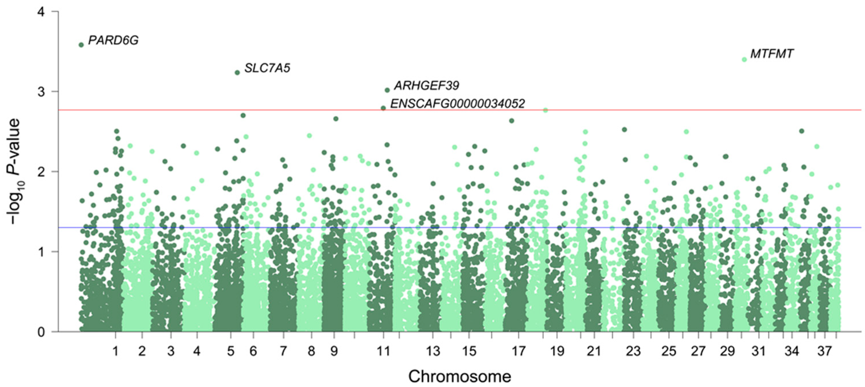
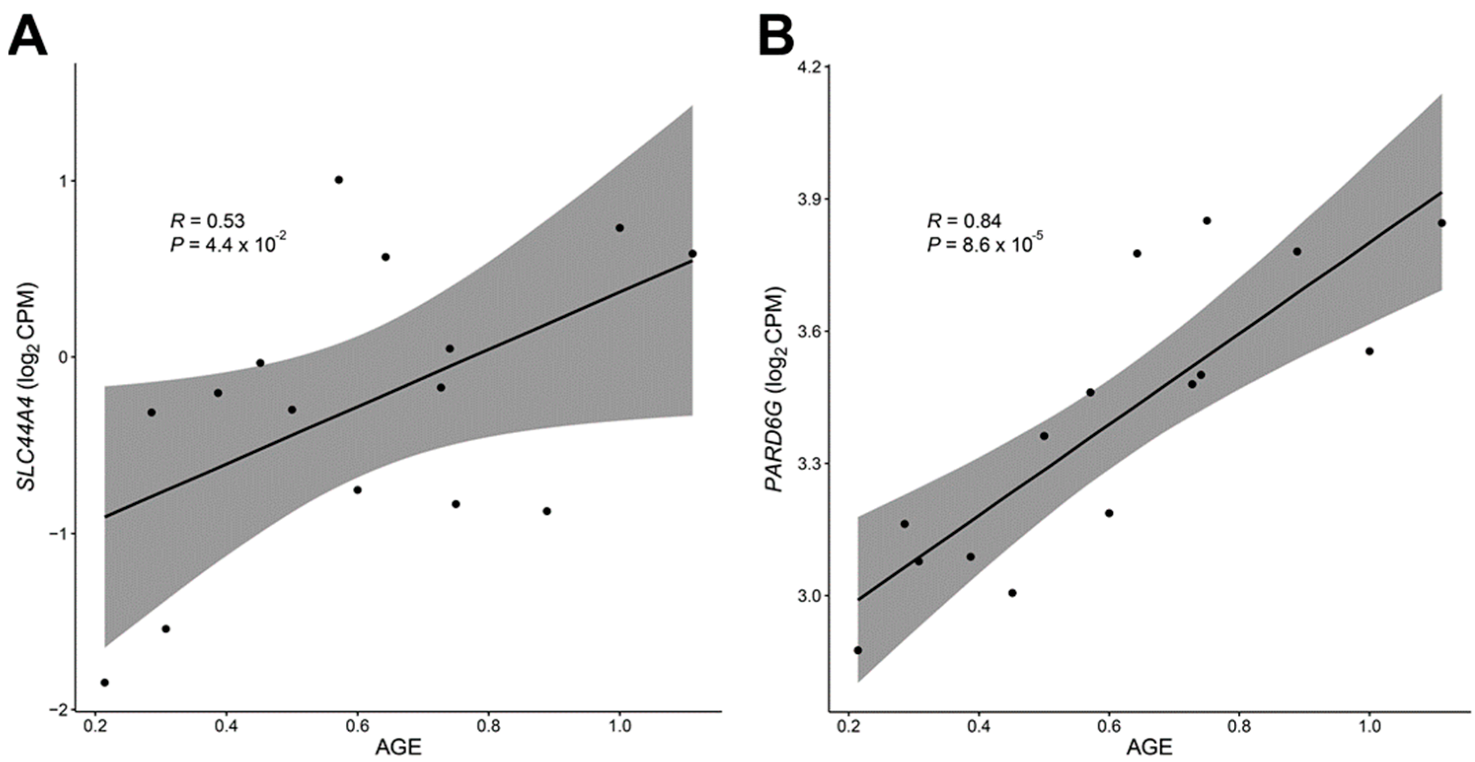
Figure 1. Manhattan plot for the transcriptome-wide significance of expression changes during aging. The threshold for transcriptome-wide significance was p < 0.05 and |log2FC| > 1 (blue line), but only the five most significant genes (above the red line) are labeled in this figure. A full list of significant genes is provided in Supplementary Table S2.
Genes 14 01131 g001
Figure 2. Candidate genes exhibiting transcriptional remodeling with age in 15 dogs. The gene expression level was found to be significantly associated with normalized age measures in the SLC44A4 (A) and PARD6G (B) genes, respectively.
Genes 14 01131 g002
Figure 3. Aging-associated DNA methylation changes in the PARD6G (A) and SLC44A4 (B) genes. For both candidate genes, hypomethylation was observed in aged Labrador retrievers (6 > years old) compared to younger ones (6 < years old).
Genes 14 01131 g003
Table 1. Significant enrichment of 154 differentially expressed genes during aging. The table presents pathways that were significantly over-represented (adjusted p-value < 0.05).
PathwaysTermN GeneN Overlapp-ValueAdj.P
GO-Biological PathwaysHormone transport31994.30 × 10−69.75 × 10⁻³
Regulation of hormone levels513115.07 × 10−69.75 × 10⁻³
Ion transport1663205.87 × 10−69.75 × 10⁻³
Embryo development986156.35 × 10−69.75 × 10⁻³
Regulation of protein modification process1826216.63 × 10−69.75 × 10⁻³
Regulation of blood circulation29381.88 × 10−52.06 × 10⁻²
Cardiac conduction14361.99 × 10−52.06 × 10⁻²
Regulation of intracellular signal transduction1824202.24 × 10−52.06 × 10⁻²
Ion transmembrane transport1125152.96 × 10−52.42 × 10⁻²
Transmembrane transport1574183.48 × 10−52.45 × 10⁻²
Maintenance of location32583.93 × 10−52.45 × 10⁻²
Embryonic organ development42394.01 × 10−52.45 × 10⁻²
Circulatory system process541104.86 × 10−52.75 × 10⁻²
Regulation of myoblast differentiation5345.27 × 10−52.77 × 10⁻²
Regulation of transmembrane transport553105.84 × 10−52.86 × 10⁻²
Regulation of transport1827197.43 × 10−53.40 × 10⁻²
Regulation of phosphorus metabolic process1677187.86 × 10−53.40 × 10⁻²
Regulation of ion transmembrane transport46798.54 × 10−53.49 × 10⁻²
Heterotypic cell–cell adhesion6149.17 × 10−53.55 × 10⁻²
Multicellular organismal homeostasis47791.00 × 10−43.68 × 10⁻²
Negative regulation of protein modification process599101.13 × 10−43.94 × 10⁻²
Heart process28771.28 × 10−44.23 × 10⁻²
Multicellular organismal signaling20161.32 × 10−44.23 × 10⁻²
Disclaimer/Publisher’s Note: The statements, opinions and data contained in all publications are solely those of the individual author(s) and contributor(s) and not of MDPI and/or the editor(s). MDPI and/or the editor(s) disclaim responsibility for any injury to people or property resulting from any ideas, methods, instructions or products referred to in the content.

