Article

Molecular Survey of Rodent-Borne Infectious Agents in the Ferlo Region, Senegal

by
Joa Braïthe Mangombi-Pambou
1,2,3,
Laurent Granjon
4,
Fabien Flirden
2,3,
Mamadou Kane
5,
Youssoupha Niang
5,
Bernard Davoust
2,3,
Florence Fenollar
3,6 and
Oleg Mediannikov
2,3,*
1
Centre Interdisciplinaire de Recherches Médicales de Franceville (CIRMF), Franceville BP 769, Gabon
2
Department of Epidemiology of Parasitic Diseases, Aix-Marseille University, IRD, AP-HM, MEPHI, 13005 Marseille, France
3
IHU Méditerranée Infection, 13005 Marseille, France
4
CBGP, IRD, CIRAD, INRAE, Institut Agro, University Montpellier, 34000 Montpellier, France
5
BIOPASS, CBGP-IRD, ISRA, UCAD, CIRAD, Campus de Bel-Air, Dakar 1386, Senegal
6
IRD, AP-HM, SSA, VITROME, Aix-Marseille University, 13005 Marseille, France
*
Author to whom correspondence should be addressed.
Genes 2023, 14(5), 1107; https://doi.org/10.3390/genes14051107
Submission received: 22 April 2023 / Revised: 15 May 2023 / Accepted: 16 May 2023 / Published: 18 May 2023
(This article belongs to the Special Issue Zoonotic Pathogen Infection and Host Defenses)

Abstract

:
Zoonotic pathogens are responsible for most infectious diseases in humans, with rodents being important reservoir hosts for many of these microorganisms. Rodents, thus, pose a significant threat to public health. Previous studies in Senegal have shown that rodents harbour a diversity of microorganisms, including human pathogens. Our study aimed to monitor the prevalence of infectious agents in outdoor rodents, which can be the cause of epidemics. We screened 125 rodents (both native and expanding) from the Ferlo region, around Widou Thiengoly, for different microorganisms. Analysis, performed on rodent spleens, detected bacteria from the Anaplasmataceae family (20%), Borrelia spp. (10%), Bartonella spp. (24%) and Piroplasmida (2.4%). Prevalences were similar between native and the expanding (Gerbillus nigeriae) species, which has recently colonised the region. We identified Borrelia crocidurae, the agent responsible for tick-borne relapsing fever, which is endemic in Senegal. We also identified two other not-yet-described bacteria of the genera Bartonella and Ehrlichia that were previously reported in Senegalese rodents. Additionally, we found a potential new species, provisionally referred to here as Candidatus Anaplasma ferloense. This study highlights the diversity of infectious agents circulating in rodent populations and the importance of describing potential new species and evaluating their pathogenicity and zoonotic potential.

1. Introduction

Rodentia is the most successful and diversified order of living mammals, representing about 40% of all mammalian species [1]. They have a very broad ecological spectrum, and their presence in different types of biotopes allows some species to live in close proximity to wildlife, livestock and humans [2,3]. Rodents are well known as important reservoirs of infectious agents, which they can transmit to humans [4]. They are the source of many zoonotic pathogens, including Borrelia spp., Leptospira spp., Bartonella spp. and Trypanosoma spp. [4,5].
Several rodent-borne zoonotic infectious agents and their associated diseases circulate in Africa. These include plague [6,7], Lassa haemorrhagic fever [8,9] and leptospirosis [10]. Many studies have described the presence not only of known zoonotic pathogens in rodents but also of microorganisms the pathogenicity and zoonotic potential of which are not yet known [11,12,13,14,15].
In Senegal, many studies have focused on small mammals, especially rodents, that inhabit the country, where they can represent reservoirs of infectious and potentially zoonotic agents [3,16]. This is the case in the Ferlo region of northern Senegal and has become evident, in particular, as part of a major multidisciplinary project connected with the pan-African Great Green Wall initiative, aimed at mitigating the effects of desertification and environmental degradation in the Sahelian environment: “https://ohmi-tessekere.in2p3.fr/ (accessed on 25 February 2023)”.
One of these studies detected the presence of several known zoonotic infectious agents (bacteria and parasites) and described new infectious agents in rodents from indoor and outdoor habitats in the Ferlo region [12]. This study concerned, among others, the Nigerian gerbil G. nigeriae, a species that has recently and rapidly colonised northern Senegal, and the domestic mouse Mus musculus sp.), an exotic invasive species currently expanding in most of the country [17,18,19,20]. Another study recently highlighted an epizootic outbreak of Q fever in rodents from the Ferlo region [21]. This outbreak was caused by a new genotype of Coxiella burnetii, the pathogenicity of which is still unknown. It was recent and is probably ongoing since previous studies of rodents from the same region revealed no evidence of the Q fever agent [12]. This study [21] contributes towards the same subject matter and was published separately and urgently due to the importance of the results.
These data show the importance of monitoring zoonotic pathogens potentially transmitted by rodents. Indeed, rodents are sentinels of infectious diseases that can allow the early detection and management of zoonoses [22]. This is the context in which we carried out this molecular epidemiological survey of microorganisms carried by rodent populations in Ferlo in order to address the public health risks for human populations in contact with these small mammals.

2. Material and Methods

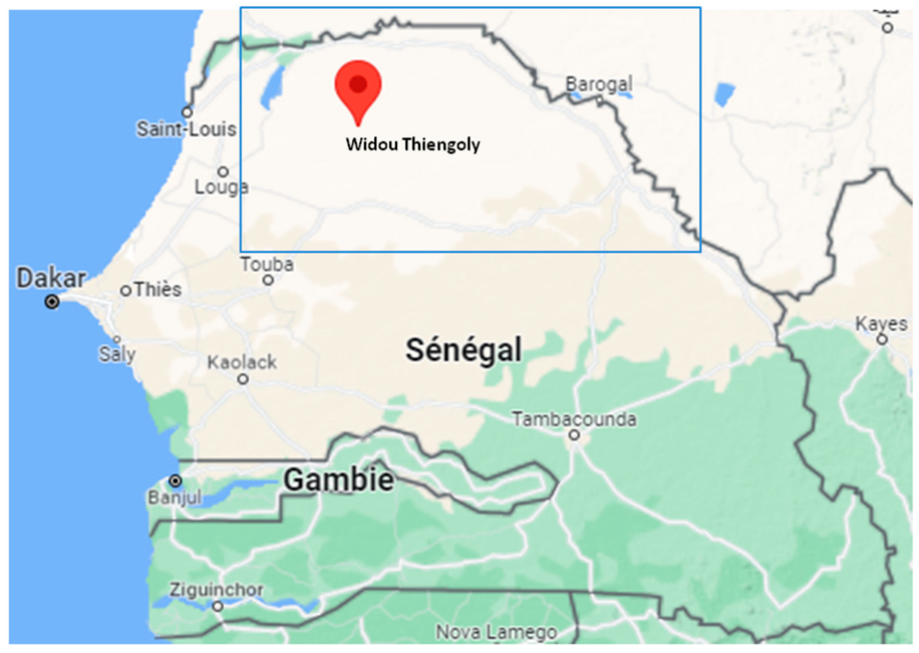
2.1. Study Area and Sample Design

Rodent sampling was conducted in the Ferlo region over two sampling periods in outdoor habitats around three temporary ponds east of Widou Thiengoly (average coordinates 15.96° N; 15.25° W), as shown in Figure 1. The first took place at the end of the 2019 rainy season (September–October 2019) and the second at the end of the 2020 dry season (June–July 2020). The trapping methodology was described elsewhere [19], with traps being placed either in lines of 20 to 40 traps spaced 10 m apart or opportunistically, depending on signs of the presence of rodents. In addition, “night hunts” were carried out at each site to catch species that are difficult to capture by hand (small Gerbillinae, jerboas, etc.). All the individuals were euthanatized by cervical dislocation, then autopsied, and samples (spleen in 95% ethanol) were extracted from each in order to search for the parasites and pathogens they harboured.

2.2. Ethical Statement Regarding Fieldwork

The fieldwork was conducted within the framework of agreements between the Institut National de Recherche pour le Développement (IRD) and the Republic of Senegal, as well as with the Senegalese Water and Forest Management Head Office of the Ministry of Environment and Sustainable Development. None of the rodent species investigated in this study have a protected status (see IUCN and CITES lists). Handling procedures were carried out under the CBGP agreement for experiments on wild animals (no. D-34-169-1) and followed the official guidelines of the American Society of Mammalogists [23]. The trapping campaigns were carried out with the explicit prior agreement of the competent local authorities.

2.3. DNA Extraction

DNA extraction was performed on a BioRobot EZ1 (Qiagen, Courtaboeuf, France) using a commercial EZ1 DNA Tissue Kit (Qiagen, Hilden, Germany) according to the manufacturer’s instructions. Total DNA was extracted from 10 mg of spleen from all rodent samples that were preserved in alcohol. DNA was eluted in 100 μL of TE buffer and stored at −20 °C until used for PCR amplification.

2.4. PCR Amplification

Real-time PCR (qPCR) was performed to screen all rodent samples using previously reported primers and probes [12] for Bartonella spp., Anaplasmataceae, Borrelia spp., Rickettsia spp., Piroplasmida, Mycoplasma spp., pan-Filaria, pan-Kinetolastidae and pan-Leishmania–Trypanosoma.
For each qPCR run, the final volume of 20 μL was composed of 10 μL of the Roche master mix (Roche Applied Science, Mannheim, Germany), 3 μL of water, 0.5 μL of each primer (20 μM) and probe (5 μM), 0.5 μL of UDG (uracil DNA glycosylase) and 5 μL of DNA extracted from the spleen. Amplification was performed in a CFX96 Real-Time PCR detection system (Bio-Rad Laboratories, Foster City, CA, USA) according to the following thermal profile: one step at 50 °C for 2 min for UDG action (eliminating PCR amplicon contaminants), an initial denaturation at 95 °C for 5 min, followed by 40 cycles at 95 °C for 15 s and 60 °C for 30 s for annealing extension. For all systems, any sample with a cycle threshold (Ct) value of less than 38 Ct was considered positive. The sequences of the primers and probes are shown in Table 1.
Conventional PCR analysis was performed in an automated DNA thermal cycler (GeneAmp PCR Systems Applied Biosystems, Courtaboeuf, France) for all qPCR-positive samples using the primers and conditions described in Table 1. The amplification reaction was conducted in a final volume of 25 μL containing 12.5 μL of Ampli Taq Gold master mix, 0.75 μL of each primer (10 μM), 5 μL of DNA template and 6 μL of water. The thermal cycling profile consisted of one incubation step at 95 °C for 5 min, 45 cycles of 30 s at 95 °C, 30 s to 1 min at the annealing temperature (Table 1) and 1 min at 72 °C, and a final extension step of 5 min at 72 °C. Successful amplification was confirmed by electrophoresis in a 1.5% agarose gel, and the amplicons were completely sequenced on both strands.
For each assay, DNA extracts of the targeted bacteria or parasites (laboratory colony) were used as positive controls and distilled water as the negative control (Table S1).

2.5. Sequencing and Phylogenetic Analysis

Sequencing analyses were performed on an ABI Prism 3130XL Genetic Analyzer (Applied Biosystems, Thermo Fisher Scientific, Illkirch-Graffenstaden, France) using a DNA sequencing BigDye Terminator V3.1 Cycle Sequencing Kit (Applied Biosystems, Foster City, CA, USA, PerkinElmer) according to the manufacturer’s instructions. The BigDye products were purified on Sefadex G-50 Superfine gel filtration resin (Cytiva, Formerly GE Healthcare Life Science, Lund, Sweden). The obtained sequences were analysed using ChromasPro version 1.3 (Technelysium Pty, Ltd., Tewantin, QLD, Australia) for assembly and were aligned with other sequences of targeted bacteria or parasite species from GenBank using CLUSTALW, implemented in BioEdit v7.2 [33]. Phylogenetic trees were constructed with MEGA software v.7 [34]. The Maximum Likelihood method based on the Hasegawa–Kishino–Yano model (HKY) was used to infer the phylogenetic analysis with 500 bootstrap replicates.

2.6. Statistical Analysis

Statistical analysis was performed with R software V4.1.2 [35] using Chi-square/Fisher’s exact tests or Wilcoxon–Mann–Whitney for data comparisons between the prevalence of infected rodents for all parasites according to their sex, age class (distinguished using the weight criteria provided in [3]), status (expanding G. nigeriae or native) and captured season. When p-values were <0.05, they were considered to be significant.

3. Results

3.1. Samples Included in the Study

A total of 125 small mammals captured in the Ferlo region were analysed in this study. The animals captured included the expanding species G. nigeriae (71/125, 56.8%), which was the most abundant species, and six native species, namely, Arvicanthis niloticus (29/125, 23.2%), Desmodilliscus braueri (3/125, 2.4%), Gerbillus nancillus (9/125, 7.2%), Jaculus jaculus (4/125, 3.2%), Taterillus sp. (corresponding most probably to Taterillus pygargus; 8/125, 6.4%) and Xerus erythropus (1/125, 0.8%) (Table 2).

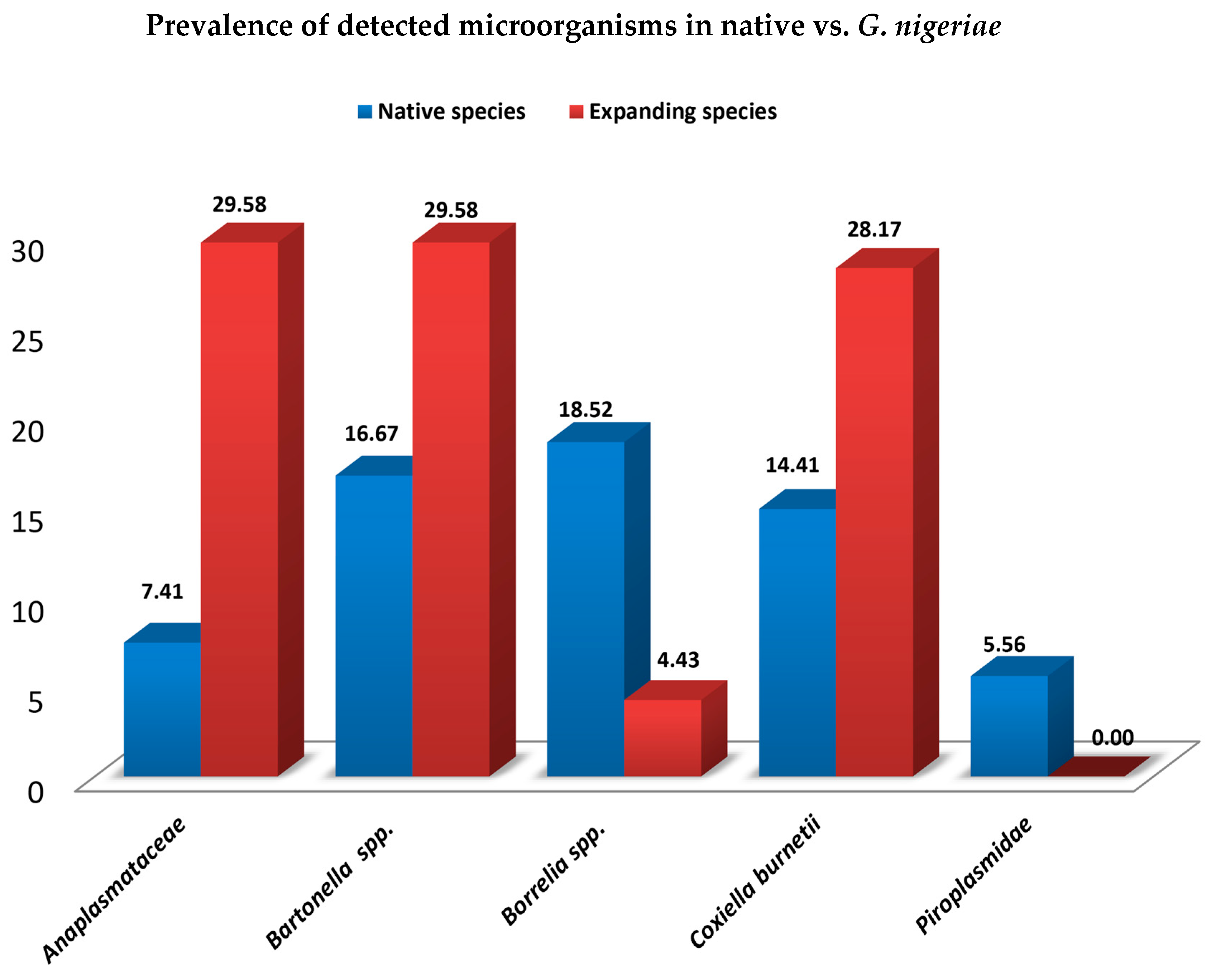
3.2. Molecular Detection of Microorganisms (Bacteria and Protozoa)

All rodents were found to be negative by qPCR for pan-Kinetoplastidae, pan-Leishmania–Trypanosoma, Rickettsia spp., Mycoplasma spp. And pan-Filaria. We also included here the results on C. burnetii, which were published separately, because these results originated from the same rodents. Thus, 67/125 (53.6%) rodents were positive for at least one of the five other qPCR screening systems (Table 2). The most common microorganism detected was Bartonella spp. at 24% (30/125) followed by C. burnetii at 22.4% (28/125), Anaplasmataceae at 20% (25/125) and Borrelia spp. at 10.4% (13/125). The less frequent pathogen was Piroplasmida at 2.4% (3/125) (Table 2).

3.3. Prevalence of Microorganisms by Sex, Age, Status and Season

In terms of host species, considering only species represented by at least ten individuals, G. nigeriae appears to be the most infected species with 59.1% (42/71) of individuals positive for at least one of the microorganisms detected (Table 2). It is also the species with the highest diversity of microorganisms, i.e., four infectious agents out of the five detected (Table 2 and Table 3). However, despite its relatively low abundance, it should be noted that the native Taterillus species has a similar profile with the highest diversity of microorganisms including five out of the five detected (Table 3).
Overall, although the prevalence of microorganisms was higher in G. nigeriae, as illustrated in Figure 2, compared to native rodents (all native species), no statistically significant differences were found (W = 20.5, p-value = 0.75). There was also no significant difference in the prevalence of microorganisms between males and females (W = 18, p-value = 1) or between sampling periods (W = 8, p-value = 0.1208). Among the age groups (juveniles vs. adults), however, a statistically significant difference was found, with adults more infected than juveniles (p-value = 0.0115; Pearson’s Chi-squared test).

3.4. Coinfections with Multiple Microorganisms

Overall, 19.2% (24/125) of rodents had mixed infections with at least two microorganisms. This included 16 double infections and 8 triple infections. Of these mixed infections, 71% (17/24) was described in G. nigeriae compared to 29% (7/24) for all other species (p-value = 0.4) (Table 2 and Table 3). The association Anaplasmataceae/C. burnetii (6/24) represented the most commonly encountered coinfection, followed by Anaplasmataceae/Bartonella/Coxiella (5/24) and Anaplasmataceae/Bartonella (4/24).

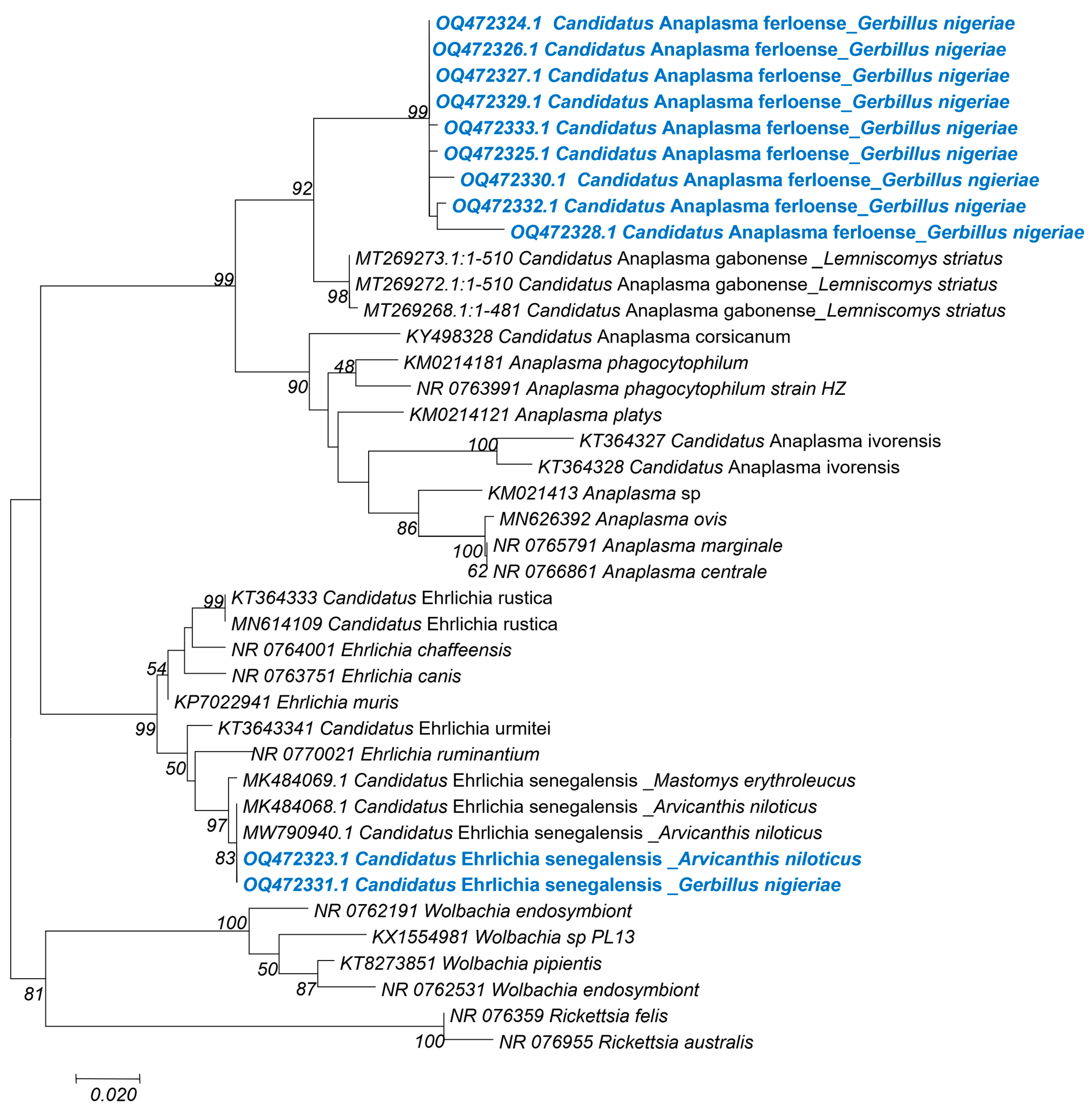
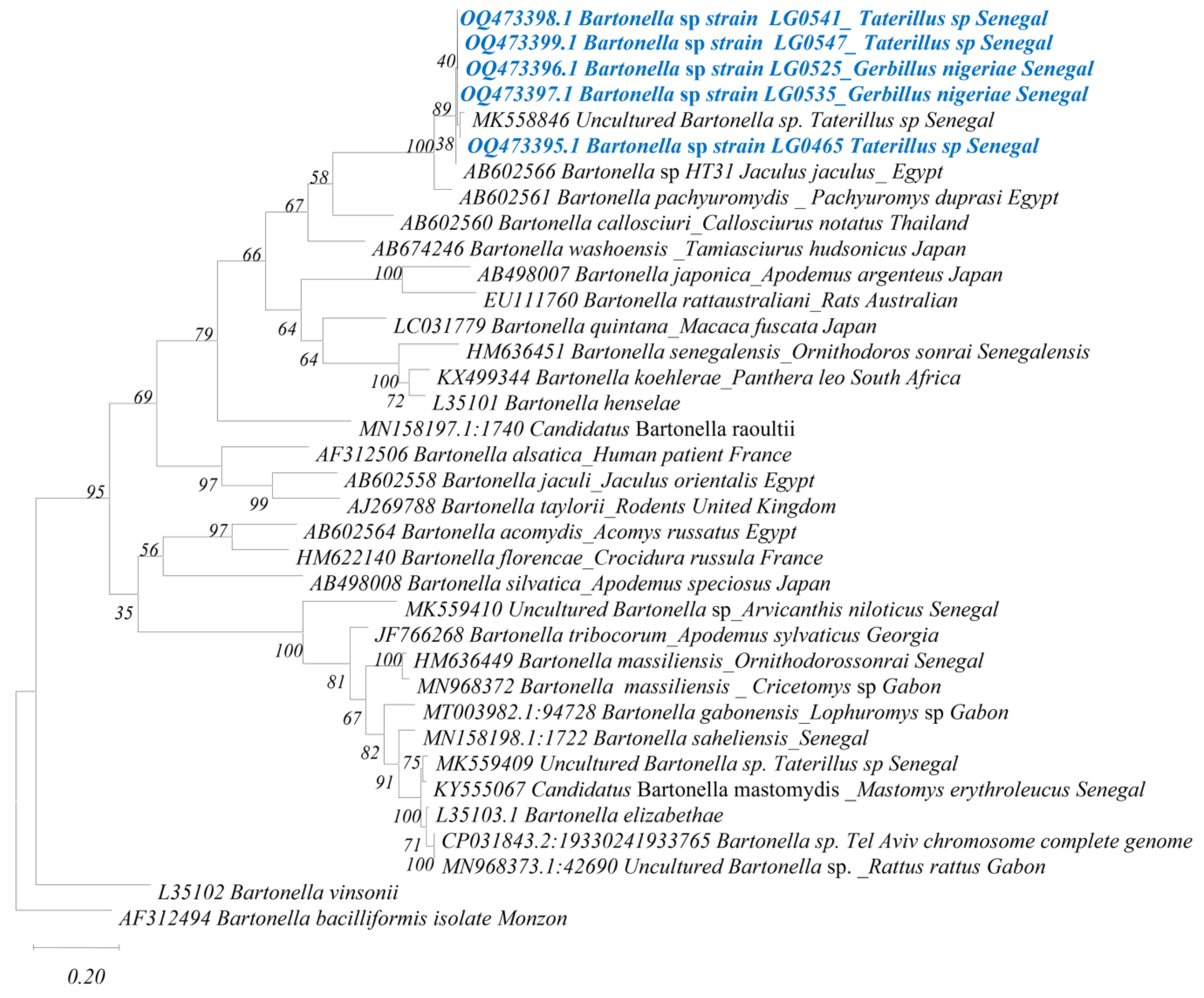
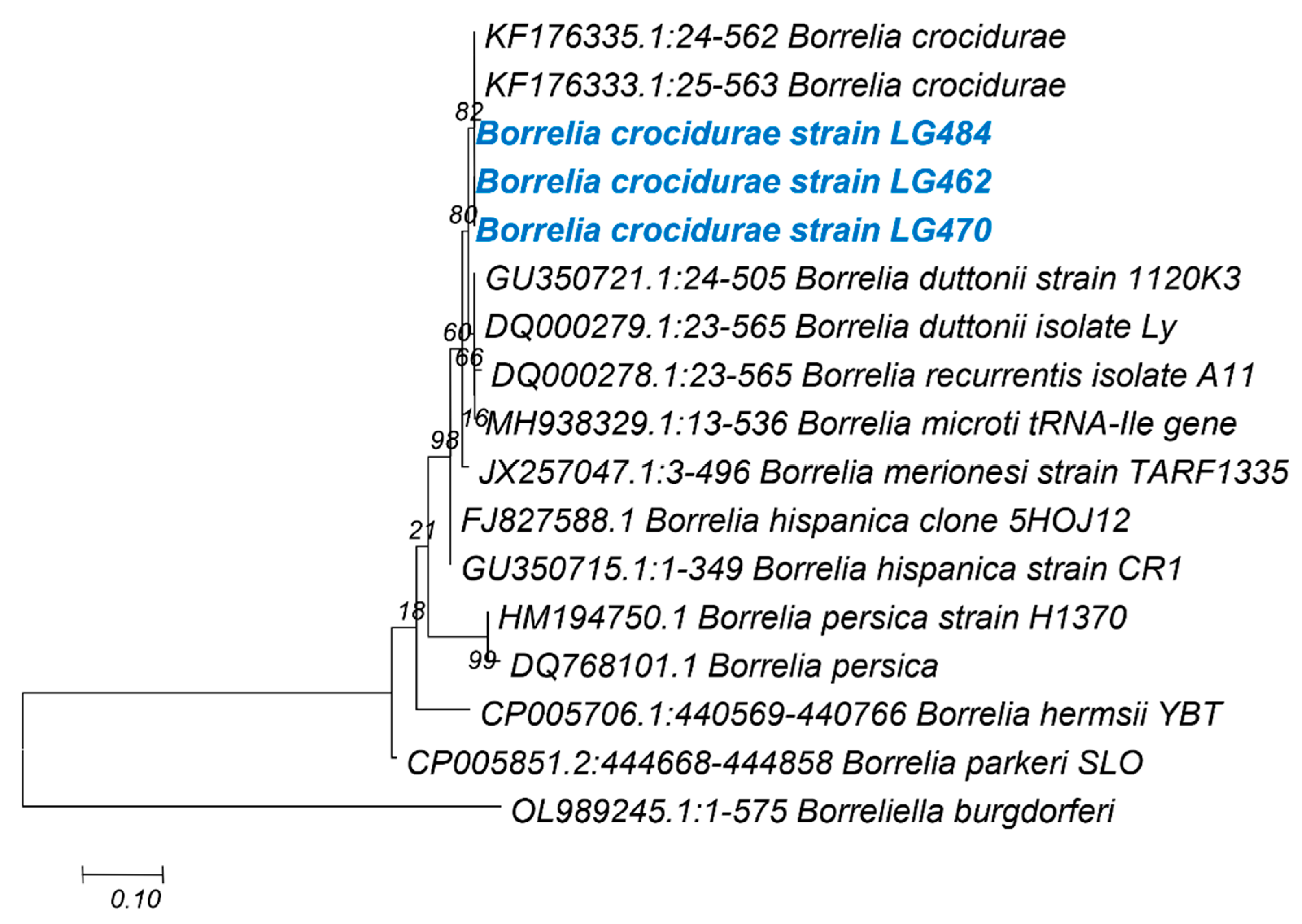
3.5. Phylogenetic Analysis for the Taxonomic Description of Detected Pathogens

Anaplasmataceae: A total of 14 23S ribosomal gene sequences, grouped into 2 clusters, were obtained from the 25 Anaplasmataceae qPCR-positive samples. The first group consisted of 12 sequences of around 420 bp (OQ472324–OQ472330, OQ472333 and OQ472334), ranging from 99% to 100% identity with one another, and presenting 91% identity with Anaplasma phagocytophilum (KM021418) and Anaplasma platys (KM021425). These sequences were all obtained from samples of G. nigeriae (Figure 3). We consider it to be a putative new species, given its position on the phylogenetic tree and the percentage of homology with the closest valid species. We propose naming it Candidatus Anaplasma ferloense. The second group consisted of two sequences (OQ472323 and OQ472331) from A. niloticus and G. nigeriae, respectively (Figure 3). They were 99% identical to one another and 95% identical to Ehrlichia ruminantium (CR92567). However, they also presented 99% homology with Candidatus Ehrlichia senegalensis (MK484068 MK484067) and uncultured Ehrlichia sp. (OP935909), previously identified in rodents [12] and in Ornithodoros sonrai [36] ticks, respectively, both from Senegal.
Bartonella spp.: Among the 30 Bartonella qPCR-positive individuals, we obtained 5 sequences with 98–100% identity to one another, including 3 sequences (OQ473395, OQ473398 and OQ473399) from Taterillus sp. And 2 sequences (OQ473396 and OQ473397) from G. nigeriae. These sequences showed 92% identity with Bartonella pachyuromydis (AB602561), described in Egyptian gerbilline rodents Pachyuromys duprasi and 98–99% identity with an uncultured Bartonella strain found in Senegalese rodents (MK558846). This is probably the same Bartonella species (Figure 4).
Borrelia spp.: A total of 3 (OQ980277-OQ980279) sequences were obtained from the 13 qPCR 16S Borrelia sp. Positive samples. Two of these sequences were from Taterillus sp. (LG0470 and LG484) and the last one was from G. nigeriae. These sequences were identical to one another and showed 99.80% identity with B. crocidurae detected in ticks from Mali (JX292946) and 99.63% identity with B. crocidurae (KF176340) detected in ticks from Senegal (Figure 5).

4. Discussion

We conducted a study based on molecular detection targeting a wide range of microorganisms in the rodents’ spleens. We compared recently arrived and expanding rodents with anciently established indigenous rodents in a region of northern Senegal in order to survey the potential occurrence of rodent-borne zoonotic epidemics. In the Ferlo region, where this investigation is being implemented, a previous study detected and described a diversity of infectious agents in rodents responsible for diseases of concern to public health [12]. This is not the only epidemiological study to have been conducted in Senegalese rodents describing a wide range of microorganisms hosted by these small mammals. Although using different techniques, several studies have revealed the circulation of many zoonotic infectious agents in Senegalese rodents, such as Toxoplasma gondii [37,38], Orthohantavirus, Mammarenavirus, Orthopoxvirus [39] and Flavivirus [40]. Taken as a whole, the results of these studies justify the need to conduct molecular surveys on the prevalence of microorganisms in small mammals in Senegal, even more so in the context of biological invasion/expansion processes where host–parasite interactions can influence the outcome of the invasion [39].
Furthermore, among the rodents included in this study, we recently highlighted the circulation of a new genotype of C. burnetii, which is potentially responsible for an epizootic in these small mammals in the Ferlo. These data, suggesting a high public health risk, were published urgently to alert policy makers to the potential threat that this could represent [21]. This result will, therefore, be included in our discussion.
Our data show the detection of five of the nine microorganisms searched for (Table 1 and Table 2) and the precise identification by sequencing and phylogenetic analysis of three of the microorganisms detected within Bartonella, Anaplasmataceae and Borrelia (Figure 3, Figure 4 and Figure 5); of all the microorganisms detected, these same three infectious agents exhibited the highest prevalence, i.e., 24%, 20% and 10%, respectively.
The Anaplasmataceae family consists of numerous genera, including Anaplasma, Aegyptianella, Ehrlichia, Neorickettsia, Neoehrlichia and Wolbachia. These are gram-negative Alphaproteobacteria, small and commonly pleomorphic, which reside in the cytoplasmic vacuoles of host cells [41,42]. Several distinct species of the Anaplasmataceae family have been identified as tick-borne human pathogens. Indeed, they infect humans, as well as domestic and wild animals, and are responsible for tick-borne diseases, which are becoming more and more common due to the increase in factors leading to contact between wild animals, their ectoparasites, domestic animals and humans [41].
In Senegal, several studies have reported the circulation of Anaplasmataceae in domestic animals [24,43] and in rodents [11,12]. Recent data show, for the first time, the detection of Ehrlichia sp. in O. sonrai ticks, known to be the only vectors of B. crocidurae, the agent of tick-borne relapsing fever (TBRF) [36]. Although there are no reported cases of human anaplasmosis, this group of infectious agents continues to receive a great deal of attention both because they affect the economy through disease outbreaks in livestock populations (while also harbouring species known to be pathogenic to humans, such as A. phagocytophilum) [43] and because new species are increasingly being identified that could be potentially pathogenic to humans. Our results showed the qPCR detection of Anaplasmataceae DNA in 25/125 rodent spleen samples with a prevalence of 20%, comparable to that previously found in the same area (18.8% [12]). Anaplasmataceae bacteria were successfully sequenced in 14 samples. The sequences obtained showed the presence of two distinct groups of bacteria. The first group, including 12 samples all from G. nigeriae, showed 91% homology with A. phagocytophilum (KM021418) and Anaplasma platys (KM021425), suggesting that this is a new or undescribed species. This putative new species, named Candidatus Anaplasma ferloense, presented 94% to 95% identity with uncultured Anaplasma strains known as Candidatus Anaplasma gabonense, identified in Gabonese rodents (MT269273) [13]. These two independent, well-supported clusters (Candidatus Anaplasma ferloense and Candidatus Anaplasma gabonenses) appear as sister groups in a moderately well-supported clade of Anaplasma species associated with rodents (Figure 3). The two sequences retrieved from the A. niloticus and G. nigeriae cluster, with others previously identified in rodents from Senegal and provisionally identified as Candidatus Ehrlichia senegalensis (Figure 3), have also been found in O. sonrai ticks [36]. This species shows 95% homology with E. ruminantium (CR925677). In a study by Dahmana et al. (2020), Candidatus Ehrlichia senegalensis was only identified in native rodents, whereas in our study it is also present in expanding G. nigeriae, suggesting a possible transfer of infectious agents between native and invasive rodents. This work highlights the identification of potentially new species of Anaplasmataceae and emphasises not only the species diversity of this family but also the magnitude of the range of hosts they parasitise.
Bartonella species are intracellular, vector-borne, blood-borne gram-negative bacteria that can induce prolonged infection in the host [44]. These infections can be persistent in domestic and wild animals, constituting a significant reservoir of Bartonella organisms in nature, which may serve as a source of human infection [44,45]. Indeed, the Bartonella genus includes several species that are responsible for zoonotic diseases [46] and are often associated with rodents as their main reservoir hosts [45]. The genus Bartonella has been reported several times in both native and invasive rodents in Senegal [11,12]. However, here, we recorded a prevalence of 24% (30/125), much higher than in a previous study by Dahmana et al. 2020: 9% in various sites in the Ferlo region. Kosoy et al. found inter-annual variations in Bartonella infection patterns [47]. Additionally, this difference in the prevalence of Bartonella infection in sites in the same region could be associated with the period of rodent collection and the diffusion or spread of infection. Indeed, in a study by Dahmana et al., invasive rodents (G. nigeriae and Mus musculus sp.) collected in 2017 did not show any infection [12], unlike the G. nigeriae specimens captured between 2019 and 2020 included in our study. In other African countries, prevalence values of 17.7% [48] and 6.6% [13] have been found in Mali and Gabon, respectively. The high prevalence in our sample rate raises questions about the risk of human infection. Furthermore, of the 30 positive samples, only 5 could be sequenced: 3 from Taterillus sp. and 2 from G. nigeriae. These five sequences obtained were identical to one another, showing the presence of the same Bartonella genotype infecting both species. This genotype showed only 92% identity with B. pachyuromydis, which is the closest valid species, indicating, by this low percentage of identity (<98%, according to La Scola et al. 2003 [49]), that it is a new genotype. This genotype has previously been found in Ferlo rodents exclusively in Taterillus sp., whereas no detection of Bartonella was found in G. nigeriae [12].
One endemic species of Borrelia in Senegal is B. crocidurae, the agent of TBRF in humans [50,51,52]. Borrelia are fastidious bacteria transmitted by ectoparasites (e.g., lice or ticks) and are responsible for various febrile presentations in humans, most often malaria-like symptoms [50]. In Senegal, TBRF caused by B. crocidurae is the most common bacterial infection affecting the human population in rural areas. It may be responsible for up to 11% of febrile illnesses recorded in dispensaries [51]. Hosts and reservoirs of this important pathogen have already been studied: previous studies have shown Crocidura sp. A. niloticus, Mastomys huberti, Mus musculus sp., G. nigeriae, M. erythroleucus and Taterillus sp. as rodent hosts for TBRF [12,53]. Our study adds D. braueri and G. nancillus to this list.
In this study, 10% (13/125) of the samples were positive for Borrelia spp. through qPCR, and 3 could be sequenced. These sequences were all identified as B. crocidurae (KF176340), previously reported in Senegal [11,12] and in other countries of the Sahelo–Saharan region [52,54]. These data show the continuous circulation of B. crocidurae and its host variability. It is, therefore, important to continue to search for and identify TBRF in Senegal. In this regard, one recent study reported the detection of Borrelia spp. DNA in human skin swabs and dust samples in rural Senegal [55].
Our study revealed the detection of multiple coinfections in rodents, the most represented being Anaplasmataceae/Coxiella (25%, 6/24), Anaplasmataceae/Bartonella/Coxiella (21%, 5/24) and Anaplasmataceae/Bartonella (17%, 4/24) (Table 3). As these infectious agents share the same reservoirs (hosts), it is not surprising to find them in association [56]. In addition, these are infectious agents transmitted by ectoparasites such as ticks, which are known to be vectors of coinfections [57]. However, despite the fact that coinfections are known in rodents [13,57,58], very few studies have reported the Anaplasma–Bartonella association [59], and in Senegal there has been no report of it in rodents to date.

5. Conclusions

Our study aimed to detect and identify infectious agents harboured by rodents in Ferlo, Senegal. Our results confirm that rodents are hosts of a large number of infectious agents that are potentially pathogenic to humans. Among the six groups of microorganisms found, three were found to present significant prevalence: Anaplasmataceae, 20%; Bartonella, 24%; and Borrelia, 10%. We found no difference in prevalence rates between native rodents and the expanding species G. nigeriae, which is currently colonising the region. However, we did observe a potential exchange of bacteria between native species and this recently arrived species. Indeed, potentially new genotypes of Anaplasmataceae and Bartonella were identified in G. nigeriae, whereas in a previous study, they were absent and present only in native rodents. In addition, we found B. crocidurae, which is responsible for TBRF and which can have a significant rate of incidence, up to 11% in Senegal. In addition to Candidatus Anaplasma ferloense, several undescribed infectious agents have been identified in Senegal in rodents. In order to determine the zoonotic potential of these genotypes and their importance for animal and public health, it would be essential to characterise them.

Supplementary Materials

The following supporting information can be downloaded at: https://www.mdpi.com/article/10.3390/genes14051107/s1. Table S1: The list of negative and positive DNA controls used to confirm the sensitivity and specificity of the PCR systems designed for this study.

Author Contributions

Conceptualization, O.M., L.G. and F.F.; methodology, O.M., J.B.M.-P., L.G. and B.D.; formal analysis, J.B.M.-P. and F.F. (Fabien Flirden); investigation, J.B.M.-P., F.F. (Fabien Flirden), Y.N. and M.K.; data curation, J.B.M.-P., F.F. (Fabien Flirden), Y.N. and M.K.; writing—original draft preparation, J.B.M.-P.; writing–review, J.B.M.-P., O.M., L.G. and F.F. (Florence Fenollar); supervision, O.M. and F.F. (Florence Fenollar); project administration, O.M., B.D., L.G. and F.F. (Florence Fenollar). All authors have read and agreed to the published version of the manuscript.

Funding

This study was supported by the Institut Hospitalo-Universitaire (IHU) Méditerranée Infection, the French National Research Agency under the Investissements d’avenir programme (reference: ANR-10-IAHU-03) and the fourth Make Our Planet Great Again programme (MOPGA 4). This work was co-funded by the Labex DRIIHM, the French Investissements d’Avenir programme (ANR-11-LABX-0010), which is managed by the ANR.

Institutional Review Board Statement

Please see Section 2.2.

Informed Consent Statement

Not applicable.

Data Availability Statement

Not applicable.

Acknowledgments

We thank Philippe Gauthier for taxonomical precisions on some rodent specimens using barcoding (based on cyt b sequences).

Conflicts of Interest

The authors declare no conflict of interest.

References

  1. Burgin, C.J.; Colella, J.P.; Kahn, P.L.; Upham, N.S. How many species of mammals are there? J. Mammal. 2018, 99, 1–14. [Google Scholar] [CrossRef]
  2. Carleton, M.D.; Musser, G.G. Order Rodentia. In Mammal Species of the World, 3rd ed.; Wilson, D.E., Reeder, D.M., Eds.; The Johns Hopkins University Press: Baltimore, MD, USA, 2005; pp. 745–752. [Google Scholar]
  3. Granjon, L.; Duplantier, J.M. Les Rongeurs de l’Afrique Sahélo-Soudanienne; Publications Scientifiques du Muséum; IRD: Marseille, France, 2009; 215p. [Google Scholar]
  4. Meerburg, B.G.; Singleton, G.R.; Kijlstra, A. Rodent-borne diseases and their risks for public health. Crit. Rev. Microbiol. 2009, 35, 221–270. [Google Scholar] [CrossRef] [PubMed]
  5. Han, B.A.; Schmidt, J.P.; Bowden, S.E.; Drake, J.M. Rodent reservoirs of future zoonotic diseases. Proc. Natl. Acad. Sci. USA 2015, 112, 201501598. [Google Scholar] [CrossRef] [PubMed]
  6. Rabaan, A.A.; Al-Ahmed, S.H.; Alsuliman, S.A.; Aldrazi, F.A.; Alfouzan, W.A.; Haque, S. The rise of pneumonic plague in Madagascar: Current plague outbreak breaks usual seasonal mould. J. Med. Microbiol. Microbiol. Soc. 2019, 68, 292–302. [Google Scholar] [CrossRef]
  7. Riehm, J.M.; Rahalison, L.; Scholz, H.C.; Thoma, B.; Pfeffer, M.; Razanakoto, L.M.; Al Dahouk, S.; Neubauer, H.; Tomaso, H. Detection of Yersinia pestis using real-time PCR in patients with suspected bubonic plague. Mol. Cell. Probes 2011, 25, 8–12. [Google Scholar] [CrossRef]
  8. Manning, J.T.; Forrester, N.; Paessler, S. Lassa virus isolates from Mali and the Ivory Coast represent an emerging fifth lineage. Front. Microbiol. 2015, 6, 1037. Available online: http://journal.frontiersin.org/article/10.3389/fmicb.2015.01037 (accessed on 16 January 2023). [CrossRef]
  9. World Health Organisation. Lassa Fever–Nigeria Disease Outbreak News; World Health Organisation: Geneva, Switzerland, 2019. [Google Scholar]
  10. Houéménou, G.; Gauthier, P.; Etougbétché, J.; Badou, S.; Dossou, H.J.; Agossou, D.; Picardeau, M.; Dobigny, G. Pathogenic Leptospira in Commensal Small Mammals from the Extensively Urbanized Coastal Benin. Urban Sci. 2019, 3, 99. [Google Scholar] [CrossRef]
  11. Diagne, C.; Galan, M.; Tamisier, L.; D’Ambrosio, J.; Dalecky, A.; Bâ, K.; Kane, M.; Niang, Y.; Diallo, M.; Sow, A.; et al. Ecological and sanitary impacts of bacterial communities associated to biological invasions in African commensal rodent communities. Sci. Rep. 2017, 7, 14995. [Google Scholar] [CrossRef]
  12. Dahmana, H.; Granjon, L.; Diagne, C.; Davoust, B.; Fenollar, F.; Mediannikov, O. Rodents as Hosts of Pathogens and Related Zoonotic Disease Risk. Pathogens 2020, 9, 202. Available online: http://www.ncbi.nlm.nih.gov/pubmed/32164206 (accessed on 25 March 2020). [CrossRef]
  13. Mangombi, J.B.; N’dilimabaka, N.; Lekana-Douki, J.-B.; Banga, O.; Maghendji-Nzondo, S.; Bourgarel, M.; Leroy, E.; Fenollar, F.; Mediannikov, O. First investigation of pathogenic bacteria, protozoa and viruses in rodents and shrews in context of forest-savannah-urban areas interface in the city of Franceville (Gabon). Markotter, W., Ed. PLoS ONE 2021, 16, e0248244. [Google Scholar] [CrossRef]
  14. Mawanda, P.; Rwego, I.; Kisakye, J.J.; Sheil, D. Rodents as potential hosts and reservoirs of parasites along the edge of a Central African forest: Bwindi impenetrable national park, South Western Uganda. Afr. Health Sci. 2020, 20, 1168. Available online: https://www.ncbi.nlm.nih.gov/pmc/articles/PMC7751523/ (accessed on 2 February 2020). [CrossRef] [PubMed]
  15. Suu-Ire, R.; Obodai, E.; Bel-Nono, S.O.; Ampofo, W.K.; Mazet, J.A.K.; Goldstein, T.; Johnson, C.K.; Smith, B.; Boaatema, L.; Asigbee, T.W.; et al. Surveillance for potentially zoonotic viruses in rodent and bat populations and behavioral risk in an agricultural settlement in Ghana. One Health Outlook 2022, 4, 6. [Google Scholar] [CrossRef] [PubMed]
  16. Duplantier, J.M.; Granjon, L.; Ba, K. Répartition biogéographique des petits rongeurs au Sénégal. J. Afr. Zool. 1997, 111, 17–26. [Google Scholar]
  17. Dalecky, A.; Bâ, K.; Piry, S.; Lippens, C.; Diagne, C.A.; Kane, M.; Sow, A.; Diallo, M.; Niang, Y.; Konečný, A.; et al. Range expansion of the invasive house mouse Mus musculus domesticus in Senegal, West Africa: A synthesis of trapping data over three decades, 1983–2014. Mamm. Rev. 2015, 45, 176–190. Available online: http://doi.wiley.com/10.1111/mam.12043 (accessed on 3 September 2015). [CrossRef]
  18. Diagne, C.; Granjon, L.; Tatard, C.; Ribas, A.; Ndiaye, A.; Kane, M.; Niang, Y.; Brouat, C. Same Invasion, Different Routes: Helminth Assemblages May Favor the Invasion Success of the House Mouse in Senegal. Front. Vet. Sci. 2021, 8, 1234. [Google Scholar] [CrossRef] [PubMed]
  19. Granjon, L.; Ba, K.; Diagne, C.; Ndiaye, A.; Piry, S.; Thiam, M. La communauté des petits rongeurs du Ferlo: Tendances historiques et caractéristiques du peuplement actuel. In La Grande Muraille Verte, une Réponse Africaine au Changement Climatique; Boëtsch, G., Duboz, P., Guissé, A., Sarr, P., Eds.; CNRS: Paris, France, 2019; pp. 1–19. [Google Scholar]
  20. Thiam, M.; Ba, K.; Duplantier, J.M. Impacts of climatic changes on small mammal communities in the Sahel (West Africa) as evidenced by owl pellet analysis|African Zoology. Afr. Zool. 2008, 43, 135–143. Available online: https://journals.co.za/doi/epdf/10.10520/EJC18091 (accessed on 4 March 2023). [CrossRef]
  21. Mangombi-Pambou, J.; Granjon, L.; Labarrere, C.; Kane, M.; Niang, Y.; Fournier, P.E.; Delerce, J.; Fenollar, F.; Mediannikov, O. New Genotype of Coxiella burnetii Causing Epizootic Q Fever Outbreak in Rodents, Northern Senegal-Volume 29, Number 5—May 2023-Emerging Infectious Diseases journal-CDC. Emerg. Infect. Dis. 2023, 29, 1078. Available online: https://www.nc.cdc.gov/eid/article/29/5/22-1034_article (accessed on 20 April 2023).
  22. Heyman, P.; Saegerman, C. Les rongeurs en tant que sentinelles dans le cadre des infections à hantavirus. Epidémiol. St. Anim. 2009, 56, 47–52. [Google Scholar]
  23. Sikes, R.S. Mammalogists the AC and UC of the AS of. 2016 Guidelines of the American Society of Mammalogists for the use of wild mammals in research and education. J. Mammal. 2016, 97, 663. Available online: https://www.ncbi.nlm.nih.gov/pmc/articles/PMC5909806/ (accessed on 17 January 2023). [CrossRef]
  24. Dahmani, M.; Davoust, B.; Tahir, D.; Raoult, D.; Fenollar, F.; Mediannikov, O. Molecular investigation and phylogeny of Anaplasmataceae species infecting domestic animals and ticks in Corsica, France. Parasites Vectors 2017, 10, 302. [Google Scholar] [CrossRef]
  25. Mediannikov, O.; Fenollar, F. Looking in ticks for human bacterial pathogens. Microb. Pathog. 2014, 77, 142–148. [Google Scholar] [CrossRef] [PubMed]
  26. Jensen, W.A.; Fall, M.Z.; Rooney, J.; Kordick, D.L.; Breitschwerdt, E.B. Rapid identification and differentiation of Bartonella species using a single-step PCR assay. J. Clin. Microbiol. 2000, 38, 1717–1722. [Google Scholar] [CrossRef] [PubMed]
  27. Mediannikov, O.; Abdissa, A.; Socolovschi, C.; Diatta, G.; Trape, J.F.; Raoult, D. Detection of a New Borrelia Species in Ticks Taken from Cattle in Southwest Ethiopia. Vector-Borne Zoonotic Dis. 2013, 13, 266–269. Available online: https://www.liebertpub.com/doi/10.1089/vbz.2011.0874 (accessed on 10 February 2023). [CrossRef] [PubMed]
  28. Bittar, F.; Keita, M.B.; Lagier, J.C.; Peeters, M.; Delaporte, E.; Raoult, D. Gorilla gorilla gorilla gut: A potential reservoir of pathogenic bacteria as revealed using culturomics and molecular tools. Sci. Rep. 2014, 4, 7174. [Google Scholar] [CrossRef]
  29. Sokhna, C.; Mediannikov, O.; Fenollar, F.; Bassene, H.; Diatta, G.; Tall, A.; Trape, J.F.; Drancourt, M.; Raoult, D. Point-of-Care Laboratory of Pathogen Diagnosis in Rural Senegal. PLoS Negl. Trop. Dis. 2013, 7, e1999. [Google Scholar] [CrossRef]
  30. Laidoudi, Y.; Davoust, B.; Varloud, M.; Niang, E.H.A.; Fenollar, F.; Mediannikov, O. Development of a multiplex qPCR-based approach for the diagnosis of Dirofilaria immitis, D. repens and Acanthocheilonema reconditum. Parasites Vectors 2020, 13, 319.AR. Available online: https://parasitesandvectors.biomedcentral.com/articles/10.1186/s13071-020-04185-0 (accessed on 3 February 2023). [CrossRef]
  31. Dahmana, H.; Amanzougaghene, N.; Davoust, B.; Normand, T.; Carette, O.; Demoncheaux, J.P.; Mulot, B.; Fabrizy, B.; Scandola, P.; Chik, M.; et al. Great diversity of Piroplasmida in Equidae in Africa and Europe, including potential new species. Vet. Parasitol. Reg. Stud. Rep. 2019, 18, 100332. [Google Scholar] [CrossRef]
  32. Medkour, H.; Varloud, M.; Davoust, B.; Mediannikov, O. New Molecular Approach for the Detection of Kinetoplastida Parasites of Medical and Veterinary Interest. Microorganisms 2020, 8, 356. Available online: https://www.mdpi.com/2076-2607/8/3/356 (accessed on 4 March 2020). [CrossRef]
  33. Hall, T.A. BioEdit: A user-friendly biological sequence alignment editor and analysis program for Windows 95/98/NT. Nucleic. Acids Symp. Ser. 1999, 41, 95–98. [Google Scholar]
  34. Kumar, S.; Stecher, G.; Tamura, K. MEGA7: Molecular Evolutionary Genetics Analysis Version 7.0 for Bigger Datasets. Mol. Biol. Evol. 2016, 33, 1870–1874. [Google Scholar] [CrossRef]
  35. R Development Core Team. R: A Language and Environment for Statistical Computing; R Foundation for Statistical Computing: Vienna, Austria, 2007; Available online: http://www.r-project.org/ (accessed on 23 August 2018).
  36. Ouarti, B.; Sall, M.; Ndiaye, E.H.I.; Diatta, G.; Zan Diarra, A.; Berenger, J.M.; Sokhna, C.; Granjon, L.; Le Fur, J.; Parola, P. Pathogen Detection in Ornithodoros sonrai Ticks and Invasive House Mice Mus musculus domesticus in Senegal. Microorganisms 2022, 10, 2367. [Google Scholar] [CrossRef] [PubMed]
  37. Brouat, C.; Diagne, C.A.; Ismaïl, K.; Aroussi, A.; Dalecky, A.; Bâ, K.; Kane, M.; Niang, Y.; Diallo, M.; Sow, A.; et al. Seroprevalence of Toxoplasma gondii in commensal rodents sampled across Senegal, West Africa. Parasite 2018, 25, 32. [Google Scholar] [CrossRef] [PubMed]
  38. Galal, L.; Schares, G.; Stragier, C.; Vignoles, P.; Brouat, C.; Cuny, T.; Dubois, C.; Rohart, T.; Glodas, C.; Dardé, M.-L.; et al. Diversity of Toxoplasma gondii strains shaped by commensal communities of small mammals. Int. J. Parasitol. 2018, 49, 267–275. [Google Scholar] [CrossRef] [PubMed]
  39. Diagne, C.; Charbonnel, N.; Henttonen, H.; Sironen, T.; Brouat, C. Serological Survey of Zoonotic Viruses in Invasive and Native Commensal Rodents in Senegal, West Africa. Vector-Borne Zoonotic Dis. 2017, 17, 730–733. Available online: http://online.liebertpub.com/doi/10.1089/vbz.2017.2135 (accessed on 10 April 2020). [CrossRef]
  40. Diagne, M.; Ndione, M.; Di Paola, N.; Fall, G.; Bedekelabou, A.; Sembène, P.; Faye, O.; Zanotto, P.M.d.A.; Sall, A.A. Usutu Virus Isolated from Rodents in Senegal. Viruses 2019, 11, 181. Available online: https://www.mdpi.com/1999-4915/11/2/181 (accessed on 30 April 2020). [CrossRef]
  41. André, M.R. Diversity of Anaplasma and Ehrlichia/Neoehrlichia Agents in terrestrial wild carnivores worldwide: Implications for human and domestic animal health and wildlife conservation. Front. Vet. Sci. Front. Media S.A. 2018, 5, 293. [Google Scholar] [CrossRef]
  42. Dumler, J.S.; Walker, D.H. Ehrlichioses and Anaplasmosis. Trop. Infect. Dis. Princ. Pathog. Pract. 2-Vol. Set CD-ROM 2006, 564–573. [Google Scholar]
  43. Lamine Djiba, M.; Mediannikov, O.; Mbengue, M.; Thiongane, Y.; Molez, J.F.; Talla Seck, M.; Fenollar, F.; Raoult, D.; Ndiaye, M. Survey of Anaplasmataceae bacteria in sheep from Senegal. Trop. Anim. Health Prod. 2013, 45, 1557–1561. Available online: http://blast.ncbi.nlm.nih.gov (accessed on 9 March 2023). [CrossRef]
  44. Angelakis, E.; Mediannikov, O.; Socolovschi, C.; Mouffok, N.; Bassene, H.; Tall, A.; Niangaly, H.; Doumbo, O.; Znazen, A.; Sarih, M.; et al. Coxiella burnetii-positive PCR in febrile patients in rural and urban Africa. Int. J. Infect. Dis. 2014, 28, 107–110. [Google Scholar] [CrossRef]
  45. Krügel, M.; Król, N.; Kempf, V.A.J.; Pfeffer, M.; Obiegala, A. Emerging rodent-associated Bartonella: A threat for human health? Parasites Vectors 2022, 15, 113. Available online: https://parasitesandvectors.biomedcentral.com/articles/10.1186/s13071-022-05162-5 (accessed on 27 January 2023). [CrossRef]
  46. Angelakis, E.; Raoult, D. Q fever. Vet. Microbiol. 2010, 140, 297–309. [Google Scholar] [CrossRef] [PubMed]
  47. Kosoy, M.; Mandel, É.; Green, D.; Marston, É.; Childs, J. Prospective Studies of Bartonella of Rodents. Part I. Demographic and Temporal Patterns in Population Dynamics. Vector-Borne Infect. Dis. 2004, 4, 285–295. [Google Scholar] [CrossRef] [PubMed]
  48. Diarra, A.Z.; Kone, A.K.; Niare, S.D.; Laroche, M.; Diatta, G.; Atteynine, S.A.; Coulibaly, M.; Sangare, A.K.; Kouriba, B.; Djimde, A.; et al. Molecular detection of microorganisms associated with small mammals and their ectoparasites in Mali. Am. J. Trop. Med. Hyg. 2020, 103, 2542–2551. [Google Scholar] [CrossRef] [PubMed]
  49. La Scola, B.; Zeaiter, Z.; Khamis, A.; Raoult, D. Gene-sequence-based criteria for species definition in bacteriology: The Bartonella paradigm. Trends Microbiol. 2003, 11, 318–321. [Google Scholar] [CrossRef]
  50. Mediannikov, O.; Socolovschi, C.; Bassene, H.; Diatta, G.; Ratmanov, P.; Fenollar, F.; Sokhna, C.; Raoult, D. Borrelia crocidurae infection in acutely febrile patients, Senegal. Emerg. Infect. Dis. 2014, 20, 1335–1338. [Google Scholar] [CrossRef] [PubMed]
  51. Parola, P.; Diatta, G.; Socolovschi, C.; Mediannikov, O.; Tall, A.; Bassene, H.; Trape, J.F.; Raoult, D. Tick-Borne Relapsing Fever Borreliosis, Rural Senegal. Emerg. Infect. Dis. 2011, 17, 883. Available online: https://www.ncbi.nlm.nih.gov/pmc/articles/PMC3321757/ (accessed on 8 March 2023). [CrossRef]
  52. Trape, J.F.; Diatta, G.; Arnathau, C.; Bitam, I.; Sarih, M.; Belghyti, D.; Bouattour, A.; Elguero, E.; Vial, L.; Mané, Y.; et al. The epidemiology and geographic distribution of relapsing fever borreliosis in West and North Africa, with a review of the Ornithodoros erraticus complex (Acari: Ixodida). PLoS ONE 2013, 8, e78473. [Google Scholar] [CrossRef]
  53. Godeluck, B.; Duplantier, J.M.; Ba, K.; Trape, J.F. A Longitudinal Survey of Borrelia crocidurae Prevalence in Rodents and Insectivores in Senegal. Am. J. Trop. Med. Hyg. 1994, 50, 165–168. Available online: https://www.ajtmh.org/view/journals/tpmd/50/2/article-p165.xml (accessed on 10 April 2023). [CrossRef]
  54. Elbir, H.; Raoult, D.; Drancourt, M. Review article: Relapsing fever borreliae in Africa. The American Society of Tropical Medicine and Hygiene. Am. J. Trop. Med. Hyg. 2013, 89, 288–292. Available online: https://www.ncbi.nlm.nih.gov/pmc/articles/PMC3741250/ (accessed on 23 February 2023). [CrossRef]
  55. Diouf, F.S.; Ndiaye, E.H.I.; Hammoud, A.; DIamanka, A.; Bassene, H.; Ndiaye, M.; Mediannikov, O.; Parola, P.; Raoult, D.; Sokhna, C.; et al. Detection of Coxiella burnetii and Borrelia spp. DNA in Cutaneous Samples and in Household Dust in Rural Areas, Senegal. Vector-Borne Zoonotic Dis. 2021, 21, 659–666. Available online: https://home.liebertpub.com/vbz (accessed on 20 March 2023). [CrossRef]
  56. Anh, L.T.L.; Balakirev, A.E.; Van Chau, N. Investigation of multiple infections with zoonotic pathogens of rodents in northern Vietnam. J. Vector Borne Dis. 2021, 58, 47. [Google Scholar] [CrossRef] [PubMed]
  57. Rocha, S.C.; Velásquez, C.V.; Aquib, A.; Al-Nazal, A.; Parveen, N. Transmission Cycle of Tick-Borne Infections and Co-Infections, Animal Models and Diseases. Pathogens 2022, 11, 1309. [Google Scholar] [CrossRef] [PubMed]
  58. Abbate, J.L.; Galan, M.; Razzauti, M.; Sironen, T.; Voutilainen, L.; Henttonen, H.; Gasqui, P.; Cosson, J.-F.; Charbonnel, N. Pathogen community composition and co-infection patterns in a wild community of rodents. bioRxiv 2021, 2020.02.09.940494. Available online: https://www.biorxiv.org/content/10.1101/2020.02.09.940494v2 (accessed on 10 March 2023).
  59. Panthawong, A.; Grieco, J.P.; Ngoen-klan, R.; Chao, C.C.; Chareonviriyaphap, T. Detection of Anaplasma spp. and Bartonella spp. from wild-caught rodents and their ectoparasites in Nakhon Ratchasima Province, Thailand. J. Vector Ecol. 2020, 45, 241–253. Available online: https://onlinelibrary.wiley.com/doi/full/10.1111/jvec.12395 (accessed on 10 March 2023). [CrossRef] [PubMed]
Figure 1. Map of the location of Widou Thiengoly, the area where rodents were collected in the Ferlo region (in the blue box), northern Senegal.
Genes 14 01107 g001
Figure 2. Prevalence of detected microorganisms among 54 native rodents (all native species) in blue and 71 expanding rodents (G. nigeriae) in red.
Genes 14 01107 g002
Figure 3. Taxonomic phylogeny of the Anaplasmatacae genera and species described in this study. In blue, the sequences of this study. The evolutionary history was inferred by using the Maximum Likelihood method based on the Hasegawa–Kishino–Yano model (HKY). This analysis involved 40 nucleotide sequences. There was a total of 497 positions in the final dataset. Evolutionary analyses were conducted in MEGA7.
Genes 14 01107 g003
Figure 4. Taxonomic tree and description of the identified Bartonella sp. In blue, the sequences of this study and after underscore, the host species of Bartonella and the country. The evolutionary history was inferred by using the Maximum Likelihood method and Hasegawa–Kishino–Yano model. This analysis involved 36 nucleotide sequences. There was a total of 610 positions in the final dataset. Evolutionary analyses were conducted in MEGA 7.
Genes 14 01107 g004
Figure 5. Taxonomic tree and description of the identified Borrelia sp. In blue, the sequences of this study. The evolutionary history was inferred by using the Maximum Likelihood method and Hasegawa–Kishino–Yano model. This analysis involved 17 nucleotide sequences. All positions containing gaps and missing data were eliminated. There was a total of 135 positions in the final dataset. Evolutionary analyses were conducted in MEGA7.
Genes 14 01107 g005
Table 1. Oligonucleotide sequences of primers and probes used for real-time PCR and conventional PCR to detect and identify bacteria and protozoa in this study. For nested PCR: * primers for first PCR, # primers for second PCR.
Target OrganismTarget GeneTechniqueNameSEQUENCES (5′-3′)Annealing TemperatureAmpliconReference
Anaplasmataceae23SBroad-range qPCRTtAna_FTGACAGCGTACCTTTTGCAT55 °C190 bp[24]
TtAna_RGTAACAGGTTCGGTCCTCCA
TtAna_P6FAM- GGATTAGACCCGAAACCAAG
Broad-range conventional PCRAna23S-212FATAAGCTGCGGGGAATTGTC58 °C960 bp[24]
Ana23S-753RTGCAAAAGGTACGCTGTCAC (for sequencing only)
Ana23S-908RGTAACAGGTTCGGTCCTCCA
Bartonella sp.ITS
(Intergenic 16S–23S)
Broad-range qPCRBarto_ITS3_FGATGCCGGGGAAGGTTTTC60 °C104 bp[25]
Barto_ITS3_RGCCTGGGAGGACTTGAACCT
Barto_ITS3_P6FAM- GCGCGCGCTTGATAAGCGTG
Broad-range conventional PCRUrbarto1CTTCGTTTCTCTTTCTTCA50 °C733 bp[26]
Urbarto2CTTCTCTTCACAATTTCAAT
Borrelia sp.16SBroad-range qPCRBor_16S_3FAGCCTTTAAAGCTTCGCTTGTAG60 °C148 bp[27]
Bor_16S_3RGCCTCCCGTAGGAGTCTGG
Bor_16S_3P 6FAM- CCGGCCTGAGAGGGTGAACGG
Broad-range conventional PCR nested PCR* Bor_ITS_FTATGTTTAGTGAGGGGGGTG56 °C1034 bpThis study
* Bor_ITS_RGATCATAGCTCAGGTGGTTAG
# Bor_ITSi_FGGGGGGTGAAGTCGTAACAAG60 °C993 bp
# Bor_ITSi_RTCTGATAAACCTGAGGTCGGA
Mycoplasma sp.ITSBroad-range qPCRMycop_ITS_FGGGAGCTGGTAATACCCAAAGT60 °C114 bp[28]
Mycop_ITS_RCCATCCCCACGTTCTCGTAG
Mycop_ITS_P6FAM-GCCTAAGGTAGGACTGGTGACTGGGG
Rickettsia sp.gltA (CS)Broad-range qPCRRKND03_FGTGAATGAAAGATTACACTATTTAT60 °C166 bp[25,29]
RKND03_RGTATCTTAGCAATCATTCTAATAGC
RKND03 P6-FAM-CTATTATGCTTGCGGCTGTCGGTTC
Pan-Filaria28S rRNABroad-range qPCRqFil-28S-FTTG TTT GAG ATT GCA GCC CA60 °C [30]
qFil-28S-RGTT TCC ATC TCA GCG GTT TC
qFil-28S-P6FAM-CAA GTA CCG TGA GGG AAA GT
Piroplasma sp.5.8SBroad-range qPCR5,8s-F5AYYKTYAGCGRTGGATGTC60 °C40 bp[31]
5,8s-R1TCGCAGRAGTCTKCAAGTC
5,8s-S6-FAM-TTYGCTGCGTCCTTCATCGTTGT
Pan-Leishmania/Trypanosoma28S LSUBroad-range qPCRF Leish/Tryp AGATCTTGGTTGGCGTAG60 °C135 bp[32]
R Leish/TrypATAACGTTGTGCTCAGTTTCC
P. Leish/TrypFAM-GGGAAGGATTTCGTGCCAACG
Pan-Kinetoplastidae 28S LSU
(24 alpha)
Broad-range qPCRF. 24a; 5198AGTATTGAGCCAAAGAAGG60 °C200 bp[32]
R. 24a; 5412TTGTCACGACTTCAGGTTCTAT
P. 24a; 5345FAM- TAGGAAGACCGATAGCGAACAAGTAG
Table 2. Prevalence and diversity of infectious agents detected following rodent species. # is for the expanding rodent species.
Rodent Species
Microorganism detected (qPCR-positive individual number)A. niloticus
N = 29
D. braueri
N = 3
T. pygargus
N = 8
G. nancillus
N = 9
J. jaculus
N = 4
# G. nigeriae
N = 71
X. erythropus
N = 1
Total
N = 125
(prevalence%)
Prevalence of microorganisms
detected
Anaplasmataceae (25)2/29 (6.9%)02/8 (25%)0021/71 (29.6%)025/125 (20%)
Bartonella spp. (30)3/29 (10.3%)1/3 (33.3%)4/8 (50%)0021/71 (29.6%)1/1 (100%)30/125 (24%)
Borrelia spp. (13)5/29 (17.2%)1/3 (33.3%)3/8 (37.5%)1/9 (11.1%)03/71 (4.22%)013/125 (10.4%)
C. burnetii (28)01/3 (33.3%)3/8 (37.5%)3/9 (33.3%)1 (25%) 20/71 (28.2%)028/125 (22.4)
Piroplasmida (3)003/8 (37.5%)00003/125 (2.4%)
Infection typeOne infection (43)Anaplasma spp.10000708
Bartonella spp.2100011115
Borrelia spp.51110109
C. burnetii001316011
Double infections (16)Anaplasmataceae/Bartonella10100204
Anaplasmataceae/Borrelia00000101
Anaplasmataceae/Coxiella00000606
Borrelia/Coxiella01000001
Bartonella/Coxiella00000202
Bartonella/Borrelia00100001
Bartonella/Piroplasmida00100001
Triple infections (8)Anaplasmataceae/Bartonella/Coxiella00000505
Anaplasmataceae/Coxiella/Piroplasmida00100001
Bartonella/Borrelia/Coxiella00000101
Bartonella/Coxiella/Piroplasmida00100001
Total infected rodents 9 (31.03%)3 (100%)7 (87.5%)4 (44.4%)1 (25%) 42 (59.1%)1 (100%)67
Table 3. Prevalence of infection by age, sex, status and season. 0: not infected; 1: infected.
LabelVariableInfectionTotalTest
01
AgeAdults37 (39.8%)56 (60.2%)93 (74.4%)p-value: 0.01
Juveniles21 (65.6%)11 (34.4%)32 (25.6%)
Total58 (46.4%)67 (53.6%)125 (100.0%)
SexFemales32 (51.6%)30 (48.4%)62 (49.6%)p-value: 1
Males26 (41.3%)37 (58.7%)63 (50.4%)
Total58 (46.4%)67 (53.6%)125 (100.0%)
StatusExpanding species29 (40.8%)42 (59.2%)71 (56.8%)p-value: 0.75
Native species29 (53.7%)25 (46.3%)54 (43.2%)
Total58 (46.4%)67 (53.6%)125 (100.0%)
SeasonDry season38 (42.2%)52 (57.8%)90 (72.0%)p-value: 0.12
Rainy season20 (57.1%)15 (42.9%)35 (28.0%)
Total58 (46.4%)67 (53.6%)125 (100.0%)
Disclaimer/Publisher’s Note: The statements, opinions and data contained in all publications are solely those of the individual author(s) and contributor(s) and not of MDPI and/or the editor(s). MDPI and/or the editor(s) disclaim responsibility for any injury to people or property resulting from any ideas, methods, instructions or products referred to in the content.

