Article

Genetic Screening of Targeted Region on the Chromosome 22q11.2 in Patients with Microtia and Congenital Heart Defect

1
State Key Laboratory of Medical Molecular Biology, Institute of Basic Medical Sciences, Chinese Academy of Medical Sciences, School of Basic Medicine Peking Union Medical College, Beijing 100005, China
2
Neuroscience Center, Chinese Academy of Medical Sciences, Beijing 100005, China
3
Department of Auricular Reconstruction, Plastic Surgery Hospital, Chinese Academy of Medical Science and Peking Union Medical College, Beijing 100144, China
4
Research Center, Plastic Surgery Hospital, Chinese Academy of Medical Science and Peking Union Medical College, Beijing 100144, China
*
Authors to whom correspondence should be addressed.
These authors contributed equally to this work.
Genes 2023, 14(4), 879; https://doi.org/10.3390/genes14040879
Submission received: 10 February 2023 / Revised: 29 March 2023 / Accepted: 4 April 2023 / Published: 7 April 2023
(This article belongs to the Special Issue Genetics and Genomics of Rare Disorders)

Abstract

:
Microtia is a congenital malformation characterized by a small, abnormally shaped auricle (pinna) ranging in severity. Congenital heart defect (CHD) is one of the comorbid anomalies with microtia. However, the genetic basis of the co-existence of microtia and CHD remains unclear. Copy number variations (CNVs) of 22q11.2 contribute significantly to microtia and CHD, respectively, thus suggesting a possible shared genetic cause embedded in this genomic region. In this study, 19 sporadic patients with microtia and CHD, as well as a nuclear family, were enrolled for genetic screening of single nucleotide variations (SNVs) and CNVs in 22q11.2 by target capture sequencing. We detected a total of 105 potential deleterious variations, which were enriched in ear- or heart-development-related genes, including TBX1 and DGCR8. The gene burden analysis also suggested that these genes carry more deleterious mutations in the patients, as well as several other genes associated with cardiac development, such as CLTCL1. Additionally, a microduplication harboring SUSD2 was validated in an independent cohort. This study provides new insights into the underlying mechanisms for the comorbidity of microtia and CHD focusing on chromosome 22q11.2, and suggests that a combination of genetic variations, including SNVs and CNVs, may play a crucial role instead of single gene mutation.

1. Introduction

Microtia is defined as a developmental malformation of the external ear, characterized by a small, abnormally shaped auricle (pinna), unilaterally or bilaterally, that ranges from mild structural abnormalities to complete absent ear. The estimated incidence rate of microtia ranges from 0.8 to 17.4 per 10,000 births [1,2,3]. Microtia may manifest as an isolated condition or as part of a spectrum of abnormalities or a syndrome, such as Velo-Cardio-Facial syndrome, CHARGE syndrome, and Goldenhar syndrome [4,5,6]. A total of 85% of patients with CHARGE syndrome also had congenital heart defect (CHD). A recent study in the Chinese Han population showed that 44% of patients with microtia have one or more comorbid anomalies, including facial and neck (40%), followed by the musculoskeletal system (35%) and cardiovascular system (11%) [7]. A high incidence of CHD (3.35~18.5%) in microtia patients has been observed in multiple populations, but the potential genetic mechanisms underlying this association are still unclear [8].
The previous genetic studies in microtia have focused on the structural variation (SV) in the chromosome 22q11.2 region. The 22q11.2 region is a hotspot for chromosomal rearrangements mediated by non-allelic homologous recombination (NAHR) between eight low-copy repeats (LCRs), known as LCR22-A to LCR22-H, which share 97–98% identity [9,10]. The abnormal rearrangements (including duplications, deletions, and translocations) of this region are associated with a variety of diseases, including microtia and CHD. Microdeletions and microduplications are the most frequent SVs found in 22q11.2. Previous studies showed that 22q11.2 deletion syndrome (22q11.2 DS), in which most patients share a ~3 Mb heterozygous deletion between LCR22-A and LCR22-D, is the most common cause of syndromic palatal anomalies and the second-most common cause of CHD and developmental delays [11,12,13,14]. The physiological phenotypes in patients with 22q11.2 microduplications are extremely variable, ranging from multiple defects to mild learning difficulties. However, they share features including a heart defect, urogenital abnormalities, and velopharyngeal insufficiency. Among the various phenotypes, both ear malformations and heart malformations are represented in 22q11.2-related syndromes, respectively [15,16].
Considering that ear and cardiac malformations are frequently present in the phenotype spectrum of complex diseases associated with the 22q11.2 region, we hypothesize that pathogenic genetic variants residing in this region may contribute to both ear and heart dysplasia. In this study, the entire 8 Mb genomic region of 22q11.2 was screened by a target capture deep sequencing strategy in patients with both microtia and CHD, to identify potential pathological SNVs and CNVs, and analyze mutational burden in 22q11.2.

2. Materials and Methods

2.1. Participants

A total of 19 sporadic patients, diagnosed with microtia and CHD, and one nuclear family were enrolled as discovery set in this study. In total, 96 healthy controls and 64 patients with isolated microtia or microtia and other abnormalities were included for validation. All patients were unrelated and diagnosed by clinical evaluation in Plastic Surgery Hospital (Institute), Chinese Academy of Medical Sciences (CAMS), Peking Union Medical College (PUMC). In addition to microtia and CHD, the child in the nuclear family was diagnosed with epilepsy and polydactyly. The severity of microtia was ascertained using the classification system proposed by Hunter [17]. First-degree microtia is defined as the presence of all the normal ear components and the median longitudinal length more than 2 SD below the mean. Second degree microtia is characterized by a median longitudinal length of the ear more than 2 SD below the mean in the presence of some, but not all, parts of the normal ear. Third degree microtia is defined as the presence of some auricular structures, but none of these structures conform to recognized ear components. Each patient from the cohort underwent a detailed physical examination by the clinical authors.
The study was approved by the Ethics Committee of the Chinese Academy of Medical Sciences and Peking Union Medical College. Informed written consent was obtained from all participants and their guardians.

2.2. Targeted Region Capture Sequencing

Genomic DNA from the peripheral blood of participants was extracted using the QuickGene DNA Whole Blood Kit L (KURABO) and quantitated by Qubit® 2.0 Fluorometer (Life Technologies, Grand Island, New York, NY, USA). The custom-targeted sequence capture probes were used to capture the 22q11.2 region (8M, including coding and non-coding regions). The construction of the Illumina sequencing libraries was performed using the SureSelectXT Custom 12–24 Mb kit in accordance with the manufacturer’s instructions. After quality control, the libraries were sequenced in the Illumina HiSeq 2500 platform to generate 150 bp paired-end reads.

2.3. Quality Control of Sequencing Data and Single Nucleotide Variant (SNV) Calling

The quality control of raw data was performed with FASTQC (version 0.11.5) [18]. Known Illumina primers and adapter sequences were removed from each read with cutadapt (version 1.18) [19]. Bases lower than a defined Phred quality score (default: 20) at the 3′ end were trimmed off from each read. Reads with more than 30% of low-quality nucleotides (≤20) and reads with more than 5% of non-determinant nucleotides (N) were filtered out using FASTX_Toolkit software. The raw reads that passed the quality control were considered clean reads and aligned to the NCBI human reference genome (hg19) using Burrows–Wheeler Aligner (BWA version 0.7.17-r1188) [20]. Picard tools (version 2.18) (https://broadinstitute.github.io/picard/, accessed on 15 March 2019) and SAMtools (version 1.8) were used to sort, mark duplicate reads and reorder the bam alignment results of each sample [21]. We then performed local realignment and recalibration of the BWA-aligned reads using the Genome Analysis Toolkit (GATK version 3.7) [22]. GATK’s HaplotypeCaller was used to call SNV across all samples simultaneously in the multi-sample calling mode, which allowed us to distinguish homozygous reference sites and sites with missing data. To obtain a reliable list of variants suitable for further analysis, single nucleotide polymorphism (SNP) and insertion/deletion (indel) were filtered separately. SNPs were filtered by the following parameters: QD < 2.0, FS > 60.0, SOR > 4.0, MQ < 40.0, MQRankSum < −12.5, ReadPosRankSum < −8.0. Indels were filtered by the following parameters: QD < 2.0, FS > 200.0, SOR > 10.0, InbreedingCoeff < −0.8, ReadPosRankSum < −20.0. Minor allele frequency (MAF) of each SNP in the patient population was computed using VCFtools (version 0.1.15) [23].

2.4. Functional Annotation and Gene Burden Analysis

Variants were considered novel if not annotated in dbSNP (release 147) and the 1000 Genomes Project (phase 3). Rare variants were defined as not exceeding 1% allele frequency based on the East Asian population in the 1000 Genomes Project (phase 3) and the ExAC (Exome Aggregation Consortium) database [24,25]. Novel and rare variants were further categorized as loss-of-function (LoF) (i.e., stop-gain, frameshift, or splice site alterations) and missense damaging (missense predicted damaging by at least two out of eight databases). Eight databases were used to predict the effect of amino acid changes on protein function, including SIFT, Polyphen2, LRT, MutationTaster, Mutation Assessor, FATHMM, GERP++, and CADD [26]. The SNVs that were predicted to be deleterious in two out of the eight databases were selected as potential candidates. Gene-based testing was performed using PLINK/SEQ (https://zzz.bwh.harvard.edu/plinkseq/, accessed on 24 July 2019) one-sided burden test or SKAT-O with Han Chinese in Beijing, China (CHB) in the 1000 Genomes Project (phase3) served as controls. The significance of the gene burden results is evaluated with 10,000 permutations. Potential deleterious variants were further annotated with the SCAN database to assess their effects on expression levels of known causal genes [27]. The Gene Ontology (GO) enrichment analysis was performed using the clusterProfiler package, setting a q-value threshold of 0.05 for statistical significance [28].

2.5. Copy Number Variant (CNV) Analysis and Validation by qRT-PCR

CNV analysis from the targeted sequencing data were performed using CNVnator with a bin size of 100 bp (version 0.3.3) [29]. CNV calls with a p-value < 0.01 and a size > 1 kb remained, and calls with >50% of q0 (zero mapping quality) reads within the CNV regions were removed. Two calls were considered concordant if they had >50% reciprocal overlap. Concordant calls were merged by selecting the largest coordinates of each bound, pursuing all concordant calls were covered by merged CNV. CNVs with a recurrence rate > 60% of patients and not present in the parents of the nuclear family were retained for further validation. CNVs were annotated with the Reference Sequence (RefSeq) database and the Database of Genomic Variants (DGV, http://dgv.tcag.ca, accessed on 16 February 2020) with BEDTools using −r 0.5 option (i.e., 50% overlap) [30,31]. A CNV was classified as rare if ≤50% of its length overlapped regions present at >1% frequency in DGV.
Six candidate CNVs were confirmed by quantitative real-time PCR (qRT-PCR) using primers specific to the region (Table S1). Each sample was tested three times and the ∆∆Ct method was used to calculate the relative copy number (CN) for each region. The linear regression analysis adjusted for age and sex was performed to assess the difference in copy number between patients and controls in the validation set. The sequences of the primers and probes used in this study are shown in Table S1.

3. Results

3.1. Clinical Description

We included 19 sporadic patients (mean age 12 years), 5 (26.32%) females and 14 (73.68%) males, with a diagnosis of microtia and CHD in the discovery set. The major clinical features of these patients are shown in Table 1. Most patients (57.89%) had second-degree microtia and six patients (31.58%) had first-degree microtia. The two patients with the most serve form of microtia (third-degree) also had craniofacial anomalies, such as facial cleft, cleft palate, and hemifacial dysplasia. The most common cardiac abnormality in the discovery set was ventricular septal defect (VSD) (observed in nine patients), followed by an atrial septal defect (ASD) (21.05%) and tetralogy of fallot (TOF) (21.05%). The mean ages for patients and controls in the validation set were 11.39 and 39.23 years, respectively. Males accounted for 57.81% and 40.63% of the patients and controls, respectively.

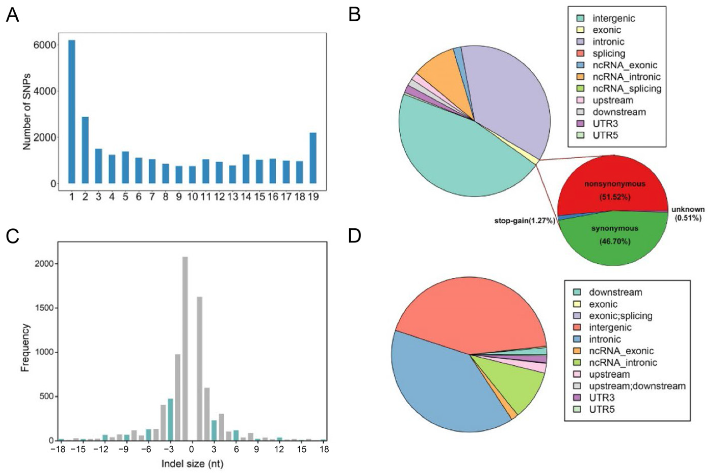
3.2. Functional Annotation of SNVs

In this study, about 5.04 Gb of raw sequencing data were generated for each sample. After stringent quality assessment and data filtering, the samples yielded approximately 28.6 million (85.18%) high-quality reads each, with depths ranging from 65× to 94× (Table S2). Based on the read depth smoothed in a 100 bp sliding window, a nearly 3 Mb deletion across LCR22-AD was observed in the child of the nuclear family (Figure 1).
After quality control, we included a total of 37,659 SNVs (29,071 SNPs and 8584 indels) in 19 sporadic cases. Figure 2a shows that 21.34% of SNPs were observed in only one case, while 7.53% (2188) of SNPs were observed in all 19 cases. There was a total of 396 SNPs distributed in the coding regions, including 203 non-synonymous sites, 184 synonymous sites, five stop-gain sites, and two unknown sites, in addition to two splice sites (Figure 2b). Figure 2c shows that the length of most indels (92.70%) was less than 15 bp. Functional annotation indicated there were ten LoF indels, including nine frameshift indels and one stop-gain indel (Figure 2d).

3.3. Identification of Genes with Potential Functional Consequences

The primary objective of the study was to detect variants with potential functional consequences associated with microtia and CHD. We identified 105 LoFs or functional missense variants from 2165 novel and 82 rare variants from 19 sporadic cases. GO enrichment analysis illustrated that genes harboring the 105 candidate SNPs were enriched in biological processes such as the cellular modified amino acid catabolic process, the leukotriene D4 metabolic process, and the glutathione catabolic process, and molecular function such as omega peptidase activity and transferase activity (Figure 3a). Of these putatively deleterious variants, 50 variants were absent in the healthy parents and resided in 38 genes, such as TBX1 and DGCR8. TBX1 harboring rs5993826 and a novel SNP, and DGCR8 harboring rs182736423 and a novel SNP were known to be associated with the development of the pharyngeal arches or CHD. Figure 3b shows that each sample harbored one to six variants of the 50 variants. Additionally, four SNPs (rs368014, rs11704009, rs9623076, rs4820360) were predicted to be eQTLs for the expression of CXCL10 (p < 0.0001) (Table S3). rs738802 and rs396330 could influence the expression levels of DGCR2, DGCR11, and DGCR12 (p < 0.0001).
A total of 74 overlapped rare and potential deleterious variations were obtained between 19 cases and 103 controls (CHB derived from the 1000 Genome Project). Gene-based testing between cases and CHB illustrated that these potential causal variants were significantly enriched in 10 genes (Table 2). The top gene on the list—SUSD2—had a nearly 22-fold higher frequency in patients than in controls. We also assessed the genetic burden based on candidate genes associated with microtia and CHD. The potential deleterious variants were also significantly enriched in TBX1 (the classical 22q11.2 DS phenotype associated gene) and CLTCL1 (the classical CHD associated gene). Additionally, 20 genes harbored potential deleterious variations that only appeared in the sporadic patients, including RIMBP3 (harbored 17 SNPs), DGCR8, PI4KA, and UFD1.

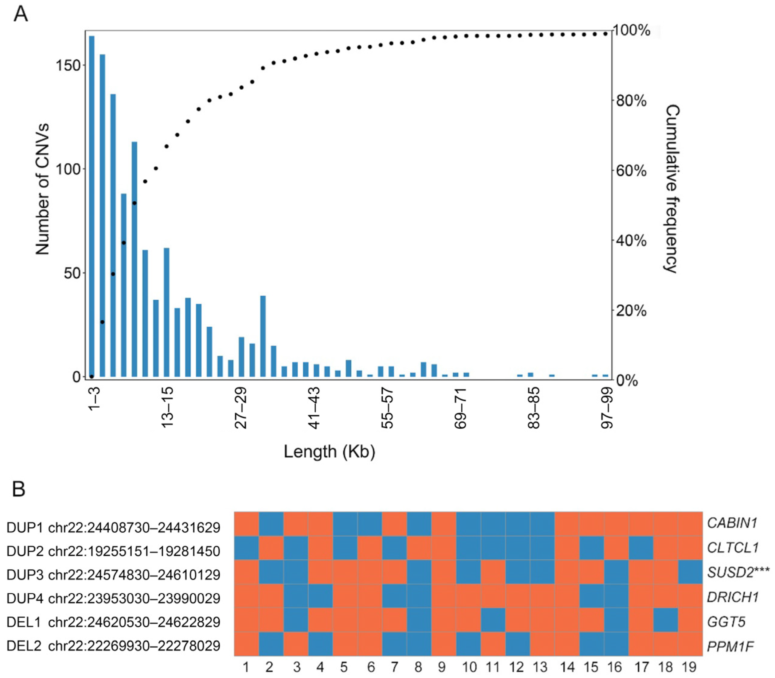
3.4. Microduplication Fragment May Associate with Microtia and CHD

The sequencing depth for each sample varied from 65× to nearly 95×, allowing sufficient power to detect CNV. For 19 sporadic cases and healthy parents, a total of unique 1135 CNVs were identified with an average of 54 calls per sample (Table S4). Calls across all samples affected nearly 28.81% (2.31 Mbp) of the targeted region. The CNV calls had an average length of 14.56 kbp, with a median length of 9.1 kbp (Figure 4a). The cohort showed more duplications than deletions (p < 0.001), where duplications had a median length of 12.95 kbp compared to deletions at 3.90 kbp. The number of sample-specific CNVs for each sample ranged from 5 to 34.
We identified recurrent CNVs between samples as calls with at least 50% reciprocal overlap. After removing merged calls present in the healthy parents, six recurrent CNV calls were retained as candidate CNVs, including four duplications and two deletions (Figure 4b). Except for DUP4, all candidate CNV calls were rare CNVs and not overlapped with segmental duplications (SDs). In our study, 11 of 19 cases had CNV amplifications (DUP3) overlapping most of the sushi domain containing 2 (SUSD2), which was validated by qPCR in the validation set (p < 0.001, Figure 4b). The results of the analysis are summarized in Table S5.

4. Discussion

Our study shows that the clinical manifestations of microtia and CHD comorbidity are highly heterogeneous. Consistent with previous studies, we found that microtia occurs more frequently in males and on the right side (unilateral) [32,33,34]. Nearly 60% of the patients were classified as having second-degree microtia. CHD is the most common congenital disorder in newborns. A previous study showed that Asia has the highest CHD birth prevalence, with 9.3 per 1000 newborns [35]. Among patients with microtia, ASD is the most common lesion, followed by VSD and PDA [8]. In our study, the top three kinds of CHD were VSD, ASD, and TOF.
Considering the high co-occurrence of these two complex diseases, it is speculated that common genetic mutations or biological pathways may play important roles in both microtia and CHD. Among the syndromes with these symptoms, Goldenhar syndrome is also associated with this region in addition to those directly caused by the 22q11.2 variations. Balcı et al. suggested that each patient with Goldenhar syndrome should be screened for 22q11.2 deletion, which could be a candidate causal gene for this syndrome [36]. The strategy of capture-based target enrichment followed by deep sequencing (~80×) enabled us to identify both SNVs and small CNVs simultaneously. Only the child in the nuclear family had a 3 Mbp fragment deletion (LCR22-A to LCR22-D), which belongs to the typical 22q11.2 DS and contributes to the complex phenotypes of the patient [11].
Among 19 sporadic cases, LoF variants and putatively functional missense SNVs were combined for a total of 105 predicted deleterious variants. These variants are predicted to have a strong effect on protein structure and function. Each patient carries at least one harmful mutation, especially three individuals who carry variants of known susceptible genes for microtia or CHD. Two patients (4,12) had a different TBX1 deleterious variant, and one patient (5) had two deleterious variants in DGCR8.
TBX1 is a member of the phylogenetically conserved T-box transcription factor family. TBX1 is highlighted as the most widely studied gene in 22q11.2 DS and is required for the normal development of the fourth pharyngeal arch arteries [37,38]. The previous study suggested that TBX1 mutation is responsible for five major phenotypes in 22q11.2 DS, including abnormal facies, cardiac defects, thymic hypoplasia, velopharyngeal insufficiency with cleft palate, and parathyroid dysfunction with hypocalcemia [39]. Although TBX1 is not expressed in NCCs, NCC patterning of both the surface ectoderm and the second heart field is affected in conditional mutants [40,41]. Haploinsufficiency of TBX1 leads to partially penetrant cardiovascular, thymic, and parathyroid defects in mice [42,43]. Another gene of interest is DGCR8, encoding an essential component of the microprocessor complex that mediates the biogenesis of microRNA [44]. The abnormal microRNA biogenesis due to DGCR8 deficiency contributes to structural abnormalities in the heart and vasculature and neuronal deficits [45,46]. In mice, loss-of-function of DGCR8 in NCCs results in congenital heart abnormalities that are characteristic of 22q11.2 DS, and heterozygous loss-of-function mutations of DGCR8 result in neuronal deficits [45,47].
Gene-based enrichment analysis showed that these variations were significantly enriched in 10 genes, such as SUSD2, TBX1, CLTCL1, and TANGO2. The ratio of deleterious mutation burden between cases and controls reached 21 in gene SUSD2. As well as a tumor-reversing protein, SUSD2 (encoding sushi domain containing 2) also plays a dual role during neural development [48,49,50]. SUSD2 can alter axon and dendritic outgrowth in young neurons, and regulate excitatory synapse numbers at a later stage. CLTCL1 encodes a major protein of the polyhedral coat of coated pits and vesicles, which is associated with multiple syndromes, including DiGeorge syndrome and Velo-Cardio-Facial syndrome. Based on whole exome sequencing and the analysis of the rare mutations burden, Shi et al. found that CLTCL1 was associated with the total anomalous pulmonary venous connection (TAPVC), and highly expressed in human umbilical vein endothelial cells and human embryonic hearts [51]. TANGO2 (transport and Golgi organization 2 homolog) encodes a protein that is involved in redistributing Golgi membranes into the endoplasmic reticulum [52]. Initial reported TANGO2-related disorders were considered metabolic encephalopathy and arrhythmias, characterized by variable clinical features [53,54]. The variability in presentations was expanded to primarily neurological presentations, such as progressive neurodegeneration and cognitive impairment [55]. Further study is needed to assess the contribution of TANGO2 to microtia and CHD, which is involved in the 22q11.2 phenotypic spectrum. In accordance with the previous study, genes harbored by these identified SNVs can only explain a fraction of the genotype–phenotype relationships underlying microtia and CHD [56].
Additionally, potential pathogenic variations that appear only in patients are mainly enriched in some genes, such as RIMBP3, DGCR8, PI4KA, and UFD1. RIMBP3 is mostly expressed outside of the brain. As a member of the Rab3-interacting molecules (RIM), binding proteins (RIMBPs), RIMBP3, together with RIMBP1 and RIMBP2, regulate the fidelity of synaptic transmission at central synapses and play a potential role in regulating the fusion and release of neuronal vesicles by stabilizing the active zone structure in the presynaptic region and locating presynaptic ion channels [57,58]. Previous studies have suggested that PI4KA is a candidate gene for schizophrenia susceptibility and plays an important role in early brain development [59,60]. Ubiquitin Fusion-Degradation Protein-1 Like (Ufd1l) encoded by the gene UFD1 serves as a ubiquitin recognition factor linking the ubiquitination and degradation process of proteins. p97 in mammals associated with the cofactor Ufd1-Npl4 forms a functional conservative complex, which can mediate the transport of endoplasmic reticulum proteins into the cytosol for degradation [61,62,63]. As a housekeeping gene, UFD1 is mainly expressed in pharyngeal arches, palatal precursors, and ears, heart, and brain during development, and the haploinsufficiency of UFD1 is supposed to be responsible for the pathogenesis of 22q11.2 DS [64,65,66]. A previous study reported the association between the bicuspid aortic valve and the reduced expression of UFD1, further supporting the role of UFD1 in normal heart formation [67].
The CNV of genomic fragments is a common phenomenon that affects about 4.8–9.5% of the human genome [68]. The enrichment of CNVs in SDs was consistent with results from the previous study [69]. The hotspots of chromosomal rearrangement defined by SDs can act as mediators of normal variation as well as genomic diseases [68]. Previous studies combined CNVs and SNPs and illustrated the association of CNVs with multiple diseases, such as autism, neuroblastoma, and severe early-onset obesity [70,71,72]. Among six CNVs after strict filtering, a novel microduplication that harbored gene SUSD2 was identified as being associated with phenotypes of patients, and confirmed by quantitative PCR. SUSD2 also plays a dual role at different stages of neural development [50]. However, the molecular mechanism underlying microtia or CHD and SUSD2 remains unknown, and further studies will be required to address this issue.
Given the high heterogeneity of microtia and CHD, the comorbidity of these two diseases is unlikely to be caused by a few specific loci but rather may be the result of the combined effects of multiple variations in multiple genes. In our study, deleterious variants were significantly enriched in serval genes (SUSD2, TBX1, DGCR8, CLTCL1, TANGO2 and UFD1) and the microduplication fragment that may present genetic factors relevant to the etiology. The candidate variants and genes deserve further exploration to better understand the specific mechanisms of microtia and CHD comorbidity. The small sample size is the main limitation of our study. To increase the statistical power of the gene-based rare variant association method, it is usually necessary to increase the sample size [73]. An in-depth study of screening for the whole genome may provide new insights for the investigation of rare and complex diseases.

Supplementary Materials

The following supporting information can be downloaded at: https://www.mdpi.com/article/10.3390/genes14040879/s1, Table S1: Primers and probes used for duplex qRT-PCR; Table S2: Summary of targeted sequencing data output title; Table S3: The association between SNPs (SNP-2) and genes; Table S4: Distribution of CNVs in samples; Table S5: Summary of findings.

Author Contributions

Conceptualization, C.Z. and Y.Y.; methodology, C.Z., Y.Y., B.P. and H.W.; software, C.Z.; validation, H.W. and J.J.; formal analysis, C.Z.; investigation, Y.Y.; resources, B.P.; data curation, Y.Y.; writing—original draft preparation, C.Z. and Y.Y.; writing—review and editing, N.S.; visualization, C.Z.; supervision, N.S. and Q.X.; project administration, Q.X.; funding acquisition, Q.X. All authors have read and agreed to the published version of the manuscript.

Funding

This research was funded by the National Natural Science Foundation of China (Grant number 82071504, 81930104 and 82101545), the STI2030-Major Projects (2021ZD0200600, 2021ZD0202000 and 2021ZD0203000), and the Fundamental Research Funds for the Central Universities and CAMS Innovation Fund for Medical Sciences (Grant number 2021-I2M-1-020, 2021-I2M-1-052).

Institutional Review Board Statement

The study was conducted in accordance with the Declaration of Helsinki, and approved by the Ethics Committee of the Chinese Academy of Medical Sciences and Peking Union Medical College.

Informed Consent Statement

Informed consent was obtained from all subjects involved in the study.

Data Availability Statement

All the raw sequence data have been deposited in the National Genomics Data Center (NGDC) Genome Sequence Archive (GSA) under the accession number: HRA000684 (https://ngdc.cncb.ac.cn/gsa-human/s/mxTu6mt8, accessed on 5 January 2023).

Acknowledgments

The authors thank the patients and their families for their participation.

Conflicts of Interest

The authors declare no conflict of interest. The funders had no role in the design of the study; in the collection, analyses, or interpretation of data; in the writing of the manuscript; or in the decision to publish the results.

References

  1. Alasti, F.; Van Camp, G. Genetics of microtia and associated syndromes. J. Med. Genet. 2009, 46, 361–369. [Google Scholar] [CrossRef] [PubMed]
  2. Suutarla, S.; Rautio, J.; Ritvanen, A.; Ala-Mello, S.; Jero, J.; Klockars, T. Microtia in Finland: Comparison of characteristics in different populations. Int. J. Pediatr. Otorhinolaryngol. 2007, 71, 1211–1217. [Google Scholar] [CrossRef]
  3. Chen, J.P.; Zhang, L.; Chen, G.; Song, X.M.; Zheng, X.Y. Capacity of monitoring system on birth defects during 1990s in China. Chin. J. Epidemiol. 2006, 27, 392. [Google Scholar]
  4. Luquetti, D.V.; Heike, C.L.; Hing, A.V.; Cunningham, M.L.; Cox, T.C. Microtia: Epidemiology and genetics†. Am. J. Med. Genet. A 2012, 158A, 124–139. [Google Scholar] [CrossRef] [Green Version]
  5. Wyse, R.K.; Al-Mahdawi, S.; Burn, J.; Blake, K. Congenital heart disease in CHARGE association. Pediatr. Cardiol. 1993, 14, 75–81. [Google Scholar] [CrossRef]
  6. Digilio, M.C.; Calzolari, F.; Capolino, R.; Toscano, A.; Sarkozy, A.; De Zorzi, A.; Dallapiccola, B.; Marino, B. Congenital heart defects in patients with oculo-auriculo-vertebral spectrum (Goldenhar syndrome). Am. J. Med. Genet. Part A 2008, 146, 1815–1819. [Google Scholar] [CrossRef]
  7. Zhang, Y.; Jiang, H.; Yang, Q.; He, L.; Yu, X.; Huang, X.; Wu, R.; Yang, M.; Li, C.; Pan, B. Microtia in a Chinese Specialty Clinic Population: Clinical Heterogeneity and Associated Congenital Anomalies. Plast. Reconstr. Surg. 2018, 142, 892e–903e. [Google Scholar] [CrossRef]
  8. Cao, T.; Chen, Q.; Wang, B.; Hu, J.; Zou, M.; Zhang, Q. Epidemiological research of microtia combined with congenital heart disease. J. Craniofacial Surg. 2021, 32, 920–924. [Google Scholar] [CrossRef] [PubMed]
  9. Shaikh, T.; Kurahashi, H.; Saitta, S.; O’Hare, A.; Hu, P.; Roe, B.; Driscoll, D.; McDonald-McGinn, D.; Zackai, E.; Budarf, M.; et al. Chromosome 22-specific low copy repeats and the 22q11.2 deletion syndrome: Genomic organization and deletion endpoint analysis. Hum. Mol. Genet. 2000, 9, 489–501. [Google Scholar] [CrossRef] [PubMed] [Green Version]
  10. Edelmann, L.; Pandita, R.K.; Spiteri, E.; Funke, B.; Goldberg, R.; Palanisamy, N.; Chaganti, R.S.K.; Magenis, E.; Shprintzen, R.J.; Morrow, B.E. A Common Molecular Basis for Rearrangement Disorders on Chromosome 22q11. Hum. Mol. Genet. 1999, 8, 1157. [Google Scholar] [CrossRef] [Green Version]
  11. McDonald-McGinn, D.M.; Sullivan, K.E.; Marino, B.; Philip, N.; Swillen, A.; Vorstman, J.A.; Zackai, E.H.; Emanuel, B.S.; Vermeesch, J.R.; Morrow, B.E. 22q11. 2 deletion syndrome. Nat. Rev. Dis. Prim. 2015, 1, 15071. [Google Scholar] [CrossRef] [PubMed] [Green Version]
  12. Di Rocco, M.; Buocompagni, A.; Picco, P.; Vignola, S.; Borrone, C.; Gimelli, G. Spectrum of clinical features associated with interstitial chromosome 22q11 deletions. J. Med. Genet. 1998, 35, 346. [Google Scholar] [CrossRef] [Green Version]
  13. McDonald-McGinn, D.M.; Sullivan, K.E. Chromosome 22q11. 2 deletion syndrome (DiGeorge syndrome/velocardiofacial syndrome). Medicine 2011, 90, 1–18. [Google Scholar] [CrossRef] [PubMed]
  14. White, L.K.; Crowley, T.B.; Finucane, B.; McClellan, E.J.; Donoghue, S.; Garcia-Minaur, S.; Repetto, G.M.; Fischer, M.; Jacquemont, S.; Gur, R.E.; et al. Gathering the Stakeholder&rsquo;s Perspective: Experiences and Opportunities in Rare Genetic Disease Research. Genes 2023, 14, 169. [Google Scholar]
  15. Tan, T.Y.; Collins, A.; James, P.A.; McGillivray, G.; Stark, Z.; Gordon, C.T.; Leventer, R.J.; Pope, K.; Forbes, R.; Crolla, J.A.; et al. Phenotypic variability of distal 22q11. 2 copy number abnormalities. Am. J. Med. Genet. A. 2011, 155, 1623–1633. [Google Scholar] [CrossRef] [PubMed]
  16. Blagowidow, N.; Nowakowska, B.; Schindewolf, E.; Grati, F.R.; Putotto, C.; Breckpot, J.; Swillen, A.; Crowley, T.B.; Loo, J.C.Y.; Lairson, L.A.; et al. Prenatal sreening and diagnostic considerations for 22q11.2 microdeletions. Genes 2023, 14, 160. [Google Scholar] [CrossRef]
  17. Hunter, A.; Frias, J.L.; Gillessen-Kaesbach, G.; Hughes, H.; Jones, K.L.; Wilson, L. Elements of morphology: Standard terminology for the ear. Am. J. Med. Genet. Part A 2009, 149a, 40–60. [Google Scholar] [CrossRef] [PubMed]
  18. Andrews, S. FastQC: A Quality Control Tool for High Throughput Sequence Data. 2010. Available online: https://www.bioinformatics.babraham.ac.uk/projects/fastqc/ (accessed on 12 November 2017).
  19. Martin, M. Cutadapt removes adapter sequences from high-throughput sequencing reads. EMBnet. J. 2011, 17, 10–12. [Google Scholar] [CrossRef]
  20. Li, H.; Durbin, R. Fast and accurate short read alignment with Burrows–Wheeler transform. Bioinformatics 2009, 25, 1754–1760. [Google Scholar] [CrossRef] [Green Version]
  21. Li, H.; Handsaker, B.; Wysoker, A.; Fennell, T.; Ruan, J.; Homer, N.; Marth, G.; Abecasis, G.; Durbin, R. The sequence alignment/map format and SAMtools. Bioinformatics 2009, 25, 2078–2079. [Google Scholar] [CrossRef] [Green Version]
  22. Van der Auwera, G.A.; Carneiro, M.O.; Hartl, C.; Poplin, R.; Del Angel, G.; Levy-Moonshine, A.; Jordan, T.; Shakir, K.; Roazen, D.; Thibault, J. From FastQ data to high-confidence variant calls: The genome analysis toolkit best practices pipeline. Curr. Protoc. Bioinform. 2013, 43, 11.10.11–11.10.33. [Google Scholar] [CrossRef] [PubMed] [Green Version]
  23. Danecek, P.; Auton, A.; Abecasis, G.; Albers, C.A.; Banks, E.; DePristo, M.A.; Handsaker, R.E.; Lunter, G.; Marth, G.T.; Sherry, S.T. The variant call format and VCFtools. Bioinformatics 2011, 27, 2156–2158. [Google Scholar] [CrossRef]
  24. Lek, M.; Karczewski, K.J.; Minikel, E.V.; Samocha, K.E.; Banks, E.; Fennell, T.; O’Donnell-Luria, A.H.; Ware, J.S.; Hill, A.J.; Cummings, B.B. Analysis of protein-coding genetic variation in 60,706 humans. Nature 2016, 536, 285. [Google Scholar] [CrossRef] [PubMed] [Green Version]
  25. Sudmant, P.H.; Rausch, T.; Gardner, E.J.; Handsaker, R.E.; Abyzov, A.; Huddleston, J.; Zhang, Y.; Ye, K.; Jun, G.; Fritz, M.H.-Y. An integrated map of structural variation in 2,504 human genomes. Nature 2015, 526, 75. [Google Scholar] [CrossRef] [Green Version]
  26. Liu, X.; Jian, X.; Boerwinkle, E. dbNSFP v2. 0: A database of human non-synonymous SNVs and their functional predictions and annotations. Hum. Mutat. 2013, 34, E2393–E2402. [Google Scholar] [CrossRef] [Green Version]
  27. Zhang, W.; Gamazon, E.R.; Zhang, X.; Konkashbaev, A.; Liu, C.; Szilágyi, K.L.; Dolan, M.E.; Cox, N.J. SCAN database: Facilitating integrative analyses of cytosine modification and expression QTL. Database 2015, 2015, bav025. [Google Scholar] [CrossRef] [Green Version]
  28. Yu, G.; Wang, L.-G.; Han, Y.; He, Q.-Y. clusterProfiler: An R package for comparing biological themes among gene clusters. OMICS J. Integr. Biol. 2012, 16, 284–287. [Google Scholar] [CrossRef]
  29. Abyzov, A.; Urban, A.E.; Snyder, M.; Gerstein, M. CNVnator: An approach to discover, genotype and characterize typical and atypical CNVs from family and population genome sequencing. Genome Res. 2011, 21, 974–984. [Google Scholar] [CrossRef] [Green Version]
  30. Quinlan, A.R.; Hall, I.M. BEDTools: A flexible suite of utilities for comparing genomic features. Bioinformatics 2010, 26, 841–842. [Google Scholar] [CrossRef] [PubMed] [Green Version]
  31. Pruitt, K.D.; Tatusova, T.; Maglott, D.R. NCBI Reference Sequence (RefSeq): A curated non-redundant sequence database of genomes, transcripts and proteins. Nucleic Acids Res. 2005, 33, D501–D504. [Google Scholar] [CrossRef] [Green Version]
  32. Forrester, M.B.; Merz, R.D. Descriptive epidemiology of anotia and microtia, Hawaii, 1986–2002. Congenit. Anom. 2005, 45, 119–124. [Google Scholar] [CrossRef]
  33. Okajima, H.; Takeichi, Y.; Umeda, K.; Baba, S. Clinical analysis of 592 patients with microtia. Acta Oto-Laryngol. Suppl. 1996, 525, 18–24. [Google Scholar]
  34. Shaw, G.M.; Carmichael, S.L.; Kaidarova, Z.; Harris, J.A. Epidemiologic characteristics of anotia and microtia in California, 1989–1997. Birth Defects Res. Part A Clin. Mol. Teratol. 2004, 70, 472–475. [Google Scholar] [CrossRef] [PubMed]
  35. van der Linde, D.; Konings, E.E.M.; Slager, M.A.; Witsenburg, M.; Helbing, W.A.; Takkenberg, J.J.M.; Roos-Hesselink, J.W. Birth Prevalence of Congenital Heart Disease Worldwide. A Syst. Rev. Meta-Anal. 2011, 58, 2241–2247. [Google Scholar] [CrossRef] [PubMed] [Green Version]
  36. Balcı, S.; Engiz, Ö. Goldenhar syndrome phenotypes and 22q11 deletion. Am. J. Med. Genet. Part A 2011, 155, 458. [Google Scholar] [CrossRef] [PubMed]
  37. Jerome, L.A.; Papaioannou, V.E. DiGeorge syndrome phenotype in mice mutant for the T-box gene, Tbx1. Nat. Genet. 2001, 27, 286. [Google Scholar] [CrossRef]
  38. Papangeli, I.; Scambler, P. The 22q11 deletion: DiGeorge and velocardiofacial syndromes and the role of TBX1. Wiley Interdiscip. Rev. Dev. Biol. 2013, 2, 393–403. [Google Scholar] [CrossRef]
  39. Yagi, H.; Furutani, Y.; Hamada, H.; Sasaki, T.; Asakawa, S.; Minoshima, S.; Ichida, F.; Joo, K.; Kimura, M.; Imamura, S.-I.; et al. Role of TBX1 in human del22q11.2 syndrome. Lancet 2003, 362, 1366–1373. [Google Scholar] [CrossRef]
  40. Calmont, A.; Ivins, S.; Van Bueren, K.L.; Papangeli, I.; Kyriakopoulou, V.; Andrews, W.D.; Martin, J.F.; Moon, A.M.; Illingworth, E.A.; Basson, M.A. Tbx1 controls cardiac neural crest cell migration during arch artery development by regulating Gbx2 expression in the pharyngeal ectoderm. Development 2009, 136, 3173–3183. [Google Scholar] [CrossRef] [Green Version]
  41. Vitelli, F.; Morishima, M.; Taddei, I.; Lindsay, E.A.; Baldini, A. Tbx1 mutation causes multiple cardiovascular defects and disrupts neural crest and cranial nerve migratory pathways. Hum. Mol. Genet. 2002, 11, 915–922. [Google Scholar] [CrossRef] [Green Version]
  42. Merscher, S.; Funke, B.; Epstein, J.A.; Heyer, J.; Puech, A.; Lu, M.M.; Xavier, R.J.; Demay, M.B.; Russell, R.G.; Factor, S. TBX1 is responsible for cardiovascular defects in velo-cardio-facial/DiGeorge syndrome. Cell 2001, 104, 619–629. [Google Scholar] [CrossRef] [Green Version]
  43. Lindsay, E.A.; Vitelli, F.; Su, H.; Morishima, M.; Huynh, T.; Pramparo, T.; Jurecic, V.; Ogunrinu, G.; Sutherland, H.F.; Scambler, P.J.; et al. Tbx1 haploinsufficiency in the DiGeorge syndrome region causes aortic arch defects in mice. Nature 2001, 410, 97–101. [Google Scholar] [CrossRef] [PubMed]
  44. Wang, Y.; Medvid, R.; Melton, C.; Jaenisch, R.; Blelloch, R. DGCR8 is essential for microRNA biogenesis and silencing of embryonic stem cell self-renewal. Nat. Genet. 2007, 39, 380. [Google Scholar] [CrossRef] [PubMed]
  45. Stark, K.L.; Xu, B.; Bagchi, A.; Lai, W.-S.; Liu, H.; Hsu, R.; Wan, X.; Pavlidis, P.; Mills, A.A.; Karayiorgou, M. Altered brain microRNA biogenesis contributes to phenotypic deficits in a 22q11-deletion mouse model. Nat. Genet. 2008, 40, 751. [Google Scholar] [CrossRef]
  46. Smith, T.; Rajakaruna, C.; Caputo, M.; Emanueli, C. MicroRNAs in congenital heart disease. Ann. Transl. Med. 2015, 3, 333. [Google Scholar]
  47. Chapnik, E.; Sasson, V.; Blelloch, R.; Hornstein, E. Dgcr8 controls neural crest cells survival in cardiovascular development. Dev. Biol. 2012, 362, 50–56. [Google Scholar] [CrossRef] [Green Version]
  48. Cheng, Y.; Wang, X.; Wang, P.; Li, T.; Hu, F.; Liu, Q.; Yang, F.; Wang, J.; Xu, T.; Han, W. SUSD2 is frequently downregulated and functions as a tumor suppressor in RCC and lung cancer. Tumor Biol. 2016, 37, 9919–9930. [Google Scholar] [CrossRef]
  49. Watson, A.P.; Evans, R.L.; Egland, K.A. Multiple functions of sushi domain containing 2 (SUSD2) in breast tumorigenesis. Mol. Cancer Res. 2013, 11, 74–85. [Google Scholar] [CrossRef] [PubMed] [Green Version]
  50. Nadjar, Y.; Triller, A.; Bessereau, J.-L.; Dumoulin, A. The Susd2 protein regulates neurite growth and excitatory synaptic density in hippocampal cultures. Mol. Cell. Neurosci. 2015, 65, 82–91. [Google Scholar] [CrossRef]
  51. Shi, X.; Huang, T.; Wang, J.; Liang, Y.; Gu, C.; Xu, Y.; Sun, J.; Lu, Y.; Sun, K.; Chen, S.; et al. Next-generation sequencing identifies novel genes with rare variants in total anomalous pulmonary venous connection. EBioMedicine 2018, 38, 217–227. [Google Scholar] [CrossRef] [Green Version]
  52. Bard, F.; Casano, L.; Mallabiabarrena, A.; Wallace, E.; Saito, K.; Kitayama, H.; Guizzunti, G.; Hu, Y.; Wendler, F.; DasGupta, R.; et al. Functional genomics reveals genes involved in protein secretion and Golgi organization. Nature 2006, 439, 604–607. [Google Scholar] [CrossRef] [PubMed]
  53. Kremer, L.S.; Distelmaier, F.; Alhaddad, B.; Hempel, M.; Iuso, A.; Küpper, C.; Mühlhausen, C.; Kovacs-Nagy, R.; Satanovskij, R.; Graf, E.; et al. Bi-allelic Truncating Mutations in TANGO2 Cause Infancy-Onset Recurrent Metabolic Crises with Encephalocardiomyopathy. Am. J. Hum. Genet. 2016, 98, 358–362. [Google Scholar] [CrossRef] [Green Version]
  54. Lalani, S.R.; Liu, P.; Rosenfeld, J.A.; Watkin, L.B.; Chiang, T.; Leduc, M.S.; Zhu, W.; Ding, Y.; Pan, S.; Vetrini, F.; et al. Recurrent Muscle Weakness with Rhabdomyolysis, Metabolic Crises, and Cardiac Arrhythmia Due to Bi-allelic TANGO2 Mutations. Am. J. Hum. Genet. 2016, 98, 347–357. [Google Scholar] [CrossRef] [PubMed] [Green Version]
  55. Dines, J.N.; Golden-Grant, K.; LaCroix, A.; Muir, A.M.; Cintrón, D.L.; McWalter, K.; Cho, M.T.; Sun, A.; Merritt, J.L.; Thies, J. TANGO2: Expanding the clinical phenotype and spectrum of pathogenic variants. Genet. Med. 2019, 21, 601. [Google Scholar] [CrossRef] [PubMed] [Green Version]
  56. Motahari, Z.; Moody, S.A.; Maynard, T.M.; LaMantia, A.-S. In the line-up: Deleted genes associated with DiGeorge/22q11.2 deletion syndrome: Are they all suspects? J. Neurodev. Disord. 2019, 11, 7. [Google Scholar] [CrossRef] [Green Version]
  57. Acuna, C.; Liu, X.; Gonzalez, A.; Südhof Thomas, C. RIM-BPs Mediate Tight Coupling of Action Potentials to Ca2+-Triggered Neurotransmitter Release. Neuron 2015, 87, 1234–1247. [Google Scholar] [CrossRef] [Green Version]
  58. Gao, T.; Zhang, Z.; Yang, Y.; Zhang, H.; Li, N.; Liu, B. Impact of RIM-BPs in neuronal vesicles release. Brain Res. Bull. 2021, 170, 129–136. [Google Scholar] [CrossRef] [PubMed]
  59. Jungerius, B.J.; Hoogendoorn, M.L.C.; Bakker, S.C.; van’t Slot, R.; Bardoel, A.F.; Ophoff, R.A.; Wijmenga, C.; Kahn, R.S.; Sinke, R.J. An association screen of myelin-related genes implicates the chromosome 22q11 PIK4CA gene in schizophrenia. Mol. Psychiatry 2008, 13, 1060–1068. [Google Scholar] [CrossRef] [Green Version]
  60. Pagnamenta, A.T.; Howard, M.F.; Wisniewski, E.; Popitsch, N.; Knight, S.J.; Keays, D.A.; Quaghebeur, G.; Cox, H.; Cox, P.; Balla, T.; et al. Germline recessive mutations in PI4KA are associated with perisylvian polymicrogyria, cerebellar hypoplasia and arthrogryposis. Hum. Mol. Genet. 2015, 24, 3732–3741. [Google Scholar] [CrossRef] [Green Version]
  61. Ye, Y.; Meyer, H.H.; Rapoport, T.A. Function of the p97–Ufd1–Npl4 complex in retrotranslocation from the ER to the cytosol: Dual recognition of nonubiquitinated polypeptide segments and polyubiquitin chains. J. Cell Biol. 2003, 162, 71–84. [Google Scholar] [CrossRef]
  62. Ye, Y.; Meyer, H.H.; Rapoport, T.A. The AAA ATPase Cdc48/p97 and its partners transport proteins from the ER into the cytosol. Nature 2001, 414, 652–656. [Google Scholar] [CrossRef]
  63. Bays, N.W.; Hampton, R.Y. Cdc48–Ufd1–Npl4: Stuck in the middle with Ub. Curr. Biol. 2002, 12, R366–R371. [Google Scholar] [CrossRef] [PubMed] [Green Version]
  64. Novelli, G.; Mari, A.; Amati, F.; Colosimo, A.; Sangiuolo, F.; Bengala, M.; Conti, E.; Ratti, A.; Bordoni, R.; Pizzuti, A. Structure and expression of the human ubiquitin fusion–degradation gene (UFD1L). Biochim. Biophys. Acta (BBA)-Gene Struct. Expr. 1998, 1396, 158–162. [Google Scholar] [CrossRef]
  65. Yamagishi, H. A Molecular Pathway Revealing a Genetic Basis for Human Cardiac and Craniofacial Defects. Science 1999, 283, 1158–1161. [Google Scholar] [CrossRef] [Green Version]
  66. Rizzu, P.; Lindsay, E.A.; Taylor, C.; O’Donnell, H.; Levy, A.; Scambler, P.; Baldini, A. Cloning and comparative mapping of a gene from the commonly deleted region of DiGeorge and Velocardiofacial syndromes conserved in C. elegans. Mamm. Genome 1996, 7, 639–643. [Google Scholar] [CrossRef] [PubMed]
  67. Mohamed, S.A.; Hanke, T.; Schlueter, C.; Bullerdiek, J.; Sievers, H.-H. Ubiquitin fusion degradation 1–like gene dysregulation in bicuspid aortic valve. J. Thorac. Cardiovasc. Surg. 2005, 130, 1531–1536. [Google Scholar] [CrossRef] [Green Version]
  68. Zarrei, M.; MacDonald, J.R.; Merico, D.; Scherer, S.W. A copy number variation map of the human genome. Nat. Rev. Genet. 2015, 16, 172–183. [Google Scholar] [CrossRef] [PubMed]
  69. Sharp, A.J.; Locke, D.P.; McGrath, S.D.; Cheng, Z.; Bailey, J.A.; Vallente, R.U.; Pertz, L.M.; Clark, R.A.; Schwartz, S.; Segraves, R.; et al. Segmental duplications and copy-number variation in the human genome. Am. J. Hum. Genet. 2005, 77, 78–88. [Google Scholar] [CrossRef] [Green Version]
  70. Glessner, J.T.; Wang, K.; Cai, G.; Korvatska, O.; Kim, C.E.; Wood, S.; Zhang, H.; Estes, A.; Brune, C.W.; Bradfield, J.P.; et al. Autism genome-wide copy number variation reveals ubiquitin and neuronal genes. Nature 2009, 459, 569–573. [Google Scholar] [CrossRef] [Green Version]
  71. Diskin, S.J.; Hou, C.; Glessner, J.T.; Attiyeh, E.F.; Laudenslager, M.; Bosse, K.; Cole, K.; Mosse, Y.P.; Wood, A.; Lynch, J.E.; et al. Copy number variation at 1q21.1 associated with neuroblastoma. Nature 2009, 459, 987–991. [Google Scholar] [CrossRef] [PubMed] [Green Version]
  72. Bochukova, E.G.; Huang, N.; Keogh, J.; Henning, E.; Purmann, C.; Blaszczyk, K.; Saeed, S.; Hamilton-Shield, J.; Clayton-Smith, J.; O’Rahilly, S.; et al. Large, rare chromosomal deletions associated with severe early-onset obesity. Nature 2010, 463, 666–670. [Google Scholar] [CrossRef] [PubMed] [Green Version]
  73. Moutsianas, L.; Agarwala, V.; Fuchsberger, C.; Flannick, J.; Rivas, M.A.; Gaulton, K.J.; Albers, P.K.; Go, T.D.C.; McVean, G.; Boehnke, M.; et al. The power of gene-based rare variant methods to detect disease-associated variation and test hypotheses about complex disease. PLoS Genet. 2015, 11, e1005165. [Google Scholar] [CrossRef] [PubMed] [Green Version]
Figure 1. Depth of several samples with a 100 bp sliding window. (A) The child in the nuclear family. The red box indicates the deletion of LCRA-D. (B,C) The healthy parents in the nuclear family. (D) Patient #1. Bases labeled as N in the reference genome are represented by blue dots, while red dots indicate bases located in other regions.
Genes 14 00879 g001
Figure 2. Distribution of SNVs by location and type. (A) Frequency distributions in terms of occurrences in 19 cases. (B) Distribution of SNPs by their gene location and type. (C) Distribution of indel lengths. (D) Distribution of indels by their gene location.
Genes 14 00879 g002
Figure 3. (A) GO enrichment analysis of genes harbored 105 LoFs and missense SNPs. (B) Distribution of candidate SNPs (SNP-1) in 19 cases. 0/0, homozygote for the reference allele; 0/1, heterozygote; 1/1, homozygote for the alternative allele.
Genes 14 00879 g003
Figure 4. CNVs called by CNVnator. (A) Length distribution of CNV calls across 19 cases and the healthy parents. (B) Distribution of candidate CNVs in 19 cases. The presence of a particular copy number variation (CNV) in the patient is indicated by red, and its absence is indicated by blue. *** p < 0.001 in qRT-PCR.
Genes 14 00879 g004
Table 1. Summary of the clinical features observed in 20 patients included in our study.
PatientSexAgeEar AnomaliesCardiac AnomaliesOther Anomalies
1M8B; degree IVSD
2M6R; degree IVSD
3F20R; degree ITOF
4M13L; degree IVSD; TI; PI
5M25L; degree IVSD
6M9L; degree ITOF
7M6R; degree IIPDA
8M12R; degree IIVSD; PH
9M11R; degree IIVSDcleft palate
10M8R; degree IITOF
11M7R; degree IIPFObilateral preauricular fistula
12F13R; degree IIASD
13M10R; degree IIVSD
14M18L; degree IIVSD
15M15L; degree IIASD
16F6L; degree IIVSD
17M7L; degree IITOF
18F35R; degree IIIASDfacial cleft; hemifacial dysplasia
19F8R; degree IIIASDhemifacial dysplasia; facial transversal cleft; left eye cyst
20 1M7B; degree IASD; PDApolydactyly; epilepsy
1 The child in the nuclear family. F, feminine; M, masculine; L, left; R, right; B, bilateral; VSD, ventricular septal defect; ASD, atrial septal defect; TOF, tetralogy of fallot; TI, tricuspid insufficiency; PDA, patent ductus arteriosus; PFO, patent foramen ovale; PH, pulmonary hypertension; PI, pulmonary insufficiency. Blank boxes indicate the absence of the phenotype. Degree I, first degree microtia; degree II, second degree microtia; degree III, third degree microtia.
Table 2. Gene-based association between cases and CHB.
GeneCounts (CHB) 1Counts (Cases)Frequency Ratiop Value
SUSD252021.68<0.0001
COMT921.20<0.0001
GGT216227.45<0.0001
TOP3B1641.36<0.0001
SCARF22961.12<0.0001
PIWIL31316.260.0001
BCR225.420.0002
TBX13881.140.0293
TANGO2115.420.0350
CLTCL1116251.170.0430
1 Counts represent the number of times that mutation is observed in cases or CHB.
Disclaimer/Publisher’s Note: The statements, opinions and data contained in all publications are solely those of the individual author(s) and contributor(s) and not of MDPI and/or the editor(s). MDPI and/or the editor(s) disclaim responsibility for any injury to people or property resulting from any ideas, methods, instructions or products referred to in the content.

