Article

Identification and Functional Analysis of ToBPI1/LBP and ToBPI2/LBP in Anti-Bacterial Infection of Trachinotus ovatus

1
Guangxi Key Laboratory of Beibu Gulf Marine Biodiversity Conservation, College of Marine Sciences, Beibu Gulf University, Qinzhou 535011, China
2
State Key Laboratory for Conservation and Utilization of Subtropical Agro-Bioresources, College of Animal Science and Technology, Guangxi University, Nanning 530004, China
*
Author to whom correspondence should be addressed.
These authors contributed equally to this work.
Genes 2023, 14(4), 826; https://doi.org/10.3390/genes14040826
Submission received: 3 March 2023 / Revised: 25 March 2023 / Accepted: 28 March 2023 / Published: 29 March 2023
(This article belongs to the Section Animal Genetics and Genomics)

Abstract

:
Bactericidal/permeability-increasing protein (BPI) and lipopolysaccharide-binding protein (LBP) are a group of antibacterial proteins that play an important role in the host’s innate immune defense against pathogen infection. In this study, two BPI/LBPs, named ToBPI1/LBP (1434 bp in length, 478 amino acids) and ToBPI2/LBP (1422 bp in length, 474 amino acids), were identified from the golden pompano. ToBPI1/LBP and ToBPI2/LBP were significantly expressed in immune-related tissues after being challenged with Streptococcus agalactiae and Vibrio alginolyticus. The two BPI/LBPs showed significant antibacterial activity against Gram-negative Escherichia coli and Gram-positive S. agalactiae and Streptococcus iniae. In contrast, the antibacterial activity against Staphylococcus aureus, Corynebacterium glutamicum, Vibrio parahaemolyticus, V. alginolyticus and Vibrio harveyi was low and decreased with time. The membrane permeability of bacteria treated with recombinant ToBPI1/LBP and ToBPI2/LBP was significantly enhanced. These results suggest that ToBPI1/LBP and ToBPI2/LBP may play important immunological roles in the immune response of the golden pompano to bacteria. This study will provide basic information and new insights into the immune response mechanism of the golden pompano to bacteria and the function of BPI/LBP.

1. Introduction

Innate immunity is an essential component of the biological immune defense system. It is crucial for maintaining host-microbial homeostasis and for the host’s defense system against the invasion of numerous pathogens such as viruses, bacteria and parasites [1,2,3]. Antimicrobial peptides are a kind of polypeptides with antibacterial activity found in many organisms [4]. There are many kinds of antimicrobial peptides, which can be divided into animal-derived antimicrobial peptides, plant-derived antimicrobial peptides and microbial-derived antimicrobial peptides [5]. Bactericidal/permeability enhancing protein (BPI) and lipopolysaccharide binding protein (LBP) are animal-derived antimicrobial peptides, also members of the lipid transfer/endotoxin binding proteins family; they play an important role in host defense against pathogens and can regulate the innate immune response via pathogen-associated molecular patterns. Structurally and functionally, BPI and LBP are very similar; they own one domain, BPI/LBP/CETP, at the N-terminal that can bind to lipopolysaccharides and one domain, BPI/LBP CETP, at the C-terminal that can transport lipopolysaccharides to other host molecules [2]. However, they also differ significantly in function [6,7]. LBP binds to LPS and enhances the cell response to LPS, whereas BPI binds to Gram-negative bacteria and kills the bacteria by neutralizing LPS and inhibiting the cellular response to LPS [7]. In addition to neutralizing lipopolysaccharide, studies have demonstrated that BPI serves physiological functions such as conditioning, anti-inflammatory and reproduction [8,9]. BPI has currently been shown to have numerous biological functions in mammals. Recombinant mouse BPI, for instance, has been demonstrated to inhibit endotoxin-mediated macrophage activation, while human BPI has been shown to have good antibacterial activity [10]. Additionally, several investigations have also discovered that BPI has a role in activating complement [11]. In teleost fish, BPI or LBP homologues are not classified as BPI or LBP, but they are referred to as BPI/LBP [3]. In fish, BPI/LBP exhibited antibacterial, immunomodulatory and endotoxin-binding properties. An example of this would be the recombinant RfBPI/LBP obtained from Sebastes schlegelii, which exhibited antibacterial activity against E. coli in vitro [12]. The recombinant MaBPI/LBP protein has shown effective bactericidal activity against E. coli and Aeromonas hydrophila in Megalobrama amblycephala [3]. The recombinant CsBPI protein increases body resistance to bacterial and viral infections in Cynoglossus semilaevis by increasing the expression of several genes involved in antibacterial and antiviral immunity [13]. SmBPI/LBP1 demonstrated a high affinity for lipopolysaccharide in Scophthalmus maximus, and its binding capacity increased significantly with an increase in protein concentration [14].
Because of its short growth cycle, tasty meat, and high economic value, the golden pompano (Trachinotus ovatus) has become a popular mariculture fish in China [15]. The T. ovatus mariculture sector has recently suffered significant financial losses as a result of disease outbreaks. Studies have shown that after the golden pompano is infected by a bacteria, immune-related pathways and genes will be activated to participate in the body’s immune response, such as immune-related pathways, the JAK-STAT signaling pathway, Th1 and Th2 cell differentiation, the TNF signaling pathway, the NF-κB signaling pathway and immune-related genes NF-κB, TNF-α, IL1, IL6, IL8, IFN-γ [16]. Wu et al. studied the bacteriostatic effect of golden pompano’s BPI/LBP on Gram-negative bacteria, but its effect on bacterial cell membrane permeability was not studied, and its bacteriostatic effect on Gram-positive bacteria was not clear [17]. Therefore, the function of the golden pompano’s BPI/LBP requires further study. In this study, two BPI/LBP genes, designated as ToBPI1/LBP and ToBPI2/LBP, were isolated from T. ovatus. In addition, we also analyzed the expression, antibacterial activity and cell membrane permeability of ToBPI1/LBP and ToBPI2/LBP in immune-associated tissues to reveal their immunological function. These results should serve as an important reference for the immune defense and function of ToBPI1/LBP and ToBPI2/LBP.

2. Materials and Methods

2.1. Fish and Bacterial Strain

Two hundred individuals of T. ovatus with an average weight of 50 ± 0.7 g were purchased from local farms in Qinzhou, Guangxi, China. The fish samples were placed in 200 L of cyclically aerated seawater and kept between 28° and 30° for 10 days. Streptococcus agalacia, V. alginolyticus, S. iniae, S. aureus, C. glutamicum, E. coli, V. harveyi, V. parahaemolyticus were all donated by Dr. Cai from Beibu Gulf University, Qinzhou, Guangxi, China, whereas E. coli DH5α and BL21 were purchased from Beijing TransGen Biotech Co., Ltd. Both S. agalactiae and V. alginolyticus were cultured in BHI medium overnight at 37°, and then inoculated in 10 mL of liquid medium for shock culture for 10 h. The bacteria were collected, cleaned with 1× PBS three times, and diluted to 1 × 10⁷ CFU/mL for later use.

2.2. Fish Challenge Experiments and Tissue Sample

Before dissection, fish were euthanized by MS-222, as previously reported in [18]. Six fish were used to determine the expression of ToBPI1/LBP and ToBPI2/LBP in healthy tissues. The collected tissues include the heart, gill, stomach, intestine, head, kidney, liver, spleen, brain and muscle. The remaining 190 fish were randomly divided into the control group, V. alginolytica group and S. agalactiae group, and each fish was intraperitoneally injected with 100 μL PBS or 1 × 10⁷ CFU/mL bacterial suspension, respectively. At 0 h, 6 h, 12 h, 24 h, 36 h, 48 h and 72 h after infection, the tissues, including head, kidney, liver and spleen of six fish in each group, were collected and frozen at −80° until further analysis. In this study, all experiments were conducted under a protocol approved by the Institutional Animal Care and Use Committee (IACUC, China).

2.3. Gene Cloning

All PCR primers used in this study are listed in Table 1. Total RNA was extracted from the spleen using a TRIzol reagent (Invitrogen, USA), and RNA quality was detected by 1% agarose gel, stored at −80 °C for backup. The first strand of cDNA was synthesized according to the manufacturer’s instructions using NovoScript Plus one-step 1st Strand cDNA Synthesis SuperMix (Novoprotein, Shanghai, China). Using the cDNA from the spleen tissue of the golden pompano as the template, the CDS sequences of ToBPI1/LBP and ToBPI2/LBP were amplified by the following primers, ToBPI1/LBP-F and ToBPI1/LBP-R, ToBPI2/LBP-F and ToBPI2/LBP-R with Premix TaqTM Hot Start Version (TaKaRa, Dalian, China). The reaction system consisted of 4 μL of cDNA, 2 μL of each primer, 24 μL of Premix Taq, and 24 μL of H2O. The reaction procedure was run at 95 °C for 30 s; 94 °C for 30 s, 53 °C/57 °C for 30 s, 72 °C for 1 min, for 35 cycles; and at 72 °C for 10 min and 4 °C forever. The amplified product was cloned into the pEASY-T1 vector and transformed into E. coli DH5α cells. The positive clones were screened and sequenced by the Dongxuan gene (Jiangsu, China).

2.4. Bioinformatics Analysis

A similarity analysis and an open reading frame determination were performed through the National Center for Biotechnology Information (NCBI), BLAST and ORF Finder programs. The expert Protein Analysis System (EXPASY) server (http://web.expasy.org/protparam/, accessed on 2 March 2023) was used for molecular weight and theoretical isoelectric point analysis. SignalP 5.0 (https://services.healthtech.dtu.dk/service.php?SignalP-5.0, accessed on 2 March 2023) was performed for the prediction of signal peptides. The Interpro program (http://www.ebi.ac.uk/interpro/, accessed on 2 March 2023) was used for protein domain prediction, and the phylogenetic tree was built using MEGA 7.0.

2.5. Expression of ToBPI1/LBP and ToBPI2/LBP in Healthy and Challenged T. ovatus

In this study, the expression patterns of ToBPI1/LBP and ToBPI2/LBP in the control and experimental groups were evaluated by real-time quantitative polymerase chain reaction, and the β-actin gene was used as an internal control in all RT-qPCR experiments. The specific primers for ToBPI1/LBP, ToBPI2/LBP and β-actin were designed using the Primer 5.0 software (Table 1). The NovoStart® SYBR qPCR SuperMix Plus Kit (Novoprotein, Shanghai, China) was used to perform a real-time quantitative polymerase chain reaction on the QuantStudioFlex 6Flex real-time fluorescent quantitative polymerase chain reaction system; the reaction system required 0.5 μL of cDNA, 0.4 μL of each primer, 5.2 μL of the SYBR qPCR Mix, and 3.5 μL of H2O. The reaction conditions were as follows: predenaturation at 95 °C for 30 s once, retention at 95 °C for 5 s, retention at 56 °C for 15 s, and retention at 72 °C for 10 s for 40 cycles. The expression results were normalized by 2−△△CT and each experiment was repeated three times.

2.6. Prokaryotic Expression, Purification and Western Blotting of the Recombinant ToBPI1/LBP(rToBPI1/LBP) and ToBPI2/LBP(rToBPI2/LBP)

To understand the specific biological functions of ToBPI1/LBP and ToBPI2/LBP, open reading frames were amplified with gene-specific primers (mToBPI1/LBP-F andmToBPI1/LBP-R, mToBPI2/LBP-F and mToBPI2/LBP-R), and then cloned into the expression vectors pSumo-mut and pET-32a. Then, the recombinant vectors (pSumo-mut -ToBPI1/LBP and pET-32a-ToBPI2/LBP) were transformed into E. coli BL21 (DE3) cells for screening and sequencing. Sequence-correct bacteria were cultured to OD600 values of 0.6–0.8 in LB medium containing 20 μg/mL kanamycin and ampicillin. Then, the recombinant strains of rToBPI1/LBP and rToBPI2/LBP were induced by 0.2 mM IPTG for 4 h, and SDS-PAGE was performed for expression analysis of the recombinant protein. The rToBPI1/LBP and ToBPI2/LBP proteins were purified by Ni Seflnose resin under denaturing and renaturation conditions according to our previous work. The recombinant proteins were verified by 12% SDS-PAGE, and protein concentrations were measured by the BCA Protein Assay kit (Beyotime).
During Western blot analysis, rToBPI1/LBP and rToBPI2/LBP were separated by 12% SDS-PAGE and electrophoretically transferred onto a PVDF membrane. After the transfer, the proteins were washed with PBST 4 times every 5 min, and then the membrane was placed in 5% skim milk powder solution and blocked at 37 °C for 1 h. Anti-his labeled mouse monoclonal antibody was incubated at room temperature for 2 h. After the incubation of the primary antibody, PBST was used to wash 4 times for 5 min each time. Then the membranes were incubated at 37 °C with diluted (1:10,000 (v/v)) horseradish peroxidase labeled goat anti-mouse IgG for 1 h. Finally, after washing the film 4 times, the ECL method was used to develop the image, and the gel imager was photographed.

2.7. Antimicrobial Activity

To detect whether rToBPI1/LBP and rToBPI2/LBP have antibacterial effects on Gram-positive bacteria (S. agalactiae, S. iniae, S. aureus, C. glutamicum) and Gram-negative bacteria (E. coli, V. harveyi, V. parahaemolyticus and V. alginolyticus), the absorbance was measured at 600 nm at different time points by using the NanoDrop 2000 [19]. The eight strains were cultured in the corresponding liquid medium overnight and then inoculated into 10 mL of the corresponding liquid medium for 10 h. After that, the bacteria were centrifugally collected and washed with 1 × PBS three times. The bacteria were precipitated and diluted to 2 × 10⁸ CFU/mL by a 10-fold dilution. rToBPI1/LBP and rToBPI2/LBP were diluted with 1 × PBS (pH = 7.4) to different concentrations (50 and 100 μg/mL); and 100 μL of the diluted rToBPI1/LBP or rToBPI2/LBP protein solution was evenly mixed with 100 μL of different bacterial suspensions. After incubation at 37 °C, the OD600 value was detected every 1 h. PBS mixed with the bacterial solution was used as a negative control. The experiment was repeated three times.

2.8. Cell Membrane Permeability Assay

Membrane permeability was measured by fluorescence staining using the Live/Dead® BacLight TM Bacterial Viability Kit (Thermo) [20]. Briefly, 150 μL of rToBPI1/LBP or rToBPI2/LBP protein (100 μg/mL) was incubated for 2 h with equal volumes of E. coli (2.2 × 10⁸ CFU/mL), V. alginolyticus (2.1 × 10⁸ CFU/mL) and S. agalactiae (1.5 × 10⁸ CFU/mL), respectively. Then 1 μL of the fluorescent staining mixture (STYO9 0.5 μL and PI 0.5 μL) was added and incubated for 30 min away from light. The samples were observed and photographed at the excitation wavelengths of 485 nm and 535 nm and the emission wavelengths of 498 nm and 617 nm. PBS mixed with bacteria was used as a control.

2.9. Statistical Analyses

All real-time quantitative PCR reactions and antibacterial experiments were repeated three times and analyzed by SPSS 26.0. The statistical p-values were calculated by a one-way analysis of variance (One-Way ANOVA). Statistically significant differences were designated at * p < 0.05 and highly significant differences at ** p < 0.01.

3. Results

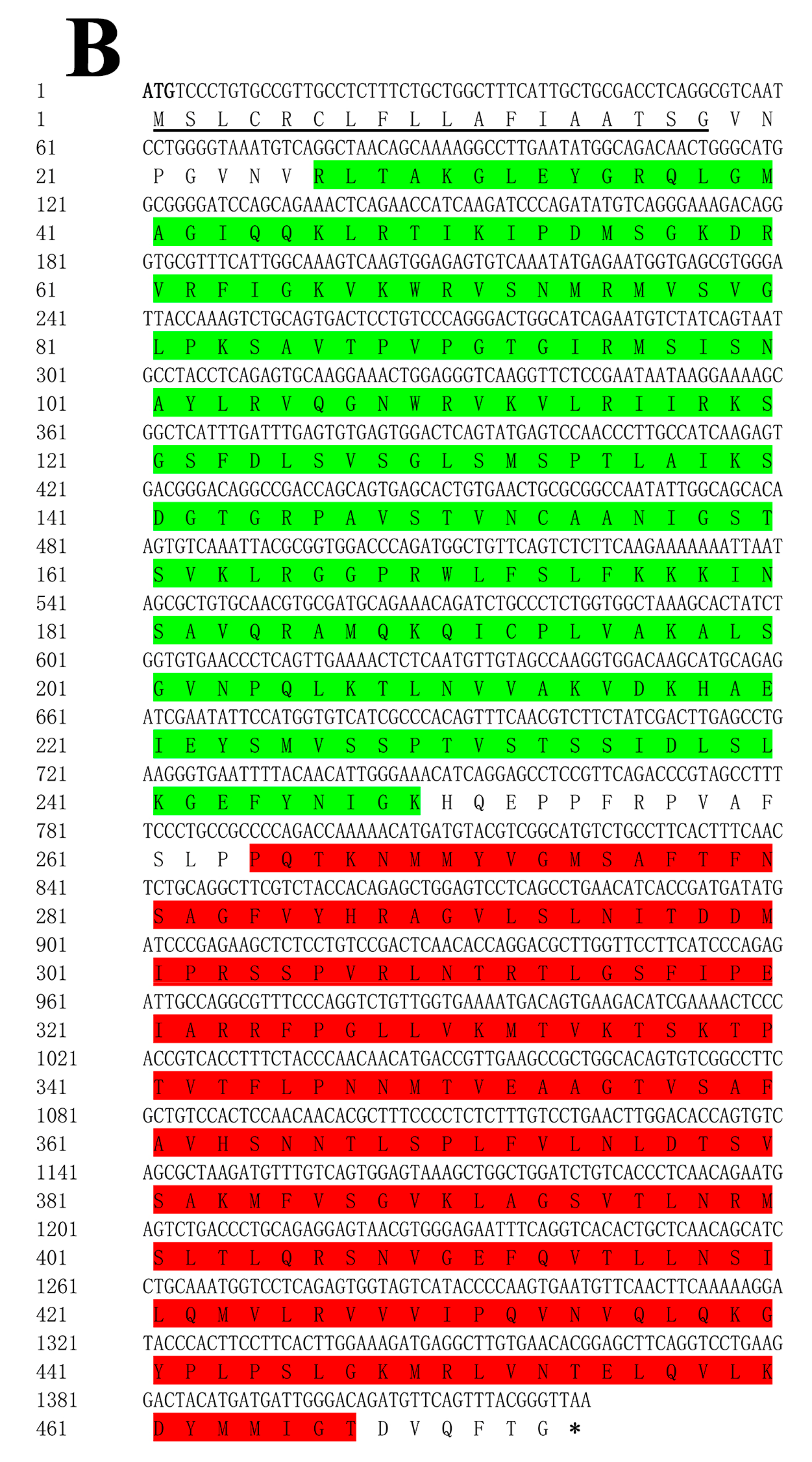
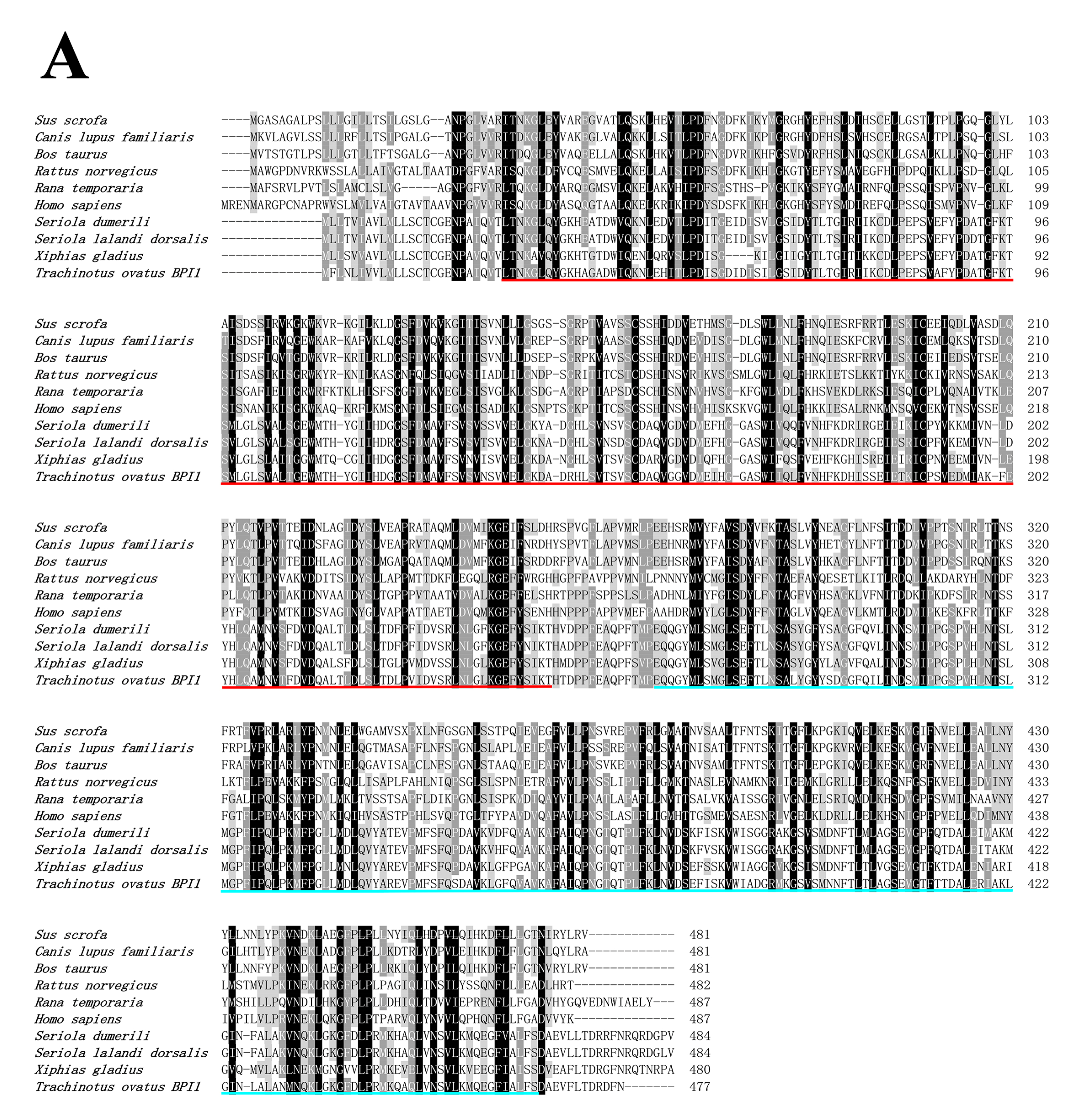
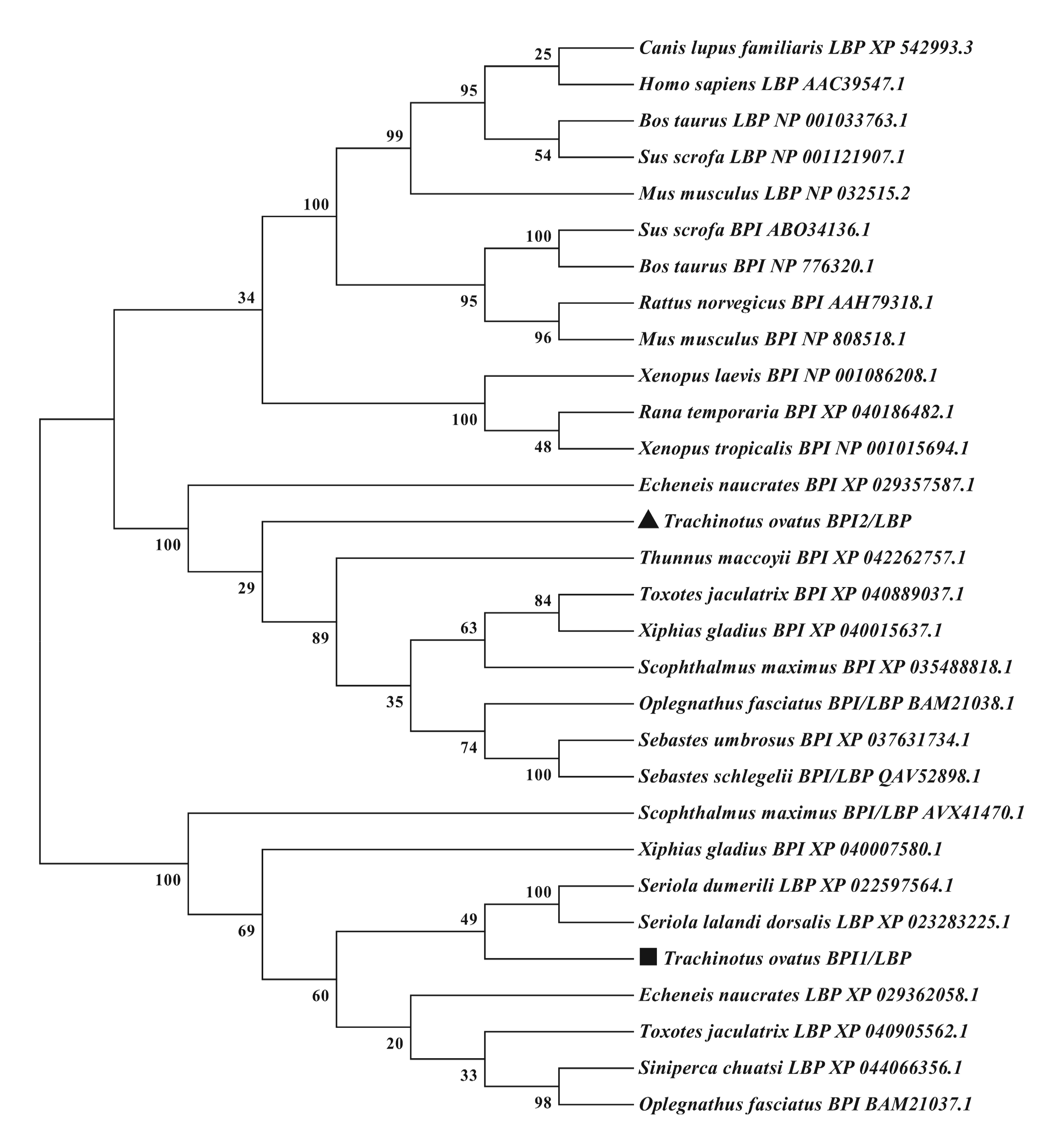
3.1. ToBPI1/LBP and ToBPI2/LBP Sequence Analysis

A sequence analysis of ToBPI1/LBP and ToBPI2/LBP revealed that their ORFs were 1434 bp and 1422 bp, containing 478 and 474 amino acids with molecular weights of 52.33 kDa and 51.49 kDa, and isoelectric points of 4.77 and 10.7, respectively. The signal peptide regions of ToBPI1/LBP and ToBPI2/LBP were 1–17 and 1–18 amino acids, respectively (Figure 1A,B). Both ToBPI1/LBP and ToBPI2/LBP possessed the typical BPI/LBP protein characteristics, including an N-terminal BPI/LBP/CETP domain and a C-terminal BPI/LBP/CETP domain (Figure 1A,B). The blast analysis revealed that ToBPI1/LBP was highly consistent with the greater amberjack Seriola dumerili (86.16%), and ToBPI2/LBP was highly consistent with the southern bluefin tuna Thunnus maccoyii (71.25%). Both genes are not comparable to the mammalian sequences (Figure 2A,B). The phylogenetic tree showed that ToBPI1/LBP and ToBPI2/LBP did not converge into one branch. ToBPI1/LBP is clustered with S. dumerili and S. lalandi dorsalis, while ToBPI2/LBP is distant from other fish, and both genes were far away from other species (Figure 3).

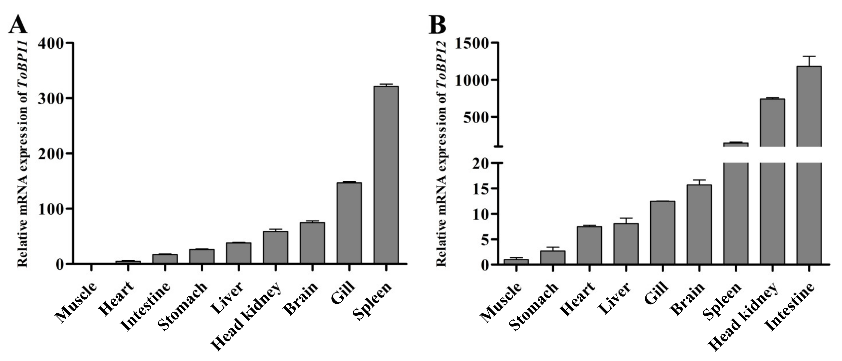
3.2. Tissue Distribution of ToBPI1/LBP and ToBPI2/LBP in Healthy T. ovatus

Both genes were found to be expressed in all tissues. The spleen had the highest levels of ToBPI1/LBP expression, while the gill, brain, cephalic kidney, liver, stomach, intestine, and heart had lower levels (Figure 4A). ToBPI2/LBP expression was highest in the intestine, followed by the head, kidney and spleen, and lowest in the brain, gill, liver, heart and stomach (Figure 4B).

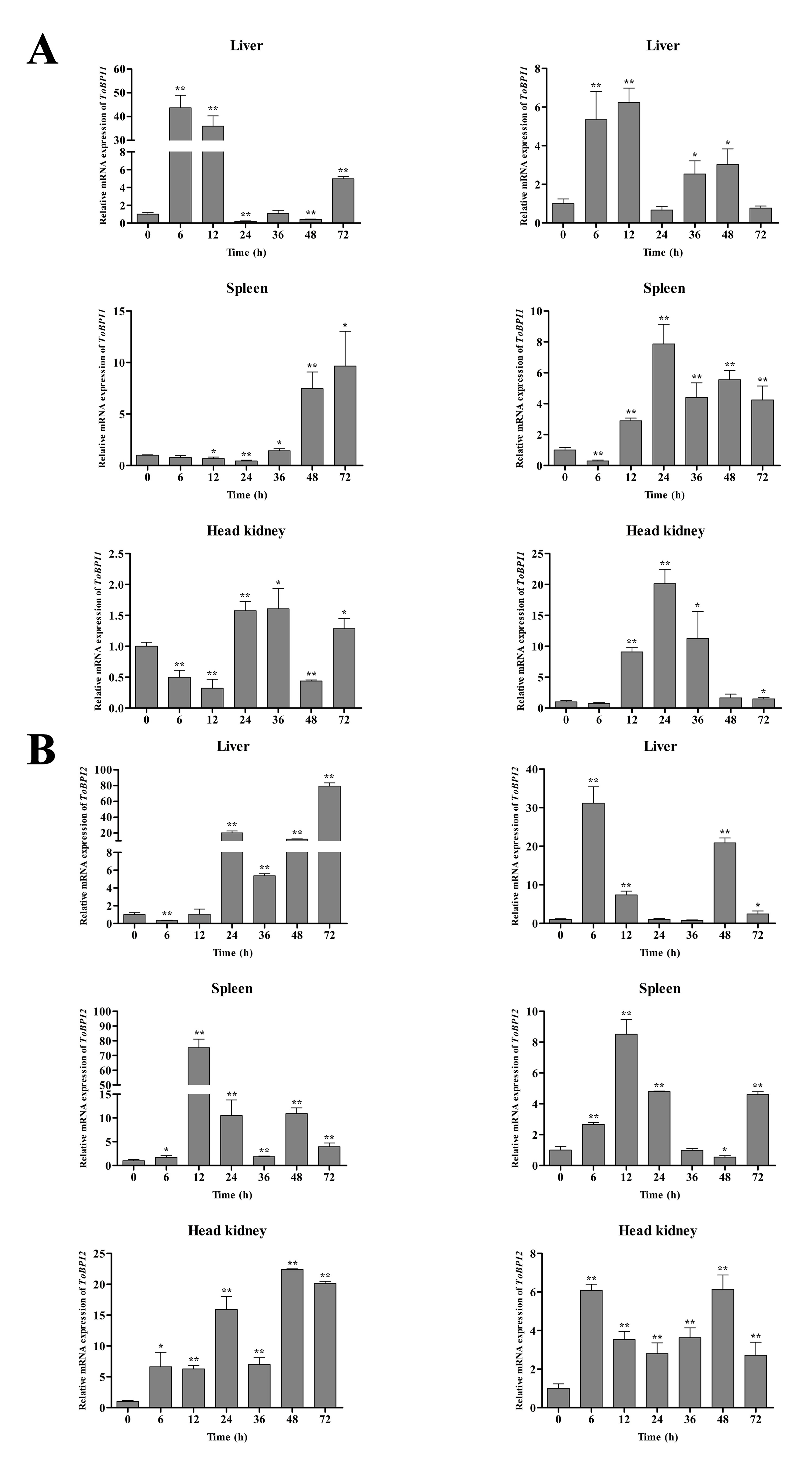
3.3. Expression of ToBPI1/LBP and ToBPI2/LBP in Different Tissues after Bacterial Infection

In the liver, the expression of ToBPI1/LBP and ToBPI2/LBP peaked at 6 h and 72 h, respectively, following the infection with V. alginolyticus (Figure 5). In the spleen, ToBPI1/LBP showed a gradual up-regulation trend and peaked at 72 h. At 12 h, ToBPI2/LBP reached a peak and began to decline. ToBPI1/LBP and ToBPI2/LBP peaked in the head and kidney at 36 h and 48 h, respectively. In the S. agalactiae group, ToBPI1/LBP and ToBPI2/LBP peaked in the liver after infection at 12 h and 6 h, respectively. ToBPI1/LBP and ToBPI2/LBP both tended to decline in the spleen after reaching peaks at 24 h and 12 h, respectively. ToBPI1/LBP and ToBPI2/LBP peaked in the head and kidney at 24 h and 48 h, respectively. These findings implied that ToBPI1/LBP and ToBPI2/LBP played a role in T. ovatus’s immunological protection against bacterial invasion.

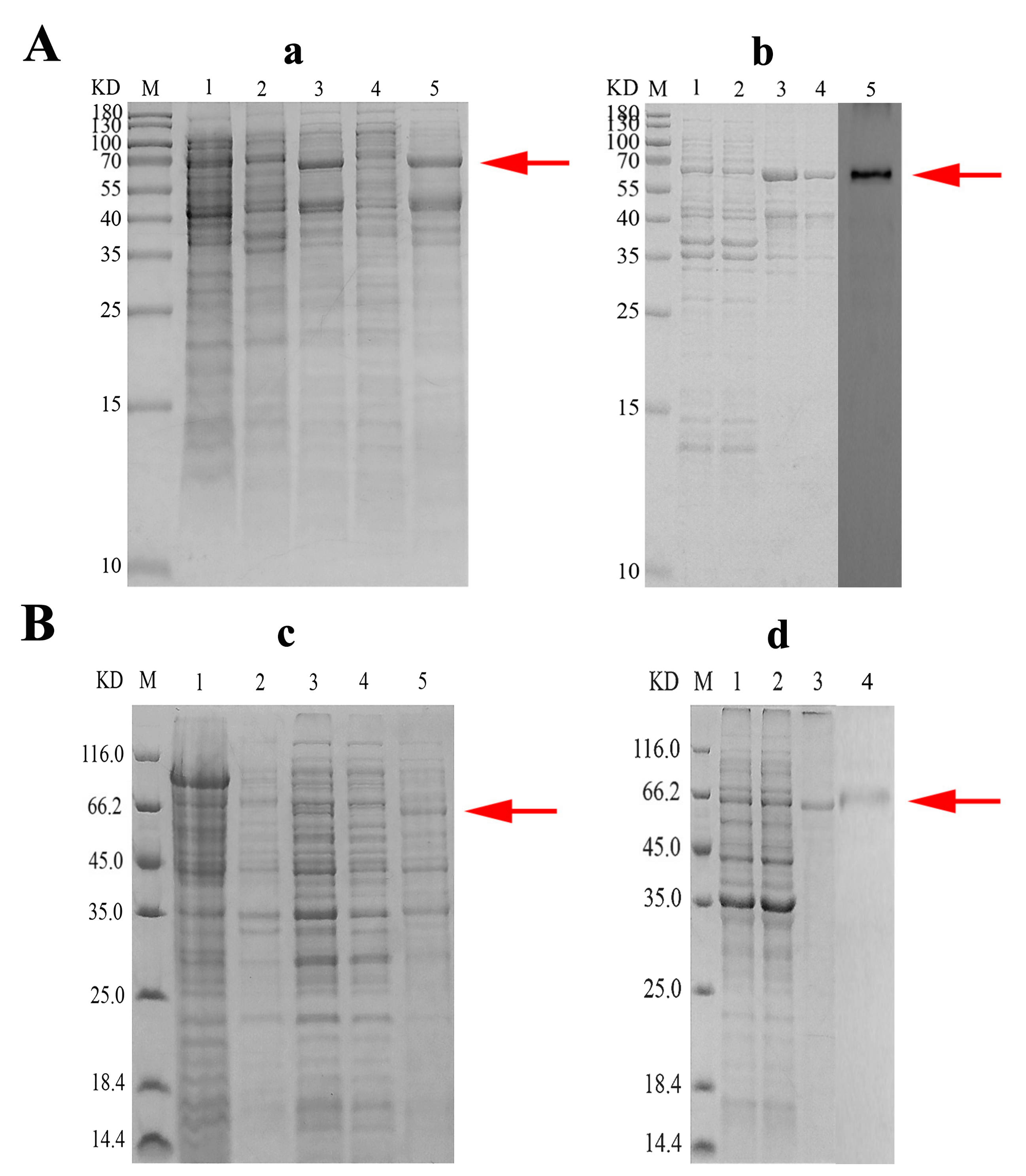
3.4. Recombinant Expression and Purification of rToBPI1/LBP and rToBPI2/LBP

Results bands of about 64.6 kDa and 66.6 kDa were seen (Figure 6a,c), which is consistent with the expected molecular weight results, while no band was found in the non-IPTG induced bacterial solution. The findings in Western blotting revealed a single strip (Figure 6b,d), indicating that it could react specifically with rToBPI1/LBP and rToBPI2/LBP.

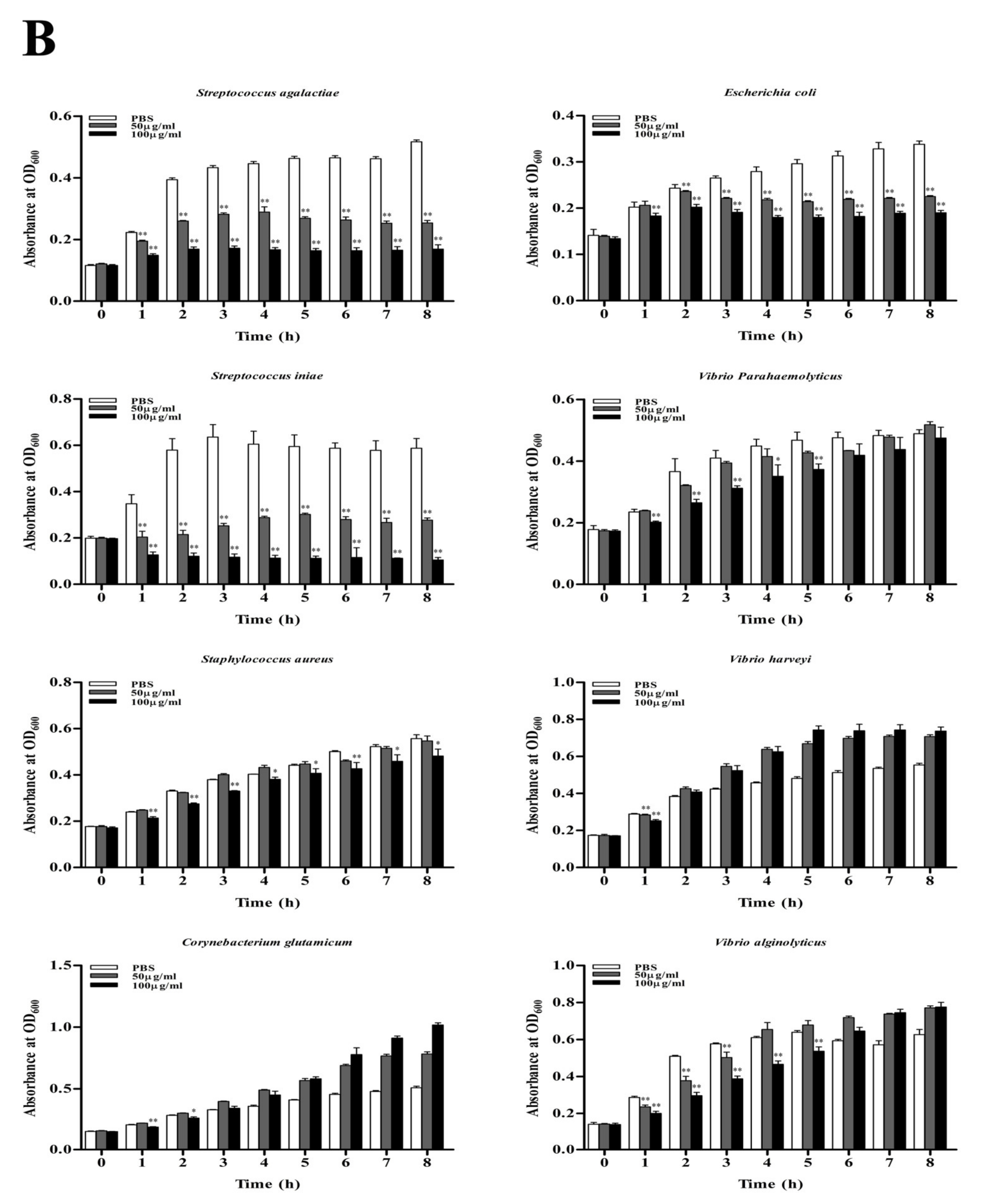
3.5. Antimicrobial Activity

To test the bacteriostatic activity of rToBPI1/LBP and rToBPI2/LBP, the OD600 of Gram-negative bacteria (E. coli, V. alginolyticus, V. parahaemolyticus and V. harveyi) and Gram-positive bacteria (S. aureus, S. agalactiae, S. iniae and C. glutamicum) were measured at various time points (Figure 7A,B). The results demonstrated that, at two doses, rToBPI1/LBP and rToBPI2/LBP significantly inhibited the growth of E. coli, S. agalactiae and S. iniae. The bacteriostatic impact on S. aureus, C. glutamicum, V. parahaemolyticus, V. harveyi and V. alginolyticus all reduced over time when rToBPI1/LBP concentration was 100 μg/mL. The RToBPI1/LBP protein had no antibacterial effects on C. glutamate and V. harveyi after 6 h and 4 h, respectively. When the concentration of rToBPI1 was 50 μg/mL, there was almost no bacteriostatic effect on S. aureus, C. glutamicum and V. harveyi, and the effect on V. parahaemolyticus and V. alginolyticus was subpar (Figure 7A). The bacteriostatic effect on S. aureus, V. parahaemolyticus and V. alginolyticus was obtained at a concentration of 100 μg/mL rToBPI2/LBP, and the bacteriostatic impact waned over time. The RToBPI2/LBP protein had an antibacterial effect on C. glutamicum and V. harveyi only at 2 h and 1 h. When the concentration of rToBPI2/LBP was 50 μg/mL, there was no bacteriostatic effect on S. aureus, C. glutamicum, V. parahaemolyticus and V. Harveyi; there was a bacteriostatic effect on V. alginolyticus only in the first 3 h (Figure 7B).

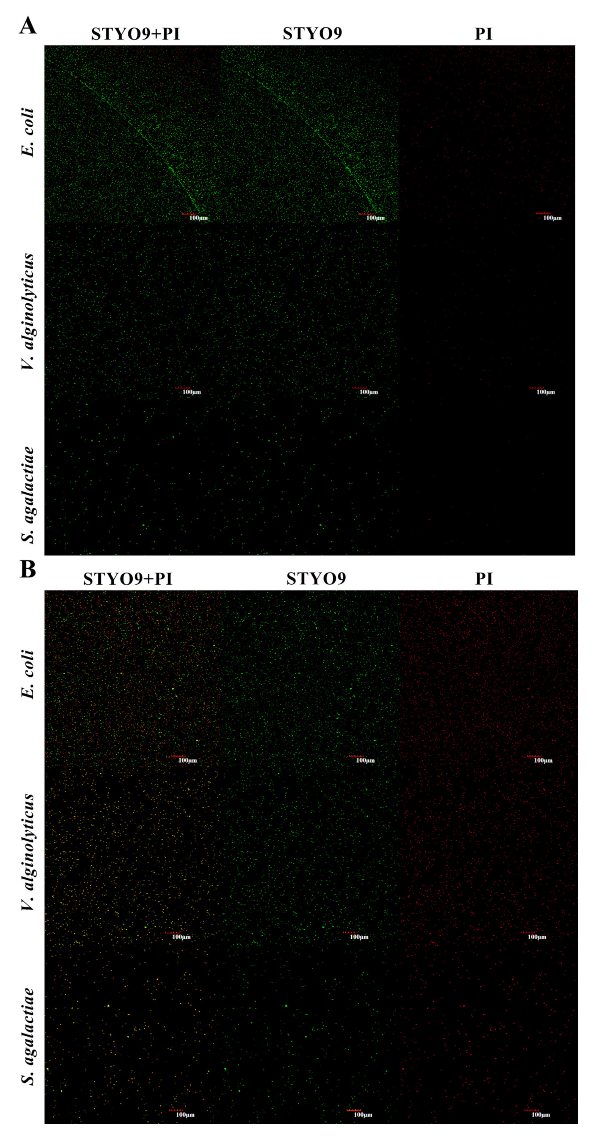
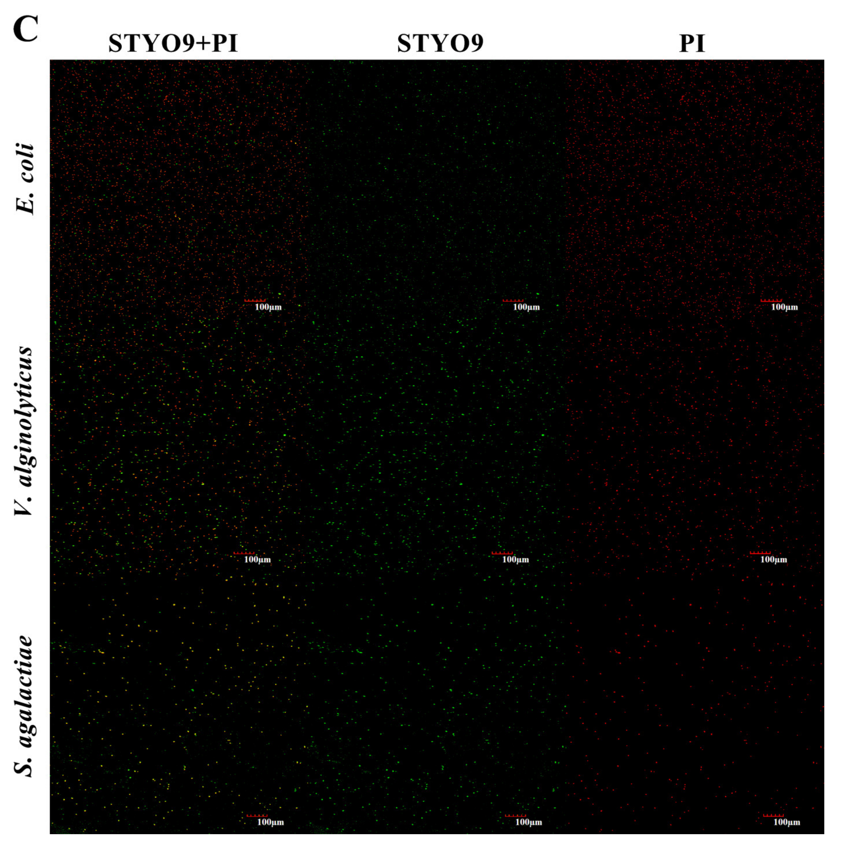
3.6. Permeability of Cell Membrane

The results of fluorescence staining showed that after rToBPI1/LBP and rToBPI2/LBP protein treatment, the red fluorescence (PI) significantly increased (Figure 8B,C), while the PI in the PBS control group did not change (Figure 8A).

4. Discussion

The BPI/LBP antimicrobial peptide is crucial for the host’s resistance to bacterial infection, which was also found in many teleost fish species, and the amount is species-specific [21,22]. In this study, two new BPI/LBP genes (ToBPI1/LBP and ToBPI2/LBP) were cloned from T. ovatus. The amino acid sequence analysis showed they had an N-terminal BPI/LBP/CETP domain and a C-terminal BPI/LBP/CETP domain. According to previous studies, the C-terminal domain may be involved in facilitating LPS binding, while the N-terminal domain is thought to be involved in bactericidal activity and neutralizing LPS [23]. This may be due to the N-terminal domain of BPI/LBP having abundant positively-charged amino acid residues that interact electrostatically with the negatively-charged group of LPS to facilitate the binding of BPI/LBP and LPS [24]. Other bony fish, including blunt snout bream M. amblycephala [3], black rockfish S. schlegelii [12] and Japanese flounder Paralichthys olivaceus [25], have been shown to have identical N-terminal domains and C-terminal domains which perform similar functions.
The present study evaluated the spatial expression of ToBPI1/LBP and ToBPI2/LBP in healthy tissues of golden pompano. The results showed that ToBPI1/LBP was highly expressed in the brain, gill, and spleen, while ToBPI2/LBP was highly expressed in the spleen, cephalic kidney, and intestine (Figure 4). In other fish, BPI/LBP also showed high expression in these immune-related organizations. In M. amblycephala, BPI/LBP is highly expressed in the kidney [3]. BPI/LBP is highly expressed in the spleen and kidney in Oplegnathus fasciatus [6]. In S. maximus, BPI/LBP is highly expressed in the spleen [14]. ToBPI1/LBP expression was the highest in the spleen, which resembled the results of S. schlegelii [12]. The spleen is considered the major immune organ for the generation of adaptive immune responses [26,27], which is regarded as a crucial immunological organ in bony fish because it has lymphocytes and macrophages that can initiate immune responses [28]. The intestine, followed by the head, kidney and spleen, all had the highest levels of ToBPI2/LBP expression. As a crucial mucosal immunological organ of fish, the intestinal tract serves as a fish’s defense against the external environment. It is essential for fending against pathogen invasion and preventing the entry of microbial and bacterial toxins into the systemic circulation. We speculate that the differential expression of ToBPI1/LBP and ToBPI2/LBP in healthy tissues may be related to their interaction with T. ovatus defense against pathogen invasion in a complex aquatic environment, but further research is required to confirm this hypothesis.
After infection with S. agalactiae and V. alginolyticus, the expressions of ToBPI1/LBP and ToBPI2/LBP in the liver, head, kidney and spleen increased considerably. Similarly, the head, kidney and spleen of S. schlegelii had much higher levels of the BPI/LBP homolog after being exposed to the Gram-positive bacterium S. iniae [12]. P. olivaceus also demonstrated a similar pattern in the spleen, head, kidney, intestine, gill, and liver after exposure to S. iniae [25]. These findings suggest that ToBPI1/LBP and ToBPI2/LBP are involved in the immune defense against pathogen invasion in T. ovatus.
Numerous investigations have demonstrated that BPI/LBP has strong antibacterial activity against Gram-negative bacteria but none against Gram-positive bacteria at this time [12,17]. In this study, rToBPI1/LBP and rToBPI2/LBP proteins were also confirmed to have significant inhibitory effects on the growth and proliferation of Gram-negative bacteria, which is consistent with the results of S. schlegelii, C. semilaevis and Paa yunnanensis [12,13,29]. The difference is that the rToBPI1/LBP and rToBPI2/LBP proteins also showed a significant antibacterial effect against the Gram-positive bacteria S. agalactiae and S. iniae. It has been discovered in earlier studies that the recombinant protein made from the human BPI N-terminal analog can inhibit the growth of the L-form S. aureus [30], and our study findings are compatible with these results.
According to various studies, LPS is one of the primary components of Gram-negative bacteria’s cell walls, and BPI can damage cell membranes by binding to LPS in Gram-negative bacteria [31], eventually killing the bacteria. Recombinant ToBPI1/LBP and ToBPI2/LBP proteins were found to increase the permeability of Gram-negative bacterial and Gram-positive bacterial cell membranes in a laboratory experiment. Additionally, it was discovered while researching C. semilaevis, that Pseudomonas fluorescens treated with recombinant CsBPI caused a considerable increase in membrane permeability [13]. However, no fish studies have demonstrated that BPI/LBP directly eliminates Gram-positive bacteria. Similarly, no research has shown that BPI/LBP can harm cell membranes and increase membrane permeability in Gram-positive bacteria. In some investigations, Lipoteichoic acid (LTA), a product of Gram-positive bacteria’s cell walls, has also been discovered to be a new BPI ligand [32]. Studies have shown that both LTA and LPS use N-terminal lipid binding pockets when competing for binding to BPI. It can bind hydrophobic acyl chains, and acyl chains are dependent on BPI and LTA binding, just as they are crucial to the binding of BPI and LPS [32]. Therefore, we speculate that BPI/LBP may interact with LTA to affect the cell membrane of Gram-positive bacteria. However, more research is required to determine the underlying mechanism by which the recombinant ToBPI1/LBP and ToBPI2/LBP proteins affect Gram-positive bacteria.

5. Conclusions

In conclusion, the ORF sequences of ToBPI1 and ToBPI2 were successfully cloned at 1434 bp (478 aa) and 1422 bp (474 aa) in length, respectively. A protein domain prediction analysis showed that ToBPI1 and ToBPI2 both contained an N-terminal signal peptide, an N-terminal domain and a C-terminal domain. The homology comparison showed that ToBPI1/LBP was highly consistent with Seriola dumerili (86.16%), and ToBPI2/LBP was highly consistent with Thunnus maccoyii (71.25%). A phylogenetic analysis showed that ToBPI1 and ToBPI2 were not clustered together. Real-time PCR analysis showed that ToBPI1 and ToBPI2 were expressed at the highest levels in the spleen and intestine of healthy golden pompano, respectively. The expression levels of ToBPI1 and ToBPI2 in immune-related tissues were significantly upregulated after stimulation with S. agalactiae and V. alginolyticus. In vitro bacteriostasis tests showed that rToBPI1 and rToBPI2 could effectively inhibit the growth of E. coli, S. agalactiae and V. alginolyticus. Cell membrane permeability experiments showed that rToBPI1 and rToBPI2 could increase bacterial cell membrane permeability. The present findings offer new perspectives and may serve as important references for the immune defense effect of ToBPI1/LBP and ToBPI2/LBP in T. ovatus.

Author Contributions

Z.-C.B.: Methodology, Investigation, Writing (original draft), Formal analysis. X.-H.C.: Writing (original draft); Supervision, Funding acquisition, Conceptualization. K.A.T.: Investigation. Y.-D.W.: Methodology. Z.H.: Visualization. K.Y.K.: Writing (review and editing), Resources, Funding acquisition. P.X.: Project administration. All authors have read and agreed to the published version of the manuscript.

Funding

This work was sponsored by the Science and Technology Base and Talent Project of Guangxi Province (Gui Ke AD19245059), Natural Science Foundation of Guangxi Province (2019GXNSFBA245062), High-level Talents Scientific Research Start-Up Fund Project of Beibu Gulf University (2019KYQD15), Guangxi Middle-aged and Young Teachers’ Basic Ability Promotion Project (2019KY0464), Guangxi Key Laboratory of Beibu Gulf Marine Biodiversity Conservation (2021ZC02), ASEAN Talent Program (ATYSP2022008), and Marine Science Program for Guangxi First-Class Discipline, Beibu Gulf University (TRA001, DRA002, ESB010).

Data Availability Statement

The data presented in this study are available in the article.

Conflicts of Interest

The authors declare no conflict of interest.

References

  1. Shao, Y.; Li, C.; Che, Z.; Zhang, P.; Zhang, W.; Duan, X.; Li, Y. Cloning and characterization of two lipopolysaccharide-binding protein/bactericidal permeability-increasing protein (LBP/BPI) genes from the sea cucumber Apostichopus japonicus with diversified function in modulating ROS production. Dev. Comp. Immunol. 2015, 52, 88–97. [Google Scholar] [CrossRef]
  2. Mao, Y.; Zhou, C.; Zhu, L.; Huang, Y.; Yan, T.; Fang, J.; Zhu, W. Identification and expression analysis on bactericidal permeability-increasing protein (BPI)/lipopolysaccharide-binding protein (LBP) of ark shell, Scapharca broughtonii. Fish Shellfish. Immunol. 2013, 35, 642–652. [Google Scholar] [CrossRef]
  3. Tang, L.; Liang, Y.; Jiang, Y.; Liu, S.; Zhang, F.; He, X.; Wang, T.; Zhou, Y.; Zhong, H.; Yan, J. Identification and expression analysis on bactericidal permeability-increasing protein/lipopolysaccharide-binding protein of blunt snout bream, Megalobrama amblycephala. Fish Shellfish. Immunol. 2015, 45, 630–640. [Google Scholar] [CrossRef]
  4. Su, J.; Li, H.; Hu, J.; Wang, D.; Zhang, F.; Fu, Z.; Han, F. LcCCL28-25, Derived from Piscine Chemokine, Exhibits Antimicrobial Activity against Gram-Negative and Gram-Positive Bacteria In Vitro and In Vivo. Microbiol. Spectr. 2022, 10, e0251521. [Google Scholar] [CrossRef]
  5. Bin Hafeez, A.; Jiang, X.; Bergen, P.J.; Zhu, Y. Antimicrobial Peptides: An Update on Classifications and Databases. Int. J. Mol. Sci. 2021, 22, 11691. [Google Scholar] [CrossRef]
  6. Kim, J.W.; Gerwick, L.; Park, C.I. Molecular identification and expression analysis of two distinct BPI/LBPs (bactericidal permeability-increasing protein/LPS-binding protein) from rock bream, Oplegnathus fasciatus. Fish Shellfish. Immunol. 2012, 33, 75–84. [Google Scholar] [CrossRef] [PubMed]
  7. Weiss, J. Bactericidal/permeability-increasing protein (BPI) and lipopolysaccharide-binding protein (LBP): Structure, function and regulation in host defence against Gram-negative bacteria. Biochem. Soc. Trans. 2003, 31, 785–790. [Google Scholar] [CrossRef] [PubMed]
  8. Schultz, H.; Weiss, J.P. The bactericidal/permeability-increasing protein (BPI) in infection and inflammatory disease. Clin. Chim. Acta 2007, 384, 12–23. [Google Scholar] [CrossRef] [PubMed]
  9. Zhou, Z.P.; Xia, X.Y.; Guo, Q.S.; Xu, C. Bactericidal/permeability-increasing protein originates in both the testis and the epididymis and localizes in mouse spermatozoa. Asian J. Androl. 2014, 16, 309–313. [Google Scholar] [CrossRef] [PubMed]
  10. Wittmann, I.; Schonefeld, M.; Aichele, D.; Groer, G.; Gessner, A.; Schnare, M. Murine bactericidal/permeability-increasing protein inhibits the endotoxic activity of lipopolysaccharide and gram-negative bacteria. J. Immunol. 2008, 180, 7546–7552. [Google Scholar] [CrossRef] [PubMed]
  11. Nishimura, H.; Gogami, A.; Miyagawa, Y.; Nanbo, A.; Murakami, Y.; Baba, T.; Nagasawa, S. Bactericidal/permeability-increasing protein promotes complement activation for neutrophil-mediated phagocytosis on bacterial surface. Immunology 2001, 103, 519–525. [Google Scholar] [CrossRef] [PubMed]
  12. Lee, S.; Elvitigala, D.A.S.; Lee, S.; Kim, H.C.; Park, H.C.; Lee, J. Molecular characterization of a bactericidal permeability-increasing protein/lipopolysaccharide-binding protein from black rockfish (Sebastes schlegelii): Deciphering its putative antibacterial role. Dev. Comp. Immunol. 2017, 67, 266–275. [Google Scholar] [CrossRef] [PubMed]
  13. Sun, Y.Y.; Sun, L. A Teleost Bactericidal Permeability-Increasing Protein Kills Gram-Negative Bacteria, Modulates Innate Immune Response, and Enhances Resistance against Bacterial and Viral Infection. PLoS ONE 2016, 11, e0154045. [Google Scholar] [CrossRef]
  14. Zhou, S.; Jiang, G.; Zhu, Y.; Liu, L.; Liu, D.; Diao, J.; Liu, H.; Xiu, Y. Molecular identification and function analysis of bactericidal permeability-increasing protein/LPS-binding protein 1 (BPI/LBP1) from turbot (Scophthalmus maximus). Fish Shellfish. Immunol. 2019, 87, 499–506. [Google Scholar] [CrossRef]
  15. Cai, X.H.; Peng, Y.H.; Wang, Z.C.; Huang, T.; Xiong, X.Y.; Huang, Y.C.; Wang, B.; Xu, L.W.; Wu, Z.H. Characterization and identification of streptococci from golden pompano in China. Dis. Aquat. Organ 2016, 119, 207–217. [Google Scholar] [CrossRef] [PubMed]
  16. Gao, J.; Guo, H.-Y.; Liu, M.-J.; Zhu, K.-C.; Liu, B.; Liu, B.-S.; Zhang, N.; Jiang, S.-G.; Zhang, D.-C. Transcriptome Analysis of the Immune Process of Golden Pompano (Trachinotus ovatus) Infected with Streptococcus agalactiae. Fishes 2023, 8, 52. [Google Scholar] [CrossRef]
  17. Wu, Y.; Du, H.H.; Zhu, L.; Zhao, N.; Zhang, S.; Cao, Z.; Zhou, Y.; Sun, Y. Bactericidal permeability-increasing protein/LPS-binding protein (BPI/LBP) enhances resistance of golden pompano Trachinotus ovatus against bacterial infection. Fish Shellfish. Immunol. 2022, 131, 872–880. [Google Scholar] [CrossRef] [PubMed]
  18. Zhou, Y.; Lei, Y.; Cao, Z.; Chen, X.; Sun, Y.; Xu, Y.; Guo, W.; Wang, S.; Liu, C. A β-defensin gene of Trachinotus ovatus might be involved in the antimicrobial and antiviral immune response. Dev. Comp. Immunol. 2019, 92, 105–115. [Google Scholar] [CrossRef] [PubMed]
  19. Ding, M.; Fan, J.; Wang, W.; Wang, H.; Liu, H. Molecular characterization, expression and antimicrobial activity of complement factor D in Megalobrama amblycephala. Fish Shellfish. Immunol. 2019, 89, 43–51. [Google Scholar] [CrossRef]
  20. Liu, Y.; Zha, H.; Han, X.; Yu, S.; Chai, Y.; Zhong, J.; Zhu, Q. Molecular characterization and functional analysis of the bactericidal permeability-increasing protein/LPS-binding protein (BPI/LBP) from roughskin sculpin (Trachidermus fasciatus). Dev. Comp. Immunol. 2021, 123, 104133. [Google Scholar] [CrossRef]
  21. Inagawa, H.; Honda, T.; Kohchi, C.; Nishizawa, T.; Yoshiura, Y.; Nakanishi, T.; Yokomizo, Y.; Soma, G. Cloning and characterization of the homolog of mammalian lipopolysaccharide-binding protein and bactericidal permeability-increasing protein in rainbow trout Oncorhynchus mykiss. J. Immunol. 2002, 168, 5638–5644. [Google Scholar] [CrossRef]
  22. Kono, T.; Sakai, M. Molecular cloning of a novel bactericidal permeability-increasing protein/lipopolysaccharide-binding protein (BPI/LBP) from common carp Cyprinus carpio L. and its expression. Mol. Immunol. 2003, 40, 269–278. [Google Scholar] [CrossRef]
  23. Theprungsirikul, J.; Skopelja-Gardner, S.; Rigby, W.F.C. Killing three birds with one BPI: Bactericidal, opsonic, and anti-inflammatory functions. J. Transl. Autoimmun. 2021, 4, 100105. [Google Scholar] [CrossRef] [PubMed]
  24. Beamer, L.J.; Carroll, S.F.; Eisenberg, D. The BPI/LBP family of proteins: A structural analysis of conserved regions. Protein Sci. 1998, 7, 906–914. [Google Scholar] [CrossRef] [PubMed]
  25. Nam, B.H.; Ahn, K.J.; Kim, Y.O.; Kong, H.J.; Kim, W.J.; Kim, H.S.; Lee, S.J.; Kim, K.K. Molecular cloning and characterization of LPS-binding protein/bactericidal permeability-increasing protein (LBP/BPI) from olive flounder, Paralichthys olivaceus. Vet. Immunol. Immunopathol. 2010, 133, 256–263. [Google Scholar] [CrossRef] [PubMed]
  26. Flajnik, M.F. A cold-blooded view of adaptive immunity. Nat. Rev. Immunol. 2018, 18, 438–453. [Google Scholar] [CrossRef] [PubMed]
  27. Ofek, T.; Lalzar, M.; Izhaki, I.; Halpern, M. Intestine and spleen microbiota composition in healthy and diseased tilapia. Anim. Microbiome 2022, 4, 50. [Google Scholar] [CrossRef]
  28. Press, C.M.; Evensen, Ø. The morphology of the immune system in teleost fishes. Fish Shellfish. Immunol. 1999, 9, 309–318. [Google Scholar] [CrossRef]
  29. Jiang, C.; Wang, S. Identification and functional characterization of bactericidal permeability/increasing protein (BPI) from frog Nanorana yunnanensis (Paa yunnanensis). Dev. Comp. Immunol. 2022, 137, 104517. [Google Scholar] [CrossRef]
  30. Horwitz, A.H.; Williams, R.E.; Liu, P.S.; Nadell, R. Bactericidal/permeability-increasing protein inhibits growth of a strain of Acholeplasma laidlawii and L forms of the gram-positive bacteria Staphylococcus aureus and Streptococcus pyogenes. Antimicrob. Agents Chemother. 1999, 43, 2314–2316. [Google Scholar] [CrossRef]
  31. Wiese, A.; Brandenburg, K.; Carroll, S.F.; Rietschel, E.T.; Seydel, U. Mechanisms of action of bactericidal/permeability-increasing protein BPI on reconstituted outer membranes of gram-negative bacteria. Biochemistry 1997, 36, 10311–10319. [Google Scholar] [CrossRef] [PubMed]
  32. Bulow, S.; Zeller, L.; Werner, M.; Toelge, M.; Holzinger, J.; Entzian, C.; Schubert, T.; Waldow, F.; Gisch, N.; Hammerschmidt, S.; et al. Bactericidal/Permeability-Increasing Protein Is an Enhancer of Bacterial Lipoprotein Recognition. Front. Immunol. 2018, 9, 2768. [Google Scholar] [CrossRef] [PubMed]
Figure 1. Sequence analysis of ToBPI1/LBP and ToBPI2/LBP. (A,B): nucleotide sequence and derived amino acid sequence of ToBPI1/LBP and ToBPI2/LBP. Signal peptide sequences are indicated by horizontal lines. The N-terminal domain is represented by green, and the C-terminal domain is represented by red.
Genes 14 00826 g001aGenes 14 00826 g001b
Figure 2. Comparison of ToBPI1/LBP (A) and ToBPI2/LBP (B) with the homologous sequences of BPI/LBP. The black part at the bottom has the same sequence, and the grey part has high repeatability. The N-terminal domains are represented by red lines and C-terminal domains by blue lines.
Genes 14 00826 g002aGenes 14 00826 g002b
Figure 3. The amino acid sequences of ToBPI1/LBP and ToBPI2/LBP were compared with the known sequences of BPI, LBP and BPI/LBP from other species. The NJ method was used to construct an evolutionary tree. Boot the NJ tree 1000 times to check the repeatability of the results. ToBPI1/LBP is denoted by a black square, and ToBPI2/LBP is indicated by a black triangle.
Genes 14 00826 g003
Figure 4. The expression of ToBPI1/LBP (A) and ToBPI2/LBP (B) in healthy tissues of T. ovatus was detected by real-time quantitative PCR. For comparison, the expression level in muscles was set at 1.0. Each T-line represents the mean standard deviation (n = 3).
Genes 14 00826 g004
Figure 5. The expressions of ToBPI1/LBP (A) and ToBPI2/LBP (B) in tissues of T. ovatus were detected by quantitative real-time PCR at different time points after infection with V. alginolyticus (left) and S. agalactiae (right). For comparison, the expression level at 0 h for each group was set as 1. Each T-line represents the mean standard deviation (n = 3). There was no significant difference p > 0.05. The difference was considered significant when * p < 0.05, highly significant when ** p < 0.01.
Genes 14 00826 g005
Figure 6. Expression and purification of rToBPI1/LBP (A) and rToBPI2/LBP (B); (a,c): Lane M: Protein molecular standard; Lane 1: pSumo-mut and pET-32a no-load plasmid; Lane 2: rToBPI1/LBP and rToBPI2/LBP without IPTG induction; Lane 3: rToBPI1/LBP and rToBPI2/LBP after 4 h induction by IPTG; Lane 4: Supernatant in rToBPI1/LBP and rToBPI2/LBP lysate; Lane 5: Precipitation in rToBPI1/LBP and rToBPI2/LBP lysate. (a,c): Lane M: Protein molecular standard; Lane 1: pSumo-mut and pET-32a no-load plasmid; Lane 2: rToBPI1/LBP and rToBPI2/LBP without IPTG induction; Lane 3: rToBPI1/LBP and rToBPI2/LBP after 4 h induction by IPTG; Lane 4: Supernatant in rToBPI1/LBP and rToBPI2/LBP lysate; Lane 5: Precipitation in rToBPI1/LBP and rToBPI2/LBP lysate. (b): Lane M: Protein molecular standard; Lane 1: Precipitation in rToBPI1/LBP lysate; Lane 2: Pre-flow purified solution; Lane 3–4: Recombinant rToBPI1/LBP purified using purified column; Lane 5: Western blot of rToBPI1/LBP; (d): Lane M: Protein molecular standard; Lane 1: Precipitation in rToBPI2/LBP lysate; Lane 2: Pre-flow purified solution; Lane 3: Recombinant rToBPI2/LBP purified using purified column; Lane 4: Western blot of rToBPI2/LBP;.
Genes 14 00826 g006
Figure 7. Bacteriostasis of rToBPI1/LBP (A) and rToBPI1/LBP (B) against Gram-positive bacteria S. agalactiae, S. iniae, S. aureus and C. glutamicum as well as Gram-negative bacteria E. coli, V. parahaemolyticus, V. harveyi and V. alginolyticus at 50 μg/mL and 100 μg/mL. Gram-positive bacteria are on the left panel, whereas Gram-negative bacteria are on the right. The data shown are the average of three independent experiments. The difference was significant when * p < 0.05, highly significant when ** p < 0.01.
Genes 14 00826 g007aGenes 14 00826 g007b
Figure 8. E. coli, V. alginolyticus and S. agalactiae treated with rToBPI1/LBP and rToBPI2/LBP proteins were used to observe the permeability of bacterial cell membrane. PBS was used as a control. (A) the bacteria treated with PBS; (B) rToBPI1/LBP treated bacteria; (C) rToBPI2/LBP treated bacteria; STYO9 is shown in green, and PI is shown in red.
Genes 14 00826 g008aGenes 14 00826 g008b
Table 1. Primers used in this study.
Primers NameSequence 5′–3′Purpose
ToBPI1/LBP -FATGTTTCTAAACCTAATAGTAGTGCTCATGCCloning
ToBPI1/LBP -RTCAGTTGAAGTCTCTGTCTGTTAGGCloning
ToBPI2/LBP -FATGTCCCTGTGCCGTTGCloning
ToBPI2/LBP -RTTAACCCGTAAACTGAACATCTGTCCloning
ToBPI1/LBP -FCCTTTGCCATCCAGCCAAATReal-time PCR
ToBPI1/LBP -RCCACTTCACTTCCTGCCAACReal-time PCR
ToBPI2/LBP -FGCCCACAGTTTCAACGTCTTReal-time PCR
ToBPI2/LBP -RGTGTTGAGTCGGACAGGAGAReal-time PCR
β-action-FTACGAGCTGCCTGACGGACAReal-time PCR
β-action-RGGCTGTGATCTCCTTCTGCReal-time PCR
mToBPI1 LBP-FCGCGGATCCGAAAATCCTGCAATACAAGTCProkaryotic expression
mToBPI1/LBP-RCCGCTCGAGGTTGAAGTCTCTGTCTGTTAGGProkaryotic expression
mToBPI2/LBP-FCGGAATTCGTCAATCCTGGGGTAAATGTProkaryotic expression
mToBPI2/LBP-RCCCTCGAGACCCGTAAACTGAACATCTGTProkaryotic expression
Disclaimer/Publisher’s Note: The statements, opinions and data contained in all publications are solely those of the individual author(s) and contributor(s) and not of MDPI and/or the editor(s). MDPI and/or the editor(s) disclaim responsibility for any injury to people or property resulting from any ideas, methods, instructions or products referred to in the content.

