Article

Chronic Stress Alters Hippocampal Renin-Angiotensin-Aldosterone System Component Expression in an Aged Rat Model of Wolfram Syndrome

1
Laboratory Animal Centre, Institute of Biomedicine and Translational Medicine, University of Tartu, 14B Ravila Street, 50411 Tartu, Estonia
2
Department of Physiology, Institute of Biomedicine and Translational Medicine, University of Tartu, 19 Ravila Street, 50411 Tartu, Estonia
*
Author to whom correspondence should be addressed.
Genes 2023, 14(4), 827; https://doi.org/10.3390/genes14040827
Submission received: 28 November 2022 / Revised: 21 March 2023 / Accepted: 27 March 2023 / Published: 30 March 2023
(This article belongs to the Section Human Genomics and Genetic Diseases)

Abstract

:
Biallelic mutations in the gene encoding WFS1 underlie the development of Wolfram syndrome (WS), a rare neurodegenerative disorder with no available cure. We have previously shown that Wfs1 deficiency can impair the functioning of the renin-angiotensin-aldosterone system (RAAS). The expression of two key receptors, angiotensin II receptor type 2 (Agtr2) and bradykinin receptor B1 (Bdkrb1), was downregulated both in vitro and in vivo across multiple organs in a rat model of WS. Here, we show that the expression of key RAAS components is also dysregulated in neural tissue from aged WS rats and that these alterations are not normalized by pharmacological treatments (liraglutide (LIR), 7,8-dihydroxyflavone (7,8-DHF) or their combination). We found that the expression of angiotensin II receptor type 1a (Agtr1a), angiotensin II receptor type 1b (Agtr1b), Agtr2 and Bdkrb1 was significantly downregulated in the hippocampus of WS animals that experienced chronic experimental stress. Treatment-naïve WS rats displayed different gene expression patterns, underscoring the effect of prolonged experiment-induced stress. Altogether, we posit that Wfs1 deficiency disturbs RAAS functioning under chronic stressful conditions, thereby exacerbating neurodegeneration in WS.

1. Introduction

Wolfram syndrome (WS; Appendix A includes a list of abbreviations used) is a rare monogenic neurodegenerative disease caused by biallelic mutations in the gene encoding the transmembrane glycoprotein Wolframin (WFS1). Disease manifestation typically begins with juvenile-onset diabetes mellitus, diabetes insipidus and loss of vision (due to optic nerve atrophy) and is often accompanied by sensorineural deafness and neuropsychiatric abnormalities, among other complications [1,2]. The incidence can vary by ethnicity, ranging from 1/770,000 in the United Kingdom to 1/68,000 in Lebanon, for instance [3,4].
Wfs1 is broadly expressed in several tissues, with higher levels in the brain, pancreas, lungs, heart and retina [5,6,7,8]. WFS1 is primarily involved in regulating Ca2+ homeostasis and the endoplasmic reticulum (ER) stress response [9,10]. Additionally, Wfs1 deficiency is associated with disruptions in mitochondrial activity, including changes in mitochondrial dynamics and degradation rate [11]. Several unfolded protein response modulators are localized in mitochondria-associated ER membranes (MAMs); these structures facilitate ER-mitochondria interactions that are critical for regulating several functions, including Ca2+ signaling and metabolism. MAM dysfunction can directly impact cell survival and has been implicated in various metabolic and neurodegenerative disorders. WFS1 also localizes in MAMs, and its absence in fibroblasts results in Ca2+ exchange disturbances and reduced ER-mitochondria contact formation in vitro [12,13].
While there are currently no curative treatments available for WS, drug-repurposing efforts have identified several promising candidates, including ER stress modulators (e.g., valproate (VPA), originally a first-choice anti-epileptic drug), chemical chaperones (e.g., sigma-1 receptor (S1R) agonists), and antidiabetics (e.g., glucagon-like peptide 1 receptor (GLP-1R) agonists). For instance, S1R agonists restored mitochondrial function and alleviated behavioral deficits in WS animal models [14]. VPA was shown to induce WFS1 expression, modulate the ER stress response and reduce apoptosis in vitro [15,16], as well as ameliorate glucose tolerance in WS mice [17]. Similarly, dantrolene (a skeletal muscle relaxant) suppressed ER stress-mediated cell death in both in vitro and in vivo WS models [18]. VPA and dantrolene are also already being explored in clinical trials (clinical trial identifiers: NCT03717909/NCT04940572 and NCT02829268, respectively; [19]). Interestingly, some drug candidates from across the neurodegenerative spectrum have also demonstrated disease-modifying potential in both in vivo and in vitro WS models. Riluzole, one of the few drugs approved for the treatment of amyotrophic lateral sclerosis (ALS), regulated aberrant glutamate transporter expression in Wfs1-deficient cerebral organoids, thereby restoring synapse formation and functionality. It also improved spatial memory and depressive behavior in Wfs1 conditional knock-out mice [20]. A combination of 4-phenylbutyrate and tauroursodeoxycholic acid, also recently approved in the United States for the treatment of ALS [21], increased WFS1 levels, alleviated ER stress and inhibited cellular apoptosis in patient-derived induced pluripotent stem cells. Moreover, this combination also stimulated insulin secretion in stem cell-derived β cells and delayed the progression of diabetes in Wfs1-deficient mice [22]. For a comprehensive overview of potential treatment strategies for WS, interested readers may refer to [23].
Antidiabetic GLP-1R agonists in particular have shown promising results by ameliorating disease progression in both rodent models [24,25,26,27,28,29] and human patients [30,31]. More specifically, our group has shown that the GLP-1R agonist liraglutide (LIR) delays the progression of diabetes, loss of vision and neurodegeneration and improves cognitive function in a rat model of WS [24,25,26,27]. An additional trial investigating combination therapy of GLP-1 and glucose-dependent insulinotropic polypeptide receptor agonists will also be underway soon (clinical trial identifier: NCT05659368). However, the mechanisms underlying LIR’s therapeutic effects remain to be elucidated.
Additionally, we have recently shown that the renin-angiotensin-aldosterone system (RAAS) is significantly affected in Wfs1-deficient rats; the expression of two key RAAS receptors, angiotensin II receptor type 2 (Agtr2) and bradykinin receptor B1 (Bdkrb1), was markedly downregulated both in vivo (heart and lungs) and in vitro (in primary cortical neurons). Furthermore, deficient rats had decreased aldosterone and increased bradykinin serum levels, both of which are important hormone modulators of the RAAS. Interestingly, LIR was able to modulate these levels [32], which is consistent with our previous findings that RAAS components can be pharmacologically modulated by LIR [33,34].
The RAAS regulates critical functions, including body fluid volume and blood pressure, and its dysregulation is implicated in many conditions, including cancer, diabetes and neurodegenerative disorders [35,36,37]. Importantly, in addition to the “classical” systemic RAAS, tissue-specific “micro-RAASs” have been described for several organs, including the brain and pancreas. These micro-RAASs participate in various cellular processes, including vasodilation and vasoconstriction, proliferation and regeneration and inflammatory responses [38,39,40].
Importantly, the RAAS is also associated with ER stress regulation, mitochondrial functioning and MAMs [41]. Key RAAS components are located in the mitochondria of various tissues, e.g., the adrenal glands, kidneys, liver, heart, and brain (specifically in dopaminergic neurons) [42,43]. To illustrate, redundant angiotensin II, one of the main hormones in the system, increased oxidative stress in microglia and accelerated the apoptosis of dopaminergic neurons [44]. Crucially, modulating the RAAS was shown to alleviate oxidative and ER stress and improve mitochondrial functioning [42,45].
In light of our previous observations and the functional overlap between WFS1 and the RAAS, we wanted to assess whether the RAAS is also altered in the central nervous system (CNS) of WS rats. The brain stem and hippocampus include some of the most notably affected regions in WS [46,47,48]. WFS1 is also highly abundant in these regions, predominating in the CA1 region of the hippocampus and in the brain stem nuclei [5,49].
Accordingly, for the current study, we used hippocampi and brain stem tissue collected as part of our previous long-term treatment study, wherein aged WS rats (9 months) were administered LIR and 7,8-dihydroxyflavone (7,8-DHF, an in vivo brain-derived neurotrophic factor, BDNF, mimetic) for 3.5 months. There, we showed that all treatment modalities (LIR only, 7,8-DHF only or combination) prevented lateral ventricle enlargement, reduced neuroinflammation, delayed optic nerve atrophy and improved visual acuity and learning in WS rats [26]. Therefore, we were additionally interested in evaluating the effect of these drugs on RAAS gene expression. Further, in order to control for stress induced by chronic experimental manipulations, treatment-naïve rats taken directly from their home cages were included as an experimental group.

2. Materials and Methods

2.1. Animals

For this study, outbred male CD® (Sprague-Dawley) IGS homozygous Wfs1-deficient (Wfs1-ex5-KO232) rats and their wild-type (WT) littermates (as controls) were used; outbred animals were selected as these are more representative of population-level heterogeneity. Wfs1-ex5-KO232 mutants have previously been extensively characterized [50]. Breeding and genotyping were executed at the Laboratory Animal Centre at the University of Tartu. Animals were housed in groups of 4 under a 12 h light/dark cycle (lights on at 7 a.m.) with unlimited access to food (Sniff universal mouse and rat maintenance diet, Ssniff #V1534, ssniff Spezialdiäten, Germany) and water. All experimental protocols were approved by the Estonian Project Authorization Committee for Animal Experiments (No 155, 6 January 2020), and all experiments were performed in accordance with the European Communities Directive of September 2010 (2010/63/EU). The study was carried out in compliance with the ARRIVE guidelines.

2.2. Treatment and Sample Collection

Nine-month-old animals were randomly allocated to the following treatment groups: liraglutide (LIR, n = 5–7), 7,8-dihydroxyflavone (7,8-DHF, n = 5–7), liraglutide + 7,8-dihydroxyflavone (LIR + 7,8-DHF, n = 6–8) or control (vehicle) group (VEH, n = 5–7). LIR (Novo Nordisk, Denmark) was prepared in 0.9% saline; 7,8-DHF (#D1916, Tokyo Chemical Industry CO., Ltd., Japan) was first dissolved in 100% dimethyl sulfoxide (DMSO) to 400 mg/mL and further diluted 1:20 with a polyethylene glycol-300 (PEG-300)/PBS mix (1:1), resulting in a final solution of 20 mg/mL 7,8-DHF in 5% DMSO/47.5% PEG-300/47.5% PBS. The animals received a daily subcutaneous dose of LIR (0.4 mg/kg), 7,8-DHF (5 mg/kg), LIR + 7,8-DHF or the corresponding vehicle (1 mL/kg for 0.9% saline or 0.25 mL/kg for 5% DMSO/47.5% PEG-300/47.5% PBS) for 3.5 consecutive months [26]. All drug injections were performed between 8 a.m. and 11 a.m.
Of note, the animals also underwent a battery of other experimental manipulations over the study period, including routine blood sugar measurements, visual acuity measurements, cataract scoring, Morris water maze and MRI imaging under isoflurane anesthesia [26].
In order to control for the effect of repeated experimental manipulations, 12.5–13-month-old naïve WS rats and their WT littermates (n = 8, both groups) were used. These animals were not subjected to any treatment or manipulation and were directly euthanized from their home cages.
Both treated (within 24 h following the last injection) and naïve animals (taken directly from their home cages for downstream analyses and hereafter referred to as “treatment-naïve”) were sacrificed by decapitation. The brains were removed, and the hippocampi and brain stems were dissected, immediately washed with 0.9% saline and snap frozen in liquid nitrogen. Tissue samples were stored at −80 °C for further analysis.

2.3. Sample Preparation and Gene Expression Analyses

Hippocampi and brain stems were homogenized (Precellys lysing Kit CK14 + Precellys homogenizer (Bertin Instruments, Montigny-le-Bretonneux, France)), and total RNA from tissue lysates was isolated using Direct-zol RNA MiniPrep (Zymo Research, Irvine, CA, USA) according to the manufacturers’ protocol. Total RNA (500 ng) was reverse-transcribed to cDNA using random hexamers and SuperScript™ III Reverse Transcriptase (Invitrogen, Carlsbad, CA, USA).
qPCR was performed on the QuantStudio 12K Flex Real-Time PCR System (Applied Biosystem, Waltham, MA, USA) using Taqman Gene Expression Mastermix (Thermo Fisher Scientific, Baltics, Vilnius, Lithuania) with the following TaqMan Gene Expression Assays: Ace (angiotensin I converting enzyme; Rn00561094_m1), Ace2 (angiotensin I converting enzyme 2; Rn01416293_m1), Agtr1a (angiotensin II receptor, type 1a; Rn02758772_s1), Agtr1b (angiotensin II receptor, type 1b; Rn02132799_s1), Agtr2 (angiotensin II receptor, type 2; Rn00560677_s1), Bdkrb1 (bradykinin receptor B1; Rn02064589_s1), Bdkrb2 (bradykinin receptor B2; Rn01430057_m1) and Mas1 (MAS1 proto-oncogene G protein-coupled receptor; Rn00562673_s1). The expression of target genes was normalized to Hprt1 (hypoxanthine-guanine phosphoribosyltransferase; Rn01527840_m1) as an endogenous reference control. Relative expression was quantified using the 2−ΔCt method [50].

2.4. Statistical Analysis

Statistical analyses were performed and data visualized using the GraphPad Prism software v9 (GraphPad Software Inc., San Diego, CA, USA). The data were compared using either a (i) one-way ANOVA followed by Dunnett’s multiple comparisons test or (ii) an unpaired t-test. The data are presented as the mean and standard error of the mean (±SEM). A p-value of <0.05 was considered statistically significant.

3. Results

3.1. Agtr1a, Agtr1b, Agtr2 and Bdkrb1 Levels Are Downregulated in the Hippocampi of WS Rats Receiving Chronic Treatment

The hippocampi of WS rats were analyzed to examine whether the expression of key RAAS components was affected and whether chronic drug treatment with LIR and 7,8-DHF can exert a modulatory effect.
First, hippocampal levels of Agtr1a, Agtr1b, Agtr2 and Bdkrb1 were significantly downregulated in vehicle-treated WS rats relative to their vehicle-treated WT littermates (Figure 1a–d) (p < 0.0001). These alterations were conserved in WS rats across all treatment groups, indicating that none of the administered drugs (LIR only, 7,8-DHF only or combination) were able to modulate this downregulation. In contrast, a treatment-induced effect was evident in WT animals; Agtr1a, Agtr1b, Agtr2 and Bdkrb1 were significantly downregulated across all treatment groups relative to the vehicle group (Figure 1a–d) (p < 0.05). Finally, no significant treatment- or genotype-driven differences were observed for Bdkrb2, Ace, Ace2 and Mas1 expression (Figure 1e–h).
In summary, hippocampal RAAS component expression significantly differed between WS rats and their WT littermates. Surprisingly, chronic drug treatment was unable to influence this difference, although it induced changes in the WT animals.

3.2. RAAS Component Expression Was Unchanged in the Brain Stems of WS Rats Receiving Chronic Treatment

Genotype- and treatment-induced differences in RAAS component expression were also examined in the brain stem. However, in contrast to the observations in the hippocampi, no significant differences for any of the target genes were noted in either between-genotype or between-treatment group comparisons (Figure 2).
Taken together, and in agreement with previous observations in the heart and lungs [32], Agtr1a, Agtr1b, Agtr2 and Bdkrb1 gene expression was substantially downregulated in the hippocampi but not in the brainstems of WS rats relative to their WT littermates exposed to long-lasting treatment. Chronic administration of LIR, 7,8-DHF or their combination induced changes in the hippocampal expression of WT animals but had no significant effect on the expression in the brain stems of either genotype (Figure 1 vs. Figure 2). This suggests that alterations in key RAAS components may be brain region specific.

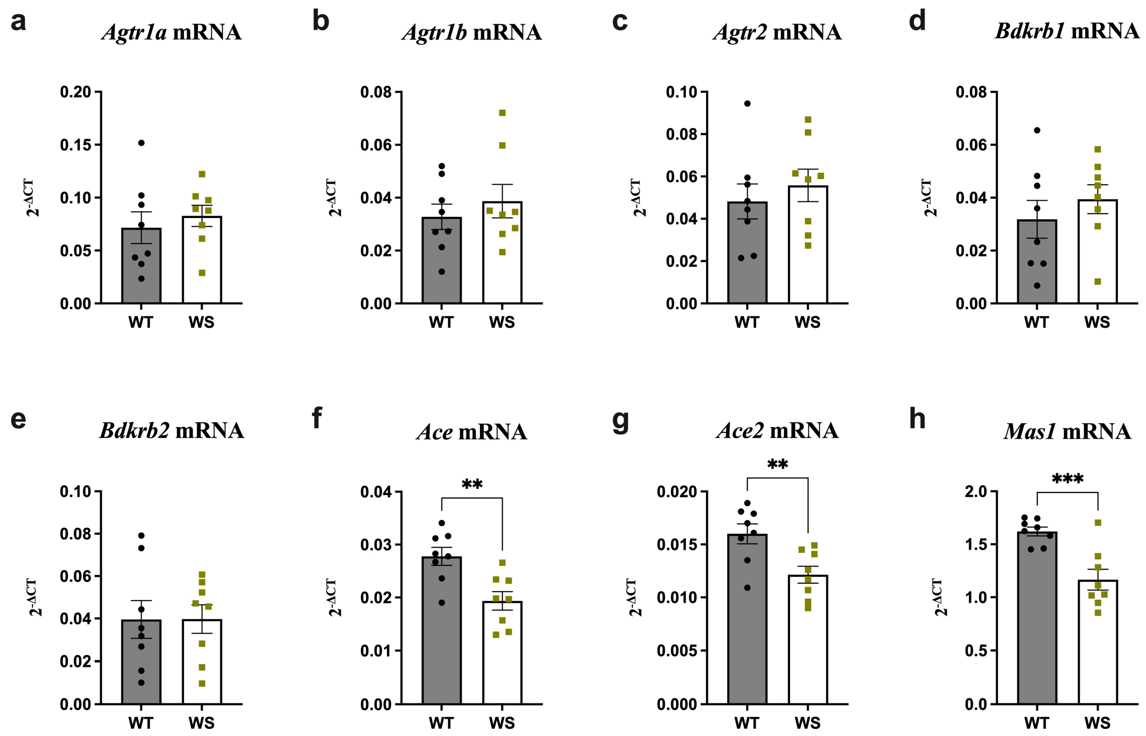
3.3. Ace, Ace2 and Mas1 Were Significantly Downregulated in the Hippocampi of Treatment-Naïve WS Rats

Several neuropsychiatric complications, including increased anxiety and depression, have been reported in both WS patients and animal models [51]. Moreover, both preclinical and clinical studies have demonstrated a link between RAAS alterations and these complications (for a comprehensive review, see [52]). In lieu of this, it was speculated that chronic treatment- and handling-induced stress may underlie the finding of administered treatments being unable to modulate the downregulated hippocampal levels of Agtr1a, Agtr1b, Agtr2 and Bdkrb1 in vehicle-treated WS rats. It was further hypothesized that fully functional WFS1 is necessary for proper functioning of the RAAS, particularly its compensatory axis, during chronic stress. To investigate this, RAAS component expression was analyzed in age-matched treatment-naïve WS and WT rats taken directly from their home cages.
Indeed, hippocampal RAAS expression in treatment-naïve rats significantly differed relative to their treated counterparts. More specifically, no differences in hippocampal Agtr1a, Agtr1b, Agtr2 and Bdkrb1 expression were noted between treatment-naïve WT and WS rats, in contrast to the finding of these being significantly downregulated in vehicle-treated WS rats. Rather, treatment-naïve WS rats had slightly elevated levels relative to their WT littermates (Figure 3a–d vs. Figure 1a–d). Treatment-naïve WS rats also displayed significantly downregulated Ace, Ace2 and Mas1 levels relative to their treatment-naïve WT littermates (Figure 3f–h) (p < 0.01).
To summarize, hippocampal RAAS expression differed considerably between treated (manipulated) and treatment-naïve (non-manipulated) WS and WT animals, indicating a potential interplay between Wfs1 deficiency and chronic (prolonged treatment- and experiment-induced) stress in RAAS regulation.

3.4. Ace Was Significantly Upregulated and Agtr2 Downregulated in the Brain Stems of Treatment-Naïve WS Rats

The analysis was extended to the brain stems to examine whether RAAS alterations in treatment-naïve rats displayed the same regional specificity as in treated rats.
Indeed, increased Ace and decreased Agtr2 expression was seen in the brain stems of treatment-naïve WS relative to WT rats (Figure 4g,c) (p < 0.05). Additionally, a slight, albeit insignificant, downregulation, was observed for Agtr1a, Agtr1b and Bdkrb1 expression in WS animals (Figure 4a,b,d). Finally, and as observed in the hippocampus, Mas1 and Ace2 expression was also slightly—although not significantly—decreased in WS rats (Figure 4f,h).
Altogether, region-specific differences in treatment-naïve rats were not as pronounced as those observed in treated animals.

4. Discussion

Mutations in a gene encoding WFS1 are the underlying cause of WS. Although WS is a monogenic disorder, pathogenic mechanisms remain poorly understood. Consequently, there is no cure for WS; nevertheless, several promising candidates, including GLP-1R agonists, have been shown to mitigate disease progression. Although this class of drugs was originally designed for the treatment of diabetes, it has demonstrated profound neuroprotective effects in preclinical models of several neurodegenerative conditions, including Alzheimer’s Disease [53], Parkinson’s Disease [54] and stroke [55].
While the functions of WFS1 remain to be fully understood, our recent study indicated a role in the modulation of the RAAS, as Wfs1 deficiency induced profound alterations in RAAS components both in vivo and in vitro [32]. Thus, the present study sought to examine (1) the expression of key RAAS components in neural tissues from WS rats and (2) whether any observed alterations can be influenced by LIR (GLP1-R agonist) and 7,8-DHF treatment, both of which have previously demonstrated neuroprotective effects in a rat model of WS [26].
Alterations in hippocampal RAAS component expression in WS animals exposed to prolonged experimental stress were similar to those previously observed in heart, lung and primary cortical neuron cultures [32]; Agtr2 and Bdkrb1 levels were significantly downregulated relative to WT animals. In addition, the levels of the AGTR1 genes Agtr1a and Agtr1b were also substantially decreased. The protective functions of AGTR2 have been well established; its stimulation exerts both anti-inflammatory and anti-fibrotic effects and can promote axonal regeneration [56]. In the CNS, AGTR2 activation can induce transactivation of the brain-derived neurotrophic factor (BDNF) receptor tropomyosin receptor kinase B (TrkB), thereby facilitating BDNF/TrkB-mediated signaling. BDNF/TrkB signal transduction can activate several downstream pathways that promote cell proliferation, survival and plasticity. Disruptions in the BDNF/TRKB axis have been implicated in several neuropsychiatric conditions [57].
Both trauma and inflammation have been shown to activate BDKRB1 [58], which subsequently exerts neuroprotective effects by mediating Ca2+-dependent bradykinin-induced microglial migration [59]. Taken together, the loss of functional WFS1 may cause disturbances in AGTR2- and BDKRB1-mediated signaling and impair their neuroprotective effects, including cell regeneration, ER stress and inflammatory responses, thereby ultimately exacerbating WS progression. Interestingly, none of the administered treatments were able to rescue the gene downregulation observed in the hippocampi of vehicle-treated WS rats. Conversely, and surprisingly, expression levels were downregulated in WT rats across all treatment groups relative to the vehicle-treated WT rats. We speculate that this phenomenon may result, at least in part, because functional WFS1 is required for these drugs to modulate the RAAS under conditions of prolonged stress caused by long-term experimental manipulation. Additionally, there is a possibility that in WT animals, the neuroprotective potential of these drugs diminishes the need for RAAS engagement, even under chronic stress conditions. Curiously, no significant changes in the RAAS were observed in the brain stems for both between-genotype and between-treatment group comparisons in the treated rats. However, this may indicate that the interplay between WFS1 and the RAAS is influenced by time, region and environmental conditions.
Micro-RAASs can be modulated pharmacologically via cognitive processes, such as learning, as well as by chronic stress [57,60]. This is relevant, since the tissues used in the present study were collected as part of a previous study where animals continuously (3.5 months) underwent several procedures, including drug administration, vision and hearing tests and MRI-based imaging, which undoubtedly induced chronic stress [26]. Considering this and our observation that none of the treatments were able to “normalize” the alterations observed in vehicle-treated WS animals, we speculate that functional WFS1 is required to support the hippocampal RAAS response to chronic stress. Thus, treatment-naïve rats were studied to control for the effects of treatment-induced stress. Indeed, we found that these rats had decreased hippocampal expression of Ace, Ace2 and Mas1, but no changes were observed for Agtr2, Agtr1a, Agtr1b and Bdkrb1, as seen in treated animals. Furthermore, as in treated animals, RAAS alterations in treatment-naïve rats displayed regional specificity when comparing the hippocampi and brain stems.
Decreased levels of hippocampal Ace and Ace2 in treatment-naïve WS rats may indicate disturbances in angiotensin processing and consequently compromised AGTR1-, AGTR2- and MAS1-facilitated signaling. Furthermore, changes in neural ACE and ACE2 activity increase neuronal vulnerability to ER stress and inflammation and facilitate the accumulation of bradykinin and proteins such as tau and amyloid-β, all of which are implicated in neurodegenerative pathologies [61,62,63,64]. Similarly, ACE inhibition can delay neurodegeneration via the retardation of tau hyperphosphorylation [65], while ACE2 and AGTR2 activation can protect against cognitive impairments [66]. ACE inhibitors may improve cognitive functioning, including learning and memory, by activating the Ang-(1–7)/Mas axis [67]. Interestingly, a recent study found that WFS1-positive neurons in the entorhinal cortex express tau and mediate its shift to the hippocampal CA1 pyramidal cells, leading to a decline in learning and memory [68,69]. Increased vulnerability to tau pathology in WS indicates that, similarly to ACE, WFS1 interacts with tau and mediates its effects [70]. To conclude, the modulation of RAAS components can influence cognitive processes.
Present and previous findings indicate that the loss of functional WFS1 might disturb RAAS functioning, as evidenced by alterations in its key components, both peripherally and in the nervous system [32]. These disturbances may consequently augment oxidative stress, impair inflammatory responses and Ca2+ homeostasis, affect cognition and contribute to the development of neuropsychiatric complications. An interaction between WFS1 and key RAAS components is further supported by their co-expression in various tissues, including the brain, retina, pancreas, heart and lungs (in humans [71]), and their somewhat overlapping roles. WFS1 may potentially affect RAAS regulation under stressful conditions and facilitate the functioning of the system’s stress-response compensatory axis; disturbances in this axis, as seen here, could therefore exacerbate the course of WS disease.
GLP1-R activation can alleviate ER stress and improve cell survival and mitochondrial function via several pathways [72,73], including the ACE2-mediated RAAS compensatory axis: Ace2/Ang-(1–7)/Mas1/Agtr2. This axis supports cellular function and survival via the induction of a strong ER stress response and anti-inflammatory and regenerative pathways [74,75]. Our previous study demonstrated that LIR treatment, in addition to exerting neuroprotective effects and supporting cognitive function, could modulate the RAAS in peripheral organs [32]. Accordingly, we hypothesized that these positive effects may lie downstream of neural RAAS modulation. Here, we found that differentially expressed RAAS genes in the neural tissues of WS animals were not normalized by LIR treatment, suggesting that LIR’s efficacy derives from the modulation of other signaling and/or homeostatic pathways. In the brain, GLP-1Rs are abundant in pyramidal neurons, and their expression is induced by injury in astrocytes and GABAergic interneurons [76,77,78]. Moreover, GLP-1R agonists have been shown to abate microglial activation in vivo in WS rats [25] and increase GABAergic neurotransmission in different disease conditions, including ischemia [78,79]. Interestingly, GABA receptor activation could significantly delay neuronal death in ischemia-induced injury [80]. Accordingly, while the exact mechanisms underlying LIR’s neuroprotective effects in WS remain to be fully elucidated, they may include ameliorating reactive gliosis by modulating GABAergic signalling and/or augmenting ACE2 activity [33].

5. Conclusions

To summarize, the present study showed that the neural RAAS is altered in WS, as evidenced by the substantial changes in the expression of two key receptors, Agtr2 and Bdkrb1. However, those alterations are not conserved across different regions, potentially owing to the differential regional, environmental and temporal modulation of the RAAS across the WS disease course.
Crucially, we showed that those changes vary depending on whether or not animals are exposed to a prolonged stressful environment (long-term animal experimentation), indicating a role played by chronic stress. Stress may further compound the effects of Wfs1 deficiency on RAAS function, and a compromised compensatory axis could ultimately exacerbate the disease process. These results emphasize once more that experimental design and environment can affect gene expression, and that there is a strong need to control for procedural stress and include treatment-naïve animals within experimental paradigms. Finally, we showed that none of the alterations observed in vehicle-treated WS rats were amenable to pharmacological modulation, despite animals experiencing symptomatic improvement in our previous study [26]. This suggests that the neuroprotective effects of these drugs in WS are likely mediated independently of the RAAS.

6. Limitations of the Study

The present study is not without its limitations; alterations were only described at the transcriptomic level, and since protein-level changes were beyond the scope of this study, as it was exploratory, we recommend that future studies address this. Furthermore, experimental tissue samples were harvested from aged rats that had already developed substantial neurological symptoms, including impaired cognitive function and hippocampal lateral ventricle enlargement. Future studies may also consider investigating transcriptomic changes within specific neuronal populations, especially in regions as diverse as the brain stem. Examining the temporal development of RAAS disruptions across the WS disease course also warrants investigation. Finally, the chronic stress conditions described in this study resulted inadvertently from prolonged experimental handling. Additional analyses using classical stress paradigms should be performed to verify the results reported here.

Author Contributions

Conceptualization, M.P. (Mario Plaas); methodology, M.P. (Mario Plaas); formal analysis, M.P. (Mario Plaas), M.P. (Marite Punapart) and K.S.; investigation, K.S., T.J., R.R., K.-L.E. and S.K.; writing—original draft preparation, M.P. (Mario Plaas), M.P. (Marite Punapart) and N.G.; writing—review and editing, M.P. (Mario Plaas), M.P. (Marite Punapart) and E.V.; visualization, M.P. (Marite Punapart) and K.S.; project administration, M.P. (Mario Plaas); funding acquisition, M.P. (Mario Plaas). All authors have read and agreed to the published version of the manuscript.

Funding

This research was supported by the Estonian Research Council via grants PSG471 (Mario Plaas) and SJD90 (Nayana Gaur), by the European Union through the European Regional Development Fund (Project No. 2014-2020.4.01.15-0012), by CELSA and by the Eye Hope Foundation.

Institutional Review Board Statement

The study was approved by the Estonian Project Authorization Committee for Animal Experiments (No 155, 6 January 2020), and all experiments were performed in accordance with the European Communities Directive of September 2010 (2010/63/EU). The study was carried out in compliance with the ARRIVE guidelines.

Informed Consent Statement

Not applicable.

Data Availability Statement

The datasets generated and analyzed during this study are available from the corresponding authors on reasonable request.

Conflicts of Interest

The authors declare no conflict of interest.

Appendix A

List of Abbreviations (in alphabetical order)
7,8-DHF7,8-dihydroxyflavone
AceAngiotensin I converting enzyme
Ace2Angiotensin I converting enzyme 2
Agtr1aAngiotensin II receptor type 1a
Agtr1bAngiotensin II receptor type 1b
Agtr2Angiotensin II receptor type 2
ARRIVEAnimal Research: Reporting of In Vivo Experiments
Bdkrb1Bradykinin receptor B1
Bdkrb2Bradykinin receptor B2
BDNFBrain-derived neurotrophic factor
CNSCentral nervous system
DMSODimethyl sulfoxide
EREndoplasmic reticulum
GABAGamma-aminobutyric acid
GLP-1RGlucagon-like peptide 1 receptor
Hprt1Hypoxanthine-guanine phosphoribosyltransferase
LIRLiraglutide
MAMMitochondria-associated ER membraane
Mas1MAS1 proto-oncogene, G protein-coupled receptor
PBSPhosphate-buffered saline
PEG-300Polyethylene glycol-300
RAASRenin-angiotensin-aldosterone system
SEMStandard error of the mean
TrkBTropomyosin receptor kinase B
VEHVehicle
WFS1Wolframin/Wolfram Syndrome 1
WSWolfram Syndrome
WTWild-type

References

  1. Barrett, T.G.; Bundey, S.E. Wolfram (DIDMOAD) Syndrome. J. Med. Genet. 1997, 34, 838–841. [Google Scholar] [CrossRef] [PubMed] [Green Version]
  2. Inoue, H.; Tanizawa, Y.; Wasson, J.; Behn, P.; Kalidas, K.; Bernal-Mizrachi, E.; Mueckler, M.; Marshall, H.; Donis-Keller, H.; Crock, P.; et al. A Gene Encoding a Transmembrane Protein Is Mutated in Patients with Diabetes Mellitus and Optic Atrophy (Wolfram Syndrome). Nat. Genet. 1998, 20, 143–148. [Google Scholar] [CrossRef] [PubMed]
  3. Barrett, T.G.; Bundey, S.E.; Macleod, A.F. Neurodegeneration and Diabetes: UK Nationwide Study of Wolfram (DIDMOAD) Syndrome. Lancet 1995, 346, 1458–1463. [Google Scholar] [CrossRef]
  4. Medlej, R.; Wasson, J.; Baz, P.; Azar, S.; Salti, I.; Loiselet, J.; Permutt, A.; Halaby, G. Diabetes Mellitus and Optic Atrophy: A Study of Wolfram Syndrome in the Lebanese Population. J. Clin. Endocrinol. Metab. 2004, 89, 1656–1661. [Google Scholar] [CrossRef] [Green Version]
  5. Luuk, H.; Koks, S.; Plaas, M.; Hannibal, J.; Rehfeld, J.F.; Vasar, E. Distribution of Wfs1 Protein in the Central Nervous System of the Mouse and Its Relation to Clinical Symptoms of the Wolfram Syndrome. J. Comp. Neurol. 2008, 509, 642–660. [Google Scholar] [CrossRef]
  6. Hofmann, S.; Philbrook, C.; Gerbitz, K.-D.; Bauer, M.F. Wolfram Syndrome: Structural and Functional Analyses of Mutant and Wild-Type Wolframin, the WFS1 Gene Product. Hum. Mol. Genet. 2003, 12, 2003–2012. [Google Scholar] [CrossRef] [PubMed] [Green Version]
  7. The Human Protein Atlas. Available online: https://www.proteinatlas.org/ (accessed on 21 February 2021).
  8. GeneCards—Human Genes|Gene Database|Gene Search. Available online: https://www.genecards.org/ (accessed on 21 February 2021).
  9. Fonseca, S.G.; Fukuma, M.; Lipson, K.L.; Nguyen, L.X.; Allen, J.R.; Oka, Y.; Urano, F. WFS1 is a Novel Component of the Unfolded Protein Response and Maintains Homeostasis of the Endoplasmic Reticulum in Pancreatic β-Cells. J. Biol. Chem. 2005, 280, 39609–39615. [Google Scholar] [CrossRef] [PubMed] [Green Version]
  10. Takei, D.; Ishihara, H.; Yamaguchi, S.; Yamada, T.; Tamura, A.; Katagiri, H.; Maruyama, Y.; Oka, Y. WFS1 Protein Modulates the Free Ca(2+) Concentration in the Endoplasmic Reticulum. FEBS Lett. 2006, 580, 5635–5640. [Google Scholar] [CrossRef] [Green Version]
  11. Cagalinec, M.; Liiv, M.; Hodurova, Z.; Hickey, M.A.; Vaarmann, A.; Mandel, M.; Zeb, A.; Choubey, V.; Kuum, M.; Safiulina, D.; et al. Role of Mitochondrial Dynamics in Neuronal Development: Mechanism for Wolfram Syndrome. PLoS Biol. 2016, 14, e1002511. [Google Scholar] [CrossRef] [Green Version]
  12. La Morgia, C.; Maresca, A.; Amore, G.; Gramegna, L.L.; Carbonelli, M.; Scimonelli, E.; Danese, A.; Patergnani, S.; Caporali, L.; Tagliavini, F.; et al. Calcium Mishandling in Absence of Primary Mitochondrial Dysfunction Drives Cellular Pathology in Wolfram Syndrome. Sci. Rep. 2020, 10, 4785. [Google Scholar] [CrossRef] [Green Version]
  13. Angebault, C.; Fauconnier, J.; Patergnani, S.; Rieusset, J.; Danese, A.; Affortit, C.A.; Jagodzinska, J.; Mégy, C.; Quiles, M.; Cazevieille, C.; et al. ER-Mitochondria Cross-Talk Is Regulated by the Ca2+ Sensor NCS1 and Is Impaired in Wolfram Syndrome. Sci. Signal. 2018, 11, eaaq1380. [Google Scholar] [CrossRef] [PubMed] [Green Version]
  14. Crouzier, L.; Danese, A.; Yasui, Y.; Richard, E.M.; Liévens, J.-C.; Patergnani, S.; Couly, S.; Diez, C.; Denus, M.; Cubedo, N.; et al. Activation of the Sigma-1 Receptor Chaperone Alleviates Symptoms of Wolfram Syndrome in Preclinical Models. Sci. Transl. Med. 2022, 14, eabh3763. [Google Scholar] [CrossRef] [PubMed]
  15. Kakiuchi, C.; Ishigaki, S.; Oslowski, C.M.; Fonseca, S.G.; Kato, T.; Urano, F. Valproate, a Mood Stabilizer, Induces WFS1 Expression and Modulates Its Interaction with ER Stress Protein GRP94. PLoS ONE 2009, 4, e4134. [Google Scholar] [CrossRef]
  16. Batjargal, K.; Tajima, T.; Jimbo, E.F.; Yamagata, T. Effect of 4-Phenylbutyrate and Valproate on Dominant Mutations of WFS1 Gene in Wolfram Syndrome. J. Endocrinol. Investig. 2020, 43, 1317–1325. [Google Scholar] [CrossRef] [PubMed]
  17. Terasmaa, A.; Soomets, U.; Oflijan, J.; Punapart, M.; Hansen, M.; Matto, V.; Ehrlich, K.; Must, A.; Kõks, S.; Vasar, E. Wfs1 Mutation Makes Mice Sensitive to Insulin-like Effect of Acute Valproic Acid and Resistant to Streptozocin. J. Physiol. Biochem. 2011, 67, 381–390. [Google Scholar] [CrossRef]
  18. Lu, S.; Kanekura, K.; Hara, T.; Mahadevan, J.; Spears, L.D.; Oslowski, C.M.; Martinez, R.; Yamazaki-Inoue, M.; Toyoda, M.; Neilson, A.; et al. A Calcium-Dependent Protease as a Potential Therapeutic Target for Wolfram Syndrome. Proc. Natl. Acad. Sci. USA 2014, 111, E5292–E5301. [Google Scholar] [CrossRef] [Green Version]
  19. Abreu, D.; Stone, S.I.; Pearson, T.S.; Bucelli, R.C.; Simpson, A.N.; Hurst, S.; Brown, C.M.; Kries, K.; Onwumere, C.; Gu, H.; et al. A Phase Ib/IIa Clinical Trial of Dantrolene Sodium in Patients with Wolfram Syndrome. JCI Insight 2021, 6, e145188. [Google Scholar] [CrossRef] [PubMed]
  20. Yuan, F.; Li, Y.; Hu, R.; Gong, M.; Chai, M.; Ma, X.; Cha, J.; Guo, P.; Yang, K.; Li, M.; et al. Modeling Disrupted Synapse Formation in Wolfram Syndrome Using HESCs-Derived Neural Cells and Cerebral Organoids Identifies Riluzole as a Therapeutic Molecule. Mol. Psychiatry 2023, 1–14. [Google Scholar] [CrossRef]
  21. Mullard, A. Amylyx’s ALS Therapy Secures FDA Approval, as Regulatory Flexibility Trumps Underwhelming Data. Nat. Rev. Drug Discov. 2022, 21, 786. [Google Scholar] [CrossRef]
  22. Kitamura, R.A.; Maxwell, K.G.; Ye, W.; Kries, K.; Brown, C.M.; Augsornworawat, P.; Hirsch, Y.; Johansson, M.M.; Weiden, T.; Ekstein, J.; et al. Multidimensional Analysis and Therapeutic Development Using Patient IPSC–Derived Disease Models of Wolfram Syndrome. JCI Insight 2022, 7, e156549. [Google Scholar] [CrossRef]
  23. Rigoli, L.; Caruso, V.; Salzano, G.; Lombardo, F. Wolfram Syndrome 1: From Genetics to Therapy. Int. J. Env. Res. Public Health 2022, 19, 3225. [Google Scholar] [CrossRef] [PubMed]
  24. Toots, M.; Seppa, K.; Jagomäe, T.; Koppel, T.; Pallase, M.; Heinla, I.; Terasmaa, A.; Plaas, M.; Vasar, E. Preventive Treatment with Liraglutide Protects against Development of Glucose Intolerance in a Rat Model of Wolfram Syndrome. Sci. Rep. 2018, 8, 10183. [Google Scholar] [CrossRef] [PubMed] [Green Version]
  25. Seppa, K.; Toots, M.; Reimets, R.; Jagomäe, T.; Koppel, T.; Pallase, M.; Hasselholt, S.; Mikkelsen, M.K.; Randel Nyengaard, J.; Vasar, E.; et al. GLP-1 Receptor Agonist Liraglutide Has a Neuroprotective Effect on an Aged Rat Model of Wolfram Syndrome. Sci. Rep. 2019, 9, 15742. [Google Scholar] [CrossRef] [Green Version]
  26. Seppa, K.; Jagomäe, T.; Kukker, K.G.; Reimets, R.; Pastak, M.; Vasar, E.; Terasmaa, A.; Plaas, M. Liraglutide, 7,8-DHF and Their Co-Treatment Prevents Loss of Vision and Cognitive Decline in a Wolfram Syndrome Rat Model. Sci. Rep. 2021, 11, 2275. [Google Scholar] [CrossRef]
  27. Jagomäe, T.; Seppa, K.; Reimets, R.; Pastak, M.; Plaas, M.; Hickey, M.A.; Kukker, K.G.; Moons, L.; De Groef, L.; Vasar, E.; et al. Early Intervention and Lifelong Treatment with GLP1 Receptor Agonist Liraglutide in a Wolfram Syndrome Rat Model with an Emphasis on Visual Neurodegeneration, Sensorineural Hearing Loss and Diabetic Phenotype. Cells 2021, 10, 3193. [Google Scholar] [CrossRef] [PubMed]
  28. Sedman, T.; Rünkorg, K.; Krass, M.; Luuk, H.; Plaas, M.; Vasar, E.; Volke, V. Exenatide Is an Effective Antihyperglycaemic Agent in a Mouse Model of Wolfram Syndrome 1. J. Diabetes Res. 2016, 2016, 9239530. [Google Scholar] [CrossRef] [Green Version]
  29. Kondo, M.; Tanabe, K.; Amo-Shiinoki, K.; Hatanaka, M.; Morii, T.; Takahashi, H.; Seino, S.; Yamada, Y.; Tanizawa, Y. Activation of GLP-1 Receptor Signalling Alleviates Cellular Stresses and Improves β Cell Function in a Mouse Model of Wolfram Syndrome. Diabetologia 2018, 61, 2189–2201. [Google Scholar] [CrossRef] [Green Version]
  30. Scully, K.J.; Wolfsdorf, J.I. Efficacy of GLP-1 Agonist Therapy in Autosomal Dominant WFS1-Related Disorder: A Case Report. Horm. Res. Paediatr. 2020, 93, 409–414. [Google Scholar] [CrossRef]
  31. Frontino, G.; Raouf, T.; Canarutto, D.; Tirelli, E.; Di Tonno, R.; Rigamonti, A.; Cascavilla, M.L.; Baldoli, C.; Scotti, R.; Leocani, L.; et al. Case Report: Off-Label Liraglutide Use in Children With Wolfram Syndrome Type 1: Extensive Characterization of Four Patients. Front. Pediatr. 2021, 9, 755365. [Google Scholar] [CrossRef]
  32. Punapart, M.; Seppa, K.; Jagomäe, T.; Liiv, M.; Reimets, R.; Kirillov, S.; Kaasik, A.; Moons, L.; De Groef, L.; Terasmaa, A.; et al. The Expression of RAAS Key Receptors, Agtr2 and Bdkrb1, Is Downregulated at an Early Stage in a Rat Model of Wolfram Syndrome. Genes 2021, 12, 1717. [Google Scholar] [CrossRef]
  33. Romaní-Pérez, M.; Outeiriño-Iglesias, V.; Moya, C.M.; Santisteban, P.; González-Matías, L.C.; Vigo, E.; Mallo, F. Activation of the GLP-1 Receptor by Liraglutide Increases ACE2 Expression, Reversing Right Ventricle Hypertrophy, and Improving the Production of SP-A and SP-B in the Lungs of Type 1 Diabetes Rats. Endocrinology 2015, 156, 3559–3569. [Google Scholar] [CrossRef]
  34. Sedman, T.; Heinla, K.; Vasar, E.; Volke, V. Liraglutide Treatment May Affect Renin and Aldosterone Release. Horm. Metab. Res. 2017, 49, 5–9. [Google Scholar] [CrossRef] [PubMed]
  35. Perini, M.V.; Dmello, R.S.; Nero, T.L.; Chand, A.L. Evaluating the Benefits of Renin-Angiotensin System Inhibitors as Cancer Treatments. Pharmacol. Ther. 2020, 211, 107527. [Google Scholar] [CrossRef] [PubMed]
  36. Ribeiro-Oliveira, A.; Nogueira, A.I.; Pereira, R.M.; Boas, W.W.V.; Dos Santos, R.A.S.; Simões e Silva, A.C. The Renin-Angiotensin System and Diabetes: An Update. Vasc. Health Risk Manag. 2008, 4, 787–803. [Google Scholar] [PubMed]
  37. Labandeira-Garcia, J.L.; Rodríguez-Perez, A.I.; Garrido-Gil, P.; Rodriguez-Pallares, J.; Lanciego, J.L.; Guerra, M.J. Brain Renin-Angiotensin System and Microglial Polarization: Implications for Aging and Neurodegeneration. Front. Aging Neurosci. 2017, 9, 129. [Google Scholar] [CrossRef] [Green Version]
  38. Guimond, M.-O.; Gallo-Payet, N. The Angiotensin II Type 2 Receptor in Brain Functions: An Update. Int. J. Hypertens. 2012, 2012, 351758. [Google Scholar] [CrossRef] [Green Version]
  39. Wright, J.W.; Harding, J.W. The Brain Renin–Angiotensin System: A Diversity of Functions and Implications for CNS Diseases. Pflug. Arch. Eur. J. Physiol. 2013, 465, 133–151. [Google Scholar] [CrossRef]
  40. Leung, P.S.; Chappell, M.C. A Local Pancreatic Renin-Angiotensin System: Endocrine and Exocrine Roles. Int. J. Biochem. Cell Biol. 2003, 35, 838–846. [Google Scholar] [CrossRef]
  41. Cao, X.; Lu, X.-M.; Tuo, X.; Liu, J.-Y.; Zhang, Y.-C.; Song, L.-N.; Cheng, Z.-Q.; Yang, J.-K.; Xin, Z. Angiotensin-Converting Enzyme 2 Regulates Endoplasmic Reticulum Stress and Mitochondrial Function to Preserve Skeletal Muscle Lipid Metabolism. Lipids Health Dis. 2019, 18, 207. [Google Scholar] [CrossRef] [Green Version]
  42. Escobales, N.; Nuñez, R.E.; Javadov, S. Mitochondrial Angiotensin Receptors and Cardioprotective Pathways. Am. J. Physiol. Heart Circ. Physiol. 2019, 316, H1426–H1438. [Google Scholar] [CrossRef]
  43. Valenzuela, R.; Costa-Besada, M.A.; Iglesias-Gonzalez, J.; Perez-Costas, E.; Villar-Cheda, B.; Garrido-Gil, P.; Melendez-Ferro, M.; Soto-Otero, R.; Lanciego, J.L.; Henrion, D.; et al. Mitochondrial Angiotensin Receptors in Dopaminergic Neurons. Role in Cell Protection and Aging-Related Vulnerability to Neurodegeneration. Cell Death Dis. 2016, 7, e2427. [Google Scholar] [CrossRef] [PubMed] [Green Version]
  44. Rodriguez-Pallares, J.; Rey, P.; Parga, J.A.; Muñoz, A.; Guerra, M.J.; Labandeira-Garcia, J.L. Brain Angiotensin Enhances Dopaminergic Cell Death via Microglial Activation and NADPH-Derived ROS. Neurobiol. Dis. 2008, 31, 58–73. [Google Scholar] [CrossRef] [PubMed]
  45. Sunanda, T.; Ray, B.; Mahalakshmi, A.M.; Bhat, A.; Rashan, L.; Rungratanawanich, W.; Song, B.-J.; Essa, M.M.; Sakharkar, M.K.; Chidambaram, S.B. Mitochondria-Endoplasmic Reticulum Crosstalk in Parkinson’s Disease: The Role of Brain Renin Angiotensin System Components. Biomolecules 2021, 11, 1669. [Google Scholar] [CrossRef] [PubMed]
  46. Scolding, N.J.; Kellar-Wood, H.F.; Shaw, C.; Shneerson, J.M.; Antount, N. Wolfram Syndrome: Hereditary Diabetes Mellitus with Brainstem and Optic Atrophy. Ann. Neurol. 1996, 39, 352–360. [Google Scholar] [CrossRef]
  47. Hershey, T.; Lugar, H.M.; Shimony, J.S.; Rutlin, J.; Koller, J.M.; Perantie, D.C.; Paciorkowski, A.R.; Eisenstein, S.A.; Permutt, M.A. Early Brain Vulnerability in Wolfram Syndrome. PLoS ONE 2012, 7, e40604. [Google Scholar] [CrossRef] [PubMed]
  48. Shannon, P.; Becker, L.; Deck, J. Evidence of Widespread Axonal Pathology in Wolfram Syndrome. Acta Neuropathol. 1999, 98, 304–308. [Google Scholar] [CrossRef]
  49. Takeda, K.; Inoue, H.; Tanizawa, Y.; Matsuzaki, Y.; Oba, J.; Watanabe, Y.; Shinoda, K.; Oka, Y. WFS1 (Wolfram Syndrome 1) Gene Product: Predominant Subcellular Localization to Endoplasmic Reticulum in Cultured Cells and Neuronal Expression in Rat Brain. Hum. Mol. Genet. 2001, 10, 477–484. [Google Scholar] [CrossRef] [Green Version]
  50. Plaas, M.; Seppa, K.; Reimets, R.; Jagomäe, T.; Toots, M.; Koppel, T.; Vallisoo, T.; Nigul, M.; Heinla, I.; Meier, R.; et al. Wfs1- Deficient Rats Develop Primary Symptoms of Wolfram Syndrome: Insulin-Dependent Diabetes, Optic Nerve Atrophy and Medullary Degeneration. Sci. Rep. 2017, 7, 10220. [Google Scholar] [CrossRef] [Green Version]
  51. Munshani, S.; Ibrahim, E.Y.; Domenicano, I.; Ehrlich, B.E. The Impact of Mutations in Wolframin on Psychiatric Disorders. Front. Pediatr. 2021, 9, 718132. [Google Scholar] [CrossRef]
  52. Mohite, S.; Sanches, M.; Teixeira, A.L. Exploring the Evidence Implicating the Renin-Angiotensin System (RAS) in the Physiopathology of Mood Disorders. Protein Pept. Lett. 2017, 27, 449–455. [Google Scholar] [CrossRef]
  53. McClean, P.L.; Jalewa, J.; Hölscher, C. Prophylactic Liraglutide Treatment Prevents Amyloid Plaque Deposition, Chronic Inflammation and Memory Impairment in APP/PS1 Mice. Behav. Brain Res. 2015, 293, 96–106. [Google Scholar] [CrossRef] [Green Version]
  54. Liu, W.; Jalewa, J.; Sharma, M.; Li, G.; Li, L.; Hölscher, C. Neuroprotective Effects of Lixisenatide and Liraglutide in the 1-Methyl-4-Phenyl-1,2,3,6-Tetrahydropyridine Mouse Model of Parkinson’s Disease. Neuroscience 2015, 303, 42–50. [Google Scholar] [CrossRef] [Green Version]
  55. Yang, X.; Qiang, Q.; Li, N.; Feng, P.; Wei, W.; Hölscher, C. Neuroprotective Mechanisms of Glucagon-Like Peptide-1-Based Therapies in Ischemic Stroke: An Update Based on Preclinical Research. Front. Neurol. 2022, 13, 844697. [Google Scholar] [CrossRef]
  56. Lucius, R.; Gallinat, S.; Rosenstiel, P.; Herdegen, T.; Sievers, J.; Unger, T. The Angiotensin II Type 2 (AT2) Receptor Promotes Axonal Regeneration in the Optic Nerve of Adult Rats. J. Exp. Med. 1998, 188, 661–670. [Google Scholar] [CrossRef] [PubMed] [Green Version]
  57. Diniz, C.R.A.F.; Casarotto, P.C.; Fred, S.M.; Biojone, C.; Castrén, E.; Joca, S.R.L. Antidepressant-like Effect of Losartan Involves TRKB Transactivation from Angiotensin Receptor Type 2 (AGTR2) and Recruitment of FYN. Neuropharmacology 2018, 135, 163–171. [Google Scholar] [CrossRef] [PubMed]
  58. Hofman, Z.; de Maat, S.; Hack, C.E.; Maas, C. Bradykinin: Inflammatory Product of the Coagulation System. Clin. Rev. Allergy Immunol. 2016, 51, 152–161. [Google Scholar] [CrossRef] [PubMed] [Green Version]
  59. Ifuku, M.; Färber, K.; Okuno, Y.; Yamakawa, Y.; Miyamoto, T.; Nolte, C.; Merrino, V.F.; Kita, S.; Iwamoto, T.; Komuro, I.; et al. Bradykinin-Induced Microglial Migration Mediated by B1-Bradykinin Receptors Depends on Ca2+ Influx via Reverse-Mode Activity of the Na+/Ca2+ Exchanger. J. Neurosci. 2007, 27, 13065–13073. [Google Scholar] [CrossRef] [Green Version]
  60. Saavedra, J.M.; Benicky, J. Brain and Peripheral Angiotensin II Play a Major Role in Stress. Stress 2007, 10, 185–193. [Google Scholar] [CrossRef]
  61. Petek, B.; Villa-Lopez, M.; Loera-Valencia, R.; Gerenu, G.; Winblad, B.; Kramberger, M.G.; Ismail, M.-A.-M.; Eriksdotter, M.; Garcia-Ptacek, S. Connecting the Brain Cholesterol and Renin–Angiotensin Systems: Potential Role of Statins and RAS-Modifying Medications in Dementia. J. Intern. Med. 2018, 284, 620–642. [Google Scholar] [CrossRef] [Green Version]
  62. Hemming, M.L.; Selkoe, D.J. Amyloid β-Protein Is Degraded by Cellular Angiotensin-Converting Enzyme (ACE) and Elevated by an ACE Inhibitor. J. Biol. Chem. 2005, 280, 37644–37650. [Google Scholar] [CrossRef] [Green Version]
  63. Kehoe, P.G.; Wong, S.; Al Mulhim, N.; Palmer, L.E.; Miners, J.S. Angiotensin-Converting Enzyme 2 Is Reduced in Alzheimer’s Disease in Association with Increasing Amyloid-β and Tau Pathology. Alzheimers Res. 2016, 8, 50. [Google Scholar] [CrossRef] [Green Version]
  64. Singh, P.K.; Chen, Z.-L.; Ghosh, D.; Strickland, S.; Norris, E.H. Increased Plasma Bradykinin Level Is Associated with Cognitive Impairment in Alzheimer’s Patients. Neurobiol. Dis. 2020, 139, 104833. [Google Scholar] [CrossRef] [PubMed]
  65. AbdAlla, S.; el Hakim, A.; Abdelbaset, A.; Elfaramawy, Y.; Quitterer, U. Inhibition of ACE Retards Tau Hyperphosphorylation and Signs of Neuronal Degeneration in Aged Rats Subjected to Chronic Mild Stress. BioMed Res. Int. 2015, 2015, 917156. [Google Scholar] [CrossRef] [PubMed] [Green Version]
  66. Fouda, A.Y.; Fagan, S.C.; Ergul, A. Brain Vasculature and Cognition. Arterioscler. Thromb. Vasc. Biol. 2019, 39, 593–602. [Google Scholar] [CrossRef] [PubMed]
  67. Hellner, K.; Walther, T.; Schubert, M.; Albrecht, D. Angiotensin-(1–7) Enhances LTP in the Hippocampus through the G-Protein-Coupled Receptor Mas. Mol. Cell. Neurosci. 2005, 29, 427–435. [Google Scholar] [CrossRef] [PubMed]
  68. Delpech, J.-C.; Pathak, D.; Varghese, M.; Kalavai, S.V.; Hays, E.C.; Hof, P.R.; Johnson, W.E.; Ikezu, S.; Medalla, M.; Luebke, J.I.; et al. Wolframin-1—Expressing Neurons in the Entorhinal Cortex Propagate Tau to CA1 Neurons and Impair Hippocampal Memory in Mice. Sci. Transl. Med. 2021, 13, eabe8455. [Google Scholar] [CrossRef]
  69. Chen, S.; Acosta, D.; Li, L.; Liang, J.; Chang, Y.; Wang, C.; Fitzgerald, J.; Morrison, C.; Goulbourne, C.N.; Nakano, Y.; et al. Wolframin Is a Novel Regulator of Tau Pathology and Neurodegeneration. Acta Neuropathol. 2022, 143, 547–569. [Google Scholar] [CrossRef]
  70. Chen, S.; Venkaraman, L.; Liang, J.; Nakano, Y.; Villegas, N.E.H.; Brown, C.; Urano, F.; Koks, S.; Serrano, G.E.; Beach, T.G.; et al. Deficiency of WFS1 Increases Vulnerability to Pathological Tau in Vitro and in Vivo. Alzheimer’s Dement. 2020, 16, e042085. [Google Scholar] [CrossRef]
  71. BioGPS—Your Gene Portal System. Available online: http://biogps.org/#goto=welcome (accessed on 2 June 2022).
  72. Chen, J.; Xie, J.-J.; Shi, K.-S.; Gu, Y.-T.; Wu, C.-C.; Xuan, J.; Ren, Y.; Chen, L.; Wu, Y.-S.; Zhang, X.-L.; et al. Glucagon-like Peptide-1 Receptor Regulates Endoplasmic Reticulum Stress-Induced Apoptosis and the Associated Inflammatory Response in Chondrocytes and the Progression of Osteoarthritis in Rat. Cell Death Dis. 2018, 9, 212. [Google Scholar] [CrossRef] [Green Version]
  73. Nuamnaichati, N.; Mangmool, S.; Chattipakorn, N.; Parichatikanond, W. Stimulation of GLP-1 Receptor Inhibits Methylglyoxal-Induced Mitochondrial Dysfunctions in H9c2 Cardiomyoblasts: Potential Role of Epac/PI3K/Akt Pathway. Front. Pharmacol. 2020, 11, 805. [Google Scholar] [CrossRef]
  74. Rodrigues Prestes, T.R.; Rocha, N.P.; Miranda, A.S.; Teixeira, A.L.; Simoes-E-Silva, A.C. The Anti-Inflammatory Potential of ACE2/Angiotensin-(1-7)/Mas Receptor Axis: Evidence from Basic and Clinical Research. Curr. Drug Targets 2017, 18, 1301–1313. [Google Scholar] [CrossRef] [PubMed]
  75. Yang, G.; Istas, G.; Höges, S.; Yakoub, M.; Hendgen-Cotta, U.; Rassaf, T.; Rodriguez-Mateos, A.; Hering, L.; Grandoch, M.; Mergia, E.; et al. Angiotensin-(1-7)-Induced Mas Receptor Activation Attenuates Atherosclerosis through a Nitric Oxide-Dependent Mechanism in ApolipoproteinE-KO Mice. Pflug. Arch. 2018, 470, 661–667. [Google Scholar] [CrossRef] [PubMed] [Green Version]
  76. Cork, S.C.; Richards, J.E.; Holt, M.K.; Gribble, F.M.; Reimann, F.; Trapp, S. Distribution and Characterisation of Glucagon-like Peptide-1 Receptor Expressing Cells in the Mouse Brain. Mol. Metab. 2015, 4, 718–731. [Google Scholar] [CrossRef] [Green Version]
  77. Hamilton, A.; Hölscher, C. Receptors for the Incretin Glucagon-like Peptide-1 Are Expressed on Neurons in the Central Nervous System. Neuroreport 2009, 20, 1161–1166. [Google Scholar] [CrossRef]
  78. Lee, C.H.; Yan, B.; Yoo, K.-Y.; Choi, J.H.; Kwon, S.-H.; Her, S.; Sohn, Y.; Hwang, I.K.; Cho, J.H.; Kim, Y.-M.; et al. Ischemia-Induced Changes in Glucagon-like Peptide-1 Receptor and Neuroprotective Effect of Its Agonist, Exendin-4, in Experimental Transient Cerebral Ischemia. J. Neurosci. Res. 2011, 89, 1103–1113. [Google Scholar] [CrossRef]
  79. Korol, S.V.; Jin, Z.; Babateen, O.; Birnir, B. GLP-1 and Exendin-4 Transiently Enhance GABAA Receptor–Mediated Synaptic and Tonic Currents in Rat Hippocampal CA3 Pyramidal Neurons. Diabetes 2014, 64, 79–89. [Google Scholar] [CrossRef] [Green Version]
  80. Zhou, C.; Li, C.; Yu, H.-M.; Zhang, F.; Han, D.; Zhang, G.-Y. Neuroprotection of γ-Aminobutyric Acid Receptor Agonists via Enhancing Neuronal Nitric Oxide Synthase (Ser847) Phosphorylation through Increased Neuronal Nitric Oxide Synthase and PSD95 Interaction and Inhibited Protein Phosphatase Activity in Cerebral Ischemia. J. Neurosci. Res. 2008, 86, 2973–2983. [Google Scholar] [CrossRef] [PubMed]
Figure 1. Expression of Agtr1a, Agtr1b, Agtr2 and Bdkrb1 was significantly downregulated in the hippocampi of chronically treated aged Wfs1-deficient rats. Gene expression was analyzed from the hippocampi of 12.5-month-old animals after 3.5 months of treatment with liraglutide (LIR), 7,8-dihydroxyflavone (DHF), liraglutide + 7,8-dihydroxyflavone (LIR + DHF) or vehicle (VEH). Relative gene expression levels of (a) Agtr1a, (b) Agtr1b, (c) Agtr2, (d) Bdkrb1, (e) Bdkrb2, (f) Ace, (g) Ace2 and (h) Mas1 (presented as 2−ΔCT relative to the housekeeper Hprt). Statistical significance was determined using one-way ANOVA followed by Dunnett’s multiple comparisons test * p < 0.05; ** p < 0.01; **** p < 0.0001. The data are presented as mean ± SEM, n  =  5–8 per group.
Genes 14 00827 g001
Figure 2. No significant between-genotype or between-treatment group differences were noted in the brain stems of chronically treated aged Wfs1-deficient rats. Gene expression was analyzed from the brain stems of 12.5-month-old animals after 3.5 months of treatment with liraglutide (LIR), 7,8-dihydroxyflavone (DHF), liraglutide + 7,8-dihydroxyflavone (LIR + DHF) or vehicle (VEH). Relative gene expression levels of (a) Agtr1a, (b) Agtr1b, (c) Agtr2, (d) Bdkrb1, (e) Bdkrb2, (f) Ace, (g) Ace2 and (h) Mas1 (presented as 2−ΔCT relative to the housekeeper Hprt). Statistical significance was determined using one-way ANOVA followed by Dunnett’s multiple comparisons test. The data are presented as mean ± SEM, n  =  5–8 per group.
Genes 14 00827 g002
Figure 3. Expression of Ace, Ace2 and Mas1 was substantially downregulated in the hippocampi of treatment-naïve aged Wfs1-deficient rats. Gene expression was analyzed from the hippocampi of 12.5–13-month-old animals taken directly from their home cages. Relative gene expression levels of (a) Agtr1a, (b) Agtr1b, (c) Agtr2, (d) Bdkrb1, (e) Bdkrb2, (f) Ace, (g) Ace2 and (h) Mas1 (presented as 2−ΔCT relative to the housekeeper Hprt). Statistical significance was determined using an unpaired t-test; ** p < 0.01; *** p < 0.001. The data are presented as mean ± SEM, n  =  8 per group.
Genes 14 00827 g003
Figure 4. Significant upregulation and downregulation of Ace and Agtr2, respectively, was noted in the brain stems of treatment-naïve aged Wfs1-deficient rats. Gene expression was analyzed from the brain stems of 12.5–13-month-old animals taken directly from their home cages. Relative gene expression levels of (a) Agtr1a, (b) Agtr1b, (c) Agtr2, (d) Bdkrb1, (e) Bdkrb2, (f) Ace, (g) Ace2 and (h) Mas1. Gene expression level is presented as 2−ΔCT relative to the housekeeper Hprt. Statistical significance was determined using an unpaired t-test; * p < 0.05 The data are presented as mean ± SEM, n  =  8 per group.
Genes 14 00827 g004
Disclaimer/Publisher’s Note: The statements, opinions and data contained in all publications are solely those of the individual author(s) and contributor(s) and not of MDPI and/or the editor(s). MDPI and/or the editor(s) disclaim responsibility for any injury to people or property resulting from any ideas, methods, instructions or products referred to in the content.

