Article

Detecting Melanocortin 1 Receptor Gene’s SNPs by CRISPR/enAsCas12a

1
Key Laboratory of Agricultural Animal Genetics, Breeding and Reproduction of Ministry of Education & Key Lab of Swine Genetics and Breeding of Ministry of Agriculture and Rural Affairs, Huazhong Agricultural University, Wuhan 430070, China
2
Guangdong Laboratory of Lingnan Modern Agriculture, Guangzhou 510642, China
3
Key Laboratory of Animal Biochemistry and Nutrition, Ministry of Agriculture and Rural Affairs and Key Laboratory of Animal Growth and Development of Henan Province, College of Veterinary Medicine, Henan Agricultural University, Zhengzhou 450046, China
*
Authors to whom correspondence should be addressed.
Genes 2023, 14(2), 394; https://doi.org/10.3390/genes14020394
Submission received: 29 December 2022 / Revised: 28 January 2023 / Accepted: 31 January 2023 / Published: 2 February 2023
(This article belongs to the Special Issue CRISPR-Based Nucleic Acid Detection and Genome Editing in Animals)

Abstract

:
Beyond its powerful genome-editing capabilities, the CRISPR/Cas system has opened up a new era of molecular diagnostics due to its highly specific base recognition and trans-cleavage activity. However, most CRISPR/Cas detection systems are mainly used to detect nucleic acids of bacteria or viruses, while the application of single nucleotide polymorphism (SNP) detection is limited. The MC1R SNPs were investigated by CRISPR/enAsCas12a and are not limited to the protospacer adjacent motif (PAM) sequence in vitro. Specifically, we optimized the reaction conditions, which proved that the enAsCas12a has a preference for divalent magnesium ion (Mg2+) and can effectively distinguish the genes with a single base difference in the presence of Mg2+, and the Melanocortin l receptor (MC1R) gene with three kinds of SNP sites (T305C, T363C, and G727A) was quantitatively detected. Since the enAsCas12a is not limited by PAM sequence in vitro, the method shown here can extend this extraordinary CRISPR/enAsCas12a detection system to other SNP targets, thus providing a general SNP detection toolbox.

1. Introduction

The clustered regularly interspaced short palindromic repeats (CRISPR) and CRISPR-associated proteins (Cas) have been widely used in molecular diagnostics due to their simple operation and rapid detection system [1,2]. After the target DNA fragment is recognized by the CRISPR/Cas complex, the Cas is activated and randomly cleaves the ssDNA reporters with quenched fluorescence to release fluorescent signals. The crRNA is one of the most important parts of the CRISPR/Cas detection system, which includes two parts: the invariant skeleton, which is universal for each Cas, and the variable part, which differs according to the target gene sequence. The other important part of the CRISPR/Cas detection system is the protospacer adjacent motif (PAM) requirement, located near the target gene and affecting Cas’s endonuclease activity. Therefore, the molecular diagnostics of the CRISPR/Cas, by interacting with crRNAs, should enable the detection of any nucleic acid fragment in the genome in theory. However, the available target sequence is limited by the need for a specific PAM sequence. Until now, the CRISPR/Cas systems, as a new generation of detection methods, have been only used for detecting the sequence of various pathogens, such as SARS-CoV-2, Zika virus, MERS, Dengue virus, Japanese encephalitis virus (JEV), Porcine reproductive and respiratory syndrome virus (PRRSV), and African swine fever virus (ASFV) [3,4,5,6,7,8,9,10]. A series of studies have been carried out on designing and optimizing components, including crRNA, Cas, and the signal output of ssDNA, to make the detection more efficient and sensitive [11,12,13,14,15,16,17]. Although great progress has been made, almost all reported CRISPR/Cas systems are developed to detect the fragment of nucleic acid with PAM nearby. Therefore, more approaches are needed to develop and optimize the CRISPR/Cas system that can detect any SNP targets beyond the targets of nucleic acid fragments and is not limited to the specific PAM sequence.
Single nucleotide polymorphisms (SNPs) mainly refer to the polymorphisms in the DNA sequence caused by the variations of a single nucleotide at the genomic level. SNPs are widespread in the pig genome, including both genetic coding regions and non-coding regions. As the third-generation molecular marker, SNP is widely used in molecular genetics, forensic material evidence testing, disease diagnosis, and treatment. In addition, SNP detection is of great significance in genetics and breeding. At present, there are four main SNP detection methods: TaqMan probe detection and molecular beacon detection, which are based on probe specificity [18]; ARMS-PCR and KASP detection, which are based on primer specificity [19,20,21]; SNaPshot; and mass spectrometry methods which are based on single base extension technology and sequencing [22]. The main problems of the first two methods are complex designs and tedious processes; then, the latter two methods must be carried out by the professional organization and the need for professional analysis. To overcome these disadvantages, the CRISPR/Cas system, based on its recognition specificity and trans-cleavage activity, has been reported in detecting SNP, but the dependence on the PAM sequence limits its application. In addition, the enzyme activity of Cas could be affected by reaction conditions, and it has the best enzyme activity only in appropriate reaction conditions [23,24,25]. To achieve the general application of CRISPR/Cas in SNP detection, we herein report an engineered Cas12a from Acidaminococcus (enAsCas12a), which was used in the detection of the melanocortin 1 receptor (MC1R) gene with SNPs.
The MC1R is an important gene that controls melanin synthesis in animals [26]. It is expressed in melanocytes, adrenal cortical cells, the nervous system, and the immune system and has different functions [27,28,29]. The melanocortin receptor in melanocytes is called “melanocortin receptor 1 (MC1R)”. In mammals, the MC1R is the main gene regulating skin and hair color. In addition, the MC1R is also related to the formation of human cancer. With the extensive development of research on the hair color of different breeds of pigs, the MC1R gene of pigs has been a useful genetic marker, which can be used to identify the purity of breeds, and genetic relationships, determine cross combinations, and evaluate product quality [30,31,32]. It has been reported that the SNPs of the Porcine MC1R gene in T305C, T363C, and G727A are directly related to skin color traits (Table 1), and its identification is broadly used in genetic breeding. Here, we chose the CRISPR/enAsCas12a system because it has been shown that enAsCas12a has higher enzyme activity [13]. It is well known that CRISPR/Cas detection technology originates from CRISPR/Cas editing technology. Based on this principle, a reporter molecule with fluorescent and quenched groups is added to the system. When the CRISPR/Cas system recognizes the target sequence, the reporter sequence will be cut, and then the fluorescence signal generated by the fluorescent group will be measured to determine whether the target gene in the sample exists. The precise identification of the CRISPR/Cas detection system brings high specificity and high sensitivity to the detection, which makes CRISPR/Cas detection technology show great potential in molecular detection. By reacting with different crRNAs, we demonstrated that the PAM sequence is not strictly dependent when enAsCas12a recognizes the target nucleic acid sequence in vitro. Subsequently, through optimization of reaction conditions, the results showed that the enzyme activity of enAsCas was increased, and the CRISPR/enAsCas12a could distinguish between the MC1R gene and MC1R with SNPs. Altogether, the results confirmed that the CRISPR/enAsCas12a assay system holds the potential to avoid the strict dependence on PAM and simplify the detection processes that will be widely used in SNP detection.

2. Materials and Methods

2.1. The Expression and Purification of enAsCas12a and LbCas12a

The gene of enAsCas12a was synthesized in pUC (GenScript, Nanjing, China) and then cloned into pET28 with a 6His tag at C-terminal. The plasmid of pET28-enAsCas12a was transformed into Escherichia coli BL21 (DE3). A single clone was selected from a Kanamycin-resistant plate and inoculated into 5 mL LB medium for 12 h at 37 °C. Then, the bacterial solution was inoculated into 200 mL LB medium at the ratio of 1/1000 and Isopropyl β-D-1-thiogalactopyranoside (IPTG) was added with the final concentration of 0.5 mM when OD600 reached 0.6–0.8. The bacterial solution was continuously cultured for another 12 h at 18 °C and harvested.
The cell pellets were washed again and then dissolved in a lysis solution. After ultra-sonication, the enAsCas12a was purified by affinity purification using an Ni–NTA (Nickel-nitrilotriacetic acid) resin column (TransGen Biotech, Beijing, China). Then, the purified enAsCas12a was concentrated by centrifugation and dialyzed to remove imidazole. At last, the purified enAsCas12a was stored in a storage buffer (20 mM PBS, pH 7.5, 20 mM NaCl, 10% (v/v) glycerol) and quantified using the BCA Protein Assay Kit (TAKARA, Beijing, China). The results are shown in Supplementary Figure S1.
The gene of Lachnospiraceae Cas12a (LbCas12a) was synthesized in pUC (GenScript, Nanjing, China) and then cloned into pET28 with a 6His tag at C-terminal. The LbCas12a was expressed and purified in the same way.

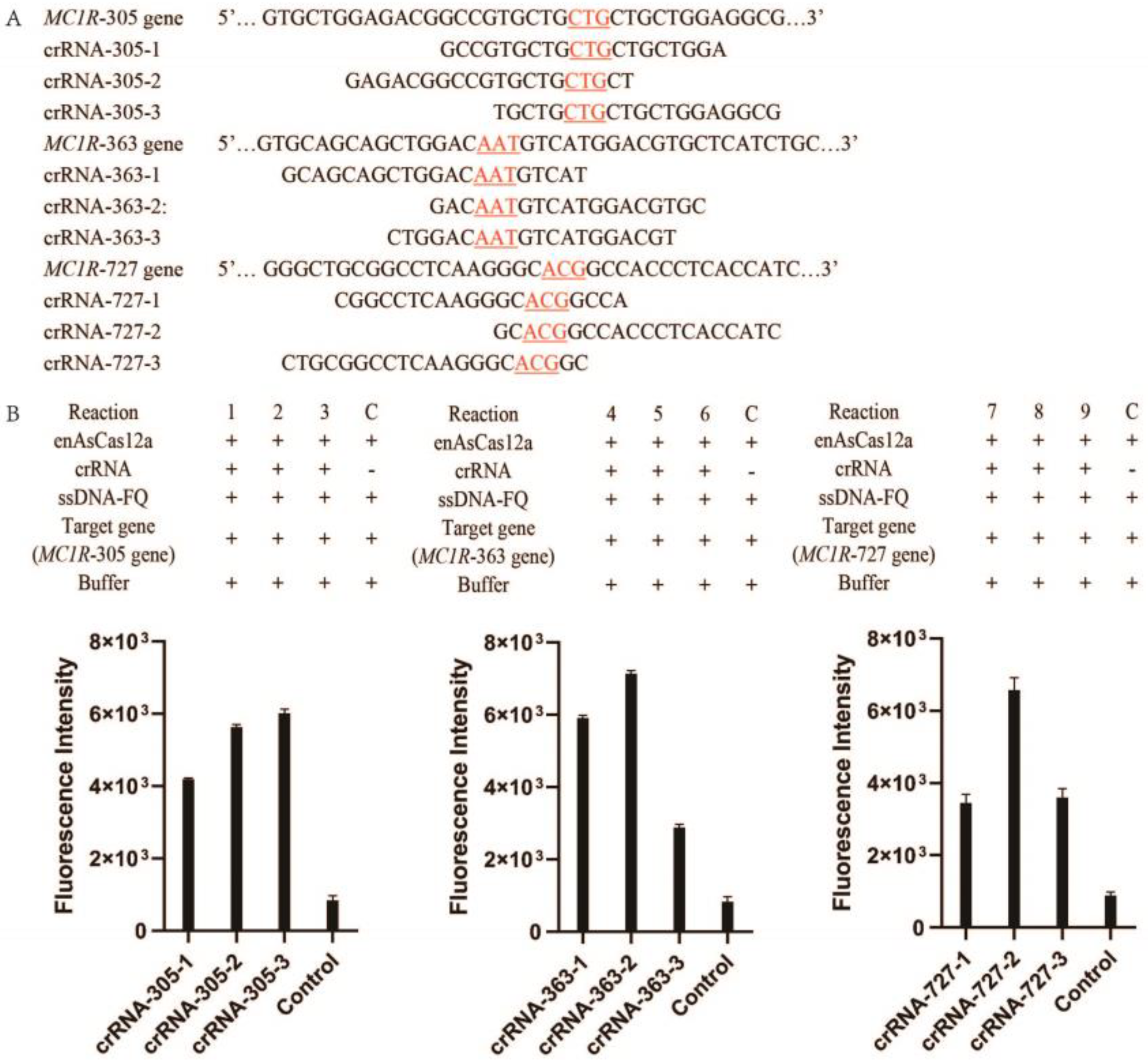
2.2. The Selection of Target Genes of MC1R

We selected three fragments of the MC1R gene, each containing one SNP site (T305C, T363C, and G727A) (Table S1). The PCR reactions were conducted as the following conditions: initial preheating to activation of Taq polymerase at 96 °C for 2 minutes (min), followed at 96 °C for 2 min, 55 °C for 30 seconds (s) and 72 °C for 30 s with 30 cycles of denaturation, annealing, and extension, and then an extra extension at 72 °C for 3 min. The lengths of the amplificated fragment of the MC1R-305, MC1R-T305C, MC1R-363, MC1R-T363C, MC1R-727, and MC1R-A727G were 96 bp, 96 bp, 98 bp, 98 bp, 94 bp, and 94 bp, respectively. The PCR reaction system was carried out in a 50 µL reaction volume, including 25 µL 2 × Taq DNA polymerase Mix (TSINGKE, China), 1 µL of the forward primer (10 pmol), 1 µL of reverse primers (10 pmol), 1 μL of template DNA (1ng), and adding 22 µL of H2O to 50 µL. The PCR products were purified (PCR purification kit, Beijing Tianmo Sci & Tech Development, Beijing, China), and the concentration of PCR products were quantified with Nano drops 2000 spectrophotometer (Thermo Fisher Scientific, Wilmington, DE, USA). It was stored at −20 °C and used as the target gene for in vitro detection.

2.3. The RNA Transcription In Vitro

The skeleton sequence of crRNA was as reported (gggAATTTCTACTGTTGTAGAT). The skeleton sequence of crRNA and the fragment of the target gene were combined by overlap PCR using a forward primer with a T7 promoter and a reverse primer with the target sequence (Figure S2). The PCR products were purified with a PCR purification kit (Beijing Tianmo Sci & Tech Development, Beijing, China), and used as a transcription template for in vitro transcription.
To obtain the crRNA with a specific target gene, the products of PCR were used as the DNA templates for in vitro transcription (HiScribe T7 High Yield RNA Synthesis Kit, New England Biolabs, Beijing, China). To remove the DNA template, DNase enzyme was added for 15 min at 37 °C (Cat. No. M0303S, New England Biolabs, Beijing, China). In the transcription reaction, a 20 µL transcription reaction mixture containing 1 µL T7 polymerase (5 U µL −1), 1 µL ATP (2.5 mM), 1 µL GTP (2.5 mM), 1 µL CTP (2.5 mM), 1 µL UTP (2.5 mM), 2 µL transcription buffer (10×), 5 µL target gene fragment (100 ng), and 8 µL H2O to the final volume. It was incubated at 37 °C for 8–10 h. Subsequently, the transcribed crRNA in vitro was purified (Monarch RNA Cleanup Kit, New England Biolabs, Beijing, China), and the concentration of crRNA was quantified with Nano drops 2000 spectrophotometer (Thermo Fisher Scientific, Wilmington, DE, USA). The copy numbers of crRNA were calculated by their concentration and length.

2.4. Optimization of the CRISPR/enAsCas12a Detection Assay

First, we optimized the concentration of PBS because the reaction buffer significantly affects the enzyme activity. The CRISPR/enAsCas12a reaction volume was 20 µL containing 1 × 10⁻³ mM crRNA-305-3, 200 ng enAsCas12a, 20 × 10−9 mM ssDNA reporter, 2 µL 10 × NEB buffer 2.1, 1 µL the target gene (20 ng/µL, MC1R-305), PBS buffer (10, 20, 50, and 100 mM), and H2O added to the final volume. The reaction mixture was incubated at 37 °C for 10 min, and the reaction was terminated by incubation at 95 °C for 5 min. Then, the 20 mM PBS with different concentration Mg2+ (0, 5, 10, 20, 30, 40, 50, 60, 70, 80, and 90 mM MgCl2) was tested.

2.5. The CRISPR/enAsCas12a Detect the MC1R Gene and the MC1R Gene with SNP

The single-strand DNA reporter with the fluorescent group at 5′ end and quenching group at 3′ ends (ssDNA-FQ), namely ROX-N12-BHQ2, was synthesized (TSINGKE, Beijing, China) and was listed in the Supplementary Table S2. The CRISPR/enAsCas12a detection assays were carried out as described in the previous report [3] and slightly optimized. In a 20 µL reaction system, it contained 200 ng purified enAsCas12a, 20 × 10−9 mM ssDNA with quenched fluorescent group sensor, 1 × 10⁻³ mM purified crRNA, and 1 µL of the target gene (MC1R-gene or MC1R-SNP gene) in the reaction buffer (20 mM PBS, 20 mM NaCl, 40 mM MgCl2, pH 7.4) and the reaction was incubated at 37 °C for 10 min. The reaction was terminated by heating at 95 °C for 10 min. For the fluorescence detection, the absorbance is 585 nm, and the emission is 605 nm.

2.6. Statistical Analysis

Statistical analysis of the florescent signals was performed using GraphPad Prism 8. The mean ± (SEM) standard error was obtained from every reaction sample of three independent experiments. The two-tailed Student’s t-test was used to compare significant differences between the two reaction samples (**** p < 0.0001, *** p < 0.001, ns: not significant at the 95% confidence level).

3. Results

3.1. The Detection of the MC1R Gene by CRISPR/enAsCas12a System

As shown in Figure 1, the CRISPR/enAsCas12a assay system is prepared by adding expressed and purified enAsCas12a, transcribed and purified crRNA, single-stranded DNA reporter with fluorophore and quencher (ssDNA-FQ), and the synthesized target gene. After incubating the CRISPR/enAsCas12a reaction system at 37 °C for 10 min, the reaction was terminated at 95 °C for 10 min. If the crRNA of the enAsCas12a-crRNA complex is complementary to the target gene, the trans-endonuclease of enAsCas12a is activated, which then cuts the ssDNA-FQ and generates a fluorescent signal. Otherwise, if the crRNA of the enAsCas12a-crRNA complex is not completely complementary to the target gene, the enAsCas12a could not be activated to produce a fluorescent signal.
For a detailed analysis of the CRISPR/enAsCas12a assay system, we designed and detected nine reaction systems with various crRNAs (reactions 1–9). Nine crRNA without PAM limitation were selected according to the MC1R gene fragments. The sequence of MC1R gene fragments and the various part of crRNAs are listed (Figure 2A). The MC1R gene was also synthesized (Table S1). In theory, the reaction without crRNA was used as the negative control and the background, and it shows that there is no background interference fluorescence and no generation of fluorescence. The reactions with crRNA, which have different a variable part, are tested and compared.
The results showed that each crRNA without additional PAM could combine with enAsCas12a and activate its trans-cleavage activity to detect specific genes even though it has different effects on Cas activity (Figure 2B). In Figure 2B, the results also showed that the fluorescence intensity of reaction 3 is better than reaction 1 and reaction 2, the fluorescence intensity of reaction 5 is better than reaction 4 and reaction 6, and the fluorescence intensity of reaction 8 is better than reaction 7 and reaction 9. LbCas12a, commonly used in molecular diagnostics in vitro, was also tested in the same way and could not be activated by crRNA without PAM (The results were shown in Supplementary Materials). Therefore, the results confirmed that the activity of enAsCas12a is mainly dependent on the crRNA and the target gene.

3.2. Optimization of the CRISPR/enAsCas12a Assay System

It is well known that buffer solution and metal ions affect enzyme activity. We aimed to optimize the solution conditions and Mg2+ concentration in the CRISPR/enAsCas12a assay system. First, we tested four different concentrations of phosphate buffer saline (PBS, 10, 20, 50, and 100 mM) that affect the sensitivity of enAsCas12a in nucleic acid detection. We found that enAsCas12a-mediated nucleic acid detection has the highest reaction efficiency in 20 mM PBS buffer compared with 10, 50, and 100 mM PBS (Figure 3A). Then, we screened divalent Mg2+ for their effect on the activity of enAsCas12a. The results showed that enAsCas12a is a kind of enzyme with strict metal ion dependence, and its activity is the best at the concentration of 40 mM Mg2+, rather than the commonly used concentration of 10 mM Mg2+ (Figure 3B).
To further confirm the influence of Mg2+ on the CRISPR/enAsCas12a assay system, the reaction efficiency of the nine reaction systems (reactions # 1–9) was tested in the 20 mM PBS with 10 or 40 mM Mg2+. The results showed that the fluorescence intensity of all reactions is improved more in 40 mM Mg2+ than in 10 mM Mg2+ (Figure 3C–E). Therefore, 40 mM Mg2+ enables higher sensitivity to the CRISPR/enAsCas12a assay in 20 mM PBS.

3.3. The SNPs of MC1R Detection by CRISPR/enAsCas12a

It has been reported that three kinds of SNPs (T305C, T363C, and G727A) of the MC1R gene affect skin color directly. The MC1R gene fragments with the SNP site were synthesized: the first fragment containing the T305C site (MC1R-T305C gene), the second fragment containing the T363C site (MC1R-T363C gene), and the third fragment containing the A727G site (MC1R-A727G gene). The corresponding MC1R gene fragments were also synthesized (Table 2). Here, we evaluated MC1R SNPs’ detection using the CRISPR/enAsCas12a assay system. The crRNA-305-3, crRNA-363-2, and crRNA-727-2, which had promoted the activity of enAsCas12a according to the previous results (Figure 2B), were selected for further detection. Each crRNA is complementary to the MC1R gene while not completely complementary to the MC1R gene with SNP.
In Figure 4, the reaction 1 (R 1) and the negative control 1 (C 1) have the same target gene (MC1R-305 gene), while C 1 is the negative control without addition of crRNA. Similarly, the reaction 2 (R 2) and the negative control 2 (C 2) have the same target gene (MC1R-T305C gene), the reaction 3 (R 3) and the negative control 3 (C 3) have the same target gene (MC1R-363 gene), the reaction 4 (R 4) and the negative control 4 (C 4) have the same target gene (MC1R-T363C gene), the reaction 5 (R 5) and the negative control 5 (C 5) have the same target gene (MC1R-727 gene), and the reaction 6 (R 6) and the negative control 6 (C 6) have the same target gene (MC1R-A727G gene). The results showed that all the negative controls have weak fluorescence as the background. The results also showed that the fluorescence intensity of reaction 1 is significantly higher than reaction 2 (Figure 4, left panel), the fluorescence intensity of reaction 3 is significantly higher than reaction 4 (Figure 4, middle panel), and the fluorescence intensity of reaction 5 is significantly higher than reaction 6 (Figure 4, right panel). Therefore, the CRISPR/enAsCas12a assay system can distinguish between the MC1R gene and the MC1R gene with an SNP site, which has only one base difference.

4. Discussion

The MC1R, a key factor in melanogenesis synthesis, is the main factor affecting hair and skin color and a major determinant of sun sensitivity. It has been reported that MC1R affects animals’ skin and fur color by controlling the synthesis of true melanin. It is also associated with many important production properties and affects the occurrence of many diseases. In addition, the SNPs of the MC1R gene can be used as an important indicator to determine the purity of varieties, breeds, and other studies, so the studies on skin and fur color and the related SNP detection have received wide attention. Many techniques for detecting SNPs have been established (as described above), but these methods cannot be widely used due to their complex operation and high technical requirements.
CRISPR/Cas diagnosis has received extensive attention in the past few years due to its accuracy, sensitivity, and rapidity. Here we used CRISPR/enAsCas12a, which is optimized to improve its detection efficiency, to detect the SNPs of the MC1R gene. To detect the MC1R gene, nine random crRNA, without adding additional PAM sequences, were designed, and the variable region of the crRNA is complementary to the MC1R gene fragments (Figure 2A). The results showed that every crRNA could activate the trans-exonuclease activity of enAsCas12a and the strong fluorescence could be produced (Figure 2B). So, unlike other Cas proteins, enAsCas12a could be activated by crRNA independently on specific PAM sequences in vitro and detect any gene fragments without PAM. We also found that CRISPR/enAsCas12a can detect all target nucleic acid sequences without PAM, but its detection efficiency is different due to the variable sequence contained in crRNA. These results confirmed that the activity of enAsCas12a is mainly affected by the target gene sequence. Therefore, we speculate that enAsCas12a has some base preference, which requires further confirmation. Furthermore, we optimized the solution buffer and the concentration of divalent Mg2+ and found that the CRISPR/enAsCas12a assay system is more sensitive in 20 mM PBS with 40 mM Mg2+. Finally, we proved that the CRISPR/enAsCas12a assay system could distinguish between genes and SNPs with a single base nucleotide difference. So, the current approach confirms the detection activity of CRISPR/enAsCas12a and the sensitivity for detecting SNPs without PAM, thus expanding the application of CRISPR/enAsCas12a in vitro detection and eliminating the limitation of PAM.
In summary, this method has more advantages than traditional technologies. First, the costs are significantly decreased. If the samples to be tested are sent to the relevant testing organization or company, each sample will cost from ten to tens of dollars, while the method we have established needs only about USD 1. So, this is a low-cost diagnostic analysis with high sensitivity. Second, this method is fast. The whole process of SNP detection based on CRISPR/Cas needs about 1–2 h, while other detection methods require large instruments and equipment, professional testing institutions, or companies, and the samples must be transported. We have to wait at least one day to receive the results; this method is rapid in detection. Third, it is an easy diagnostic approach. If the reagents for detection are provided, they can be carried out in general laboratories. Thus, this technology is a low-cost, convenient, and rapid detection method.

Supplementary Materials

The following supporting information can be downloaded at: https://www.mdpi.com/article/10.3390/genes14020394/s1, Figure S1: The expression and purification of enAsCas12a and LbCas12a; Figure S2: The amplification results of crRNA (crRNA-102-1/2/3, crRNA-121-1/2/3, and crRNA-243-1/2/3); Figure S3: The MC1R detection by the CRISPR/LbCas12a; Table S1: the gene of MC1R, the fragment of MC1R-102/121 gene, and the fragment of MC1R-243 gene; Table S2: the sequence of ssDNA-FQ.

Author Contributions

Conceptualization, W.Y.; methodology, W.Y. and D.T.; software, W.Y.; validation, S.Z. and Y.Z.; formal analysis, W.Y. and B.X.; investigation, Y.Z.; resources, W.Y.; data curation, W.Y., D.T. and B.X.; writing—original draft preparation, W.Y.; writing—review and editing, Y.Z.; visualization, W.Y. and D.T.; supervision, S.Z.; project administration, S.Z. All authors have read and agreed to the published version of the manuscript.

Funding

This research was funded by the Laboratory of Lingnan Modern Agriculture Project (No. NZ2021005), the Natural Science Foundation of China (32072685) and the Fund of Modern Industrial Technology System of Pigs (CARS-35).

Institutional Review Board Statement

Not applicable.

Informed Consent Statement

Not applicable.

Data Availability Statement

Not applicable.

Conflicts of Interest

The authors declare no conflict of interest.

References

  1. Aman, R.; Mahas, A.; Mahfouz, M. Nucleic Acid Detection Using CRISPR/Cas Biosensing Technologies. ACS Synth. Biol. 2020, 9, 1226–1233. [Google Scholar] [CrossRef]
  2. Chen, J.S.; Ma, E.; Harrington, L.B.; Da Costa, M.; Tian, X.; Palefsky, J.M.; Doudna, J.A. CRISPR-Cas12a target binding unleashes indiscriminate single-stranded DNase activity. Science 2018, 360, 436–439. [Google Scholar] [CrossRef]
  3. Xie, S.; Tao, D.; Fu, Y.; Xu, B.; Tang, Y.; Steinaa, L.; Hemmink, J.D.; Pan, W.; Huang, X.; Nie, X.; et al. Rapid Visual CRISPR Assay: A Naked-Eye Colorimetric Detection Method for Nucleic Acids Based on CRISPR/Cas12a and a Convolutional Neural Network. ACS Synth. Biol. 2022, 11, 383–396. [Google Scholar] [CrossRef]
  4. Fozouni, P.; Son, S.; Diaz de Leon Derby, M.; Knott, G.J.; Gray, C.N.; D’Ambrosio, M.V.; Zhao, C.; Switz, N.A.; Kumar, G.R.; Stephens, S.I.; et al. Amplification-free detection of SARS-CoV-2 with CRISPR-Cas13a and mobile phone microscopy. Cell 2021, 184, 323–333.e329. [Google Scholar] [CrossRef]
  5. Soh, J.H.; Balleza, E.; Abdul Rahim, M.N.; Chan, H.M.; Mohd Ali, S.; Chuah, J.K.C.; Edris, S.; Atef, A.; Bahieldin, A.; Ying, J.Y.; et al. CRISPR-based systems for sensitive and rapid on-site COVID-19 diagnostics. Trends Biotechnol. 2022, 40, 1346–1360. [Google Scholar] [CrossRef]
  6. Wang, J.; Xia, Q.; Wu, J.; Lin, Y.; Ju, H. A sensitive electrochemical method for rapid detection of dengue virus by CRISPR/Cas13a-assisted catalytic hairpin assembly. Anal. Chim. Acta 2021, 1187, 339131. [Google Scholar] [CrossRef]
  7. Xu, B.; Gong, P.; Zhang, Y.; Wang, Y.; Tao, D.; Fu, L.; Khazalwa, E.M.; Liu, H.; Zhao, S.; Zhang, X.; et al. A one-tube rapid visual CRISPR assay for the field detection of Japanese encephalitis virus. Virus Res. 2022, 319, 198869. [Google Scholar] [CrossRef]
  8. Liu, S.; Tao, D.; Liao, Y.; Yang, Y.; Sun, S.; Zhao, Y.; Yang, P.; Tang, Y.; Chen, B.; Liu, Y.; et al. Highly Sensitive CRISPR/Cas12a-Based Fluorescence Detection of Porcine Reproductive and Respiratory Syndrome Virus. ACS Synth. Biol. 2021, 10, 2499–2507. [Google Scholar] [CrossRef]
  9. Ghosh, N.; Saha, I.; Sharma, N. Palindromic target site identification in SARS-CoV-2, MERS-CoV and SARS-CoV-1 by adopting CRISPR-Cas technique. Gene 2022, 818, 146136. [Google Scholar] [CrossRef]
  10. Talwar, C.S.; Park, K.H.; Ahn, W.C.; Kim, Y.S.; Kwon, O.S.; Yong, D.; Kang, T.; Woo, E. Detection of Infectious Viruses Using CRISPR-Cas12-Based Assay. Biosensors 2021, 11, 301. [Google Scholar] [CrossRef]
  11. Ma, P.; Meng, Q.; Sun, B.; Zhao, B.; Dang, L.; Zhong, M.; Liu, S.; Xu, H.; Mei, H.; Liu, J.; et al. MeCas12a, a Highly Sensitive and Specific System for COVID-19 Detection. Adv. Sci. 2020, 7, 2001300. [Google Scholar] [CrossRef] [PubMed]
  12. Swarts, D.C.; Jinek, M. Mechanistic Insights into the cis- and trans-Acting DNase Activities of Cas12a. Mol. Cell 2019, 73, 589–600.e584. [Google Scholar] [CrossRef] [PubMed]
  13. Ooi, K.H.; Liu, M.M.; Tay, J.W.D.; Teo, S.Y.; Kaewsapsak, P.; Jin, S.; Lee, C.K.; Hou, J.; Maurer-Stroh, S.; Lin, W.; et al. An engineered CRISPR-Cas12a variant and DNA-RNA hybrid guides enable robust and rapid COVID-19 testing. Nat. Commun. 2021, 12, 1739. [Google Scholar] [CrossRef] [PubMed]
  14. Nguyen, L.T.; Smith, B.M.; Jain, P.K. Enhancement of trans-cleavage activity of Cas12a with engineered crRNA enables amplified nucleic acid detection. Nat. Commun. 2020, 11, 4906. [Google Scholar] [CrossRef] [PubMed]
  15. Huyke, D.A.; Ramachandran, A.; Bashkirov, V.I.; Kotseroglou, E.K.; Kotseroglou, T.; Santiago, J.G. Enzyme Kinetics and Detector Sensitivity Determine Limits of Detection of Amplification-Free CRISPR-Cas12 and CRISPR-Cas13 Diagnostics. Anal. Chem. 2022, 94, 9826–9834. [Google Scholar] [CrossRef]
  16. Ramachandran, A.; Santiago, J.G. CRISPR Enzyme Kinetics for Molecular Diagnostics. Anal. Chem. 2021, 93, 7456–7464. [Google Scholar] [CrossRef]
  17. Rossetti, M.; Merlo, R.; Bagheri, N.; Moscone, D.; Valenti, A.; Saha, A.; Arantes, P.R.; Ippodrino, R.; Ricci, F.; Treglia, I.; et al. Enhancement of CRISPR/Cas12a trans-cleavage activity using hairpin DNA reporters. Nucleic Acids Res. 2022, 50, 8377–8391. [Google Scholar] [CrossRef]
  18. Zhang, L.; Jiang, Z.; Zhou, Z.; Sun, J.; Yan, S.; Gao, W.; Shao, Y.; Bai, Y.; Wu, Y.; Yan, Z.; et al. A TaqMan Probe-Based Multiplex Real-Time PCR for Simultaneous Detection of Porcine Epidemic Diarrhea Virus Subtypes G1 and G2, and Porcine Rotavirus Groups A and C. Viruses 2022, 14, 1819. [Google Scholar] [CrossRef] [PubMed]
  19. Islam, M.T.; Alam, A.R.U.; Sakib, N.; Hasan, M.S.; Chakrovarty, T.; Tawyabur, M.; Islam, O.K.; Al-Emran, H.M.; Jahid, M.I.K.; Anwar Hossain, M. A rapid and cost-effective multiplex ARMS-PCR method for the simultaneous genotyping of the circulating SARS-CoV-2 phylogenetic clades. J. Med. Virol. 2021, 93, 2962–2970. [Google Scholar] [CrossRef]
  20. Dema, A.; Ganji, V.K.; Yella, N.R.; Putty, K. A novel one-step amplification refractory mutation system PCR (ARMS-PCR) for differentiation of canine parvovirus-2 variants. Virus Genes 2021, 57, 426–433. [Google Scholar] [CrossRef]
  21. Alyethodi, R.R.; Singh, U.; Kumar, S.; Alex, R.; Sengar, G.S.; Raja, T.V.; Deb, R.; Prakash, B. Designing, optimization, and validation of whole blood direct T-ARMS PCR for precise and rapid genotyping of complex vertebral malformation in cattle. BMC Biotechnol. 2021, 21, 36. [Google Scholar] [CrossRef]
  22. Bookari, K.; Arrish, J.; Zaher, S.; Alkhalaf, M.; Alharbi, M.; Alkhattaf, N.; Harb, Z.; Al Hinai, E.; Alanqodi, N.; Almajed, S.; et al. A Snapshot of the Experience of Dietitians during the COVID-19 Crisis in Five Arab Countries: Findings from a Regional Cross-Sectional Study. Nutrients 2022, 14, 4904. [Google Scholar] [CrossRef]
  23. Li, L.; Li, S.; Wu, N.; Wu, J.; Wang, G.; Zhao, G.; Wang, J. HOLMESv2: A CRISPR-Cas12b-Assisted Platform for Nucleic Acid Detection and DNA Methylation Quantitation. ACS Synth. Biol. 2019, 8, 2228–2237. [Google Scholar] [CrossRef]
  24. Chen, Y.; Mei, Y.; Jiang, X. Universal and high-fidelity DNA single nucleotide polymorphism detection based on a CRISPR/Cas12a biochip. Chem. Sci. 2021, 12, 4455–4462. [Google Scholar] [CrossRef]
  25. Wang, M.; Liu, X.; Yang, J.; Wang, Z.; Wang, H.; Wang, X. CRISPR/Cas12a-based biosensing platform for the on-site detection of single-base mutants in gene-edited rice. Front. Plant Sci. 2022, 13, 944295. [Google Scholar] [CrossRef]
  26. Kijas, J.M.; Wales, R.; Tornsten, A.; Chardon, P.; Moller, M.; Andersson, L. Melanocortin receptor 1 (MC1R) mutations and coat color in pigs. Genetics 1998, 150, 1177–1185. [Google Scholar] [CrossRef]
  27. Ji, R.L.; Tao, Y.X. Melanocortin-1 receptor mutations and pigmentation: Insights from large animals. Prog. Mol. Biol. Transl. Sci. 2022, 189, 179–213. [Google Scholar] [CrossRef]
  28. Zhong, H.; Zhang, J.; Tan, C.; Shi, J.; Yang, J.; Cai, G.; Wu, Z.; Yang, H. Pig Coat Color Manipulation by MC1R Gene Editing. Int. J. Mol. Sci. 2022, 23, 356. [Google Scholar] [CrossRef]
  29. Wang, W.; Guo, D.Y.; Lin, Y.J.; Tao, Y.X. Melanocortin Regulation of Inflammation. Front. Endocrinol. 2019, 10, 683. [Google Scholar] [CrossRef]
  30. Switonski, M.; Mankowska, M.; Salamon, S. Family of melanocortin receptor (MCR) genes in mammals-mutations, polymorphisms and phenotypic effects. J. Appl. Genet. 2013, 54, 461–472. [Google Scholar] [CrossRef] [Green Version]
  31. Chen, S.; Zhu, B.; Yin, C.; Liu, W.; Han, C.; Chen, B.; Liu, T.; Li, X.; Chen, X.; Li, C.; et al. Palmitoylation-dependent activation of MC1R prevents melanomagenesis. Nature 2017, 549, 399–403. [Google Scholar] [CrossRef] [PubMed]
  32. Almathen, F.; Elbir, H.; Bahbahani, H.; Mwacharo, J.; Hanotte, O. Polymorphisms in MC1R and ASIP Genes are Associated with Coat Color Variation in the Arabian Camel. J. Hered. 2018, 109, 700–706. [Google Scholar] [CrossRef]
Figure 1. The CRISPR/enAsCas12a assay system in vitro.
Genes 14 00394 g001
Figure 2. The MC1R detection by the CRISPR/enAsCas12a. (A). The MC1R gene fragment containing 305, 363, and 727 site; the variable part of the crRNA sequence (the red parts translate to the 102nd, the 121st, and the 243rd amino acid of MC1R); (B). the fluorescence detection of every reaction (C, control).
Genes 14 00394 g002
Figure 3. Optimization of the CRISPR/enAsCas12a assay system. (A) enAsCas12a-mediated MC1R detection in 10, 20, 50, and 100 mM PBS buffer with different concentrations of Mg2+; (B) enAsCas12a-mediated MC1R detection in 20 mM PBS buffer with different concentrations of Mg2+; (CE). enAsCas12a-mediated nucleic acid fragment detection of MC1R (containing 102, 121, and 243 site) in different buffer solutions (Buffer1: 20 mM PBS, 10 mM Mg2+; Buffer2: 20 mM PBS, 40 mM Mg2+).
Genes 14 00394 g003
Figure 4. MC1R SNPs detection by CRISPR/enAsCas12a assay system. The CRISPR/enAsCas12a assay system to detect MC1R-305-gene fragment and MC1R-T305C-SNP gene fragment in vitro (the left panel); the CRISPR/enAsCas12a assay system to detect MC1R-363-gene fragment and MC1R-T363C-SNP gene fragment in vitro (the middle panel); the CRISPR/enAsCas12a assay system to detect MC1R-727-gene fragment and MC1R-G727A-SNP gene fragment in vitro (the right panel) (**** p < 0.0001).
Genes 14 00394 g004
Table 1. The dominant SNP sites of the MC1R gene.
AlleteCoat ColorCodon
102121243
MC1R*1(E+)Wild type, European wild boarCTG
Leu
AAT
Asn
GCG
Ala
MC1R*1(E+)Wild type, Japanese wild boar---
-
--C
-
---
-
MC1R*2(ED1)Dominant black-C-
-
--C
-
--A
-
MC1R*4(e)Recessive red---
-
---
-
A--
Thr
E, the Extension locus in pig; E+, wild type; ED, the dominant black; e, the recessive red. The dash (-) indicates the identity to the top base or amino acid.
Table 2. The sequence of the MC1R gene and its SNPs’ sequence.
GeneSequence
MC1R-305 gene…5’-GAGACGGCCGTGCTGCTGCTGCTGGAGGCGGGC-3’…
MC1R-T305C SNP…5’-GAGACGGCCGTGCTGCcGCTGCTGGAGGCGGGC-3’…
MC1R-363 gene…5’-GTGCAGCAGCTGGACAATGTCATGGACGTGCTC-3’…
MC1R-T363C SNP…5’-GTGCAGCAGCTGGACAAcGTCATGGACGTGCTC-3’…
MC1R-727 gene…5’-TGCGGCCTCAAGGGCACGGCCACCCTCACCATC-3’…
MC1R-A727G SNP…5’-TGCGGCCTCAAGGGCgCGGCCACCCTCACCATC -3’…
Disclaimer/Publisher’s Note: The statements, opinions and data contained in all publications are solely those of the individual author(s) and contributor(s) and not of MDPI and/or the editor(s). MDPI and/or the editor(s) disclaim responsibility for any injury to people or property resulting from any ideas, methods, instructions or products referred to in the content.

