Article

Homologous Recombination and Repair Functions Required for Mutagenicity during Yeast Meiosis

by
Liat Morciano
1,†,
Renana M. Elgrabli
1,†,
Drora Zenvirth
1 and
Ayelet Arbel-Eden
1,2,*
1
Department of Genetics, Hebrew University of Jerusalem, Jerusalem 91904, Israel
2
The Medical Laboratory Sciences Department, Hadassah Academic College, Jerusalem 91010, Israel
*
Author to whom correspondence should be addressed.
These authors contributed equally to this work.
Genes 2023, 14(11), 2017; https://doi.org/10.3390/genes14112017
Submission received: 29 September 2023 / Revised: 22 October 2023 / Accepted: 23 October 2023 / Published: 28 October 2023
(This article belongs to the Special Issue Mechanisms of DNA Damage, Repair and Mutagenesis 2023)

Abstract

:
Several meiotic events reshape the genome prior to its transfer (via gametes) to the next generation. The occurrence of new meiotic mutations is tightly linked to homologous recombination (HR) and firmly depends on Spo11-induced DNA breaks. To gain insight into the molecular mechanisms governing mutagenicity during meiosis, we examined the timing of mutation and recombination events in cells deficient in various DNA HR-repair genes, which represent distinct functions along the meiotic recombination process. Despite sequence similarities and overlapping activities of the two DNA translocases, Rad54 and Tid1, we observed essential differences in their roles in meiotic mutation occurrence: in the absence of Rad54, meiotic mutagenicity was elevated 8-fold compared to the wild type (WT), while in the tid1Δ mutant, there were few meiotic mutations, nine percent compared to the WT. We propose that the presence of Rad54 channels recombinational repair to a less mutagenic pathway, whereas repair assisted by Tid1 is more mutagenic. A 3.5-fold increase in mutation level was observed in dmc1∆ cells, suggesting that single-stranded DNA (ssDNA) may be a potential source for mutagenicity during meiosis. Taken together, we suggest that the introduction of de novo mutations also contributes to the diversification role of meiotic recombination. These rare meiotic mutations revise genomic sequences and may contribute to long-term evolutionary changes.

Graphical Abstract

1. Introduction

A central role of meiosis is to generate haploid gametes with new combinations of genetic material. Genetic variation is achieved by recombination and independent assortment of chromosomes. These meiotic events contribute to genome heterogeneity by scrambling and shuffling preexisting sequences. Rare, de novo mutations that occur during meiosis further increase variation and are the only source of new genetic information and novel alleles. About sixty years ago, Magni and Von Borstel (1962) [1] found that the rate of de novo mutation in Saccharomyces cerevisiae meiosis is higher than the rate in mitosis, and they coined the term “the meiotic effect” [2]. Since their discovery, we and others have confirmed that the rates of meiotic mutation are elevated in yeast cells [3,4,5] and in humans [6,7]. Depending on the locus examined, the mutation rates in meiosis are 6- to 22-fold higher than in mitotically growing cells [1,5].
Several lines of evidence link meiotic mutations to the recombination process: An early finding was that colonies with de novo mutations at his4 showed a high frequency of recombination of markers bracketing the gene [8]. Likewise, meiotic mutations at the CAN1 reporter had a high frequency of recombination between two adjacent markers, covering 2.2 kb around this reporter, compared to non-recombinant colonies or compared to vegetatively growing cells [5]. Meiotic mutations were found to depend strongly on DNA double-strand breaks (DSBs), induced by the meiosis-specific endonuclease Spo11. We further showed that most meiotic mutations do not occur during S-phase, which is the phase where most DNA synthesis takes place [5]. Rather, elevated meiotic mutagenicity appears at the same time and by the same kinetics as recombination events [5]. These findings imply that breakage, followed by recombination, is an intrinsic factor enhancing de novo mutagenicity. Nonetheless, we found that approximately half of the meiotic mutations were not associated with recombination events between closely flanking markers [5], and we therefore suggested that mutations may result not only from recombinational repair but also from sister chromatid repair of meiotic DSBs [9]. This leaves an open question: What are the relative contributions to meiotic mutagenicity of the various repair functions that operate during meiosis? We address this question in the current study.
The meiotic recombination process in the yeast S. cerevisiae is well characterized, with an orderly sequence of events in which many genes participate. Meiotic recombination is initiated by massive, tightly controlled breakage of the DNA via the Spo11 endonuclease and its assisting factors [10,11,12,13]. Next, the MRX (Mre11/Rad50/Xrs2) protein complex plays an essential role as a DNA damage sensor, and the resulting DNA ends are digested to create single-strand 3′ DNA ends (ssDNA), which are ~820 nts long [14]. Following end-resection, strand invasion is mediated by two recombinases, Rad51p and the meiosis-specific Dmc1p [15]; both act concurrently to generate interhomolog bias during the HR process [16,17]. The presence of the Rad51 protein, but not its strand invasion activity, is required for proper Dmc1-mediated strand invasion [18]. Rad51’s action requires interactions with Rad54, a DNA translocase, which assists in strand invasion [19,20] and stimulates Rad51 to preferentially utilize the sister chromatid for repair [21,22,23]. Likewise, the Rad54 paralog Tid1/Rdh54 is needed to stimulate Dmc1 activity to bind recombinogenic sites [24], and together, Tid1 and DMC1 preferentially act to promote interhomolog recombination [23]. In normal meiosis, the Hed1 protein inhibits Rad51-mediated recombination [25,26] by competing with Rad54 [27]. The direct phosphorylation of both Rad54 and Hed1 by Mek1 [28] prevents Rad51/Rad54 complex formation [29,30]. The invaded intermediates are substrates for DNA synthesis and the subsequent ligation of joint molecules (JMs). These JMs generate several types of recombination intermediates that mainly resolve to either non-crossover (NCO) or crossover (CO) products.
In the present study, we focus on the HR process, which is a core event enhancing meiotic mutagenicity, to better define recombinational repair functions required for mutagenicity during meiosis. We generated a series of mutants deleted in genes involved in meiotic recombination (namely SPO11, MRE11, DMC1, HED1, RAD54, TID1, and MUS81) and examined their effects on the occurrence of meiotic mutations. Combining mutant analysis and time course experiments, we revisited the meiotic HR repair pathway to study the kinetics of meiotic mutagenicity, recombination, and the interactions between these processes. Our results indicate that some HR genes are involved in mutagenic DSB repair pathways and others are involved in repair pathways that reduce mutagenesis. ssDNA that is exposed prior to JM formation and the various ways by which DSB repair is handled contribute to generate balanced meiotic mutagenicity that, on one hand, provide genome variability and, on the other hand, do not overload the genome with excessive deleterious variants.

2. Materials and Methods

2.1. Strain Construction

All strains used for this study are of the SK1 genetic background and are listed in Table 1. Gene deletions, insertions, and gene replacements were obtained using standard polymerase chain reaction (PCR)-based methods.
The original “founder” strains for all strains generated in this study are OMY78-1 and OMY75-2 [5]. A detailed description and a thorough characterization of the wild-type (WT) strain is found in Mansour et al. (2020). In brief, we inserted a copy of the ADE2 gene ~400 bps upstream to the functional copy of this hemizygous reporter and we selected for mutations in the functional CAN1 allele, which lead to resistance to canavanine, by spreading ascospores on plates containing canavanine and lacking adenine (Can −Ade). To enable detection of intragenic recombination events, these strains also contained heteroallelic mutations in HIS4 (his4-X/his4-B) [31], and recombination was measured by recovering colonies on −His plates.
Strains deleted for MRE11, DMC1, HED1, HED1DMC1, TID1, and MUS81 were obtained by generating the relevant PCR-amplified gene product from the kanMX4-deletion library (http://chemogenomics.pharmacy.ubc.ca/hiplab/GGCN_Lab/SGDP/group/yeast_deletion_project/deletions3.html, accessed on 20 September 2023) and the transformation of this PCR fragment into the haploid YRA10 strain, followed by further crosses and ascus dissections.
SPO11 was deleted in the haploid strain by transformation with the corresponding PCR-amplified product from the hisG-URA3-hisG plasmid. URA3 was “popped out” by growing the transformed colonies on 5FOA.
The RAD54-deleted strain (ALY92) was obtained by haploid rad54∆::URA3 strain with YRA12 and selecting the desired Ura+ spore colony following tetrad dissection.
The RAD54-deleted strain was manipulated to carry the cdc7-as3 allele (ALY170). Haploids of the latter were obtained by mating the RAD54 cdc7-as3 haploid strain OMY78-1 with ALY92 (rad54Δ), followed by tetrad dissection. The cdc7-as3 allele was detected by PCR.

2.2. Media

YPD (complete medium with glucose as a carbon source), YPA (complete medium with acetate as a carbon source, used for pre-sporulation), SPM (sporulation medium), and SPO (solid sporulation medium) were prepared as described previously [32]. The drop-out media (various amino acid and nucleotide mixes) were prepared as described by Rose et al. (1990), except that lysine, leucine, tryptophan, arginine, and adenine were added at concentrations of 80 mg/L each, when required. Can −Ade medium: canavanine (Sigma C9758) was added at 60 mg/L to the synthetic complete medium lacking arginine and adenine. SCLG-2 medium: synthetic complete [33], with 0.5% glucose and without adenine and arginine.

2.3. Determining Mutation Rates during Mitotic Cell Divisions with Fluctuation Analysis

Yeast strains were thawed from frozen stocks and streaked to single colonies on YPD plates. In each fluctuation assay, single colonies from the appropriate strains were patched on YPD plates, and after overnight growth at 30 °C, the patches were lightly replica-plated onto the selective medium, Can −Ade (60 mg/L). After 4 days, areas with no mutant colonies were chosen, and cells from the corresponding YPD plate were cultured in liquid SCLG-2 medium [34]. Cultures were shaken overnight at 30 °C, diluted into SCLG-2 medium, and dispensed into 96-well plates, 100 µL per well. When testing the CAN1 mutation rate, ~1000 cells were cultured per well. Cultures in 96-cell plates were grown for 48 h at 30 °C without shaking in a high humidity incubator, to prevent evaporation. For each culture, six wells were pooled and diluted, and cells were counted using a Neubauer counting chamber. To determine the Nt value, which reflects the actual number of viable cells per subculture (well) at the end of the experiment, 100 µL aliquots of the pooled and diluted cultures were plated on each of 4 YPD plates. Then, 100 µL of double-distilled water was added to each of the remaining 90 subcultures. For each subculture, 100 µL was spot-plated onto dried Can −Ade plates to identify mutant colonies (9 spots per plate). Plates were allowed to dry for 3 h at room temperature and then incubated at 30 °C for 5 days, after which the number of spots without resistant colonies was recorded.
The method chosen to calculate the mutation rate in these mitotically growing cultures was the P-zero (P0) method [35]. As the number of mutations per culture follows the Poisson distribution, the proportion of cultures without mutant colonies, P0, is the zero term of the Poisson, given by the following equation [36]:
a = l n ( p 0 ) N t
where a is the mutation rate, p0 is the fraction of subcultures (spots) with zero colonies, and Nt is the average number of cells per subculture at the time of application of the selective agent.

2.4. Determination of Meiotic Mutation and Recombination Rates

To determine mutation and recombination rates during and after meiosis, cells from −80 °C stocks were streaked to single colonies onto YPD plates. Several single colonies were then picked and patched (~2 cm × 2 cm patch) onto another YPD plate and incubated overnight. This plate was replica-plated onto three different plates: SPO, −His (to exclude preexisting recombinants), and Can −Ade (to exclude preexisting mutants). The patch that showed the highest sporulation efficiency on the SPO plate was chosen, and a clean section that did not show any colonies on the remaining two plates (−His and Can −Ade) was marked. A culture of each relevant strain, from the corresponding YPD plate, was inoculated into 2 mL liquid YPD, grown overnight at 30 °C, and then resuspended in 100 mL of liquid YPA at a dilution of 1:2500, followed by incubation with vigorous shaking (200 rpm) at 30 °C for ~18 h to reach a titer of 1–2 × 10⁷ cells/mL. In cases where strains grew more slowly (such as in mre11Δ and rad54Δ strains), the cells were resuspended in YPA at a dilution of 1:1000. Cells were then washed twice in water and resuspended in the same volume (100 mL) of liquid SPM, which was pre-warmed to 30 °C. Upon resuspension, a sample was taken and, after appropriate dilutions, spread on the selection plates (−His or Can −Ade); this represented the meiotic time zero sample. The remaining culture was then vigorously shaken (200 rpm) at 30 °C. The volume of the shaking flasks was ten times greater than the volume of the liquid medium (YPA or SPM) to allow efficient aeration. Cells were taken for analysis after 2–8 h in sporulation conditions and spread on selection plates, which were then incubated at 30 °C for 2–7 days, after which recombinant and mutant colonies were counted. The number of colonies appearing on selection plates from time zero was always subtracted from the numbers obtained at later time points during meiosis, as the time zero colonies reflect events that occurred in the mitotic divisions prior to meiosis. To determine the number of cells that had been spread on the selection plates, serial 1:10 dilutions were made, until we obtained a cell suspension of ~5 × 10³ cells/mL (as determined by counting the cells in a Neubauer counting chamber). Three aliquots of 100 µL each were then spread on YPD plates to score for cell viability at each time point. To deduce the original number of cells in each sample, the average number of colonies that grew on YPD plates was multiplied by the dilution factor. Recombination and mutation rates were then calculated. Sporulation efficiency after 24 h in sporulation conditions was determined microscopically by counting monads, dyads, and 3–4-spore asci vs. non-sporulated cells.

2.5. Meiotic Time Course Experiments

Meiotic time course experiments were performed to follow the kinetics and rates of recombination and/or mutation and to monitor cell viability along the meiotic process.
In time course experiments, the same protocol was followed as described above. Usually, 4 samples were collected after 0, 4, 6, and 8 h in sporulation medium (SPM), and whenever needed, a 2 h sample was also collected. Cells were diluted in 100 mL of liquid YPA and resuspended in the same volume of pre-warmed SPM in 1 L flasks. Cell samples were collected and treated as explained above.
In earlier studies using similar meiotic conditions, we showed that in our SK1 strains, global DNA synthesis at S-phase takes place at around 2 h after transfer to SPM, DSBs begin to appear at 3 h, recombination occurs between 4 and 6 h, and meiosis is completed after about 8 h [37,38]. Diploid dmc1Δ cultures embark on meiosis but become arrested in meiotic prophase I [39] and do not reach the haploidization commitment point. Return to mitotic growth (RTG) rescues the viability of these dmc1Δ cultures and allows one to assess the levels of recombination and mutations, although the cultures remain diploid.

2.6. Meiotic Experiments Using the Inhibitor PP1

For strains bearing the cdc7-as3 allele, meiosis was induced in the presence of the inhibitor PP1 [4-amino-1-tert-butyl-3-(p-methylyphenyl) pyrazolo [3,4-d] pyrimidine], which halts cells after meiotic S-phase [40]. Following thawing from −80 °C and patching as described above, cells were inoculated in 2 mL liquid YPD, grown overnight (at 30 °C), and then resuspended in 60 mL of liquid YPA at a dilution of 1:2500 for the RAD54 strain, or of 1:1000 for rad54Δ, followed by incubation with vigorous shaking (200 rpm) at 30 °C for ~18 h to reach a titer of 1–2 × 10⁷ cells/mL. Cells were then washed twice in water and resuspended in 60 mL and divided into two flasks of liquid SPM. Upon transfer to SPM, PP1 was added to half the culture at a final concentration of 15 µM. The other half was left untreated to proceed normally through meiosis. After 8 h in sporulation conditions, both PP1-treated and untreated cultures were spread on YPD plates, as well as on selection plates (as described above), and rates of recombination and mutation were calculated.

2.7. Statistical Treatments

Bars of mutation rates in the figures represent standard errors (SE), calculated from n independent experiments with each strain, performed on different days. Most meiotic experiments were based on at least 8 independent experiments. In the WT, we based our analytical calculations on at least 19 meiotic experiments. Comparisons of mutation rates between strains were based on t-tests (at 0.05 level).

3. Results

De novo meiotic mutations lead to genetic variability by generating novel alleles. To date, much of our knowledge regarding de novo mutations derives from the analysis of somatic mutations appearing during mitotic cell divisions in normal and in cancerous cells [41]. In the present work, we focus on mutations that occur during meiosis, which show a marked enhancement compared to mitotic mutations, and study the occurrence of meiotic mutagenesis in relation to recombination functions.
We first characterized the parental reference wild-type (WT) diploid strain (YRA23-1), which shows rapid and synchronous meiosis upon transfer to SPM, as described in the Materials and Methods and [37,38]. YRA23-1 showed a high sporulation efficiency (~90%) and maintained 100% and 80% viability 8 h and 24 h following the induction of meiosis, respectively. Spores recovered after tetrad dissection were 96% viable (366 viable spores out of 380, Table 2).
In order to measure the frequency of mutagenesis, we used the reporter gene CAN1; mutations in this gene enable growth on canavanine medium (Can −Ade) [2,34]. Since these mutations are recessive, we constructed a diploid strain in which CAN1 is expressed only from one copy of chromosome V, in the SK1 genetic background [4,5]. Thus, mutations in the single functional copy of CAN1 allow growth on canavanine medium. To follow recombination around the CAN1 reporter, we inserted the ADE2 gene upstream of the functional CAN1 allele and the gene URA3 downstream of the promoterless can1 allele (see Materials and Methods). Colonies that could grow without adenine and without uracil could only have been produced by crossing over within the 2210 bp spanning the CAN1 reporter [5]. Our yeast strains are also heterozygous for two separate alleles of the HIS4 gene (his4-X/his4-B; Table 1) to enable the detection of intragenic (heteroallelic) recombination events; only recombinant products could grow on minimal medium that lacked histidine [31]. The meiotic mutation and recombination rates for WT YRA23-1 are shown in Table 2 and were used as the baseline reference. In WT cells that were transferred to SPM (aliquot taken at time zero in meiosis), a frequency of can1-mutated colonies of ~6.65 × 10⁻⁷ was recorded. This number of colonies, appearing on Can −Ade plates at the onset (time zero) of all experiments, was subtracted from the numbers obtained at each later time point during meiosis, as the time zero colonies reflect events that occurred prior to meiosis. Thus, all graphs show zero mutation rate at the X axes at the beginning of the time course experiment and show only the net meiotic mutation rates.

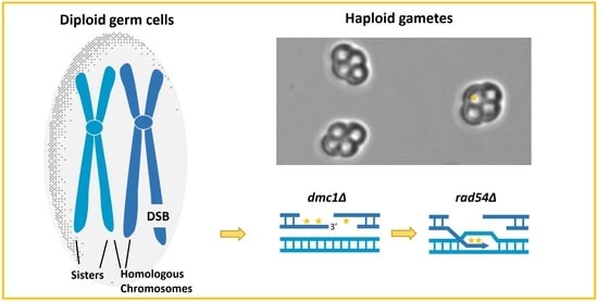
3.1. Meiotic Mutations Depend on the Generation of DSBs

spo11Δ and mre11Δ strains: As we and others have shown, de novo mutations during meiosis firmly depend on SPO11-induced DSBs [4,5]. A null mutation in MRE11 also eliminates meiotic recombination (reviewed in reference [42]). To confirm the dependence of meiotic mutations on DSB formation, we analyzed meiotic mutagenicity in spo11Δ and mre11Δ strains. Strains homozygous for either spo11∆ or mre11Δ were induced into meiosis (see Materials and Methods for details), and aliquots were taken at the time of initiation of meiosis and 8 h afterwards. The rates of mutagenesis and recombination were determined and compared to the WT strain, YRA23-1 (Table 2). In a diploid strain homozygous for spo11Δ, cell viability stayed high until 4 h into meiosis [5] and gradually decreased afterwards, reaching 36% at 8 h (Table 2). The spo11Δ strain showed no increase in the rate of recombination at HIS4 during meiosis compared to WT. When meiotic mutagenicity was assessed in the spo11 mutant, only a few mutated colonies were observed at 8 h (~10% of the mutations accumulated in the WT strain (Table 2 and [5]). Cell viability in diploids homozygous for mre11 dropped to 56% 8 h after induction of meiosis (Table 2). Similar to spo11Δ cells, no recombination was observed at HIS4 in the mre11Δ strain during meiosis (Table 2).The mutation rate observed at CAN1 in mre11-deleted cells was ~20% of the WT level at 8 h (Table 2). Hence, the dependence of meiotic mutations on DSBs was confirmed not only in cultures bearing the spo11 deletion but also in mre11Δ cells.

3.2. Recombinases and Proteins Involved in Strand Exchange Affect Meiotic Mutagenicity

Both the Rad51 recombinase and the meiosis-specific Dmc1 recombinase play roles in meiotic DSB repair. Rad51 catalyzes DNA strand exchange and is essential for both mitotic and meiotic HR [43]. Yeast mutants that are deleted for RAD51 show low viability, poor recombination [44], and no DSB repair during meiosis [38]. Due to its poor meiotic phenotype, we did not examine rad51Δ cells directly; instead, we examined the involvement of gene functions that regulate the activity of Rad51 (and of Dmc1), namely those coded by RAD54, TID1, and HED1.
dmc1Δ, hed1Δ, and hed1Δ dmc1Δ strains: We performed time course experiments in meiosis (see Materials and Methods) using strains homozygous for dmc1Δ, hed1Δ, or the double-mutant hed1Δdmc1Δ. Cell viability in dmc1Δ and hed1Δ mutants, and in the hed1Δdmc1Δ double mutant, remained high, around 100%, until 8 h following the induction of meiosis, similar to the WT strain (Figure 1A). The rates of mutagenesis in CAN1 and recombination events at HIS4 were examined in each strain and compared to the WT. Wherever possible, recombination was also recorded around the CAN1 reporter itself (this is possible only when cells complete meiosis and form viable haploid products). In total, 84% of the cells had sporulated in hed1∆ and in the hed1Δdmc1Δ cultures [25]. 93% of hed1Δ spores were viable, and 55% of the spores in the hed1Δdmc1Δ double mutant showed germination capacity, compared to 96% viable spores in WT cells (Table 2). Similar results were previously reported for other strains from the SK1 background; 96% in hed1Δ and 70% in the double mutant, compared to 96% in the WT [45].
Upon 8 h in meiosis, the rates of recombination at HIS4 in dmc1Δ and in hed1Δ mutants were 43% and 49%, respectively, compared to the WT, while in the hed1Δdmc1Δ double mutant, recombination dropped to 12% (Figure 1B and Table 2). Our observation regarding recombination frequencies at the HIS4-LEU2 hotspot is consistent with previous findings of a 2-fold decrease in interhomolog bias in hed1∆ diploids [16,45]. However, little effect on recombination levels was observed for another hotspot [46]. We could not measure recombination levels around CAN1 in dmc1Δ cells since these cells arrest in meiosis and remain diploid following RTG [39]. In canavanine-resistant (CanR) colonies obtained from hed1Δ cells after 8 h in meiosis, we observed 40% recombination between the CAN1-flanking markers URA3 and ADE2 (69 Ura+ Ade+ colonies, out of 171 CanR haploid colonies examined). In hed1Δ dmc1Δ cells, the rate of recombination around CAN1 was 25% (38 Ura+ Ade+ colonies, out of 153 CanR haploid colonies examined). These results are summarized in Table 3.
rad54 and tid1 strains: Rad54 and Tid1 both operate as chromatin remodeling proteins during vegetative growth and in meiosis [22,47,48,49]. We therefore examined mutation rates during both mitotic and meiotic divisions. To determine mutation rates during vegetative growth, we conducted fluctuation experiments (as described in the Materials and Methods) in rad54∆ and in tid1∆ strains as well as in WT, using CAN1 as a reporter gene (Table 4). In WT cells, we observed a rate of 0.2 mutations per million mitotic cell divisions, and in tid1-deleted cells, the mutation rate was 1.5-fold higher. The mitotic mutation rate in rad54Δ cells was ~26-fold higher than in the WT strain (Table 4).
Mutation and recombination rates during meiosis were determined by meiotic time course experiments in diploids homozygous for rad54∆ or tid1∆. Cell viability gradually decreased during meiosis, reaching 84% viability for the rad54∆ strain and 58% for the tid1∆ strain at 8 h (Figure 2A).
Examination of recombination rates and kinetics at HIS4 in rad54Δ cells showed levels and timing comparable to the WT (Figure 2B and [21]). In the tid1Δ strain, however, clear retardment and overall low recombinant levels of 35% compared to the WT were recorded (as previously observed [21]). Moreover, when mutation rates at the CAN1 reporter were measured, a considerable difference between the rad54Δ and tid1Δ strains was observed: at 4 h, the mutation rates in rad54Δ cells and in tid1Δ cells were 7.1 and 0.1 mutations per 1 million cells (the WT with 0.27 mutations per 1 million cells). At later time points, the mutation rate in rad54Δ cells was higher than in the WT by 8-fold and reached 9.7 mutations per 1 million cells at 8 h (Figure 2C and Table 2). In tid1Δ cultures, however, mutation levels remained low, 0.09-fold of the WT level at 8 h (Figure 2C and Table 2). Interestingly, at the meiotic start point, when the time zero aliquot was taken, frequencies of 2.12 × 10⁻⁵ and 1.06 × 10⁻⁸ CanR-mutated colonies were recorded in rad54Δ and in tid1Δ cells, respectively. The rad54Δ strain showed a 32 times higher mutation level than the WT basal level (~6.65 × 10⁻⁷) at the same time point. As in all strains examined in this study, this value (which represents mutation events that had occurred prior to meiosis) was subtracted from the numbers obtained at each later time point during the rad54Δ meiotic time course.

3.3. Evaluation of Mutation Occurrence during Meiotic S-Phase in rad54∆ Cells

As described above, the rad54Δ strain showed a pronounced elevated meiotic mutation level, 8-fold higher compared to the WT (Table 2). As can be seen in the time course experiments that follow the kinetics of mutation enhancement in rad54Δ strains (Figure 2C), the increase in mutation levels is well observed at 4 h, but only minor (if any) elevation is detected at 2 h. In WT SK1 strains, such as YRA23-1, the S-phase takes place at around 2 h [37]. We tested the hypothesis that the meiotic S-phase is not the source for the elevated meiotic mutation levels observed in the rad54Δ mutant. To check this, we took advantage of the cdc7-as3 mutation, which is a conditional allele of the protein kinase CDC7 [40]. The Cdc7-as3 kinase is inactivated by the addition of the inhibitor PP1 [4-amino-1-tert-butyl-3-(p-methylyphenyl) pyrazolo [3,4-d] pyrimidine] to the sporulation medium; in the presence of PP1, cells homozygous for cdc7-as3 complete DNA replication but arrest before DSB formation and remain diploids. The cdc7-as3 mutant, therefore, is used as a tool for obtaining synchronous cell cultures [40] and for separating meiotic S-phase mutations from DSB-induced mutations during HR [5]. Strains bearing the cdc7-as3 allele allow us to distinguish between mutations that occurred prior to recombination, in cells that underwent meiotic DNA replication (in the presence of PP1) and arrested (in the presence of PP1), and mutations in cells of the same strain that underwent both replication and recombination (without PP1). We previously confirmed that DNA replication is indeed completed in WT cells of the SK1 genetic background that are arrested upon exposure to PP1. This was achieved by FACS analysis to track the DNA content in WT cells undergoing meiosis and by visualizing the cells with microscopy, 8 h upon PP1 addition [5].
We introduced the cdc7-as3 allele into the rad54Δ strain (ALY94) to obtain a diploid strain that is homozygous for both cdc7-as3 and the rad54Δ-deleted gene (see Table 1 for full genotypes). This diploid (ALY170) and the corresponding WT strain, OMY93, were induced into meiosis in the presence or absence of PP1 (for details, see the Materials and Methods). Samples were spread on Can −Ade medium (as well as on YPD) upon transferring the culture to SPM to induce meiosis and after 8 h in SPM. Exposure to PP1 for 8 h did not affect cell viability in any of the strains (Figure 3A). As expected, after 8 h in SPM, meiotic cell cultures that contained PP1 showed no detectable recombination in WT cdc7-as3 as well as in rad54Δ cdc7-as3 cells, since cells that are treated with PP1 are arrested prior to prophase I with no DSBs [5,40]. Cells that were not treated with PP1 reached high levels of recombination in WT and in rad54Δ cdc7-as3 cells (1.3 × 10⁻² and 0.8 × 10⁻², respectively, see also in Figure 2B). Meiotic mutation levels upon exposure to PP1 in WT cells were very low at the time of entry into meiosis and remained low after 8 h (5.2 × 10⁻⁷), which means that essentially no new mutated cells were generated during the meiotic S-phase in the WT culture, as we previously reported [5]. Similarly, in the rad54Δ cdc7-as3 strain, very few CanR mutant colonies (2.3 × 10⁻⁶) were detected in cultures that were arrested with PP1 upon meiosis (Figure 3C). This indicates that the mutations that had accumulated during the meiotic process in WT and in rad54Δ cells did not result from replication failure or from mismatched bases introduced during the meiotic S-phase DNA synthesis. These experiments using the PP1 inhibitor complement the time course experiments described above (Figure 2). Taken together, the data suggest that the elevated mutagenicity observed in the rad54Δ strain results from the meiotic recombinational repair process that is triggered by DSBs.

3.4. The Involvement of Mus81 in Mutagenicity of Meiotic Joint Molecule Resolution

The mus81 strain: Mus81p endonuclease plays a role in joint molecule (JM) formation and resolution during meiosis in S. cerevisiae [50,51] and directs meiotic recombination towards interhomolog interactions [50]. To evaluate mutagenicity levels of JM resolution that depend on Mus81, we examined meiotic mutagenicity and recombination rates in mus81∆ cells (Figure 2B,C). Following the induction of meiosis, the cell viability of mus81∆ cells was maintained around 94% until 4 h and gradually dropped to 58% at 8 h. Recombination frequencies remained lower than in the WT throughout the experiment and reached 38% of the WT level after 8 h (Figure 2B and Table 2). Recombination levels were also measured in CanR colonies that were obtained 8 h into the meiosis of mus81Δ cultures. Of 229 CanR haploid colonies examined, 34 colonies showed the exchange of flanking markers (URA3 and ADE2) around can1, which is 15% recombination (Table 3). Meiotic mutagenicity levels in mus81Δ cells were lower by ~30% of the WT level at 8 h (Figure 2C and Table 2) and reached 0.85 mutated colonies per 1 million cells.

4. Discussion

De novo mutations are found at elevated levels following meiotic recombination in yeast cells and are triggered by DSBs [4,5,8]. In humans, crossovers bear more de novo mutations than non-crossovers in male sperm [6] and mutations are increased 50-fold around crossovers, as observed with whole-genome sequencing of many human families [7]. These findings suggest that meiotic DNA repair by HR is inherently mutagenic. The molecular mechanism that contributes to the enhanced occurrence of new mutations in meiosis is not clear [52,53]. In the present study, we explore the effect of several recombinational repair genes on the occurrence of new mutations during meiosis. We examine not only the rates of mutagenicity at the meiotic endpoint but also the timing and kinetics of the appearance of new meiotic mutations in relation to recombination and, wherever relevant, compare meiotic mutagenesis to mutation rates obtained during mitotic cell divisions, as determined the same CAN1 reporter gene.
DSB formation is a prerequisite for most meiotic mutations, as evident from the deleted strains of the SPO11 or MRE11 genes (Table 2). What other recombination functions affect mutagenicity?
Rad51 recombinase is essential for both mitotic and meiotic HR (reviewed by Bishop [54]), whereas Dmc1 is meiosis-specific [15]. Rad51 catalyzes recombination directly in mitosis and indirectly, via Dmc1, during meiosis [18], so that the interplay between these two recombinases leads to homologue bias repair during meiosis, as previously reviewed [17]. Since the deletion of RAD51 causes cell death and severe impairment of recombination during meiosis [38,44] and the deletion of DMC1 leads to meiotic arrest in S. cerevisiae [39], we chose to explore mutagenicity during the HR process by analyzing the involvement of genes that assist and regulate the prime recombinases without severe interruption of the recombination process, namely RAD54, TID1, and HED1. The corresponding recombination-defective deletion mutants proceed into meiosis, maintain reasonable viability, and generate (at least to some extent) recombinant products.

4.1. Rad54 Restrains Mutagenicity during Meiosis and in Mitotically Cycling Cells

High mutation rates are observed in mitotically cycling cells that are deleted of the RAD54 gene (Table 4). Meiotic mutations also increased substantially in the rad54∆ strain (~8-fold over the WT level), while the recombination rate and kinetics remained fairly similar to the WT (Figure 2B,C). As determined by experiments with the rad54Δ cdc7-as3 strain (Figure 3C), in which cells are arrested after the meiotic S-phase, the latter is not the source of meiotic mutagenicity observed in rad54∆ cells; rather, mutations appear in tight correlation with recombination events. We therefore conclude that the source of the elevated mutagenicity seen in rad54Δ mutants is triggered by DNA breakage and is associated with recombination.
Tid1 is a paralog of Rad54. Although they share structural homology, Tid1 and Rad54 have non-redundant functions [47,55]. Tid1 is needed to stimulate Dmc1 activity, and together, they act to preferentially support interhomolog recombination [23,56]. We found that in the tid1∆ strain meiotic recombination dropped considerably to 35% (Figure 2B and [22]). Meiotic mutations decreased to 9% of the WT (Figure 2C and Table 2). From our findings, we suggest that the disruption of the Rad51–Rad54 pathway (in rad54Δ strain) leads to higher mutagenesis during meiosis. On the other hand, when the meiotic Dmc1 pathway becomes less functional (as in tid1∆ cells), DSB repair is channeled to a less mutagenic track. Therefore, one may suggest that upon the deletion of TID1, DSB repair may rely mainly on the sister chromatid and that type of repair pathway is less mutagenic than interhomolog repair. Nevertheless, our results show opposite and complementary effects of these two paralogs (Rad54, Tid1), not only in meiotic recombinational repair but also in the levels of mutagenicity obtained during meiosis and in mitotically dividing cells.
Hed1 prevents functional cooperation between Rad54 and Rad51 during the HR process [26]. Therefore, in the absence of Hed1, the Rad51/Rad54 complex is more active in DSB repair. If active Rad54 is involved in generating less mutations during DSB repair (Figure 2C), hed1Δ cells should utilize more frequently the Rad51/Rad54 repair pathway and mutations should appear at a lower level than in WT cells. Indeed, the mutation rate in hed1Δ cells was 33% of the WT level (Figure 1B,C and Table 2), while recombination surrounding the CAN1 reporter was similar to the WT level (Table 3). Deletion of HED1 efficiently overcomes the DSB repair defects caused by the dmc1Δ deletion [25], akin to RAD51 overexpression [57]. Along the same line, in our time course experiments of the double-mutant hed1Δ dmc1Δ, recombination rates were reduced to 12% of the WT level (Figure 1B and Table 2) and mutations at CAN1 were lowered to 15% of the WT level (Figure 1C and Table 2). In addition, a moderate drop (of 38%) in recombination levels was observed in hed1Δdmc1Δ CanR colonies examined for the exchange of flanking markers (Table 3). Similarly, whole-genome sequencing of tetrads showed a mild reduction of about 25% in crossing overs in the dmc1∆ hed1∆ mutant [58].
To draw a more global view, our findings indicate that recombinational repair pathways differ in the degree to which they generate mutations. We propose that Rad54 may have a moderating effect on mutagenicity in meiosis and in mitotically cycling cells. Since meiosis aims to create genetic diversity, and mutagenesis is another source of genetic variation, the inhibition of the Rad51/Rad54 pathway (by Hed1) during meiosis leads to the generation of more mutations, while in mitotically dividing cells, HR repair makes extensive use of the Rad51/Rad54 pathway and mutagenesis is minimized. The enhanced mutagenicity observed during meiosis reflects a fine balance between factors that enhance mutagenesis and factors that restrain it, such as Rad54.

4.2. Single-Stranded DNA May Enhance Meiotic Mutagenicity

Investigation of the dmc1∆ single mutant is challenging, since dmc1∆ cells arrest during meiosis and colonies are obtained only upon return to mitotic growth (RTG). Therefore, in dmc1∆ cells, meiotic and mitotic repair are mixed in various proportions. Nevertheless, our exploration of mutation occurrence in dmc1Δ cells allows us to propose another source for meiotic mutagenicity; in our time course experiments, dmc1∆ cells maintained full viability upon RTG, with reduced recombination, but showed a robust increase in mutagenicity (Figure 1B,C and Table 2). Yeast cells lacking DMC1 fail to complete prophase I and arrest during meiosis with excessive amount of DSBs [39] but retain full viability upon RTG as diploids and do not generate spores [38]. What could trigger mutagenicity in dmc1∆ cells?
Bishop et al. (1992) examined the appearance and repair of DSBs in dmc1∆ cells and showed that DSBs are generated at the same time as in WT cells, but the broken DNA ends accumulate at a higher level and undergo additional 5′ end digestion, generating ssDNA beyond what is normally observed in the WT [39]. dmc1 mutants accumulate DSBs with long ssDNA for a prolonged meiotic time, since they are not converted to mature JM structures [59]. Furthermore, Dmc1p normally forms a long nucleofilament that protects the ssDNA prior to invasion [60,61]. Excessive ssDNA tails (in the absence of DMC1) that are left exposed and do not invade the homologous partner during meiosis may be subjected to mutations, explaining the elevated mutagenicity in dmc1Δ cells. Early in vitro studies showed that ssDNA is more prone to the accumulation of mutations than dsDNA [62]. More recently, in vivo studies in yeast and in human cancer cell lines demonstrated hypermutability in ssDNA [63]. In yeast, the density of mutations in ssDNA can exceed 100–1000-fold over the rest of the double-stranded genome (reviewed in [64] and references therein). In addition, Tiemann-Boege and colleagues have shown that recombination hotspots are mutagenic in human sperm and speculated that the formation of ssDNA at methylated CpG sites is the main driver for de novo mutations [6].
Based on our findings in dmc1∆ cells, we hypothesize that ssDNA associated with recombination could be another source for mutagenicity during meiosis. Mimitou et al. (2017) determined an average of 822 nts resection tail length around recombination hotspot centers in normal yeast cells undergoing meiosis. Given that ~ 150 DSBs are normally generated in a single cell going through meiosis, we have previously estimated that ~310,000 nucleotides of ssDNA are present in each yeast WT cell during meiosis, which is about 2.3% of the entire genome [53]. Excess ssDNA may result not only in increased mutagenesis but may also impact the spectrum of mutations obtained in various recombination sites. Particularly in dmc1Δ cells, the extra ssDNA may lead to increased loss-of-function can1 mutants due to deletions and other rearrangements. It will be interesting to test the mutation signature and spectrum at the molecular level, comparing dmc1Δ to WT cells. Indeed, the low mutation rate seen in hed1Δdmc1Δ cells further supports this scenario; HED1 deletion bypasses the meiotic arrest of dmc1Δ cells, in which ssDNA tails are presumably shorter and remain exposed for shorter periods, and therefore, the mutation rate in hed1Δdmc1Δ mutants is considerably lower.
Alternatively, it is possible that the usage of mixed HR repair pathways in dmc1Δ cells that are arrested for long periods during meiosis or some yet unknown mechanism, generates the elevated mutagenicity observed in cells lacking DMC1.

4.3. Is Meiotic Joint Molecule Resolution a Mutagenic Process?

In the current study, the only resolvase that was examined for involvement in meiotic mutagenesis was MUS81. The kinetics of mutagenicity in mus81∆ cells were similar to those of the WT, with the mutation rate in mus81∆ reaching maximal levels at 8 h (0.85 mutations per 1 million cells at the CAN1 reporter), which is 71% relative to the WT level (Figure 2C and Table 2). However, despite six independent experiments performed with this mutant, we could not detect a statistically significant difference between mus81∆ and WT cells. Mus81 is not the only resolvase operating in meiosis. In fact, in S. cerevisiae, meiotic crossover resolution is less dependent on Mus81/Mms4 [65]; mus81∆ cells show only a modest CO reduction and Mus81 may act to process 3′ flaps of over-replication during meiotic recombination [66]. If the reduction in mutation rate (~30%) observed in mus81∆ cells is a true result, then Mus81-mediated resolution is somewhat mutagenic. However, even if there is no difference between mus81∆ and WT cells, it is still possible that the resolution that occurs via some other resolvase is mutagenic. It leaves open the issue whether the resolution of joint molecules is a mutagenic event during meiosis. Testing the Mlh1-Mlh3/Exo1 resolvase complex (which has a more central function in meiotic Holliday junction resolution) may be interesting but needs to be examined with caution, since MLH1 or MLH3 deletion is associated with several meiotic dysfunctions [67].

4.4. Concluding Remarks

Sexual reproduction and meiosis provide an effective mechanism to shuffle existing genetic information. We consider meiotic mutagenicity as another source of genetic diversity that passes on from parents to offspring. We provide, for the first time to our knowledge, experimental data showing that different recombination pathways disclose different mutagenicity loads during meiosis. We place these new findings regarding meiotic mutagenicity in the context of previous observations concerning recombination and DSBs repair. Mechanisms that enhance meiotic mutagenicity alter sequences in the genome of germ cells and may lead to alternations in allele frequencies in populations over evolutionary timescales.

Author Contributions

Conceptualization, A.A.-E.; methodology, A.A.-E. and D.Z.; formal analysis, L.M. and R.M.E.; investigation, L.M. and R.M.E.; data curation, A.A.-E.; writing—original draft preparation, A.A.-E., L.M. and R.M.E.; review and editing, A.A.-E., D.Z., L.M. and R.M.E.; supervision, A.A.-E.; funding acquisition, A.A.-E. All authors have read and agreed to the published version of the manuscript.

Funding

Our work was supported by funds provided by the Israel Science Foundation (Grant # 501/16 and Grant # 397/20) and by Hadassah Academic College.

Institutional Review Board Statement

Not applicable.

Informed Consent Statement

Not applicable.

Data Availability Statement

Not applicable.

Acknowledgments

We are grateful to Giora Simchen for his careful reading and helpful comments on the manuscript and for the fruitful partnership. We thank Shoshana Klein for kindly providing the inhibitor PP1 [4-amino-1-tert-butyl-3-(p-methylyphenyl) pyrazolo [3,4-d] pyrimidine] and for her helpful suggestions while reading the manuscript.

Conflicts of Interest

The authors declare no conflict of interest.

References

  1. Magni, G.E.; Von Borstel, R.C. Different Rates of Spontaneous Mutation during Mitosis and Meiosis in Yeast. Genetics 1962, 47, 1097–1108. [Google Scholar] [CrossRef] [PubMed]
  2. Magni, G.E. Origin and Nature of Spontaneous Mutations in Meiotic Organisms. J. Cell. Physiol. 1964, 64, 65–71. [Google Scholar] [CrossRef]
  3. Kiktev, D.A.; Sheng, Z.; Lobachev, K.S.; Petes, T.D. GC content elevates mutation and recombination rates in the yeast Saccharomyces cerevisiae. Proc. Natl. Acad. Sci. USA 2018, 115, E7109–E7118. [Google Scholar] [CrossRef] [PubMed]
  4. Rattray, A.; Santoyo, G.; Shafer, B.; Strathern, J.N. Elevated mutation rate during meiosis in Saccharomyces cerevisiae. PLOS Genet. 2015, 11, e1004910. [Google Scholar] [CrossRef]
  5. Mansour, O.; Morciano, L.; Zion, K.; Elgrabli, R.; Zenvirth, D.; Simchen, G.; Arbel-Eden, A. Timing of appearance of new mutations during yeast meiosis and their association with recombination. Curr. Genet. 2020, 66, 577–592. [Google Scholar] [CrossRef] [PubMed]
  6. Arbeithuber, B.; Betancourt, A.J.; Ebner, T.; Tiemann-Boege, I. Crossovers are associated with mutation and biased gene conversion at recombination hotspots. Proc. Natl. Acad. Sci. USA. 2015, 112, 2109–2114. [Google Scholar] [CrossRef]
  7. Halldorsson, B.V.; Palsson, G.; Stefansson, O.A.; Jonsson, H.; Hardarson, M.T.; Eggertsson, H.P.; Gunnarsson, B.; Oddsson, A.; Halldorsson, G.H.; Zink, F.; et al. Characterizing mutagenic effects of recombination through a sequence-level genetic map. Science 2019, 363, eaau1043. [Google Scholar] [CrossRef]
  8. Magni, G.E. The Origin of Spontaneous Mutations during Meiosis. Proc. Natl. Acad. Sci. USA 1963, 50, 975–980. [Google Scholar] [CrossRef]
  9. Simchen, G.; Mansour, O.; Morciano, L.; Zenvirth, D.; Arbel-Eden, A. Mutagenicity in haploid yeast meiosis resulting from repair of DSBs by the sister chromatid. Curr. Genet. 2021, 67, 799–806. [Google Scholar] [CrossRef]
  10. Claeys Bouuaert, C.; Pu, S.; Wang, J.; Oger, C.; Daccache, D.; Xie, W.; Patel, D.J.; Keeney, S. DNA-driven condensation assembles the meiotic DNA break machinery. Nature 2021, 592, 144–149. [Google Scholar] [CrossRef]
  11. Székvölgyi, L.; Nicolas, A. From meiosis to postmeiotic events: Homologous recombination is obligatory but flexible. FEBS J. 2010, 277, 571–589. [Google Scholar] [CrossRef] [PubMed]
  12. Pan, J.; Sasaki, M.; Kniewel, R.; Murakami, H.; Blitzblau, H.G.; Tischfield, S.E.; Zhu, X.; Neale, M.J.; Jasin, M.; Socci, N.D.; et al. A hierarchical combination of factors shapes the genome-wide topography of yeast meiotic recombination initiation. Cell 2011, 144, 719–731. [Google Scholar] [CrossRef]
  13. Lam, I.; Keeney, S. Mechanism and regulation of meiotic recombination initiation. Cold Spring Harb. Perspect. Biol. 2015, 7, a016634. [Google Scholar] [CrossRef]
  14. Mimitou, E.P.; Yamada, S.; Keeney, S. A global view of meiotic double-strand break end resection. Science 2017, 355, 40–45. [Google Scholar] [CrossRef] [PubMed]
  15. Neale, M.J.; Keeney, S. Clarifying the mechanics of DNA strand exchange in meiotic recombination. Nature 2006, 442, 153–158. [Google Scholar] [CrossRef]
  16. Liu, Y.; Gaines, W.A.; Callender, T.; Busygina, V.; Oke, A.; Sung, P.; Fung, J.C.; Hollingsworth, N.M. Down-Regulation of Rad51 Activity during Meiosis in Yeast Prevents Competition with Dmc1 for Repair of Double-Strand Breaks. PLoS Genet. 2014, 10, e1004005. [Google Scholar] [CrossRef] [PubMed]
  17. Schwacha, A.; Kleckner, N. Interhomolog bias during meiotic recombination: Meiotic functions promote a highly differentiated interhomolog-only pathway. Cell 1997, 90, 1123–1135. [Google Scholar] [CrossRef] [PubMed]
  18. Cloud, V.; Chan, Y.-L.; Grubb, J.; Budke, B.; Bishop, D.K. Rad51 Is an Accessory factor for dmc1-mediated joint molecule formation during meiosis. Science 2012, 337, 1222–1225. [Google Scholar] [CrossRef] [PubMed]
  19. Mazin, A.V.; Mazina, O.M.; Bugreev, D.V.; Rossi, M.J. Rad54, the motor of homologous recombination. DNA Repair 2010, 9, 286–302. [Google Scholar] [CrossRef]
  20. Sanchez-Rebato, M.H.; Bouatta, A.M.; Gallego, M.E.; White, C.I.; Da Ines, O. RAD54 is essential for RAD51-mediated repair of meiotic DSB in Arabidopsis. PLoS Genet. 2021, 17, e1008919. [Google Scholar] [CrossRef]
  21. Arbel, A.; Zenvirth, D.; Simchen, G. Sister chromatid-based DNA repair is mediated by RAD54, not by DMC1 or TID1. Embo J. 1999, 18, 2648–2658. [Google Scholar] [CrossRef]
  22. Shinohara, M.; Shita-Yamaguchi, E.; Buerstedde, J.M.; Shinagawa, H.; Ogawa, H.; Shinohara, A. Characterization of the roles of the Saccharomyces cerevisiae RAD54 gene and a homologue of RAD54, RDH54/TID1, in mitosis and meiosis. Genetics 1997, 147, 1545–1556. [Google Scholar] [CrossRef] [PubMed]
  23. Nimonkar, A.V.; Dombrowski, C.C.; Siino, J.S.; Stasiak, A.Z.; Stasiak, A.; Kowalczykowski, S.C. Saccharomyces cerevisiae Dmc1 and Rad51 proteins preferentially function with Tid1 and Rad54 proteins, respectively, to promote DNA strand invasion during genetic recombination. J. Biol. Chem. 2012, 287, 28727–28737. [Google Scholar] [CrossRef]
  24. Holzen, T.M.; Shah, P.P.; Olivares, H.A.; Bishop, D.K. Tid1/Rdh54 promotes dissociation of Dmc1 from nonrecombinogenic sites on meiotic chromatin. Genes Dev. 2006, 20, 2593–2604. [Google Scholar] [CrossRef]
  25. Tsubouchi, H.; Roeder, G.S. Budding yeast Hed1 down-regulates the mitotic recombination machinery when meiotic recombination is impaired. Genes Dev. 2006, 20, 1766–1775. [Google Scholar] [CrossRef] [PubMed]
  26. Busygina, V.; Saro, D.; Williams, G.; Leung, W.K.; Say, A.F.; Sehorn, M.G.; Sung, P.; Tsubouchi, H. Novel Attributes of Hed1 Affect Dynamics and Activity of the Rad51 Presynaptic Filament during Meiotic Recombination. J. Biol. Chem. 2012, 287, 1566–1575. [Google Scholar] [CrossRef] [PubMed]
  27. Crickard, J.B.; Kaniecki, K.; Kwon, Y.; Sung, P.; Lisby, M.; Greene, E.C. Regulation of Hed1 and Rad54 binding during maturation of the meiosis-specific presynaptic complex. EMBO J. 2018, 37, e98728. [Google Scholar] [CrossRef]
  28. Niu, H.; Wan, L.; Busygina, V.; Kwon, Y.; Allen, J.A.; Li, X.; Kunz, R.C.; Kubota, K.; Wang, B.; Sung, P.; et al. Regulation of Meiotic Recombination via Mek1-Mediated Rad54 Phosphorylation. Mol. Cell 2009, 36, 393–404. [Google Scholar] [CrossRef]
  29. Hollingsworth, N.M. Phosphorylation and the creation of interhomolog bias during meiosis in yeast. Cell Cycle 2010, 9, 436–437. [Google Scholar] [CrossRef]
  30. Busygina, V.; Sehorn, M.G.; Shi, I.Y.; Tsubouchi, H.; Roeder, G.S.; Sung, P. Hed1 regulates Rad51-mediated recombination via a novel mechanism. Genes Dev. 2008, 22, 786–795. [Google Scholar] [CrossRef]
  31. Cao, L.; Alani, E.; Kleckner, N. A pathway for generation and processing of double-strand breaks during meiotic recombination in S. cerevisiae. Cell 1990, 61, 1089–1101. [Google Scholar] [CrossRef] [PubMed]
  32. Kassir, Y.; Simchen, G. Monitoring meiosis and sporulation in Saccharomyces cerevisiae. Methods Enzymol. 1991, 194, 94–110. [Google Scholar] [PubMed]
  33. Rose, M.; Winston, F.; Hieter, P. Methods Yeast Genetics—A Laboratory Course Manual; Cold Spring Harbor: New York, NY, USA, 1990. [Google Scholar]
  34. I Lang, G.; Murray, A.W. Estimating the per-base-pair mutation rate in the yeast Saccharomyces cerevisiae. Genetics 2008, 178, 67–82. [Google Scholar] [CrossRef] [PubMed]
  35. Luria, S.E.; Delbrück, M. Mutations of Bacteria from Virus Sensitivity to Virus Resistance. Genetics 1943, 28, 491–511. [Google Scholar] [CrossRef] [PubMed]
  36. Rosche, W.A.; Foster, P.L. Determining mutation rates in bacterial populations. Methods 2000, 20, 4–17. [Google Scholar] [CrossRef]
  37. Arbel-Eden, A.; Joseph-Strauss, D.; Masika, H.; Printzental, O.; Rachi, E.; Simchen, G. Trans-Lesion DNA Polymerases May Be Involved in Yeast Meiosis. G3 Genes Genomes Genet. 2013, 3, 633–644. [Google Scholar] [CrossRef]
  38. Zenvirth, D.; Loidl, J.; Klein, S.; Arbel, A.; Shemesh, R.; Simchen, G. Switching yeast from meiosis to mitosis: Double-strand break repair, recombination and synaptonemal complex. Genes Cells 1997, 2, 487–498. [Google Scholar] [CrossRef]
  39. Bishop, D.K.; Park, D.; Xu, L.; Kleckner, N. DMC1: A meiosis-specific yeast homolog of E. coli recA required for recombination, synaptonemal complex formation, and cell cycle progression. Cell 1992, 69, 439–456. [Google Scholar] [CrossRef]
  40. Wan, L.; Zhang, C.; Shokat, K.M.; Hollingsworth, N.M. Chemical inactivation of cdc7 kinase in budding yeast results in a reversible arrest that allows efficient cell synchronization prior to meiotic recombination. Genetics 2006, 174, 1767–1774. [Google Scholar] [CrossRef]
  41. Alexandrov, L.B.; Kim, J.; Haradhvala, N.J.; Huang, M.N.; Ng, A.W.T.; Wu, Y.; Boot, A.; Covington, K.R.; Gordenin, D.A.; Bergstrom, E.N.; et al. The repertoire of mutational signatures in human cancer. Nature 2020, 578, 94–101. [Google Scholar] [CrossRef]
  42. Keeney, S. Mechanism and control of meiotic recombination initiation. Curr. Top. Dev. Biol. 2001, 52, 1–53. [Google Scholar]
  43. Brown, M.S.; Bishop, D.K. DNA strand exchange and RecA homologs in meiosis. Cold Spring Harb. Perspect. Biol. 2014, 7, a016659. [Google Scholar] [CrossRef]
  44. Shinohara, A.; Ogawa, H.; Ogawa, T. Rad51 protein involved in repair and recombination in S. cerevisiae is a RecA-like protein. Cell 1992, 69, 457–470. [Google Scholar] [CrossRef]
  45. Lao, J.P.; Cloud, V.; Huang, C.-C.; Grubb, J.; Thacker, D.; Lee, C.-Y.; Dresser, M.E.; Hunter, N.; Bishop, D.K. Meiotic Crossover Control by Concerted Action of Rad51-Dmc1 in Homolog Template Bias and Robust Homeostatic Regulation. PLoS Genet. 2013, 9, e1003978. [Google Scholar] [CrossRef] [PubMed]
  46. Ziesel, A.; Weng, Q.; Ahuja, J.S.; Bhattacharya, A.; Dutta, R.; Cheng, E.; Börner, G.V.; Lichten, M.; Hollingsworth, N.M. Rad51-mediated interhomolog recombination during budding yeast meiosis is promoted by the meiotic recombination checkpoint and the conserved Pif1 helicase. PLoS Genet. 2022, 18, e1010407. [Google Scholar] [CrossRef]
  47. Klein, H.L. RDH54, a RAD54 homologue in Saccharomyces cerevisiae, is required for mitotic diploid-specific recombination and repair and for meiosis. Genetics 1997, 147, 1533–1543. [Google Scholar] [CrossRef] [PubMed]
  48. Alexeev, A.; Mazin, A.; Kowalczykowski, S.C. Rad54 protein possesses chromatin-remodeling activity stimulated by the Rad51–ssDNA nucleoprotein filament. Nat. Struct. Biol. 2003, 10, 182–186. [Google Scholar] [CrossRef]
  49. Prasad, T.K.; Robertson, R.B.; Visnapuu, M.L.; Chi, P.; Sung, P.; Greene, E.C. A DNA-translocating Snf2 Molecular Motor: Saccharomyces cerevisiae Rdh54 Displays Processive Translocation and Extrudes DNA Loops. J. Mol. Biol. 2007, 369, 940–953. [Google Scholar] [CrossRef] [PubMed]
  50. Jessop, L.; Lichten, M. Mus81/Mms4 Endonuclease and Sgs1 Helicase Collaborate to Ensure Proper Recombination Intermediate Metabolism during Meiosis. Mol. Cell. 2008, 31, 313–323. [Google Scholar] [CrossRef] [PubMed]
  51. Oh, S.D.; Lao, J.P.; Taylor, A.F.; Smith, G.R.; Hunter, N. RecQ Helicase, Sgs1, and XPF Family Endonuclease, Mus81-Mms4, Resolve Aberrant Joint Molecules during Meiotic Recombination. Mol. Cell 2008, 31, 324–336. [Google Scholar] [CrossRef]
  52. Bergero, R.; Ellis, P.; Haerty, W.; Larcombe, L.; Macaulay, I.; Mehta, T.; Mogensen, M.; Murray, D.; Nash, W.; Neale, M.J.; et al. Meiosis and beyond—Understanding the mechanistic and evolutionary processes shaping the germline genome. Biol. Rev. 2021, 96, 822–841. [Google Scholar] [CrossRef]
  53. Arbel-Eden, A.; Simchen, G. Elevated Mutagenicity in Meiosis and Its Mechanism. BioEssays 2019, 41, 272–281. [Google Scholar] [CrossRef]
  54. Bishop, D.K. Rad51, the lead in mitotic recombinational DNA repair, plays a supporting role in budding yeast meiosis. Cell Cycle 2012, 11, 4105–4106. [Google Scholar] [CrossRef] [PubMed]
  55. Shinohara, M.; Gasior, S.L.; Bishop, D.K.; Shinohara, A. Tid1/Rdh54 promotes colocalization of rad51 and dmc1 during meiotic recombination. Proc. Natl. Acad. Sci. USA 2000, 97, 10814–10819. [Google Scholar] [CrossRef] [PubMed]
  56. Shinohara, M.; Sakai, K.; Shinohara, A.; Bishop, D.K. Crossover interference in Saccharomyces cerevisiae requires a T1D1/RDH54- and DMC1-dependent pathway. Genetics 2003, 163, 1273–1286. [Google Scholar] [CrossRef] [PubMed]
  57. Tsubouchi, H.; Roeder, G. The importance of genetic recombination for fidelity of chromosome pairing in meiosis. Dev. Cell 2003, 5, 915–925. [Google Scholar] [CrossRef] [PubMed]
  58. Callender, T.L.; Laureau, R.; Wan, L.; Chen, X.; Sandhu, R.; Laljee, S.; Zhou, S.; Suhandynata, R.T.; Prugar, E.; Gaines, W.A.; et al. Mek1 Down Regulates Rad51 Activity during Yeast Meiosis by Phosphorylation of Hed1. PLoS Genet. 2016, 12, e1006226. [Google Scholar] [CrossRef]
  59. Johnson, R.; Borde, V.; Neale, M.J.; Bishop-Bailey, A.; North, M.; Harris, S.; Nicolas, A.; Goldman, A.S.H. Excess single-stranded DNA inhibits meiotic double-strand break repair. PLoS Genet. 2007, 3, e223. [Google Scholar] [CrossRef] [PubMed]
  60. Hong, E.L.; Shinohara, A.; Bishop, D.K. Saccharomyces cerevisiae Dmc1 Protein Promotes Renaturation of Single-strand DNA (ssDNA) and Assimilation of ssDNA into Homologous Super-coiled Duplex DNA. J. Biol. Chem. 2001, 276, 41906–41912. [Google Scholar] [CrossRef]
  61. MacQueen, A.J. Catching a (Double-Strand) Break: The Rad51 and Dmc1 Strand Exchange Proteins Can Co-occupy Both Ends of a Meiotic DNA Double-Strand Break. PLoS Genet. 2015, 11, e1005741. [Google Scholar] [CrossRef]
  62. Lindahl, T. Instability and decay of the primary structure of DNA. Nature 1993, 362, 709–715. [Google Scholar] [CrossRef] [PubMed]
  63. Roberts, S.A.; Sterling, J.; Thompson, C.; Harris, S.; Mav, D.; Shah, R.; Klimczak, L.J.; Kryukov, G.V.; Malc, E.; Mieczkowski, P.A.; et al. Clustered Mutations in Yeast and in Human Cancers Can Arise from Damaged Long Single-Strand DNA Regions. Mol. Cell 2012, 46, 424–435. [Google Scholar] [CrossRef]
  64. Saini, N.; Gordenin, D.A. Hypermutation in single-stranded DNA. DNA Repair 2020, 91–92, 102868. [Google Scholar] [CrossRef]
  65. Hollingsworth, N.M.; Brill, S.J. The Mus81 solution to resolution: Generating meiotic crossovers without Holliday junctions. J. Bone Jt. Surg. 2004, 18, 117–125. [Google Scholar] [CrossRef] [PubMed]
  66. De los Santos, T.; Hunter, N.; Lee, C.; Larkin, B.; Loidl, J.; Hollingsworth, N.M. The MUS81/MMS4 endonuclease acts independently of double-holliday junction resolution to promote a distinct subset of crossovers during meiosis in budding yeast. Genetics 2003, 164, 81–94. [Google Scholar] [CrossRef] [PubMed]
  67. Dai, J.; Sanchez, A.; Adam, C.; Ranjha, L.; Reginato, G.; Chervy, P.; Tellier-Lebegue, C.; Andreani, J.; Guérois, R.; Ropars, V.; et al. Molecular basis of the dual role of the Mlh1-Mlh3 endonuclease in MMR and in meiotic crossover formation. Proc. Natl. Acad. Sci. USA 2021, 118, e2022704118. [Google Scholar] [CrossRef] [PubMed]
Figure 1. Kinetics of viability, recombination, and mutation events in WT, dmc1Δ, hed1∆, and double-mutant hed1Δdmc1Δ strains during meiosis. Aliquots of meiotic cells were taken at the times indicated and assayed for (A) cell viability (%) on YEPD plates, (B) recombination at HIS4 on −His plates, and (C) mutation at CAN1 on Can −Ade plates. WT (n = 19, in blue), dmc1∆ (n = 8, in red), hed1∆ (n = 9, in green), and hed1∆dmc1∆ (n = 9, in purple). n denotes the number of independent experiments for each strain. Recombination rate—recombinations per 100 meiotic events. Mutation rate—mutations per million meiotic events. The number of colonies appearing on selection plates from time zero (either on −His or on Can −Ade plates) was always subtracted from the numbers obtained at later time points. Therefore, the net meiotic contribution at the beginning of the experiment is zero.
Genes 14 02017 g001
Figure 2. Kinetics of viability, recombination, and mutation events in WT, rad54Δ, tid1∆, and mus81Δ strains during meiosis. Aliquots of meiotic cells were taken at the times indicated, returned to vegetative growth, and assayed for (A) cell viability (%) on YEPD plates, (B) recombination at HIS4 on −His plates, and (C) mutation at CAN1 on Can −Ade plates. WT (n = 19, in blue), tid1∆ (n = 7, in red), rad54∆ (n = 7, in green), and mus81∆ (n = 6, in purple). In C, the Y axis is split so that the lower half of the plot covers 0 to 1.5 and the upper half of the plot covers 2 to 20 regarding CAN1 mutation rates ×10−6. n denotes the number of independent experiments for each strain. Recombination rate—recombinations per 100 meiotic events. Mutation rate—mutations per million meiotic events. The number of colonies appearing on selection plates from time zero (either on −His or on Can −Ade plates) was always subtracted from the numbers obtained at later time points. Therefore, the net meiotic contribution at the beginning of the experiment is zero.
Genes 14 02017 g002
Figure 3. Viability, recombination, and mutations obtained at 8 h in WT and in rad54∆ cells upon treatment with the PP1 inhibitor. Aliquots of meiotic cells were taken at 8 h following transfer to SPM, treated with the PP1 inhibitor (dotted bars) or without PP1 (solid bars). (A) Cell viability (%) on YEPD plates. (B) Recombination at HIS4 on −His plates. (C) Mutation at CAN1 on Can −Ade plates. WT (blue); rad54∆ (green).
Genes 14 02017 g003
Table 1. Yeast strains used and constructed in this study and their relevant genotypes.
Strain NameRelevant MutationsGenotype
YRA10WT, MATaMATa, ho∆::LYS2, lys2, leu2, his4X, trp1∆::hisG, ura3, ade2∆::hisG, ADE2-CAN1 (ADE2 inserted upstream of CAN1), TRP1-LYP1 (TRP1 inserted upstream of LYP1)
YRA12WT, MATαMATα, ho∆::LYS2, lys2, leu2, his4B::LEU2, trp1∆::hisG, ura3, ade2∆::hisG, avt2∆::URA3 (URA3 inserted downstream of can1), can1::kanMX6, lyp1::natNT2
YRA23-1diploid WT, CDC7YRA10 × YRA12
YRA17spo11∆YRA23-1 with homozygous spo11∆::hisG
YRA34-1mre11∆YRA23-1 with homozygous mre11∆::kanMX4
YRA20dmc1∆YRA23-1 with homozygous dmc1∆::kanMX4
YRA55hed1∆YRA23-1 with homozygous hed1∆::kanMX4 (ydr015c)
YRA56hed1∆ dmc1∆YRA23-1 with homozygous hed1∆::kanMX4 (ydr015c), dmc1∆::kanMX4
#3512rad54∆, MATaMATa, ho∆::LYS2, lys2, ura3, leu2∆::hisG, his4X::LEU2-BamHI-URA3, rad54∆::URA3
ALY77rad54∆, MATaMATa, ho∆::LYS2, lys2, leu2, his4X, trp1∆::hisG, ura3, ade2:∆::kanMX4, ADE2-CAN1 (ADE2 inserted upstream of CAN1), TRP1-LYP1 (TRP1 inserted upstream of LYP1), rad54∆::URA3
ALY92rad54∆, MATαMATα, ho∆::LYS2, lys2, leu2, his4B::LEU2, trp1∆::hisG, ura3, ade2∆::kanMX4, avt2∆::URA3 (URA3 inserted downstream of can1), can1::kanMX6, lyp1::natNT2, rad54∆::URA3
ALY94diploid, rad54∆ALY77 × ALY92
YAS3tid1∆YRA23-1 with homozygous tid1∆::kanMX4
YRA51mus81∆YRA23-1 with homozygous mus81∆::kanMX4
OMY78-1WT, MATa, cdc7-as3-9mycMATa, hoΔ::LYS2, leu2Δ::hisG, his4x, lys2, trp1∆::hygB, ura3∆::hisG, ade2∆::kanMX4, cdc7-as3-9myc, ADE2-CAN1 (ADE2 inserted upstream of CAN1), TRP1-LYP1 (TRP1 inserted upstream of LYP1)
OMY75-2WT, MATα, cdc7-as3-9mycMATα, ho∆::LYS2, ade2∆::kanMX4, trp1∆::hph, his4B::LEU2, avt2∆::URA3 (URA3 inserted downstream of can1), cdc7-as3-9myc, can1::kanMX6, lyp1::natNT2
OMY93diploid WT, cdc7-ac3-9mycOMY78-1 × OMY75-2
ALY170rad54∆, cdc7-as3-9mycALY94 with homozygous cdc7-as3-9myc
In can1R::kanMX6 allele, the promoter and the first six codons of the ORF of the CAN1 gene are replaced by kanMX6. In the lyp1R::natNT2 allele, the promoter and the first six codons of the ORF of the LYP1 gene are replaced by kanMX6.
Table 2. Summary of viability, spore germination, recombination, and mutations obtained at 8 h in WT and in various mutants examined in the current study.
Mutated Gene% Cell
Viability
(at 8 h)
Spore
Germination
Recombination at HIS4 (at 8 h)
(×10−2)
Mutation at CAN1 (8 h)
Meiotic Mutations
(×10−6)
Fold Change
Mutant/ WT
WT10096% (366/380)1.871.201.00
spo11∆360% (0/150)0.000.120.10
mre11∆560% (0/146)0.010.240.20
dmc1∆108Φ0.814.333.60
hed1∆9693% (164/176)0.910.390.33
hed1∆dmc1∆9355% (173/312)0.220.180.15
rad54∆8466% (105/160)1.97 *9.708.07
tid1∆58N.D0.650.110.09
mus81∆5856% (158/280)0.710.85 *0.71
From left to right: cell viability, spore germination, recombination frequencies at HIS4, and mutation rates at CAN1. * Results shown with asterisk (*) do not differ from the WT and are not statistically significant. All other values presented in this table are statistically significant (with a p value less than 0.05). Φ spores are not generated due to meiotic arrest of dmc1Δ cells.
Table 3. % Recombinants of markers flanking the CAN1 reporter in CanR-mutated colonies of WT, hed1∆, hed1∆dmc1∆, and mus81∆ strains.
Mutated Gene StrainRecombinants around CAN1 Reporter in CANR MutantsFold Change
Mutant/WT
WTYRA23-139% (95/243)1
hed1∆YRA5540% (69/171)1
hed1∆dmc1∆YRA5625% (38/153)0.64
mus81∆YRA5115% (34/229)0.38
Sporulating cultures of WT, hed1∆, hed1∆dmc1∆, and mus81∆ from SPO plates were placed on YPD plates, and four-spore asci were dissected by micromanipulation. YPD plates were incubated at 30 °C for 4 days, and spore germination was assessed. Germinated spores were then replica-plated onto -Ura and –Ade plates to examine recombination around the CAN1 reporter. The numbers in parentheses show recombinant colonies out of the number of CanR haploid colonies. Fold change (compared to the WT) of recombination around the CAN1 reporter is shown on the right.
Table 4. Spontaneous mitotic mutation rate at the CAN1 reporter gene in WT, rad54∆, and in tid1∆ strains.
Mutated Gene StrainMitotic Mutation Rate
at CAN1 (×10−6)
In Mitotic Cells
Fold Change
Mutant/WT
WTYRA100.21
rad54∆ALY945.1425.7
tid1∆YAS30.31.5
Each value (×10−6) represents the average of at least four independent experiments. All values are statistically significant according to a t-test with a p value less than 0.05. Fold change (compared to the WT) of mutation rate in vegetatively cycling cells is denoted on the right.
Disclaimer/Publisher’s Note: The statements, opinions and data contained in all publications are solely those of the individual author(s) and contributor(s) and not of MDPI and/or the editor(s). MDPI and/or the editor(s) disclaim responsibility for any injury to people or property resulting from any ideas, methods, instructions or products referred to in the content.

