Review

DOCK3-Associated Neurodevelopmental Disorder—Clinical Features and Molecular Basis

by
Matthew S. Alexander
1,2,3,4,5 and
Milen Velinov
6,*
1
Department of Pediatrics, Division of Neurology, University of Alabama at Birmingham and Children’s of Alabama, Birmingham, AL 35294, USA
2
UAB Center for Exercise Medicine, University of Alabama at Birmingham, Birmingham, AL 35294, USA
3
Department of Genetics, University of Alabama at Birmingham, Birmingham, AL 35294, USA
4
UAB Civitan International Research Center (CIRC), University of Alabama at Birmingham, Birmingham, AL 35233, USA
5
UAB Center for Neurodegeneration and Experimental Therapeutics (CNET), University of Alabama at Birmingham, Birmingham, AL 35294, USA
6
Department of Pediatrics, Division of Genetics, Rutgers Robert Wood Johnson Medical School, New Brunswick, NJ 08901, USA
*
Author to whom correspondence should be addressed.
Genes 2023, 14(10), 1940; https://doi.org/10.3390/genes14101940
Submission received: 11 September 2023 / Revised: 9 October 2023 / Accepted: 12 October 2023 / Published: 14 October 2023

Abstract

:
The protein product of DOCK3 is highly expressed in neurons and has a role in cell adhesion and neuronal outgrowth through its interaction with the actin cytoskeleton and key cell signaling molecules. The DOCK3 protein is essential for normal cell growth and migration. Biallelic variants in DOCK3 associated with complete or partial loss of function of the gene were recently reported in six patients with intellectual disability and muscle hypotonia. Only one of the reported patients had congenital malformations outside of the CNS. Further studies are necessary to better determine the prevalence of DOCK3-associated neurodevelopmental disorders and the frequency of non-CNS clinical manifestations in these patients. Since deficiency of the DOCK3 protein product is now an established pathway of this neurodevelopmental condition, supplementing the deficient gene product using a gene therapy approach may be an efficient treatment strategy.

1. Introduction

Neurodevelopmental disorders (NDDs) are a large and heterogeneous group of conditions affecting cognitive and motor functioning with typical childhood onset [1]. Recent developments in genomic technologies have led to the discovery of a large number of chromosomal rearrangements and single-gene disorders associated with NDDs [2,3,4]. These discoveries have allowed connecting multiple genes into neurodevelopmental networks and disease pathways [5]. Most importantly, identifying new disease-associated genes has allowed for the development of novel treatment strategies using downstream targets of disease-associated pathways or gene therapy approaches. There are now several FDA-approved protocols for the medical treatment of NDDs. Examples of such protocols approved for clinical use are delivering the entire gene or modifying the RNA product [6] in patients with spinal muscular atrophy, and downstream regulation using a small molecule in patients with PIK3CA mutations [7]. Finally, identifying the genetic cause for a specific disorder allows for better-informed family planning for the families and makes possible prenatal or pre-implantation diagnoses in future pregnancies.
Mutations in the gene DOCK3 associated with complete or partial deficiency of gene function have recently been identified and may lead to the future development of specific therapy protocols for these patients.

2. History of DOCK3-Deficiency

In 2003, Efron et al. reported a 10-year-old male patient with moderate intellectual disability and severe impulse control problems with features of ADHD and conduct disorder [8]. This individual’s routine chromosomal analysis identified an apparently balanced chromosomal inversion in chromosome 3: inv (3) (p14q21). Afterward, 10 additional family members were found to have the same chromosome 3 inversion and similar clinical manifestations. The average IQ of the family members who carried the family inversion was 76.6 while non-carrier family members had an average IQ of 93.6 [8]. Follow-up studies were carried out to characterize the inversion breakpoints [9]. The 3p breakpoint was found to be between exons 19 and 20 of DOCK3 while the 3q breakpoint was between exons 13 and 14 of gene SLC9A9. The authors showed that both genes were expressed in the brain. They concluded that one or both genes may be associated with a genetic form of developmental disability [9].
In 2017, we reported a brother and a sister who presented with the phenotype of intellectual disability and muscle hypotonia [10]. Both siblings had a paternally inherited chromosomal deletion involving DOCK3 and a maternally inherited loss of function mutation in this gene. Functional studies were not performed. However, Dock3 knockout mice were previously shown to exhibit gait abnormalities and ataxia, limb weakness, and impairment in learning ability [11]. We, therefore, concluded that the affected siblings’ symptoms recapitulate the phenotype of the Dock3 knockout mouse model and represent the first reported cases of complete DOCK3 loss-of-function, causing disease in humans [10].

3. Clinical Features of DOCK3-Deficiency

The proband in our report [10] was a 12-year-old girl. She is one of seven children of her parents with four sisters and two brothers. The parents are of Ashkenazi and Yemeni Jewish ancestry. Her mother had a difficult labor during her daughter’s birth and needed vacuum extraction. She was born post-mature at 42 weeks. Her neonatal period was uncomplicated. Her development was delayed. She started walking at the age of 5 and had an unsteady gait. At age 12, she needed support while walking. She was nonverbal and not toilet trained. Upon physical examination, her height was 144 cm (15th percentile), her weight was 37 kg (25th percentile), and her head circumference was 54.5 cm (75th percentile). She had a crouched gait and was looking for support while walking. She had a prominent chin, high arched palate, malocclusion, and long fingers. She had decreased deep tendon reflexes in her knees. She did not have spasticity. The rest of her exam was unremarkable.
She had an 11-year-old brother with developmental delays. He was born at 41 weeks without any complications during labor. Sepsis was suspected after birth, but the blood cultures were negative. He started walking at 2.5 years of age. He said his first words at 4 years. His speech at age 11 years was still difficult to understand. He was partially toilet trained. Upon physical examination, his height was 133 cm (8th percentile), his weight was 34 kg (40th percentile), and his head circumference was 51.5 cm (30th percentile). He had an unsteady gait like his sister but did not need support to walk. He had down-slanting palpebral fissures, a long face, and a pointed chin. His deep tendon reflexes were normal and symmetric. There was no spasticity. The rest of his physical exam was unremarkable.
After the initial report, four additional patients from four unrelated families were reported [12,13]. A summary of the clinical features of patients reported to date is shown in Table 1. No common mutations were identified in these reports. All patients had moderate to severe intellectual disability. Two were nonverbal and one had some speech that was later lost. The age of walking ranged from 22 months to 5 years. Variability of clinical severity was observed even within the same family [10]. Four of the reported patients (indicated in Table 1 as P1, P2, P3, and P4) have loss of function mutations, while patients P5 and P6 have missense mutations that were shown to decrease DOCK3 activity [13]. Multiple congenital anomalies outside the CNS were only present in P4. Except for P3, who had homozygous chromosomal deletion, all patients were diagnosed using whole exome sequencing (WES).
Of interest is the fact that 4 out of the 12 genetic variants in the reported individuals were chromosomal rearrangements involving larger portions of the gene. This brings the question as to whether the DOCK3 chromosomal region is prone to rearrangements. Accordingly, WES alone may miss some of the patients with chromosomal rearrangements who may be difficult to detect with this technology. Our search of various genetic databases and published reports did not identify additional patients with rearrangements involving DOCK3 that had similar phenotypes, possibly because biallelic changes are necessary for the development of the DOCK3-associated phenotype, while chromosomal rearrangements typically involve only one of the DOCK3 alleles.

4. Etiology and Molecular Pathways

4.1. DOCK3 Is Essential for Normal Cell Growth, Proliferation, and Migration

DOCK3 was first identified as MOCA (modifier of cellular adhesion) or (presenilin-binding protein, PBP) and was shown to be highly expressed in neurons and to function as a guanine nucleotide exchange factor (GEF) by activating the Rho GTPase RAC1 [14]. DOCK3 has been shown to associate with cellular adhesion, specifically by associating with Alzheimer’s disease tangles through the regulation of the accumulation of amyloid precursor protein and β-amyloid plaques [15]. The same study demonstrated that DOCK3 promoted neuronal outgrowth through interactions with N-cadherin and F-actin by promoting cell-to-cell adhesion. There is strong evidence that DOCK factors promote cellar fusion and cell differentiation in multiple cell types, and DOCK3 is not the exception. DOCK3 transgenic overexpression in mice promoted axonal outgrowth by recruiting the WAVE1 signaling complex to the outer membrane [16]. Neuronal outgrowth is subsequently stimulated by BDNF signaling and RAC1 activation, thereby causing cytoskeletal rearrangement and DOCK3-FYN protein–protein association. RAC1 activation and downstream signaling are one of the key functions of DOCK3. Cellular movement and migration require DOCK3 as siRNA DOCK3 in tumorigenic cell lines showed poor motility [17]. RAC1 activity is a key function of DOCK3 in neuronal disease states as found in epileptic patients and models; DOCK3 levels are increased but activated RAC1 (RAC1-GTP) levels are decreased [18]. In Duchenne muscular dystrophy (DMD), a progressive X-linked neuromuscular disorder, DOCK3 expression is increased; however, the increased expression is observed along with a subsequent decrease in the levels of activated RAC1 due to skeletal myofiber membrane instability [19,20]. Evaluation of DOCK3 patient pathogenic variants found that most of the damaging alleles were those that disrupted RAC1 binding, which suggests that DOCK3–RAC1 interactions are essential for normal cellular functions in neurons and other important tissues [12,13].
DOCK3 has other key interacting molecular partners that likely play important roles in cellular signaling and development processes within neurons. DOCK3 interacts with ELMO1/2 (engulfment and cell motility 1/2) to regulate axon guidance and cellular polarity after stimulation by Sonic Hedgehog (SHH) [21]. More extensive probing of this complex revealed RhoG as another key interacting member of this complex that also regulates neurite outgrowth as part of a ternary complex [22]. Additional interactions with N-methyl-D-aspartate receptors (NMDARs) play essential cytoprotective roles in preventing cell death in retinal ganglion cells (RGCs) [23]. In non-neuronal cell types, DOCK3 also has key protein–protein interactions. In skeletal muscle myotubes, DOCK3 interacts with SORBS1, a key regulator of glucose and insulin signaling [24]. There are likely additional DOCK3-interacting proteins that may be identified on a tissue-specific and temporal basis that have yet to be identified and may also play significant roles in cellular growth and signaling processes.

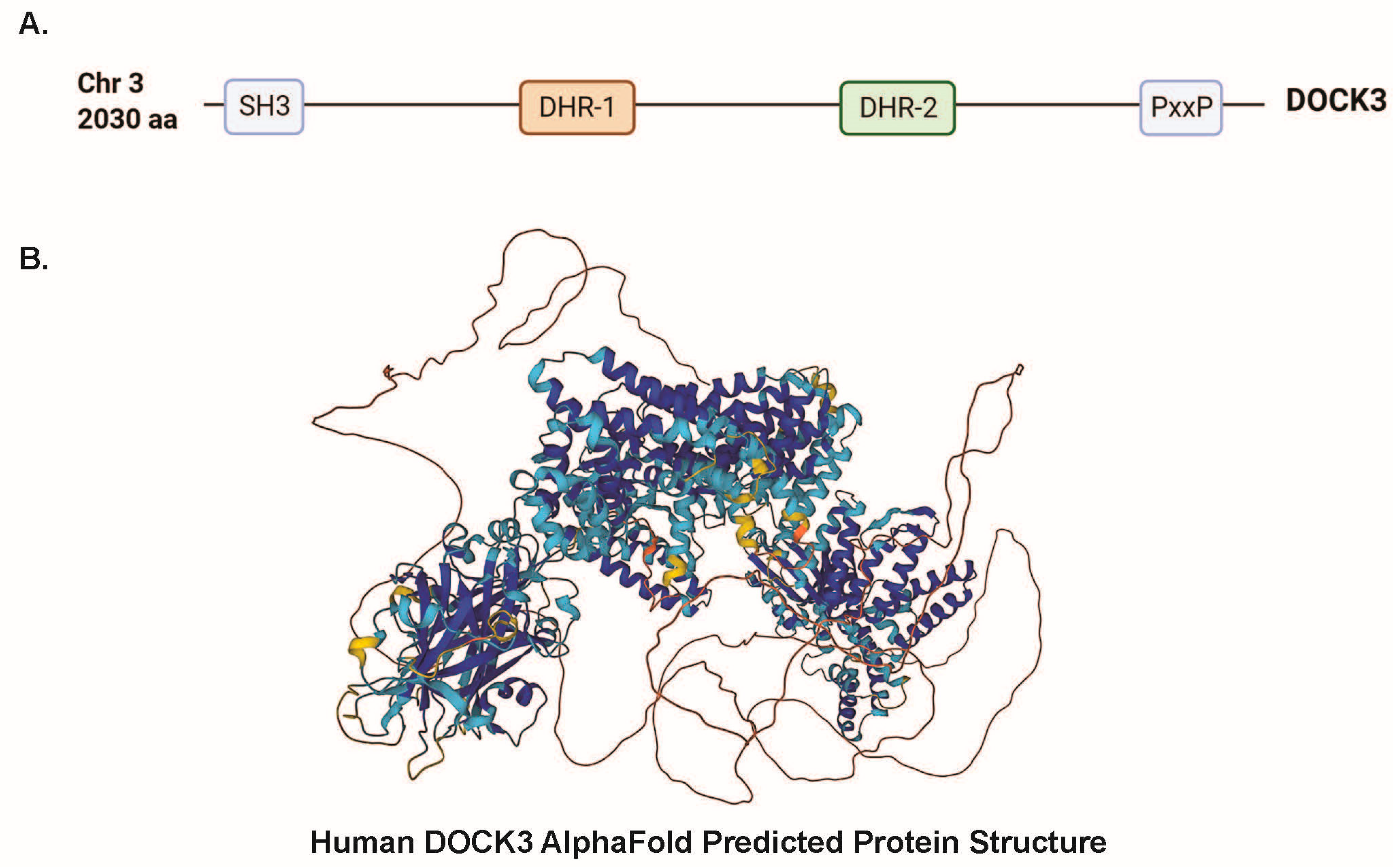
4.2. DOCK3 Is Comprises Key Evolutionary Conserved Domains Essential for Protein–Protein Interactions

Mammalian DOCK3 is a member of the Dock-B subclass of DOCK proteins along with DOCK4, and the longest human DOCK3 protein isoform is approximately 2030 amino acids (Figure 1A). The human DOCK3 protein consists of a conserved SH3 domain, DHR-1/2 domains, and a PxxP domain. The DHR-2 (DOCK homology region 2) domain of DOCK3 is essential for GEF activity and binds to the WAVE proteins via their DHR-1 domains, thus subsequently activating RAC1 [16]. Comparisons of protein structures among DOCK family members have revealed commonalities that both distinguish DOCK proteins from other GEFs and define the mechanism by which a DOCK catalytic DHR2 domain elicits nucleotide dissociation from small RhoGTPase (especially, RAC1) [25]. The SH3 domain in DOCK proteins is likely responsible for the signal transduction of tyrosine phosphorylation signals to the actin cytoskeleton in regulating cell motility [25,26,27,28]. Subsequently, the SH3 domain also interacts with the PxxP domain within each of the DOCKs (DOCK1-5) to initiate actin signals [29,30]. As more information emerges on the structure of DOCK3, DOCK proteins, and DOCK3 protein interactions, increased knowledge of DOCK3’s functional role in different cell types and the consequences of loss-of-function variants will be better understood.
A full protein structure of mammalian DOCK3 does not exist; however, several AI-predicted structures for DOCK3 exist including one for human DOCK3 from AlphaFold (Figure 1B) [31]. Insights from the published cryo-EM structure of the DOCK2-ELMO2 protein–protein complex may yield clues into commonalities for protein binding mechanisms between DOCK3 and ELMO binding [32]. DOCK2–ELMO1 interactions mutually relieve their autoinhibition for the activation of RAC1 for lymphocyte chemotaxis, which might explain similar DOCK3-ELMO1/2 regulation of cellular migration [33]. DOCK3 binding to ELMO1/2 likely similarly regulates GEF activity and promotes subsequent cellular growth. While DOCK3 is conserved in vertebrate species, in Drosophila, a DOCK3/4 counterpart is the sponge (spg)/CG31048 gene, which also interacts with ELMO and regulates RAC1 activity [34]. Knockdown of sponge in Drosophila eye imaginal discs induced abnormal eye morphology in adult flies and reduced ERK signaling [35]. Follow-up Drosophila studies demonstrated a critical role for sponge in air sac promordium development and tracheal cell viability that was mediated by the ERK signaling pathway. Further study of DOCK3 domains in other species and cell models may yield additional clues into the function of DOCK3 in a cellular and tissue signaling context.
Evaluation of the functional role(s) of DOCK4 may also yield clues into novel approaches in corrective therapies for DOCK3 patients. An interesting finding from a recent evaluation of Dock4-deficient (Dock4 KO) mice showed that overexpression of active RAC1 viral vectors could partially rescue neuronal functional tests [36]. Although it is not desirable to fully activate RAC1 in every tissue due to its role as a known cancer-driving oncogenic factor [37], temporal and tissue-specific modulation of active RAC1 may be beneficial for rescuing some of the DOCK3-affected pathways that are altered in patients. Indeed, some pharmacological modulators of RAC1 activity are being explored for cognitive disorders and may be worth pursuing in Dock3-deficient mice and cell lines [38]. If RAC1 modulators were both tissue-specific and reversible, there may be benefits towards exploring the effects of RAC1 modulation in DOCK3-deficiency in animal models with the end goal of patient trials should efficacy and safety be established.

4.3. Dock3-Deficient Mice Have Significant Developmental and Regenerative Defects

Dock3 knockout (Dock3 KO) mice were generated and showed progressive movement defects and accumulation of autophagic vacuoles accumulating in the spinal cords of aged Dock3 KO mice [11]. Dock3 KO mice develop clasping pathologies and abnormal aggregates of neurofilament protein along with the disorganization of the axonal cytoskeleton. Electron microscopy of Dock3 KO mice revealed impaired axonal transport of and a general accumulation of polyubiquitinated proteins, a hallmark of neurodegenerative disorders [11,39]. Genetic ablation of Dock3 on the dystrophin-deficient (mdx) background further exacerbates dystrophic pathologies including increasing muscle fibrosis and loss of muscle physiological force [20]. Interestingly, haploinsufficiency of Dock3 (Dock3 +/−) on the mdx background restores the DOCK3 protein to normal levels and partially improves dystrophic muscle pathologies and histology. Conditional disruption of Dock3 in skeletal muscle (Dock3 mKO) results in mild impairment of muscle pathology and histology including impairment of glucose processing [20]. Additional use of conditional Dock3 knockout mice to evaluate DOCK3′s functional role in other cell lineages may yield clues in other tissue and cell types.

4.4. RAC1-Affected Pathways Affected by DOCK3 Disruption

As previously mentioned, genetic disruption of DOCK3 in patients results in decreased RAC1 activation [13]. This DOCK3-deficiency and decrease in global RAC1 activity likely has profound affects throughout several tissues as RAC1 is a key regulator of cellular migration, fusion, differentiation, proliferation, and viability as a GTPase effector protein [21,40]. RAC1 is a dynamic molecule that can have profoundly different effects in a tissue-specific and cellular context. Patients with ultra-rare RAC1 missense mutations have reported symptoms consisting of developmental delay, and macrocephaly, although some reported instances of microcephaly depended on whether the RAC1 mutation was a dominant negative activating or inactivating mutation [22,41]. Interestingly, those same RAC1 variants were able to be effectively modeled in zebrafish through the use of mRNA overexpression and the quantification of zebrafish larvae head circumference, highlighting the utility of animal models in dissecting RAC1-specific mutational consequences.
Conversely, complete loss of Rac1 expression in Rac1 knockout (Rac1 KO) mice is embryonic lethal due defects in the formation of the embryonic germ layers resulting from the cellular disruption of lamellipodia formation, cell adhesion, and cell migration processes [23,42]. Further elucidation of RAC1’s functional roles occurred with the generation of Rac1 conditional knockout mice (Rac1 cKO) which demonstrated the tissue-specific requirements of RAC1. For example, cardiomyocyte-specific Rac1 knockout mice (c-Rac1) demonstrated an essential requirement for RAC1 in cardiac hypertrophy [24,43]. This study revealed that Rac1 expression in cardiomyocytes was essential for NADPH oxidase activity and myocardial oxidative stress in response to angiotensin II treatment. This study along with others was one of the first indicators of tissue-specific roles for RAC1 and highlighted the utility of the Rac1 cKO mouse.
Follow-up work from the laboratory of Lykke Sylow and colleagues further expanded upon this model through the conditional ablation of Rac1 in skeletal myofibers using an inducible muscle-specific promoter (Rac1 mKO) [25,44]. Rac1 mKO mice given a high-fat diet (HFD) experienced insulin resistance in their skeletal muscles and in vivo insulin-stimulated glucose uptake was reduced in the triceps, soleus, and gastrocnemius muscles. Interestingly, whole-body glucose uptake was unaffected when assessed using a 2-deoxy-glucose uptake challenge, as was AKT activation. These results suggest that Rac1 genetic loss is detrimental to insulin-stimulated muscle glucose uptake independent of skeletal muscle AKT signaling pathway activation. Interestingly, studies of these Rac1 mKO mice also showed that exercise enhanced whole-body insulin sensitivity by 40% in WT mice and rescued insulin intolerance in Rac1 mKO mice by improving whole-body insulin sensitivity by 230% [26,45]. What was equally important from these studies was that exercise improved insulin muscle sensitivity in both WT and Rac1 mKO mouse muscles, suggesting that RAC1 dysfunction may be dispensable for the correction of muscle insensitivity. More recent work from the Sylow group has identified RhoGDIα phosphorylation as a critical step towards skeletal muscle GLUT4 translocation and RAC1 activation [27,46]. When bound to RAC1, RhoGDIα inhibits RAC1 activation, although it is unclear if DOCK proteins interact directly or indirectly with this complex.
Additional studies by other groups have focused on the dynamic regulation of RAC1 to Rho signaling in the context of other cell populations that reside within the skeletal muscle. In quiescence muscle satellite cells (MuSCs), activated RAC1 inhibits activated RhoA, which is inversely switched in early activated MuSCs [28,47]. These activities control cellular projections and filopodia formation within the MuSC population via F/G-actin polymerization. In an adeno-associated viral vector (AAV)-mediated motor neuron Rac1 knockout mouse model, these mice developed abnormal dendritic spine morphology associated with hyperexcitability disorder, increased mature, mushroom dendritic spines, and an increased level in overall spine length and spine head size [29,48]. Interestingly, after a spinal cord injury (SCI), three-weeks post injury, it was observed that there was significant restoration of rate-dependent depression (RDD) and reduced H-reflex excitability in these Rac1 motor neuron knockout mice. More recently, the topic of RAC1 regulation has been of interest as a means for understanding the dynamics of its regulation. In skeletal muscle, a muscle-enriched microRNA signaling pathway “myomiRs” (miR-1/206/133) has been shown to regulate DOK7-CRK-RAC1, which is critical for the stabilization and anchoring of postsynaptic acetylcholine receptor AChRs during NMJ development and maintenance [30,49]. The authors concluded that the failure to properly modulate RAC1 activity severely compromises NMJ function, causing respiratory failure in neonates and neuromuscular symptoms in adult mice. These findings highlight the dynamic nature of RAC1 expression and the regulation of RAC1 signaling in different muscle cell populations. Nevertheless, the dynamics of RAC1 activation and its protein–protein binding partners in skeletal muscle and neuronal tissues remains to be fully elucidated.
The role of RAC1 activation and DOCK proteins also appears to be that of a tissue- and cell-specific nature. RhoG was shown to activate RAC1 via DOCK1 and ELMO1 to affect mammalian cell shape and migration [31,32,50]. Follow-up studies demonstrated that activation of RAC1 by RhoG is required for normal lamellipodia formation at the leading edge during cellular migration [33,51]. In contrast, this study also demonstrated that the interaction of DOCK1 with CRK was dispensable for the activation of RAC1 and the promotion of cell migration by RhoG. The generation of additional Dock1 and Dock5 knockout mice revealed essential, non-overlapping functions for each factor in myoblast fusion and muscle differentiation [34,52]. Similar work from our group not only demonstrated that Dock3 knockout mice also had impaired muscle myoblast differentiation and regeneration following injury through downregulation of the fusogen Myomixer (Mymx) but also failed RAC1 activation [19,20]. Interestingly, recent findings from the work of Jean-François Côté and colleagues demonstrated that dysferlinopathy (limb girdle muscular dystrophy type 2B/R2) muscle pathologies can be partially mitigated by manipulating ELMO2 conformational regulation [35,53]. Whereas the authors demonstrated that both ELMO1 and ELMO2 muscle expression were required for myoblast fusion, manipulation of key regulatory domains could affect muscle outcomes. ELMO proteins are normally in a closed conformation and the DOCK1 DHR-2 domain is blocked by the Ras-binding domain (RBD) domain of ELMO, subsequently preventing RAC1 activation and the binding of interacting proteins to ELMO proteins. Upon activation of the ELMO-DOCK1 complex, protein–protein binding sites open up to allow for RAC1 activation. It remains unclear if a similar process occurs in DOCK3 or other DOCK proteins that bind to both ELMO proteins and RAC1 regulators; however, these studies have laid the foundation for future work in this field. Newer, exciting drug screens for small molecule compounds that modulate DOCK3 conformational changes affecting DOCK3 and ELMO1 interactions have recently been published and could be explored for treating axonal injury and neurodegenerative diseases [36,54].

4.5. Disease Phenotypes Associated with Deficient Functioning of Genes Interacting with DOCK3

The RAC1 gene is activated by DOCK3. It encodes an RHO GTPase involved in the modulation of the cytoskeleton, which plays a role in multiple cellular functions, including phagocytosis, mesenchymal-like migration, neuronal polarization, axonal growth, and the differentiation of multiple cell types. RAC1 is also involved in cellular growth and cell-cycle regulation [41]. De novo, heterozygous variants in RAC1 are associated with a distinct NDD that includes dysmorphic features, moderate/severe intellectual disability, seizures, hypotonia, CNS anomalies including cerebellar dysplasia, hypoplasia of the corpus callosum, enlarged ventricles, mega cisterna magna, a thin brainstem, white matter abnormalities, and polymicrogyria [41,55]. In a study, 14 cases with de novo, heterozygous RAC1 mutations were reported [41,55]. Of the six cases that had functional studies, four were shown to be gain-of-function type and two had a dominant-negative effect. While there are significant similarities in the manifestations associated with DOCK3 and RAC1 variants, there are also differences. For instance, the RAC1 group showed brain malformations not described in association with DOCK3 deficiency. In summary, the relationship between DOCK3 disfunction and RAC1 is not fully understood and needs further study.
Another disease phenotype is associated with the gene ELMO2 which interacts with DOCK3. The ELMO2-associated condition includes vascular malformations with recurrent and sometimes life-threatening bleeding in different organs. This phenotype was reported in eight individuals from five families and was associated with biallelic loss of function variants in ELMO2 and autosomal recessive inheritance [56]. The relationship between DOCK3 deficiency and ELMO2 functioning also remains to be clarified in future studies. The clinical consequences of DOCK3 deficiency seem to be very different from those associated with ELMO2 deficiency.
Finally, the DOCK3 protein was initially characterized as a presenilin-binding protein [14]. Variants in presenilin 1 and 2 are associated with autosomal dominant Alzheimer’s disease and dilated cardiomyopathy [57].

5. Future Research

The future of DOCK3-related research is likely tied to that of other DOCK factors in determining the function of DOCK3 on a tissue-specific basis. Research into DOCK3 and its other DOCK-B family member DOCK4 may yield clues into its function in other tissue types in development and disease [58]. The use of conditional knockout animals and CRISPR-genomic editing approaches will likely yield clues into its function in a cell-type and temporal-specific manner. DOCK3 is an important factor in many tissues outside of neurons and improved DOCK3 antibody and protein detection strategies will yield better insight into the localization of DOCK3 within mammalian healthy and disease tissues. Probing of DOCK3 expression in cancer and other atlas databases (e.g., The Cancer Genome Atlas—TCGA) may yield insights into DOCK3 dysregulation in disease contexts.
Given the large size of the human DOCK3 gene (53 exons; 6 kb open reading frame), traditional overexpression restorative gene therapies are unlikely due to viral packaging limitations. However, one can look towards the neuromuscular field for insight into strategies for genetic correction of partially functional DOCK3 proteins using exon-skipping phosphorodiamidate morpholino oligomers (PMOs). In Duchenne muscular dystrophy (DMD), exon-skipping PMOs have been FDA-approved and lead to long-term improvements in DMD patient functional outcomes [59,60,61]. One can envision that exon-skipping PMOs might be a reasonable strategy towards making DOCK3 patient variants amenable by restoring the DOCK3 reading frame in one or both pathogenic alleles. Another strategy that may be promising would be a split intein adeno-associated viral (AAV) vector approach to allow for the recombination of the full-length DOCK3 open reading frame within the cell or tissue of interest. These approaches rely on the reconstitution of separate AAV viral vectors into a single large construct, often using a base-editor approach within the tissue of interest within an organism [62,63]. This approach has shown promise in correcting liver metabolic disease in the Pahenu2 mouse model [60]. With improvements in CRISPR base editing, gene therapy delivery systems, and AAV expression vectors, one can envision testing corrective gene therapy approaches in Dock3-deficient mice as a viable strategy for eventual DOCK3 patient testing.
An additional hurdle in the development of an efficient protocol for gene therapy is the limited efficiency of the viral vectors to cross the blood–brain barrier (BBB). We have recently shown that AAV-based vectors cross the BBB with very high efficiency in a mouse model of fragile X syndrome [64]. While the vector used by us provides such high efficiency only for selected mouse strains, more recently developed AAV vectors show promise for high BBB crossing efficiency in humans [65]. When vectors with high efficiency are used, another potential problem may be oversupply of the gene product. Using highly efficient AAV vectors, we observed a potential decrease in the efficiency to correct the disease phenotype due to oversupply of the protein product of the FMR1 gene, FMRP [64]. Following these concerns, it is important to remember that DOCK3 activates RAC1, and at least some of the RAC1 pathogenic variants were associated with gain of function [41,55].
Alternative to gene therapy, small-molecule agents may be used to correct DOCK3 deficiency. In a recent study, the authors screened 462,169 low-molecular-weight compounds and identified compounds that stimulate the interaction between DOCK3 and Elmo1, resulting in neurite outgrowth in vitro. Some of these compounds stimulated neuroprotection and axon regeneration in a mouse model of optic nerve injury. These findings may be the basis of the development of new treatments for DOCK3 deficiency using small-molecule compounds that may be more efficient in crossing the BBB [54].
From a clinical point of view, one important question regarding phenotype–genotype correlations that may need further clarification is the association of DOCK3 mutations with congenital anomalies. Multiple congenital anomalies outside the CNS were present only in one out of the six of the reported patients to date (P4). In addition, in most reported patients, diagnosis was achieved with WES. Since all reported patients to date have muscle hypotonia and an abnormal gait, the gene may be potentially included in a more targeted muscle weakness test panel. Questions remain as to how to best treat DOCK3 loss-of-function patients as these patients suffer from intellectual disability and muscle hypotonia from an early age, with them often having difficulty obtaining an official genetic diagnosis without genomic testing. Documentation as to the region of DOCK3 pathogenic mutation(s) would likely provide insight as to the degree of damage influencing protein function. Early assessments of biallelic DOCK3 pathogenic variant families appear to indicate that larger, multi-exon deletions in the N-terminus of the DOCK3 gene affect patient outcomes more severely than those with C-terminal DOCK3 variants or deletions [13]. As more patients are identified with DOCK3 pathogenic variants and other DOCK gene loss-of-function variants, strategies for the treatment of these disorders need to be addressed [66,67]. Gene therapy approaches may need to be refined as traditional overexpression approaches may need to be optimized given the large size of DOCK3 gene open reading frames.

Author Contributions

M.S.A. and M.V. wrote and edited the manuscript. M.V. collected all DOCK3 patient genetic information. All authors have read and agreed to the published version of the manuscript.

Funding

The research reported in this publication was supported by the Eunice Kennedy Shriver National Institute of Child Health and Human Development, NIH, HHS of the National Institutes of Health under award number R01HD095897 awarded to M.S. Alexander. M.S. Alexander is supported by an NIH Office of the Director (OD) U54 grant U54OD030167.

Institutional Review Board Statement

Not applicable.

Informed Consent Statement

Not applicable.

Data Availability Statement

No new data were created or analyzed in this study. Data sharing is not applicable to this article.

Acknowledgments

The authors wish to acknowledge the DOCK3 patients for their contribution to this research and providing a better understanding of their condition. The authors also wish to acknowledge members of their laboratories for the critical analysis of the manuscript.

Conflicts of Interest

The authors declare no conflict of interest.

References

  1. Thapar, A.; Cooper, M.; Rutter, M. Neurodevelopmental disorders. Lancet Psychiatry 2017, 4, 339–346. [Google Scholar] [CrossRef]
  2. Velinov, M. Genomic Copy Number Variations in the Autism Clinic-Work in Progress. Front. Cell Neurosci. 2019, 13, 57. [Google Scholar] [CrossRef] [PubMed]
  3. Wang, W.; Corominas, R.; Lin, G.N. De novo Mutations from Whole Exome Sequencing in Neurodevelopmental and Psychiatric Disorders: From Discovery to Application. Front. Genet. 2019, 10, 258. [Google Scholar] [CrossRef]
  4. Malpass, K. Neurodevelopmental disorders: Unlocking the secrets of autism through whole-exome sequencing. Nat. Rev. Neurol. 2012, 8, 295. [Google Scholar] [CrossRef]
  5. Wang, T.; Kim, C.N.; Bakken, T.E.; Gillentine, M.A.; Henning, B.; Mao, Y.; Gilissen, C.; SPARK Consortium; Nowakowski, T.J.; Eichler, E.E. Integrated gene analyses of de novo variants from 46,612 trios with autism and developmental disorders. Proc. Natl. Acad. Sci. USA 2022, 119, e2203491119. [Google Scholar] [CrossRef] [PubMed]
  6. Aslesh, T.; Yokota, T. Restoring SMN Expression: An Overview of the Therapeutic Developments for the Treatment of Spinal Muscular Atrophy. Cells 2022, 11, 417. [Google Scholar] [CrossRef]
  7. Canaud, G.; Gutierrez, J.C.L.; Irvine, A.D.; Vabres, P.; Hansford, J.R.; Ankrah, N.; Branle, F.; Papadimitriou, A.; Ridolfi, A.; O’Connell, P.; et al. Alpelisib for Treatment of Patients with PIK3CA-Related Overgrowth Spectrum (PROS). Genet. Med. 2023, 25, 100969. [Google Scholar] [CrossRef] [PubMed]
  8. Efron, D.; Delatycki, M.B.; de Silva, M.G.; Langbein, A.; Slaghuis, W.; Larson, A.; Dahl, H.H.; Forrest, S.M. A novel pericentric inversion of chromosome 3 cosegregates with a developmental-behavioral phenotype. J. Med. Genet. 2003, 40, E15. [Google Scholar] [CrossRef]
  9. de Silva, M.G.; Elliott, K.; Dahl, H.H.; Fitzpatrick, E.; Wilcox, S.; Delatycki, M.; Williamson, R.; Efron, D.; Lynch, M.; Forrest, S. Disruption of a novel member of a sodium/hydrogen exchanger family and DOCK3 is associated with an attention deficit hyperactivity disorder-like phenotype. J. Med. Genet. 2003, 40, 733–740. [Google Scholar] [CrossRef]
  10. Helbig, K.L.; Mroske, C.; Moorthy, D.; Sajan, S.A.; Velinov, M. Biallelic loss-of-function variants in DOCK3 cause muscle hypotonia, ataxia, and intellectual disability. Clin. Genet. 2017, 92, 430–433. [Google Scholar] [CrossRef] [PubMed]
  11. Chen, Q.; Peto, C.A.; Shelton, G.D.; Mizisin, A.; Sawchenko, P.E.; Schubert, D. Loss of Modifier of Cell Adhesion Reveals a Pathway Leading to Axonal Degeneration. J. Neurosci. 2009, 29, 118–130. [Google Scholar] [CrossRef]
  12. Iwata-Otsubo, A.; Ritter, A.L.; Weckselbatt, B.; Ryan, N.R.; Burgess, D.; Conlin, L.K.; Izumi, K. DOCK3-related neurodevelopmental syndrome: Biallelic intragenic deletion of DOCK3 in a boy with developmental delay and hypotonia. Am. J. Med. Genet. Part A 2018, 176, 241–245. [Google Scholar] [CrossRef]
  13. Wiltrout, K.; Ferrer, A.; van de Laar, I.; Namekata, K.; Harada, T.; Klee, E.W.; Zimmerman, M.T.; Cousin, M.A.; Kempainen, J.L.; Babovic-Vuksanovic, D.; et al. Variants in DOCK3 cause developmental delay and hypotonia. Eur. J. Hum. Genet. 2019, 27, 1225–1234. [Google Scholar] [CrossRef]
  14. Namekata, K.; Enokido, Y.; Iwasawa, K.; Kimura, H. MOCA Induces Membrane Spreading by Activating Rac1. J. Biol. Chem. 2004, 279, 14331–14337. [Google Scholar] [CrossRef]
  15. Chen, Q.; Chen, T.J.; Letourneau, P.C.; Costa Lda, F.; Schubert, D. Modifier of cell adhesion regulates N-cadherin-mediated cell-cell adhesion and neurite outgrowth. J. Neurosci. 2005, 25, 281–290. [Google Scholar] [CrossRef] [PubMed]
  16. Namekata, K.; Harada, C.; Taya, C.; Guo, X.; Kimura, H.; Parada, L.F.; Harada, T. Dock3 induces axonal outgrowth by stimulating membrane recruitment of the WAVE complex. Proc. Natl. Acad. Sci. USA 2010, 107, 7586–7591. [Google Scholar] [CrossRef] [PubMed]
  17. Sanz-Moreno, V.; Gadea, G.; Ahn, J.; Paterson, H.; Marra, P.; Pinner, S.; Sahai, E.; Marshall, C.J. Rac Activation and Inactivation Control Plasticity of Tumor Cell Movement. Cell 2008, 135, 510–523. [Google Scholar] [CrossRef] [PubMed]
  18. Li, J.; Mi, X.; Chen, L.; Jiang, G.; Wang, N.; Zhang, Y.; Deng, W.; Wang, Z.; Chen, G.; Wang, X. Dock3 Participate in Epileptogenesis through rac1 Pathway in Animal Models. Mol. Neurobiol. 2016, 53, 2715–2725. [Google Scholar] [CrossRef] [PubMed]
  19. Alexander, M.S.; Casar, J.C.; Motohashi, N.; Vieira, N.M.; Eisenberg, I.; Marshall, J.L.; Gasperini, M.J.; Lek, A.; Myers, J.A.; Estrella, E.A.; et al. MicroRNA-486–dependent modulation of DOCK3/PTEN/AKT signaling pathways improves muscular dystrophy–associated symptoms. J. Clin. Investig. 2014, 124, 2651–2667. [Google Scholar] [CrossRef]
  20. Reid, A.L.; Wang, Y.; Samani, A.; Hightower, R.M.; Lopez, M.A.; Gilbert, S.R.; Ianov, L.; Crossman, D.K.; Dell’Italia, L.J.; Millay, D.P.; et al. DOCK3 is a dosage-sensitive regulator of skeletal muscle and Duchenne muscular dystrophy-associated pathologies. Hum. Mol. Genet. 2020, 29, 2855–2871. [Google Scholar] [CrossRef] [PubMed]
  21. Makihara, S.; Morin, S.; Ferent, J.; Côté, J.-F.; Yam, P.T.; Charron, F. Polarized Dock Activity Drives Shh-Mediated Axon Guidance. Dev. Cell 2018, 46, 410–425.e417. [Google Scholar] [CrossRef] [PubMed]
  22. Namekata, K.; Watanabe, H.; Guo, X.; Kittaka, D.; Kawamura, K.; Kimura, A.; Harada, C.; Harada, T. Dock3 regulates BDNF-TrkB signaling for neurite outgrowth by forming a ternary complex with Elmo and RhoG. Genes Cells 2012, 17, 688–697. [Google Scholar] [CrossRef] [PubMed]
  23. Bai, N.; Hayashi, H.; Aida, T.; Namekata, K.; Harada, T.; Mishina, M.; Tanaka, K. Dock3 interaction with a glutamate-receptor NR2D subunit protects neurons from excitotoxicity. Mol. Brain 2013, 6, 22. [Google Scholar] [CrossRef]
  24. Samani, A.; Karuppasamy, M.; English, K.G.; Siler, C.A.; Wang, Y.; Widrick, J.J.; Alexander, M.S. DOCK3 regulates normal skeletal muscle regeneration and glucose metabolism. FASEB J. 2023, 37, e23198. [Google Scholar] [CrossRef]
  25. Thompson, A.P.; Bitsina, C.; Gray, J.L.; von Delft, F.; Brennan, P.E. RHO to the DOCK for GDP disembarking: Structural insights into the DOCK GTPase nucleotide exchange factors. J. Biol. Chem. 2021, 296, 100521. [Google Scholar] [CrossRef] [PubMed]
  26. Ruan, W.; Pang, P.; Rao, Y. The SH2/SH3 Adaptor Protein Dock Interacts with the Ste20-like Kinase Misshapen in Controlling Growth Cone Motility. Neuron 1999, 24, 595–605. [Google Scholar] [CrossRef]
  27. Schlessinger, J. SH2/SH3 signaling proteins. Curr. Opin. Genet. Dev. 1994, 4, 25–30. [Google Scholar] [CrossRef]
  28. Rao, Y.; Zipursky, S.L. Domain requirements for the Dock adapter protein in growth- cone signaling. Proc. Natl. Acad. Sci. USA 1998, 95, 2077–2082. [Google Scholar] [CrossRef] [PubMed]
  29. Liu, X.; Li, F.; Pan, Z.; Wang, W.; Wen, W. Solution structure of the SH3 domain of DOCK180. Proteins Struct. Funct. Bioinform. 2013, 81, 906–910. [Google Scholar] [CrossRef]
  30. Aitio, O.; Hellman, M.; Kazlauskas, A.; Vingadassalom, D.F.; Leong, J.M.; Saksela, K.; Permi, P. Recognition of tandem PxxP motifs as a unique Src homology 3-binding mode triggers pathogen-driven actin assembly. Proc. Natl. Acad. Sci. USA 2010, 107, 21743–21748. [Google Scholar] [CrossRef] [PubMed]
  31. Jumper, J.; Evans, R.; Pritzel, A.; Green, T.; Figurnov, M.; Ronneberger, O.; Tunyasuvunakool, K.; Bates, R.; Žídek, A.; Potapenko, A.; et al. Highly accurate protein structure prediction with AlphaFold. Nature 2021, 596, 583–589. [Google Scholar] [CrossRef] [PubMed]
  32. Chang, L.; Yang, J.; Jo, C.H.; Boland, A.; Zhang, Z.; McLaughlin, S.H.; Abu-Thuraia, A.; Killoran, R.C.; Smith, M.J.; Côté, J.-F.; et al. Structure of the DOCK2−ELMO1 complex provides insights into regulation of the auto-inhibited state. Nat. Commun. 2020, 11, 3464. [Google Scholar] [CrossRef] [PubMed]
  33. Hanawa-Suetsugu, K.; Kukimoto-Niino, M.; Mishima-Tsumagari, C.; Akasaka, R.; Ohsawa, N.; Sekine, S.-I.; Ito, T.; Tochio, N.; Koshiba, S.; Kigawa, T.; et al. Structural basis for mutual relief of the Rac guanine nucleotide exchange factor DOCK2 and its partner ELMO1 from their autoinhibited forms. Proc. Natl. Acad. Sci. USA 2012, 109, 3305–3310. [Google Scholar] [CrossRef] [PubMed]
  34. Biersmith, B.; Liu, Z.; Bauman, K.; Geisbrecht, E.R. The DOCK Protein Sponge Binds to ELMO and Functions in Drosophila Embryonic CNS Development. PLoS ONE 2011, 6, e16120. [Google Scholar] [CrossRef]
  35. Eguchi, K.; Yoshioka, Y.; Yoshida, H.; Morishita, K.; Miyata, S.; Hiai, H.; Yamaguchi, M. The Drosophila DOCK family protein sponge is involved in differentiation of R7 photoreceptor cells. Exp. Cell Res. 2013, 319, 2179–2195. [Google Scholar] [CrossRef] [PubMed]
  36. Guo, D.; Peng, Y.; Wang, L.; Sun, X.; Wang, X.; Liang, C.; Yang, X.; Li, S.; Xu, J.; Ye, W.C.; et al. Autism-like social deficit generated by Dock4 deficiency is rescued by restoration of Rac1 activity and NMDA receptor function. Mol. Psychiatry 2019, 26, 1505–1519. [Google Scholar] [CrossRef]
  37. Zhou, C.; Licciulli, S.; Avila, J.L.; Cho, M.; Troutman, S.; Jiang, P.; Kossenkov, A.V.; Showe, L.C.; Liu, Q.; Vachani, A.; et al. The Rac1 splice form Rac1b promotes K-ras-induced lung tumorigenesis. Oncogene 2013, 32, 903–909. [Google Scholar] [CrossRef] [PubMed]
  38. Tejada-Simon, M.V. Modulation of actin dynamics by Rac1 to target cognitive function. J. Neurochem. 2015, 133, 767–779. [Google Scholar] [CrossRef]
  39. Jain, A.; Vale, R.D. RNA phase transitions in repeat expansion disorders. Nature 2017, 546, 243–247. [Google Scholar] [CrossRef]
  40. Bosco, E.E.; Mulloy, J.C.; Zheng, Y. Rac1 GTPase: A “Rac” of all trades. Cell. Mol. Life Sci. 2009, 66, 370–374. [Google Scholar] [CrossRef]
  41. Reijnders, M.R.F.; Ansor, N.M.; Kousi, M.; Yue, W.W.; Tan, P.L.; Clarkson, K.; Clayton-Smith, J.; Corning, K.; Jones, J.R.; Lam, W.W.K.; et al. RAC1 Missense Mutations in Developmental Disorders with Diverse Phenotypes. Am. J. Hum. Genet. 2017, 101, 466–477. [Google Scholar] [CrossRef]
  42. Sugihara, K.; Nakatsuji, N.; Nakamura, K.; Nakao, K.; Hashimoto, R.; Otani, H.; Sakagami, H.; Kondo, H.; Nozawa, S.; Aiba, A.; et al. Rac1 is required for the formation of three germ layers during gastrulation. Oncogene 1998, 17, 3427–3433. [Google Scholar] [CrossRef] [PubMed]
  43. Satoh, M.; Ogita, H.; Takeshita, K.; Mukai, Y.; Kwiatkowski, D.J.; Liao, J.K. Requirement of Rac1 in the development of cardiac hypertrophy. Proc. Natl. Acad. Sci. USA 2006, 103, 7432–7437. [Google Scholar] [CrossRef] [PubMed]
  44. Raun, S.H.; Ali, M.; Kjøbsted, R.; Møller, L.L.V.; Federspiel, M.A.; Richter, E.A.; Jensen, T.E.; Sylow, L. Rac1 muscle knockout exacerbates the detrimental effect of high-fat diet on insulin-stimulated muscle glucose uptake independently of Akt. J. Physiol. 2018, 596, 2283–2299. [Google Scholar] [CrossRef] [PubMed]
  45. Sylow, L.; Møller, L.L.; D’Hulst, G.; Schjerling, P.; Jensen, T.E.; Richter, E.A. Rac1 in Muscle Is Dispensable for Improved Insulin Action After Exercise in Mice. Endocrinology 2016, 157, 3009–3015. [Google Scholar] [CrossRef]
  46. Møller, L.L.V.; Ali, M.S.; Davey, J.; Raun, S.H.; Andersen, N.R.; Long, J.Z.; Qian, H.; Jeppesen, J.F.; Henriquez-Olguin, C.; Frank, E.; et al. The Rho guanine dissociation inhibitor α inhibits skeletal muscle Rac1 activity and insulin action. Proc. Natl. Acad. Sci. USA 2023, 120, e2211041120. [Google Scholar] [CrossRef]
  47. Kann, A.P.; Hung, M.; Wang, W.; Nguyen, J.; Gilbert, P.M.; Wu, Z.; Krauss, R.S. An injury-responsive Rac-to-Rho GTPase switch drives activation of muscle stem cells through rapid cytoskeletal remodeling. Cell Stem Cell 2022, 29, 933–947.e6. [Google Scholar] [CrossRef]
  48. Benson, C.A.; Olson, K.L.; Patwa, S.; Reimer, M.L.; Bangalore, L.; Hill, M.; Waxman, S.G.; Tan, A.M. Conditional RAC1 knockout in motor neurons restores H-reflex rate-dependent depression after spinal cord injury. Sci. Rep. 2021, 11, 7838. [Google Scholar] [CrossRef]
  49. Klockner, I.; Schutt, C.; Gerhardt, T.; Boettger, T.; Braun, T. Control of CRK-RAC1 activity by the miR-1/206/133 miRNA family is essential for neuromuscular junction function. Nat. Commun. 2022, 13, 3180. [Google Scholar] [CrossRef]
  50. Gumienny, T.L.; Brugnera, E.; Tosello-Trampont, A.C.; Kinchen, J.M.; Haney, L.B.; Nishiwaki, K.; Walk, S.F.; Nemergut, M.E.; Macara, I.G.; Francis, R.; et al. CED-12/ELMO, a novel member of the CrkII/Dock180/Rac pathway, is required for phagocytosis and cell migration. Cell 2001, 107, 27–41. [Google Scholar] [CrossRef] [PubMed]
  51. Katoh, H.; Hiramoto, K.; Negishi, M. Activation of Rac1 by RhoG regulates cell migration. J. Cell Sci. 2006, 119, 56–65. [Google Scholar] [CrossRef]
  52. Laurin, M.; Fradet, N.; Blangy, A.; Hall, A.; Vuori, K.; Côté, J.F. The atypical Rac activator Dock180 (Dock1) regulates myoblast fusion in vivo. Proc. Natl. Acad. Sci. USA 2008, 105, 15446–15451. [Google Scholar] [CrossRef] [PubMed]
  53. Tran, V.; Nahlé, S.; Robert, A.; Desanlis, I.; Killoran, R.; Ehresmann, S.; Thibault, M.P.; Barford, D.; Ravichandran, K.S.; Sauvageau, M.; et al. Biasing the conformation of ELMO2 reveals that myoblast fusion can be exploited to improve muscle regeneration. Nat. Commun. 2022, 13, 7077. [Google Scholar] [CrossRef] [PubMed]
  54. Namekata, K.; Tsuji, N.; Guo, X.; Nishijima, E.; Honda, S.; Kitamura, Y.; Yamasaki, A.; Kishida, M.; Takeyama, J.; Ishikawa, H.; et al. Neuroprotection and axon regeneration by novel low-molecular-weight compounds through the modification of DOCK3 conformation. Cell Death Discov. 2023, 9, 166. [Google Scholar] [CrossRef]
  55. Banka, S.; Bennington, A.; Baker, M.J.; Rijckmans, E.; Clemente, G.D.; Ansor, N.M.; Sito, H.; Prasad, P.; Anyane-Yeboa, K.; Badalato, L.; et al. Activating RAC1 variants in the switch II region cause a developmental syndrome and alter neuronal morphology. Brain 2022, 145, 4232–4245. [Google Scholar] [CrossRef] [PubMed]
  56. Cetinkaya, A.; Xiong, J.R.; Vargel, I.; Kosemehmetoglu, K.; Canter, H.I.; Gerdan, O.F.; Longo, N.; Alzahrani, A.; Camps, M.P.; Taskiran, E.Z.; et al. Loss-of-function mutations in ELMO2 cause intraosseous vascular malformation by impeding RAC1 signaling. Am. J. Hum. Genet. 2016, 99, 299–317. [Google Scholar] [CrossRef]
  57. Clark, R.F.; Hutton, M.; Talbot, C.; Wragg, M.; Lendon, C.; Busfield, F.; Han, S.W.; Perez-Tur, J.; Adams, M.; Fuldner, R.; et al. The role of presenilin 1 in the genetics of Alzheimer’s disease. Cold Spring Harbor Symp. Quant. Biol. 1996, 61, 551–558. [Google Scholar]
  58. Laurin, M.; Côté, J.-F. Insights into the biological functions of Dock family guanine nucleotide exchange factors. Genes Dev. 2014, 28, 533–547. [Google Scholar] [CrossRef]
  59. Charleston, J.S.; Schnell, F.J.; Dworzak, J.; Donoghue, C.; Lewis, S.; Chen, L.; Young, G.D.; Milici, A.J.; Voss, J.; DeAlwis, U.; et al. Eteplirsen treatment for Duchenne muscular dystrophy. Neurology 2018, 90, e2146–e2154. [Google Scholar] [CrossRef]
  60. Mendell, J.R.; Goemans, N.; Lowes, L.P.; Alfano, L.N.; Berry, K.; Shao, J.; Kaye, E.M.; Mercuri, E.; for the Eteplirsen Study Group; Telethon Foundation DMD Italian Network. Longitudinal effect of eteplirsen versus historical control on ambulation in Duchenne muscular dystrophy. Ann. Neurol. 2016, 79, 257–271. [Google Scholar] [CrossRef]
  61. Iff, J.; Gerrits, C.; Zhong, Y.; Tuttle, E.; Birk, E.; Zheng, Y.; Paul, X.; Henricson, E.K.; McDonald, C.M.; Investigators, C.-D. Delays in pulmonary decline in eteplirsen-treated patients with Duchenne muscular dystrophy. Muscle Nerve 2022, 66, 262–269. [Google Scholar] [CrossRef] [PubMed]
  62. Vila-Perelló, M.; Muir, T.W. Biological Applications of Protein Splicing. Cell 2010, 143, 191–200. [Google Scholar] [CrossRef] [PubMed]
  63. Stevens, A.J.; Sekar, G.; Shah, N.H.; Mostafavi, A.Z.; Cowburn, D.; Muir, T.W. A promiscuous split intein with expanded protein engineering applications. Proc. Natl. Acad. Sci. USA 2017, 114, 8538–8543. [Google Scholar] [CrossRef] [PubMed]
  64. Chadman, K.K.; Adayev, T.; Udayan, A.; Ahmed, R.; Dai, C.L.; Goodman, J.H.; Meeker, H.; Dolzhanskaya, N.; Velinov, M. Efficient Delivery of FMR1 across the Blood Brain Barrier Using AAVphp Construct in Adult FMR1 KO Mice Suggests the Feasibility of Gene Therapy for Fragile X Syndrome. Genes 2023, 14, 505. [Google Scholar] [CrossRef] [PubMed]
  65. Goertsen, D.; Flytzanis, N.C.; Goeden, N.; Chuapoco, M.R.; Cummins, A.; Chen, Y.; Fan, Y.; Zhang, Q.; Sharma, J.; Duan, Y.; et al. AAV capsid variants with brain-wide transgene expression and decreased liver targeting after intravenous delivery in mouse and marmoset. Nat. Neurosci. 2022, 25, 106–115. [Google Scholar] [CrossRef]
  66. Villiger, L.; Grisch-Chan, H.M.; Lindsay, H.; Ringnalda, F.; Pogliano, C.B.; Allegri, G.; Fingerhut, R.; Häberle, J.; Matos, J.; Robinson, M.D.; et al. Treatment of a metabolic liver disease by in vivo genome base editing in adult mice. Nat. Med. 2018, 24, 1519–1525. [Google Scholar] [CrossRef]
  67. Samani, A.; English, K.G.; Lopez, M.A.; Birch, C.L.; Brown, D.M.; Kaur, G.; Worthey, E.A.; Alexander, M.S. DOCKopathies: A systematic review of the clinical pathologies associated with human DOCK pathogenic variants. Hum. Mutat. 2022, 43, 1149–1161. [Google Scholar] [CrossRef]
Figure 1. DOCK3 is comprised of key evolutionary conserved domains essential for protein-protein interactions. (A) Conserved protein domains for the largest isoform of the human DOCK3 protein (2030 amino acids, aa). These domains consist of an SH3 domain, a DHR-1 and DHR-2 domain, and a PxxP domain. (B) The human DOCK3 AlphaFold Predicted Protein Structure for DOCK3 is shown here. AlphaFold AF-Q8IZD9-F1 model is displayed here.
Genes 14 01940 g001
Table 1. Summary of clinical features of patients with DOCK3 mutations.
PatientDOCK3 VariantReferenceSexAge at EvaluationBirth HistoryFamily HistoryDevelopmental MilestonesGrowth (%)Dysmorphic FeaturesCongenital AnomaliesStudies Prior to Diagnosis
P1del3:50789040- 51247265/c.382C>T[10]F12 yearsBorn at 42 weeks gestationSimilarly affected siblingSevere Developmental Delay, walked at 5
years, unstable crouched, ataxic gait, non-
verbal, and not toilet trained at 12y
WT = 25;
Ht = 15;
HC = 75
Prominent chin, high arched
palate, malocclusion, long
fingers
NoneNormal metabolic screen, EEG, BEAR, brain MRI
P2del3:50789040- 51247265/c.382C>T[10]M11 yearsBorn at 41 weeks gestationSimilarly affected siblingWalked at 2.5 years, first word at 4 years, single words at TOE, unstable, ataxic gaitWt = 40; Ht = 8; HC = 30Pointed chin, down slanting palpebral fissures, long faceNoneNone
P3homozygous del. 3:51,062,402–51,232,768 [12]M28 monthsNRParents are first cousinsStarted sitting at 14 months, walked at 22 months, unsteady gait, few specific words at TOE, Bayley score <50 (at TOE)Wt = 4; Ht = 5; HC = 14Epicanthal folds, up-turned nasal tip, prominent cheeksNoneBrain MRI-dysmorphic Corpus Callosum, ECHO-normal
P4c.1038-2A>G:IVS12- 2A>G/c.3107_3110delACTT[13]M5 yearsBorn at 37 weeks gestationUnremarkableStarted walking at 36 months, 5-10 single words at 5Wt ≥ 99; Ht = 66; HC = 85Broad forehead, deep set, hooded eyesTE fistula with esophageal atresia, vertebral anomalies, rib anomalies, single
kidney
Negative microarray, brain MRI- shallow sulci, hypoplastic white matter, spine MRI-syrinx, abnormal EEG
P5c.1175G>A/c.3887A>G[13]M5.5 yearsFull termUnremarkableWas able to sit at 30 months, walked at 48 months, non-verbal, autism, unprovoked laughter, hypotoniaWt = 50; Ht = 25; HC = 7Brachicephaly, plagiocephaly, prominent philtrumPhimosisBrain MRI-diminished white matter, hypoplastic CC, negative macroarray, UBE3A, MECP2,
meth-Angelman
P6c.5020A>T/5020A>T[13]F3 yearsborn at 35 weeks gestation, feeding difficultiesNRWalked at 18 months, and said the first word at 15 months, but then lost her speech, autismWt ≥ 99; Ht ≥ 99; HC ≥ 99Macrocephaly, frontal bossingSpina bifidaBrain MRI-resolved Chiari malformation, negative CMA, PTEN, FXS
Disclaimer/Publisher’s Note: The statements, opinions and data contained in all publications are solely those of the individual author(s) and contributor(s) and not of MDPI and/or the editor(s). MDPI and/or the editor(s) disclaim responsibility for any injury to people or property resulting from any ideas, methods, instructions or products referred to in the content.

