Article

Clustering of Genetic Anomalies of Cilia Outer Dynein Arm and Central Apparatus in Patients with Transposition of the Great Arteries

by
Marlon De Ita
1,2,
Javier Gaytán-Cervantes
3,
Bulmaro Cisneros
2,
María Antonieta Araujo
4,
Juan Carlos Huicochea-Montiel
4,
Alan Cárdenas-Conejo
4,
Charles César Lazo-Cárdenas
5,
César Iván Ramírez-Portillo
5,
Carina Feria-Kaiser
6,
Leoncio Peregrino-Bejarano
7,
Lucelli Yáñez-Gutiérrez
8,
Carolina González-Torres
3,* and
Haydeé Rosas-Vargas
1,*
1
Unidad de Investigación Médica en Genética Humana, UMAE Hospital de Pediatría, Centro Médico Nacional Siglo XXI, IMSS, Ciudad de México 06720, Mexico
2
Dpto de Genética y Biología Molecular, CINVESTAV Zacatenco IPN, Ciudad de México 07360, Mexico
3
Laboratorio de Secuenciación, División de Desarrollo de la Investigación, IMSS, Ciudad de México 06720, Mexico
4
Departamento clínico de Genética Médica, UMAE Hospital de Pediatría, Centro Médico Nacional Siglo XXI, IMSS, Ciudad de México 06720, Mexico
5
Departamento clínico de Cardiología, UMAE Hospital de Pediatría, Centro Médico Nacional Siglo XXI, IMSS, Ciudad de México 06720, Mexico
6
Unidad de Cuidados Intensivos Neonatales, UMAE Hospital de Pediatría, Centro Médico Nacional Siglo XXI, IMSS, Ciudad de México 06720, Mexico
7
UMAE Hospital de Pediatría, Centro Médico Nacional Siglo XXI, IMSS, Ciudad de México 06720, Mexico
8
Clínica de Cardiopatías Congénitas, UMAE Hospital de Cardiología, CMN Siglo XXI, Ciudad de México 06720, Mexico
*
Authors to whom correspondence should be addressed.
Genes 2022, 13(9), 1662; https://doi.org/10.3390/genes13091662
Submission received: 11 August 2022 / Revised: 10 September 2022 / Accepted: 11 September 2022 / Published: 16 September 2022
(This article belongs to the Special Issue Genetics of Complex Human Disease)

Abstract

:
Transposition of the great arteries (TGA) is a congenital heart defect with a complex pathogenesis that has not been fully elucidated. In this study, we performed whole-exome sequencing (WES) in isolated TGA-diagnosed patients and analyzed genes of motile and non-motile cilia ciliogenesis and ciliary trafficking, as well as genes previously associated with this heart malformation. Deleterious missense and splicing variants of genes DNAH9, DNAH11, and ODAD4 of cilia outer dynein arm and central apparatus, HYDIN, were found in our TGA patients. Remarkable, there is a clustering of deleterious genetic variants in cilia genes, suggesting it could be an oligogenic disease. Our data evidence the genetic diversity and etiological complexity of TGA and point out that population allele determination and genetic aggregation studies are required to improve genetic counseling.

Graphical Abstract

1. Introduction

Transposition of the great arteries (TGA) is a severe congenital heart disease (CHD) characterized by concordant atrioventricular and ventriculoarterial discordant connections in situs solitus. Although several monogenic syndromes have been associated with this defect, including Kabuki, Carpenter, and MRFACD, the etiology in its isolated and sporadic form is still a matter of debate [1,2,3]. Knockout mouse models of genes involved in left-right (LR) establishment have helped link TGA pathogenesis to an abnormal NODAL/GDF1-DAND5 developmental pathway (NGD).
NGD signaling depends on fluid flow gradients, with beating cilia being relevant to establishing LR asymmetry. Besides this, cilia have been associated with laterality establishment and heart development in other ways: the bending of primary cilia by hydrodynamic forces, the position of ciliary cells in the organizer, and the ciliary cell polarization in the posterior region of the embryo for LR establishment [4]. This evidence suggests that cilia function could be relevant in TGA pathogenesis.
In this regard, several cilia genes have been associated with this abnormal LR establishment and CHD. Abnormalities in the OFD1 gene, which is involved in ciliogenesis and the function of motile and non-motile cilia, have been associated with abnormal node development and the establishment of LR [5]. In motile cilia, alterations in heavy chain dyneins such as DNAH9 and DNAH11 have been associated with abnormal cilia beating and linked to congenitally corrected TGA [6,7,8]. Similarly, abnormalities in the intermediate strand DNAI1, required for cilia motility, have also been reported in patients with TGA [9,10]. In addition to these functions, primary cilia modulate several signaling pathways involved in cardiogenesis, such as TGF β and Sonic Hedgehog, whose alterations are related to outflow tract defects [11,12,13].
In general, congenital heart diseases, including TGA, have been linked with ciliopathies, such as Ellis van Creveld and Simpson–Golabi–Behmel syndrome [5,6,7]. However, a massive WES analysis recently suggested that cilia genetic anomalies could be relevant to TGA etiology [8]. Furthermore, the interaction and clustering of several deleterious genetic variants could lead to a predisposition with the transposition of the great arteries, as has been previously suggested for other CHD, such as the tetralogy of Fallot [9]. This study shows a high throughput analysis of genes previously linked with TGA (NODAL-GDF1-DAND5 signaling pathway) and motile (inner, outer dynein arms, and central apparatus) and non-motile cilia genes, which providence evidence for the identification of the etiology underlying this congenital heart disease.

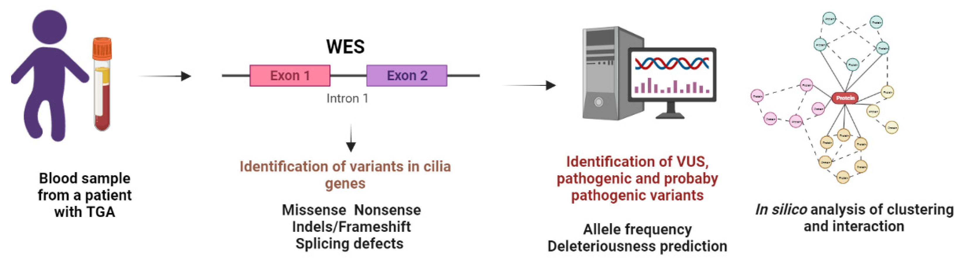
2. Materials and Methods

2.1. Patients

Eleven patients, from newborn to 16 years, with transposition of the great arteries, were recruited from Pediatric and Cardiology Hospitals from National Medical Center XXI century, Mexican Social Security Institute (IMSS), Mexico. Clinical data relevant to this study were obtained from medical records. Given the presence of clinical manifestations suggestive of congenital heart disease, the diagnosis of transposition of the great arteries was made by pediatric cardiologists and described by ICD categorization. The patients were also evaluated by clinical geneticists to rule out the presence of other phenotypic alterations that would make it possible to integrate the diagnosis of various syndromic entities that may involve this type of congenital heart disease. Patients with congenital anomalies suggestive of syndromic entities were excluded from the study. The Research Ethics Board of the Mexican Social Security Institute, México, approved this study (R-2018-785-047). Prior to conducting this study, the parents of the affected children were informed and asked to give their consent via a signed written consent before the beginning of the study. Children eight years old and older signed an assent/consent document.

2.2. Sample Collection and Processing DNA Extraction

Blood samples were collected from 11 subjects diagnosed with TGA for WES. According to the manufacturer’s instructions, the genomic DNA was extracted using a QIAamp DNA Blood Mini Kit I (Qiagen, Hilden, Germany). DNA concentration and purity were measured using a Nanodrop Spectrophotometer (Thermo Fisher Scientific, Waltham, MA, USA) and Qubit 2.0 Fluorometer (Thermo Fisher Scientific, Waltham, MA, USA), while integrity was established by capillary electrophoresis using the TapeStation4200 (Agilent Technologies, Santa Clara, CA, USA).

2.3. Whole-Exome Sequencing

A total of 50 ng of genomic DNA per sample was used for DNA library preparation. DNA libraries were prepared by fragmentation using a Nextera DNA Exome sample prep kit (Illumina Inc., San Diego, CA, USA; Cat N. 20020616), involving end repair, A-tailing, and adapter ligation, as instructed by the manufacturer. Libraries were quantified using Qubit (dsDNA HS assays, ThemoFisher Scientific, Waltham, MA, USA, Cat.N. Q33230), pooled in equimolar amounts, and subjected to exome capture using XGen Research Panel version 1.0 (Integrated DNA Technologies, IDT, Coralville, IA, USA), with blocking adapters (xGen Blockers, IDT, Coralville, IA, USA), designed to match the Nextera library constructs following the manufacturer’s instructions. The libraries were diluted for sequencing on an Illumina Nextseq 500 platform using 2 × 146 paired-end cycles. BCL2 data were transformed to fastq through Bcl2fastq software, for further analysis, and the read quality of fastq files per sample was carried out using FAST-QC v0.11.9 under default parameters.

2.4. Variant Analysis

After WES, a DRAGEN Germline Pipeline (v3.2.8, Illumina, Inc., Waltham, MA, USA) was used to align the remaining high-quality sequencing reads to the reference human genome (hg19), and Germline Variant Small Hard Filtering (Illumina, Inc. Waltham, MA, USA) was used to annotate the identified single-nucleotide variants and InDels. The variant interpretation was performed with the Variant Interpreter software (Illumina Inc., Waltham, MA, USA). In the previously related genes, all non-synonymous variants were studied, whereas only VUS, pathogenic, or possibly pathogenic cilia genes were analyzed.
Nonsynonymous variants were then evaluated, determining their deleteriousness by SIFT, PolyPhen2, and a Mutation Assessor medium or high score. For splicing, nonsense, and indel variants, a LoF-Tool or CADD PHRED score was determined; a ≥20 value indicates that a variant is among the top 1% of damaging variants in the genome. Variant Effect Predictor was used to identify the effect on the canonic transcript.
To predict interactions, the STRING database was used (V11.5). First, a Markov Clustering Algorithm was employed to determine the global gene aggregation. Secondly, an interaction enrichment analysis was performed (EPv), where a p-value less than 0.05 was interpreted as indicative of a functional connection between proteins [10]. Protein domains were determined using Uniprot. Variants were subsetted to the GnomAD V2.1.1 to describe the Latin allele population frequency. As genetic variants presence interpretation is challenging, deleteriousness was defined using several algorithms. In silico analysis can predict whether a genetic anomaly will result in protein abnormalities that could lead to a malfunction or even its absence. The presence of deleterious variants may elevate the individual’s susceptibility or predisposition to the disease. Concerning missense variants, deleteriousness was determined by the presence of two abnormalities by SIFT, PolyPhen2, and Mutation Assessor, or the presence of one abnormality if a new variant was described. Although WES analysis is focused on exons, we analyzed proximal splicing variants when possible. Online Mendelian Inheritance in Man, OMIM® McKusick-Nathans Institute of Genetic Medicine, Johns Hopkins University (Baltimore, MD, USA), (06-01-2022) was used to determine gene relevance in a clinical context. URL: https://omim.org/ (accessed on 30 November 2021). A list of the analyzed genes and their general relationship with motile and non-motile cilia, ciliogenesis, and ciliary trafficking can be found in Table S1.

2.5. Statistical Analysis
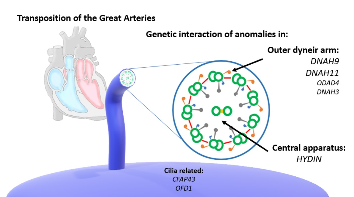
Probabilistic binomial analysis was performed to determine the probability of two or more variants in our cohort using the Minitab statistical software (Minitab19, Minitab LLC, State College, PA, USA). The variant expected population probability used in this analysis was obtained from frequency data from GnomAD. To compare the allele frequency between sexes, Exact Fisher Test was employed.

3. Results

3.1. Genetic Analysis Related to Syndromic Entities

We examined 11 unrelated probands with sporadic TGA, levocardia, and situs solitus. After WES analysis, a median of 160k variants per patient was obtained, and missense, nonsense, indels, or altered splicing regions were identified. A flow chart of the genetic analysis performed is presented in Figure 1.
First, genes previously associated with TGA in syndromic entities were analyzed (Table 1: Abnormalities in genes previously related to TGA). Genetic anomalies associated with Kabuki syndrome were found in three probands: the first one carries a KDM6A hemizygous variant c.232C>T (p.R78C) OMIM Gene (OMIM-G) 300128, which was classified as non-deleterious (Table S2: Complete gene abnormalities of ciliary genes in TGA). The two remaining probands carry the autosomic KMT2D (OMIM-G 602113) deleterious variants c.547C>T (p.P183S) and c.15686G>A (p.R5229H). The first variant is in PHD finger 1 (PHD1), a module involved in the epigenetic regulation of gene expression via the recruitment of chromatin regulators and transcription factors [11]. The second variant is related to the FYR-C terminal domain of KMT2D, characterized by phenylalanine/tyrosine-rich regions found in various chromatin-associated proteins. The p.P183S is a newly described variant, while KMT2D p.R5229H is infrequently found in Latin American populations. It is worth noting that none of the patients displayed phenotypical characteristics of Kabuki syndrome.
On the other hand, variants of MEGF8 (OMIM-G 604267), a gene related to TGA in Carpenter syndrome, were found in two patients. The first one carries the p.R439W (c.1315C>T) deleterious variant, while the second one carries the p.R782W non-deleterious variant, which is rare in the Latin American population (Table 1). However, both patients have heterozygous anomalies, whereas this entity is a recessive inheritance disorder.

3.2. NGD and Previously TGA-Related Genes

Next, TGA syndromic genes were analyzed, namely those related to LR establishment CHD (NODAL, GDF1, and ACVR2B) (Table 1). Several algorithms found two missense variants of the FOXH1 and GDF1 genes, which were classified as non-deleterious. Concerning ciliary genes previously associated with TGA, deleterious DNAI1 and DISC1 in T181101 and SLC4A1 and CLASP1 in T181201 variants were identified (Table 1). Since both patients were heterozygous carriers, we presume that different heterozygous deleterious anomalies in ciliary genes could have a summative effect, supporting an oligogenic etiology.

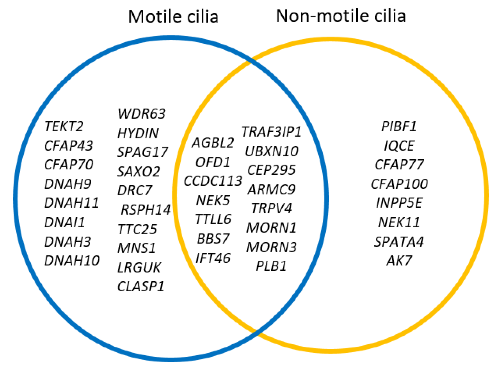
3.3. Motile and Non-Motile Genes, Ciliogenesis, and Cilia Trafficking Gene Analysis

To ascertain whether ciliary genes could be related to this CHD, we examined genes previously implicated with either functional or structural functions of motile cilia, non-motile cilia, and ciliary trafficking [12,13]. Several variants, including missense, nonsense codon, frameshift, and splicing alterations, were identified in DNAH9, OFD1, BBS7, and other genes (Table 2, Table 3, Table 4 and Table S2). Most ciliary deleterious genetic variants were found in heterozygous probands, except for homozygous probands for HYDIN and PIBF1 and a hemizygous proband for OFD1. Only deleterious and possibly pathogenic variants were analyzed.
The most common anomalies were within the profile of the motile cilia (Table 2: Anomalies in motile ciliary genes in TGA patients), where patients displayed abnormalities in the outer dynein arm and central pair proteins. Deleterious variants of DNAH9 and DNAH11 genes were found in outer dynein arms genes. Four DNAH9 variants were identified in three patients: a p.Y1017C and a p.G2384R variant; a splice donor splicing variant c.5151+1G>A and two missense changes in c.3050A>G. Although two patients displayed the same variant, the presence of 2 out of 22 alleles c.3050A>G in our sample has a probability of 0.2184, according to binomial probability calculation. Interestingly, the T180401 proband carries both missense and splicing variants that could have a summative effect in these patients. The latter variant was described as deleterious; as a result, the patient was compound heterozygous for the DNAH9 gene. On the other hand, T180201 presented a DNAH11 nonsense heterozygous variant located in the C terminal domain, after the AAA6 region, in an area likely associated with a deleterious effect. As the allele presents a frequency of 2.897 × 10⁻⁵ in the Latin American population, a relationship with TGA is plausible in this case.
Additional deleterious genetic variants of outer dynein arms genes were found (Table 2). Two patients were hemizygous with deleterious variants in OFD1, a gene associated with Oral-Facial-Digital syndrome type 1 (OMIM Phenotype 311200), which has a sex-linked inheritance. A female proband carried an OFD1 c.2610G>C (p.Q870H) variant, and a male proband had a c.1819C>T variant (p.F828V). Although p.Q870H has been observed in a male subject, the variant does not present a population difference between the sexes (exact Fisher test, p = 0.520), suggesting a lesser effect on a lethal cardiovascular phenotype. On the other side, the OFD1 p.Q870H is a newly reported deleterious anomaly; however, it has been found in a female, so the effect of its probable deleteriousness could be masked. Finally, the presence of a novel homozygous HYDIN c.3332C>T (p.P1111L) deleterious variant in a T180401 proband was revealed by algorithm analysis.
Regarding non-motile cilia genes (Table 3: Genetic anomalies in non-motile cilia genes in TGA), a subject presented several abnormalities in genes related to this organelle as INPPP5E and IQCE, which are related to the modulation of Hedgehog signaling that could be relevant to outflow tract development [14,15,16]. On the other hand, a previously described pathogenic variant of the PIBF1 gene was identified in three patients; two homozygous probands and a single heterozygous proband for c.1214G>A (p.R405Q) (Table 3). This gene has been associated with Joubert syndrome, a ciliopathy not associated with abnormal organ disposition (OMIM-P 617767). It is worth mentioning that finding 5 out of 22 alleles with a p < 0.05 according to binomial probability calculation with data of GnomAD for the Latin American population suggests that this is not a stochastic finding and could be associated with this disease.
Concerning ciliogenesis and cilia trafficking gene analysis (Table 4, genetic anomalies in ciliogenesis and ciliary trafficking in TGA patients), the patients presented several anomalies related to these processes. In addition to the presence of previously described OFD1 and PIBF1 genetic variants, IFT46 anomalies were also observed. This gene is part of the IFT subcomplex B that is required for retrograde transport in the cilia but is also related to ciliogenesis [17,18]. In particular, Ift46 KO mouse embryos displayed randomization of the embryo heart looping, a hallmark of defective lateralization; this effect has been associated with a lack of cilia in node cells [19]. The absence of nodal flow, which can be caused by the loss of nodal cilia and abnormal LR patterning, as seen in Ift46 embryos, has been consistently related to TGA [3].

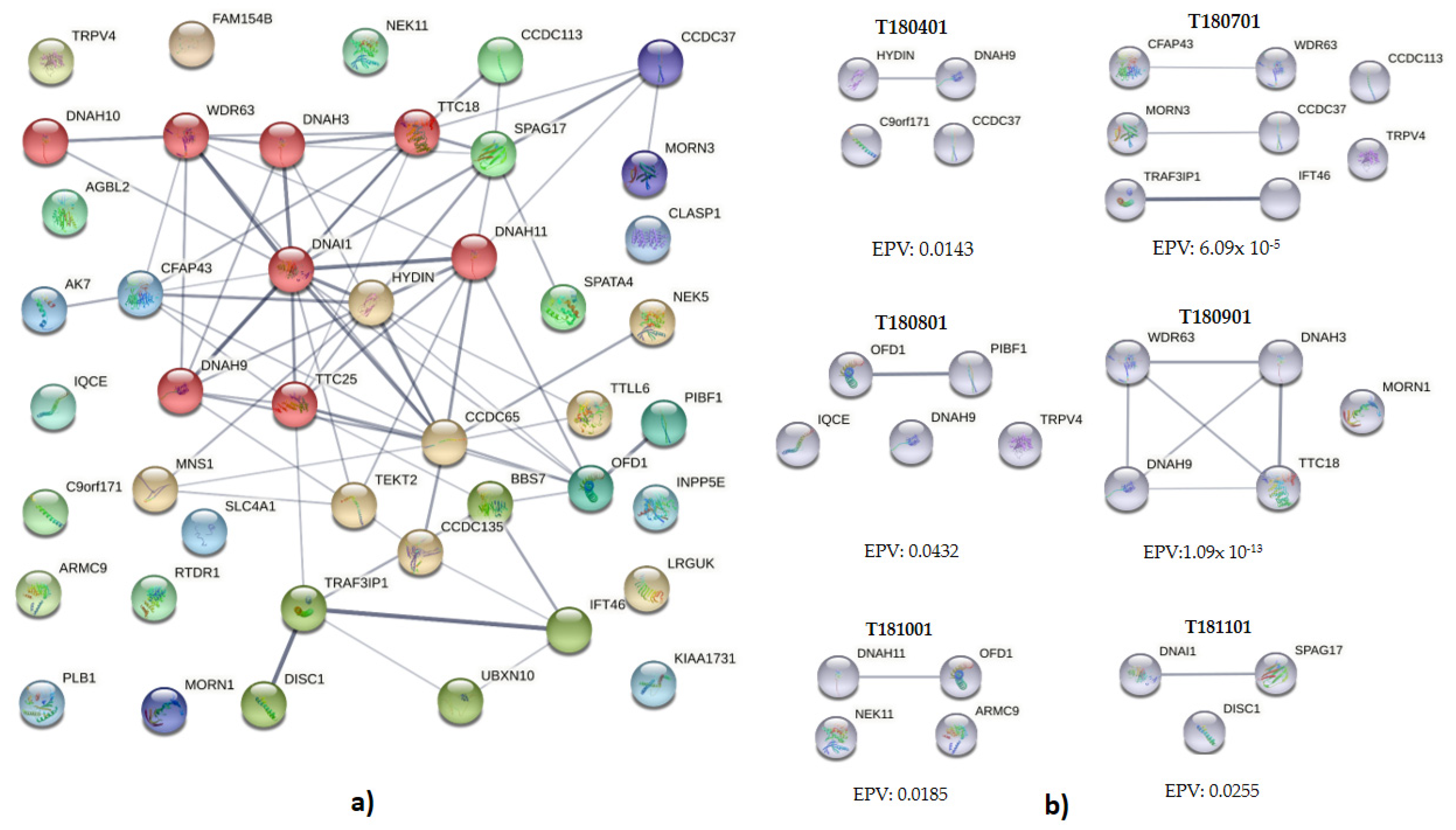
3.4. Interaction and Clustering Analysis

Apart from DNAH9 and HYDIN variants found in some patients, most of the genetic variants were observed in genes related to autosomic recessive diseases. Thus, we hypothesized that clustering and interaction analyses might uncover an abnormal cilia function genotype by presenting several risk variants (Figure 2, genetic abnormality aggregation in patients with transposition of the great arteries). The interactions described by STRING software include direct (physical) and indirect (functional) associations, using knowledge transfer between organisms, and cluster interactions from other databases [10]. An interactive protein network was obtained in 29 out of 44 genes (65.9%) with deleterious variants (Figure 2a). Markov Clustering Algorithm displayed the presence of 7 different clusters, suggesting that dynamic protein complexes favor aggregation in static gene interactions (Figure 2a and Table S3). Further, to determine the cluster relevance, STRING interaction enrichment analysis was performed (EPV); a p-value of less than 0.05 was interpreted as indicative of a functional connection between proteins [10]. Using the EPV comprehensive analysis, a clustering includes deleterious variants of BBS7, TRAF3IP1, OFD1, and PIBF1 as part of primary cilia development (WP4536, STRING False Discovery Rate: 3.57 × 10⁻⁸). Interestingly, a cluster formed by 12 elements, including WDR63, DNAH3, DNAI1, TTC18, SPAG17, TTC25, and CCDC113, was found to be a cluster related to primary ciliary dyskinesia and COPI-independent Golgi-to-ER (Cluster 10689, STRING False Discovery Rate: 1.0–12) (Figure S1).
We next examined deleterious variants of TGA genes using an individual in silico functional interaction analysis. In 6 out of 11 analyzed probands (54.5%), clustering and interaction of anomalies were observed (Figure 2b). Abnormalities of DNAH9 and HYDIN interact in proband T180401, suggesting an oligogenic context in this patient. To T180701, several pairs of clustering anomalies, including CFAP43 and WDR63; CCDC36 and MORN3; TRAF3IP1 and IFT46. Studies using the single-celled model Chlamydomonas showed that the complex CFAP43/CFAP44 could regulate IDAF/I1(IC140), an orthologue of WDR63, hence, modulating the cilia beating [20,21]. Despite this, abnormalities of these genes have been implicated in nervous system malformations and infertility but not in heart defects [22,23,24,25], suggesting a poor association with TGA.
Further, the interaction between OFD1 and PIBF1 was observed for proband T180801. Both genes have previously been implicated congenital defects in related syndromic entities OMIM Phenotype 311,200 and 617,767, respectively; thus, their implication in TGA requires further analysis. A complex interaction among DNAH9, DNAH3, WDR63, and CFAP70/TTC18 gene variants was revealed for the T180901 proband, suggesting a deleterious aggregation integration. Among them, DNHA9 had been associated with motile cilia function and abnormal heart defects [26,27,28]; therefore, this interaction could be relevant to the pathogenesis of TGA in this patient. Regarding the T181001, a weak association between DNAH11 and OFD1 gene variants was found. Finally, DNAI1 and SPAG17 variants were biologically associated with the T181101 proband. DNAI1 anomalies have been previously associated with TGA [8], while the central pair associated protein SPAG17 has been associated only with Primary Ciliary Dyskinesia (PCD). Finally, the relationship between gene anomalies related to motile and non-motile cilia can be observed in Figure 3. Several genes could be related to motility or signaling in primary cilia in general; others are related to ciliogenesis, and signaling can be found in both types of cilia. This evidence is relevant and suggest that a single type of cilia cannot rule the etiopathogenesis of the transposition of the great arteries.
Finally, the effect of cilia functions on the described genes can be found in Table 5; in general, deletion models displayed several effects on cilia function or organization; despite DNAH9 defects, IFT46 and OFD1 anomalies have been associated with node cilia defects in embryo development. Other motile cilia found in this study have been associated with abnormal beating, a process that could be related to laterality and eventually to heart defects.

4. Discussion

The transposition of the great arteries is a complex genetic disease whose pathogenesis has not been fully elucidated. Although this disease has been associated with genetic anomalies related to the laterality establishment, its relevance in subjects with situs solitus is still a matter of discussion [2,3]. The presence of several genetic anomalies in our patients provides the context for a change in the paradigm in the genetic counseling of TGA.
Firstly, we observed gene anomalies related to Joubert and Kabuki syndromes. The presence of KMT2D deleterious variants suggests that epigenetic alterations could be relevant to TGA pathogenesis, as has previously been suggested for other genetic syndromes, such as MRFACD (OMIM-P 608771) [38]. To support this notion, it is necessary to analyze the presence of KMT2D variants in TGA patients—even those lacking the distinctive phenotype. With respect to the Joubert (Jb) syndrome-related gene, PIBF1, the fact that a deleterious variant c.1214G>A (p.R405Q) was found in two heterozygous patients (p = 6.02 × 10⁻³) suggests its relevance in TGA etiology. Although this variant is likely-pathogenic and associated with Joubert syndrome in ClinVar, it has been reported in a heterozygous compound context with a genomic deletion encompassing PIBF1 coding regions [39].
Despite the consistent implication of NODAL-GDF1-DAND5 genetic anomalies in TGA etiology [40,41], none of our patients displayed genetic defects in this developmental pathway. Since genetic clustering analyses suggest that the etiology of CHD disorders is extended beyond a single-gene alteration [9], we analyzed cilia anomalies aggregation in our probands. In general, cilia dysfunction triggers defects in the left–right animal plan patterning, leading to the development of CHD [2,42]; moreover, genes encoding cilia proteins could have non-ciliary functions that might also be significant to CHD, as has previously been revealed in the modulation of outflow tract signaling pathways [12].
Interestingly, mutations in primary cilia genes have been linked to motile dysfunction. For instance, WDR35 heterozygous mutations, a gene involved in retrograde ciliary transport, ciliogenesis, and ciliary protein trafficking, caused motile cilia dysfunction in Sensenbrenner syndrome [43]. In this regard, we found a deleterious variant of HYDIN; this central apparatus protein is related to Primary Ciliary Dyskinesia (OMIM-P 608647, Ciliary Dyskinesia, Primary 5) but not to the randomization of the left-right body or CHD. Thus, the biological relevance of this variant should be interpreted in combination with the DNAH9 missense and splicing variants found in the same proband.
On the other hand, outer dynein arm anomalies are the most common cause of PCD, a disorder related to TGA patients with abnormal LRA organ disposition [44]. DNAH9 and DNAH11 are required for the assembly and function of distal outer dynein arms; in fact, DNAH11 mutations could result in abnormal ciliary ultrastructure and hyperkinetic ciliary beating associated with congenitally corrected TGA (ccTGA) [45], while DNAH9 genetic anomalies are linked to either laterality defects, subtle respiratory ciliary-beating defects, and ccTGA [27,46]. Intriguingly, the situs solitus T180401 proband, which carries both a heterozygous compound DNAH9 variant and a HYDIN homozygous variant, did not display any observable clinical manifestation of a ciliopathy.
We also found some anomalies in other genes related to motile ciliary function. A heterozygous nonsense variant was found in the DNAH11, which was linked to primary ciliary dyskinesia 7 (CILD7; OMIM-P 611884). Furthermore, a heterozygous variant was found in the CILD35-associated gene, ODAD4/TTC25 (OMIM-P 617092). It is worth noting that defective TTC25 has been associated with immotile nodal cilia and missing leftward flow via particle image velocimetry, lack of ODAs, and the ODA docking complex [47]. Therefore, this variant might affect cilia function somehow, and further investigation is required to determine the mechanism. The physical interaction of abnormal genetic variants suggested needs validation using assays such as a yeast two-hybrid one-on-one screening, as was previously described for DNAH9 anomalies [27]. Parallelly, the clustering biological relevance requires further study using molecular dissection in models such as Paramecium, Chlamydomonas, or engineered retinal pigment epithelial tissue [48,49,50], which have been shown to be relevant for deciphering ciliary beating mechanisms, structures, and anchoring and ciliogenesis; and these could be valuable tools to validate candidate genes for ciliopathies. Examples of this dissection are the identification of the cilia localization of ARMC9 and CEP104 or the role of CFAP43 and CFAP70 in cilia, both using the single-celled models, and the use of techniques such as CRISPR-Cas9 to mimic the genetic variants to identify their roles in cilia function [20,28,51,52].
Furthermore, the PIBF1 aggregation in the cohort is interesting. PIBF1 had been associated with non-motile cilia assembly, and its absence has been associated with a diminished number of ciliary cells [35]. The presence of a deleterious PIBF1 variant could be suggestive of the role of cilia in TGA downstream of the early laterality establishment. In the heart, primary cilia regulate cardiogenesis via the modulation of several signaling pathways, including platelet-derived growth factor receptor-α (Pdgfra). Pdgfra and its absence has been related to this CHD [53,54].
Despite this, a central role for cilia in TGA pathogenesis was previously suggested by clinical studies where ciliary dysfunction was a predominant feature in patients [55]. Consistent with this hypothesis, a TGA exome-based analysis unveiled the enrichment of cilia-related pathways [8]. Although apparent stochastic findings and interaction were reported in half of our patients, globally we found no enrichment of any ciliary processes shared between subjects, neither in terms of motile or non-motile cilia, ciliogenesis, ciliary compartmentalization, or ciliary trafficking, that could be consistently related to TGA. Although a couple of patients displayed interactions between ciliary genes, other cilia-associated anomalies, such as airway dysfunction, were not evaluated; hence, its functional relevance requires more analysis.
Intriguingly, a recent WGS analysis showed that abnormalities in cilia-related genes were similar between TGA patients and controls [56]; in this context, the clustering of several variants in cilia genes could be relevant to identifying susceptibility genotypes that could increase the familiar risk related to cilia defects. Interestingly, a recent report described familial co-segregation of ccTGA and dextro-TGA [57]. ccTGA has been consistently associated with laterality defects [2,3], and these findings suggest a common pathogenic pathway involving laterality genes in both defects. This pathway could be related to other CHD, such as atrial/ventricular septal defects. Furthermore, a genetic anomalies integration analysis in each patient is required to determine the relevance in a personalized way as other cilia abnormalities could be relevant to diagnosis and the patient’s prognosis. As a result, in isolated and sporadic TGA, the pathogenesis could be more closely associated with an intrinsic abnormal heart development than a cilia-related abnormality.
In general, it is essential to state that it is widely accepted that variant findings require Sanger sequencing validation despite the constant improving accuracy of next-generation sequencing (NGS). Despite this, it has been suggested that high-quality reads in NGS permit a high concordance of the findings with Sanger analysis, which can be as high as 100% [58,59,60]. This concordance is essential as the translational impact of NGS variant findings could be earlier, faster, and cheaper as strict NGS quality controls standards are set.
Even though this study has limitations, the evidence presented suggests that genetic anomalies clustering is more of a risk-influencing genetic factor for non-syndromic CHD than a causative one in the transposition of the great arteries. Therefore, TGA-associated heterozygous genetic anomalies in cilia genes might cause only subtle alterations that may predispose one to the appearance of the TGA. However, these findings require confirmation and further analysis in situs solitus patients and patients with isomerism features.

5. Conclusions

Our study provides evidence that the clustering of anomalies in cilia genes involved in motile and non-motile cilia could underlie TGA pathogenesis, suggesting a complex and heterogeneous genetic architecture and underpinning the genetic interaction analyses as part of the genetic counseling strategy.

Supplementary Materials

The following supporting information can be downloaded at: https://www.mdpi.com/article/10.3390/genes13091662/s1, Table S1: List of cilia genes and previously reported genes analyzed in the patients with transposition of the great arteries; Table S2: Complete gene abnormalities of ciliary genes in TGA; Table S3: Protein clusters of selected genes in TGA patients. Figure S1: Genetic anomalies aggregation of transposition of the great arteries by String.

Author Contributions

Conceptualization, M.D.I. and H.R.-V.; methodology, M.D.I., C.G.-T., J.G.-C., M.A.A., J.C.H.-M., A.C.-C., C.C.L.-C., C.I.R.-P., C.F.-K., L.P.-B. and L.Y.-G.; writing—original draft preparation, M.D.I. and H.R.-V.; writing—review and editing, B.C., C.G.-T. and H.R.-V. All authors have read and agreed to the published version of the manuscript.

Funding

MDI has a CONACYT postdoctoral fellowship (390453). This work was supported by Instituto Mexicano del Seguro Social, grant FIS/IMSS/PROT/PRIO/19/131 and FIS/IMSSPROT/PRIO/16/056.

Institutional Review Board Statement

The study was conducted under the Declaration of Helsinki and approved by the Research Ethics Board of the National Medical Center, Mexican Social Security Institute, México (R-2018-785-047).

Informed Consent Statement

Patients were recruited from the Pediatric and Cardiology hospital from CMN SXXI IMSS. Parents of affected children were informed and asked to give their consent by a signed written form before the beginning of the study. Eight years old and older children signed an assent consent. Written informed consent has been obtained from the patients to publish this paper.

Data Availability Statement

The data supporting this study’s findings are available at https://www.ncbi.nlm.nih.gov/sra/PRJNA865734.

Acknowledgments

We thank all our patients and their families for collaborating in this study.

Conflicts of Interest

The authors declare no conflict of interest. The funders had no role in the design of the study, in the collection, analyses, or interpretation of data, in the writing of the manuscript, or in the decision to publish the results.

References

  1. Unolt, M.; Putotto, C.; Silvestri, L.M.; Marino, D.; Scarabotti, A.; Massaccesi, V.; Caiaro, A.; Versacci, P.; Marino, B. Transposition of great arteries: New insights into the pathogenesis. Front. Pediatr. 2013, 1, 11. [Google Scholar] [CrossRef] [PubMed]
  2. Versacci, P.; Pugnaloni, F.; Digilio, M.C.; Putotto, C.; Unolt, M.; Calcagni, G.; Baban, A.; Marino, B. Some Isolated Cardiac Malformations Can Be Related to Laterality Defects. J. Cardiovasc. Dev. Dis. 2018, 5, 24. [Google Scholar] [CrossRef] [PubMed]
  3. De Ita, M.; Cisneros, B.; Rosas-Vargas, H. Genetics of Transposition of Great Arteries: Between Laterality Abnormality and Outflow Tract Defect. J. Cardiovasc. Transl. Res. 2021, 14, 390–399. [Google Scholar] [CrossRef] [PubMed]
  4. Dasgupta, A.; Amack, J.D. Cilia in vertebrate left-right patterning. Philos. Trans. R. Soc. Lond. B Biol. Sci. 2016, 371, 20150410. [Google Scholar] [CrossRef]
  5. Cottereau, E.; Mortemousque, I.; Moizard, M.-P.; Bürglen, L.; Lacombe, D.; Gilbert-Dussardier, B.; Sigaudy, S.; Boute, O.; David, A.; Faivre, L.; et al. Phenotypic Spectrum of Simpson-Golabi-Behmel Syndrome in a Series of 42 Cases with a Mutation in GPC 3 and Review of the Literature. Am. J. Med. Genet. Part C Semin. Med. Genet. 2013, 163, 92–105. [Google Scholar] [CrossRef]
  6. Chen, C.-P.; Su, Y.-N.; Hsu, C.-Y.; Chern, S.-R.; Tsai, F.-J.; Wu, P.-C.; Chen, P.-T.; Wang, W. Ellis-van Creveld syndrome: Prenatal diagnosis, molecular analysis and genetic counseling. Taiwan J. Obstet. Gynecol. 2010, 49, 481–486. [Google Scholar] [CrossRef]
  7. Chen, C.-P.; Chen, C.-Y.; Chern, S.-R.; Su, J.-W.; Wang, W. First-trimester prenatal diagnosis of Ellis-van Creveld syndrome. Taiwan J. Obstet. Gynecol. 2012, 51, 643–648. [Google Scholar] [CrossRef]
  8. Liu, X.; Chen, W.; Li, W.; Priest, J.R.; Fu, Y.; Pang, K.-J.; Ma, B.; Han, B.; Liu, X.; Hu, S.; et al. Exome-Based Case-Control Analysis Highlights the Pathogenic Role of Ciliary Genes in Transposition of the Great Arteries. Circ. Res. 2020, 126, 811–821. [Google Scholar] [CrossRef]
  9. Reuter, M.S.; Chaturvedi, R.R.; Jobling, R.K.; Pellecchia, G.; Hamdan, O.; Sung, W.W.; Nalpathamkalam, T.; Attaluri, P.; Silversides, C.K.; Wald, R.M.; et al. Clinical Genetic Risk Variants Inform a Functional Protein Interaction Network for Tetralogy of Fallot. Circ. Genom. Precis. Med. 2021, 14, e003410. [Google Scholar] [CrossRef]
  10. Szklarczyk, D.; Gable, A.L.; Nastou, K.C.; Lyon, D.; Kirsch, R.; Pyysalo, S.; Doncheva, N.T.; Legeay, M.; Fang, T.; Bork, P.; et al. The STRING database in 2021: Customizable protein-protein networks, and functional characterization of user-uploaded gene/measurement sets. Nucleic Acids Res. 2021, 49, D605–D612. [Google Scholar] [CrossRef]
  11. Sanchez, R.; Zhou, M.M. The PHD finger: A versatile epigenome reader. Trends Biochem. Sci. 2011, 36, 364–372. [Google Scholar] [CrossRef] [PubMed]
  12. Reiter, J.F.; Leroux, M.R. Genes and molecular pathways underpinning ciliopathies. Nat. Rev. Mol. Cell Biol. 2017, 18, 533–547. [Google Scholar] [CrossRef] [PubMed]
  13. Patir, A.; Fraser, A.M.; Barnett, M.W.; McTeir, L.; Rainger, J.; Davey, M.G.; Freeman, T.C. The transcriptional signature associated with human motile cilia. Sci. Rep. 2020, 10, 10814. [Google Scholar] [CrossRef]
  14. Pusapati, G.; Hughes, C.E.; Dorn, K.V.; Zhang, D.; Sugianto, P.; Aravind, L.; Rohatgi, R. EFCAB7 and IQCE Regulate Hedgehog Signaling by Tethering the EVC-EVC2 Complex to the Base of Primary Cilia. Dev. Cell 2014, 28, 483–496. [Google Scholar] [CrossRef] [PubMed]
  15. Dyson, J.M.; Conduit, S.E.; Feeney, S.J.; Hakim, S.; DiTommaso, T.; Fulcher, A.J.; Sriratana, A.; Ramm, G.; Horan, K.A.; Gurung, R.; et al. INPP5E regulates phosphoinositide-dependent cilia transition zone function. J. Cell Biol. 2016, 216, 247–263. [Google Scholar] [CrossRef] [PubMed]
  16. Smoak, I.W.; Byrd, N.A.; Abu-Issa, R.; Goddeeris, M.M.; Anderson, R.; Morris, J.; Yamamura, K.; Klingensmith, J.; Meyers, E.N. Sonic hedgehog is required for cardiac outflow tract and neural crest cell development. Dev. Biol. 2005, 283, 357–372. [Google Scholar] [CrossRef]
  17. Park, I.; Lee, H.-K.; Kim, C.; Ismail, T.; Kim, Y.-K.; Park, J.-W.; Kwon, O.-S.; Kang, B.S.; Lee, D.-S.; Park, T.-J.; et al. IFT46 plays crucial roles in craniofacial and cilia development. Biochem. Biophys. Res. Commun. 2016, 477, 419–425. [Google Scholar] [CrossRef]
  18. Shi, L.; Shi, X.; Shen, Y. Intraflagellar transport 46 (IFT46) is essential for trafficking IFT proteins between cilia and cytoplasm in Paramecium. Sci. Rep. 2018, 8, 9259. [Google Scholar] [CrossRef]
  19. Lee, M.-S.; Hwang, K.-S.; Oh, H.-W.; Ji-Ae, K.; Kim, H.-T.; Cho, H.-S.; Lee, J.-J.; Ko, J.Y.; Choi, J.-H.; Jeong, Y.-M.; et al. IFT46 plays an essential role in cilia development. Dev. Biol. 2015, 400, 248–257. [Google Scholar] [CrossRef]
  20. Urbanska, P.; Joachimiak, E.; Bazan, R.; Fu, G.; Poprzeczko, M.; Fabczak, H.; Nicastro, D.; Wloga, D. Ciliary proteins Fap43 and Fap44 interact with each other and are essential for proper cilia and flagella beating. Cell. Mol. Life Sci. 2018, 75, 4479–4493. [Google Scholar] [CrossRef] [Green Version]
  21. Kubo, T.; Hou, Y.; Cochran, D.A.; Witman, G.B.; Oda, T. A microtubule-dynein tethering complex regulates the axonemal inner dynein f (I1). Mol. Biol. Cell 2018, 29, 1060–1074. [Google Scholar] [CrossRef] [PubMed]
  22. Morimoto, Y.; Yoshida, S.; Kinoshita, A.; Satoh, C.; Mishima, H.; Yamaguchi, N.; Matsuda, K.; Sakaguchi, M.; Tanaka, T.; Komohara, Y.; et al. Nonsense mutation in CFAP43 causes normal-pressure hydrocephalus with ciliary abnormalities. Neurology 2019, 92, e2364–e2374. [Google Scholar] [CrossRef]
  23. Hofmeister, W.; Pettersson, M.; Kurtoglu, D.; Armenio, M.; Eisfeldt, J.; Papadogiannakis, N.; Gustavsson, P.; Lindstrand, A. Targeted copy number screening highlights an intragenic deletion of WDR63 as the likely cause of human occipital encephalocele and abnormal CNS development in zebrafish. Hum. Mutat. 2018, 39, 495–505. [Google Scholar] [CrossRef] [PubMed]
  24. Tang, S.; Wang, X.; Li, W.; Yang, X.; Li, Z.; Liu, W.; Li, C.; Zhu, Z.; Wang, L.; Wang, J.; et al. Biallelic Mutations in CFAP43 and CFAP44 Cause Male Infertility with Multiple Morphological Abnormalities of the Sperm Flagella. Am. J. Hum. Genet. 2017, 100, 854–864. [Google Scholar] [CrossRef] [PubMed]
  25. Zhang, H.; Shi, Q.-H.; Khan, I.; Shah, B.; Dil, S.; Ullah, N.; Zhou, J.-T.; Zhao, D.-R.; Zhang, Y.-W.; Jiang, X.-H.; et al. Novel biallelic loss-of-function mutations in CFAP43 cause multiple morphological abnormalities of the sperm flagellum in Pakistani families. Asian J. Androl. 2021, 23, 627–632. [Google Scholar] [CrossRef] [PubMed]
  26. Chen, W.; Zhang, Y.; Shen, L.; Zhu, J.; Cai, K.; Lu, Z.; Zeng, W.; Zhao, J.; Zhou, X. Biallelic DNAH9 mutations are identified in Chinese patients with defective left–right patterning and cilia-related complex congenital heart disease. Qual. Life Res. 2022, 141, 1339–1353. [Google Scholar] [CrossRef] [PubMed]
  27. Loges, N.T.; Antony, D.; Maver, A.; Deardorff, M.A.; Güleç, E.Y.; Gezdirici, A.; Nöthe-Menchen, T.; Höben, I.M.; Jelten, L.; Frank, D.; et al. Recessive DNAH9 Loss-of-Function Mutations Cause Laterality Defects and Subtle Respiratory Ciliary-Beating Defects. Am. J. Hum. Genet. 2018, 103, 995–1008. [Google Scholar] [CrossRef]
  28. Shamoto, N.; Narita, K.; Kubo, T.; Oda, T.; Takeda, S. CFAP70 Is a Novel Axoneme-Binding Protein That Localizes at the Base of the Outer Dynein Arm and Regulates Ciliary Motility. Cells 2018, 7, 124. [Google Scholar] [CrossRef]
  29. Rachev, E.; Schuster-Gossler, K.; Fuhl, F.; Ott, T.; Tveriakhina, L.; Beckers, A.; Hegermann, J.; Boldt, K.; Mai, M.; Kremmer, E.; et al. CFAP43 modulates ciliary beating in mouse and Xenopus. Dev. Biol. 2019, 459, 109–125. [Google Scholar] [CrossRef]
  30. Dougherty, G.W.; Loges, N.T.; Klinkenbusch, J.A.; Olbrich, H.; Pennekamp, P.; Menchen, T.; Raidt, J.; Wallmeier, J.; Werner, C.; Westermann, C.; et al. DNAH11 Localization in the Proximal Region of Respiratory Cilia Defines Distinct Outer Dynein Arm Complexes. Am. J. Respir. Cell Mol. Biol. 2016, 55, 213–224. [Google Scholar] [CrossRef] [Green Version]
  31. Zheng, R.; Yang, W.; Wen, Y.; Xie, L.; Shi, F.; Lu, D.; Luo, J.; Li, Y.; Zhang, R.; Chen, T.; et al. Dnah9 mutant mice and organoid models recapitulate the clinical features of patients with PCD and provide an excellent platform for drug screening. Cell Death Dis. 2022, 13, 559. [Google Scholar] [CrossRef] [PubMed]
  32. Ostrowski, L.E.; Yin, W.; Rogers, T.D.; Busalacchi, K.B.; Chua, M.; O’Neal, W.K.; Grubb, B.R. Conditional Deletion of Dnaic1 in a Murine Model of Primary Ciliary Dyskinesia Causes Chronic Rhinosinusitis. Am. J. Respir. Cell Mol. Biol. 2010, 43, 55–63. [Google Scholar] [CrossRef] [PubMed]
  33. Lechtreck, K.F.; Delmotte, P.; Robinson, M.L.; Sanderson, M.J.; Witman, G.B. Mutations in Hydin impair ciliary motility in mice. J. Cell Biol. 2008, 180, 633–643. [Google Scholar] [CrossRef]
  34. Ferrante, M.I.; Zullo, A.; Barra, A.; Bimonte, S.; Messaddeq, N.; Studer, M.; Dollé, P.; Franco, B. Oral-facial-digital type I protein is required for primary cilia formation and left-right axis specification. Nat. Genet. 2005, 38, 112–117. [Google Scholar] [CrossRef]
  35. Kim, K.; Lee, K.; Rhee, K. CEP90 is required for the assembly and centrosomal accumulation of centriolar satellites, which is essential for primary cilia formation. PLoS ONE 2012, 7, e48196. [Google Scholar] [CrossRef] [PubMed]
  36. Teves, M.E.; Zhang, Z.; Costanzo, R.M.; Henderson, S.C.; Corwin, F.D.; Zweit, J.; Sundaresan, G.; Subler, M.; Salloum, F.N.; Rubin, B.K.; et al. Sperm-Associated Antigen–17 Gene Is Essential for Motile Cilia Function and Neonatal Survival. Am. J. Respir. Cell Mol. Biol. 2013, 48, 765–772. [Google Scholar] [CrossRef] [PubMed]
  37. Berbari, N.F.; Kin, N.W.; Sharma, N.; Michaud, E.J.; Kesterson, R.A.; Yoder, B.K. Mutations in Traf3ip1 reveal defects in ciliogenesis, embryonic development, and altered cell size regulation. Dev. Biol. 2011, 360, 66–76. [Google Scholar] [CrossRef]
  38. Muncke, N.; Jung, C.; Rüdiger, H.; Ulmer, H.; Roeth, R.; Hubert, A.; Goldmuntz, E.; Driscoll, D.; Goodship, J.; Schön, K.; et al. Missense Mutations and Gene Interruption in PROSIT240, a Novel TRAP240-like Gene, in Patients with Congenital Heart Defect (Transposition of the Great Arteries). Circulation 2003, 108, 2843–2850. [Google Scholar] [CrossRef]
  39. Shen, Y.; Wang, H.; Liu, Z.; Luo, M.; Ma, S.; Lu, C.; Cao, Z.; Yu, Y.; Cai, R.; Chen, C.; et al. Identification of two novel pathogenic variants of PIBF1 by whole exome sequencing in a 2-year-old boy with Joubert syndrome. BMC Med. Genet. 2020, 21, 192. [Google Scholar] [CrossRef]
  40. Lei, L.; Lin, H.; Zhong, S.; Zhang, Z.; Chen, J.; Li, X.-X.; Yu, X.; Liu, X.; Zhuang, J. Analysis of mutations in 7 candidate genes for dextro-Transposition of the great arteries in Chinese population. J. Thorac. Dis. 2014, 6, 491–496. [Google Scholar] [CrossRef]
  41. Digilio, M.C.; Casey, B.; Toscano, A.; Calabrò, R.; Pacileo, G.; Marasini, M.; Banaudi, E.; Giannotti, A.; Dallapiccola, B.; Marino, B. Complete transposition of the great arteries: Patterns of congenital heart disease in familial precurrence. Circulation 2001, 104, 2809–2814. [Google Scholar] [CrossRef] [PubMed]
  42. Watkins, W.S.; Hernandez, E.J.; Wesolowski, S.; Bisgrove, B.W.; Sunderland, R.T.; Lin, E.; Lemmon, G.; Demarest, B.L.; Miller, T.A.; Bernstein, D.; et al. De novo and recessive forms of congenital heart disease have distinct genetic and phenotypic landscapes. Nat. Commun. 2019, 10, 4722. [Google Scholar] [CrossRef] [PubMed]
  43. Li, Y.; Garrod, A.S.; Madan-Khetarpal, S.; Sreedher, G.; McGuire, M.; Yagi, H.; Klena, N.T.; Gabriel, G.C.; Khalifa, O.; Zahid, M.; et al. Respiratory motile cilia dysfunction in a patient with cranioectodermal dysplasia. Am. J. Med. Genet. Part A 2015, 167, 2188–2196. [Google Scholar] [CrossRef]
  44. Bitar, F.F.; Shbaro, R.; Mroueh, S.; Yunis, K.; Obeid, M. Dextrocardia and corrected transposition of the great arteries (I,D,D) in a case of Kartagener’s syndrome: A unique association. Clin. Cardiol. 1998, 21, 298–299. [Google Scholar] [CrossRef]
  45. Liu, S.; Chen, W.; Zhan, Y.; Li, S.; Ma, X.; Ma, D.; Sheng, W.; Huang, G. DNAH11 variants and its association with congenital heart disease and heterotaxy syndrome. Sci. Rep. 2019, 9, 6683. [Google Scholar] [CrossRef] [PubMed]
  46. Fassad, M.R.; Shoemark, A.; Legendre, M.; Hirst, R.A.; Koll, F.; le Borgne, P.; Louis, B.; Daudvohra, F.; Patel, M.P.; Thomas, L.; et al. Mutations in Outer Dynein Arm Heavy Chain DNAH9 Cause Motile Cilia Defects and Situs Inversus. Am. J. Hum. Genet. 2018, 103, 984–994. [Google Scholar] [CrossRef]
  47. Wallmeier, J.; Shiratori, H.; Dougherty, G.W.; Edelbusch, C.; Hjeij, R.; Loges, N.T.; Menchen, T.; Olbrich, H.; Pennekamp, P.; Raidt, J.; et al. TTC25 Deficiency Results in Defects of the Outer Dynein Arm Docking Machinery and Primary Ciliary Dyskinesia with Left-Right Body Asymmetry Randomization. Am. J. Hum. Genet. 2016, 99, 460–469. [Google Scholar] [CrossRef]
  48. Kukic, I.; Rivera-Molina, F.; Toomre, D. The IN/OUT assay: A new tool to study ciliogenesis. Cilia 2016, 5, 23. [Google Scholar] [CrossRef]
  49. Bayless, B.A.; Navarro, F.M.; Winey, M. Motile Cilia: Innovation and Insight from Ciliate Model Organisms. Front. Cell Dev. Biol. 2019, 7, 265. [Google Scholar] [CrossRef]
  50. Wan, K.Y.; Jekely, G. On the unity and diversity of cilia. Philos. Trans. R. Soc. Lond. B Biol. Sci. 2020, 375, 20190148. [Google Scholar] [CrossRef] [Green Version]
  51. Coutton, C.; Vargas, A.S.; Amiri-Yekta, A.; Kherraf, Z.-E.; Ben Mustapha, S.F.; Le Tanno, P.; Wambergue-Legrand, C.; Karaouzène, T.; Martinez, G.; Crouzy, S.; et al. Mutations in CFAP43 and CFAP44 cause male infertility and flagellum defects in Trypanosoma and human. Nat. Commun. 2018, 9, 686. [Google Scholar] [CrossRef] [PubMed]
  52. Louka, P.; Vasudevan, K.K.; Guha, M.; Joachimiak, E.; Wloga, D.; Tomasi, R.F.-X.; Baroud, C.N.; Dupuis-Williams, P.; Galati, D.F.; Pearson, C.G.; et al. Proteins that control the geometry of microtubules at the ends of cilia. J. Cell Biol. 2018, 217, 4298–4313. [Google Scholar] [CrossRef] [PubMed]
  53. Aghajanian, H.; Cho, Y.K.; Rizer, N.W.; Wang, Q.; Li, L.; Degenhardt, K.; Jain, R. Pdgfralpha functions in endothelial-derived cells to regulate neural crest cells and the development of the great arteries. Dis. Model Mech. 2017, 10, 1101–1108. [Google Scholar] [PubMed]
  54. Gerhardt, C.; Lier, J.M.; Kuschel, S.; Ruther, U. The ciliary protein Ftm is required for ventricular wall and septal development. PLoS ONE 2013, 8, e57545. [Google Scholar] [CrossRef] [PubMed]
  55. Zahid, M.; Bais, A.; Tian, X.; Devine, W.; Lee, D.M.; Yau, C.; Sonnenberg, D.; Beerman, L.; Khalifa, O.; Lo, C.W. Airway ciliary dysfunction and respiratory symptoms in patients with transposition of the great arteries. PLoS ONE 2018, 13, e0191605. [Google Scholar] [CrossRef]
  56. Blue, G.M.; Mekel, M.; Das, D.; Troup, M.; Rath, E.; Ip, E.; Gudkov, M.; Perumal, G.; Harvey, R.P.; Sholler, G.F.; et al. Whole genome sequencing in transposition of the great arteries and associations with clinically relevant heart, brain and laterality genes. Am. Heart J. 2022, 244, 1–13. [Google Scholar] [CrossRef] [PubMed]
  57. Tortigue, M.; Nield, L.E.; Karakachoff, M.; McLeod, C.J.; Belli, E.; Babu-Narayan, S.V.; Prigent, S.; Boet, A.; Conway, M.; Elder, R.W.; et al. Familial Recurrence Patterns in Congenitally Corrected Transposition of the Great Arteries: An International Study. Circ. Genom. Precis. Med. 2022, 15, e003464. [Google Scholar] [CrossRef] [PubMed]
  58. Arteche-López, A.; Ávila-Fernández, A.; Romero, R.; Riveiro-Álvarez, R.; López-Martínez, M.A.; Giménez-Pardo, A.; Vélez-Monsalve, C.; Gallego-Merlo, J.; García-Vara, I.; Almoguera, B.; et al. Sanger sequencing is no longer always necessary based on a single-center validation of 1109 NGS variants in 825 clinical exomes. Sci. Rep. 2021, 11, 5697. [Google Scholar] [CrossRef]
  59. Mu, W.; Lu, H.M.; Chen, J.; Li, S.; Elliott, A.M. Sanger Confirmation Is Required to Achieve Optimal Sensitivity and Specificity in Next-Generation Sequencing Panel Testing. J. Mol. Diagn. 2016, 18, 923–932. [Google Scholar] [CrossRef]
  60. Baudhuin, L.M.; Lagerstedt, S.A.; Klee, E.W.; Fadra, N.; Oglesbee, D.; Ferber, M.J. Confirming Variants in Next-Generation Sequencing Panel Testing by Sanger Sequencing. J. Mol. Diagn. 2015, 17, 456–461. [Google Scholar] [CrossRef]
Figure 1. Genetic analysis abnormalities flow chart in patients with transposition of the great arteries. WES: Whole exome sequencing. VUS: Variants of Uncertain Significance.
Genes 13 01662 g001
Figure 2. Genetic abnormality aggregation in patients with transposition of the great arteries. Global interaction of anomalies found in patients (a) The shared colors represent the clustering according to the Markov algorithm. Ciliary genetic anomalies are observed in genes involved in diverse processes such as ciliogenesis, ciliary trafficking, and the function of motile cilia. Individual patient aggregation of missense, nonsense, frameshift, and splicing deleterious variants is observed (b). In both, the line thickness indicates the strength of data support and the confidence of interactions; the thinnest line represents low confidence (0.150), next, medium confidence (0.400); then, high confidence (0.700), and, finally, the thickest line is the highest value (0.900). EPV. Enrichment p-value.
Genes 13 01662 g002
Figure 3. Summary of genetic anomalies found in pediatric patients with TGA. Genetic defects could be observed in the motile and non-motile cilia genes. DNAH3 and MORN3 had been associated with cilia function, although no cilia description of deletion had been reported in models.
Genes 13 01662 g003
Table 1. Abnormalities in genes previously related to TGA. Missense and splicing anomalies were found in patients with transposition of the great arteries. NODAL/GDF1-DAND5 genetic anomalies are mostly non-deleterious: while other previously cilia-related genes present features of deleteriousness.
PatientsGenedbSNPNT ChangeCQAA ChangeSIFTPP2. HumDivPP2. HumVarMutation AssessorAllele FreqMendOMIMDel
T180101KDM6Ars780238270c.232C>TMp.R78CT (0.15)B (0.05)B (0.009)L (1.31)3.95 × 10⁻⁴ *HEMYXLDNo
FOXH1rs899189505c.187G>AMp.V63IT (0.69)B (0.251)B (0.083)N (−1.435)5.81 × 10⁻⁵HENRNo
T180201GDF1rs944730356c.404C>TMp.A135VT (0.54)B (0.015)B (0.008)N (0.6)0.001301HEAD/ARNo
KMT2DNewc.547C>TMp.P183ST (0.62)PD (0.959)PD (0.6)N (0.55)NewHEADYes
MEGF8rs769862975c.1315C>TMp.R439WD (0)PD (1)PD (0.948)M (2.27)6.13 × 10⁻⁴HEARYes
T180301KMT2Drs201628357c.15686G>AMp.R5229HD(0.04)PD (1)PD (0.98)N (0.255)2.49 × 10⁻⁵ *HEADYes
T181001MEGF8rs1281253733c.2344C>TMp.R782WD (0.085)PD (0.962)D (0.898)N (0.345)8.11 × 10⁻⁶HEARYes
T181101DNAI1rs771320807c.203G>AMp.R68QD (0.0)PD (0.998)PD (0.917)M (2.455)2.89 × 10⁻⁵HEARYes
DISC1rs753171376c.1852C>GMp.P618AT (0.08)PD (0.995)PD (0.865)M (2.24)2.83 × 10⁻⁴ *HEARYes
CCDC65rs200575863c.470+3A>GSSplice acceptor16.55 CAD PHRED (No deleterious)8.52 × 10⁻⁴HEARNo
T181201SLC4A1rs2285644c.2561C>TMp.P854LD (0.04)PD (0.823)B (0.140)M (2.66)0.04128HEYesYes
CLASP1rs373752835c.170C>TMp.S57FT (0.08)PD (0.985)PD (0.55)M (2.08)2.89 × 10⁻⁵HENoYes
T181401PLB1rs745799206c.2089-2A>GSSplice acceptor33 CAD PHRED (Deleterious)8.674 × 10⁻⁵HENRYes
CLASP1Newc.1110T>GMp.D370ET (0.92)B (0)B (0.001)N (−0.175)NewHENoNo
dbSNP: Single Nucleotide Polymorphism database; AA: Amino acid; NT: Nucleotide; CQ: Consequence of genetic variant; M: Missense, N: Nonsense; F: Frameshift SIFT: PP2: Polyphen2; Freq: Frequency; Mend: Mendelian inheritance; OMIM: Online Mendelian Inheritance of Man; PD: Probably/Possibly damaging; B: Benign; L: Lo; M: Medium; H: High; N: Neutral; T: Tolerated; Del/D: Deleterious. AD: Autosomic dominant; AR: Autosomic recessive; NF: Not found; HETO: Heterozygous; HOMO: Homozygous; HEMY: Hemizygous; Allele Freq: Allele frequency in Latin population or if data is not available an asterisk (*) represent global frequency.
Table 2. Anomalies in motile ciliary genes in TGA patients. Missense, splicing, and nonsense anomalies of ciliary genes found in patients.
PatientGenedbSNPNT ChangeCQAA ChangeSIFTPP2. HumDivPP2. HumVarMutation AssessorAllele FreqOMIMMendVariant Associated
T180101
Male
DRC7rs199828087c.1819C>TMp.R607CD (0.04)D (0.91)PD (0.99)L (1.81)2.83 × 10⁻⁵NRHENo
TTC25rs782333806c.218C>TMp.S73LD (0)PD (1)PD (0.97)M (2.54)5.94 × 10⁻⁴ARHENo
RSPH14rs780971104c.488A>GMp.E163GD (0)B (0.02)B (0.02)M (2.71)4.63 × 10⁻⁴NRHENo
T180201
Male
NEK5rs35465612c.1420C>TMp.R474CD (0)PD (1)PD (0.95)M (2.44)9.27 × 10⁻³NRHENo
LRGUKrs140175129c.2044C>TMp.R682CD (0)PD (0.96)B (0.27)N (0)3.44 × 10⁻²NRHENo
CFAP43rs150378110c.3935G>AMp.R1312HT (0.12)PD (0.99)PD (0.85)M (2.59)1.21 × 10⁻³AD/RHENo
DNAH11rs1243678738c.12577C>TNp.Q4193/Stop48 CAD PHRED (Deleterious)2.897 ×10⁻⁵HEARNo
T180301
Male
DNAH10rs779897384c.1468C>AMp.P490TD (0.01)PD (0.99)PD (0.97)M (3.04)1.98 × 10⁻³NRHENo
DNAH3rs182462514c.608T>CMp.M203TT (0.1)PD (0.45)B (0.07)M (2.17)3.95 × 10⁻³NRHENo
T180401
Male
HYDINNewc.3332C>TMp.P1111LD (0)PD (0.99)PD (0.98)M (2.76)NewARHONo
DNAH9rs139596704c.3050A>GMp.Y1017CD (0)PD (0.98)PD (0.82)M (2.93)5.30 × 10⁻²ARHENo
DNAH9rs777167537c.5151+1G>ASSplice acceptor34 CAD PHRED (Deleterious)1.1 × 10⁻⁴ARHENo
T180701
Female
WDR63rs1056616254c.1742C>AMp.T581ND (0)PD (0.98)PD (0.64)M (2.58)1.19 × 10⁻⁵ *NRHENo
CFAP43rs117768807c.589G>AMp.V197MD (0)PD (0.99)PD (0.97)L (1.76)3.05 × 10⁻⁴AD/RHENo
CCDC113rs144246110c.300A>TMp.K100ND (0)PD (1.0)PD (0.99)M (2.85)2.03 × 10⁻³NRHENo
T180801
Male
DNAH10rs755673190c.8228C>TMp.P2743LD (0.05)B (0.005)B (0.005)M (2.53)2.83 × 10⁻⁵NRHENo
OFD1rs779051357c.2482T>GMp.F828VT (0.07)PD (0.90)PD (0.59)M (2.43)5.17 × 10⁻⁵XLDHEMYNo
T180901
Male
WDR63rs138379333c.922G>AMp.A308TD (0.04)PD (0.79)B (0.14)M (2.49)5.39 × 10⁻²NRHENo
CFAP70rs575812060c.3079T>AMp.C1027SD (0.02)PD (0.98)PD (0.90)M (2.43)2.60 × 10⁻³ARHENo
DNAH3rs141197402c.8597A>GMp.H2866RD (0)PD (0.83)PD (0.49)L (1.29)1.87 × 10⁻³NRHENo
DNAH9rs267604735c.7150G>AMp.G2384RD (0.04)PD (0.99)D (0.91)H (3.71)5.79 × 10⁻⁵ARHENo
ARMC9rs386656198c.878C>TMp.T293MT (1)PD (1.0)D (0.98)M (2.33)5.24 × 10⁻⁴ARHENo
T181001
Female
DNAH11rs199789835c.8521A>GMp.S2841GD (0.02)PD (0.95)PD (0.79)M (2.95)2.23 × 10⁻⁴ARHENo
OFD1Newc.2610G>CMp.Q870HD (0.04)PD (0.89)PD (0.63)M (2.12)NewXLHEMYNo
CEP295rs763108226c.512C>TMp.P171LD (0.04)PD (0.72)B (0.25)NF5.37 × 10−3NDHENo
T181101
Male
SPAG17rs140959339c.430C>TMp.R144WD (0)PD (1.0)PD (0.99)M (2.70)2.37 × 10⁻²NRHENo
DNAI1rs771320807c.203G>AMp.R68QD (0)PD (0.99)PD (0.91)M (2.45)2.89 × 10⁻⁴ARHENo
MNS1rs549395315c.605delAFp.L202SfsTer29.2 CAD PHRED (Deleterious)NFARHENo
T181201
Male
SPAG17rs1028261558c.1700C>AMp.P567QD (0.01)PD (0.97)PD (0.84)M (2.62)1.74 × 10⁻⁴NRHENo
DNAH9rs139596704c.3050A>GMp.Y1017CD (0)PD (0.98)PD (0.82)M (2.93)5.30 × 10⁻²ARHENo
T181401
Male
TEKT2rs144497984c.1114C>TMp.R372WD (0.01)PD (1.0)PD (0.99)M (2.86)2.21 × 10⁻²NRHENo
Allele frequency in the Latin population or if data is unavailable, an asterisk (*) represents global frequency. dbSNP: Single Nucleotide Polymorphism database; AA: Amino acid; NT: Nucleotide; CQ: Consequence of genetic variants; M: Missense, N: Nonsense; F: Frameshift; SIFT: PP2: Polyphen2; Freq: Frequency; Mend: Mendelian inheritance; OMIM: Online Mendelian Inheritance of Man; PD: Probably/Possibly damaging; B: Benign; L: Lo; M: Medium; H: High; N: Neutral; T: Tolerated; Del/D: Deleterious. AD: Autosomic dominant; AR: Autosomic recessive; NF: Not found; HETO: Heterozygous; HOMO: Homozygous; HEMY: Hemizygous; Jb: Joubert; NEDY: Neuromuscular dysplasia.
Table 3. Genetic anomalies in non-motile cilia genes in TGA. Missense, splicing, and nonsense anomalies of ciliary genes found in patients with transposition of the great arteries.
PatientGenedbSNPNT ChangeCQAA ChangeSIFTPP2. HumDivPP2. HumVarMutation AssessorAllele FreqOMIMMendVariant Associated
T180101
Male
PIBF1rs17089782c.1214G>AMp.R405QD (0)PD (1)PD (0.99)M (2.56)0.1021ARHEJb
T180201
Male
IQCErs200648086c.1688T>CMp.L563SD (0)B (0.19)B (0.20)M (2.31)5.23 × 10⁻⁴ARHENo
T180301
Male
TTLL6rs184362955c.517C>TMp.R173WD (0)PD (1.0)PD (1.0)H (4.64)8.89 × 10⁻³NRHENo
T180401
Male
CFAP100rs149511023c.589G>AMp.A197TT (0.04)PD (0.99)PD (0.85)M (2.71)6.85 × 10⁻³NDHENo
T180701
Female
CFAP77rs11243798c.551G>AMp.R184HD (0.04)PD (1)PD (0.99)M (2.65)5.03 × 10⁻³NDHENo
CFAP100rs754767651c.1292G>CMp.R430TD (0)PD (0.99)PD (0.84)L (1.76)3.76 × 10⁻⁴NDHENo
T180801
Male
PIBF1rs17089782c.1214G>AMp.R405QD (0)PD (1.0)PD (0.96)M (2.56)0.1021ARHOJb
IQCErs375144768c.784C>TMp.L262FD (0)PD (1.0)PD (0.99)M (2.59)2.60 × 10⁻⁴ARHENo
INPP5Ers138150684c.1360G>AMp.D454NT (0.06)PD (1.0)PD (0.88)M (1.99)1.96 × 10⁻⁴ *ARHENo
OFD1rs779051357c.2482T>GMp.F828VT (0.07)PD (0.90)PD (0.59)M (2.43)5.17 × 10⁻⁵XLDHEMYNo
T181001
Female
OFD1Newc.2610G>CMp.Q870HD (0.04)PD (0.89)PD (0.63)M (2.12)NewXLHEMYNo
AK7rs746369518c.159_170delFp.(Glu53_Glu56del)0.514 LoF-Tool PD3 × 10−3ARHENo
T181201
Male
PIBF1rs17089782c.1214G>AMp.R405QD (0)PD (1.0)PD (0.99)M (2.56)0.1021ARHOJb
Allele frequency in the Latin population or if data is unavailable, an asterisk (*) represents global frequency. dbSNP: Single Nucleotide Polymorphism database; AA: Amino acid; NT: Nucleotide; CQ: Consequence; M: Missense, N: Nonsense; F: Frameshift; SIFT: PP2: Polyphen2; Freq: Frequency; Mend: Mendelian inheritance; OMIM: Online Mendelian Inheritance of Man; PD: Probably/Possibly damaging; B: Benign; L: Lo; M: Medium; H: High; N: Neutral; T: Tolerated; Del/D: Deleterious. AD: Autosomic dominant; AR: Autosomic recessive; NF: Not found; HETO: Heterozygous; HOMO: Homozygous; HEMY: Hemizygous; Jb: Joubert; NEDY: Neuromuscular dysplasia.
Table 4. Genetic anomalies in ciliogenesis and ciliary trafficking in TGA patients. Missense, splicing, and nonsense anomalies of ciliary genes found in patients with transposition of the great arteries.
PatientGenedbSNPNT ChangeCQAA ChangeSIFTPP2. HumDivPP2. HumVarMutation AssesorAllele FreqOMIMMendVariant Associated
T180101
Male
SAXO2rs116324279c.1111T>CMp.S371PT (0.04)PD (0.87)PD (0.63)M (2.14)5.65 × 10⁻⁴NDHENo
T180201
Male
PIBF1rs17089782c.1214G>AMp.R405QD (0)PD (1)PD (0.99)M (2.56)0.1021ARHEJb
UBXN10rs11556959c.794A>GMp.H265RD (0.01)PD (0.98)PD (0.82)M (2.32)2.60 × 10⁻⁴NRHENo
T180301
Male
TTLL6rs184362955c.517C>TMp.R173WD (0)PD (1.0)PD (1.0)H (4.64)8.89 × 10⁻³NRHENo
T180701
Female
TRPV4rs187864727c.649G>TMp.A217ST (0.13)PD (1.0)PD (0.99)M (2.00)6.85 × 10⁻²ADHENEDY
IFT46rs145438119c.454C>GMp.P152AD (0.01)PD (1.0)PD (1.0)M (3.25)3.16 × 10⁻³ARHENo
MORN3rs782293129c.616G>CMp.A206PD (0)PD (1.0)PD (1.0)M (2.87)2.03 × 10⁻⁴NDHENo
TRAF3IP1rs761035757c.838C>TMp.R280WD (0.01)PD (0.99)PD (0.91)L (1.79)2.56 × 10⁻⁴ARHENo
T180801
Male
TRPV4rs187864727c.649G>TMp.A217ST (0.13)PD (1.0)PD (0.99)M (2.00)6.85 × 10⁻²ADHENEDY
PIBF1rs17089782c.1214G>AMp.R405QD (0)PD (1.0)PD (0.96)M (2.56)0.1021ARHOJb
OFD1rs779051357c.2482T>GMp.F828VT (0.07)PD (0.90)PD (0.59)M (2.43)5.17 × 10⁻⁵XLDHEMYNo
T180901
Male
MORN1rs34587196c.757C>TMp.R253WD(0)PD (1.0)PD (0.99)M (2.25)6.57 × 10⁻³NRHENo
ARMC9rs386656198c.878C>TMp.T293MT (1)PD (1.0)D (0.98)M (2.33)5.24 × 10⁻⁴ARHENo
T181001
Female
NEK11rs140058289c.127G>CMp.V43LD (0.01)PD (0.93)PD (0.52)M (3.41)4.80 × 10⁻²NRHENo
OFD1Newc.2610G>CMp.Q870HD (0.04)PD (0.89)PD (0.63)M (2.12)NewXLHEMYNo
T181201
Male
AGBL2rs7941404c.956G>AMp.R319HT (0.09)PD (0.99)PD (0.91)M (2.49)9.9 × 10⁻⁵ *NRHENo
PIBF1rs17089782c.1214G>AMp.R405QD (0)PD (1.0)PD (0.99)M (2.56)0.1021ARHOJb
T181401
Male
BBS7rs199891330c.508A>GMp.R170GD (0.02)PD (1.0)PD (0.99)M (2.66)9.84 × 10⁻⁴ARHENo
SPATA4rs765034017c.599A>CMp.N200TD (0.02)PD (0.98)PD (0.88)M (2.17)8.67 × 10⁻⁵NDHENo
Allele frequency in the Latin population or if data is unavailable, an asterisk (*) represents global frequency. dbSNP: Single Nucleotide Polymorphism database; AA: Amino acid; NT: Nucleotide; CQ: Consequence; M: Missense, N: Nonsense; F: Frameshift; SIFT: PP2: Polyphen2; Freq: Frequency; Mend: Mendelian inheritance; OMIM: Online Mendelian Inheritance of Man; PD: Probably/Possibly damaging; B: Benign; L: Lo; M: Medium; H: High; N: Neutral; T: Tolerated; Del/D: Deleterious. AD: Autosomic dominant; AR: Autosomic recessive; NF: Not found; HETO: Heterozygous; HOMO: Homozygous; HEMY: Hemizygous; Jb: Joubert; NEDY: Neuromuscular dysplasia.
Table 5. Global effect of genetic anomalies in cilia. Deletion of cilia genes influences the process of cilia function and ciliogenesis. ODA: Outer dynein arm, IDA: Inner dynein arm.
Cilia GeneRegion AffectedGenetic Alteration EffectReferences
CFAP43AxonemalLower beating frequency[29]
CFAP70/TTC18ODA-Central pairReduced beating frequency[28]
DNAH11ODAHyperkinetic beating [30]
DNAH9ODALower beating frequency, loss of outer dynein arm structures[27,31]
DNAI1ODAFewer actively beating cilia, loss of outer dynein arm structures[32]
HYDINCentral pairCilia is unable to bend normally; reduced beat frequency [33]
IFT46IFT subcomplex BReduced length and number of cilia[18]
OFD1CentrioleLack of cilia in the embryonic node[34]
PIBF1Cilia assemblyNon-motile cilia assembly, Reduced number of cilia[35]
SPAG17Central pairCentral pair structural abnormalities[36]
TRAF3IP1/IFT154IFT subcomplex BAbsence of cilia, abnormal ciliogenesis[37]
WDR63/DNAI3IDADisorganization of cilia[23]
Publisher’s Note: MDPI stays neutral with regard to jurisdictional claims in published maps and institutional affiliations.

