Review

SR Splicing Factors Promote Cancer via Multiple Regulatory Mechanisms

1
Department of Pathology, Research Unit of Intelligence Classification of Tumor Pathology and Precision Therapy of Chinese Academy of Medical Sciences (2019RU042), Zhejiang University School of Medicine, Hangzhou 310058, China
2
Cold Spring Harbor Laboratory, Cold Spring Harbor, NY 11724, USA
3
Department of Pathology, First Peoples Hospital Fuyang, Hangzhou 311400, China
*
Authors to whom correspondence should be addressed.
Genes 2022, 13(9), 1659; https://doi.org/10.3390/genes13091659
Submission received: 29 July 2022 / Revised: 9 September 2022 / Accepted: 14 September 2022 / Published: 16 September 2022
(This article belongs to the Special Issue RNA Splicing in Cancer and Targeted Therapies)

Abstract

:
Substantial emerging evidence supports that dysregulated RNA metabolism is associated with tumor initiation and development. Serine/Arginine-Rich proteins (SR) are a number of ultraconserved and structurally related proteins that contain a characteristic RS domain rich in arginine and serine residues. SR proteins perform a critical role in spliceosome assembling and conformational transformation, contributing to precise alternative RNA splicing. Moreover, SR proteins have been reported to participate in multiple other RNA-processing-related mechanisms than RNA splicing, such as genome stability, RNA export, and translation. The dysregulation of SR proteins has been reported to contribute to tumorigenesis through multiple mechanisms. Here we reviewed the different biological roles of SR proteins and strategies for functional rectification of SR proteins that may serve as potential therapeutic approaches for cancer.

1. Introduction

Advances in our understanding and therapy of cancer have been obtained from coordinated efforts to characterize genomic alterations in cancer through large-scale and ultra-deep sequencing and delicate models recapitulating clinical cancer features. However, unlike the heavily interrogated cancer genome, our understanding of RNA metabolism and translation is still in development. RNA-binding proteins (RBPs) recognize and assemble with RNA in RNA transcription. Different RBPs are recruited to or released from RNA following sequential events, such as 5′ capping, 3′ polyadenylation, RNA splicing, modification, intracellular trafficking, translation, and degradation. RBPs have various RNA-binding domains (RBD) that recognize specific RNA cis-elements or structures and auxiliary non-RNA binding domains with low amino acid complexity associated with protein–protein interaction [1,2,3]. RBPs can specifically interact with hundreds and thousands of transcripts and bind to sequence motifs and/or structures in RNA, thereby forming extensive regulatory networks and contributing to cell homeostasis [2].
Serine/Arginine-Rich proteins (SR) belong to a family of RNA-binding proteins that consist of one or two RNA recognition motifs (RRMs) that specifically bind to RNA and a C-terminal RS domain enriched with arginine and serine residues involved in protein–protein interaction (Figure 1) [4,5]. SR proteins were first identified as potential nucleoproteins during transcription [6], and they were soon characterized as the splicing factors that mediate both constitutive and alternative splicing [7,8,9,10,11]. Additionally, several other SR protein functions have been reported, such as genomic stability, promoter selection, 3′ end processing, mRNA export, mRNA stability, and translation [12,13,14]. Here we review recent advances in our understanding of the SR proteins’ functions in RNA metabolism and translation and their roles in tumorigenesis. Targeting SR proteins and their downstream splicing changes should provide potential cancer therapeutic strategies.

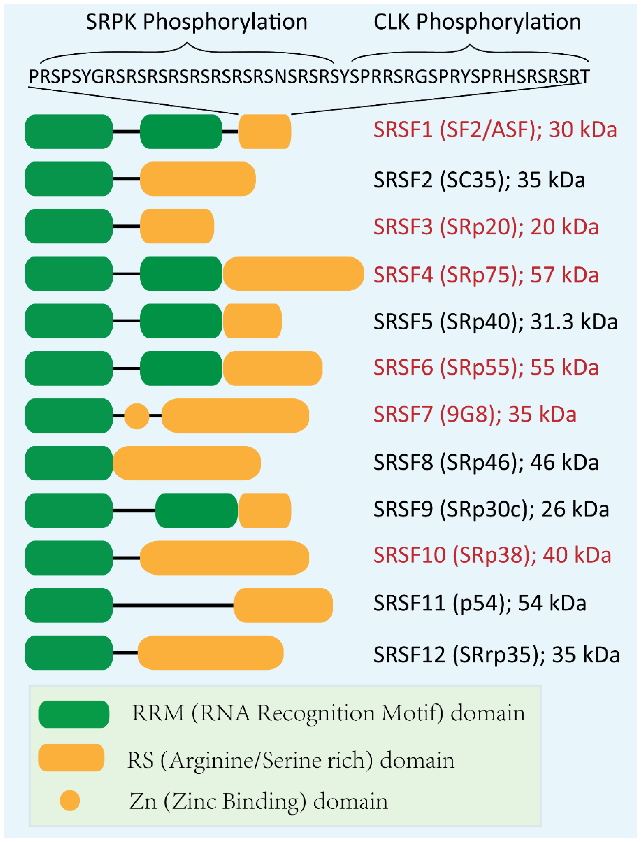
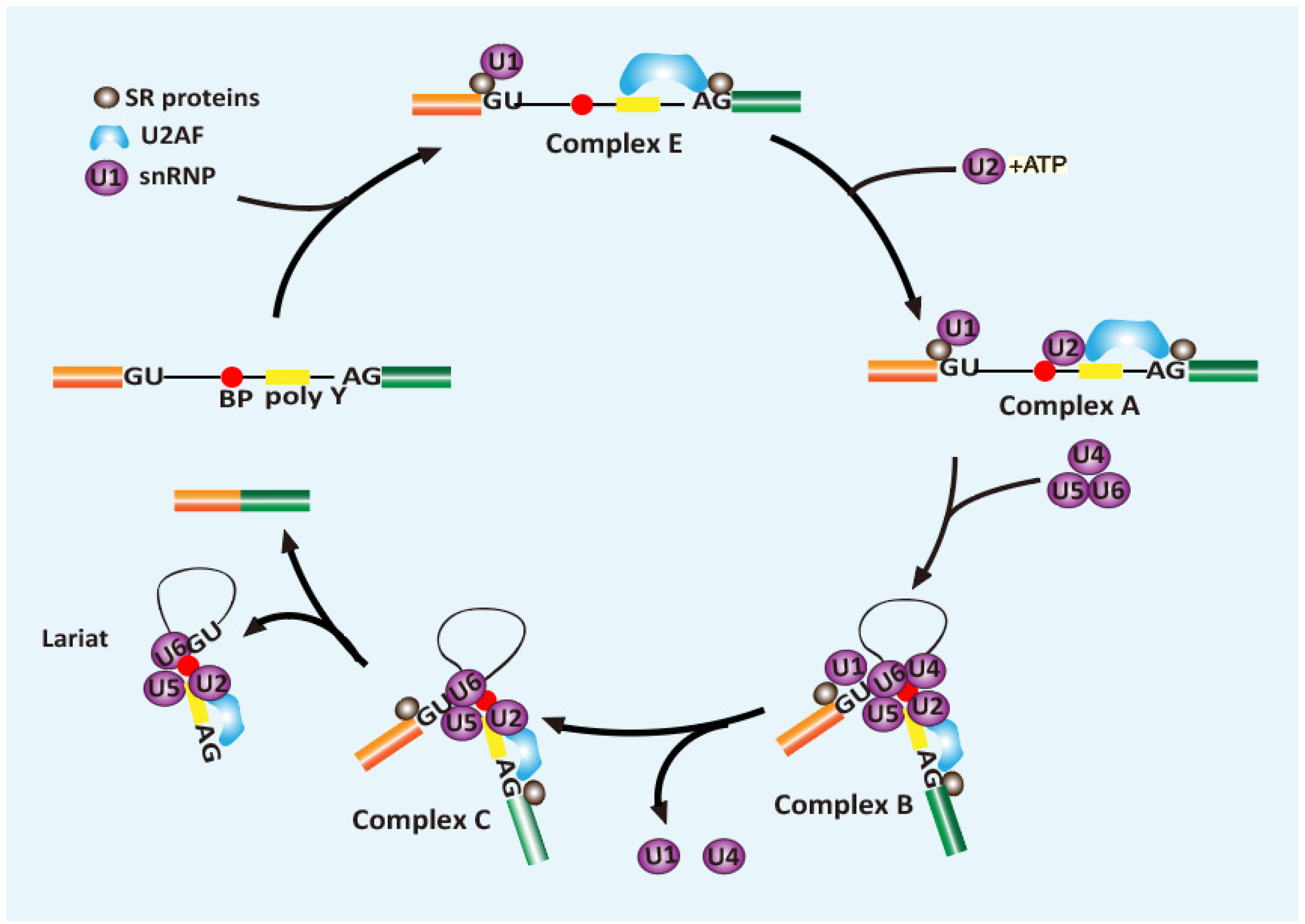
2. Role of SR Proteins in Splicing

RNA splicing is a critical process for the expression of >95% of human genes, during which the introns (non-coding sequences) dispersing throughout the primary transcripts are excised, and exons are ligated together [15,16]. RNA splicing involves two consecutive transesterification reactions that occur under the regulation of the spliceosome, a dynamic macromolecular machine that is comprised of proteins and small nuclear RNAs. The spliceosome contains over 300 components that were recruited and assembled onto the pre-mRNA, undergoing a series of dynamic changes via RNA–RNA, protein–protein, RNA–protein, and recently discovered protein–phosphoinositide interaction during the splicing reaction (Figure 2) [17,18]. The splicing reaction starts with U1 small nuclear ribonucleoprotein (snRNP) recognizing the 5′ splice site in an ATP-dependent manner; together with SF1, U2 auxiliary factors (U2AF1 and U2AF2) recruited to the branch point site (BPS), the polypyrimidine tract (PPT), and the 3′ splice site, respectively. U2 snRNP then binds to the BPS to displace and release SF1 in an ATP-dependent manner. This interaction is stabilized by the assembling of SF3a and SF3b protein complexes, as well as the U2AF that recognizes the 3’ splice site. Then the pre-assembled U4/U6/U5 tri-snRNP joins the pre-splicing complex to form a fully assembled spliceosome (complex B). The complex B further conformationally transforms into the catalytically active spliceosome by releasing U1 and U4. This active spliceosome cleaves the 5′ splice site and allows the ligation between the intron and branch point site, forming a lariat. Next, the spliceosome further transforms to bring the two exons close together and catalyzes the joining of the exons and the release of the lariat [19].
Though stoichiometric RNA and protein components are present in the spliceosome, some splice sites are generally not competitive to recruit a functional spliceosome. They need auxiliary elements nearby to facilitate regulating the splicing events, such as ESEs and ESSs (exonic splicing enhancers and silencers, respectively) in the exons, and ISEs and ISSs (intronic splicing enhancers and silencers, respectively) in the introns. As pivotal recognizers of these elements, SR proteins can bind to these elements and facilitate spliceosome assembling [11]. The function of SR proteins has been identified in nearly every step of the splicing procedure. Firstly, SR proteins generally recognize the ESE elements and promote the formation of E complex and its binding to the 5’-splice site by assisting U1 snRNA in recognizing the splicing site [11]. Next, SR proteins promote U2 snRNA interacting with pre-mRNA at the branch point region to form complex A. Then SR proteins, such as SRSF2, enforce the interaction between U2AF and U1 70K to bridge complexes across two splice sites, which further recruits U4/U6 and U5 tri snRNP complex to form complex B (Figure 2) [20]. Among these steps, the most critical function of SR proteins is documented in the early splice site recognition and initiation of spliceosome assembly. Through binding with a preferable sequence, SR proteins assist adjacent splice sites to compete in recruiting spliceosomes, which results in enhanced RNA splicing.

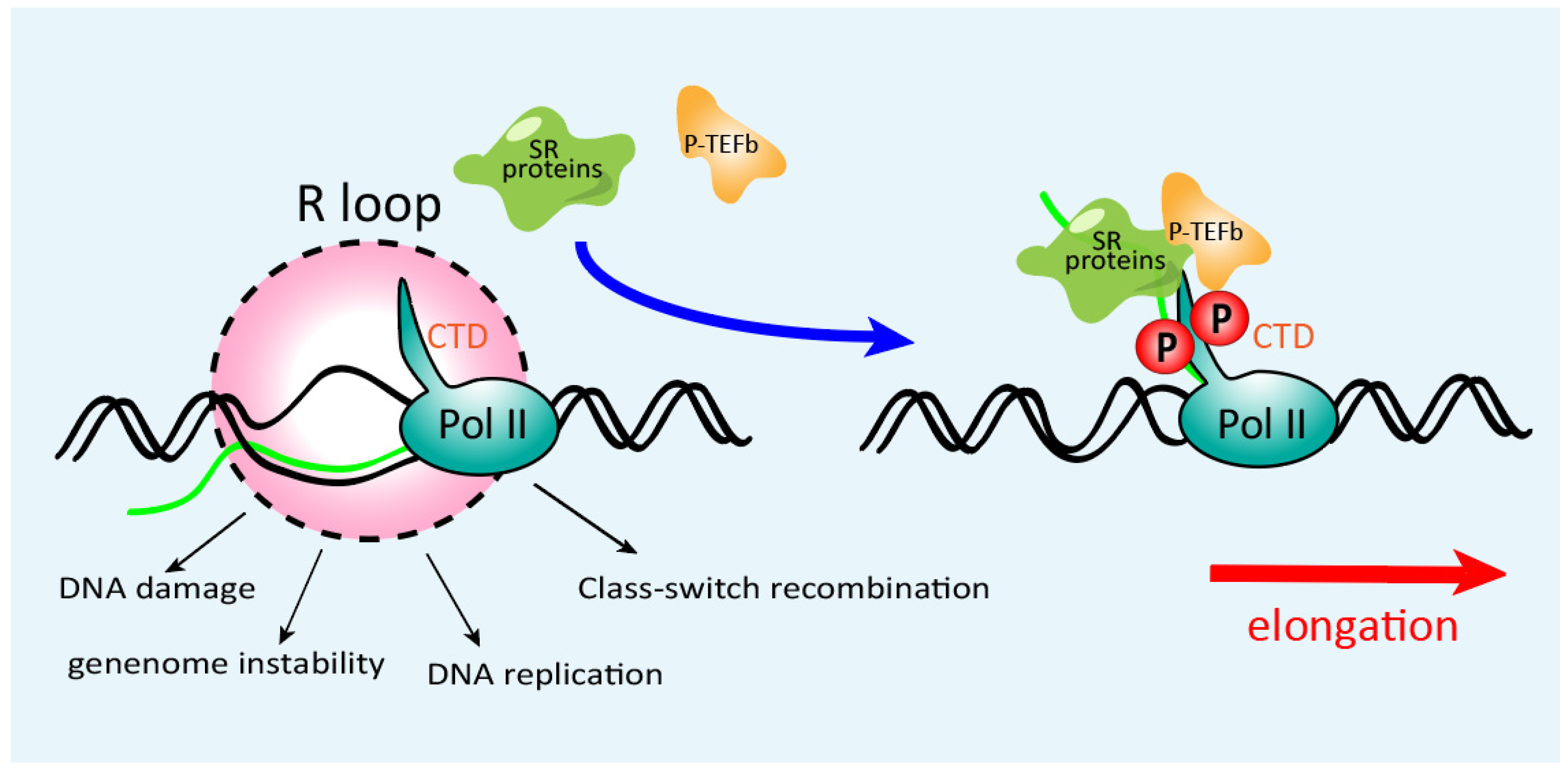
3. Role of SR Proteins in Transcriptional Elongation and Genomic Stability

During transcriptional elongation, nascent RNA can displace the non-template strand and bind to template DNA to form an RNA:DNA hybrid (R-loop) that extends for about nine nucleotides [21]. R-loops have been extensively implicated in genome instability: the exposed non-template ssDNA becomes a vulnerable target to nucleases or other DNA modification enzymes [22]. Blockage or termination of transcription elongation gives rise to stabilized R-loops behind the stalled Pol II complex [22,23]. Transcription is processed functionally coupled with RNA transcript maturation, including 5′ capping, pre-mRNA splicing, cleavage/polyadenylation of nascent transcripts, and ribonucleoprotein assembly [24,25,26]. In addition to their functions in the spliceosome, SR protein family members also participate in the Pol II complex, contributing to transcriptional elongation (Figure 3) [27,28]. For example, SRSF2 can facilitate P-TEFb binding to Pol II, increasing Pol II phosphorylation at Ser2 positions in its C-terminal repeat domain (CTD) and promoting transcriptional elongation [29].
Additionally, SR proteins can attach to and prevent the pre-mRNA binding to template DNA. These features of SR proteins can suppress the R-loops formation, guarantee the correct transcriptional elongation, and thereby increase genome stability (Figure 3). SRSF1 knockout in chicken DT40 cells results in genome-wide DNA breaks and gross DNA recombination [12]. Moreover, knocking out SRSF2 in mouse embryo fibroblasts also results in catastrophic DBS, which may block the cell cycle at the S-phase checkpoint [30]. Overall, SR proteins hinder the formation of the R-loop by promoting transcriptional elongation and binding to nascent RNA to help release RNA from the template DNA when a splicing signal emerges, thereby safeguarding the genome stability.

4. Post-Splicing Activities of SR Proteins

In addition to their activities in the nucleus, the nucleo-cytoplasmic shuttling property of SR proteins confers their noncanonical function in mRNA export, translation, and decay (Figure 1 and Figure 4) [11,31].

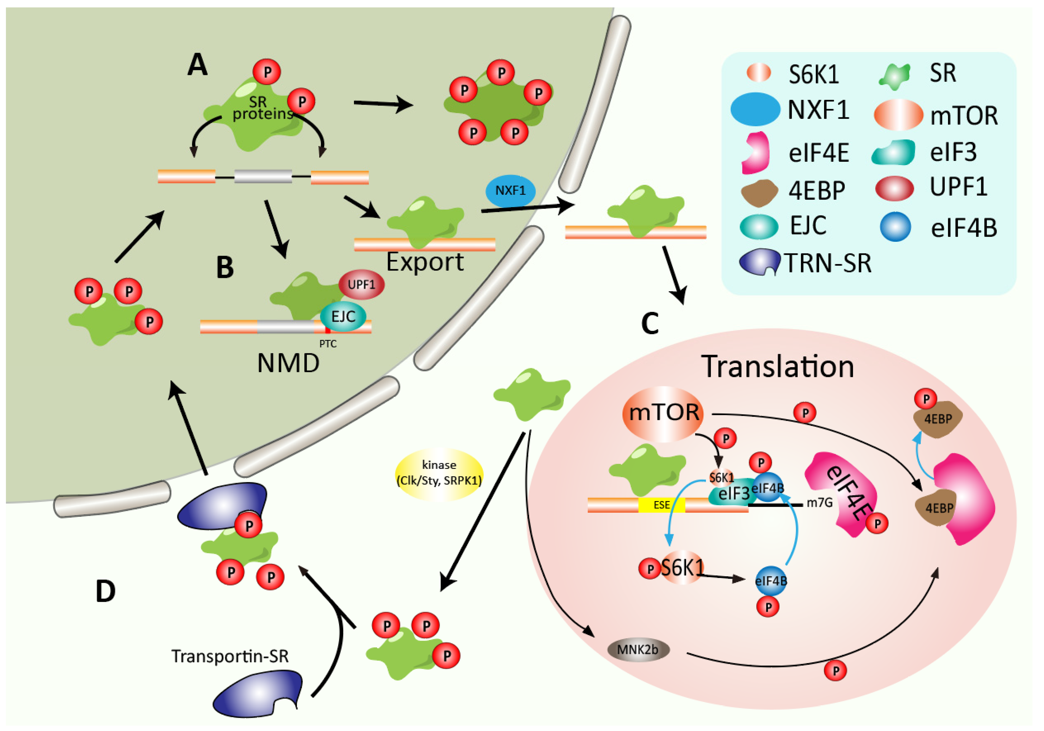
4.1. SR Proteins Promote Mature RNA Export from the Nucleus

After processing, mRNA is exported to the cytoplasm, where the translation is highly regulated to synthesize polypeptide chains. Nuclear export factor 1 (NXF1) binds to and transports the processed mRNA through the nuclear pore complex (Figure 4A) [32]. Free NXF1 forms a closed loop that hides its RNA binding domain and inhibits RNA binding. Therefore, coordinated binding of adaptors is required to transform NXF1 conformation to expose its RNA binding domain.
It has been reviewed that RNA-splicing is associated with nuclear export since the spliceosome can facilitate export factors binding to the mature RNA [33,34]. Canonical SR proteins exhibit different nucleo-cytoplasmic shuttling properties, grouped by shuttling to the cytoplasm (SRSF1, SRSF3, SRSF4, SRSF6, SRSF7, and SRSF10) or not (SRSF2, SRSF5, SRSF8, SRSF9, SRSF11, and SRSF12) (Figure 1) [11,35]. Therefore, SR proteins’ specific RNA-binding capacity and nucleo-cytoplasmic shuttling property endow them with functions in mRNA export. Recent work by Müller-McNicoll et al. analyzed transcriptomic-binding profiles of NXF1 and SRSF1–7 and suggested that, though all the examined SR proteins exhibit partial RNase A resistance in their interaction with NXF1, SRSF3 exhibits the most robust binding. Its motif is most similar to that of NXF1 in 3’ UTR, supporting the mechanism that SRSF3 serves as an adaptor in the recruitment of NXF1 to specific mRNA 3’ ends, coordinating RNA export [31].

4.2. Regulating mRNA Decay and Translation

Nonsense-mediated mRNA decay (NMD) has been considered a crucial strategy for protecting cells from the disastrous effects of truncated proteins or degraded transcripts containing premature termination codons (PTC) [36]. Interestingly, SR proteins can cross-interact with the exon junction complex (EJC)—the function of which is well-characterized in NMD, which suggests that SR proteins perform a significant role in NMD (Figure 4B) [37]. Additionally, several studies reported that overexpression of SRSF1, SRSF2, and other SR proteins, respectively, increase NMD, which is not observed with the overexpression of the splicing factor hnRNP A1 [38]. SRSF1 promotes NMD both after mRNA is exported to the cytoplasm and before mRNA is released from the nucleus [38]. Mechanistically, binding downstream of PTC, SRSF1 enhances the UPF1 recruiting to the spliced mRNA, which then promotes the NMD in the nucleus. Additionally, once the PTC is encountered, UPF1 is phosphorylated at its N and C termini, facilitating its binding with mRNA decay factors, e.g., SMG6 and SMG5/SMG7, and triggering RNA degradation. SRSF1 expedites UPF1 dephosphorylation by recruiting SMG7 and PP2A, facilitating UPF1’s releasing and recycling into the SURF complex [39].
In addition to the effect on NMD, SRSF1 is implicated in translational regulation. SRSF1 could increase the efficiency of eIF4E-initiated mRNA translation by facilitating the pioneer round of translation [40]. Another study revealed that SRSF1 co-sediments with the 80S fragment from the ribosome and polysomes, improving the translation of intronless reporter in HeLa cell-free translation system, which is dependent on the RS domain of SRSF1, but not on the RRM domain [41]. High-throughput sequencing of polysomal fractions further identified that mRNA translational targets of SRSF1 are associated with cell cycle regulation, including kinetochore formation, spindle assembly, and synthesis of M phase proteins, ensuring chromosome segregation (Figure 4C) [42]. Other shuttling SR proteins such as SRSF3 and SRSF7 can also function in the mRNA translation [43,44]. SRSF3, for instance, has been reported to interact with the internal ribosomal entry site (IRES), regulating poliovirus translation initiation (Figure 4C) [44]. In conclusion, SR proteins regulate gene expression by controlling multiple mechanisms throughout RNA processing.

5. Dysregulation of SR Proteins in Cancers

For their multiple functions involved in transcriptional and post-translational regulations, dysregulated SR proteins catastrophically disrupt genome stability, RNA metabolism, and transcriptome, thereby promoting tumorigenesis. Multiple lines of evidence suggest that SR proteins act as oncoproteins in different tumor types. SR proteins’ activities can be regulated by various mechanisms, including epigenetics, genomic mutation, expression level, and protein phosphorylation. Here we review how misregulated SR proteins facilitate tumor initiation and progression.

5.1. Aberrant SR Proteins Expression Is Associated with Cell Transformation and Tumorigenesis

The expression of SR proteins is generally dysregulated both in solid tumors such as colon, kidney, lung, liver, pancreas, and breast cancers [45,46,47,48,49] and in non-solid tumors such as leukemia and acute lymphoblastic leukemia (Table 1) [50,51,52]. As an oncoprotein, elevated SRSF1 expression has been reported in various cancers. Slight overexpression of SRSF1 can sufficiently transform fibroblast and mammary epithelial cells [47,53]. Won Cheol Park et al. pointed out an increased SRSF7 expression in gastric cancer in contrast to normal gastric mucosa [54]. Furthermore, colorectal cancer also exhibits increased SRSF3, SRSF5, and SRSF6 expression.
Several lines of evidence indicate frequent amplification of genes encoding SR proteins in different cancers, at least partially contributing to the high expression of SR proteins in tumors [45,47,69]. For example, extensive amplification of SRSF6 has been identified in colon, lung, and breast cancers [69]. Meanwhile, SRSF1 copy number gain is associated with DNA repair and chemo-sensitivity, which predicts poor survival in small cell lung cancers (SCLC) [45].
In addition to gene amplification, transcriptional regulations have also been reported to increase SR protein expression. MYC is a potent oncogenic transcription factor frequently hyper-activated in cancers [77]. Positive correlations between MYC and SRSF1 expression have been reported across different malignant contexts, including lung and breast cancers. Mechanistically, CHIP data show that MYC can bind to the SRSF1 promoter directly and enhance SRSF1 transcription [78]. Moreover, SRSF1 knockdown impaired MYC-induced cell transformation of Rat1a fibroblast, which in turn suggests the critical role of SRSF1 in MYC’s oncogenic functions [78]. Moreover, SR proteins expression can also be auto-regulated by individual SR proteins themselves or be cross-regulated by other SR protein family members via alternative splicing of ultra-conserved “poison exons” that are localized between protein-coding exons or in 3′UTR, and that contain a premature termination codon (PTC) associated with NMD-mediated mRNA degradation [9,79]. Further investigation of the auto- and cross-regulation mechanisms of SR proteins in cancer and how the loss of their regulation contributes to cancer should improve our understanding of the feedback loops of SR proteins expression and provide potential therapeutic opportunities.
Due to their canonical function in alternative splicing, the mechanism of dysregulated SR proteins promoting cancer progression has been well elucidated through aberrant regulation of alternative splicing and gene expression. For example, highly expressed SRSF1 in breast cancer promotes cancer progression via the oncogenic splicing switch of PTPMT1. In vivo CLIP assays identified a direct SRSF1 binding motif in PTPMT1 exon3 associated with exon inclusion. Expression of PTPMT1 isoforms respectively suggests that overexpression of exon3-included PTPMT1 isoform promotes, while exon3-excluded isoform suppresses tumor growth and metastasis [49]. Additionally, upregulated SRSF1, SRSF2, and SRSF3, respectively, are responsible for aberrant CD44 splicing in ovarian cancer [80]. Besides promoting tumor progression via aberrant regulation of alternative splicing, the function of highly expressed SR proteins has been implicated in aberrant translation in cancer. SRSF1 and SRSF9 have been suggested to facilitate β-catenin mRNA translation and promote β-catenin accumulation through mTOR signaling, which drives colon cancer progression [81].

5.2. Aberrant Phosphorylation of SR Protein in Cancer

Phosphorylation of protein can regulate their biological activity, subcellular localization, half-life, and docking with other proteins [82]. Subcellular localization of SR proteins is strictly regulated within mammalian cells via phosphorylation at multiple sites in the RS domain. Initially, phosphorylation in the RS domain could destabilize the α-helical structure, forming the “arginine claw” structures, which can be recognized by an SR-specific transportin (TRN-SR) that transports the splicing factors to the nucleus where the splicing occurs (Figure 4D) [83,84]. During spliceosomal assembling, the RS domain is appropriately phosphorylated [85]. Along with the splicing procedure, SR proteins are partially dephosphorylated during the late stage of splicing and then can be exported to the cytoplasm [86].
As the upstream regulator of SR proteins, the serine-arginine protein kinase (SRPK) can efficiently phosphorylate the RS domains of SR proteins and promote their nuclear import [87]. Recent studies pointed out that the phosphorylation of the RS domain is not random. SRPK1 phosphorylates the RS domain of its physiological substrate SRSF1 in a directional (C to N) mechanism. Pedro Serrano et al. revealed that, even though the RRM2 domain of SRSF1 is not the phosphorylation target of SRPK1, it coordinates the phosphorylation of the RS domain through cross-interaction between these domains. RRM2 interacts with the N-terminal RS domain to expose its C terminal for initiation of phosphorylation [88].
Aberrant SRPK1 expression can dysregulate SR proteins’ function, resulting in aberrant pre-mRNA splicing, which contributes to cancer progression [89]. SRPK1 contributes to lung and brain metastasis in patients with breast cancer, which is associated with prognosis [90]. Inhibition of SRPK1 excludes SRSF1 from the nucleus and affects alternative splicing of VEGF to inhibit angiopoiesis, suppressing the prostate cancer cell line PC-3 forming tumors in vivo [91].
Dysregulated expression of another SR protein kinase, Cdc2-like kinases 1 (CLK1), has been reported in pancreatic ductal adenocarcinoma, which enhances phosphorylation on SRSF5250-Ser that is associated with alternative splicing of METTL14 and Cyclin L2. Aberrant METTL14 exon 10 exclusion enhances the N6-methyladenosine modification in PDAC cells which promotes tumor metastasis, while aberrant splicing of Cyclin L2 exon 6.3 promotes tumor proliferation [67].
Protein kinase A (PKA), a novel tumor biomarker, phosphorylates SRSF1 at serine 119 in the RRM, which enhances the RNA-binding properties of SRSF1, and increases SRSF1′s activity in regulating the Minx transcript splicing in vitro [92]. Hyperactive PKA signaling pathway has been reported in different cancers and can be exploited as potential cancer therapeutic and diagnosis [93].
Hypoxia is commonly recognized as a prognostic factor negatively related to therapeutic response and cancer patients’ survival [94]. Increasing hypoxia-inducible factor HIF-1 in hypoxic cells upregulates the transcription level of CDC-like kinase 1 (CLK1), which subsequently generates hyperphosphorylated RS domain of SR proteins and decreases their RNA-binding specificity. Consequently, infrequent or new isoforms were generated in hypoxic tumors compared with the normoxic environment, contributing to tumor progression and resistance to radiation therapy.
Overall, SR proteins can be phosphorylated at multisite by different kinases, which comprehensively adjust SR proteins’ activity. Further investigating the underlying mechanisms of the spatiotemporal phosphorylation of SR proteins associated with subcellular distribution and protein–protein interaction should improve our understanding of SR proteins’ role in tumorigenesis and progression.

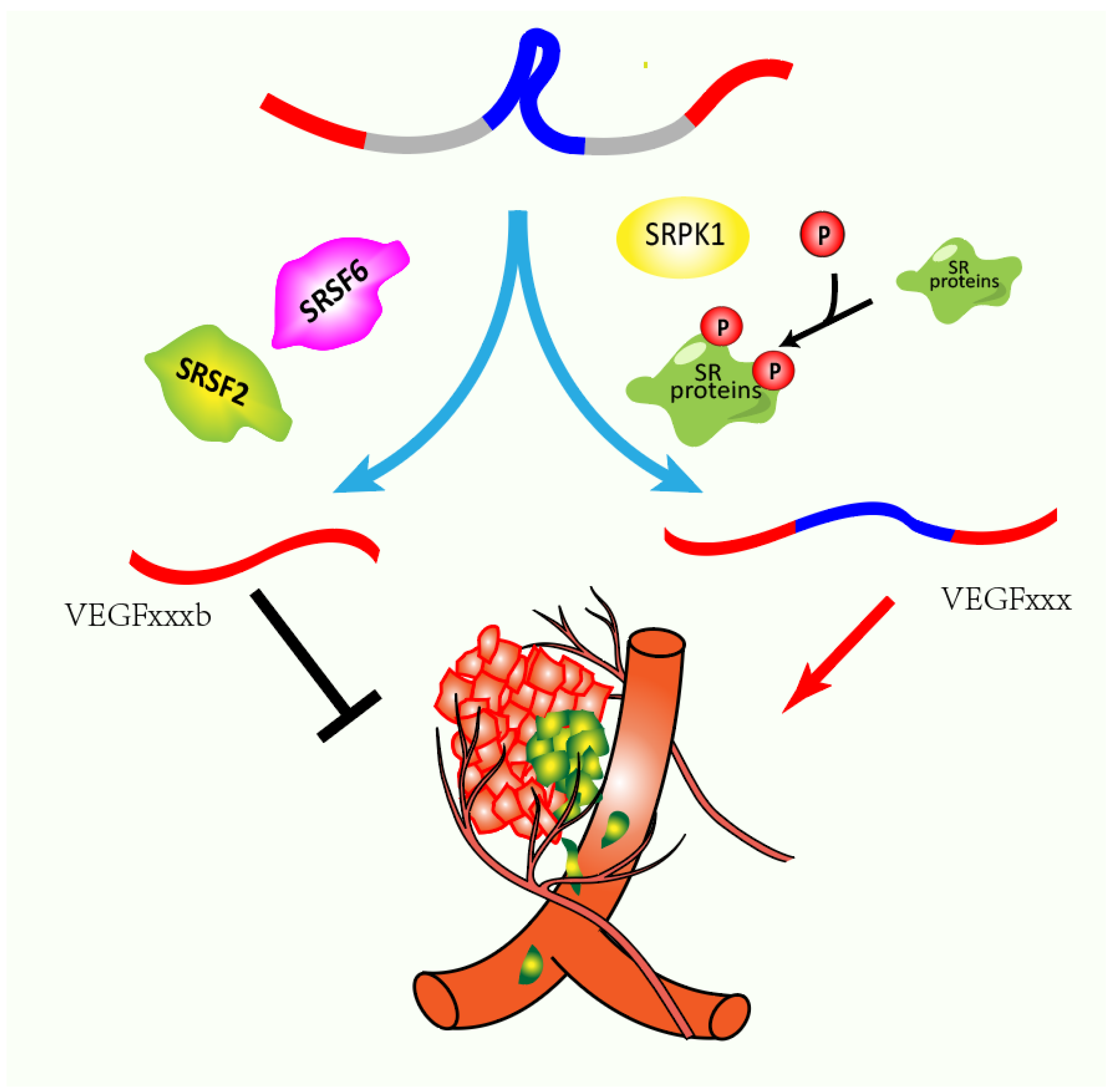
5.3. SR proteins Link Alternative Splicing and Epigenetics Promoting Cancer Development

Aberrant epigenetic regulation, a susceptible adapter of multiple biological changes, increases the risk of cancer [95]. SR proteins have been implicated as potent mediators for aberrant epigenetic-mediated tumorigenesis and metastasis. For example, histone methylation affects the affinity of SR proteins to their specific RNA binding sites. DNA methylation allows for the H3K9me3 modification of histone corresponding to methylated alternative exons. As a histone methylation reader, HP1 protein recruits SR proteins to the transcribed nascent RNA, thus leading to specific alternative splicing outcomes [96]. Then SRSF1 and SRSF3 bind directly to HP1α and HP1 via RNA-independent interaction. However, in the absence of HP1, overexpression of SRSF3 resulted in a diminished effect on splicing and vice versa. These results indicate that SR proteins provide bridging between alternative splicing and epigenetics. Further research dissecting the underlying mechanism of the SR proteins as executors of epigenetic changes should improve our understanding of SR proteins’ roles in physiological conditions and tumor development.

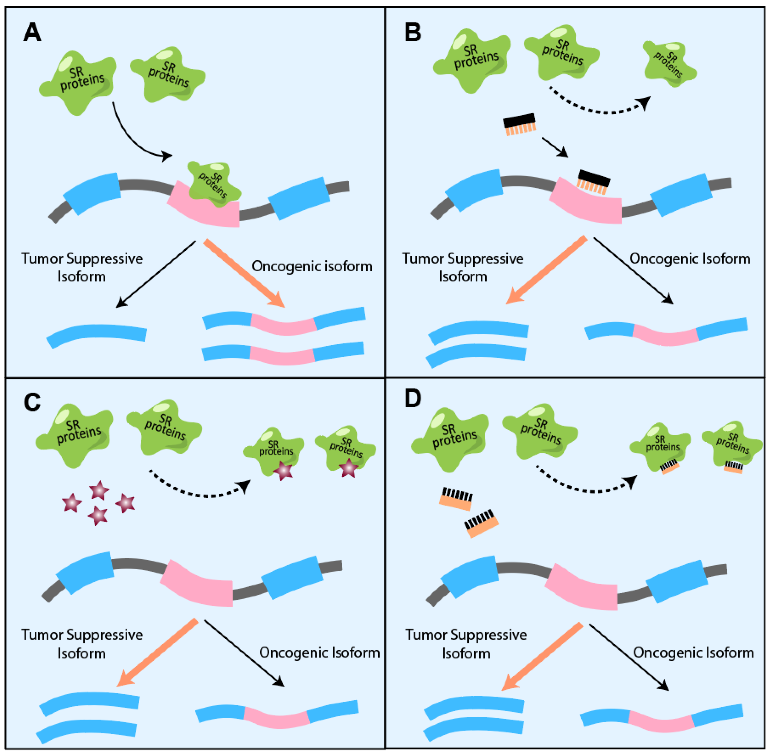
6. Competition or Coordination between Different SR Proteins

Specific splicing target is often regulated synergically by multiple splicing factor proteins [97]. As a typical target for SR proteins, alternative splicing of VEGF mRNA leads to different isoforms with diverse functions. Particularly, the VEGFxxx isoforms and the VEGFxxxb isoforms (x denotes the number of amino acids) show opposite roles with pro- or anti-angiogenic properties, respectively (Figure 5) [98,99]. It has been reported that dysregulated VEGF splicing is associated with cancer progression. Decreased VEGFxxxb has been reported in several cancers, including renal [100], melanoma [101], and colorectal carcinoma [102]. Different SR proteins regulate VEGF splicing via multiple mechanisms. Aberrant expressions of multiple SR proteins disrupt the VEGF splicing. For example, it has been found that increased expression of SRSF2 can upregulate the VEGF165b/VEGF ratio and suppress tumor neovascularization [103]. Similarly, SRSF6 can bind directly to the exon 8b of VEGF pre-mRNA favoring distal splice site utilization and VEGF165b expression [104]. On the contrary, increasing SRPK1 in tumor phosphorylates SRSF1 and stimulates its nuclear import, which favors the splicing at the proximal splice site of VEGF pre-mRNA and leads to the expression of the VEGFxxx isoform and neovascularization. The mechanism for the competition or coordination between different SR proteins during individual splicing events needs to be further explored to clarify the exact roles of SR proteins and their cross-interaction in splicing. Moreover, investigating other cross-interactions between SR proteins during RNA processing, mRNA export, mRNA stability, and translation will expand our understanding of their functions in physiological and pathological conditions.

7. Targeting SR Proteins as Potential Cancer Therapeutics

To target SR-proteins-mediated aberrant alternative splicing in cancer, a vaccine approach has recently been developed to apply antibodies specifically to VEGFxxx, and not to VEGFxxxb isoforms, providing a promising strategy for cancer therapy. A prophylactic immunization using purified immunoglobulins against the pro-angiogenic isoform of VEGF suppresses melanoma and renal cell carcinoma tumor growth in vivo [105].
Importantly, with advances in the development of antisense oligonucleotides (ASOs)—synthetic molecules with 15–30 nt in length, base-pair binding to the splice sites or regulatory sequences [106]—have been under pre-clinical evaluation for correcting aberrant alternative splicing in cancer (Figure 6A,B). SRSF3 recognizes and enhances the inclusion of the exon 10 of the pyruvate kinase M (PKM) that plays a crucial role in regulating the Warburg effect, favoring the M2 isoform that promotes aerobic glycolysis and tumor growth [107,108]. Another study suggested that highly expressed SRSF10 in head and neck cancer is also associated with aberrant PKM splicing resulting in the expression of PKM2 isoform [109]. ASO targeting PKM splicing increases the PKM1 to PKM2 ratio, thereby redirecting glucose carbons from anabolic processes to the tricarboxylic acid (TCA) cycle in hepatocellular carcinoma and suppressing the liver-tumor formation and growth [110].
In addition to RNA alternative splicing, dysregulated SR proteins can disrupt genome stability, transcription, and further RNA processing, contributing to tumorigenesis and progression. Therefore, targeting SR proteins instead of their splicing targets may modulate gene expression extensively and benefit tumor therapeutics. In order to modulate SR protein phosphorylation, small molecules have been developed to target the kinases associated with splicing regulation [111,112,113]. NB-506, a complete inhibitor of topoisomerase I, is repositioned to reduce SRSF1 phosphorylation and regulates gene expression by modulating pre-mRNA splicing. In addition, it has been described that chimeric antibodies of SRPK1 have the capacity to suppress non-small cell lung cancer growth and metastasis [114]. Meanwhile, small molecules targeting SRPK1 can switch splicing of VEGF mRNA to generate the VEGF165b isoform and decrease tumor growth [91]. These studies suggest that inhibition of SRPK1 may provide a strategy for cancer therapeutic.
Modulating the SR protein kinases’ activities tends to change multiple SR proteins’ functions simultaneously. Several efforts have been made to specifically target the individual hyperactive SR proteins in tumors (Figure 6C,D). Virtual screening of 4855 FDA-approved drugs to target the domain-specific role of SRSF6 repurposed a β2-adrenergic receptor agonist indacaterol, which is approved for chronic obstructive pulmonary disease (COPD) treatment, for blocking SRSF6′s regulation on its downstream splicing targets, which suppresses colorectal cancer [48]. In addition, Denichenko et al. developed decoy oligonucleotides that can compete for RNA binding proteins, such as splicing factors, including RBFOX1/2, SRSF1, and PTBP1, rather than their specific RNA targets. SRSF1 splicing targets, such as INSR, U2AF1, MKNK2, and USP8, respond to decoy oligonucleotides for SRSF1 while not to those for PTBP1 and RBFOX1/2. Considering the non-RNA-binding functions of SR proteins, strategies targeting the splicing regulation activity of SR proteins may provide more precise on-target therapeutic benefits.

8. Conclusions

In summary, SR proteins’ activities are regulated via multiple mechanisms, including gene copy number [47], transcriptional regulation [50], alternative splicing [9,79], and phosphorylation [91], which affects a variety of cellular processes, such as genome stability [12,115], RNA metabolism [116], and translation [117]. Connecting upstream regulators and downstream targets, multiple SR proteins shuttle between the nucleus and cytoplasm to maintain the precise net of the biological pathways. Dysregulated SR proteins drive tumorigenesis and cancer development [45,46,47]. To date, a few therapeutic strategies have been developed to regulate the activity of SR proteins [48,111,112,113,118]. Among them, targeting the regulators of SR proteins will result in extensive modulation of various SR protein functions simultaneously, which may elicit unwanted off-target effects. On the other hand, ASOs targeting specific aberrant alternative splicing changes may confer limited clinical benefit in cancer therapy, considering that hundreds and thousands of transcripts are aberrantly spliced in cancer due to the dysregulated SR splicing proteins. Therefore, we speculate that the protein-specific competitive inhibitor or decoy oligonucleotides targeting individual SR proteins can achieve more significant benefits for cancer therapeutics.
Further investigation of the biological function and regulatory mechanisms of SR proteins in RNA metabolism and distinguishing the cross-interaction among different high-homology SR proteins should provide a foundation for the rational development of precise cancer therapy (Table 1). It has been reported that SR proteins such as SRSF1, SRSF4, SRSF5, SRSF6, and SRSF9 contain one more RRM domain than the other family members [119]. Further insights into the functional discrimination of two RRM domains are needed to specify their respective roles in RNA recognition and other RNA processing. With further understanding of the biological function of the protein domains in SR proteins, SR proteins may serve as a precise medicinal target for cancer therapy.

Author Contributions

L.W. and H.Z. designed the manuscript; L.W., M.D. and H.Z. collected the related papers, wrote the manuscript, and drew the figures. All authors have read and agreed to the published version of the manuscript.

Funding

This work is supported by the Natural Science Foundation of Zhejiang Province (LZ21H160001), CAMS Innovation Fund for Medical Sciences (CIFMS, 2019-I2M-5-044), Medical Health Scientific Research Fund of Zhejiang Province (2020PY022), and Agricultural and Social Development Scientific Research Fund of Hangzhou (20191231Y200).

Institutional Review Board Statement

Not applicable.

Informed Consent Statement

Not applicable.

Data Availability Statement

Not applicable.

Acknowledgments

The authors apologize to those colleagues whose work was not cited due to space limitation.

Conflicts of Interest

The authors declare no conflict of interest.

References

  1. Hentze, M.W.; Castello, A.; Schwarzl, T.; Preiss, T. A brave new world of RNA-binding proteins. Nat. Rev. Mol. Cell Biol. 2018, 19, 327–341. [Google Scholar] [CrossRef]
  2. Gebauer, F.; Schwarzl, T.; Valcárcel, J.; Hentze, M.W. RNA-binding proteins in human genetic disease. Nat. Rev. Genet. 2021, 22, 185–198. [Google Scholar] [CrossRef] [PubMed]
  3. Sanchez de Groot, N.; Armaos, A.; Graña-Montes, R.; Alriquet, M.; Calloni, G.; Vabulas, R.M.; Tartaglia, G.G. RNA structure drives interaction with proteins. Nat. Commun. 2019, 10, 3246. [Google Scholar] [CrossRef]
  4. Long, J.C.; Caceres, J.F. The SR protein family of splicing factors: Master regulators of gene expression. Biochem. J. 2008, 417, 15–27. [Google Scholar] [CrossRef]
  5. Rahman, M.A.; Krainer, A.R.; Abdel-Wahab, O. SnapShot: Splicing Alterations in Cancer. Cell 2020, 180, 208-208.e1. [Google Scholar] [CrossRef] [PubMed]
  6. Champlin, D.T.; Frasch, M.; Saumweber, H.; Lis, J.T. Characterization of a Drosophila protein associated with boundaries of transcriptionally active chromatin. Genes Dev. 1991, 5, 1611–1621. [Google Scholar] [CrossRef] [PubMed]
  7. Ge, H.; Manley, J.L. A protein factor, ASF, controls cell-specific alternative splicing of SV40 early pre-mRNA in vitro. Cell 1990, 62, 25–34. [Google Scholar] [CrossRef]
  8. Krainer, A.R.; Conway, G.C.; Kozak, D. The essential pre-mRNA splicing factor SF2 influences 5′ splice site selection by activating proximal sites. Cell 1990, 62, 35–42. [Google Scholar] [CrossRef]
  9. Leclair, N.K.; Brugiolo, M.; Urbanski, L.; Lawson, S.C.; Thakar, K.; Yurieva, M.; George, J.; Hinson, J.T.; Cheng, A.; Graveley, B.R.; et al. Poison Exon Splicing Regulates a Coordinated Network of SR Protein Expression during Differentiation and Tumorigenesis. Mol. Cell 2020, 80, 648–665.e649. [Google Scholar] [CrossRef]
  10. Kraus, M.E.; Lis, J.T. The concentration of B52, an essential splicing factor and regulator of splice site choice in vitro, is critical for Drosophila development. Mol. Cell. Biol. 1994, 14, 5360–5370. [Google Scholar]
  11. Jeong, S. SR Proteins: Binders, Regulators, and Connectors of RNA. Mol. Cells 2017, 40, 1–9. [Google Scholar] [CrossRef] [PubMed] [Green Version]
  12. Li, X.; Manley, J.L. Inactivation of the SR Protein Splicing Factor ASF/SF2 Results in Genomic Instability. Cell 2005, 122, 365–378. [Google Scholar] [CrossRef] [PubMed]
  13. Huang, Y.; Gattoni, R.; Stévenin, J.; Steitz, J.A. SR Splicing Factors Serve as Adapter Proteins for TAP-Dependent mRNA Export. Mol. Cell 2003, 11, 837–843. [Google Scholar] [CrossRef]
  14. Wagner, R.E.; Frye, M. Noncanonical functions of the serine-arginine-rich splicing factor (SR) family of proteins in development and disease. Bioessays 2021, 43, 2000242. [Google Scholar] [CrossRef]
  15. Baralle, F.E.; Giudice, J. Alternative splicing as a regulator of development and tissue identity. Nat. Rev. Mol. Cell Biol. 2017, 18, 437–451. [Google Scholar] [CrossRef]
  16. Angarola, B.L.; Anczuków, O. Splicing alterations in healthy aging and disease. Wiley Interdiscip. Rev. RNA 2021, 12, e1643. [Google Scholar] [CrossRef]
  17. Hegele, A.; Kamburov, A.; Grossmann, A.; Sourlis, C.; Wowro, S.; Weimann, M.; Will, C.L.; Pena, V.; Lührmann, R.; Stelzl, U. Dynamic Protein-Protein Interaction Wiring of the Human Spliceosome. Mol. Cell 2012, 45, 567–580. [Google Scholar] [CrossRef]
  18. Sztacho, M.; Šalovská, B.; Červenka, J.; Balaban, C.; Hoboth, P.; Hozák, P. Limited Proteolysis-Coupled Mass Spectrometry Identifies Phosphatidylinositol 4,5-Bisphosphate Effectors in Human Nuclear Proteome. Cells 2021, 10, 68. [Google Scholar] [CrossRef]
  19. Wilkinson, M.E.; Charenton, C.; Nagai, K. RNA Splicing by the Spliceosome. Annu. Rev. Biochem. 2020, 89, 359–388. [Google Scholar] [CrossRef]
  20. Ma, X.; He, F. Advances in the Study of SR Protein Family. Genom. Proteom. Bioinform. 2003, 1, 2–8. [Google Scholar] [CrossRef]
  21. Petermann, E.; Lan, L.; Zou, L. Sources, resolution and physiological relevance of R-loops and RNA–DNA hybrids. Nat. Rev. Mol. Cell Biol. 2022, 23, 521–540. [Google Scholar] [CrossRef] [PubMed]
  22. Gowrishankar, J.; Leela, J.K.; Anupama, K. R-loops in bacterial transcription. Transcription 2013, 4, 153–157. [Google Scholar] [CrossRef] [PubMed]
  23. Zatreanu, D.; Han, Z.; Mitter, R.; Tumini, E.; Williams, H.; Gregersen, L.; Dirac-Svejstrup, A.B.; Roma, S.; Stewart, A.; Aguilera, A.; et al. Elongation Factor TFIIS Prevents Transcription Stress and R-Loop Accumulation to Maintain Genome Stability. Mol. Cell 2019, 76, 57–69.e9. [Google Scholar] [CrossRef] [PubMed]
  24. Bentley, D.L. Coupling mRNA processing with transcription in time and space. Nat. Rev. Genet. 2014, 15, 163–175. [Google Scholar] [CrossRef]
  25. Saldi, T.; Cortazar, M.A.; Sheridan, R.M.; Bentley, D.L. Coupling of RNA Polymerase II Transcription Elongation with Pre-mRNA Splicing. J. Mol. Biol. 2016, 428, 2623–2635. [Google Scholar] [CrossRef]
  26. Drexler, H.L.; Choquet, K.; Churchman, L.S. Splicing Kinetics and Coordination Revealed by Direct Nascent RNA Sequencing through Nanopores. Mol. Cell 2020, 77, 985–998.e8. [Google Scholar] [CrossRef]
  27. Das, R.; Yu, J.; Zhang, Z.; Gygi, M.P.; Krainer, A.R.; Gygi, S.P.; Reed, R. SR Proteins Function in Coupling RNAP II Transcription to Pre-mRNA Splicing. Mol. Cell 2007, 26, 867–881. [Google Scholar] [CrossRef]
  28. Muniz, L.; Nicolas, E.; Trouche, D. RNA polymerase II speed: A key player in controlling and adapting transcriptome composition. EMBO J. 2021, 40, e105740. [Google Scholar] [CrossRef]
  29. Lin, S.; Coutinho-Mansfield, G.; Wang, D.; Pandit, S.; Fu, X.-D. The splicing factor SC35 has an active role in transcriptional elongation. Nat. Struct. Mol. Biol. 2008, 15, 819–826. [Google Scholar] [CrossRef]
  30. Xiao, R.; Sun, Y.; Ding, J.-H.; Lin, S.; Rose, D.W.; Rosenfeld, M.G.; Fu, X.-D.; Li, X. Splicing Regulator SC35 Is Essential for Genomic Stability and Cell Proliferation during Mammalian Organogenesis. Mol. Cell. Biol. 2007, 27, 5393–5402. [Google Scholar] [CrossRef]
  31. Müller-McNicoll, M.; Botti, V.; de Jesus Domingues, A.M.; Brandl, H.; Schwich, O.D.; Steiner, M.C.; Curk, T.; Poser, I.; Zarnack, K.; Neugebauer, K.M. SR proteins are NXF1 adaptors that link alternative RNA processing to mRNA export. Genes Dev. 2016, 30, 553–566. [Google Scholar] [CrossRef] [PubMed] [Green Version]
  32. Tretyakova, I.; Zolotukhin, A.S.; Tan, W.; Bear, J.; Propst, F.; Ruthel, G.; Felber, B.K. Nuclear Export Factor Family Protein Participates in Cytoplasmic mRNA Trafficking. J. Biol. Chem. 2005, 280, 31981–31990. [Google Scholar] [CrossRef] [PubMed]
  33. Palazzo, A.F.; Lee, E.S. Sequence determinants for nuclear retention and cytoplasmic export of mRNAs and lncRNAs. Front. Genet. 2018, 9, 440. [Google Scholar] [CrossRef] [PubMed]
  34. Cullen, B.R. Connections between the processing and nuclear export of mRNA: Evidence for an export license? Proc. Natl. Acad. Sci. USA 2000, 97, 4–6. [Google Scholar] [CrossRef] [PubMed]
  35. Cáceres, J.F.; Screaton, G.R.; Krainer, A.R. A specific subset of SR proteins shuttles continuously between the nucleus and the cytoplasm. Genes Dev. 1998, 12, 55–66. [Google Scholar] [CrossRef]
  36. Sato, H.; Singer, R.H. Cellular variability of nonsense-mediated mRNA decay. Nat. Commun. 2021, 12, 7203. [Google Scholar] [CrossRef]
  37. Singh, G.; Kucukural, A.; Cenik, C.; Leszyk, J.D.; Shaffer, S.A.; Weng, Z.; Moore, M.J. The Cellular EJC Interactome Reveals Higher Order mRNP Structure and an EJC-SR Protein Nexus. Cell 2012, 151, 750–764. [Google Scholar] [CrossRef]
  38. Zhang, Z.; Krainer, A.R. Involvement of SR Proteins in mRNA Surveillance. Mol. Cell 2004, 16, 597–607. [Google Scholar] [CrossRef]
  39. Aznarez, I.; Nomakuchi, T.T.; Tetenbaum-Novatt, J.; Rahman, M.A.; Fregoso, O.; Rees, H.; Krainer, A.R. Mechanism of Nonsense-Mediated mRNA Decay Stimulation by Splicing Factor SRSF1. Cell Rep. 2018, 23, 2186–2198. [Google Scholar] [CrossRef]
  40. Sato, H.; Hosoda, N.; Maquat, L.E. Efficiency of the Pioneer Round of Translation Affects the Cellular Site of Nonsense-Mediated mRNA Decay. Mol. Cell 2008, 29, 255–262. [Google Scholar] [CrossRef]
  41. Sanford, J.R.; Gray, N.K.; Beckmann, K.; Cáceres, J.F. A novel role for shuttling SR proteins in mRNA translation. Genes Dev. 2004, 18, 755–768. [Google Scholar] [CrossRef] [PubMed] [Green Version]
  42. Maslon, M.M.; Heras, S.R.; Bellora, N.; Eyras, E.; Cáceres, J.F. The translational landscape of the splicing factor SRSF1 and its role in mitosis. Elife 2014, 3, e02028. [Google Scholar] [CrossRef]
  43. Swartz, J.E.; Bor, Y.-C.; Misawa, Y.; Rekosh, D.; Hammarskjold, M.-L. The Shuttling SR Protein 9G8 Plays a Role in Translation of Unspliced mRNA Containing a Constitutive Transport Element. J. Biol. Chem. 2007, 282, 19844–19853. [Google Scholar] [CrossRef] [PubMed]
  44. Bedard, K.M.; Daijogo, S.; Semler, B.L. A nucleo-cytoplasmic SR protein functions in viral IRES-mediated translation initiation. EMBO J. 2007, 26, 459–467. [Google Scholar] [CrossRef]
  45. Jiang, L.; Huang, J.; Higgs, B.W.; Hu, Z.; Xiao, Z.; Yao, X.; Conley, S.; Zhong, H.; Liu, Z.; Brohawn, P.; et al. Genomic Landscape Survey Identifies SRSF1 as a Key Oncodriver in Small Cell Lung Cancer. PLoS Genet. 2016, 12, e1005895. [Google Scholar] [CrossRef] [PubMed]
  46. Anczuków, O.; Akerman, M.; Cléry, A.; Wu, J.; Shen, C.; Shirole, N.H.; Raimer, A.; Sun, S.; Jensen, M.A.; Hua, Y.; et al. SRSF1-regulated alternative splicing in breast cancer. Mol. Cell 2015, 60, 105–117. [Google Scholar] [CrossRef]
  47. Karni, R.; de Stanchina, E.; Lowe, S.W.; Sinha, R.; Mu, D.; Krainer, A.R. The gene encoding the splicing factor SF2/ASF is a proto-oncogene. Nat. Struct. Mol. Biol. 2007, 14, 185–193. [Google Scholar] [CrossRef]
  48. Wan, L.; Yu, W.; Shen, E.; Sun, W.; Liu, Y.; Kong, J.; Wu, Y.; Han, F.; Zhang, L.; Yu, T.; et al. SRSF6-regulated alternative splicing that promotes tumour progression offers a therapy target for colorectal cancer. Gut 2019, 68, 118–129. [Google Scholar] [CrossRef]
  49. Du, J.-X.; Luo, Y.-H.; Zhang, S.-J.; Wang, B.; Chen, C.; Zhu, G.-Q.; Zhu, P.; Cai, C.-Z.; Wan, J.-L.; Cai, J.-L.; et al. Splicing factor SRSF1 promotes breast cancer progression via oncogenic splice switching of PTPMT1. J. Exp. Clin. Cancer Res. 2021, 40, 171. [Google Scholar] [CrossRef]
  50. Liu, J.; Huang, B.; Xiao, Y.; Xiong, H.M.; Li, J.; Feng, D.Q.; Chen, X.M.; Zhang, H.B.; Wang, X.Z. Aberrant Expression of Splicing Factors in Newly Diagnosed Acute Myeloid Leukemia. Oncol. Res. Treat. 2012, 35, 335–340. [Google Scholar] [CrossRef]
  51. Sinnakannu, J.R.; Lee, K.L.; Cheng, S.; Li, J.; Yu, M.; Tan, S.P.; Ong, C.C.H.; Li, H.; Than, H.; Anczuków-Camarda, O.; et al. SRSF1 mediates cytokine-induced impaired imatinib sensitivity in chronic myeloid leukemia. Leukemia 2020, 34, 1787–1798. [Google Scholar] [CrossRef] [PubMed]
  52. Zou, L.; Zhang, H.; Du, C.; Liu, X.; Zhu, S.; Zhang, W.; Li, Z.; Gao, C.; Zhao, X.; Mei, M.; et al. Correlation of SRSF1 and PRMT1 expression with clinical status of pediatric acute lymphoblastic leukemia. J. Hematol. Oncol. 2012, 5, 42. [Google Scholar] [CrossRef] [PubMed]
  53. Anczuków, O.; Rosenberg, A.Z.; Akerman, M.; Das, S.; Zhan, L.; Karni, R.; Muthuswamy, S.K.; Krainer, A.R. The splicing factor SRSF1 regulates apoptosis and proliferation to promote mammary epithelial cell transformation. Nat. Struct. Mol. Biol. 2012, 19, 220–228. [Google Scholar] [CrossRef] [PubMed]
  54. Park, W.C.; Kim, H.-R.; Kang, D.B.; Ryu, J.-S.; Choi, K.-H.; Lee, G.-O.; Yun, K.J.; Kim, K.Y.; Park, R.; Yoon, K.-H.; et al. Comparative expression patterns and diagnostic efficacies of SR splicing factors and HNRNPA1 in gastric and colorectal cancer. BMC Cancer 2016, 16, 358. [Google Scholar] [CrossRef] [PubMed]
  55. Sheng, J.; Zhao, Q.; Zhao, J.; Zhang, W.; Sun, Y.; Qin, P.; Lv, Y.; Bai, L.; Yang, Q.; Chen, L.; et al. SRSF1 modulates PTPMT1 alternative splicing to regulate lung cancer cell radioresistance. EBioMedicine 2018, 38, 113–126. [Google Scholar] [CrossRef]
  56. Lv, Y.; Zhang, W.; Zhao, J.; Sun, B.; Qi, Y.; Ji, H.; Chen, C.; Zhang, J.; Sheng, J.; Wang, T.; et al. SRSF1 inhibits autophagy through regulating Bcl-x splicing and interacting with PIK3C3 in lung cancer. Signal Transduct. Target. Ther. 2021, 6, 108. [Google Scholar] [CrossRef]
  57. Shultz, J.C.; Goehe, R.W.; Wijesinghe, D.S.; Murudkar, C.; Hawkins, A.J.; Shay, J.W.; Minna, J.D.; Chalfant, C.E. Alternative Splicing of Caspase 9 Is Modulated by the Phosphoinositide 3-Kinase/Akt Pathway via Phosphorylation of SRp30a. Cancer Res. 2010, 70, 9185–9196. [Google Scholar] [CrossRef]
  58. Chen, L.; Luo, C.; Shen, L.; Liu, Y.; Wang, Q.; Zhang, C.; Guo, R.; Zhang, Y.; Xie, Z.; Wei, N.; et al. SRSF1 Prevents DNA Damage and Promotes Tumorigenesis through Regulation of DBF4B Pre-mRNA Splicing. Cell Rep. 2017, 21, 3406–3413. [Google Scholar] [CrossRef]
  59. Zhou, X.; Wang, R.; Li, X.; Yu, L.; Hua, D.; Sun, C.; Shi, C.; Luo, W.; Rao, C.; Jiang, Z.; et al. Splicing factor SRSF1 promotes gliomagenesis via oncogenic splice-switching of MYO1B. J. Clin. Investig. 2019, 129, 676–693. [Google Scholar] [CrossRef]
  60. Sokół, E.; Kędzierska, H.; Czubaty, A.; Rybicka, B.; Rodzik, K.; Tański, Z.; Bogusławska, J.; Piekiełko-Witkowska, A. microRNA-mediated regulation of splicing factors SRSF1, SRSF2 and hnRNP A1 in context of their alternatively spliced 3′UTRs. Exp. Cell Res. 2018, 363, 208–217. [Google Scholar] [CrossRef]
  61. Abou Faycal, C.; Gazzeri, S.; Eymin, B. A VEGF-A/SOX2/SRSF2 network controls VEGFR1 pre-mRNA alternative splicing in lung carcinoma cells. Sci. Rep. 2019, 9, 336. [Google Scholar] [CrossRef] [PubMed] [Green Version]
  62. Luo, C.; Cheng, Y.; Liu, Y.; Chen, L.; Liu, L.; Wei, N.; Xie, Z.; Wu, W.; Feng, Y. SRSF2 Regulates Alternative Splicing to Drive Hepatocellular Carcinoma Development. Cancer Res. 2017, 77, 1168–1178. [Google Scholar] [CrossRef] [PubMed]
  63. He, X.; Arslan, A.D.; Pool, M.D.; Ho, T.T.; Darcy, K.M.; Coon, J.S.; Beck, W.T. Knockdown of splicing factor SRp20 causes apoptosis in ovarian cancer cells and its expression is associated with malignancy of epithelial ovarian cancer. Oncogene 2011, 30, 356–365. [Google Scholar] [CrossRef] [PubMed]
  64. Chen, Y.; Yang, M.; Meng, F.; Zhang, Y.; Wang, M.; Guo, X.; Yang, J.; Zhang, H.; Zhang, H.; Sun, J.; et al. SRSF3 Promotes Angiogenesis in Colorectal Cancer by Splicing SRF. Front. Oncol. 2022, 12, 270. [Google Scholar] [CrossRef] [PubMed]
  65. Zhou, L.; Guo, J.; Jia, R. Oncogene SRSF3 suppresses autophagy via inhibiting BECN1 expression. Biochem. Biophys. Res. Commun. 2019, 509, 966–972. [Google Scholar] [CrossRef] [PubMed]
  66. Song, X.; Wan, X.; Huang, T.; Zeng, C.; Sastry, N.; Wu, B.; James, C.D.; Horbinski, C.; Nakano, I.; Zhang, W.; et al. SRSF3-Regulated RNA Alternative Splicing Promotes Glioblastoma Tumorigenicity by Affecting Multiple Cellular Processes. Cancer Res. 2019, 79, 5288–5301. [Google Scholar] [CrossRef] [PubMed]
  67. Chen, S.; Yang, C.; Wang, Z.-W.; Hu, J.-F.; Pan, J.-J.; Liao, C.-Y.; Zhang, J.-Q.; Chen, J.-Z.; Huang, Y.; Huang, L.; et al. CLK1/SRSF5 pathway induces aberrant exon skipping of METTL14 and Cyclin L2 and promotes growth and metastasis of pancreatic cancer. J. Hematol. Oncol. 2021, 14, 60. [Google Scholar] [CrossRef]
  68. Gautrey, H.L.; Tyson-Capper, A.J. Regulation of Mcl-1 by SRSF1 and SRSF5 in Cancer Cells. PLoS ONE 2012, 7, e51497. [Google Scholar] [CrossRef]
  69. Cohen-Eliav, M.; Golan-Gerstl, R.; Siegfried, Z.; Andersen, C.L.; Thorsen, K.; Ørntoft, T.F.; Mu, D.; Karni, R. The splicing factor SRSF6 is amplified and is an oncoprotein in lung and colon cancers. J. Pathol. 2013, 229, 630–639. [Google Scholar] [CrossRef]
  70. Choi, N.; Jang, H.N.; Oh, J.; Ha, J.; Park, H.; Zheng, X.; Lee, S.; Shen, H. SRSF6 Regulates the Alternative Splicing of the Apoptotic Fas Gene by Targeting a Novel RNA Sequence. Cancers 2022, 14, 1990. [Google Scholar] [CrossRef]
  71. Fu, Y.; Wang, Y. SRSF7 knockdown promotes apoptosis of colon and lung cancer cells. Oncol. Lett. 2018, 15, 5545–5552. [Google Scholar] [CrossRef] [PubMed] [Green Version]
  72. Saijo, S.; Kuwano, Y.; Masuda, K.; Nishikawa, T.; Rokutan, K.; Nishida, K. Serine/arginine-rich splicing factor 7 regulates p21-dependent growth arrest in colon cancer cells. J. Med. Investig. 2016, 63, 219–226. [Google Scholar] [CrossRef] [PubMed]
  73. Wang, X.; Lu, X.; Wang, P.; Chen, Q.; Xiong, L.; Tang, M.; Hong, C.; Lin, X.; Shi, K.; Liang, L.; et al. SRSF9 promotes colorectal cancer progression via stabilizing DSN1 mRNA in an m6A-related manner. J. Transl. Med. 2022, 20, 198. [Google Scholar] [CrossRef]
  74. Wang, R.; Su, Q.; Yin, H.; Wu, D.; Lv, C.; Yan, Z. Inhibition of SRSF9 enhances the sensitivity of colorectal cancer to erastin-induced ferroptosis by reducing glutathione peroxidase 4 expression. Int. J. Biochem. Cell Biol. 2021, 134, 105948. [Google Scholar] [CrossRef] [PubMed]
  75. Liu, F.; Dai, M.; Xu, Q.; Zhu, X.; Zhou, Y.; Jiang, S.; Wang, Y.; Ai, Z.; Ma, L.; Zhang, Y.; et al. SRSF10-mediated IL1RAP alternative splicing regulates cervical cancer oncogenesis via mIL1RAP-NF-κB-CD47 axis. Oncogene 2018, 37, 2394–2409. [Google Scholar] [CrossRef]
  76. Chang, C.; Rajasekaran, M.; Qiao, Y.; Dong, H.; Wang, Y.; Xia, H.; Deivasigamani, A.; Wu, M.; Sekar, K.; Gao, H.; et al. The aberrant upregulation of exon 10-inclusive SREK1 through SRSF10 acts as an oncogenic driver in human hepatocellular carcinoma. Nat. Commun. 2022, 13, 1363. [Google Scholar] [CrossRef]
  77. Madden, S.K.; de Araujo, A.D.; Gerhardt, M.; Fairlie, D.P.; Mason, J.M. Taking the Myc out of cancer: Toward therapeutic strategies to directly inhibit c-Myc. Mol. Cancer 2021, 20, 3. [Google Scholar] [CrossRef]
  78. Das, S.; Anczuków, O.; Akerman, M.; Krainer, A.R. Oncogenic Splicing Factor SRSF1 Is a Critical Transcriptional Target of MYC. Cell Rep. 2012, 1, 110–117. [Google Scholar] [CrossRef]
  79. Sun, S.; Zhang, Z.; Sinha, R.; Karni, R.; Krainer, A.R. SF2/ASF autoregulation involves multiple layers of post-transcriptional and translational control. Nat. Struct. Mol. Biol. 2010, 17, 306–312. [Google Scholar] [CrossRef]
  80. Fischer, D.-C.; Noack, K.; Runnebaum, I.B.; Watermann, D.O.; Kieback, D.G.; Stamm, S.; Stickeler, E. Expression of splicing factors in human ovarian cancer. Oncol. Rep. 2004, 11, 1085–1090. [Google Scholar] [CrossRef]
  81. Fu, Y.; Huang, B.; Shi, Z.; Han, J.; Wang, Y.; Huangfu, J.; Wu, W. SRSF1 and SRSF9 RNA binding proteins promote Wnt signalling-mediated tumorigenesis by enhancing β-catenin biosynthesis. EMBO Mol. Med. 2013, 5, 737–750. [Google Scholar] [CrossRef] [PubMed]
  82. Cohen, P. The regulation of protein function by multisite phosphorylation—A 25 year update. Trends Biochem. Sci. 2000, 25, 596–601. [Google Scholar] [CrossRef]
  83. Hamelberg, D.; Shen, T.; McCammon, J.A. A proposed signaling motif for nuclear import in mRNA processing via the formation of arginine claw. Proc. Natl. Acad. Sci. USA 2007, 104, 14947–14951. [Google Scholar] [CrossRef] [PubMed]
  84. Lai, M.-C.; Lin, R.-I.; Tarn, W.-Y. Transportin-SR2 mediates nuclear import of phosphorylated SR proteins. Proc. Natl. Acad. Sci. USA 2001, 98, 10154–10159. [Google Scholar] [CrossRef]
  85. Xiao, S.H.; Manley, J.L. Phosphorylation of the ASF/SF2 RS domain affects both protein-protein and protein-RNA interactions and is necessary for splicing. Genes Dev. 1997, 11, 334–344. [Google Scholar] [CrossRef]
  86. Huang, Y.; Yario, T.A.; Steitz, J.A. A molecular link between SR protein dephosphorylation and mRNA export. Proc. Natl. Acad. Sci. USA 2004, 101, 9666–9670. [Google Scholar] [CrossRef]
  87. Velazquez-Dones, A.; Hagopian, J.C.; Ma, C.-T.; Zhong, X.-Y.; Zhou, H.; Ghosh, G.; Fu, X.-D.; Adams, J.A. Mass Spectrometric and Kinetic Analysis of ASF/SF2 Phosphorylation by SRPK1 and Clk/Sty. J. Biol. Chem. 2005, 280, 41761–41768. [Google Scholar] [CrossRef]
  88. Serrano, P.; Aubol, B.E.; Keshwani, M.M.; Forli, S.; Ma, C.-T.; Dutta, S.K.; Geralt, M.; Wüthrich, K.; Adams, J.A. Directional Phosphorylation and Nuclear Transport of the Splicing Factor SRSF1 Is Regulated by an RNA Recognition Motif. J. Mol. Biol. 2016, 428, 2430–2445. [Google Scholar] [CrossRef]
  89. Wu, Q.; Chang, Y.; Zhang, L.; Zhang, Y.; Tian, T.; Feng, G.; Zhou, S.; Zheng, Q.; Han, F.; Huang, F. SRPK1 Dissimilarly Impacts on the Growth, Metastasis, Chemosensitivity and Angiogenesis of Glioma in Normoxic and Hypoxic Conditions. J. Cancer 2013, 4, 727–735. [Google Scholar] [CrossRef]
  90. van Roosmalen, W.; Le Dévédec, S.E.; Golani, O.; Smid, M.; Pulyakhina, I.; Timmermans, A.M.; Look, M.P.; Zi, D.; Pont, C.; de Graauw, M.; et al. Tumor cell migration screen identifies SRPK1 as breast cancer metastasis determinant. J. Clin. Investig. 2015, 125, 1648–1664. [Google Scholar] [CrossRef]
  91. Mavrou, A.; Brakspear, K.; Hamdollah-Zadeh, M.; Damodaran, G.; Babaei-Jadidi, R.; Oxley, J.; Gillatt, D.A.; Ladomery, M.R.; Harper, S.J.; Bates, D.O.; et al. Serine-arginine protein kinase 1 (SRPK1) inhibition as a potential novel targeted therapeutic strategy in prostate cancer. Oncogene 2015, 34, 4311–4319. [Google Scholar] [CrossRef] [PubMed] [Green Version]
  92. Aksaas, A.K.; Eikvar, S.; Akusjärvi, G.; Skålhegg, B.S.; Kvissel, A.K. Protein Kinase A–Dependent Phosphorylation of Serine 119 in the Proto-Oncogenic Serine/Arginine-Rich Splicing Factor 1 Modulates Its Activity as a Splicing Enhancer Protein. Genes Cancer 2011, 2, 841–851. [Google Scholar] [CrossRef] [PubMed]
  93. Naviglio, S.; Caraglia, M.; Abbruzzese, A.; Chiosi, E.; Di Gesto, D.; Marra, M.; Romano, M.; Sorrentino, A.; Sorvillo, L.; Spina, A.; et al. Protein kinase A as a biological target in cancer therapy. Expert Opin. Ther. Targets 2009, 13, 83–92. [Google Scholar] [CrossRef] [PubMed]
  94. Gardner, L.; Corn, P.G. Hypoxic regulation of mRNA expression. Cell Cycle 2008, 7, 1916–1924. [Google Scholar] [CrossRef] [PubMed]
  95. Chen, J.F.; Yan, Q. The roles of epigenetics in cancer progression and metastasis. Biochem. J. 2021, 478, 3373–3393. [Google Scholar] [CrossRef]
  96. Yearim, A.; Gelfman, S.; Shayevitch, R.; Melcer, S.; Glaich, O.; Mallm, J.-P.; Nissim-Rafinia, M.; Cohen, A.-H.S.; Rippe, K.; Meshorer, E.; et al. HP1 Is Involved in Regulating the Global Impact of DNA Methylation on Alternative Splicing. Cell Rep. 2015, 10, 1122–1134. [Google Scholar] [CrossRef]
  97. Pandit, S.; Zhou, Y.; Shiue, L.; Coutinho-Mansfield, G.; Li, H.; Qiu, J.; Huang, J.; Yeo, G.W.; Ares, M.; Fu, X.-D. Genome-wide Analysis Reveals SR Protein Cooperation and Competition in Regulated Splicing. Mol. Cell 2013, 50, 223–235. [Google Scholar] [CrossRef]
  98. Ourradi, K.; Blythe, T.; Jarrett, C.; Barratt, S.L.; Welsh, G.I.; Millar, A.B. VEGF isoforms have differential effects on permeability of human pulmonary microvascular endothelial cells. Respir. Res. 2017, 18, 116. [Google Scholar] [CrossRef]
  99. Perrin, R.M.; Konopatskaya, O.; Qiu, Y.; Harper, S.; Bates, D.O.; Churchill, A.J. Diabetic retinopathy is associated with a switch in splicing from anti- to pro-angiogenic isoforms of vascular endothelial growth factor. Diabetologia 2005, 48, 2422–2427. [Google Scholar] [CrossRef]
  100. Woolard, J.; Wang, W.-Y.; Bevan, H.S.; Qiu, Y.; Morbidelli, L.; Pritchard-Jones, R.O.; Cui, T.-G.; Sugiono, M.; Waine, E.; Perrin, R.; et al. VEGF165b, an Inhibitory Vascular Endothelial Growth Factor Splice Variant: Mechanism of Action, In vivo Effect On Angiogenesis and Endogenous Protein Expression. Cancer Res. 2004, 64, 7822–7835. [Google Scholar] [CrossRef]
  101. Pritchard-Jones, R.O.; Dunn, D.B.A.; Qiu, Y.; Varey, A.H.R.; Orlando, A.; Rigby, H.; Harper, S.J.; Bates, D.O. Expression of VEGF(xxx)b, the inhibitory isoforms of VEGF, in malignant melanoma. Br. J. Cancer 2007, 97, 223–230. [Google Scholar] [CrossRef] [PubMed] [Green Version]
  102. Díaz, R.; Peña, C.; Silva, J.; Lorenzo, Y.; García, V.; García, J.M.; Sánchez, A.; Espinosa, P.; Yuste, R.; Bonilla, F.; et al. p73 isoforms affect VEGF, VEGF165b and PEDF expression in human colorectal tumors: VEGF165b downregulation as a marker of poor prognosis. Int. J. Cancer 2008, 123, 1060–1067. [Google Scholar] [CrossRef] [PubMed]
  103. Merdzhanova, G.; Gout, S.; Keramidas, M.; Edmond, V.; Coll, J.L.; Brambilla, C.; Brambilla, E.; Gazzeri, S.; Eymin, B. The transcription factor E2F1 and the SR protein SC35 control the ratio of pro-angiogenic versus antiangiogenic isoforms of vascular endothelial growth factor-A to inhibit neovascularization in vivo. Oncogene 2010, 29, 5392–5403. [Google Scholar] [CrossRef] [PubMed]
  104. Manetti, M.; Guiducci, S.; Romano, E.; Ceccarelli, C.; Bellando-Randone, S.; Conforti, M.L.; Ibba-Manneschi, L.; Matucci-Cerinic, M. Overexpression of VEGF165b, an Inhibitory Splice Variant of Vascular Endothelial Growth Factor, Leads to Insufficient Angiogenesis in Patients With Systemic Sclerosis. Ital. J. Anat. Embryol. 2011, 109, e14–e26. [Google Scholar]
  105. Guyot, M.; Hilmi, C.; Ambrosetti, D.; Merlano, M.; Lo Nigro, C.; Durivault, J.; Grépin, R.; Pagès, G. Targeting the pro-angiogenic forms of VEGF or inhibiting their expression as anti-cancer strategies. Oncotarget 2016, 8, 9174. [Google Scholar] [CrossRef] [PubMed]
  106. Havens, M.A.; Hastings, M.L. Splice-switching antisense oligonucleotides as therapeutic drugs. Nucleic Acids Res. 2016, 44, 6549–6563. [Google Scholar] [CrossRef]
  107. Wang, Z.; Chatterjee, D.; Jeon, H.Y.; Akerman, M.; Vander Heiden, M.G.; Cantley, L.C.; Krainer, A.R. Exon-centric regulation of pyruvate kinase M alternative splicing via mutually exclusive exons. J. Mol. Cell Biol. 2011, 4, 79–87. [Google Scholar] [CrossRef]
  108. Kuranaga, Y.; Sugito, N.; Shinohara, H.; Tsujino, T.; Taniguchi, K.; Komura, K.; Ito, Y.; Soga, T.; Akao, Y. SRSF3, a Splicer of the PKM Gene, Regulates Cell Growth and Maintenance of Cancer-Specific Energy Metabolism in Colon Cancer Cells. Int. J. Mol. Sci. 2018, 19, 3012. [Google Scholar] [CrossRef]
  109. Yadav, S.; Pant, D.; Samaiya, A.; Kalra, N.; Gupta, S.; Shukla, S. ERK1/2-EGR1-SRSF10 Axis Mediated Alternative Splicing Plays a Critical Role in Head and Neck Cancer. Front. Cell Dev. Biol. 2021, 9, 2468. [Google Scholar] [CrossRef]
  110. Ma, W.K.; Voss, D.M.; Scharner, J.; Costa, A.S.H.; Lin, K.-T.; Jeon, H.Y.; Wilkinson, J.E.; Jackson, M.; Rigo, F.; Bennett, C.F.; et al. ASO-Based PKM Splice-Switching Therapy Inhibits Hepatocellular Carcinoma Growth. Cancer Res. 2022, 82, 900–915. [Google Scholar] [CrossRef]
  111. Fukuhara, T.; Hosoya, T.; Shimizu, S.; Sumi, K.; Oshiro, T.; Yoshinaka, Y.; Suzuki, M.; Yamamoto, N.; Herzenberg, L.A.; Herzenberg, L.A.; et al. Utilization of host SR protein kinases and RNA-splicing machinery during viral replication. Proc. Natl. Acad. Sci. USA 2006, 103, 11329–11333. [Google Scholar] [CrossRef]
  112. Pilch, B.; Allemand, E.; Facompré, M.; Bailly, C.; Riou, J.-F.; Soret, J.; Tazi, J. Specific Inhibition of Serine- and Arginine-rich Splicing Factors Phosphorylation, Spliceosome Assembly, and Splicing by the Antitumor Drug NB-506. Cancer Res. 2001, 61, 6876–6884. [Google Scholar]
  113. Bakkour, N.; Lin, Y.-L.; Maire, S.; Ayadi, L.; Mahuteau-Betzer, F.; Nguyen, C.H.; Mettling, C.; Portales, P.; Grierson, D.; Chabot, B.; et al. Small-Molecule Inhibition of HIV pre-mRNA Splicing as a Novel Antiretroviral Therapy to Overcome Drug Resistance. PLoS Pathog. 2007, 3, e159. [Google Scholar] [CrossRef] [PubMed]
  114. Wu, F.; Li, J.; Du, X.; Zhang, W.; Lei, P.; Zhang, Q. Chimeric antibody targeting SRPK-1 in the treatment of non-small cell lung cancer by inhibiting growth, migration and invasion. Mol. Med. Rep. 2017, 16, 2121–2127. [Google Scholar] [CrossRef] [PubMed]
  115. Jiménez, M.; Urtasun, R.; Elizalde, M.; Azkona, M.; Latasa, M.U.; Uriarte, I.; Arechederra, M.; Alignani, D.; Bárcena-Varela, M.; Álvarez-Sola, G.; et al. Splicing events in the control of genome integrity: Role of SLU7 and truncated SRSF3 proteins. Nucleic Acids Res. 2019, 47, 3450–3466. [Google Scholar] [CrossRef] [PubMed]
  116. Savisaar, R.; Hurst, L.D. Purifying Selection on Exonic Splice Enhancers in Intronless Genes. Mol. Biol. Evol. 2016, 33, 1396–1418. [Google Scholar] [CrossRef] [PubMed]
  117. Michlewski, G.; Sanford, J.R.; Cáceres, J.F. The Splicing Factor SF2/ASF Regulates Translation Initiation by Enhancing Phosphorylation of 4E-BP1. Mol. Cell 2008, 30, 179–189. [Google Scholar] [CrossRef]
  118. Denichenko, P.; Mogilevsky, M.; Cléry, A.; Welte, T.; Biran, J.; Shimshon, O.; Barnabas, G.D.; Danan-Gotthold, M.; Kumar, S.; Yavin, E.; et al. Specific inhibition of splicing factor activity by decoy RNA oligonucleotides. Nat. Commun. 2019, 10, 1590. [Google Scholar] [CrossRef]
  119. Shepard, P.J.; Hertel, K.J. The SR protein family. Genome Biol. 2009, 10, 242. [Google Scholar] [CrossRef] [Green Version]
Figure 1. Schematic representation of the 12 human SR proteins. SR proteins are presented as SRSFs with aliases indicated in the parenthesis. Shuttling SR proteins (red letters) are reported to shuttle between nucleus and cytoplasm, whereas the others (black letters) have no shuttling activity. Phosphorylation of RS domain of SRSF1 by SRPK and CLK kinases is indicated as a representative at the top.
Genes 13 01659 g001
Figure 2. The role of SR proteins in spliceosome assembly. Many studies have identified SR proteins involved in nearly every step of the spliceosome assembly. Firstly, SR proteins could promote the formation of E complex and its binding to the 5’-splice site to facilitate base pairing between U1 snRNA with the splice site. Next, SR proteins recruit U2 snRNA to the branch point region of pre-mRNA to form complex A. Then SR proteins form a bridging complex across two splice sites via binding to U2AF at the 3′-splice site and U1 70K at the 5′-splice site, facilitating the recruitment of U4/U6 and U5 tri snRNP complex to form complex B.
Genes 13 01659 g002
Figure 3. SR proteins in transcriptional elongation and genome stabilization. Through binding to nascent RNA, SR proteins hinder the R-loop formation and dynamically bridge P-TEFb to Pol II, which further catalyzes Pol II phosphorylation in its C-terminal repeat domain (CTD), promoting transcriptional elongation in many genes.
Genes 13 01659 g003
Figure 4. Regulation of SR proteins and their roles in mRNA export, translation, and decay. (A) SR proteins require phosphorylation to mediate spliceosome complex formation. With further dephosphorylation, SR proteins bind and export the mature mRNA into the cytoplasm. (B) SR proteins perform a significant role in NMD, which can alter NMD from taking place after mRNA export to the cytoplasm to coming up before mRNA release from the nucleus. (C) SR protein-bound mRNAs recruit the mTOR kinase resulting in the phosphorylation and release of 4E-BP, leading to enhanced translation initiation. SR proteins can enhance the mTOR kinase phosphorylating S6K1, which promotes translation initiation. Meanwhile, The SF2/ASF-dependent alternative splicing leads to Mnk2b isoform, which can activate translation. (D) Phosphorylated by kinase, such as SRPK1 or/and Clk, SR proteins are specifically recognized and transported into the nucleus by Transportin-SR protein.
Genes 13 01659 g004
Figure 5. Co-regulation of SR proteins on common target through multiple mechanisms. As a contributor to angiogenesis, dysregulation of VEGF splicing contributes to cancer progression and is highly correlated with acquired drug resistance. For example, upregulated expression of SRSF2 or SRSF6, as a result of transcription increased or copy number amplified, is responsible for splicing in favor of VEGF165b, which is antagonistic to tumor neovascularization in vivo. However, overactivity of SRPK1 in the tumor could phosphorylate SRSF1 and stimulate its nuclear import, which favors the selection of the proximal splice site of VEGF pre-mRNA and promotes neovascularization.
Genes 13 01659 g005
Figure 6. Strategies for correcting SR-protein-mediated aberrant alternative splicing. (A) Elevated SR proteins alter RNA splicing. (BD) Principles for targeting SR proteins induced aberrant RNA splicing in cancer. (B) Antisense oligonucleotides bind to RNA and block SR proteins’ effect on target RNA alternative splicing. (C) Small molecular inhibitors bind to RRM domain of SR proteins and inhibit RNA binding. Stars represent small molecular inhibitors. (D) Decoy oligonucleotides composed of SR protein binding sequence bind to SR proteins and block their access to the target RNA.
Genes 13 01659 g006
Table 1. The effects of SR proteins in cancer cells.
Splicing Factor NameCancer TypeChanges of SR Proteins in CancerSR Proteins’ Effect
SRSF1Lung CancerProtein [55,56]
Phosphorylation [57]
Radioresistance [55]
Autophagy [56]
Apoptosis [57]
Breast CancerProtein [53]
mRNA [49]
Apoptosis [49,53]
Cell cycle arrest [49]
Colon CancermRNA [58]DNA Damage [58]
GliomamRNA and Protein [59]Cytoskeleton reorganization [59]
Renal CancermRNA [60]Apoptosis [60]
SRSF2Lung CancermRNA [61]Angiopoiesis [61]
Liver CancermRNA and Protein [62]Proliferation [62]
Renal CancermRNA [60]Apoptosis [60]
SRSF3Ovarian CancermRNA [63]Apoptosis [63]
Colon CancerProtein [64]Angiogenesis [64]
Oral CancerProtein [65]Autophagy [65]
GliomamRNA [66]
Protein [66]
Cell Mitosis [66]
SRSF4Acute myeloid leukemiamRNA [50]Apoptosis [50]
SRSF5Pancreatic Cancer
Breast Cancer
Phosphorylation [67]
Protein [68]
Cell Cycle [67]
Apoptosis [68]
SRSF6Colon Cancer
Lung Cancer
mRNA [69]
Protein [48]
Tumorigenesis [69]
Apoptosis [70]
Cell–cell junction [48]
SRSF7Colon Cancer
Lung Cancer
Protein [71]
mRNA [72]
Apoptosis [71]
Growth Arrest [72]
SRSF9Colon CancermRNA [73]
Protein [73]
Ferroptosis [74]
m6A Modification [73]
SRSF10Cervical CancermRNA [75,76]
Protein [75]
Macrophage Phagocytosis [75]
Nonsense-mediated mRNA decay [76]
Publisher’s Note: MDPI stays neutral with regard to jurisdictional claims in published maps and institutional affiliations.

