Review

Molecular Factors and Mechanisms Driving Multidrug Resistance in Uropathogenic Escherichia coli—An Update

by
Marcin Rozwadowski
1,2,* and
Damian Gawel
2,*
1
Department of Pediatrics, Bielanski Hospital, Ceglowska 80, 01-809 Warsaw, Poland
2
Department of Cell Biology and Immunology, Centre of Postgraduate Medical Education, 01-813 Warsaw, Poland
*
Authors to whom correspondence should be addressed.
Genes 2022, 13(8), 1397; https://doi.org/10.3390/genes13081397
Submission received: 30 June 2022 / Revised: 28 July 2022 / Accepted: 3 August 2022 / Published: 6 August 2022
(This article belongs to the Special Issue Genetics and Genomics of Antimicrobial Resistance)

Abstract

:
The rapid emergence of multidrug-resistant (MDR) bacteria indisputably constitutes a major global health problem. Pathogenic Escherichia coli are listed among the most critical group of bacteria that require fast development of new antibiotics and innovative treatment strategies. Among harmful extraintestinal Enterobacteriaceae strains, uropathogenic E. coli (UPEC) pose a significant health threat. UPEC are considered the major causative factor of urinary tract infection (UTI), the second-most commonly diagnosed infectious disease in humans worldwide. UTI treatment places a substantial financial burden on healthcare systems. Most importantly, the misuse of antibiotics during treatment has caused selection of strains with the ability to acquire MDR via miscellaneous mechanisms resulting in gaining resistance against many commonly prescribed antibiotics like ampicillin, gentamicin, cotrimoxazole and quinolones. Mobile genetic elements (MGEs) such as transposons, integrons and conjugative plasmids are the major drivers in spreading resistance genes in UPEC. The co-occurrence of various bacterial evasion strategies involving MGEs and the SOS stress response system requires further research and can potentially lead to the discovery of new, much-awaited therapeutic targets. Here, we analyzed and summarized recent discoveries regarding the role, mechanisms, and perspectives of MDR in the pathogenicity of UPEC.

1. Introduction

More than 130 years ago, the first description of Escherichia coli was delivered by Austrian pediatrician Theodor Escherich. The object of his discovery still remains a subject of interest for scientists and clinicians. E. coli has earned the status of a classic experimental model organism. Commensal E. coli strains constitute one of the cornerstones of the gut microbiome, while some of the pathogenic E. coli pose a significant threat to human health [1,2,3].
E. coli is a Gram-negative, facultative anaerobic, rod-shaped bacterium. Over 160 serological types of E. coli have been classified based on the major surface antigens: O (polymer of oligosaccharides), K (capsular polysaccharides), and H (flagella components) [4,5]. The long-chain O-antigen of bacterial lipopolysaccharide (LPS) presents great structural variability between strains. Until now, over 180 distinct “ohne Hauch” O-antigens, which demonstrate nonspreading growth on agar have been described in E. coli. Likewise, over 80 separate types of “Kapsel” (K) capsular polysaccharides creating a mucous-like layer surrounding some E.coli and 50 types of “Hauch” (H) flagellum antigens, major constituents of flagella, have been identified [5,6,7].
Another classification of E. coli is based on the presence of pathogenicity islands in the genome and according to it, E. coli can be divided into A, B1, B2, C, D, E, and F groups, and clade I. Non-harmful E. coli strains are predominantly designated to the A or B1 group, while pathogenic E. coli causing extraintestinal infections are classified into groups B2 and D. Groups E and F are related to the main groups B2 and D, respectively. Interestingly, E. coli strains that present an indistinguishable phenotype, but are not genetically identical, are assigned to the cryptic clade I group [7]. Additionally, based on acquired virulence factors and the type of the caused disease, pathogenic E. coli can be classified into eight pathotypes, six of which are intestinal and two are extraintestinal (Figure 1) [5,7,8].
Among extraintestinal pathogenic E. coli (ExPEC), uropathogenic E. coli (UPEC) strains pose a significant health threat, as they cause one of the most common bacterial-born infection in humans—urinary tract infections (UTIs). Treating UTIs is becoming more and more challenging due to increasing resistance of UPEC strains to commonly used antimicrobial agents [9,10].

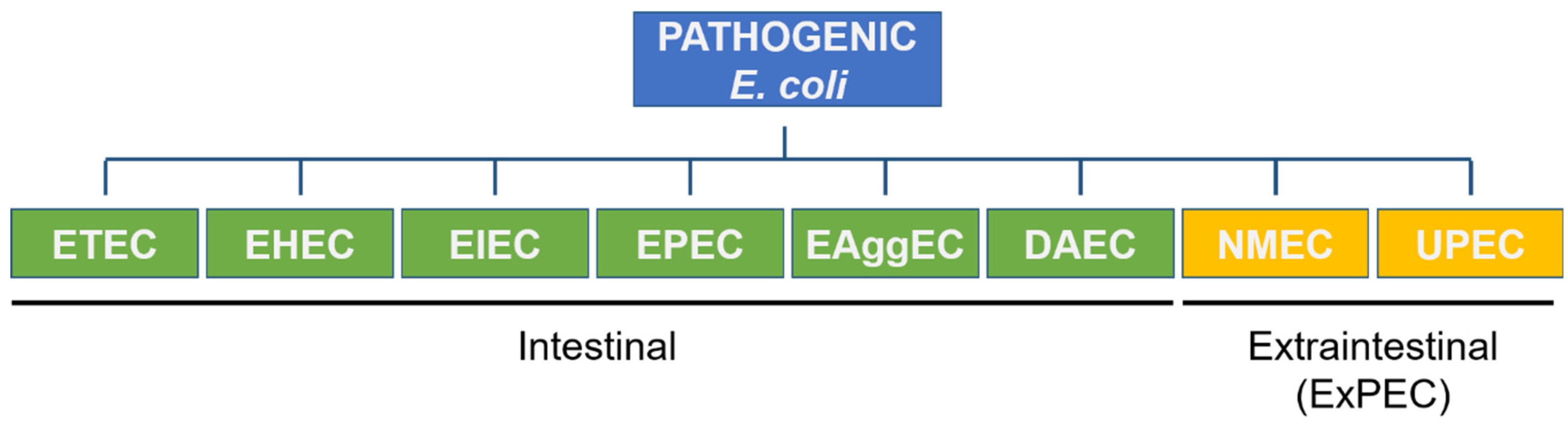
2. Urinary Tract Infections and Uroptahogenic E. coli

2.1. Epidemiology and Burden of UTI

Every year over 150 million people are diagnosed with UTIs, making them the second most common bacterial infection [11]. The number of cases is certainly underestimated as UTIs are non-reportable diseases in many countries [12]. It is predicted that 50–60% of adult women will develop a UTI at least once in their lifetime [13]. Other studies suggest that the frequency of UTIs in young women is about 0.5 episodes per person, yearly, of which 27% is recurrent, making the number even higher [14,15].
UPEC is the main etiological factor of UTIs and it is responsible for approximately 80–90% of all cases. According to published data, 81% of UPEC strains isolated from patients belong to groups B2 and D [10,11,16,17]. Both groups are characterized by higher distribution of the fyuA, pap, and yfcV genes encoding the ferric yersiniabactin uptake receptor, pyelonephritis-associated pili, and fimbrial-like protein, respectively. These virulence factors take part in iron uptake and cell adhesion [18]. Other virulence genes such as sfa (S fimbria), cnf (an operon encoding for cytotoxic necrotizing factor type I—CNF-1), and pic (factor PIC) were only found within the B2 phylogroup [18]. This is consistent with the recent report by Dadi et al. who also highlighted a strong association between the B2 group and the virulence genes listed above. However, at the same time, the prevalence of the B2 and D groups was reported to be slightly lower than the data presented above and accounted for 57.5% of all UPEC isolates [19].
In the face of the spread of multi-drug resistant (MDR) bacteria and the lack of alternative effective treatment methods, management of recurrent UTIs poses a significant challenge. Treatment of kidney-transplant patients, as well as life-threatening manifestations of ExPEC infections (e.g., sepsis or meningitis) are still associated with high mortality [20,21].
Therefore, curing UTIs is also associated with a substantial economic consequence. Medical consultations for UTIs constitute 0.9–6% of all outpatient visits and absences from work, summing up to $3.5 billion per year, in the United States alone [11,15,22].

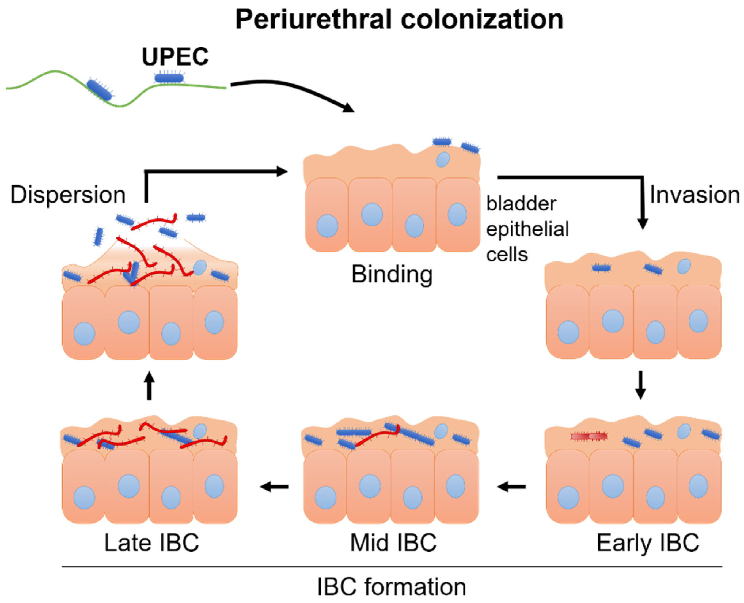
2.2. Pathomechanism of UTI

The general mechanism of the UTI cascade is relatively well understood. An infection begins when UPEC enter through the urethra and reach the epithelial cells of the bladder. Next, bacteria bind to the surface epithelium with use of various adhesins and then the invasion step occurs. As bacteria enter the cytoplasm of the urinary bladder epithelial cells, they actively replicate and form biofilm-like intracellular bacterial communities (IBCs), which hinder an effective immune response and enables further UPEC cell divisions. Lastly, dispersion of uroepithelial cells closes the cycle and the freed UPEC bugs can renew the UTI process (Figure 2) [11,23].
Understanding the infection mechanisms has led to the discovery of potential targets in UPEC therapy. For example, three types of fimbriae—Type 1, P, and Curli—are prerequisites for development of UTIs. These virulence factors are critical during uroepithelial cell invasion and acute pyelonephritis, and are associated with adhesion and the IBC formation capabilities of UPEC [24]. In a recent report, Yang et al. showed that administration of dictamine, a plant alkaloid, significantly impairs UPEC’s virulence via downregulation of the expression of fimbriae-encoding genes. This makes dictamine a promising non-antibiotic drug in treatment of UTIs [25]. Nevertheless, the general therapeutic options are limited as antibiotic therapy remains the mainstay of UTI treatment with other non-antibiotic measures such as cranberry products or probiotics playing a supporting role, although of uncertain effectiveness [26,27,28,29].

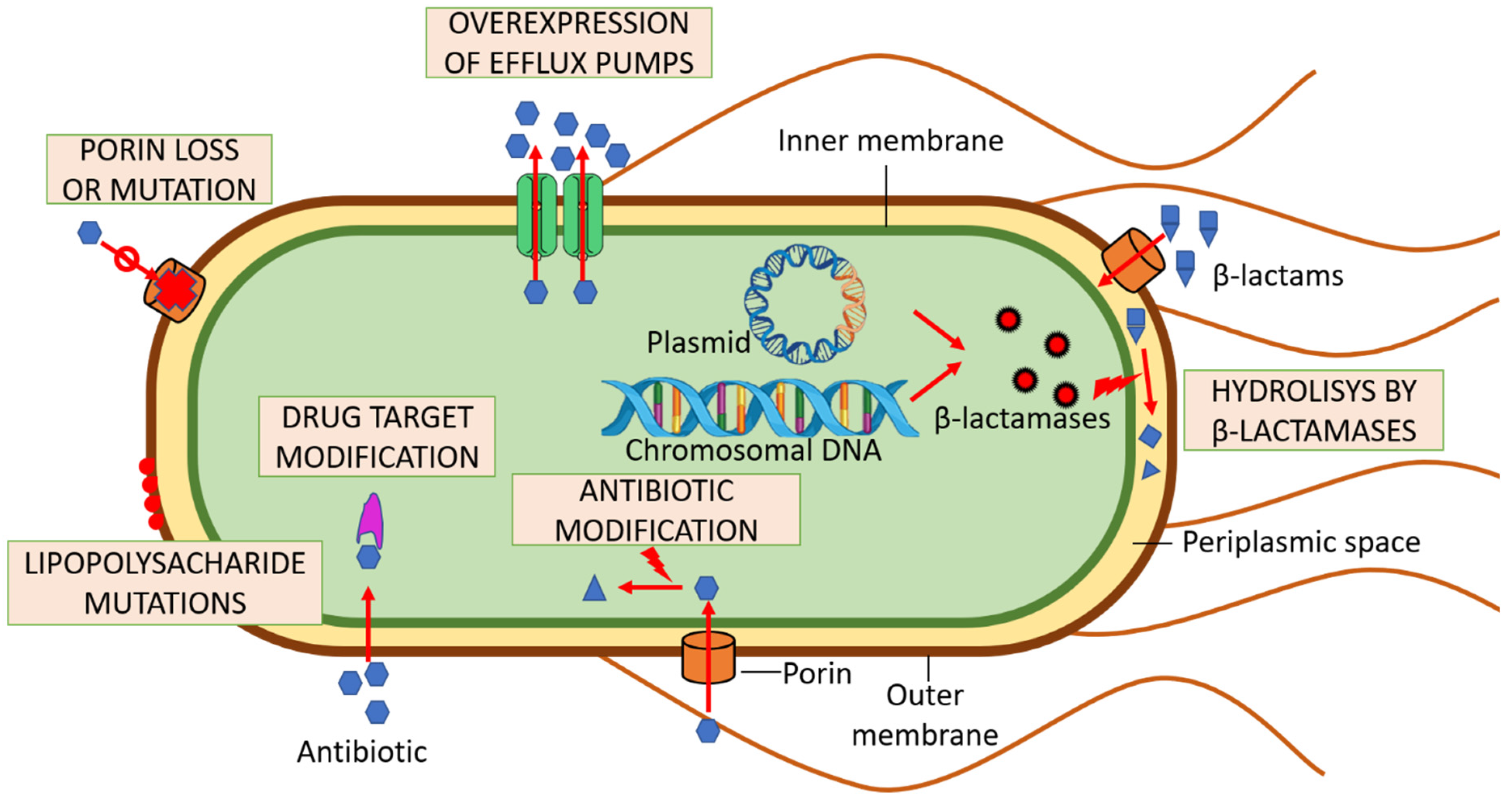
3. Multi-Drug Resistance and UPEC

Increasing antibiotic resistance of bacteria strains observed worldwide remains a burning issue. The MDR, which refers to acquired nonsusceptibility to at least one agent in three or more antimicrobial categories, currently constitutes a major obstacle to successful treatment of patients [30]. Based on various studies, the prevalence of MDR UPEC isolates in developing countries is high, varying from 42% (China) through 49.8% (Iran) and reaching an alarming 68% in Pakistan or even 98% in Mexico [31,32,33,34,35,36,37]. Rising rates of antimicrobial resistance result from irrational antibiotic policies, mainly because of inadequate broad spectrum antibiotic empirical therapies prescribed without collecting urine cultures and determining antibiotic susceptibility. MDR bacteria, UPEC included, are responsible for 25 thousand deaths every year in Europe alone, and for 700,000 deaths worldwide [38,39].
The phenomenon of antibiotic resistance among bacteria is often attributed exclusively to misuse and overuse of antimicrobial agents. Nevertheless, studies regarding environmental bacteria and human bacterial commensals also revealed that various resistance factors were present within their genomes prior to the commencement of the antibiotic era. The natural presence of antibiotic resistance is referred to as intrinsic antibiotic resistome and occurs in all bacterial species [24,40]. E. coli owes its intrinsic resistance mainly to the outer membrane and the expression of efflux pumps [41,42,43]. In contrast, extrinsic antibiotic resistance mainly results from human activity and antibiotic overuse and may be achieved by bacteria in many ways (discussed below).

3.1. Bacterial Efflux Pumps

Antimicrobial resistance can be reached through miscellaneous mechanisms (Figure 3). One of them are bacterial efflux pumps, which can regulate the intracellular environment by removing toxic substances, such as drugs, hence avoiding intracellular accumulation of antimicrobial molecules on their targets. Generally, bacteria contain two-domain half-transporters, which consist of transmembrane domains (TMD) and nucleotide-binding domains (NBD).
There are several efflux systems in E. coli associated with drug resistance, which can export drugs and other substrates out of bacterial cells. The seven major efflux pump superfamilies are: (i) the ATP-binding cassette (ABC); (ii) the major facilitator (MFS); (iii) the multidrug and toxic compound extrusion (MATE); (iv) the small multidrug resistance (SMR); (v) the resistance-nodulation-cell division (RND); (vi) the proteobacterial antimicrobial compound efflux (PACE); and (vii) the p-aminobenzoyl-glutamate transporter (AbgT) [44]. ABC transporters are ATP-dependent proteins, while other efflux systems use proton kinetic potential as their driving force.

3.1.1. The ABC Transporters

The ABC transporters are one of the most investigated efflux proteins. Among them, a special place belongs to the lactococcal homodimeric ABC-type protein (LmrA), which is considered as the classic, most-studied MDR pump, capable of transporting various drugs including antimicrobials and chemotherapeutic agents. It mainly removes drugs from the cell membrane, but not from the cytoplasm, and can be inactivated by reserpine or verapamil. P-glycoprotein 1 (P-gp) is a human transporter protein which is homologic to LmrA, sharing its structural and functional features. The activation of MDR pumps is considered as one of the causative factors of ineffectiveness of cancer chemotherapy [45,46,47]. LmrA is also homologous to MsbA, an essential ABC transporter in E. coli involved in the trafficking of lipids, including lipid A (endotoxin), which acts as the hydrophobic anchor of lipopolysaccharide (LPS) [48]. MsbA is a homodimeric half-transporter that allows for unidirectional LPS transport. MsbA confers multidrug resistance in E. coli and demonstrates overlapping substrate specificities when compared with LmrA. It also displays daunomycin, vinblastine, and Hoechst 33342-stimulated vanadate-sensitive ATPase activity. Interestingly, deletion of MsbA causes membrane disorganization, resulting in cell death [49]. This observation indicates that MsbA might be considered as a potential target of antibacterial therapy, including UPEC [49].

3.1.2. The MacAB–TolC Pump Complex

Another emphasized drug efflux system of E. coli that has been reported belongs to the MacAB–TolC pump complex. The system acts as a single drug efflux transporter of macrocyclic lipids, therefore it confers resistance to macrolides. The MacAB–TolC efflux system consists of three main parts: the outer membrane channel (TolC), creating passage for antimicrobials through the outer membrane; the inner membrane transporter, which recognizes drug molecules and activates efflux (AcrB or MacB); and the periplasmatic membrane fusion protein (MPF). Interactions between the TolC and inner member transporter are mediated by the MPF [50].

3.1.3. The AcrAB–TolC and MexAB–OprM Pump Complexes

AcrAB–TolC and MexAB–OprM are other efflux systems essential for effective bacterial colonization of the host as they provide protection against toxic substances. AcrAB–TolC genes and mutations in their efflux repressor AcrR were proposed to cause omeprazole resistance in UPEC [51]. The AcrAB–TolC system is a major player in creating intrinsic resistance to erythromycin and fusidic acid. Moreover, it protects bacterial cells from dyes and detergents. Its importance may be indicated by the fact that it is constitutive among E. coli [51]. The AcrAB–TolC system also plays a role in tetracycline resistance along with a variety of other drugs. A strong correlation between AcrA overexpression and ertapenem was recently reported by Chetri et al. [52]. They also observed that the AcrB system is overexpressed when bacteria are exposed to imipenem. Such boosted expression of the mentioned systems is a cause of concern, as it leads to increased non-susceptibility to carbapenems.
Overall, the E. coli genome is predicted to encode 69 ABC transporters. Eleven of them are presumed to be exporters, of which seven (i.e., YddA, YbhFSR, YhiH/YhhJ, YadGH, MdlAB) possibly export drugs. Nevertheless, the bacterial efflux pumps remain largely unexplored, and their role in developing the MDR phenomenon and revealing the full spectrum of substrates needs to be elucidated, as there are limited data regarding not only K1 UPEC, but also K12 E. coli.

3.2. Enzymatic Decomposition of Antibiotics

Another crucial mechanism resulting in MDR is the bacteria’s ability to enzymatically decompose antibiotic molecules. A perfect example of this phenomenon is the hydrolytic activity of β-lactamases produced by bacteria, which can inactivate β-lactam antibiotics [53].

3.2.1. β-lactams

β-lactams are widely administered antimicrobial agents that inhibit the biosynthesis of the bacterial cell wall, which prevents the multiplication of subsequent generations of bacteria. They can be divided into four main groups: (i) penicillin derivatives, (ii) cephalosporins, (iii) monobactams, and (iv) carbapenems. The first two are usually first-choice antibiotics in management of UTIs (amoxicillin or cefuroxime axetil are the most commonly prescribed), while the next two groups are reserved to treat patients with life-threatening conditions [54]. BlaTEM, the first group of β-lactamases described in Gram-negative bacteria, along with BlaCTX-M are the most common β-lactamases among E. coli detected worldwide. Genes encoding both these groups are mostly transferred by plasmids, but differ in their spectrum of action. BlaTEM genes mostly encode β-lactamases that are able to decompose penicillin and first-generation cephalosporins, while blaCTX-M encode enzymes which can hydrolyze even third-generation cephalosporins [55]. However, the most worrisome phenomenon is the emergence of broad-spectrum β-lactamases, which confer resistance to all β-lactams, including carbapenems.

3.2.2. Carbapenems

Carbapenems are the last resort antibiotics in treatment of extended spectrum β-lactamase (ESBL)-producing E. coli. According to the recent Iraqi study concerning UTI patients, β-lactamase prevalence among E. coli is associated with phylogroups [56]. The B2 phylogroup presented the highest antimicrobial resistance with 100% of isolates described as MDR. Moreover, 76.2% of them possessed carbapenemase genes with the most frequent one being blaOXA-48 (57.8%), followed by blaPER (47.3%), while other genes such as blaKPC, blaVEB, and blaVIM were detected in about 10–15% of UTI isolates. Similar findings were reported by Kuznetsova et al. who noticed that blaOXA genes are present in 58.6% of ESBL phenotype-positive isolates. However, the most common β-lactamases belonged to the BlaCTX-M group (79.3%) [57]. Importantly, the B2 phylogroup is characterized as the most virulent type and, as mentioned before, together with the D phylogroup is responsible for the majority of UTIs [56]. Therefore, coexistence of high virulence and UPEC antimicrobial resistance may potentially pose a serious threat to human health [58].

3.3. Drug Target Modifications

Bacteria are also able to alternate or protect the target site of antimicrobial drugs, preventing effective treatment. This mechanism can be observed in quinolone resistance. It was thought that quinolone resistance genes (qnr) do not exist naturally, as the developed quinolones were fully synthetic antimicrobials. Nevertheless, insusceptibility to quinolones was attributed to either changes in cell wall permeability or point mutations of genes encoding DNA gyrase and DNA topoisomerase IV, which are main quinolone targets [59]. The first chromosomal qnr gene, mfpA, was reported by Montero et al. in Mycobacterium smegmatis [60]. Up until now, mfpA homologs have been identified in many organisms, including UPEC (mcbG gene) [61].
Qnr proteins are members of the pentapeptide repeat protein family. Qnr proteins competitively attach themselves to the binding site of the DNA gyrase and topoisomerase IV. This prevents fluoroquinolones from forming and stabilizing the gyrase-cleaved DNA-quinolone complex, leading to cell death. Qnr proteins themselves confer low-level quinolone resistance. Additionally, researchers reported that Qnr proteins facilitate the selection of quinolone-resistant mutants; however, the exact molecular mechanism of this phenomenon remains unclear and needs to be elucidated [62,63]. Quinolone resistance among UPEC is mainly plasmid-mediated and is coded by the qnrA, qnrB, qnrC, qnrD, and qnrS genes [64].

3.4. Antibiotic Molecule Modification

Lack of hydrolytic enzyme inactivation by aminoglycoside acetyl transferase enzymes is another mechanism contributing to antibiotic resistance. Proteins taking part in this process in E. coli are coded by the aacA4, aadA2, and aacC2 genes. The mentioned genes encode acetyltransferases, which catalyze transfer of an acetyl group from acetyl-CoA to a 6′-amino group of aminoglycoside molecules. This confers resistance to antibiotics containing the purpurosamine ring, such as amikacin or kanamycin. Insensitivity to aminoglycosides in UPEC is mostly spread by plasmids, such as pGUE-NDM [36,65,66,67,68].
The UPEC antibiotic resistance genes (ARGs), their mechanism of action, and the antimicrobials they affect are summarized in Table 1.
The abovementioned mechanisms do not cover all the possible origins of antibiotic resistance in bacteria. Antibiotic resistance may also originate from random point mutations or can be the result of changes in cell membrane permeability [69,70]. Bacteria may acquire resistance to various antimicrobials by horizontal gene transfer (HGT) and mobile genetic elements (discussed below).

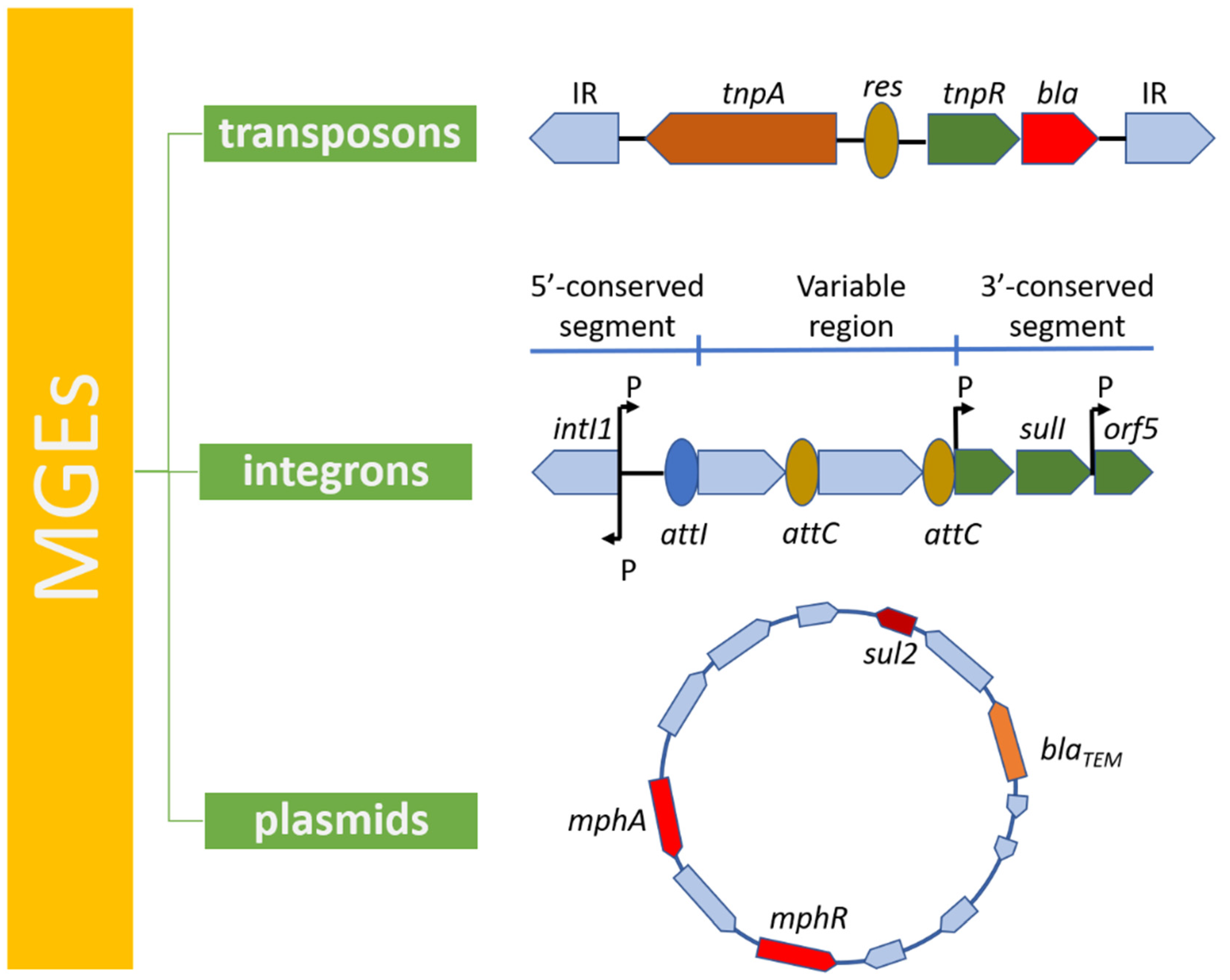
4. Mobile Genetic Elements in Antibiotic Resistance

The emergence of new MDR strains that pose a global health threat can be attributed to the circulation and acquisition of already existing ARGs among different populations of bacteria rather than to the emergence of novel resistance genes. Dissemination of ARGs is greatly facilitated by mobile genetic elements (MGEs) which are recognized as the leading drivers in spreading antimicrobial resistance in many bacteria, including UPEC. MGEs are a type of genetic material that can shift within a genome or can be transferred from one strain or replicon to another, allowing the capture, accumulation, and dissemination of resistance genes. MGEs include integrons, transposons, and plasmids [71,72] (Figure 4).
MGEs are found in all organisms and can be identified using various whole-genome alignment systems. One of the examples is the Mauve open software package for constructing multiple genome alignments in the presence of large-scale evolutionary events such as inversions and rearrangements [73]. Multiple genome alignments create foundation for research into comparative genomics and the observation of genome-wide evolutionary dynamics. The Mauve tool has its limitations as it is only capable of detecting large insertions. In order to find smaller MGEs, the breseq tool can be used [74]. Whole-genome alignment-based methods have their drawbacks, as they are unable to detect insertions of repetitive elements and are dependable on already known MGE sequences. Lately, Durrant et al. developed a bioinformatic toolbox—a MGE finder capable of detecting the MGEs’ genomic sites by using short-read sequencing data, which can overcome the mentioned obstacles [75,76].

4.1. Transposons

Transposable elements (TEs) are discrete DNA sequences that can change their position within a genome in almost random pattern. TEs can be assigned to one of two classes based on their mechanism of transposition. Class I transposons (also known as retrotransposons or copy and paste TEs) are copied in a two-step process. Firstly, they are transcribed from DNA to RNA, and then the newly synthetized RNA, with the use of reverse transcriptase, is transcribed back to DNA and can then be inserted back into the genome at the new locus. Class II transposons (named cut and paste TEs) act in a different way as they do not require an RNA intermediate. The process of transposition in this case is mediated by transposase enzymes, which can bind to specific or any target DNA sequence, depending on their type. The enzyme creates a staggered cut at the target DNA, resulting in formation of sticky ends, then it cuts out the DNA transposon and merges it into the target sequence. The process of transposition can occur autonomously if the transposon itself encodes reverse transcriptase or transposase in Class I and Class II TEs, respectively. Otherwise, presence of another (non-autonomous) TE is required for transposition to happen. TEs are responsible for spreading antibiotic resistance since they may be involved in conferring resistance to multiple classes of antimicrobial drugs, from the commonly used β-lactams to last-resort antibiotics such as colistin [77,78,79].
Recently, Carvalho et al. characterized an MDR UPEC BH100 sub-strain. This strain was obtained from the urinary tract of a Brazilian woman in 1974, but was not fully described until now. The usage of the Next Generation Sequencing (NGS) technique allowed assessing the entire sequence of the genome of the strain and it was found that the bacterium harbors two plasmids carrying MDR cassettes: pBH100 and pAp, presenting conjugative and mobilization properties, respectively [80]. The authors indicated transposon Tn21 as the one likely responsible for the mobilization of the streptomycin resistance gene—aadA (encoding for streptomycin 3′-adenylyltransferase). Tn21, along with a group of closely linked transposons, forms the Tn21 family, which is greatly involved in the spreading of antibiotic resistance factors in gram-negative bacteria worldwide [81]. The Tn21 family carries the integron In1 sequence, encoding a site-specific system for the acquisition and accumulation of multiple antibiotic resistance gene cassettes. Moreover, Tn21 also carries genes for its own transposition, making it an autonomous TE.
Carvalho et al. [80] also described the Tn3 transposon, associated with the blaTEM gene, which encodes the β-lactamase TEM-1 type enzyme. Members of the Tn3 transposon family form a widespread and homogeneous group of bacterial TEs that play a major role in disseminating antibiotic resistance. Tn3 members use DD (E/D) transposases in the process of replicative transposition in order to create a cointegrate structure, which is later resolved by means of site-specific recombination occurring between specific DNA sequences on both transposon copies. These regions also include promoters, which regulate expression of the recombinase and transposase. Moreover, they also stated that some Tn3 members encode a type II toxin-antitoxin (TA) system, which improves plasmid maintenance within a bacterial cell. The TA system usually consists of a stable toxin and a labile antitoxin that binds the toxin, therefore preventing it from being lethal for the bacteria [80,82,83,84].
A typical representative of the Tn3 family consists of three genes: tnpA, tnpR, and bla, which encode trasposase, resolvase, and β-lactamase, respectively. Recently, Lima-Mendez et al. reported that the members of the Tn3 family are able to carry a variety of passenger mutations caused by incorporation of nucleic acid fragments into their sequences [84]. Other members of the Tn3 family are Tn1546 and Tn5393, which carry vancomycin (vanA) and streptomycin (strAB) resistance, respectively. However, lately there have been no reports of them among UPEC [71].
Roy Chowdhury et al. recently described E. coli ST405 as an emerging pathogen of urosepsis. ST405 presents broad multidrug resistance, as it is unaffected by many antimicrobial agents including ampicillin, azithromycin, kanamycin, streptomycin, trimethoprim, and sulphafurazole [85]. Resistance genes encoding insusceptibility factors to the mentioned antibiotics are: blaTEM-1b (β-lactamase), mphR-mrx-mphA (macrolide inactivation gene cluster), aadA5 (aminoglycoside adenyltransferase), dfrA17 (dihydrofolate reductase), and sul1 (dihydropteroate synthase). They reside within a novel, mobile transposon named Tn6242, the structure of which is composed of four modules. Each module can comprise different antimicrobial resistance genes and is flanked with insertion sequences 26 (IS26). The IS are small (<2.5 kilobase long) DNA fragments encoding transposases. IS26 plays an important role in accumulation and spreading of multiple antibiotic resistance [86]. Tn6242 is able to generate smaller translocatable units, and a combination of different modules is also remarkable [85,87].
TEs also take part in spreading resistance to tetracyclines. Rafaque et al. described an emerging, highly resistant UPEC ST38 O1:H15 strain, which possess various ARGs and MGEs, among which there are two transposons, Tn2 and Tn3, carrying the tetA gene [88]. Other Tn3-like TEs involved in tetracycline resistance in MDR ST131-O25b-H30 UPEC and causing community-acquired UTIs were also reported by the same authors [89].
Importantly, Tn6330, a composite transposon present on both the pHS30-1 plasmid and the chromosome, was highlighted by Li et al. [90]. Tn6330 was found to be associated with the mcr-1 gene, which encodes phosphatidylethanolamine transferase. This enzyme is responsible for resistance to colistin, a last-resort antibiotic used in life-threatening infections caused by Gram-negative bacteria [90].

4.2. Integrons

Integrons belong to MGEs and are composed of three basic elements: (i) a gene encoding for a site-specific recombinase—integrase (intI); (ii) a recombination site (attI), which may be associated with various gene cassettes and is recognized by the mentioned integrase; and (iii) a promoter (Pc), the role of which is to direct transcription of cassette-encoded genes. It has been discovered that integrase plays a part in recruiting new gene cassettes into integrons, and can also modify those already inserted. Integrons are able to acquire new genes by a reversible cassette-associated recombination process that leads to incorporation, and subsequently expression, of new genetic material in bacteria. This phenomenon is responsible not only for bacterial evolution, as it enables bacteria to adapt to the changing surrounding environment, but also plays a role in acquiring and dissemination of antibiotic resistance, the latter one being a matter of concern in medicine [91,92,93,94]. Integrons can be classified into three main classes based on the intI gene. Class-1 integrons are a pivotal driver in spreading antimicrobial resistance genes. They are characterized by considerable diversity and high prevalence, as they have been detected in 22–95% of Gram-negative clinical isolates [95,96,97]. According to Poey and Lavina, class-1 integrons virtually always confer sulfonamide resistance as the already mentioned sul1 integrates the IntI-conserved platform [96]. Furthermore, this class of integrons is also usually associated with cassettes encoding resistance to streptomycin (aadA gene) and trimethoprim (dfrA gene) [67,98]. This stays in accordance with the recent study performed by Gonzalez-Villalobos et al. on MDR UPEC, where the presence of class-1 integrons was connected with dfrA5, dfrA12, dfrA17, dfrB4 (trimethoprim/sulfametaxazol), aadA1, aadA2, aadA5 (aminoglycosides), and ereA2 (erythromycin) resistance genes [99]. EreA2 is an integron-encoded erythromycin esterase that is able to hydrolyze erythromycin’s lactone ring. Nevertheless, class-1 integrons have been associated with more than 130 antibiotic resistance factors; therefore, resistance to all classes of antimicrobial agents has been reported [98]. Oliveira-Pinto et al. detected class-1 integrons in 65% of UPEC populations, but only in 11.9% of commensal E. coli, which suggests that these integrons may not be an indigenous part of E. coli [98]. This is consistent with the data from Gonzalez-Villalobos et al., who detected class-1 integrons in 63.9% of UPEC pathogenic phylogroups (B2 and D), and not in commensal ones. Similar data were reported by Poey et al., where a strong association between class-1 integrons and the phylogenetic group D was observed [96,99].
Class-2 integrons are less common, but also play a substantial role in the spread of resistance genes. They are usually found in strains from phylogroups A, B1, and B2 [96]. Shams et al. studied drug-resistant E. coli obtained from various clinical specimens and observed an association between the presence of integrons and insensitivity to various antimicrobial agents. They discovered that presence of class-1 integrons is related to resistance to ceftriaxone, ceftazidime, gentamicin, and nalidixic acid, whereas class-2 integrons were associated with insensitivity to imipenem, nalidixic acid, and co-trimoxazole.
It is worth mentioning that there is concern about high co-prevalence of class I and II integrons with ESBL and MDR in quinolone-resistant isolates [95]. The described association between class-1 integrons and antimicrobial resistance is partially consistent with more recent reports. For example, Mirnezami et al. studied UPEC strains and showed a statistically significant relationship between presence of class-1 integrons and resistance to gentamicin, ciprofloxacin, co-trimoxazole, ampicillin, and nalidixic acid. However, no significant association with ceftazidime resistance was observed [100].
Class-3 comprises the least frequently-observed integrons and are less often associated with antibiotic resistance. Until now, only one study by Xicohtencatl-Cortes et al. described class-3 integrons associated with MDR in the UPEC B2 phylogroup [101].

4.3. Plasmids

Plasmids are also considered as members of MGEs and are defined as small, extrachromosomal DNA circular molecules that possess the ability to replicate independently. They are most common among bacteria, however, plasmids may also be created artificially, and, therefore, play an important role in genetic engineering as vectors. Plasmids may be classified according to their function into five main classes: (i) virulence, (ii) resistance (R), (iii) fertility (F), (iv) degradative, and (v) colicin (Col) plasmids. The latter one harbors genes encoding for proteins toxic to other bacteria (bacteriocins). Nevertheless, the most used plasmid classification method currently is incompatibility (Inc) typing [102].
Most plasmids are double-stranded DNA (dsDNA), but some consist of single-stranded DNA (ssDNA) or double-stranded RNA (dsRNA). Plasmids play a substantial role in spread of antibiotic resistance worldwide, as some of them carry antimicrobial resistance genes that can be transferred between bacteria. Based on the Inc plasmid typing scheme, until now, 28 types of antibiotic resistance plasmids have been described. The IncF type plasmids are the most common in human and animal sources, and are mainly found in E. coli, including UPEC [103]. Unlike most plasmids, the IncF group has the ability to encode several replicons. IncF plasmids can carry a variety of resistance genes: ESBL, genes encoding carbapenemases, aminoglycoside-modifying enzymes, and plasmid-mediated quinolone resistance (PMQR) genes (discussed later). Importantly, IncFs are most often associated with ESBL resistance, as they spread blaCTX-M, blaTEM-1, and blaNDM genes [102].
Resistance plasmids can also be assigned one of two groups based on their mobility: narrow host range plasmids, which are usually transferred between the same species, and broad host range plasmids that can easily be harbored by different bacteria. The second group is associated with dissemination of MDR [104].
A recent study by Muriuki et al. from Kenya indicated that the main mechanism of resistance to β-lactam antibiotics in UPEC is the production of ESBLs and plasmid-mediated β -lactamases (pAmpCs) [105]. The occurrence of ESBLs depends on the geographical region and positively correlates with the overuse of antimicrobial agents. Prevalence rates in developing countries vary from 5% to 44.3%. AmpC β-lactamases confer insensitivity to penicillins, cephems, and monobactams, but the bacteria remain susceptible to carbapenems. Addition of β-lactamase inhibitors clavulanate, sulbactam, or tazobactam does not overcome antibiotic resistance in these cases [105]. PAmpCs originate from chromosomally-encoded AmpCs of multiple genres among the Enterobacteriaceae family, do not create a homogenous group, and the most frequent one is the CMY type.
Another worrying phenomenon is the growing resistance of bacteria to fluoroquinolones. According to Sadeghi et al. and Ramirez-Castillo et al., who examined UPEC strains obtained from kidney transplant patients (KTPs) and non-KTPs, plasmid-mediated quinolone resistance (PMQR) is increasingly identified [36,106]. Quinolone resistance is caused by various mechanisms, with PMQR being one of the main players. PMQR is associated with the qnrA, qnrB, qnrC, qnrD, and qnrS genes, which encode proteins that protect quinolone targets—gyrase and topoisomerase IV—from being affected by this group of antimicrobial agents.
PMQR is also associated with genes encoding efflux pumps qepA and qqxAB. QepA was reported to be linked with decreased susceptibility, and 8- to 32-fold increase of minimum inhibitory concentration (MIC) values of hydrophilic quinolones, such as norfloxacin, ciprofloxacin, and enrofloxacin [107]. QqxAB was characterized by a wider substrate specificity, including chloramphenicol, trimethoprim, and quinolones (ciprofloxacin, norfloxacin, and nalidixic acid) [108]. Even though PMQR is responsible for inducing only low-level resistance to quinolones, it could spread horizontally among other bacteria and elicit the selection of resistant strains, facilitating the acquisition of other quinolone-resistance mechanisms, thus resulting in high-level resistance. Acquiring quinolone resistance is especially dangerous for KTPs, as quinolones are used in UTI prophylaxis.
Moreover, PMQR strains often co-harbor genes encoding ESBL proteins, resulting in creation and selection of MDR UPEC [36,106,109]. Co-resistance to β-lactams and quinolones was also investigated in the study by Basu et al. [110]; 85% of strains collected from UTI patients were classified as MDR, where 86% of them were resistant to ciprofloxacin —a first-line drug in UPEC treatment. PMQR was detected in 50% of quinolone-resistant strains; moreover, all the isolates co-harbored at least one gene encoding β-lactamase, where blaTEM was the most common. This study also indicated that resistance genes can be transferred by conjugation upon ciprofloxacin selection.
Another example of plasmid-mediated antibiotic resistance in UPEC is azithromycin-resistance associated with presence of the mphA gene reported in cohorts of UTI patients previously not exposed to antibiotics. The spread of macrolide-resistant E. coli in the environment may become a threat to the treatment of other diseases, such as diarrheas caused by Salmonella and Shigella spp., as plasmids can be easily transferred [111].

5. Obstacles in UTI Treatment

The rapid dissemination of MDR UPEC observed worldwide could partially be attributed to gene migration. However, the mainspring of increasing antibiotic resistance is overuse and unreasonable antimicrobial policies. Frequent antibiotic exposure provides ideal conditions for selection of multi-drug resistant strains, which can later transfer resistance genes to other non-pathogenic or pathogenic bacteria present in the environment. UTI treatment may soon pose a challenge, as the situation is made worse by the lack of new antimicrobials. Recently, only three major pharmaceutical companies have been conducting research on developing new antibiotics [38,112,113].
It is observed that antimicrobial agents are less effective at killing intracellularly-localized bacteria; hence, IBC-forming cells are more likely to survive the antibiotic course and re-infect other bladder cells [114,115,116]. Therefore, increasing interest is now paid to inhibitors, which are considered as a potential tool to fight UPEC IBC’s. An example of such a compound is mannoside, capable of inhibiting FimH lectin, which plays a role in forming IBCs. According to Cusumano et al., they seem promising for patients with recurrent UTIs. However, until now, no FimH inhibitors have undergone full clinical trials [117]. Another recent trial addressed inhibitors (ZINC000104153710; ZINC000000217308) of universal stress protein A (UspA) as they present the capability to bind to the UspA molecule, and affect IBCs [118].
One of the mechanisms engaged in formation of UPEC IBCs, manifesting morphological flexibility, is filamentation. It is believed that development of filamentous cells is one of the many bacterial strategies implemented to avoid host immune responses, as it provides protection against phagocytosis. It is also a natural way to increase the gene copy number and it is a hallmark of IBCs. The process of filamentation is stimulated by stress factors, including the activated host immune system, but it can also be induced by antimicrobials. β-lactams inhibit penicillin-binding protein-3 (PBP-3), which takes part in septa formation, therefore facilitating filamentation.
The process of filamentation is controlled by the SOS response system. SOS is common and conserved in various bacteria; it is recruited in case of significant DNA damage and induces multiple proteins to protect integrity of DNA. The SOS pathway plays an essential role in increasing the resistance to stress and, therefore, to antibiotics. However, it also stimulates error-prone polymerases, allowing for enhanced survival, but also increasing mutagenesis at the same time [119,120]. The SOS response and its association with antibiotic resistance have recently been highlighted [121,122]. It has been proven that antimicrobials such as trimethoprim, fluoroquinolones, β-lactams, and aminoglycosides are potent activators of the SOS system by interfering with DNA replication or cell wall synthesis [123]. Activation of the SOS pathway results in increased mutational frequency, facilitating the rise of mutations in genes encoding antimicrobial targets, such as gyrA (DNA gyrase subunit A) in case of fluoroquinolones, making them ineffective. Antibiotics like quinolones, β-lactams, and aminoglycosides can kill bacteria by inducing ROS formation, which damages DNA. In response, the SOS pathway is recruited as DinF, a DNA-damage-inducible SOS response protein is able to decrease intracellular ROS levels, preventing bacteria from oxidative injuries and cellular death. The SOS system is also associated with HGT and MGEs, especially integrons, as it can induce the recombination of gene cassettes by controlling expression of the intI gene [124].
The highlighted close link between antibiotic resistance and the SOS system may be a potential target of new drugs—SOS modulators, which might be a tool to fight MDR bacteria [122]. Inhibition of the SOS pathway can be achieved either by LexA or RecA inactivation and results in a reduction in MIC for some antibiotics. RecA can be inhibited by Suramin, N6-ADP, curcumin, or zinc, while LexA only has a few potential inhibitors—small boron-containing compounds [125]. Crane et al. discovered that SOS-inducing drugs, such as ciprofloxacin or zidovudine, cause increased resistance to other antibiotics—rifampin, minocycline, and fosfomycin—by triggering hypermutations. Zinc, in vivo, was able to inhibit this SOS-induced appearance of antimicrobial resistance [122,126]. A similar effect was also described for N-acetylcysteine [127]. Moreover, Recacha et al. found that suppression of the SOS system can even reverse antibiotic resistance among highly-resistant quinolone strains [128]. The potential ability of SOS inhibitors to block the emergence of antimicrobial resistance or increase antibiotic effectiveness in vivo requires further research.

6. Conclusions

Mobile genetic elements play a major role in dissemination of antimicrobial resistance genes among various bacteria. The growing antibiotic resistance of pathogenic bugs resulting from overly prescribed antibiotics may soon pose an enormous challenge. In the absence of new classes of antimicrobial drugs and the bacteria’s ability to rapidly adapt to various stress conditions with the use of the SOS pathway, we should seek alternative methods of treatment of bacterial infections. SOS inhibitors seem a promising alternative, as they have not only been proven to be effective, but also the pathway they target is conserved in many different bacteria species. Nonetheless, SOS modulators require further study and for now strategies should focus on implementing rational antibiotic policies.

Author Contributions

Conceptualization, M.R. and D.G.; writing—original draft preparation, M.R.; writing—review and editing, M.R. and D.G.; visualization, M.R.; supervision, D.G.; funding acquisition, D.G. All authors have read and agreed to the published version of the manuscript.

Funding

This study was funded by National Science Centre, grant number 2019/35/B/NZ6/03996.

Conflicts of Interest

The authors declare no conflict of interest.

Abbreviation

ABCATP-binding cassette
CNF-1Cytotoxic Necrotizing Factor type I
DAECDiffusely adherent Escherichia coli
dsDNADouble-stranded DNA
dsRNADouble-stranded RNA
EAggECEnteroaggregative Escherichia coli
EHECEnterohaemorrhagic Escherichia coli
EIECEnteroinvasive Escherichia coli
EPECEnteropathogenic Escherichia coli
ESBLExtended spectrum β-lactamase
ETECEnterotoxigenic Escherichia coli
ExPECExtraintestinal Escherichia coli
HGTHorizontal gene transfer
IBCIntracellular bacterial communities
ISInsertion sequence
KTPKindey transplant patients
MATEMultidrug and toxic compound extrusion
MDRMulti drug resistant
MGEMobile genetic element
MICMinimum inhibitory concentration
MPFMembrane fusion protein
NBDNucleotide-binding domain
NGSNext Generation Sequencing
NMECNeonatal Meningitis Escherichia coli
PMQRPlasmid-mediated quinolone resistance
QnrQuinolone resistance
ROSReactive oxygen species
SMRSmall multidrug resistance
ssDNASingle-stranded DNA
TETransposable element
TMDTransmembrane domain
UPECUropathogenic Escherichia coli
UTIUrinary tract infection

References

  1. Hacker, J.; Blum-Oehler, G. In appreciation of Theodor Escherich. Nat. Rev. Microbiol. 2007, 5, 902. [Google Scholar] [CrossRef] [Green Version]
  2. Blount, Z.D. The unexhausted potential of E. coli. Elife 2015, 4, e05826. [Google Scholar] [CrossRef] [PubMed]
  3. Augusto-Pinto, L.; Silva, C.G.; Lopes Dde, O.; Machado-Silva, A.; Machado, C.R. Escherichia coli as a model system to study DNA repair genes of eukaryotic organisms. Genet. Mol. Res. 2003, 2, 77–91. [Google Scholar] [PubMed]
  4. Evans, D.J., Jr.; Evans, D.G. Classification of pathogenic Escherichia coli according to serotype and the production of virulence factors, with special reference to colonization-factor antigens. Rev. Infect. Dis. 1983, 5, S692–S701. [Google Scholar] [CrossRef] [PubMed]
  5. Stenutz, R.; Weintraub, A.; Widmalm, G. The structures of Escherichia coli O-polysaccharide antigens. FEMS Microbiol. Rev. 2006, 30, 382–403. [Google Scholar] [CrossRef] [Green Version]
  6. Whitfield, C.; Williams, D.M.; Kelly, S.D. Lipopolysaccharide O-antigens-bacterial glycans made to measure. J. Biol. Chem. 2020, 295, 10593–10609. [Google Scholar] [CrossRef]
  7. Sarowska, J.; Futoma-Koloch, B.; Jama-Kmiecik, A.; Frej-Madrzak, M.; Ksiazczyk, M.; Bugla-PLoSkonska, G.; Choroszy-Krol, I. Virulence factors, prevalence and potential transmission of extraintestinal pathogenic Escherichia coli isolated from different sources: Recent reports. Gut Pathog. 2019, 11, 10. [Google Scholar] [CrossRef] [Green Version]
  8. Manges, A.R.; Geum, H.M.; Guo, A.; Edens, T.J.; Fibke, C.D.; Pitout, J.D.D. Global Extraintestinal Pathogenic Escherichia coli (ExPEC) Lineages. Clin. Microbiol. Rev. 2019, 32, e00135-18. [Google Scholar] [CrossRef]
  9. Sanchez, G.V.; Babiker, A.; Master, R.N.; Luu, T.; Mathur, A.; Bordon, J. Antibiotic Resistance among Urinary Isolates from Female Outpatients in the United States in 2003 and 2012. Antimicrob. Agents Chemother. 2016, 60, 2680–2683. [Google Scholar] [CrossRef] [Green Version]
  10. Terlizzi, M.E.; Gribaudo, G.; Maffei, M.E. UroPathogenic Escherichia coli (UPEC) Infections: Virulence Factors, Bladder Responses, Antibiotic, and Non-antibiotic Antimicrobial Strategies. Front. Microbiol. 2017, 8, 1566. [Google Scholar] [CrossRef]
  11. Flores-Mireles, A.L.; Walker, J.N.; Caparon, M.; Hultgren, S.J. Urinary tract infections: Epidemiology, mechanisms of infection and treatment options. Nat. Rev. Microbiol. 2015, 13, 269–284. [Google Scholar] [CrossRef]
  12. Foxman, B. Epidemiology of urinary tract infections: Incidence, morbidity, and economic costs. Dis. Mon. 2003, 49, 53–70. [Google Scholar] [CrossRef] [PubMed]
  13. Medina, M.; Castillo-Pino, E. An introduction to the epidemiology and burden of urinary tract infections. Ther. Adv. Urol. 2019, 11, 1756287219832172. [Google Scholar] [CrossRef] [PubMed] [Green Version]
  14. Hooton, T.M.; Scholes, D.; Hughes, J.P.; Winter, C.; Roberts, P.L.; Stapleton, A.E.; Stergachis, A.; Stamm, W.E. A prospective study of risk factors for symptomatic urinary tract infection in young women. N. Engl. J. Med. 1996, 335, 468–474. [Google Scholar] [CrossRef] [PubMed]
  15. Al-Badr, A.; Al-Shaikh, G. Recurrent Urinary Tract Infections Management in Women: A review. Sultan Qaboos Univ. Med. J. 2013, 13, 359–367. [Google Scholar] [CrossRef] [PubMed]
  16. Foxman, B. Urinary tract infection syndromes: Occurrence, recurrence, bacteriology, risk factors, and disease burden. Infect. Dis. Clin. N. Am. 2014, 28, 1–13. [Google Scholar] [CrossRef]
  17. Yun, K.W.; Kim, D.S.; Kim, W.; Lim, I.S. Molecular typing of uropathogenic Escherichia coli isolated from Korean children with urinary tract infection. Korean J. Pediatr. 2015, 58, 20–27. [Google Scholar] [CrossRef] [Green Version]
  18. Lara, F.B.; Nery, D.R.; de Oliveira, P.M.; Araujo, M.L.; Carvalho, F.R.; Messias-Silva, L.C.; Ferreira, L.B.; Faria-Junior, C.; Pereira, A.L. Virulence Markers and Phylogenetic Analysis of Escherichia coli Strains with Hybrid EAEC/UPEC Genotypes Recovered from Sporadic Cases of Extraintestinal Infections. Front. Microbiol. 2017, 8, 146. [Google Scholar] [CrossRef] [Green Version]
  19. Dadi, B.R.; Abebe, T.; Zhang, L.; Mihret, A.; Abebe, W.; Amogne, W. Distribution of virulence genes and phylogenetics of uropathogenic Escherichia coli among urinary tract infection patients in Addis Ababa, Ethiopia. BMC Infect. Dis. 2020, 20, 108. [Google Scholar] [CrossRef] [Green Version]
  20. Halaji, M.; Shahidi, S.; Atapour, A.; Ataei, B.; Feizi, A.; Havaei, S.A. Characterization of Extended-Spectrum beta-Lactamase-Producing Uropathogenic Escherichia coli Among Iranian Kidney Transplant Patients. Infect. Drug Resist. 2020, 13, 1429–1437. [Google Scholar] [CrossRef]
  21. Yuan, X.; Liu, T.; Wu, D.; Wan, Q. Epidemiology, susceptibility, and risk factors for acquisition of MDR/XDR Gram-negative bacteria among kidney transplant recipients with urinary tract infections. Infect. Drug Resist. 2018, 11, 707–715. [Google Scholar] [CrossRef] [PubMed] [Green Version]
  22. Litwin, M.S.; Saigal, C.S.; Yano, E.M.; Avila, C.; Geschwind, S.A.; Hanley, J.M.; Joyce, G.F.; Madison, R.; Pace, J.; Polich, S.M.; et al. Urologic diseases in America Project: Analytical methods and principal findings. J. Urol. 2005, 173, 933–937. [Google Scholar] [CrossRef] [PubMed]
  23. Justice, S.S.; Hung, C.; Theriot, J.A.; Fletcher, D.A.; Anderson, G.G.; Footer, M.J.; Hultgren, S.J. Differentiation and developmental pathways of uropathogenic Escherichia coli in urinary tract pathogenesis. Proc. Natl. Acad. Sci. USA 2004, 101, 1333–1338. [Google Scholar] [CrossRef] [PubMed] [Green Version]
  24. Pawlowski, A.C.; Wang, W.; Koteva, K.; Barton, H.A.; McArthur, A.G.; Wright, G.D. A diverse intrinsic antibiotic resistome from a cave bacterium. Nat. Commun. 2016, 7, 13803. [Google Scholar] [CrossRef] [PubMed] [Green Version]
  25. Yang, W.; Liu, P.; Chen, Y.; Lv, Q.; Wang, Z.; Huang, W.; Jiang, H.; Zheng, Y.; Jiang, Y.; Sun, L. Dictamnine Inhibits the Adhesion to and Invasion of Uropathogenic Escherichia coli (UPEC) to Urothelial Cells. Molecules 2022, 27, 272. [Google Scholar] [CrossRef]
  26. Scharf, B.; Schmidt, T.J.; Rabbani, S.; Stork, C.; Dobrindt, U.; Sendker, J.; Ernst, B.; Hensel, A. Antiadhesive natural products against uropathogenic E. coli: What can we learn from cranberry extract? J. Ethnopharmacol. 2020, 257, 112889. [Google Scholar] [CrossRef]
  27. Loubet, P.; Ranfaing, J.; Dinh, A.; Dunyach-Remy, C.; Bernard, L.; Bruyere, F.; Lavigne, J.P.; Sotto, A. Alternative Therapeutic Options to Antibiotics for the Treatment of Urinary Tract Infections. Front. Microbiol. 2020, 11, 1509. [Google Scholar] [CrossRef]
  28. Hutt, P.; Shchepetova, J.; Loivukene, K.; Kullisaar, T.; Mikelsaar, M. Antagonistic activity of probiotic lactobacilli and bifidobacteria against entero- and uropathogens. J. Appl. Microbiol. 2006, 100, 1324–1332. [Google Scholar] [CrossRef]
  29. Reid, G.; Bruce, A.W.; Fraser, N.; Heinemann, C.; Owen, J.; Henning, B. Oral probiotics can resolve urogenital infections. FEMS Immunol. Med. Microbiol. 2001, 30, 49–52. [Google Scholar] [CrossRef]
  30. Magiorakos, A.P.; Srinivasan, A.; Carey, R.B.; Carmeli, Y.; Falagas, M.E.; Giske, C.G.; Harbarth, S.; Hindler, J.F.; Kahlmeter, G.; Olsson-Liljequist, B.; et al. Multidrug-resistant, extensively drug-resistant and pandrug-resistant bacteria: An international expert proposal for interim standard definitions for acquired resistance. Clin. Microbiol. Infect. 2012, 18, 268–281. [Google Scholar] [CrossRef] [Green Version]
  31. Rishabh, R.S.S. Significance of Regional Antibiogram and MDR of ESBL Producing Uropathogens Infecting Non-hospitalized Patients. Gurugram. Int. J. Curr. Microbiol. 2018, 7, 1114–1126. [Google Scholar] [CrossRef] [Green Version]
  32. Javed, S.; Mirani, Z.A.; Pirzada, Z.A. Study of class 1 integrons and plasmid profile among multiple drug resistant uropathogenic Escherichia coli. Pak. J. Pharm. Sci. 2020, 33, 2643–2649. [Google Scholar] [PubMed]
  33. Ali, I.; Rafaque, Z.; Ahmed, S.; Malik, S.; Dasti, J.I. Prevalence of multi-drug resistant uropathogenic Escherichia coli in Potohar region of Pakistan. Asian Pac. J.Trop. Biomed. 2016, 6, 60–66. [Google Scholar] [CrossRef] [Green Version]
  34. Contreras-Alvarado, L.M.; Zavala-Vega, S.; Cruz-Cordova, A.; Reyes-Grajeda, J.P.; Escalona-Venegas, G.; Flores, V.; Alcazar-Lopez, V.; Arellano-Galindo, J.; Hernandez-Castro, R.; Castro-Escarpulli, G.; et al. Molecular Epidemiology of Multidrug-Resistant Uropathogenic Escherichia coli O25b Strains Associated with Complicated Urinary Tract Infection in Children. Microorganisms 2021, 9, 2299. [Google Scholar] [CrossRef]
  35. Hadifar, S.; Moghoofei, M.; Nematollahi, S.; Ramazanzadeh, R.; Sedighi, M.; Salehi-Abargouei, A.; Miri, A. Epidemiology of Multidrug Resistant Uropathogenic Escherichia coli in Iran: A Systematic Review and Meta-Analysis. Jpn. J. Infect. Dis. 2017, 70, 19–25. [Google Scholar] [CrossRef] [Green Version]
  36. Ramirez-Castillo, F.Y.; Moreno-Flores, A.C.; Avelar-Gonzalez, F.J.; Marquez-Diaz, F.; Harel, J.; Guerrero-Barrera, A.L. An evaluation of multidrug-resistant Escherichia coli isolates in urinary tract infections from Aguascalientes, Mexico: Cross-sectional study. Ann. Clin. Microbiol. Antimicrob. 2018, 17, 34. [Google Scholar] [CrossRef]
  37. Moeinizadeh, H.; Shaheli, M. Frequency of hlyA, hlyB, hlyC and hlyD genes in uropathogenic Escherichia coli isolated from UTI patients in Shiraz. GMS Hyg. Infect. Control 2021, 16, 25. [Google Scholar] [CrossRef]
  38. Dutescu, I.A.; Hillier, S.A. Encouraging the Development of New Antibiotics: Are Financial Incentives the Right Way Forward? A Systematic Review and Case Study. Infect. Drug Resist. 2021, 14, 415–434. [Google Scholar] [CrossRef]
  39. Colomb-Cotinat, M.; Lacoste, J.; Brun-Buisson, C.; Jarlier, V.; Coignard, B.; Vaux, S. Estimating the morbidity and mortality associated with infections due to multidrug-resistant bacteria (MDRB), France, 2012. Antimicrob. Resist. Infect. Control 2016, 5, 56. [Google Scholar] [CrossRef] [Green Version]
  40. Paun, V.I.; Lavin, P.; Chifiriuc, M.C.; Purcarea, C. First report on antibiotic resistance and antimicrobial activity of bacterial isolates from 13,000-year old cave ice core. Sci. Rep. 2021, 11, 514. [Google Scholar] [CrossRef]
  41. Sommer, M.O.A.; Dantas, G.; Church, G.M. Functional characterization of the antibiotic resistance reservoir in the human microflora. Science 2009, 325, 1128–1131. [Google Scholar] [CrossRef] [Green Version]
  42. Cox, G.; Wright, G.D. Intrinsic antibiotic resistance: Mechanisms, origins, challenges and solutions. Int. J. Med. Microbiol. 2013, 303, 287–292. [Google Scholar] [CrossRef] [PubMed]
  43. Nikaido, H.; Rosenberg, E.Y.; Foulds, J. Porin channels in Escherichia coli: Studies with beta-lactams in intact cells. J. Bacteriol. 1983, 153, 232–240. [Google Scholar] [CrossRef] [PubMed] [Green Version]
  44. Chitsaz, M.; Brown, M.H. The role played by drug efflux pumps in bacterial multidrug resistance. Essays Biochem. 2017, 61, 127–139. [Google Scholar] [CrossRef] [PubMed]
  45. Veen, H.W.; Margolles, A.; Muller, M.; Higgins, C.F.; Konings, W.N. The homodimeric ATP-binding cassette transporter LmrA mediates multidrug transport by an alternating two-site (two-cylinder engine) mechanism. EMBO J. 2000, 19, 2503–2514. [Google Scholar] [CrossRef] [Green Version]
  46. Veen, H.W.; Venema, K.; Bolhuis, H.; Oussenko, I.; Kok, J.; Poolman, B.; Driessen, A.J.; Konings, W.N. Multidrug resistance mediated by a bacterial homolog of the human multidrug transporter MDR1. Proc. Natl. Acad. Sci. USA 1996, 93, 10668–10672. [Google Scholar] [CrossRef] [Green Version]
  47. Ughachukwu, P.; Unekwe, P. Efflux pump-mediated resistance in chemotherapy. Ann. Med. Health Sci. Res. 2012, 2, 191–198. [Google Scholar] [CrossRef] [Green Version]
  48. Reuter, G.; Janvilisri, T.; Venter, H.; Shahi, S.; Balakrishnan, L.; van Veen, H.W. The ATP binding cassette multidrug transporter LmrA and lipid transporter MsbA have overlapping substrate specificities. J. Biol. Chem. 2003, 278, 35193–35198. [Google Scholar] [CrossRef] [Green Version]
  49. Ho, H.; Miu, A.; Alexander, M.K.; Garcia, N.K.; Oh, A.; Zilberleyb, I.; Reichelt, M.; Austin, C.D.; Tam, C.; Shriver, S.; et al. Structural basis for dual-mode inhibition of the ABC transporter MsbA. Nature 2018, 557, 196–201. [Google Scholar] [CrossRef]
  50. Lu, S.; Zgurskaya, H.I. MacA, a periplasmic membrane fusion protein of the macrolide transporter MacAB-TolC, binds lipopolysaccharide core specifically and with high affinity. J. Bacteriol. 2013, 195, 4865–4872. [Google Scholar] [CrossRef] [Green Version]
  51. Chowdhury, N.; Suhani, S.; Purkaystha, A.; Begum, M.K.; Raihan, T.; Alam, M.J.; Islam, K.; Azad, A.K. Identification of AcrAB-TolC Efflux Pump Genes and Detection of Mutation in Efflux Repressor AcrR from Omeprazole Responsive Multidrug-Resistant Escherichia coli Isolates Causing Urinary Tract Infections. Microbiol. Insights 2019, 12, 1178636119889629. [Google Scholar] [CrossRef] [PubMed] [Green Version]
  52. Chetri, S.; Bhowmik, D.; Paul, D.; Pandey, P.; Chanda, D.D.; Chakravarty, A.; Bora, D.; Bhattacharjee, A. AcrAB-TolC efflux pump system plays a role in carbapenem non-susceptibility in Escherichia coli. BMC Microbiol. 2019, 19, 210. [Google Scholar] [CrossRef] [PubMed] [Green Version]
  53. Tooke, C.L.; Hinchliffe, P.; Bragginton, E.C.; Colenso, C.K.; Hirvonen, V.H.A.; Takebayashi, Y.; Spencer, J. beta-Lactamases and beta-Lactamase Inhibitors in the 21st Century. J. Mol. Biol. 2019, 431, 3472–3500. [Google Scholar] [CrossRef]
  54. Buettcher, M.; Trueck, J.; Niederer-Loher, A.; Heininger, U.; Agyeman, P.; Asner, S.; Berger, C.; Bielicki, J.; Kahlert, C.; Kottanattu, L.; et al. Swiss consensus recommendations on urinary tract infections in children. Eur. J. Pediatr. 2021, 180, 663–674. [Google Scholar] [CrossRef]
  55. Xiao, L.; Wang, X.; Kong, N.; Zhang, L.; Cao, M.; Sun, M.; Wei, Q.; Liu, W. Characterization of Beta-Lactamases in Bloodstream-Infection Escherichia coli: Dissemination of blaADC–162 and blaCMY-2 Among Bacteria via an IncF Plasmid. Front. Microbiol. 2019, 10, 2175. [Google Scholar] [CrossRef]
  56. Gatya Al-Mayahie, S.M.; Al-Guranie, D.R.T.; Hussein, A.A.; Bachai, Z.A. Prevalence of common carbapenemase genes and multidrug resistance among uropathogenic Escherichia coli phylogroup B2 isolates from outpatients in Wasit Province/Iraq. PLoS ONE 2022, 17, e0262984. [Google Scholar] [CrossRef]
  57. Kuznetsova, M.V.; Gizatullina, J.S. Epidemiological characteristics of uropatogenic iso-lates of Escherichia coli in hospitals. Russ. Clin. Lab. Diagn. 2021, 66, 248–256. [Google Scholar] [CrossRef]
  58. Pan, Y.; Zeng, J.; Li, L.; Yang, J.; Tang, Z.; Xiong, W.; Li, Y.; Chen, S.; Zeng, Z. Coexistence of Antibiotic Resistance Genes and Virulence Factors Deciphered by Large-Scale Complete Genome Analysis. mSystems 2020, 5, e00821-19. [Google Scholar] [CrossRef] [PubMed]
  59. Li, X.; Zhang, Y.; Zhou, X.; Hu, X.; Zhou, Y.; Liu, D.; Maxwell, A.; Mi, K. The plasmid-borne quinolone resistance protein QnrB, a novel DnaA-binding protein, increases the bacterial mutation rate by triggering DNA replication stress. Mol. Microbiol. 2019, 111, 1529–1543. [Google Scholar] [CrossRef] [Green Version]
  60. Montero, C.; Mateu, G.; Rodriguez, R.; Takiff, H. Intrinsic resistance of Mycobacterium smegmatis to fluoroquinolones may be influenced by new pentapeptide protein MfpA. Antimicrob. Agents Chemother. 2001, 45, 3387–3392. [Google Scholar] [CrossRef] [Green Version]
  61. Merens, A.; Matrat, S.; Aubry, A.; Lascols, C.; Jarlier, V.; Soussy, C.J.; Cavallo, J.D.; Cambau, E. The pentapeptide repeat proteins MfpAMt and QnrB4 exhibit opposite effects on DNA gyrase catalytic reactions and on the ternary gyrase-DNA-quinolone complex. J. Bacteriol. 2009, 191, 1587–1594. [Google Scholar] [CrossRef] [PubMed] [Green Version]
  62. Vinue, L.; Corcoran, M.A.; Hooper, D.C.; Jacoby, G.A. Mutations That Enhance the Ciprofloxacin Resistance of Escherichia coli with qnrA1. Antimicrob. Agents Chemother. 2015, 60, 1537–1545. [Google Scholar] [CrossRef] [PubMed] [Green Version]
  63. Redgrave, L.S.; Sutton, S.B.; Webber, M.A.; Piddock, L.J. Fluoroquinolone resistance: Mechanisms, impact on bacteria, and role in evolutionary success. Trends Microbiol. 2014, 22, 438–445. [Google Scholar] [CrossRef]
  64. Munita, J.M.; Arias, C.A. Mechanisms of Antibiotic Resistance. Microbiol. Spectr. 2016, 4, 15. [Google Scholar] [CrossRef] [PubMed] [Green Version]
  65. Alekshun, M.N.; Levy, S.B. Molecular mechanisms of antibacterial multidrug resistance. Cell 2007, 128, 1037–1050. [Google Scholar] [CrossRef] [PubMed] [Green Version]
  66. Cag, Y.; Caskurlu, H.; Fan, Y.; Cao, B.; Vahaboglu, H. Resistance mechanisms. Ann. Transl. Med. 2016, 4, 326. [Google Scholar] [CrossRef] [PubMed] [Green Version]
  67. Ochoa, S.A.; Cruz-Cordova, A.; Luna-Pineda, V.M.; Reyes-Grajeda, J.P.; Cazares-Dominguez, V.; Escalona, G.; Sepulveda-Gonzalez, M.E.; Lopez-Montiel, F.; Arellano-Galindo, J.; Lopez-Martinez, B.; et al. Multidrug- and Extensively Drug-Resistant Uropathogenic Escherichia coli Clinical Strains: Phylogenetic Groups Widely Associated with Integrons Maintain High Genetic Diversity. Front. Microbiol. 2016, 7, 2042. [Google Scholar] [CrossRef] [Green Version]
  68. Bonnin, R.A.; Poirel, L.; Carattoli, A.; Nordmann, P. Characterization of an IncFII plasmid encoding NDM-1 from Escherichia coli ST131. PLoS ONE 2012, 7, e34752. [Google Scholar] [CrossRef] [Green Version]
  69. Long, H.; Miller, S.F.; Strauss, C.; Zhao, C.; Cheng, L.; Ye, Z.; Griffin, K.; Te, R.; Lee, H.; Chen, C.C.; et al. Antibiotic treatment enhances the genome-wide mutation rate of target cells. Proc. Natl Acad. Sci. USA 2016, 113, e2498–e2505. [Google Scholar] [CrossRef] [Green Version]
  70. Delcour, A.H. Outer membrane permeability and antibiotic resistance. Biochim. Biophys. Acta 2009, 1794, 808–816. [Google Scholar] [CrossRef] [Green Version]
  71. Partridge, S.R.; Kwong, S.M.; Firth, N.; Jensen, S.O. Mobile Genetic Elements Associated with Antimicrobial Resistance. Clin. Microbiol. Rev. 2018, 31, e00088-17. [Google Scholar] [CrossRef] [PubMed] [Green Version]
  72. Rijavec, M.; Starcic Erjavec, M.; Ambrozic Avgustin, J.; Reissbrodt, R.; Fruth, A.; Krizan-Hergouth, V.; Zgur-Bertok, D. High prevalence of multidrug resistance and random distribution of mobile genetic elements among uropathogenic Escherichia coli (UPEC) of the four major phylogenetic groups. Curr. Microbiol. 2006, 53, 158–162. [Google Scholar] [CrossRef] [PubMed]
  73. Darling, A.C.; Mau, B.; Blattner, F.R.; Perna, N.T. Mauve: Multiple alignment of conserved genomic sequence with rearrangements. Genome Res. 2004, 14, 1394–1403. [Google Scholar] [CrossRef] [PubMed] [Green Version]
  74. Deatherage, D.E.; Barrick, J.E. Identification of mutations in laboratory-evolved microbes from next-generation sequencing data using breseq. Methods Mol. Biol. 2014, 1151, 165–188. [Google Scholar] [CrossRef] [Green Version]
  75. Barrick, J.E.; Colburn, G.; Deatherage, D.E.; Traverse, C.C.; Strand, M.D.; Borges, J.J.; Knoester, D.B.; Reba, A.; Meyer, A.G. Identifying structural variation in haploid microbial genomes from short-read resequencing data using breseq. BMC Genom. 2014, 15, 1039. [Google Scholar] [CrossRef]
  76. Durrant, M.G.; Li, M.M.; Siranosian, B.A.; Montgomery, S.B.; Bhatt, A.S. A Bioinformatic Analysis of Integrative Mobile Genetic Elements Highlights Their Role in Bacterial Adaptation. Cell Host Microbe 2020, 27, 140–153. [Google Scholar] [CrossRef]
  77. Sousa, A.; Bourgard, C.; Wahl, L.M.; Gordo, I. Rates of transposition in Escherichia coli. Biol. Lett. 2013, 9, 20130838. [Google Scholar] [CrossRef] [Green Version]
  78. Finnegan, D. Eukaryotic transposable elements and genome evolution. Trends Genetics 1989, 5, 103–107. [Google Scholar] [CrossRef]
  79. Babakhani, S.; Oloomi, M. Transposons: The agents of antibiotic resistance in bacteria. J. Basic Microbiol. 2018, 58, 905–917. [Google Scholar] [CrossRef]
  80. Carvalho, R.; Aburjaile, F.; Canario, M.; Nascimento, A.M.A.; Chartone-Souza, E.; Jesus, L.; Zamyatnin, A.A., Jr.; Brenig, B.; Barh, D.; Ghosh, P.; et al. Genomic Characterization of Multidrug-Resistant Escherichia coli BH100 Sub-strains. Front. Microbiol. 2020, 11, 549254. [Google Scholar] [CrossRef]
  81. Liebert, C.A.; Hall, R.M.; Summers, A.O. Transposon Tn21, flagship of the floating genome. Microbiol. Mol. Biol. Rev. 1999, 63, 507–522. [Google Scholar] [CrossRef] [PubMed] [Green Version]
  82. Nicolas, E.; Lambin, M.; Dandoy, D.; Galloy, C.; Nguyen, N.; Oger, C.A.; Hallet, B. The Tn3-family of Replicative Transposons. Microbiol. Spectr. 2015, 3, 3–4. [Google Scholar] [CrossRef] [PubMed] [Green Version]
  83. Heffron, F.; McCarthy, B.J.; Ohtsubo, H.; Ohtsubo, E. DNA sequence analysis of the transposon Tn3: Three genes and three sites involved in transposition of Tn3. Cell 1979, 18, 1153–1163. [Google Scholar] [CrossRef]
  84. Lima-Mendez, G.; Oliveira Alvarenga, D.; Ross, K.; Hallet, B.; Van Melderen, L.; Varani, A.M.; Chandler, M. Toxin-Antitoxin Gene Pairs Found in Tn3 Family Transposons Appear To Be an Integral Part of the Transposition Module. mBio 2020, 11, e00452-20. [Google Scholar] [CrossRef] [Green Version]
  85. Roy Chowdhury, P.; McKinnon, J.; Liu, M.; Djordjevic, S.P. Multidrug Resistant Uropathogenic Escherichia coli ST405 With a Novel, Composite IS26 Transposon in a Unique Chromosomal Location. Front. Microbiol. 2018, 9, 3212. [Google Scholar] [CrossRef] [Green Version]
  86. Varani, A.; He, S.; Siguier, P.; Ross, K.; Chandler, M. The IS6 family, a clinically important group of insertion sequences including IS26. Mob. DNA 2021, 12, 11. [Google Scholar] [CrossRef]
  87. Zhao, Q.Y.; Zhu, J.H.; Cai, R.M.; Zheng, X.R.; Zhang, L.J.; Chang, M.X.; Lu, Y.W.; Fang, L.X.; Sun, J.; Jiang, H.X. IS26 Is Responsible for the Evolution and Transmission of blaNDM-Harboring Plasmids in Escherichia coli of Poultry Origin in China. mSystems 2021, 6, e0064621. [Google Scholar] [CrossRef]
  88. Rafaque, Z.; Dasti, J.I.; Andrews, S.C. Draft genome sequence of a uropathogenic Escherichia coli isolate (ST38 O1:H15) from Pakistan, an emerging multidrug-resistant sequence type with a high virulence profile. New Microbes New Infect. 2019, 27, 1–2. [Google Scholar] [CrossRef]
  89. Rafaque, Z.; Dasti, J.I.; Andrews, S.C. Draft genome sequence of a multidrug-resistant CTX-M-15 beta-lactamase-producing uropathogenic Escherichia coli isolate (ST131-O25b-H30) from Pakistan exhibiting high potential virulence. J. Glob. Antimicrob. Resist. 2018, 15, 164–165. [Google Scholar] [CrossRef] [Green Version]
  90. Li, R.; Chen, K.; Chan, E.W.; Chen, S. Characterization of the stability and dynamics of Tn6330 in an Escherichia coli strain by nanopore long reads. J. Antimicrob. Chemother. 2019, 74, 1807–1811. [Google Scholar] [CrossRef]
  91. Kaushik, M.; Kumar, S.; Kapoor, R.K.; Virdi, J.S.; Gulati, P. Integrons in Enterobacteriaceae: Diversity, distribution and epidemiology. Int. J. Antimicrob. Agents 2018, 51, 167–176. [Google Scholar] [CrossRef] [PubMed]
  92. Larsson, D.G.J.; Andremont, A.; Bengtsson-Palme, J.; Brandt, K.K.; de Roda Husman, A.M.; Fagerstedt, P.; Fick, J.; Flach, C.F.; Gaze, W.H.; Kuroda, M.; et al. Critical knowledge gaps and research needs related to the environmental dimensions of antibiotic resistance. Environ. Int. 2018, 117, 132–138. [Google Scholar] [CrossRef] [PubMed]
  93. Messier, N.; Roy, P.H. Integron integrases possess a unique additional domain necessary for activity. J. Bacteriol. 2001, 183, 6699–6706. [Google Scholar] [CrossRef] [PubMed] [Green Version]
  94. Le-Vo, H.N.; Tran, P.T.; Le, L.; Matsumoto, Y.; Motooka, D.; Nakamura, S.; Jones, J.W.; Iida, T.; Cao, V. Complex Class 1 Integron in a Clinical Escherichia coli Strain From Vietnam Carrying Both mcr-1 and bla NDM-1. Front. Microbiol. 2019, 10, 2472. [Google Scholar] [CrossRef]
  95. Shams, F.; Hasani, A.; Rezaee, M.A.; Nahaie, M.R.; Hasani, A.; Haghi, M.H.S.B.; Pormohammad, A.; Arbatan, A.E. Carriage of Class 1 and 2 Integrons in Quinolone, Extended-Spectrum-beta-Lactamase-Producing and Multi Drug Resistant, E.coli and K.pneumoniae: High Burden of Antibiotic Resistance. Adv. Pharm. Bull. 2015, 5, 335–342. [Google Scholar] [CrossRef] [Green Version]
  96. Poey, M.E.; Lavina, M. Horizontal transfer of class 1 integrons from uropathogenic Escherichia coli to E. coli K12. Microb. Pathog. 2018, 117, 16–22. [Google Scholar] [CrossRef]
  97. Poey, M.E.; Lavina, M. Integrons in uropathogenic Escherichia coli and their relationship with phylogeny and virulence. Microb. Pathog. 2014, 77, 73–77. [Google Scholar] [CrossRef] [PubMed]
  98. Oliveira-Pinto, C.; Diamantino, C.; Oliveira, P.L.; Reis, M.P.; Costa, P.S.; Paiva, M.C.; Nardi, R.M.D.; Magalhaes, P.P.; Chartone-Souza, E.; Nascimento, A.M.A. Occurrence and characterization of class 1 integrons in Escherichia coli from healthy individuals and those with urinary infection. J. Med. Microbiol. 2017, 66, 577–583. [Google Scholar] [CrossRef]
  99. Gonzalez-Villalobos, E.; Ribas-Aparicio, R.M.; Belmont-Monroy, L.; Aparicio-Ozores, G.; Manjarrez-Hernandez, H.A.; Gavilanes-Parra, S.; Balcazar, J.L.; Molina-Lopez, J. Identification and characterization of class 1 integrons among multidrug-resistant uropathogenic Escherichia coli strains in Mexico. Microb. Pathog. 2022, 162, 105348. [Google Scholar] [CrossRef]
  100. Mirnezami, M.; Ranjbar, R.; Niakan, M.; Ahmadi, M.H. Frequency of Antimicrobial Resistance and Class 1 and 2 Integrons in Escherichia coli Strains Isolated from Urinary Tract Infections. Iran. J. Pharm. Res. 2020, 19, 282–287. [Google Scholar] [CrossRef]
  101. Xicohtencatl-Cortes, J.; Cruz-Cordova, A.; Cazares-Dominguez, V.; Escalona-Venegas, G.; Zavala-Vega, S.; Arellano-Galindo, J.; Romo-Castillo, M.; Hernandez-Castro, R.; Ochoa, S.A.; Luna-Pineda, V.M. Uropathogenic Escherichia coli strains harboring tosA gene were associated to high virulence genes and a multidrug-resistant profile. Microb. Pathog. 2019, 134, 103593. [Google Scholar] [CrossRef] [PubMed]
  102. Rozwandowicz, M.; Brouwer, M.S.M.; Fischer, J.; Wagenaar, J.A.; Gonzalez-Zorn, B.; Guerra, B.; Mevius, D.J.; Hordijk, J. Plasmids carrying antimicrobial resistance genes in Enterobacteriaceae. J. Antimicrob. Chemother. 2018, 73, 1121–1137. [Google Scholar] [CrossRef] [PubMed] [Green Version]
  103. Tarlton, N.J.; Moritz, C.; Adams-Sapper, S.; Riley, L.W. Genotypic analysis of uropathogenic Escherichia coli to understand factors that impact the prevalence of beta-lactam-resistant urinary tract infections in a community. J. Glob. Antimicrob. Resist. 2019, 19, 173–180. [Google Scholar] [CrossRef] [PubMed]
  104. Mitra, S.D.; Irshad, P.; Anusree, M.; Rekha, I.; Shailaja, S.; Suresh, J.; Aishwarya, G.; Shrestha, S.; Shome, B.R. Whole genome global insight of antibiotic resistance gene repertoire and virulome of high–risk multidrug-resistant Uropathogenic Escherichiacoli. Microb. Pathog. 2021, 161, 105256. [Google Scholar] [CrossRef] [PubMed]
  105. Muriuki, C.W.; Ogonda, L.A.; Kyanya, C.; Matano, D.; Masakhwe, C.; Odoyo, E.; Musila, L. Phenotypic and Genotypic Characteristics of Uropathogenic Escherichia coli Isolates from Kenya. Microb. Drug Resist. 2022, 28, 31–38. [Google Scholar] [CrossRef]
  106. Sadeghi, A.; Halaji, M.; Fayyazi, A.; Havaei, S.A. Characterization of Plasmid-Mediated Quinolone Resistance and Serogroup Distributions of Uropathogenic Escherichia coli among Iranian Kidney Transplant Patients. Biomed. Res. Int. 2020, 2020, 2850183. [Google Scholar] [CrossRef]
  107. Rahman, Z.; Islam, A.; Rashid, M.U.; Johura, F.T.; Monira, S.; Watanabe, H.; Ahmed, N.; Camilli, A.; Alam, M. Existence of a novel qepA variant in quinolone resistant Escherichia coli from aquatic habitats of Bangladesh. Gut Pathog. 2017, 9, 58. [Google Scholar] [CrossRef] [Green Version]
  108. El-Badawy, M.F.; Tawakol, W.M.; El-Far, S.W.; Maghrabi, I.A.; Al-Ghamdi, S.A.; Mansy, M.S.; Ashour, M.S.; Shohayeb, M.M. Molecular Identification of Aminoglycoside-Modifying Enzymes and Plasmid-Mediated Quinolone Resistance Genes among Klebsiella pneumoniae Clinical Isolates Recovered from Egyptian Patients. Int. J. Microbiol. 2017, 2017, 8050432. [Google Scholar] [CrossRef] [Green Version]
  109. Paniagua-Contreras, G.L.; Monroy-Perez, E.; Bautista, A.; Reyes, R.; Vicente, A.; Vaca-Paniagua, F.; Diaz, C.E.; Martinez, S.; Dominguez, P.; Garcia, L.R.; et al. Multiple antibiotic resistances and virulence markers of uropathogenic Escherichia coli from Mexico. Pathog. Glob. Health 2018, 112, 415–420. [Google Scholar] [CrossRef]
  110. Basu, S.; Mukherjee, M. Incidence and risk of co-transmission of plasmid-mediated quinolone resistance and extended-spectrum beta-lactamase genes in fluoroquinolone-resistant uropathogenic Escherichia coli: A first study from Kolkata, India. J. Glob. Antimicrob. Resist. 2018, 14, 217–223. [Google Scholar] [CrossRef]
  111. Nuesch-Inderbinen, M.T.; Baschera, M.; Zurfluh, K.; Hachler, H.; Nuesch, H.; Stephan, R. Clonal Diversity, Virulence Potential and Antimicrobial Resistance of Escherichia coli Causing Community Acquired Urinary Tract Infection in Switzerland. Front. Microbiol. 2017, 8, 2334. [Google Scholar] [CrossRef] [PubMed] [Green Version]
  112. Ventola, C.L. The antibiotic resistance crisis: Part 1: Causes and threats. Pharm. Ther. 2015, 40, 277–283. [Google Scholar]
  113. Klug, D.M.; Idiris, F.I.M.; Blaskovich, M.A.T.; von Delft, F.; Dowson, C.G.; Kirchhelle, C.; Roberts, A.P.; Singer, A.C.; Todd, M.H. There is no market for new antibiotics: This allows an open approach to research and development. Wellcome Open Res. 2021, 6, 146. [Google Scholar] [CrossRef] [PubMed]
  114. Sharma, K.; Dhar, N.; Thacker, V.V.; Simonet, T.M.; Signorino-Gelo, F.; Knott, G.W.; McKinney, J.D. Dynamic persistence of UPEC intracellular bacterial communities in a human bladder-chip model of urinary tract infection. Elife 2021, 10, e66481. [Google Scholar] [CrossRef]
  115. Gonzalez, M.J.; Da Cunda, P.; Notejane, M.; Zunino, P.; Scavone, P.; Robino, L. Fosfomycin tromethamine activity on biofilm and intracellular bacterial communities produced by uropathogenic Escherichia coli isolated from patients with urinary tract infection. Pathog. Dis. 2019, 77, ftz022. [Google Scholar] [CrossRef]
  116. Blango, M.G.; Mulvey, M.A. Persistence of uropathogenic Escherichia coli in the face of multiple antibiotics. Antimicrob. Agents Chemother. 2010, 54, 1855–1863. [Google Scholar] [CrossRef] [Green Version]
  117. Cusumano, C.K.; Pinkner, J.S.; Han, Z.; Greene, S.E.; Ford, B.A.; Crowley, J.R.; Henderson, J.P.; Janetka, J.W.; Hultgren, S.J. Treatment and prevention of urinary tract infection with orally active FimH inhibitors. Sci. Transl. Med. 2011, 3, 109–115. [Google Scholar] [CrossRef] [Green Version]
  118. Bandyopadhyay, D.; Mukherjee, M. Combination of bactericidal antibiotics and inhibitors of Universal stress protein A (UspA): A potential therapeutic alternative against multidrug resistant Escherichia coli in urinary tract infections. J. Antibiot. 2022, 75, 21–28. [Google Scholar] [CrossRef]
  119. Maslowska, K.H.; Makiela-Dzbenska, K.; Fijalkowska, I.J. The SOS system: A complex and tightly regulated response to DNA damage. Environ. Mol. Mutagen. 2019, 60, 368–384. [Google Scholar] [CrossRef] [Green Version]
  120. Gawel, D.; Seed, P.C. Urinary tract infection drives genome instability in uropathogenic Escherichia coli and necessitates translesion synthesis DNA polymerase IV for virulence. Virulence 2011, 2, 222–232. [Google Scholar] [CrossRef] [Green Version]
  121. Yakimov, A.; Bakhlanova, I.; Baitin, D. Targeting evolution of antibiotic resistance by SOS response inhibition. Comput. Struct. Biotechnol. J. 2021, 19, 777–783. [Google Scholar] [CrossRef] [PubMed]
  122. Crane, J.K.; Alvarado, C.L.; Sutton, M.D. Role of the SOS Response in the Generation of Antibiotic Resistance In Vivo. Antimicrob. Agents Chemother. 2021, 65, e0001321. [Google Scholar] [CrossRef] [PubMed]
  123. Adamus-Bialek, W.; Wawszczak, M.; Arabski, M.; Majchrzak, M.; Gulba, M.; Jarych, D.; Parniewski, P.; Gluszek, S. Ciprofloxacin, amoxicillin, and aminoglycosides stimulate genetic and phenotypic changes in uropathogenic Escherichia coli strains. Virulence 2019, 10, 260–276. [Google Scholar] [CrossRef] [PubMed] [Green Version]
  124. Hocquet, D.; Llanes, C.; Thouverez, M.; Kulasekara, H.D.; Bertrand, X.; Plesiat, P.; Mazel, D.; Miller, S.I. Evidence for induction of integron-based antibiotic resistance by the SOS response in a clinical setting. PLoS Pathog. 2012, 8, e1002778. [Google Scholar] [CrossRef] [Green Version]
  125. Memar, M.Y.; Yekani, M.; Celenza, G.; Poortahmasebi, V.; Naghili, B.; Bellio, P.; Baghi, H.B. The central role of the SOS DNA repair system in antibiotics resistance: A new target for a new infectious treatment strategy. Life Sci. 2020, 262, 118562. [Google Scholar] [CrossRef]
  126. Bunnell, B.E.; Escobar, J.F.; Bair, K.L.; Sutton, M.D.; Crane, J.K. Zinc blocks SOS-induced antibiotic resistance via inhibition of RecA in Escherichia coli. PLoS ONE 2017, 12, e0178303. [Google Scholar] [CrossRef] [Green Version]
  127. Rodriguez-Rosado, A.I.; Valencia, E.Y.; Rodriguez-Rojas, A.; Costas, C.; Galhardo, R.S.; Rodriguez-Beltran, J.; Blazquez, J. N-acetylcysteine blocks SOS induction and mutagenesis produced by fluoroquinolones in Escherichia coli. J. Antimicrob. Chemother. 2019, 74, 2188–2196. [Google Scholar] [CrossRef]
  128. Recacha, E.; Machuca, J.; Diaz Alba, P.; Ramos-Guelfo, M.; Docobo-Perez, F.; Rodriguez-Beltran, J.; Blazquez, J.; Pascual, A.; Rodriguez-Martinez, J.M. Quinolone Resistance Reversion by Targeting the SOS Response. mBio 2017, 8, e00971-17. [Google Scholar] [CrossRef] [Green Version]
Figure 1. Pathogenic subtypes of E. coli. ETEC—Enterotoxigenic E. coli; EHEC—Enterohaemorrhagic E. coli; EIEC—Enteroinvasive E. coli; EPEC—Enteropathogenic E. coli; EAggEC—Enteroaggregative E. coli; DAEC—Diffusely Adherent E. coli; NMEC—Neonatal Meningitis E. coli; UPEC—Uropathogenic E. coli.
Genes 13 01397 g001
Figure 2. Model of the urinary tract infection cascade. UPEC—uropathogenic E. coli; IBC—intracellular bacterial communities.
Genes 13 01397 g002
Figure 3. Various mechanisms of multi-drug resistance in E. coli. The resistance mechanisms and examples of affected antibiotic agents are: (i) efflux pumps: quinolones, tetracyclines, macrolides; (ii) hydrolysis: β-lactams, macrolides; (iii) antibiotic modification: aminoglycosides, macrolides; (iv) drug target modification: aminoglycosides, quinolones, sulfonamides; (v) lipopolysacharide mutations: colistin, aminoglycosides; (vi) porin loss or mutation: β-lactams, quinolones, tetracyclines.
Genes 13 01397 g003
Figure 4. Mobile Genetic Elements (MGEs): transposons, integrons and plasmids are the leading drivers in spreading antimicrobial resistance in many bacteria.
Genes 13 01397 g004
Table 1. The UPEC antibiotic resistance genes—ARGs, their mechanisms of action, and antimicrobials they affect.
Antimicrobial Class/AgentARGMechanism of Resistance
β-lactamsblaTEMhydrolysis of the antibiotic molecule
blaOXA
blaCTX-M
blaSHV
blaVEB
blaPER
blaKPC
blaVIM
ampC
AminoglycosidesaadAantibiotic molecule modification
(adenylyltransferase)
armAdrug target modification
(methylase)
rmtB
aaC(3)—IIaantibiotic molecule modification
(acetyltransferase)
aacA2
aacA4
QuinolonesqnrAdrug target modification
qnrB
qnrC
qnrD
qnrS
mfpA
qepAefflux pump
oqxAB
Tetracyclinestet(a) efflux pump
tet(b)
SulfonamidesdfrA1drug target modification
sul1
sul2
Macrolidesere(2) hydrolysis of the antibiotic molecule
acrBefflux pump
acrA
macB
mph(A) Antibiotic molecule modification
(phosphotransferase)
VancomycinvanAdrug target modification
Colistinmcr-1drug target modification
(phosphatidylethanolamine transferase)
Publisher’s Note: MDPI stays neutral with regard to jurisdictional claims in published maps and institutional affiliations.

