
Endoscopic Surveillance and Treatment of Upper GI Tract Lesions in Patients with Familial Adenomatous Polyposis—A New Perspective on an Old Disease

 , ,
, , 
Abstract
:1. Introduction
2. History
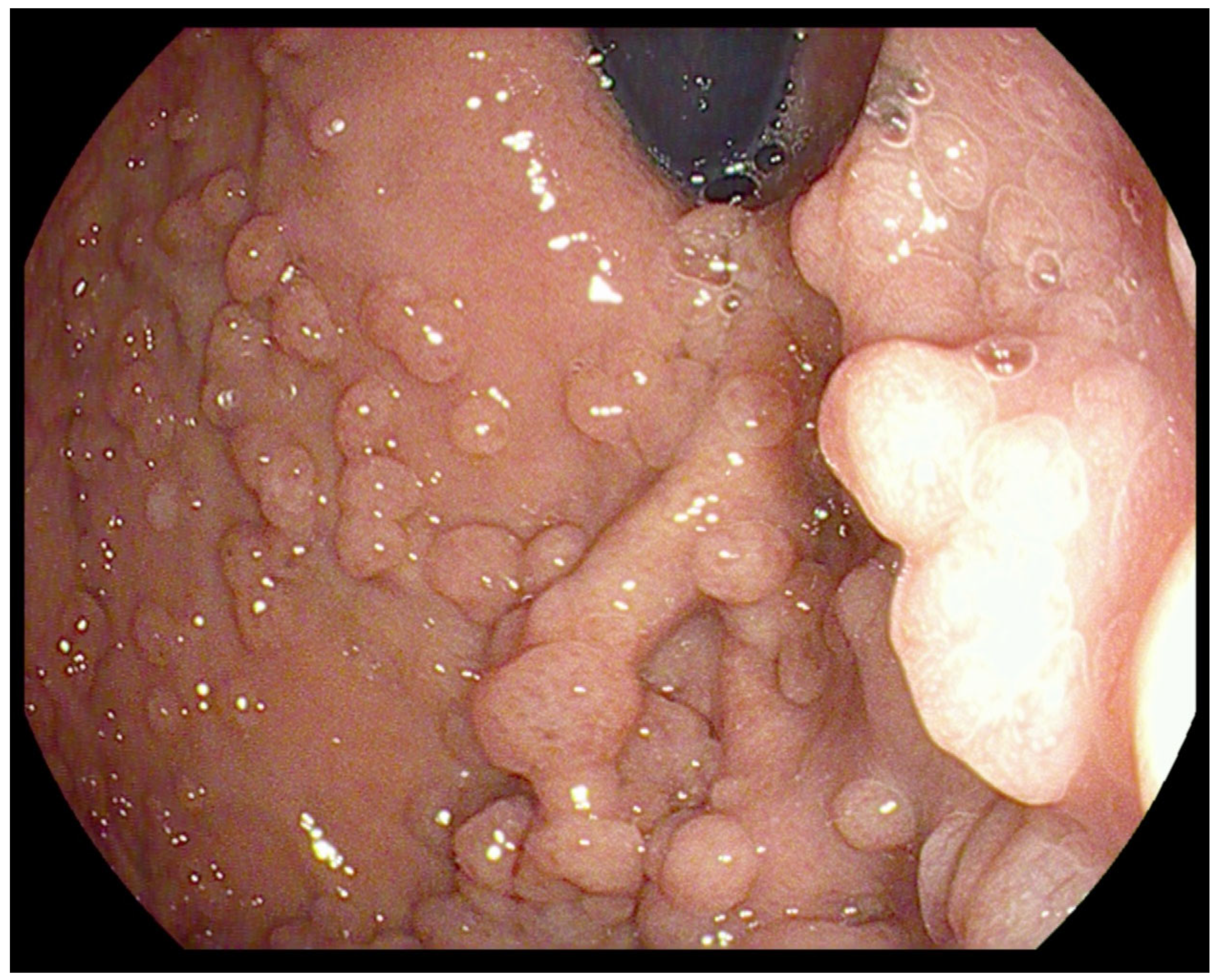
3. Stomach Lesions
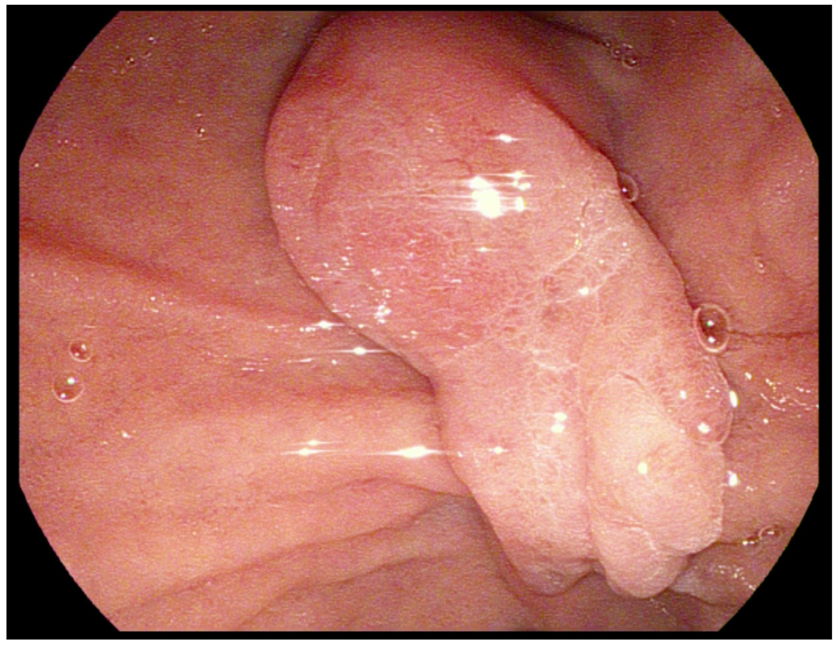
4. Duodenal Lesions
5. Endoscopic Treatment
6. Conclusions
- Early diagnosis and genetic identification of mutation carriers in FAP families with appropriate endoscopic surveillance have decreased the incidence of malignancy-related deaths during the last decades.
- More care should be taken to describe not only duodenal but also gastric findings in EGD in patients with FAP, with careful evaluation of polyps, particularly in the setting of gastric polyposis and large gastric mounds.
- There should be a heightened awareness of the risk of sessile gastric polyps and gastric cancer in patients with FAP.
- Previous surveillance recommendations might not be completely effective. They still require more data and, as a consequence, need improvement.
- Technological improvements (HR endoscopy, NBI) delivered very important tools to obtain a diagnosis and make treatment decisions in the precancerous stomach and duodenal lesions in FAP patients.
- Recently, the endoscopic treatment methods for duodenal lesions have been significantly improved. One of the most important is the cold-snare technique, which is now highly recommended in the resection of lesions <6 mm, but we already have some strong evidence for its effectiveness and safety in piece-meal resection of larger benign duodenal adenomas.
Author Contributions
Funding
Institutional Review Board Statement
Informed Consent Statement
Data Availability Statement
Conflicts of Interest
References
- Bisgaard, M.L.; Fenger, K.; Bülow, S.; Niebuhr, E.; Mohr, J. Familial adenomatous polyposis (FAP): Frequency, penetrance, and mutation rate. Hum. Mutat. 1994, 3, 121–125. [Google Scholar] [CrossRef] [PubMed]
- Mankaney, G.; Leone, P.; Cruise, M.; La Guardia, L.; O’Malley, M.; Bhatt, A.; Burke, C.A. Gastric cancer in FAP: A concerning rise in incidence. Fam. Cancer 2017, 16, 371–376. [Google Scholar] [CrossRef] [PubMed]
- Shibata, C.; Ogawa, H.; Miura, K.; Naitoh, T.; Yamauchi, J.I.; Unno, M. Clinical characteristics of gastric cancer in patients with familial adenomatous polyposis. Tohoku J. Exp. Med. 2013, 229, 143–146. [Google Scholar] [CrossRef] [PubMed] [Green Version]
- Park, S.Y.; Ryu, J.K.; Park, J.H.; Yoon, H.; Kim, J.Y.; Yoon, Y.B.; Park, J.G.; Lee, S.H.; Kang, S.B.; Park, J.W.; et al. Prevalence of gastric and duodenal polyps and risk factors for duodenal neoplasm in Korean patients with familial adenomatous polyposis. Gut Liver 2011, 5, 46–51. [Google Scholar] [CrossRef] [PubMed] [Green Version]
- Cannon, A.R.; Keener, M.; Neklason, D.; Pickron, T.B. Surgical interventions, malignancies, and couses of death in FAP patient registry. J. Gastrointest. Surg. 2021, 25, 452–456. [Google Scholar] [CrossRef]
- Hyer, W.; Cohen, S.; Attard, T.; Vila-Miravet, V.; Pienar, C.; Auth, M.; Septer, S.; Hawkins, J.; Durno, C.; Latchford, A. Management of Familial Adenomatous Polyposis in Children and Adolescents: Position Paper From the ESPGHAN Polyposis Working Group. J. Pediatr. Gastroenterol. Nutr. 2019, 68, 428–441. [Google Scholar] [CrossRef] [Green Version]
- Yang, J.; Gurudu, S.R.; Koptiuch, C.; Agrawal, D.; Buxbaum, J.L.; Abbas Fehmi, S.M.; Fishman, D.S.; Khashab, M.A.; Jamil, L.H.; Jue, T.L.; et al. American Society for Gastrointestinal Endoscopy guideline on the role of endoscopy in familial adenomatous polyposis syndromes. Gastrointest. Endosc. 2020, 91, 963–982.e2. [Google Scholar] [CrossRef]
- Amnon Sonnenberg, A.; Genta, R.M. Prevalence of benign gastric polyps in a large pathology database. Dig. Liver Dis. 2015, 47, 164–169. [Google Scholar] [CrossRef]
- Abraham, S.C.; Nobukawa, B.; Giardiello, F.M.; Hamilton, S.R.; Wu, T.T. Fundic gland polyps in familial adenomatous polyposis: Neoplasms with frequent somatic adenomatous polyposis coli gene alterations. Am. J. Pathol. 2000, 157, 747–754. [Google Scholar] [CrossRef] [Green Version]
- Bianchi, L.K.; Burke, C.A.; Bernnett, A.E.; Lopez, R.; Hasson, H.; Church, J.M. Fundic gland polyp dysplasia is common in familial adenomatous polyposis. Clin. Gastroenterol. Hepatol. 2008, 6, 180–185. [Google Scholar] [CrossRef]
- Wood, L.D.; Salaria, S.N.; Cruise, M.W. Upper GI tract Lesions in Familial Adenomatous Polyposis (FAP): Enrichment of Pyloric Gland Adenomas and Other Gastric and Duodental Neoplasms. Am. J. Surg. Pathol. 2014, 38, 389–393. [Google Scholar] [CrossRef] [PubMed] [Green Version]
- Campos, F.G.; Martinez, C.A.R.; Sulbaran, M.; Bustamante-Lopez, L.A.; Safatle-Ribeiro, A.V. Upper gastrointestinal neoplasia in familial adenomatous polyposis: Prevalence, endoscopic features and management. J. Gastrointest. Oncol. 2019, 10, 734–744. [Google Scholar] [CrossRef]
- Lami, G.; Galli, A.; Macri, G.; Dabizzi, E.; Biagini, M.R.; Tarocchi, M.; Messerini, L.; Valanzano, R.; Milani, S.; Polvani, S. Gastric and duodenal polyps in familial adenomatous polyposis patients: Conventional endoscopy vs virtual chromoendoscopy (fujinon intelligent color enhancement) in dysplasia evaluation. World J. Clin. Oncol. 2017, 8, 96–177. [Google Scholar] [CrossRef]
- Abraham, S.C.; Montgomery, E.A.; Singh, V.K.; Yardley, J.H.; Wu, T.T. Gastric adenomas: Intestinal-type and gastric-type adenomas differ in the risk of adenocarcinoma and presence of background mucosal pathology. Am. J. Surg. Pathol. 2002, 26, 1276–1285. [Google Scholar] [CrossRef]
- Khaykis, I.; Pachter, L.; Friedlander, C. Pyloric Gland Adenoma: Uncommon or Unrecognized? Am. J. Gastroenterol. 2018, 113, S1471–S1472. [Google Scholar] [CrossRef]
- Hashimoto, T.; Ogawa, R.; Matsubara, A.; Taniguchi, H.; Sugano, K.; Ushiama, M.; Yoshida, T.; Kanai, Y.; Sekine, S. Familial adenomatous polyposis-associated and sporadic pyloric gland adenomas of the upper gastrointestinal tract share common genetic features. Histopathology 2015, 67, 689–698. [Google Scholar] [CrossRef]
- Banks, M.; Graham, D.; Jansen, M.; Gotoda, T.; Coda, S.; di Pietro, M.; Uedo, N.; Bhandari, P.; Pritchard, D.M.; Kuipers, E.J.; et al. British Society og Gastroenterology guidelines on the diagnosis and management of patients at risk of gastric adenocarcinoma. Gut 2019, 68, 1545–1575. [Google Scholar] [CrossRef] [Green Version]
- Evans, J.A.; Chandrasekhara, V.; Chathadi, K.V.; Decker, G.A.; Early, D.S.; Fisher, D.A.; Foley, K.; Hwang, J.H.; Jue, T.L.; Lightdale, J.R.; et al. The role of endoscopy in the management of premalignant and malignant conditions of the stomach. Gastrointest. Endosc. 2015, 82, 1–8. [Google Scholar] [CrossRef]
- Mankaney, G.N.; Cruise, M.; Sarvepalli, S. Identifying factors associated with detection of sessile gastric polyps in patients with familial adenomatous polyposis. Endosc. Int. Open 2022, 10, E1080–E1087. [Google Scholar] [CrossRef]
- Mankaney, G.N.; Burke, C.A.; Cruise, M.; Church, J.; Wadhwa, V.; Chahal, P.; Vargo, J.; Bhatt, A. Endoscopic ultrasound imaging detection of gastric cancer in familial adenomatous polyposis. Gastroenterology 2017, 153, 353–354. [Google Scholar] [CrossRef]
- Soons, E.; Bisseling, T.; van Kouwen, M.; Möslein, G.; Siersema, P. Endoscopic management of duodenal adenomatosis in familial adenomatous polyposis—A case-based review. United Eur. Gastroenterol. J. 2021, 9, 461–468. [Google Scholar] [CrossRef] [PubMed]
- Groves, C.J.; Saunders, B.P.; Spigelman, A.D.; Phillips, R.K.S. Duodenal cancer in patients with familial adenomatous polyposis (FAP): Results of a 10 year prospective study. Gut 2002, 50, 636–641. [Google Scholar] [CrossRef] [PubMed]
- Bulow, S.; Bjork, J.; Christensen, I.J.; Fausa, O.; Järvinen, H.; Moesgaard, F.; Vasen, H.F.; DAF Study Group. Duodenal adenomatosis in familial adenomatous polyposis. Gut 2004, 53, 381–386. [Google Scholar] [CrossRef] [Green Version]
- Ma, M.X.; Bourke, M.J. Management of duodenal polyps. Best Pract. Res. Clin. Gastroenterol. 2017, 31, 389–399. [Google Scholar] [CrossRef]
- Vanbiervliet, G.; Strijker, M.; Arvanitakis, M.; Aelvoet, A.; Arnelo, U.; Beyna, T.; Busch, O.; Deprez, P.H.; Kunovsky, L.; Larghi, A.; et al. Endoscopic management of ampullary tumors: European Society of Gastrointestinal Endoscopy (ESGE) Guideline. Endoscopy 2021, 53, 429–448. [Google Scholar] [CrossRef]
- Syngal, S.; Brand, R.E.; Church, J.M.; Giardiello, F.M.; Hampel, H.L.; Burt, R.W. ACG clinical guideline: Genetic testing and management of hereditary gastrointestinal cancer syndromes. Am. J. Gastroenterol. 2015, 110, 223–262. [Google Scholar] [CrossRef] [Green Version]
- Spigelman, A.; Talbot, I.C.; Williams, C.B.; Domizio, P.; Phillips, R.K.S. Upper gastrointestinal cancer in patients with familial adenomatous polyposis. Lancet 1989, 334, 783–785. [Google Scholar] [CrossRef]
- Nakagawa, K.; Sho, M.; Fujishiro, M.; Kakushima, N.; Horimatsu, T.; Okada, K.I.; Iguchi, M.; Uraoka, T.; Kato, M.; Yamamoto, Y.; et al. Clinical practice guidelines for duodenal cancer 2021. J. Gastroenterol. 2022, 57, 927–941. [Google Scholar] [CrossRef]
- Sourrouille, I.; Lefèvre, J.H.; Shields, C.; Colas, C.; Bellanger, J.; Desaint, B.; Paye, F.; Tiret, E.; Parc, Y. Surveillance of duodenal polyposis in familial adenomatous polyposis: Should the Spigelman score Be modified? Dis. Colon. Rectum. 2017, 60, 1137–1146. [Google Scholar] [CrossRef]
- Mathus-Vliegen, E.M.; Boparai, K.S.; Dekker, E.; van Geloven, N. Progression of duodenal adenomatosis in familial adenomatous polyposis: Due to ageing of subjects and advances in technology. Fam. Cancer 2011, 10, 491–499. [Google Scholar] [CrossRef]
- Lopez-Ceron, M.; van den Broek, F.J.; Mathus-Vliegen, E.M.; Boparai, K.S.; van Eeden, S.; Fockens, P.; Dekker, E. The role of high-resolution endoscopy and narrow-band imaging in the evaluation of upper GI neoplasia in familial adenomatous polyposis. Gastrointest. Endosc. 2013, 77, 542–550. [Google Scholar] [CrossRef] [PubMed]
- Dutta, A.K.; Chacko, A. Emerging role of narrow band imaging in duodenum. World J. Gastrointest. Endosc. 2015, 7, 1216–1221. [Google Scholar] [CrossRef] [PubMed]
- Valitutti, F.; Oliva, S.; Iorfida, D.; Aloi, M.; Gatti, S.; Trovato, C.M.; Montuori, M.; Tiberti, A.; Cucchiara, S.; Di Nardo, G. Narrow band imaging combined with water immersion technique in the diagnosis of celiac disease. Dig. Liver Dis. 2014, 46, 1099–1102. [Google Scholar] [CrossRef] [PubMed]
- Silva, L.C.; Arruda, R.M.; Botelho, P.F.R.; Taveira, L.N.; Giardina, K.M.; de Oliveira, M.A.; Dias, J.; Oliveira, C.Z.; Fava, G.; Guimarães, D.P. Cap-assisted endoscopy increases ampulla of Vater visualization in high-risk patients. BMC Gastroenterol. 2020, 20, 214. [Google Scholar] [CrossRef] [PubMed]
- Hew, W.Y.; Joo, K.R.; Cha, J.M.; Shin, H.P.; Lee, J.I.; Park, J.J.; Lim, J.U. Feasibility of forward-viewing upper endoscopy for detection of the major duodenal papilla. Dig. Dis. Sci. 2011, 56, 2895–2899. [Google Scholar] [CrossRef]
- Kallenberg, F.G.J.; Bastiaansen, B.A.J.; Dekker, E. Cap-assisted forward-viewing endoscopy to visualize the ampulla of Vater and the duodenum in patients with familial adenomatous polyposis. Endoscopy 2017, 49, 181–185. [Google Scholar] [CrossRef] [Green Version]
- Shi, X.; Luo, H.; Ning, B.; Wang, X.; Tao, Q.; Liang, S.; Zhang, R.; Chen, J.; Luo, B.; Yao, S.; et al. Effect of cap-assisted esophagogastroduodenoscopy on examination of the major duodenal papilla: A noninferior, randomized controlled trial. Endoscopy 2019, 51, 427–435. [Google Scholar] [CrossRef]
- Hara, Y.; Goda, K.; Dobashi, A.; Ohya, T.R.; Kato, M.; Sumiyama, K.; Mitsuishi, T.; Hirooka, S.; Ikegami, M.; Tajiri, H. Short- and long-term outcomes of endoscopically treated superficial non-ampullary duodenal epithelial tumors. World J. Gastroenterol. 2019, 25, 707–718. [Google Scholar] [CrossRef]
- Choksi, N.; Elmunzer, B.J.; Stidham, R.W.; Shuster, D.; Piraka, C. Cold snare piecemeal resection of colonic and duodenal polyps ≥1 cm. Endosc. Int. Open 2015, 3, E508-13. [Google Scholar] [CrossRef] [Green Version]
- Roos, V.H.; Bastiaansen, B.A.; Kallenberg, F.G.J.; Aelvoet, A.S.; Bossuyt, P.M.M.; Fockens, P.; Dekker, E. Endoscopic management of duodenal adenomas in patients with familial adenomatous polyposis. Gastrointest. Endosc. 2021, 93, 457–466. [Google Scholar] [CrossRef]
- Yamasaki, Y.; Uedo, N.; Takeuchi, Y.; Ishihara, R.; Okada, H.; Iishi, H. Current Status of Endoscopic Resection for Superficial Nonampullary Duodenal Epithelial Tumors. Digestion 2018, 97, 45–51. [Google Scholar] [CrossRef] [Green Version]
- Dang, D.T.; Suresh, S.; Vance, R.B.; Singla, S.; Javia, S.; Watson, A.; Chathadi, K.V.; Katukuri, V.; Pompa, R.; Stidham, R.W.; et al. Outcomes of cold snare piecemeal endoscopic mucosal resection for nonampullary small-bowel adenomas larger than 1 centimeter: A retrospective study. Gastrointest. Endosc. 2022, 95, 1176–1182. [Google Scholar] [CrossRef]
- Trivedi, M.; Klapheke, R.; Youssef, F.; Wolfe, S.; Jih, L.; Chang, M.A.; Fehmi, S.A.; Krinsky, M.L.; Kwong, W.; Savides, T.; et al. Comparison of cold snare and hot snare polypectomy for the resection of sporadic nonampullary duodenal adenomas. Gastrointest. Endosc. 2022, 96, 657–664.e2. [Google Scholar] [CrossRef]
- Amoyel, M.; Belle, A.; Dhooge, M.; Ali, E.A.; Pellat, A.; Hallit, R.; Terris, B.; Prat, F.; Chaussade, S.; Coriat, R.; et al. Outcomes of endoscopic mucosal resection for large superficial non-ampullary duodenal adenomas. Sci. Rep. 2022, 12, 14592. [Google Scholar] [CrossRef]
- Binmoeller, K.F.; Shah, J.N.; Bhat, Y.M.; Kane, S.D. “Underwater” EMR of sporadic laterally spreading nonampullary duodenal adenomas (with video). Gastrointest. Endosc. 2013, 78, 496–502. [Google Scholar] [CrossRef]
- Okimoto, K.; Maruoka, D.; Matsumura, T.; Kanayama, K.; Akizue, N.; Ohta, Y.; Taida, T.; Saito, K.; Inaba, Y.; Kawasaki, Y.; et al. Utility of underwater EMR for nonpolypoid superficial nonampullary duodenal epithelial tumors ≤20 mm. Gastrointest. Endosc. 2022, 95, 140–148. [Google Scholar] [CrossRef]
- Flynn, M.M.; Wang, A.Y. Underwater endoscopic mucosal resection of large duodenal adenomas. Video J. Encyclop. GI Endosc. 2014, 3–4, 84–86. [Google Scholar] [CrossRef] [Green Version]
- Binmoeller, K.F. Underwater endoscopic mucosal resection. J. Interv. Gastroenterol. 2014, 4, 113–116. [Google Scholar] [CrossRef]
- Marques, J.; Baldaque-Silva, F.; Pereira, P.; Arnelo, U.; Yahagi, N.; Macedo, G. Endoscopic mucosal resection and endoscopic submucosal dissection in the treatment of sporadic nonampullary duodenal adenomatous polyps. World J. Gastrointest. Endosc. 2015, 7, 720–727. [Google Scholar] [CrossRef]
- Nonaka, S.; Oda, I.; Tada, K.; Mori, G.; Sato, Y.; Abe, S.; Suzuki, H.; Yoshinaga, S.; Nakajima, T.; Matsuda, T.; et al. Clinical outcome of endoscopic resection for nonampullary duodenal tumors. Endoscopy 2015, 47, 129–135. [Google Scholar] [CrossRef]
- Gaspar, J.P.; Stelow, E.B.; Wang, A.Y. Approach to the endoscopic resection of duodenal lesions. World J. Gastroenterol. 2016, 22, 600–617. [Google Scholar] [CrossRef] [PubMed]
- Wei, Y.; Zhou, Q.; Ji, M.; Zhang, S.; Li, P. Over-the-scope clip-assisted endoscopic full-thickness resection has potential to treat complex nonampullary duodenal lesions: A single-center case series. BMC Gastroenterol. 2021, 21, 476. [Google Scholar] [CrossRef] [PubMed]
- Ceppa, E.P.; Burbridge, R.A.; Rialon, K.L.; Omotosho, P.A.; Emick, D.; Jowell, P.S.; Branch, M.S.; Pappas, T.N. Endoscopic versus surgical ampullectomy: An algorithm to treat disease of the ampulla of Vater. Ann. Surg. 2013, 257, 315–322. [Google Scholar] [CrossRef] [PubMed]
- Nair, S.S.; Abdelrahim, M.; Varytimiadis, L.; Al-Kandari, A.; Goggin, P.; Bhandari, P. PTU-53 Efficacy and safety of endoscopic ampullectomy in the UK. Gut 2021, 70, A69. [Google Scholar]
- Campos, F.G. Surgical treatment of familial adenomatous polyposis: Dilemmas and current recommendations. World J. Gastroenterol. 2014, 20, 16620–16629. [Google Scholar] [CrossRef]
- Moussata, D.; Napoleon, B.; Lepilliez, V.; Klich, A.; Ecochard, R.; Lapalus, M.G.; Nancey, S.; Cenni, J.C.; Ponchon, T.; Chayvialle, J.A.; et al. Endoscopic treatment of severe duodenal polyposis as an alternative to surgery for patients with familial adenomatous polyposis. Gastrointest. Endosc. 2014, 80, 817–825. [Google Scholar] [CrossRef]
- Kemp Bohan, P.M.; Mankaney, G.; Vreeland, T.J.; Chick, R.C.; Hale, D.F.; Cindass, J.L.; Hickerson, A.T.; Ensley, D.C.; Sohn, V.; Clifton, G.T.; et al. Chemoprevention in familial adenomatous polyposis: Past, present and future. Fam. Cancer 2021, 20, 23–33. [Google Scholar] [CrossRef]
- Rad, E.; Murray, J.T.; Tee, A.R. Oncogenic Signalling through Mechanistic Target of Rapamycin (mTOR): A Driver of Metabolic Transformation and Cancer Progression. Cancers 2018, 10, 5. [Google Scholar] [CrossRef]


Publisher’s Note: MDPI stays neutral with regard to jurisdictional claims in published maps and institutional affiliations. |
© 2022 by the authors. Licensee MDPI, Basel, Switzerland. This article is an open access article distributed under the terms and conditions of the Creative Commons Attribution (CC BY) license (https://creativecommons.org/licenses/by/4.0/).
Share and Cite
Paszkowski, J.; Samborski, P.; Kucharski, M.; Cwaliński, J.; Banasiewicz, T.; Pławski, A. Endoscopic Surveillance and Treatment of Upper GI Tract Lesions in Patients with Familial Adenomatous Polyposis—A New Perspective on an Old Disease. Genes 2022, 13, 2329. https://doi.org/10.3390/genes13122329
Paszkowski J, Samborski P, Kucharski M, Cwaliński J, Banasiewicz T, Pławski A. Endoscopic Surveillance and Treatment of Upper GI Tract Lesions in Patients with Familial Adenomatous Polyposis—A New Perspective on an Old Disease. Genes. 2022; 13(12):2329. https://doi.org/10.3390/genes13122329
Chicago/Turabian StylePaszkowski, Jacek, Paweł Samborski, Marcin Kucharski, Jarosław Cwaliński, Tomasz Banasiewicz, and Andrzej Pławski. 2022. "Endoscopic Surveillance and Treatment of Upper GI Tract Lesions in Patients with Familial Adenomatous Polyposis—A New Perspective on an Old Disease" Genes 13, no. 12: 2329. https://doi.org/10.3390/genes13122329