Article

Changes in Expression of Specific mRNA Transcripts after Single- or Re-Irradiation in Mouse Testes

1
Department of Anatomy, Tokyo Medical University, Tokyo 160-8402, Japan
2
Department of Anatomy, Division of Basic Medical Science, Tokai University School of Medicine, Kanagawa 259-1193, Japan
3
Immuno Regulation, Institute of Medical Science, Tokyo Medical University, Tokyo 160-8402, Japan
*
Author to whom correspondence should be addressed.
Genes 2022, 13(1), 151; https://doi.org/10.3390/genes13010151
Submission received: 13 December 2021 / Revised: 4 January 2022 / Accepted: 13 January 2022 / Published: 15 January 2022
(This article belongs to the Special Issue Male Infertility: From Genes to Genomes 2022)

Abstract

:
Alkylating agents and irradiation induce testicular damage, which results in prolonged azoospermia. Even very low doses of radiation can significantly impair testis function. However, re-irradiation is an effective strategy for locally targeted treatments and the pain response and has seen important advances in the field of radiation oncology. At present, little is known about the relationship between the harmful effects and accumulated dose of irradiation derived from continuous low-dose radiation exposure. In this study, we examined the levels of mRNA transcripts encoding markers of 13 markers of germ cell differentiation and 28 Sertoli cell-specific products in single- and re-irradiated mice. Our results demonstrated that re-irradiation induced significantly decreased testicular weights with a significant decrease in germ cell differentiation mRNA species (Spo11, Tnp1, Gfra1, Oct4, Sycp3, Ddx4, Boll, Crem, Prm1, and Acrosin). In the 13 Sertoli cell-specific mRNA species decreased upon irradiation, six mRNA species (Claudin-11, Espn, Fshr, GATA1, Inhbb, and Wt1) showed significant differences between single- and re-irradiation. At the same time, different decreases in Sertoli cell-specific mRNA species were found in single-irradiation (Aqp8, Clu, Cst12, and Wnt5a) and re-irradiation (Tjp1, occludin, ZO-1, and ZO-2) mice. These results indicate that long-term aspermatogenesis may differ after single- and re-irradiated treatment.

1. Introduction

Radiotherapy and chemotherapy are commonly used as treatments for cancer. However, their use is associated with serious side effects, including temporary or permanent infertility. Recently, we demonstrated that traditional Japanese medicine, Goshajinkigan, can rescue mice aspermatogenesis after chemotherapy and radiotherapy [1,2]. In these two studies, we found that the busulfan- and irradiation-induced impaired reproductive functions were related to the different immune-pathophysiological conditions. In particular, Goshajinkigan was found to have different therapeutic effectiveness in response to this aspermatogenesis. Although supplementation with Goshajinkigan in irradiation-mice was found to rescue the disrupted inter-Sertoli tight junctions via the normalization of claudin11, occludin, and ZO-1 expression, as well as reduce serum anti-germ cell autoantibodies, the recovery of spermatogenesis (day 150) was more pronounced than that of Goshajinkigan supplementation in busulfan experiments (day 120) [1,2]. Sertoli cell tight junctions are a physical barrier in the blood-testis barrier (BTB) [3,4,5], resulting in the provision of an environment within the adluminal region that is both immune protective and of a controlled biochemical nature, which allows meiotic and post-meiotic germ cell maturation to proceed. The breakdown of BTB by irradiation can cause the germ cell autoantigens inside BTB to leak out repeatedly. The subsequent autoimmune reaction against germ cells after irradiation may affect the delayed recovery of spermatogenesis damage [6].
Total body irradiation, used as conditioning for bone marrow transplants, is associated with appreciable gonadal toxicity. Previous studies have shown that 99.5% of men who received 12 Gy total body irradiation experience permanent infertility [7], with irradiation doses as low as 5–6 Gy causing a decrease in spermatogenesis in the seminiferous tubules of mice [8,9]. With significant technological advances in the field of radiation oncology, integrated body imaging with accurate treatment delivery methods, such as stereotactic body radiotherapy, could improve the efficacy, shorten the overall treatment time, and potentially reduce treatment-related toxicities, indicating that re-irradiation is an effective treatment for local control and pain response [10]. However, little is known about the relationship between the harmful effects and accumulated dose derived from continuous low-dose radiation exposure. In the present study, we examined the impaired testicular functions induced by single- or re-irradiation in mice to address the immune-pathophysiological differences between these two treatment types.

2. Materials and Methods

2.1. Animals

C57BL/6J male mice at 4 weeks of age (weighing 16–20 g) were purchased from SLC (Shizuoka, Japan) and kept in the Laboratory Animal Center of Tokyo Medical University (Tokyo Medical University Animal Committee; no. H28016-01042017). Mice were maintained at a temperature of 22–24 °C and relative humidity of 50–60% with a 12-h light–dark cycle.

2.2. Experimental Design

In the present study, we examined the impaired testicular functions induced by low-dose radiation exposure in mice to address the immune-pathophysiological differences between single- or re-irradiation. Mice were irradiated with 4 or 6 Gy using a 60Co gamma ray unit (MBR-1520A-TWZ; HITACHI KE Systems Ltd, Tokyo, Japan). Radiation was administered to the body without anesthesia. Then, the study mice were randomly divided into the following two experimental groups: (1) group I: mice in the single-irradiated group (n = 20) were administered a single dose of 4 Gy (4 GyI; n = 10), or 6 Gy (6 GyI; n = 10); (2) group II: mice in the re-irradiated group (n = 20) were exposed to X-ray radiation with 1 Gy four times (4 GyII; n = 10) or six times (6 GyII; n = 10) with an interval of one day. The doses were monitored during each irradiation procedure. The general condition, food intake, and body weight were documented for all mice one week after the final irradiation. The protocol used in the present study was approved by the Institutional Animal Care and Use Committee at the Dongnam Institute of Radiological and Medical Sciences and the Tokyo Medical University Animal Committee (no. H28016). The animals were maintained in accordance with the Guidelines of the National Institutes of Health for Animal Experiments. Non-irradiated mice (ConI and ConII; n = 20) were housed on shelves in the same facility and shielded from radiation.
To examine spermatogenesis after irradiation, the mice from each group (n = 10) were anesthetized with pentobarbital (65 mg/kg body weight), after which the testes and epididymides were removed for gravimetry one week after the final irradiation exposure.

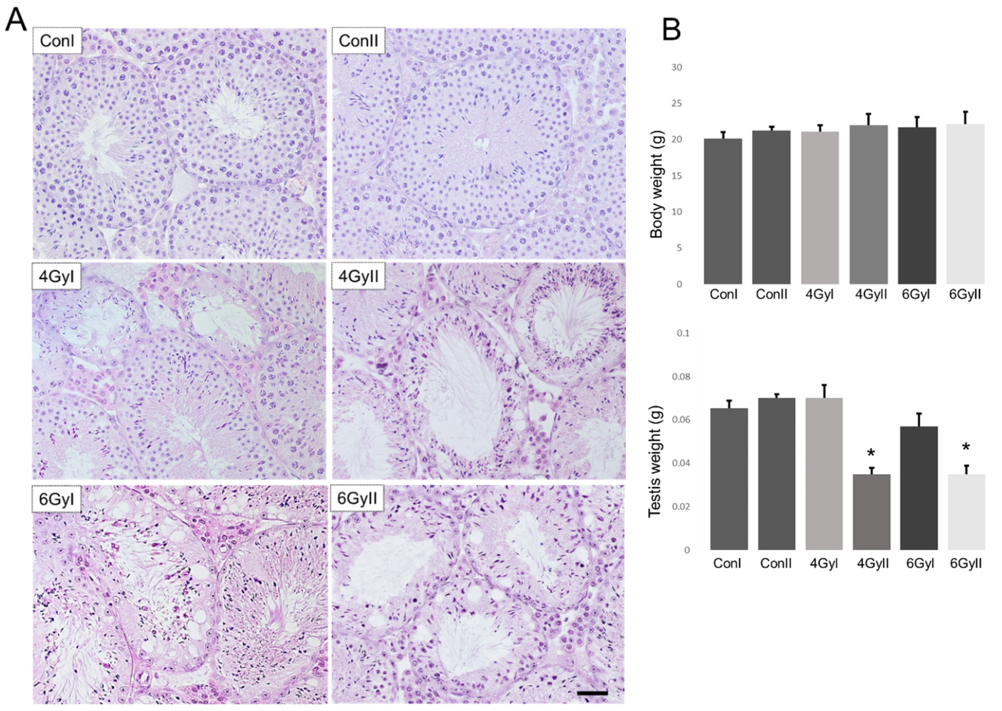
2.3. Histological Examination of the Testes

The testes of mice from each group (n = 5) were fixed with Bouin’s solution and embedded in plastic (Technovit7100; Kulzer & Co., Wehrheim, Germany). Sections (5 μm) were obtained at 15–20-μm intervals and were stained with Gill’s hematoxylin and 2% eosin Y (Muto PC, Tokyo, Japan) for observation under a light microscope.

2.4. Detection of Serum Anti-Germ Cell Antibodies

To prepare the antigens, testes from normal mice (8-week-old; n = 5) were homogenized in carbonate–bicarbonate buffer (Sigma-Aldrich, MO, USA) and germ cell concentrations were adjusted to 50 µg/200 µL. The antigens were added to 96-well microtiter plates (Nunc 96-microwell plate; Thermo Fisher Scientific, Kanagawa, Japan) and incubated at 37 °C for 30 min. After removing the coating solution, the wells were washed in triplicate with PBS. Then, 200 µL of sera (1:160 dilution) from each group of mice (n = 10) was added to micro-ELISA wells in duplicate and incubated for 2 h at room temperature. Next, the wells were washed three times with PBS–Tween-20 (0.05% v/v) and incubated for 2 h at room temperature with 200 µL of HRP-conjugated goat anti-mouse IgM (1:10,000) (Cappel). One o-phenylenediamine dihydrochloride (OPD) tablet dissolved in 20 ml of water with one phosphate–citrate tablet (Sigma-Aldrich) was used as a soluble substrate to detect peroxidase activity. The wells were washed five times with PBS–Tween-20, and 200 µL of freshly prepared OPD solution was added to each well. After 30 minutes, the sample absorbance at 450 nm was measured using MPR-A4 (Tokyo, Japan) [11].
For the detection of serum anti-germ cell antibodies, normal testes from normal 8-week-old mice were placed in OCT compound (Miles Laboratories, IL, USA), frozen in liquid nitrogen, and stored at −80 °C until further use. Sections (6 µm) were cut using a cryostat (CM1900; Leica, Wetzlar, Germany), dried in the air, fixed in 95% ethanol for 10 min at −20 °C, rinsed in PBS, and then incubated with 50-fold serial dilutions of the collected serum samples from experimental mice of each group (n = 10) for 60 min at RT. After rinsing in PBS, the cryostat sections were incubated for 60 min with HRP-conjugated goat anti-mouse IgM (1:500 dilution) (ZyMax, CA, USA) at room temperature. After washing with PBS, the HRP-binding sites were detected with 0.05% DAB and 0.01% H2O2.

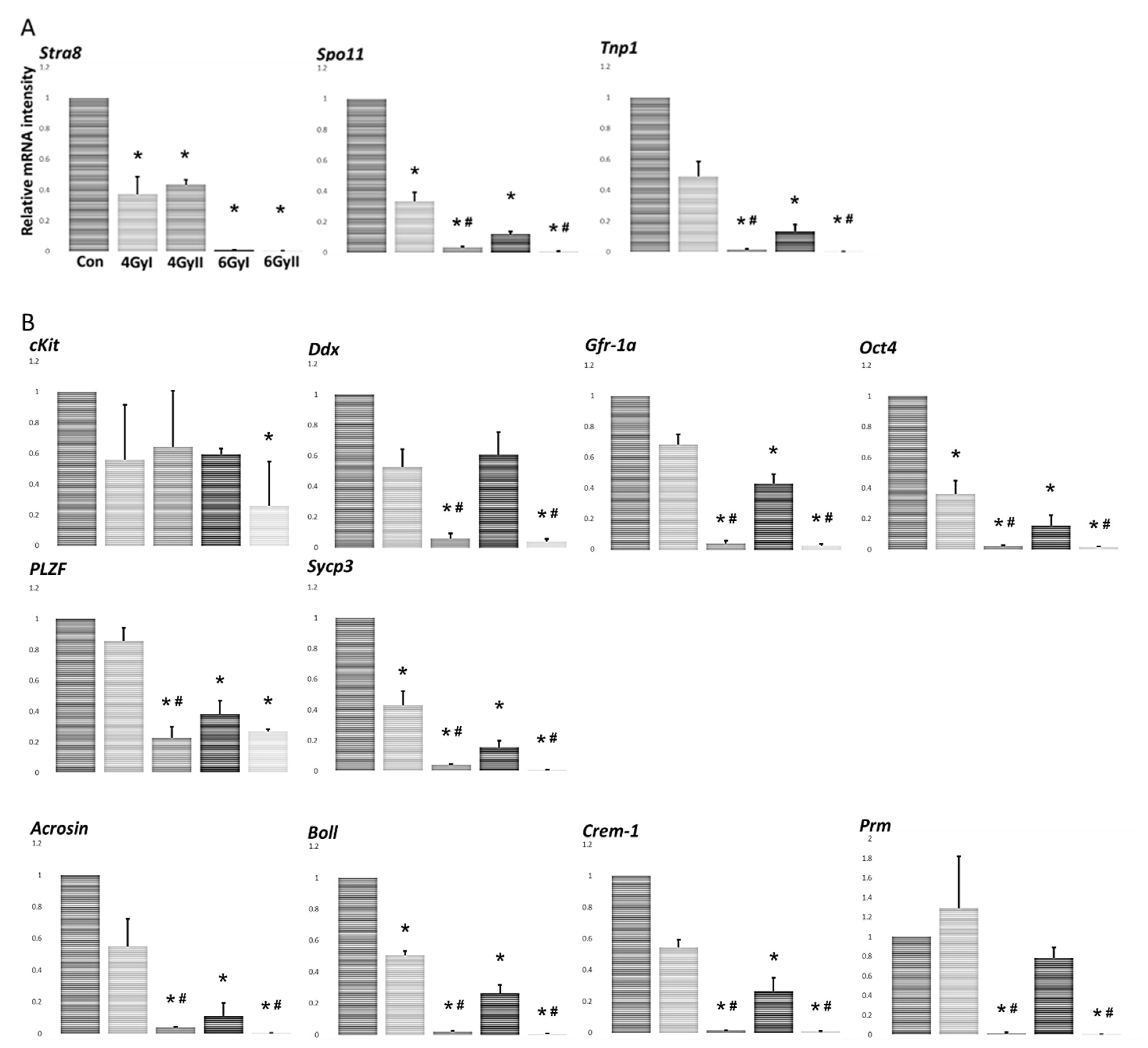
2.5. Analysis of the Specific mRNA Species in Testes Using Real-Time RT-PCR

Testes from mice in each group (n = 5) were examined. The iCycler thermal cycler (Bio-Rad, Hercules, CA, USA) was used to perform the PCR reactions, and the mixtures were stored at −80 °C until analysis. Real-time RT-PCR was performed on 3 ng cDNA using the validated SYBR Green gene expression assay in combination with SYBR Premix Ex TaqTM (TaKaRa, Bio Inc., Ohtsu, Japan) for measuring 13 germ cell genes (Stra8, Spo11, Tnp1, cKit, Gfra1, Oct4, PLZF, Sycp3, Ddx4, Boll, Crem, Prm1, and Acrosin), 28 Sertoli cell genes (Amh, Aqp8, Ccnd2, Clu, Claudin-11, Cst12, Cst9, Dhh, Espn, Fshr, Fyn, GATA1, Inhba, Inhbb, Msi1, Occlidin, Rhox5, Testin, Spata2, shbg,Sox9, sympk, Tjp1, Trf, Wt1, Wnt5a, ZO-1, and ZO-2) and GAPDH. Quantitative real-time PCR was performed in duplicate using the Thermal Cycler Dice Real-time System TP800 (TaKaRa). The Thermal Cycler Dice Real-time System software Ver.5.1.1 (TaKaRa) was used to analyze the data, and the comparative Ct method (2-∆∆Ct) was used to quantify gene expression levels. The results are expressed relative to the amount of GAPDH transcript used as an internal control. Relative mRNA intensity was calculated, and the expression in the control group for each point was normalized to 1. Data are presented as the mean ± standard deviation (SD). The primers used in the analysis are listed in Table 1.

2.6. Statistical Analysis

ANOVA and Tukey-Kramer post-hoc tests were used to analyze the differences between multiple groups. Statistical significance was set at p < 0.05.

3. Results

3.1. Histopathological Changes after Irradiation Exposure

To examine histopathological changes after irradiation, we documented the testes’ weight and observed the testes sections of mice from each group under a light microscope. Non-irradiated mice showed spermatogenesis maturity (Figure 1A) and increased body and testis weights (Figure 1B) from the ages of five weeks (ConI) to six weeks (ConII). On the other hand, atrophic seminiferous tubules with disrupted spermatogenesis were observed in the testes of all irradiated mice, with significantly decreased testicular weights in the mice of group II after re-irradiation with 4 or 6 Gy. In contrast, no significant differences in the body weight were observed between group I by single-irradiated (4 and 6 GyI), group II by re-irradiation (4 and 6 GyII), and the non-irradiated groups (ConI and ConII) (Figure 1B). These data showed re-irradiation significantly decreased testicular weight with atrophic seminiferous tubules, as well as disrupting spermatogenesis in the testes.

3.2. Effect of Irradiation Exposure on Levels of mRNA Transcripts Encoding Markers of Germ Cell Differentiation Products

For the detection of the effect of single- and re-irradiation on testicular spermatogenesis, we compared the expression levels of mRNA species encoding the spermatogonial marker Stra8, the spermatocyte marker of Spo11, the spermatid marker Tnp1 (Figure 2A), premeiotic cells (cKit, Gfra1, Oct4, PLZF, Sycp3, and Ddx4), and meiotic and postmeiotic cells (Boll, Crem, Prm1, and Acrosin) (Figure 2B) by real-time RT-PCR analysis in each group [12,13,14,15,16,17,18,19,20]. The levels of almost all examined spermatogenesis markers, excluding cKit, were significantly reduced in group II, compared to five spermatogenesis markers (Stra8, Spo11, Oct4, Sycp3, and Boll) that were significantly reduced in group I (Figure 2A,B). Furthermore, the spermatocyte marker of Spo11, the spermatid marker Tnp1, and eight differentiation markers (Gfra1, Oct4, Sycp3, Ddx4, Boll, Crem, Prm1, and Acrosin) were significantly reduced in group II compared to those in group I. These data showed the spermatocyte marker, the spermatid marker, and some mRNA species of germ cell differentiation markers were significantly affected by re-irradiation.

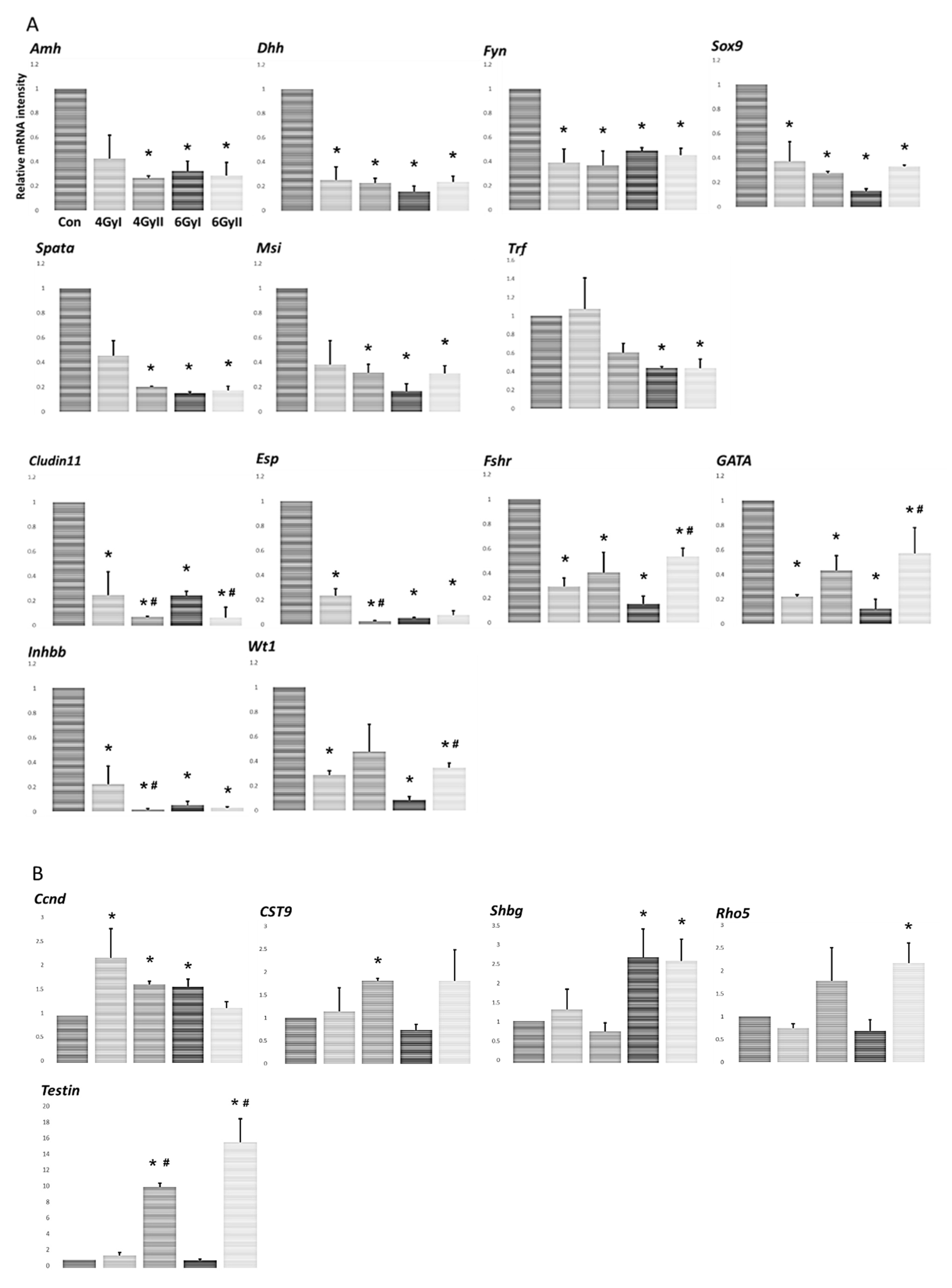
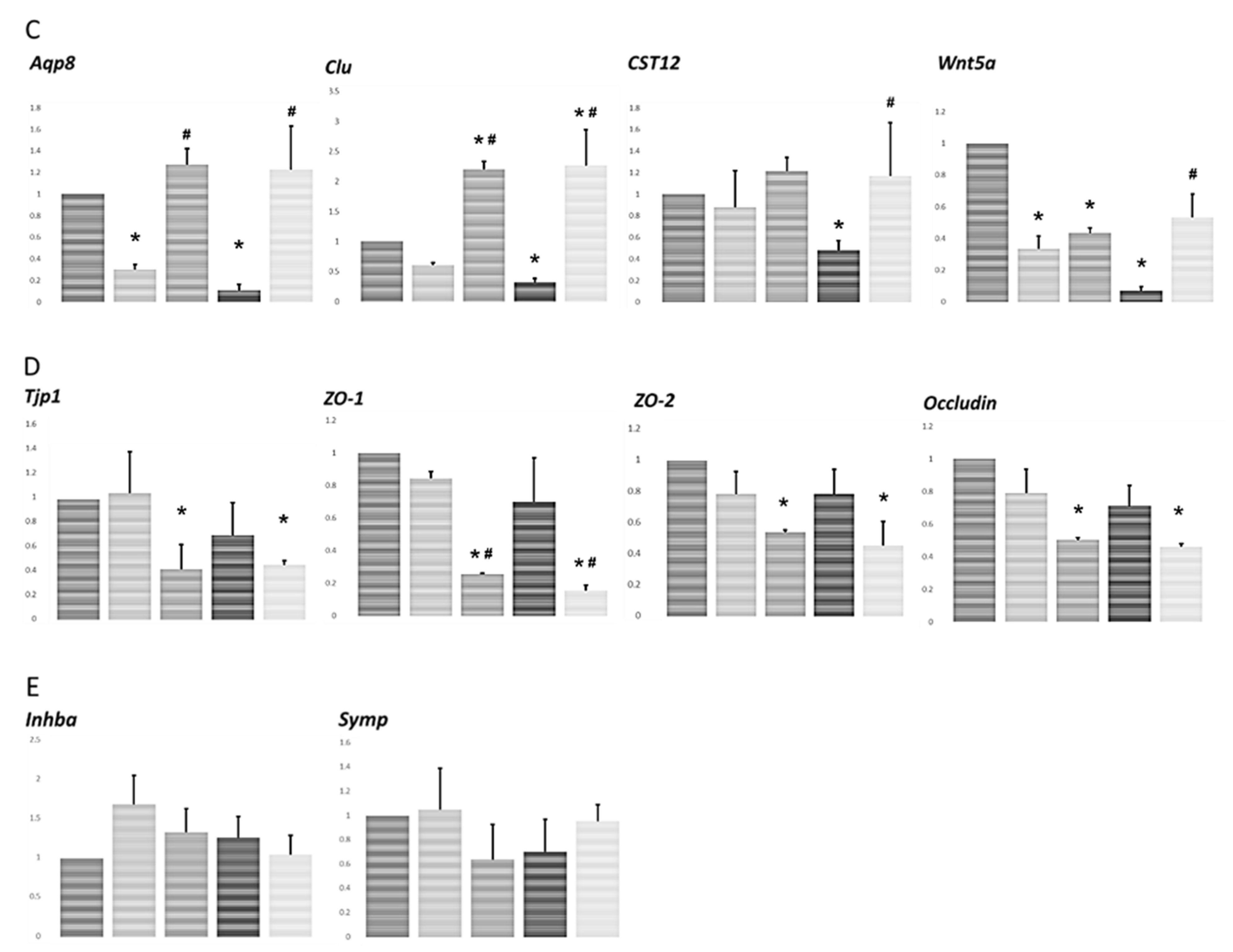
3.3. Effect of Irradiation on Levels of mRNA Transcripts Encoding Markers of Sertoli Cell-Specific Products

To examine the Sertoli cell function regulated by irradiation-induced germ cell depletion, the levels of 28 Sertoli cell-specific mRNA species were measured in each group (Figure 3). Thirteen mRNA species (Amh, Claudin-11, Dhh, Espn, Fshr, Fyn, GATA1, Inhbb, Msi1, Spata2, Sox9, Trf, and Wt1) showed significantly decreased expression (Figure 3A) in response to irradiation (group I and II) and five mRNA species (Ccnd2, Cst9, Rhox5, Shbg, and Testin) showed increased expression in response to irradiation (group I and II) (Figure 2B). Furthermore, four mRNA species (Aqp8, Clu, Cst12, and Wnt5a) showed a significant decrease in levels only in group I (single irradiation) but not in group II (re-irradiation) (Figure 3C). In contrast, the other four mRNA species (Tjp1, occlidin, ZO-1, and ZO-2) showed a significant decrease in levels only in group II after re-irradiation but not in group I after single irradiation (Figure 3D). No significant changes in the expression levels of the remaining two mRNA species (Inhba and symp) were observed after irradiation treatment (group I and II) (Figure 3E). The above data showed that single- and re-irradiation treatment induced different increased or decreased in Sertoli cell-specific mRNA species, and six mRNA species (Claudin-11, Espn, Fshr, GATA1, Inhbb, and Wt1) showed significant differences between single- and re-irradiation in all 13 decreased Sertoli cell-specific mRNA species expression after irradiation.

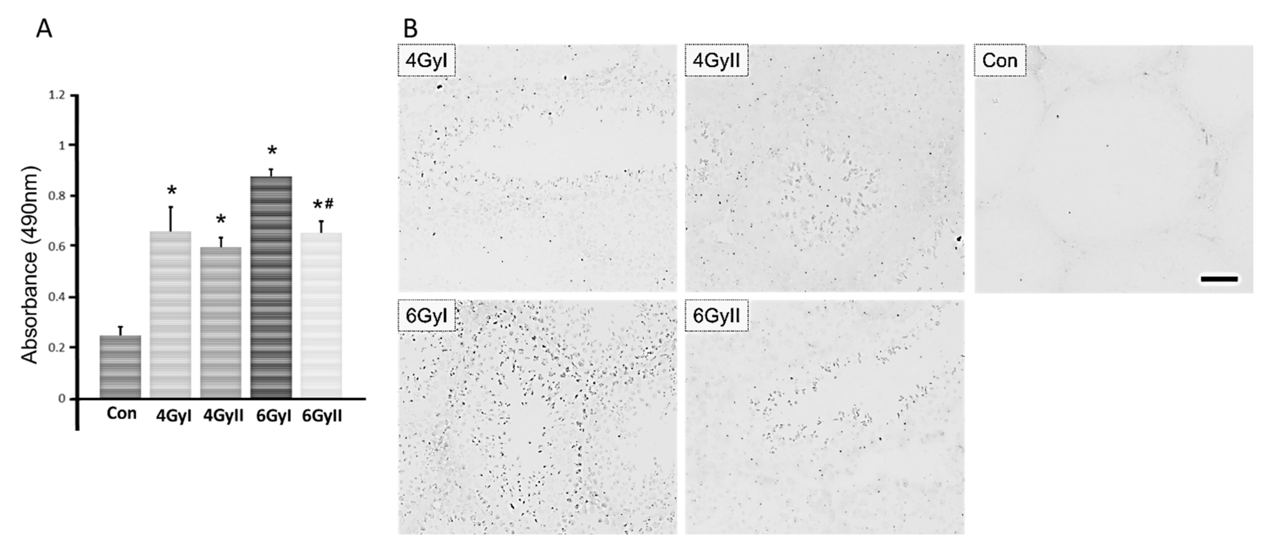
3.4. Detection of Serum Anti-Germ Cell Antibodies

For the detection of serum anti-germ cell antibodies in mice from each group, we measured the level of autoantibodies by ELISA and immunohistochemistry analysis. As shown in Figure 4A, the serum IgM levels in mice after irradiation treatment were significantly higher than those in control mice. Additionally, a significant difference was observed in the mice in group 6 GyI and 6 GyII. The serum IgM levels in the mice in group 6 GyII were significantly lower than those in group 6 GyI.
Upon assessing the reaction of sera with normal frozen seminiferous tubule section, anti-germ cell antibodies were detected in all irradiation groups (group I and II), but not in the normal group (Figure 4B). Serum autoantibodies preferentially reacted with mature spermatids and spermatozoa; concurrently, faint immunostaining was detected in spermatogonia and immature spermatids.

4. Discussion

In this study, we examined 13 markers of germ cell differentiation and 28 Sertoli cell-specific products in single- or re-irradiated mice to clarify the pathophysiological differences between single- and re-irradiation. We found that re-irradiation significantly decreased testicular weight with atrophic seminiferous tubules, as well as disrupting spermatogenesis in the testes (Figure 1). Furthermore, the spermatocyte marker of Spo11, the spermatid marker Tnp1, and eight mRNA species of differentiation markers (Gfra1, Oct4, Sycp3, Ddx4, Boll, Crem, Prm1, and Acrosin) were significantly reduced in re-irradiated mice compared to single-irradiated mice (Figure 2A,B). At the same time, irradiation treatment induced a decrease in Sertoli cell-specific mRNA species after single irradiation (Aqp8, Clu, Cst12, and Wnt5a) (Figure 3C) and after re-irradiation (Tjp1, occlidin, ZO-1, and ZO-2) (Figure 3D). In all 13 Sertoli cell-specific mRNA species in which expression was decreased, six mRNA species (Claudin-11, Espn, Fshr, GATA1, Inhbb, and Wt1) showed significant differences between single- and re-irradiation. These decreased Sertoli cell-specific mRNA species were accompanied by increased serum anti-germ cell antibody levels. In all five Sertoli cell-specific mRNA species in which expression was increased, only Testin showed a significant difference between single- and re-irradiation.
Infertility and gonadal dysfunction caused by cancer treatments have become a significant concern for cancer survivors, with substantial improvements in anticancer therapy. It is well known that spermatogenesis is a complex and tightly regulated process that leads to the continuous production of male gametes, i.e., spermatozoa. The depletion of spermatogonial stem cells is the apparent cause of infertility in oncological patients after chemo- and radiotherapy [1,21,22,23], and may play a leading role in the oligo- and azoospermia of infertility patients [24,25]. Meiotic and post-meiotic germ cells (spermatocytes and especially spermatids) are much less sensitive to cytotoxic treatment and radiation than spermatogonia [26]. Although radiotherapy-induced aspermatogenesis has been extensively studied, no study has yet addressed the differences between single- and re-irradiation-induced male infertility. In this study, we found that some atrophic seminiferous tubules with disrupted spermatogenesis were observed in the testes of all irradiated mice (Figure 1). In particular, we noted that the testicular weights, the spermatocyte marker of Spo11, the spermatid marker Tnp1, the pre-meiotic markers (Gfra1, Oct4, Sycp3, and Ddx4), and all the meiotic and post-meiotic markers (Boll, Crem, Prm1, and Acrosin) were significantly reduced in re-irradiated mice compared to single-irradiated mice (Figure 1 and Figure 2). These results demonstrate that re-irradiation damaged not only spermatogonia but also meiotic and post-meiotic germ cells (spermatocytes and spermatids). In particular, all the meiotic and post-meiotic markers examined in this study were significantly reduced by re-irradiation, indicating that the long-term aspermatogenesis in patients with longer survival may differ between single- and re-irradiated treatments.
In this study, we further examined 28 Sertoli cell-specific marker products to clarify the different effects of single- and re-irradiation-induced infertility. It is well known that Sertoli cells are the somatic cells of the testes, supporting the architectural stability of germ cells and conserving the microenvironment and BTB [27,28,29,30]. The testis is a radiosensitive organ, and significant impairment of testis function could be caused by very low dose radiation [31]. In particular, with the breakdown of BTB by irradiation, germ cell autoantigens inside BTB may leak out repeatedly, leading to a continuous supply of autoantigens for immune stimulation with resultant anti-sperm antibody production and the prolongation of testicular inflammation [6]. In this study, 13 mRNA species (Amh, Claudin-11, Dhh, Espn, Fshr, Fyn, GATA1, Inhbb, Msi1, Spata2, Sox9, Trf, and Wt1) showed significantly decreased expression (Figure 3A) in response to single- and re-irradiation, accompanied by significantly higher serum IgM levels in mice after single- and re-irradiation. Furthermore, among the above decreased thirteen mRNA species, six mRNA species (Claudin-11, Espn, Fshr, GATA1, Inhbb, and Wt1) showed significant differences between single- and re-irradiation treatments. Compared to group I, only Claudin-11 expression was significantly decreased in 4 GyII and 6 GyII mice and significantly decreased expression of Espn and Inhbb was only found in 4 GyII mice. On the other hand, the expression of Fshr, GATA1, and Wt1 was significantly increased in 6 GyII mice compared to 6 GyI mice. Furthermore, four Sertoli cell-specific mRNA species (Tjp1, occludin, ZO-1, and ZO-2) were detected only in re-irradiated mice but not in single-irradiated mice (Figure 3C,D). Various integral tight junction proteins have been described in Sertoli cell junctions, including members of the claudin family (claudin-1 [32], claudin-3 [33,34,35], claudin-5 [36], claudin-12 [37], claudin-11 [38,39], claudin-13 [35], and occludin [40,41]). These proteins are linked to the actin cytoskeleton via cytoplasmic plaque proteins, including zonula occludens-1, -2, and -3 (ZO-1, ZO-2, and ZO-3) and provide links to the gap- and adherens-junctional types in the BTB. In these junction proteins, only claudin-11 is essential for spermatogenesis, as the claudin-11 knockout is infertile [38,39]. In this study, we demonstrated that only Claudin-11 expression was significantly decreased in all re-irradiated mice compared to that in single-irradiation mice. This indicates that it may be an important factor associated with testicular aspermatogenesis induced by irradiation. Concurrently, the transformation variability of these Sertoli cell-specific markers after single- or re-irradiation may be the reason for the significantly higher serum IgM levels in 6 GyI mice.
In particular, two decreased Sertoli cell-specific mRNA species (Aqp8 and Clu) after single irradiation were significantly increased in re-irradiated mice (Figure 3C). Furthermore, in all five increased expressions of Sertoli cell-specific mRNA species, only Testin showed a significant increase in re-irradiated mice compared to that in single-irradiation mice (Figure 3B). It has been demonstrated that Testin localizes on the Sertoli cell surface that contacts germ cells, and the depletion of germ cells leads to an increase in Testin mRNA level [42]. Some studies have demonstrated that the AQP8 transcript is expressed in Sertoli cells and occurs in all stages of spermatogenesis [43,44,45]. Similar reports have demonstrated that clusterin (Clu) is secreted by Sertoli cells and deposited onto the membranes of elongating spermatids and mature spermatozoa [46,47]. Recently, we demonstrated that Testin and Clu mRNA species increased with germ cell depletion by busulfan treatment and restored with completely recovered spermatogenesis [48]. In the present study, we further demonstrated that AQP8, Testin, and Clu mRNA species were significantly increased following germ cell depletion by re-irradiation treatment. Some studies have suggested that Sertoli cells play a key role in mediating testicular immunology in irradiation-induced aspermatogenesis, while our study showed the altered Sertoli cell function after irra-treatment with intact spermatogenesis. These findings suggest that these Sertoli cell factors are more intimately interrelated with spermatogenesis after radiotherapy, providing a basis for the estimation of male fertility.
This is the first report that compared the germ cell- and Sertoli cell-specific mRNA species after single- or re-irradiation treatment in mice. Although the aspermatogenesis after radiotherapy have been extensively studied, there is little information regarding the noxious role of single- and re-irradiation-induced male infertility. Knowledge of this impaired testicular immunopathologic microenvironment will be useful to understand infertility as adverse effects of radiotherapy. The above limited experimental data would address further information about this important clinical problem.

Author Contributions

Experiments were designed by N.Q., K.S. (Kou Sakabe), T.Y. and M.I.; K.N., N.Q. and M.K. performed the experiments. Histological and histopathological analyses of the testes and epididymis were performed by M.K., K.N., Y.O. and N.Q. Statistical analyses were performed by S.H., K.N. and N.Q. drafted the original manuscript and discussed the manuscript with M.I. Secretarial and technical assistance were performed by K.S. (Kaori Suyama) and D.K. All authors have read and agreed to the published version of the manuscript.

Funding

This work was supported by a Grant-in-Aid for General Science Research (C: 19K07876) from the Ministry of Education, Science, Sports, and Culture in Japan.

Institutional Review Board Statement

The Institutional Animal Care and Use Committee at Dongnam Institute of Radiological and Medical Sciences approved the protocol used in the present study, and the animals were maintained in accordance with the Guidelines of the National Institutes of Health for Animal Experiments. The experimental study protocol was approved by the Tokyo Medical University Animal Committee (no. H28016).

Conflicts of Interest

The authors declare no conflict of interest.

References

  1. Qu, N.; Kuramasu, M.; Hirayanagi, Y.; Nagahori, K.; Hayashi, S.; Ogawa, Y.; Terayama, H.; Suyama, K.; Naito, M.; Sakabe, K.; et al. Gosha-Jinki-Gan recovers spermatogenesis in mice with busulfan-induced aspermatogenesis. Int. J. Mol. Sci. 2018, 19, 2606. [Google Scholar] [CrossRef] [PubMed] [Green Version]
  2. Takahashi, K.; Nagahori, K.; Qu, N.; Kuramasu, M.; Hirayanagi, Y.; Hayashi, S.; Ogawa, Y.; Hatayama, N.; Terayama, H.; Suyama, K.; et al. The traditional Japanese medicine Goshajinkigan makes a recovery from irradiation-induced aspermatogenesis in mice. BMC Complement. Altern. Med. 2019, 19, 362. [Google Scholar] [CrossRef]
  3. Kaur, G.; Thompson, L.A.; Dufour, J.M. Sertoli cells–immunological sentinels of spermatogenesis. Semin. Cell Dev. Biol. 2014, 30, 36–44. [Google Scholar] [CrossRef] [Green Version]
  4. Mital, P.; Hinton, B.T.; Dufour, J.M. The blood-testis and blood-epididymis barriers are more than just their tight junctions. Biol. Reprod. 2011, 84, 851–858. [Google Scholar] [CrossRef] [PubMed] [Green Version]
  5. Plöen, L.; Setchell, B.P. Blood-testis barriers revisited. A homage to Lennart Nicander. Int. J. Androl. 1992, 15, 1–4. [Google Scholar] [CrossRef] [PubMed]
  6. Qu, N.; Itoh, M.; Sakabe, K. Effects of chemotherapy and radiotherapy on spermatogenesis: The role of testicular immunology (Review). Int. J. Mol. Sci. 2019, 20, 957. [Google Scholar] [CrossRef] [Green Version]
  7. Sanders, J.E.; Hawley, J.; Levy, W.; Gooley, T.; Buckner, C.D.; Deeg, H.J.; Doney, K.; Storb, R.; Sullivan, K.; Witherspoon, R.; et al. Pregnancies following high-dose cyclophosphamide with or without high-dose busulfan or total-body irradiation and bone marrow transplantation. Blood 1996, 87, 3045–3052. [Google Scholar] [CrossRef]
  8. Vergouwen, R.P.F.A.; Huiskamp, R.; Bas, R.J.; Roepers-Gajadien, H.L.; de Jong, F.H.; van Eerdenburg, F.J.C.M.; Davids, J.A.G.; de Rooij, D.G. Radiosensitivity of testicular cells in the prepubertal mouse. Radiat. Res. 1994, 139, 316–326. [Google Scholar] [CrossRef]
  9. Khan, S.; Adhikari, J.S.; Alam Rizvi, M.; Chaudhury, N.K. Radioprotective potential of melatonin against ⁶⁰Co γ-ray-induced testicular injury in male C57BL/6 mice. J. Biomed. Sci. 2015, 22, 61. [Google Scholar] [CrossRef] [Green Version]
  10. Gabryś, D. Re-irradiation of vertebral bodies. Phys. Medica. 2019, 64, 311–316. [Google Scholar] [CrossRef] [Green Version]
  11. Hirai, S.; Naito, M.; Terayama, H.; Hatayama, N.; Qu, N.; Musha, M.; Itoh, M. Serum autoantibodies in mice immunized with syngeneic testiscular germs cell alone. Am. J. Reprod. Immunol. 2013, 70, 509–517. [Google Scholar] [CrossRef]
  12. Yelick, P.C.; Kwon, Y.H.; Flynn, J.F.; Borzorgzadeh, A.; Kleene, K.C.; Hecht, N.B. Mouse transition protein 1 is translationally regulated during the postmeiotic stages of spermatogenesis. Mol. Reprod. Dev. 1989, 1, 193–200. [Google Scholar] [CrossRef]
  13. Oulad-Abdelghani, M.; Bouillet, P.; Decimo, D.; Gansmuller, A.; Heyberger, S.; Dolle, P.; Bronner, S.; Lutz, Y.; Chambon, P. Characterization of a premeiotic germ cell-specific cytoplasmic protein encoded by Stra8, a novel retinoic acid-responsive gene. J. Cell. Biol. 1996, 135, 469–477. [Google Scholar] [CrossRef]
  14. Shannon, M.; Richardson, L.; Christian, A.; Handel, M.A.; Thelen, M.P. Differential gene expression of mammalian SPO11/TOP6A homologs during meiosis. FEBS Lett. 1999, 462, 329–334. [Google Scholar] [CrossRef] [Green Version]
  15. O’Shaughnessy, P.J.; Hu, L.; Baker, P.J. Effect of germ cell depletion on levels of specific mRNA transcripts in mouse Sertoli cells and Leydig cells. Reproduction 2008, 135, 839–850. [Google Scholar] [CrossRef] [Green Version]
  16. De França, L.R.; Bartke, A.; Borg, K.E.; Oecim, M.; Fadden, C.T.; Yagi, A.; Russell, L.D. Sertoli cells in testes containing or lacking germ cells: A comparative study of paracrine effects using the W (c-kit) gene mutant mouse model. Anat. Rec. Adv. Integr. Anat. Evol. Biol. 1994, 240, 225–232. [Google Scholar] [CrossRef] [PubMed]
  17. Yeh, J.R.; Zhang, X.; Nagano, M.C. Wnt5a Is a Cell-Extrinsic Factor That Supports Self-Renewal of Mouse Spermatogonial Stem Cells. J. Cell Sci. 2011, 124, 2357–2366. [Google Scholar] [CrossRef] [PubMed] [Green Version]
  18. Teixeira, J.; Maheswaran, S.; Donahoe, P.K. Mullerian inhibiting substance: An instructive developmental hormone with diagnostic and possible therapeutic applications. Endocr. Rev. 2001, 22, 657–674. [Google Scholar] [CrossRef]
  19. Pérez, C.V.; Sobarzo, C.M.; Jacobo, P.V.; Pellizzari, E.H.; Cigorraga, S.B.; Denduchis, B.; Lustig, L. Loss of Occludin Expression and Impairment of Blood-Testis Barrier Permeability in Rats with Autoimmune Orchitis: Effect of Interleukin 6 on Sertoli Cell Tight Junctions. Biol. Reprod. 2012, 87, 122. [Google Scholar] [CrossRef] [PubMed]
  20. Cimino, I.; Casoni, F.; Liu, X.; Messina, A.; Parkash, J.; Jamin, S.P.; Catteau-Jonard, S.; Collier, F.; Baroncini, M.; Dewailly, D.; et al. Novel role for anti-mullerian hormone in the regulation of GnRH neuron excitability and hormone secretion. Nat. Commun. 2016, 7, 10055. [Google Scholar] [CrossRef] [Green Version]
  21. Schlatt, S.; von Schönfeldt, V.; Schepers, A.G. Male germ cell transplantation: An experimental approach with a clinical perspective. Br. Med. Bull. 2000, 56, 824–836. [Google Scholar] [CrossRef] [Green Version]
  22. Radford, J. Restoration of fertility after treatment for cancer. Horm. Res. 2003, 59, 21–23. [Google Scholar] [CrossRef]
  23. Wallace, W.H.; Thompson, A.B. Preservation of fertility in children treated for cancer. Arch. Dis. Child. 2003, 88, 493–496. [Google Scholar] [CrossRef]
  24. Takagi, S.; Itoh, N.; Kimura, M.; Sasao, T.; Tsukamoto, T. Spermatogonial proliferation and apoptosis in hypospermatogenesis associated with nonobstructive azoospermia. Fertil. Steril. 2001, 76, 901–907. [Google Scholar] [CrossRef]
  25. Yakirevich, E.; Sabo, E.; Dirnfeld, M.; Sova, Y.; Spagnoli, G.C.; Resnick, M.B. Morphometrical quantification of spermatogonial germ cells with the 57B anti-MAGE-A4 antibody in the evaluation of testicular biopsies for azoospermia. Appl. Immunohistochem. Mol. Morphol. 2003, 11, 37–44. [Google Scholar] [CrossRef] [PubMed]
  26. Meistrich, M.L.; Hunter, N.R.; Suzuki, N.; Trostle, P.K.; Withers, H.R. Gradual regeneration of mouse testicular stem cells after exposure to ionizing radiation. Radiat. Res. 1978, 74, 349–362. [Google Scholar] [CrossRef] [PubMed]
  27. Wang, C.Q.; Cheng, C.Y. A seamless trespass: Germ cell migration across the seminiferous epithelium during spermatogenesis. J. Cell Biol. 2007, 178, 549–556. [Google Scholar] [CrossRef] [Green Version]
  28. Meinhardt, A.; Hedger, M.P. Immunological, paracrine and endocrine aspects of testicular immune privilege. Mol. Cell Endocrinol. 2011, 335, 60–68. [Google Scholar] [CrossRef] [PubMed]
  29. Cheng, C.Y.; Mruk, D.D. The blood-testis barrier and its implications for male contraception. Pharmacol. Rev. 2012, 64, 16–64. [Google Scholar] [CrossRef] [PubMed] [Green Version]
  30. Wang, X.-W.; Ding, G.-R.; Shi, C.-H.; Zeng, L.-H.; Liu, J.-Y.; Li, J.; Zhao, T.; Chen, Y.-B.; Guo, G.-Z. Mechanisms involved in the blood-testis barrier increased permeability induced by EMP. Toxicology 2010, 276, 58–63. [Google Scholar] [CrossRef] [PubMed]
  31. Howell, S.J.; Shalet, S.M. Spermatogenesis after cancer treatment: Damage and recovery. J. Natl. Cancer. Inst. Monogr. 2005, 34, 12–17. [Google Scholar] [CrossRef] [PubMed] [Green Version]
  32. Dietze, R.; Shihan, M.; Stammler, A.; Konrad, L.; Scheiner-Bobis, G. Cardiotonic steroid ouabain stimulates expression of blood-testis barrier proteins claudin-1 and -11 and formation of tight junctions in Sertoli cells. Mol Cell Endocrinol. 2015, 405, 1–13. [Google Scholar] [CrossRef]
  33. Komljenovic, D.; Sandhoff, R.; Teigler, A.; Heid, H.; Just, W.W.; Gorgas, K. Disruption of blood-testis barrier dynamics in ether-lipid-deficient mice. Cell Tissue Res. 2009, 337, 281–299. [Google Scholar] [CrossRef] [PubMed]
  34. Smith, B.E.; Braun, R.E. Germ cell migration across Sertoli cell tight junctions. Science 2012, 338, 798–802. [Google Scholar] [CrossRef] [Green Version]
  35. Chakraborty, P.; William Buaas, F.; Sharma, M.; Smith, B.E.; Greenlee, A.R.; Eacker, S.M.; Braun, R.E. Androgen-dependent sertoli cell tight junction remodeling is mediated by multiple tight junction components. Mol. Endocrinol. 2014, 28, 1055–1072. [Google Scholar] [CrossRef] [Green Version]
  36. Morrow, C.M.; Tyagi, G.; Simon, L.; Carnes, K.; Murphy, K.M.; Cooke, P.S.; Hofmann, M.-C.C.; Hess, R.A. Claudin 5 expression in mouse seminiferous epithelium is dependent upon the transcription factor ets variant 5 and contributes to blood-testis barrier function. Biol. Reprod. 2009, 81, 871–879. [Google Scholar] [CrossRef] [Green Version]
  37. Haverfield, J.T.; Meachem, S.J.; Nicholls, P.K.; Rainczuk, K.E.; Simpson, E.R.; Stanton, P.G. Differential permeability of the blood-testis barrier during reinitiation of spermatogenesis in adult male rats. Endocrinology 2014, 155, 1131–1144. [Google Scholar] [CrossRef]
  38. Gow, A.; Southwood, C.M.; Li, J.S.; Pariali, M.; Riordan, G.P.; Brodie, S.E.; Danias, J.; Bronstein, J.M.; Kachar, B.; Lazzarini, R. CNS myelin and sertoli cell tight junction strands are absent in Osp/claudin-11 null mice. Cell 1999, 99, 649–659. [Google Scholar] [CrossRef] [Green Version]
  39. Mazaud-Guittot, S.; Meugnier, E.; Pesenti, S.; Wu, X.; Vidal, H.; Gow, A.; Le Magueresse-Battistoni, B. Claudin 11 deficiency in mice results in loss of the sertoli cell epithelial phenotype in the testis. Biol. Reprod. 2009, 109, 202–213. [Google Scholar] [CrossRef] [Green Version]
  40. Moroi, S.; Saitou, M.; Fujimoto, K.; Sakakibara, A.; Furuse, M.; Ypshida, O.; Tsukita, S. Occludin is concentrated at tight junctions of mouse/rat but not human/guinea pig Sertoli cells in testes. Am. J. Physiol. 1998, 274, C1708–C1717. [Google Scholar] [CrossRef] [PubMed] [Green Version]
  41. Saitou, M.; Ando-Akatsuka, Y.; Itoh, M.; Furuse, M.; Inazawa, J.; Fujimoto, K.; Tsukita, S. Mammalian occludin in epithelial cells: Its expression and subcellular distribution. Eur. J. Cell Biol. 1997, 73, 222–231. [Google Scholar]
  42. Grima, J.; Zhu, L.-J.; Cheng, C.Y. Testin is tightly associated with testicular cell membrane upon its secretion by Sertoli cells whose steady-state mRNA level in the testis correlates with the turnover and integrity of inter-testicular cell junctions. J. Biol. Chem. 1997, 272, 6499–6509. [Google Scholar] [CrossRef] [Green Version]
  43. Calamita, G.; Mazzone, A.; Bizzoca, A.; Svelto, M. Possible involvement of aquaporin-7 and -8 in rat testis development and spermatogenesis. Biochem. Biophys. Res. Commun. 2001, 288, 619–625. [Google Scholar] [CrossRef]
  44. Kageyama, Y.; Ishibashi, K.; Hayashi, T.; Xia, G.; Sasaki, S.; Kihara, K. Expression of aquaporins 7 and 8 in the developing rat testis. Andrologia 2001, 33, 165–169. [Google Scholar] [CrossRef] [PubMed]
  45. Klein, C.; Troedsson, M.H.T.; Rutllant, J. Region-Specific Expression of Aquaporin Subtypes in Equine Testis, Epididymis, and Ductus Deferens. Anat. Evol. Biol. 2013, 296, 1115–1126. [Google Scholar] [CrossRef] [PubMed] [Green Version]
  46. Clark, A.M.; Griswold, M.D. Expression of clusterin/sulfated glycoprotein-2 under conditions of heat stress in rat Sertoli cells and a mouse Sertoli cell line. J. Androl. 1997, 18, 257–263. [Google Scholar] [PubMed]
  47. Sylvester, S.R.; Morales, C.; Oko, R.; Griswold, M.D. Localization of sulfated glycoprotein-2 (clusterin) on spermatozoa and in the reproductive tract of the male rat. Biol. Reprod. 1991, 45, 195–207. [Google Scholar] [CrossRef] [Green Version]
  48. Qu, N.; Nagahori, K.; Kuramasu, M.; Ogawa, Y.; Suyama, K.; Hayashi, S.; Sakabe, K.; Itoh, M. Effect of Gosha-Jinki-Gan on levels of specific mRNA transcripts in mouse testes after busulfan treatment. Biomedicines 2020, 8, 432. [Google Scholar] [CrossRef]
Figure 1. Testicular histology, testis weight, and body weight in each group (n = 5). (A) Testes sections show morphology in groups I and II. Intact seminiferous tubules showing normal germinal epithelium from the spermatogonia to spermatozoa are observed in ConI and ConII. Damaged seminiferous tubules with azoospermia are observed in the other groups of 4GyI, 4GyII, 6GyI, and 6GyII. The presence of both atrophic and intact seminiferous tubules with spermatogenesis was observed in the group IV mice. Normal-appearing seminiferous tubules are observed in the group V mice. Scale bar = 50 µm. (B) The body weight and testis weight in each group. * p < 0.05 vs. control group.
Genes 13 00151 g001
Figure 2. The levels of mRNA transcripts encoding markers of germ cell differentiation in each group (n = 5). Expression was measured by real-time RT-PCR, and the results were expressed relative to the internal control GAPDH. (A) Data show expression of the spermatogonial marker Stra8, the spermatocyte marker Spo11, and the spermatid marker Tnp1 expressed as the mean ± SD of five mice in each group. The y-axis shows the relative mRNA intensity. (B) The expression of the premeiotic markers (cKit, Gfra1, Oct4, PLZF, Sycp3, and Ddx4) and the meiotic and postmeiotic markers (Boll, Crem, Prm1, and Acrosin). The results are expressed as the mean ± SD of five mice in each group, and the y-axis shows the relative mRNA intensity. * p < 0.05 vs. control group; # p < 0.05, vs. group I.
Genes 13 00151 g002
Figure 3. The levels of 27 mRNA transcripts encoding markers of Sertoli cell-specific products in each group. Expression was measured by real-time RT PCR, and the results are expressed relative to the internal control GAPDH, expressed as the mean ± SD of five mice in each group. The y-axis shows relative mRNA intensity. (A) Transcripts showing a significant decrease in levels after irradiation treatment compared to control values. (B) Transcripts showing a significant increase in levels after irradiation treatment compared to control values. (C) Transcripts showing a significant decrease in levels only in group I but not group II compared to control values. (D) Transcripts showing a significant decrease in levels only in group II but not group I compared to control values. (E) Transcripts showing no change in levels after irradiation treatment compared to control values. * p < 0.05 vs. control group; # p < 0.05 vs. group I.
Genes 13 00151 g003aGenes 13 00151 g003b
Figure 4. Detection of serum anti-germ cell antibody levels in each group. (A) ELISA measurements of autoantibodies in each group reactive with germ cells antigens. The results are expressed as the mean ± SD of five mice in each group. * p < 0.05 vs. control group; # p < 0.05 vs. group I. (B) Normal frozen sections of seminiferous tubules were reacted with diluted sera obtained from each group mice followed by incubation with horseradish peroxidase (HRP)-conjugated anti-mouse IgM. Scale bar, 50 mm.
Genes 13 00151 g004
Table 1. Primers used for real-time RT-PCR.
GeneForward PrimerReverse Primer
GAPDHTGTGTCCGTCGTGGATCTGATTGCTGTTGAAGTCGCAGGAG
Sertoli cell
Amh TCCTACATCTGGCTGAAGTGATATGGG AGGTTCTGTGTGCCCCGCAG
Aqp8GCTGGCAGTCACAGTGATCGGACCTGGACGATGGCAAAGGCTG
Ccnd2GGAACCTGGCCGCAGTCACCAATCATCGACGGCGGGTACATG
Claudin-11TCACAACGTCCACCAATGACTGGGCACATACAGGAAACCAGATG
CluCCACGCCATGAAGATTCTCCTGCCTCCCTGGACGGCGTTCTGA
Cst12GGATGACGATTTTGCCTACAAGTTCCTTTCTCTCTCCTGGACCTTCCTGCA
Cst9 GATATTTGCCCCTTTCAGGAGAGCCAGAGAAGTACGTGACCAGTCCATGGG
DhhGGCGCAGACCGCCTGATGAAGGCACGGCCTTCGTAGTGG
EspnGCTTCTGGTCGGGCATTACCCTGTGTCATGCCGTCTTGGGCG
FshrGGCCAGGTCAACATACCGCTTGTGCCTTGAAATAGACTTGTTGCAAATTG
FynGAAGCGGCCCTGTATGGAAGGTTTGTGGGCAGGGCATCCTATAGC
GATA1ATGGTCAGAACCGGCCTCTCATCGAGCTTGAAATAGAGGCCGCAGG
InhbaCATGGAGCAGACCTCGGAGATCATGGTCCTGGTTCTGTTAGCCTTGG
InhbbGAGCGCGTCTCCGAGATCATCACGTACCTTCCTCCTGCTGCCCTT
Msi1TCACTTTCATGGACCAGGCGGGTTCACAGACAGCCCCCCCA
OcclidinCTTCTGCTTCATCGCTTCCCTTGCCCTTTCCTGCTTTC
Rhox5AGGTTCGCCCAGCATCGACTGGCCGCAGCCCTCCTGATCTT
ShbgGACATTCCCCAGCCTCATGCATGCCTCGGAAGACAGAACCACG
Sox9CGCGGAGCTCAGCAAGACTCTGTGTCCGTTCTTCACCGACTTCCTC
Spata2GCCGTGTGGGCCTGTGCTTTTCCCCAAATCAAACCCAAGGG
sympkCAAGAAGAAGGGCCAAGCATCGAAGGAAGTTGTCAAGCAGGGTGGG
TestinAAAGACAATGGCGGCCTCGCGGCCCCACTTTAGCCACTGCC
Tjp1GCGGAGAGAGACAAGATGTCCGCCTCTGAAAATGAGGAT-
TATCTCTTCCACCA
TrfCAAATGCATCAGCTTCCGTGACCCGGCATCGTACACCCAACCC
Wnt5aCTGCTTCTACCATGCGTTTGCTGGGCCATGGGACAGTGCGGC
Wt1GCTCCAGCTCAGTGAAATGGACAGAAGGCCACTCCAGATACACGCCG
ZO-1ACAAACAGCCCTACCAACCCCATCCTCATCTTCATCTTCTTC
ZO-2GTTTTTCTTCGTCCTAGTCCCCATCCATCCCTTCCATCTTTC
Germ Cell
AcrosinTGTCCGTGGTTGCCAAGGATAACAAATCCGGGTACCTGCTTGTGAGTT
BouleAACCCAACAAGTGGCCCAAGATACCTTTGGACACTCCAGCTCTGTCAT
c-KitGCATCACCATCAAAAACGTGGATAGTCAGCGTCTCCTGGC
CREM-1TTCTTTCACGAAGACCCTCATGTTAGGTGGTGTCCCTTCT
Ddx4AAGCAGAGGGTTTTCCAAGCGCCTGATGCTTCTGAATCG
Gfr-α1CAGTTTTCGTCTGCTGAGGTTGTCTGCTCAAAGTGGCTCCAT
OCT4TGCGGAGGGATGGCATACTGGCACAGGGCTCAGAGGAGGTTC
ProtamineTCCATCAAAACTCCTGCGTGAAGGTGGCATTGTTCCTTAGCA
Spo11CGCGTGGCCTCTAGTTCTGAGGGGTATCATCCGAAGGCCGACAGAAT
Stra8GAAGGTGCATGGTTCACCGTGGGCTCGATCGGCGGGCCTGTG
Sycp3TGGAGCTGACATCAACAAAGCCCCACTGCTGCAACACATTC
Tnp1GGCGATGATGCAAGTCGCAACCACTCTGATAGGATCTTTGGCTTTTGG
Zbtb16AACGGTTCCTGGACAGTTTGCCACACAGCAGACAGAAGA
Publisher’s Note: MDPI stays neutral with regard to jurisdictional claims in published maps and institutional affiliations.

