Review

Bardet–Biedl Syndrome—Multiple Kaleidoscope Images: Insight into Mechanisms of Genotype–Phenotype Correlations

1
Department of Nephrology-Internal Medicine, Faculty of Medicine, “Grigore T. Popa” University of Medicine and Pharmacy, 16 University Street, 700115 Iasi, Romania
2
Department of Medical Genetics, Faculty of Medicine, “Grigore T. Popa” University of Medicine and Pharmacy, 16 University Street, 700115 Iasi, Romania
*
Author to whom correspondence should be addressed.
Genes 2021, 12(9), 1353; https://doi.org/10.3390/genes12091353
Submission received: 30 July 2021 / Revised: 22 August 2021 / Accepted: 27 August 2021 / Published: 29 August 2021
(This article belongs to the Collection Genotype-Phenotype Study in Disease)

Abstract

:
Bardet–Biedl Syndrome is a rare non-motile primary ciliopathy with multisystem involvement and autosomal recessive inheritance. The clinical picture is extremely polymorphic. The main clinical features are retinal cone-rod dystrophy, central obesity, postaxial polydactyly, cognitive impairment, hypogonadism and genitourinary abnormalities, and kidney disease. It is caused by various types of mutations, mainly in genes encoding BBSome proteins, chaperonins, and IFT complex. Variable expressivity and pleiotropy are correlated with the existence of multiple genes and variants modifiers. This review is focused on the phenomena of heterogeneity (locus, allelic, mutational, and clinical) in Bardet–Biedl Syndrome, its mechanisms, and importance in early diagnosis and proper management.

1. Introduction

Ciliopathies are diseases caused by the dysfunction of motile and non-motile primary cilium [1]. Primary cilia are involved in numerous cellular processes. Such as cell cycle control, development, migration, polarity, differentiation, stimuli transduction, proliferation, and maintenance of stem cells [2,3,4]. Primary cilia also intervenes in the cellular signaling pathways of development and homeostasis: hedgehog, Wnt (wingless-related integration site), Notch, Hippo, GPCR (G protein-coupled receptors), PDGF (platelet-derived growth factor), mTOR (mammalian target of rapamycin), and TGF-βeta (βeta transforming growth factor-β) [5,6].
Cilia are microtubule-based organelles. They are anchored to the cytoskeleton and protrude at the cell surface [7]. They have three main components: axoneme, basal body and transition zone. The axoneme consists of nine peripheral microtubule doublets, and a central part that could be formed by a pair of microtubules (model 9 + 2) or by the absence of this pair of microtubules (model 9 + 0) [8]. The transport in and out of cilia is allowed by three complexes: intra-flagella transport (IFT) complex A (IFT-A), IFT complex B (IFT-B) and BBSome [4]. IFT-A is involved in the retrograde transport (tip to the base), IFT-B is involved in the anterograde transport (base to the tip) while BBSome is a multiprotein complex implied in ciliary trafficking activity [9].
Ciliopathies are characterized by a high clinical and molecular heterogeneity and a large clinical overlap between entities [10]. The clinical expression of the cilia dysfunction is correlated with the activity of cilia. Motile cilia dysfunction causes hydrocephalus, infertility, chronic respiratory issues, but also congenital heart defects and organ laterality defects (the last two are common manifestations of non-motile cilia dysfunction) [11]. Non-motile cilia dysfunction determines: retinal dystrophy, anosmia, hearing loss, central obesity, skeletal abnormalities (polydactyly, rib cage), hypogonadism, genital abnormalities, ataxia, epilepsy, mental disability, brain malformations, facial abnormalities, renal abnormalities (polycystic kidney disease-PKD, nephronophthisis-NPHP), and liver disease (liver fibrosis) [11].
Bardet–Biedl syndrome (BBS, OMIM 209900) is a rare autosomal recessive multisystem non-motile ciliopathy primarily characterized by heterogeneous clinical manifestations. The prevalence of BBS is high in inbred/consanguineous populations. In the general population, BBS has a prevalence of 0.7/100,000 and a prevalence at birth of 0.5/100,000 [12]. However, the incidence of BBS is variable: 1 in 160,000 in North America and Europe, 1 in 17,000 in Kuwait—Bedouin populations; 1 in 3700 individuals in the Faroe Islands [13,14,15,16].

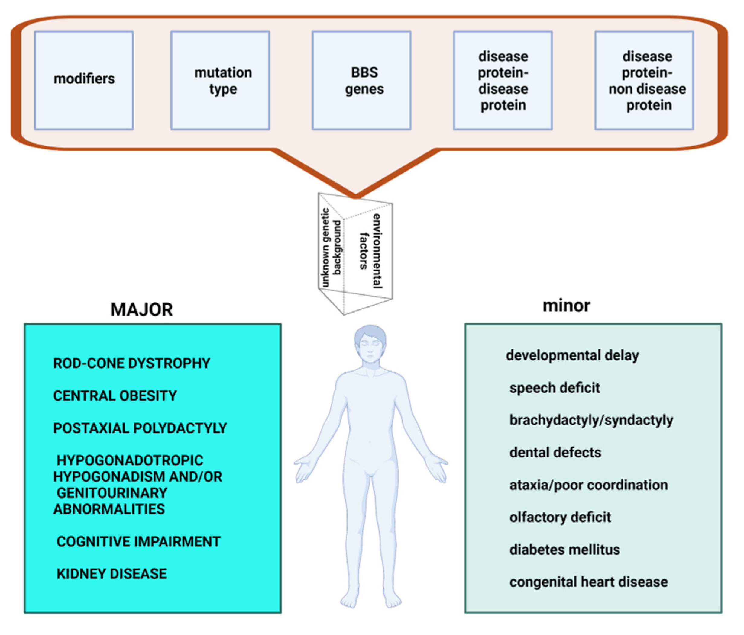
2. Pleiotropy and Variable Expressivity in Bardet–Biedl Syndrome

BBS is characterized by a high genetic heterogeneity (locus, mutational, clinical), variable expressivity and pleiotropy. There are 24 loci involved and the mutational profile is diverse. There are modifying variants and numerous interactions between the BBS proteins (interactions disease protein—disease protein) or between the BBS protein and another protein (interactions disease protein—non disease protein). These explain the extremely polymorphic clinical picture that includes both major and minor features (Figure 1).
This pleiotropic disorder has a constellation of features, which are divided into major features (rod-cone dystrophy, central obesity, postaxial polydactyly, hypogonadotropic hypogonadism and/or genitourinary abnormalities, cognitive impairment, kidney disease) and minor features (developmental delay, speech deficit, brachydactyly or syndactyly, dental defects, ataxia or poor coordination, olfactory deficit, diabetes mellitus, and congenital heart disease [17,18,19,20].
Before the discovery of BBS genes, Beales et al. established a diagnostic algorithm based on phenotypic presentations of this syndrome [20].

2.1. Major Features

One of the most important features of BBS is the rod-cone dystrophy, affecting 94–100% of individuals [21,22]. The patients can present night blindness, peripheral vision loss, diminution of color, and overall loss of visual acuity [23]. Rod and cone photoreceptor cells are modified ciliated cells responsible for the night and day vision. The main protein of rod cells is rhodopsin. The transport of this protein from the inside to the outside of rod cells is allowed by BBSome complex. Mutations in BBSome genes lead to mislocalization and accumulation of rhodopsin in rod cells. In this way the cellular homeostasis is disturbed and the degeneration of the photoreceptors is produced [24].
Obesity is a frequent feature (89%), it affects the thorax and abdomen and has an early onset (from the age of 2–3 years) [22]. Obesity is produced by dysregulation of appetite, altered leptin resistance, impaired leptin receptor signaling, altered neuroendocrine signaling from ciliated neurons to fat storage tissues, reduced number of cilia due to BBS gene mutations and alteration ways involved in the differentiating preadipocytes [25]. Common adult obesity was associated with nucleotide polymorphism in BBS2 (rs4784675). Early onset childhood obesity and common adult morbid obesity was associated with BBS4 (rs7178130) and BBS6 (rs6108572 and rs221667) changes [26].
Postaxial polydactyly is frequently reported in 79% of patients, with polydactyly of toes being more common than the polydactyly of fingers [22]. The gene BBS17, which is a negative regulator for the ciliary trafficking mediated by BBSome and the Shh (Sonic hedgehog) signaling, was associated with mesoaxial polydactyly [27].
Cognitive impairment is present in 66% cases with BBS [22]. Patients can present either intellectual disability or impairments in verbal fluency, attention capacity, poor reasoning, and emotional immaturity. The connection between BBS proteins and cognitive impairments remains unknown [22].
Hypogonadism and genitourinary malformations have an incidence of 59% [22]. Hypogonadism may be apparent at puberty. Male anomalies can vary from micro penis, small-volume testes, cryptorchidism to hypogonadotropic hypogonadism [28]. Female anatomic anomalies are uterine, fallopian, ovarian or vaginal hypoplasia, or atresia [22]. The rate of fertility is low, but both sexes can have biological children [20].
Kidney disease is present in 52% of BBS patients [22]. The spectrum of kidney disease varies from urinary tract malformations (vesicoureteral reflux, hydronephrosis, dysplastic cystic disease, absent, duplex, horseshoe or ectopic kidneys, neurogenic bladder) to chronic glomerulonephritis and defective tubular concentrating ability [18]. The main consequence is chronic kidney disease (CKD), which contributes to morbidity and mortality in patients with BBS. The majority of cases with renal disease is completely diagnosed at the age of five, but some features could be discovered in the first year of life [29]. Progression of CKD is increased by arterial hypertension or type 2 diabetes mellitus. End stage renal disease requires dialysis or a transplant. After a renal transplant, favorable long-term outcomes have been reported [30].

2.2. Minor Features

Minor features are represented by various anomalies in different systems and organs: the nervous system, sensorial changes, cardiovascular system, gastrointestinal changes, different endocrine glands, and cutaneous or musculoskeletal changes [20].
Neurodevelopmental abnormalities observed in patients with BBS include ataxia and poor coordination with mild hypertonia of all four extremities, seizures, speech abnormalities, behavioral, and psychiatric abnormalities [20].
The dysmorphism in BBS is not specific, and the most frequent reported features are: a narrow forehead, brachycephaly or macrocephaly, large ears, short, narrow, and downslanted palpebral fissures, deep and widely set eyes, malar flattening, depressed nasal bridge, long and smooth philtrum, retrognathia [20].
Anosmia/hyposmia are related to defects in olfactory cilia or the olfactory bulb [31]. Oral/dental abnormalities (50%), such as hypodontia or microdontia, high-arched palate, enamel hypoplasia, posterior crossbite, dental crowding, short roots, and taurodontism are reported [20].
Cardiovascular anomalies are relatively frequent (up to 29% of individuals) and are represented by atrioventricular septal defects, bilateral persistent superior vena cava, interrupted inferior vena cava, and hemiazygos vein continuation. Moreover, there are reports of laterality defects, such as situs inversus totalis, midline abdominal organs, asplenia, or polysplenia [22].
Gastrointestinal abnormalities can range from Hirschsprung disease, anatomic anomalies of the gastrointestinal tract (bifid epiglottis, laryngeal, and esophageal webs, bowel atresia, imperforate anus) to inflammatory bowel disease, celiac disease, and liver disease [32].
Endocrine/metabolic abnormalities include metabolic syndrome (54.3%), obesity, hyperlipidemia (usually hypertriglyceridemia), insulin resistance, and elevated fasting plasma glucose with or without type 2 diabetes mellitus (15.8%), polycystic ovarian syndrome (14.7%), and subclinical hypothyroidism (19.4%). Subclinical hypothyroidism is a mild form of hypothyroidism characterized by peripheral thyroid hormone levels within normal reference laboratory range but serum thyroid-stimulating hormone (TSH) levels are mildly elevated. Hashimoto’s thyroiditis is the most common form of subclinical hypothyroidism and is more prevalent in BBS patients compared to the general population [28,32,33].
In addition other features were reported: cutaneous dermatoses (seborrheic dermatitis, keratosis pilaris, striae, hidradenitis suppurativa, acanthosis nigricans), subclinical sensorineural hearing loss, musculoskeletal abnormalities (scoliosis, leg length discrepancy, club foot, Blount disease, and joint laxity) [20,32].
The clinical diagnosis of BBS is made in the presence of either four major features or three major features and two minor features [20]. To determine the genetic cause in BBS, gene-targeted testing (multigene panel) or comprehensive genomic testing (exome sequencing) is recommended. BBS-specific panels or larger ciliopathy genes panels can be used. In general single-gene testing by sequence analysis and deletion/duplication analysis is not recommended [32]. This genetic testing approach is based on the fact that BBS is characterized by locus and allelic heterogeneity, and because of the clinical overlap between ciliopathies [10].

3. Determinants of Clinical Effects

3.1. Locus Heterogeneity

In Table 1, there is a summary of data concerning the genes involved in BBS in correlation with tissue and single cell type specificity [34,35,36,37].
There are three categories of gene effects: primary effect (at the molecular level—protein), secondary effect (at the cellular level), and multiple tertiary effects (pleiotropic)—at the phenotypic level of organ/organism (signs and symptoms) [38]. Genes are expressed in all tissues, but there are differences in expression levels (Table 1).
The majority of genes involved in BBS presents a low tissue specificity. However, the LZTFL1 gene is mainly expressed in lymphoid tissue while the BBIP1 gene is specific to testis and the NPHP1 gene has an expression in skeletal muscle (Table 1). For single cell type specificity, a cell type enhanced expression has been described in: ciliated cells (BBS1, BBS2, ARL6, BBS4, WDPCP, LZTL1, IFT27, IFT74, NPHP1), rod photoreceptor cells (BBS1, ARL6, BBS4, BBS5, BBS7, TTC8, BBS9, BBS12, CEP290, NPHP1, SCAPER), cone photoreceptor cells (BBS1, ARL6, BBS4, BBS5, TTC8, BBS9, SCAPER), spermatocytes (ARL6, LZTFL1, BBIP1, IFT74, NPHP1), early spermatids (BBS5, BBS12, BBIP1, NPHP1), late spermatids (BBIP1), alveolar cells type 1 (WDPCP), alveolar cells type 2 (WDPCP), club cells (WDPCP) [36].
Some BBS forms are considered chaperonopathies because three genes involved in BBS encode chaperon-like proteins. The three chaperonins-like proteins—MKKS, BBS12, BBS7—are involved in assembling BBSome, a multistep process [4,39,40]. Chaperonin like BBS proteins are involved in the first stage of assembly of the BBSome and mutations in genes that encode these proteins block the formation of functional complexes [4]. BBS caused by mutations in chaperonin genes has more severe forms characterized by an earlier onset (especially BBS10), higher prevalence of primary diagnostic signs and some borderline signs with other ciliopathies, such as McKusick–Kaufman syndrome (MKKS) and Alström syndrome [4,39,41]. This increased severity could be linked to residual activity/function gain of BBSome genes [4,42].
Chaperonin-like BBS genes are characterized by a small number of coding exons (one to four) and, thus, a mutational screening of these genes be applied before a more complex analysis [4]. Mutations in MKKS are found in 3–5% of families in which there are disease-causing mutations (Bardet–Biedl Syndrome or McKusick-Kaufman syndrome) [40,43]. Mutations in BBS10 represent about 20% of all cases of BBS with certain ethnic variations: 43% in a Danish group and 8.3% in a Spanish cohort [44,45].

3.2. Mutational Heterogeneity

Mutational heterogeneity is important because testing strategies are consistent with mutation types. According to the Human Gene Mutation Database Professional (HGMD®) 2020.1 database (accessed in April 2021), 647 pathogenic variants are described. Figure 2 and Figure 3 show the implication of different types of genetic modifications in genes of BBSome, respectively the other genes implied in BBS.
The gene of BBSome could present different types of mutations like: missense/nonsense, splicing substitutions, small deletions, gross deletions, small insertions/duplications, small indels, gross insertions/duplications, complex rearrangement. (Figure 2). For the other genes implied in BBS the most frequent type of mutations are: missense/nonsense, small deletions, and small insertions/duplications (Figure 3). [46,47,48]. Some BBS forms are produced by mutations with loss-of-function (LOF): nonsense, frameshift, copy number variants, and splicing variants [49].
BBSome is a multisubunit complex with eight proteins coding by the genes BBS1, BBS2, BBS4, BBS5, BBS7, TTC8, BBS9 and BBIP1 [13,35]. The most common mutations are in BBS1 (responsible for 23% of BBS) and BBS10 (identified in 20% of patients with BBS) [20]. Founder mutations have also been described: M390R in BBS1 and C91fsX95 in BBS10 [13]. MKKS is required for BBSome assembly, and TTC8 is required for ciliary trafficking [50,51,52]. Mutations in the first 18 genes listed in the table occur in 70–80% of BBS affected families [53]. The BBS1 and BBS10 genes are the most common mutated genes in Europe and North America [54].

3.3. Modifiers

The phenotype of BBS can be changed by modifier genes. The PhenoModifier database contains 12 modifying variants described in 6 genes (TMEM67, MKS1, MKKS, CCDC28B-coiled-coil domain containing 28B, C8orf37, BBS1). Modifying genes influence expressivity or pleiotropy [55]. The pleiotropy is modified by some variants of MKS1 gene: MKS1:c.1112_1114del (p.Phe371del), MKS1:c.1476T>G (p.Cys492Trp), MKS1:c.248A>G (p.Asp83Gly), MKS1:c.368G>A (p.Arg123Gln), MKS1:p.Ile450Thr in association with mutations in BBS1 and BBS10 genes [55,56]. All of these patients had seizures, non-specific sign for BBS or MKS (Meckel–Gruber Syndrome). Therefore, the interaction between the products of the two genes could explain this pleiotropic effect [56].
With an effect on expressivity we mention variants in TMEM67, MKKS, CCDC28B, C8orf37, BBS1 genes [55,57,58,59]. For example, Bölükbaşı et al. found that most severe phenotype of BBS was allowed by following changes: C8orf37: c.533C>T (p.Ala178Val), CCDC28B: c.330C>T (p.Phe110Phe), MKKS: c.1015A>G (p.Ile339Val), and TMEM67: p.Asp799Asp [57]. Badano et al. showed that CCDC28B:c.430C/T variant (in heterozygote state) determines the introduction of a premature termination codon and the reduction of CCDC28B messenger RNA levels [58]. Badano et al. have identified another mutant alleles (BBS1: IVS115 + 2T→C) which is correlated with a more severe phenotype in a patient with homozygous mutation in BBS2 gene [59].

3.4. Disease Protein–Non-Disease Protein Interconnectivity

There is functional interaction between complexes, which explains locus heterogeneity [8,60].
In an analysis conducted by Keith et al. in 2014, the authors showed the existence of multiple interconnectivity networks [61]. These relationships involve both the disease protein-disease protein connection, but also the disease protein–non disease protein. In the latter case, it appears that there are proteins which are related to more than one of the proteins encoded by the genes involved in BBS syndrome [61]. Examples of such proteins are: ALDOB (fructose-bisphosphate aldolase B) (in interaction with genes products BBS1, BBS2, BBS4, BBS7), HSCB (Iron-sulfur cluster co-chaperone protein HscB) (in interaction with genes products BBS2, BBS4, BBS1), ACY1 (Aminoacylase-1) (in interaction with genes products BBS2, BBS4, BBS7), EXOC7 (Exocyst complex component 7) (in interaction with genes products BBS1, BBS7, BBS4, BBS2), RAB3IP (Rab-3A-interacting protein) (in interaction with genes products BBS1, BBS4), FHOD1 (FH1/FH2 domain-containing protein 1) (in interaction with genes products BBS1, BBS2, BBS4, BBS7), PCM1 (Pericentriolar material 1 protein) in interaction with genes products BBS1, BBS2, BBS4, BBIP1), CCDC28B (Coiled-coil domain-containing protein 28B) (in interaction with genes products MKKS, TTC8, BBS2, BBS4, BBS7, BBS1, BBS5) [61].
Keith postulated that there is an interconnectivity between proteins in the case of locus heterogeneity. Thus, the BBS proteins in the vicinity of proteins involved in locus heterogeneity could be involved in the pathogenesis of similar diseases or may themselves be causes of disease [61].
The cellular signaling pathways involved in BBS are: hedgehog, Wnt (wingless-related integration site), GPCR (G protein-coupled receptors), mTOR (mammalian target of rapamycin) [5,6].
The primary cilium is involved in Hedgehog signaling pathway and can act as both positive and negative regulator of this pathway [5]. Defects in cilia/intraflagellar transport lead to loss of Hh phenotype function in the neural tube and gain of function in the limbs. This explains the common Hh phenotype (especially polydactyly) present in ciliopathies, including BBS [5,62].
Several BBS-associated proteins intervene in the Wnt signaling pathway and play a role in regulating Wnt signaling by degrading Wnt effectors [5,63]. Disheveled is an essential protein in basal body docking, ciliogenesis and planar cell polarity. Meckelin (TMEM67), TMEM216, MKS1, BBS10, and BBS12 also intervene in the same processes [5,51,64,65]. The lack of these proteins leads to planar cell polarity defects also found in BBS [5].
G-protein coupled receptors (GPCRs) are important for cilia structures and function. Neuronal cilia integrity is required for brain development and the adequate interaction in the adult brain [5]. Neuronal cilium formation begins prenatally with pro-cilium formation and continues in the first 8–12 weeks after birth. During postnatal development at the level of the primary neuronal membrane cilium GPCRs appear: somatostatin receptor 3 (SSTR3), melanin-concentrating hormone receptor 1 (MCHR1), serotonin receptor 6 (5HTR6), kisspeptin 1 receptor (KISS1R), dopamine receptors 1, 2, and 5 (D1, D2, and D5), neuropeptide Y receptors, NPY2R and NPY5R. The location of these receptors is different being consistent with the neuronal type. The length of the cilium is important for proper function. For example, the shortening of the cilia of hypothalamic neurons has been correlated with obesity in mice induced by high-fat diet, so that the dysfunction of neural cilia is correlated with childhood obesity in BBS [5,66,67]. GPCRs bind to b-arrestin2 and BBS proteins associated with Arl6 and thus trigger ciliary trafficking. When some of these receptors are missing or lacking motifs of recognition by BBSome and b-arrest, an accumulation of receptors and their ectocytosis occurs [68]. Rhodopsin and opsin are other examples of GPCRs in rods and cones. Their role is to absorb light and transmit the electrical signal to the brain. If the integrity of the cilia is not adequate, retinal degeneration will occur.
Mammalian target of rapamycin is also involved in BBS. CCDC28B interacts with the SN1 subunit of the mammalian target of rapamycin complex 2 (mTORC2). In this way it regulates the length of the cilia by affecting its assembly/stability [2].

3.5. Disease Protein-Disease Protein Interconnectivity

The eight subunits of BBSome are assembled in two parts: head and body. The head is formed by BBS2 which interacts with BBS7. The body is formed by the others proteins (BBS1, BBS4, BBS5, TTC8, BBS9, BBIP1). The BBIP1 is the central part of the BBSome core [68,69]. BBIP1 interacts with BBS4 and TTC8 and thus achieves proper assembly and structural stability of the BBSome complex [70]. BBS4 and TTC8 have TPR (tetratricopeptide repeats) subunits. They interact with the domain β propeller of BBS1 and BBS9, respectively [70]. These interactions are essential for a functional BBSome and explain why mutations at the BBS4-BBS1 and BBS8-BBS9 interface lead to disease in patients [70].
In a study that analyzed the effects of the proteome of the three modules (BBSome, chaperonin complex, transition zone) and three more genes that are not yet included in a module (ARL6, TRIM32, WDPCP) six epistatic interaction effects were identified: BBS10-BBS5; BBS10-BBS12; BBS10-BBS1; BBS10-MKKS; BBS10-BBS4; BBS12-MKKS. Moreover, 12 additive interaction effects were found between the main complexes (BBSome, chaperonin and transition zone) and 1 between proteins of transition zone and others gene BBS1-BBS5, BBS1-BBS2, BBS1-NPHP1, BBS10-NPHP1, BBS2-NPHP1, BBS9-NPHP1, BBS7-NPHP1, BBS2-BBS4, BBS2-MKKS, BBS4-MKKS, BBS4-BBS9, BBS4-BBS7. On the other hand, no interaction effect was demonstrated in the following genes (nor additive or epistatic effect): TRIM32, WDPCP, CEP290, MKS1 [71].

4. Conclusions

Bardet–Biedl Syndrome is a rare ciliopathy characterized by a high degree of clinical heterogeneity. The mechanisms of heterogeneity are complex and explained by multiple loci, ubiquitous expression of genes, and multiple interaction of proteins coding by genes implicated in BBS. However, the deciphering of the entire mechanism implied in Bardet–Biedl Syndrome is a complex process that will require further research in the future.

Author Contributions

All authors contributed equally to this article. All authors have read and agreed to the published version of the manuscript.

Funding

This research received no external funding.

Institutional Review Board Statement

Not applicable.

Informed Consent Statement

Not applicable.

Data Availability Statement

Some data were obtained from Human Gene Mutation Database (HGMD® Professional 2021.1) available online: http://www.hgmd.cf.ac.uk/ac/all.php, accessed on 1 April 2021.

Conflicts of Interest

The authors declare no conflict of interest.

References

  1. Strong, A.; Li, D.; Mentch, F.; Bedoukian, E.; Hartung, E.A.; Meyers, K.; Skraban, C.; Wen, J.; Medne, L.; Glessner, J.; et al. Ciliopathies: Coloring outside of the lines. Am. J. Med. Genet. Part A 2021, 185, 687–694. [Google Scholar] [CrossRef] [PubMed]
  2. Cardenas-Rodriguez, M.; Badano, J.L. Ciliary biology: Understanding the cellular and genetic basis of human ciliopathies. Am. J. Med. Genet. Part C Semin. Med. Genet. 2009, 151C, 263–280. [Google Scholar] [CrossRef] [PubMed]
  3. Christensen, S.T.; Morthorst, S.; Mogensen, J.B.; Pedersen, L.B. Primary cilia and coordination of receptor tyrosine kinase (RTK) and transforming growth factor β (TGF-β) signaling. Cold Spring Harb. Perspect. Biol. 2017, 9, a028167. [Google Scholar] [CrossRef] [PubMed] [Green Version]
  4. Álvarez-Satta, M.; Cástro-Sánchez, S.; Valverde, D. Bardet-Biedl syndrome as a chaperonopathy: Dissecting the major role of chaperonin-like BBS proteins (BBS6-BBS10-BBS12). Front. Mol. Biosci. 2017, 4, 55. [Google Scholar] [CrossRef] [Green Version]
  5. Wheway, G.; Nazlamova, L.; Hancock, J.T. Signaling through the Primary Cilium. Front. Cell Dev. Biol. 2018, 6, 8. [Google Scholar] [CrossRef] [PubMed]
  6. Singla, V. The primary cilium as the cell’s antenna: Signaling at a sensory organelle. Science 2006, 313, 629–633. [Google Scholar] [CrossRef] [Green Version]
  7. Adamiok-Ostrowska, A.; Piekiełko-Witkowska, A. Ciliary genes in renal cystic diseases. Cells 2020, 9, 907. [Google Scholar] [CrossRef] [Green Version]
  8. Mitchison, H.M.; Valente, E.M. Motile and non-motile cilia in human pathology: From function to phenotypes. J. Pathol. 2017, 241, 294–309. [Google Scholar] [CrossRef]
  9. Ma, M. Cilia and polycystic kidney disease. Semin. Cell Dev. Biol. 2021, 110, 139–148. [Google Scholar] [CrossRef] [PubMed]
  10. Zaki, M.S.; Sattar, S.; Massoudi, R.A.; Gleeson, J.G. Co-occurrence of distinct ciliopathy diseases in single families suggests genetic modifiers. Am. J. Med. Genet. Part A 2011, 155, 3042–3049. [Google Scholar] [CrossRef] [PubMed]
  11. Reiter, J.F.; Leroux, M.R. Genes and molecular pathways underpinning ciliopathies. Nat. Rev. Mol. Cell Biol. 2017, 18, 533–547. [Google Scholar] [CrossRef]
  12. Prevalence and Incidence of Rare Diseases: Bibliographic Data. Orphanet Report Series Number 1 January 2020. Available online: https://www.orpha.net/orphacom/cahiers/docs/GB/Prevalence_of_rare_diseases_by_alphabetical_list.pdf (accessed on 5 November 2020).
  13. Khan, S.; Muhammad, N.; Khan, M.A.; Kamal, A.; Rehman, Z.U.; Alam Khan, S. Genetics of human Bardet-Biedl syndrome, an updates. Clin. Genet. 2016, 90, 3–15. [Google Scholar] [CrossRef]
  14. Ajmal, M.; Khan, M.I.; Neveling, K.; Tayyab, A.; Jaffar, S.; Sadeque, A.; Ayub, H.; Abbasi, N.M.; Riaz, M.; Micheal, S.; et al. Exome sequencing identifies a novel and a recurrent BBS1 mutation in Pakistani families with Bardet–Biedl syn-drome. Mol. Vis. 2013, 19, 644–653. [Google Scholar] [PubMed]
  15. Farag, T.I.; Teebi, A.S. High incidence of Bardet Biedl syndrome among the Bedouin. Clin. Genet. 2008, 36, 463–464. [Google Scholar] [CrossRef]
  16. Hjortshøj, T.D.; Grønskov, K.; Brondum-Nielsen, K.; Rosenberg, T. A novel founder BBS1 mutation explains a unique high prevalence of Bardet-Biedl syndrome in the Faroe Islands. Br. J. Ophthalmol. 2008, 93, 409–413. [Google Scholar] [CrossRef]
  17. Sheffield, V.C.; Carml, R.; Kwltek-Black, A.; Rokhlina, T.; Nishlmura, D.; Duyk, G.M.; Elbedour, K.; Sunden, S.L.; Stone, E.M. Identification of a Bardet-Biedl syndrome locus on chromosome 3 and evaluation of an efficient approach to homozygosity mapping. Hum. Mol. Genet. 1994, 3, 1331–1335. [Google Scholar] [CrossRef]
  18. Beales, P.L.; Elcioglu, N.; Woolf, A.; Parker, D.; Flinter, F. New criteria for improved diagnosis of Bardet-Biedl syndrome: Results of a population survey. J. Med. Genet. 1999, 36, 437–446. [Google Scholar]
  19. Sheffield, V.C.; Zhang, Q.; Heon, E.; Drack, A.V.; Stone, E.M.; Carmi, R. The Bardet-Biedl syndrome. Epstein’s Inborn Errors Dev. 2016, 237–240. [Google Scholar] [CrossRef]
  20. Forsythe, E.; Beales, P.L. Bardet–Biedl syndrome. Eur. J. Hum. Genet. 2013, 21, 8–13. [Google Scholar] [CrossRef]
  21. Denniston, A.K.; Beales, P.L.; Tomlins, P.J.; Good, P.; Langford, M.; Foggensteiner, L.; Williams, D.; Tsaloumas, M.D. Evaluation of visual function and needs in adult patients with Bardet–Biedl syndrome. Retina 2014, 34, 2282–2289. [Google Scholar] [CrossRef] [PubMed]
  22. Niederlova, V.; Modrak, M.; Tsyklauri, O.; Huranova, M.; Stepanek, O. Meta-analysis of genotype-phenotype associations in Bardet-Biedl syndrome uncovers differences among causative genes. Hum. Mutat. 2019, 40, 2068–2087. [Google Scholar] [CrossRef]
  23. Weihbrecht, K.; Goar, W.A.; Pak, T.; Garrison, J.E.; DeLuca, A.P.; Stone, E.M.; Scheetz, T.E.; Sheffield, V.C. Keeping an eye on Bardet-Biedl syndrome: A comprehensive review of the role of Bardet-Biedl syndrome genes in the eye. Med. Res. Arch. 2017, 5, 1–21. [Google Scholar] [CrossRef]
  24. Mockel, A.; Perdomo, Y.; Stutzmann, F.; Letsch, J.; Marion, V.; Dollfus, H. Retinal dystrophy in Bardet-Biedl syndrome and related syndromic ciliopathies. Prog. Retin. Eye Res. 2011, 30, 258–274. [Google Scholar] [CrossRef] [PubMed]
  25. Guo, D.-F.; Rahmouni, K. Molecular basis of the obesity associated with Bardet–Biedl syndrome. Trends Endocrinol. Metab. 2011, 22, 286–293. [Google Scholar] [CrossRef] [Green Version]
  26. Benzinou, M.; Walley, A.; Lobbens, S.; Charles, M.A.; Jouret, B.; Fumeron, F.; Balkau, B.; Meyre, D.; Froguel, P. Bardet-Biedl syndrome gene variants are associated with both childhood and adult common obesity in French Caucasians. Diabetes 2006, 55, 2876–2882. [Google Scholar] [CrossRef] [PubMed] [Green Version]
  27. Schaefer, E.; Lauer, J.; Durand, M.; Pelletier, V.; Obringer, C.; Claussmann, A.; Braun, J.-J.; Redin, C.; Mathis, C.; Muller, J.; et al. Mesoaxial polydactyly is a major feature in Bardet-Biedl syndrome patients withLZTFL1(BBS17) mutations. Clin. Genet. 2014, 85, 476–481. [Google Scholar] [CrossRef]
  28. Mujahid, S.; Hunt, K.F.; Cheah, Y.S.; Forsythe, E.; Hazlehurst, J.M.; Sparks, K.; Mohammed, S.; Tomlinson, J.A.; Amiel, S.; Carroll, P.V.; et al. The endocrine and metabolic characteristics of a large Bardet-Biedl syndrome clinic population. J. Clin. Endocrinol. Metab. 2018, 103, 1834–1841. [Google Scholar] [CrossRef]
  29. Forsythe, E.; Sparks, K.; Best, S.; Borrows, S.; Hoskins, B.; Sabir, A.; Barrett, T.; Williams, D.; Mohammed, S.; Goldsmith, D.; et al. Risk factors for severe renal disease in Bardet–Biedl syndrome. J. Am. Soc. Nephrol. 2016, 28, 963–970. [Google Scholar] [CrossRef] [PubMed]
  30. Haws, R.M.; Joshi, A.; Shah, S.A.; Alkandari, O.; Turman, M.A. Renal transplantation in Bardet–Biedl Syndrome. Pediatr. Nephrol. 2016, 31, 2153–2161. [Google Scholar] [CrossRef]
  31. Braun, J.; Noblet, V.; Kremer, S.; Molière, S.; Dollfus, H.; Marion, V.; Goetz, N.; Muller, J.; Riehm, S. Value of MRI Olfactory Bulb evaluation in the assessment of olfactory dysfunction in Bardet Biedl syndrome. Clin. Genet. 2016, 90, 79–83. [Google Scholar] [CrossRef]
  32. Forsyth, R.L.; Gunay-Aygun, M. Bardet-Biedl Syndrome Overview. In GeneReviews® [Internet]; Adam, M.P., Ardinger, H.H., Pagon, R.A., Wallace, S.E., Bean, L.J.H., Mirzaa, G., Amemiya, A., Eds.; University of Washington: Seattle, WA, USA, 1993; 14 July 2003 (Updated 23 July 2020). Available online: https://www.ncbi.nlm.nih.gov/books/NBK1363/ (accessed on 27 June 2021).
  33. Tsyklauri, O.; Niederlova, V.; Forsythe, E.; Prasai, A.; Drobek, A.; Kasparek, P.; Sparks, K.; Trachtulec, Z.; Prochazka, J.; Sedlacek, R.; et al. Bardet–Biedl Syndrome ciliopathy is linked to altered hematopoiesis and dysregulated self-tolerance. EMBO Rep. 2021, 22, e50785. [Google Scholar] [CrossRef] [PubMed]
  34. Stenson, P.D.; Mort, M.; Ball, E.V.; Evans, K.; Hayden, M.; Heywood, S.; Hussain, M.; Phillips, A.D.; Cooper, D.N. The human gene mutation database: Towards a comprehensive repository of inherited mutation data for medical research, genetic diagnosis and next-generation sequencing studies. Qual. Life Res. 2017, 136, 665–677. [Google Scholar] [CrossRef] [PubMed] [Green Version]
  35. HGNC, HUGO Gene Nomenclature Committee Home Page. Available online: http://www.genenames.org/ (accessed on 1 August 2012).
  36. Proteinatlas the Human Protein Atlas. Available online: https://www.proteinatlas.org (accessed on 19 December 2019).
  37. UniProt. Available online: https://www.uniprot.org/ (accessed on 26 April 2021).
  38. Covic, M.; Ştefănescu, D.; Sandovici, I.; Gorduza, E.V. Genetică Medicală, 3rd ed.; Polirom Iaşi: Iasi, Romania, 2017. [Google Scholar]
  39. Billingsley, G.; Bin, J.; Fieggen, K.; Duncan, J.L.; Gerth-Kahlert, C.; Ogata, K.; Wodak, S.I.; Traboulsi, E.A.; Fishman, G.; Paterson, A.; et al. Mutations in chaperonin-like BBS genes are a major contributor to disease development in a multiethnic Bardet-Biedl syndrome patient population. J. Med. Genet. 2010, 47, 453–463. [Google Scholar] [CrossRef]
  40. Deveault, C.; Billingsley, G.; Duncan, J.L.; Bin, J.; Theal, R.; Vincent, A.; Fieggen, K.; Gerth-Kahlert, C.; Noordeh, N.; Traboulsi, E.I.; et al. BBS genotype-phenotype assessment of a multiethnic patient cohort calls for a revision of the disease definition. Hum. Mutat. 2011, 32, 610–619. [Google Scholar] [CrossRef] [PubMed]
  41. Imhoff, O.; Marion, V.; Stoetzel, C.; Durand, M.; Holder, M.; Sigaudy, S.; Sarda, P.; Hamel, C.P.; Brandt, C.; Dollfus, H.; et al. Bardet-Biedl syndrome: A study of the renal and cardiovascular phenotypes in a French cohort. Clin. J. Am. Soc. Nephrol. 2010, 6, 22–29. [Google Scholar] [CrossRef] [PubMed] [Green Version]
  42. Zhang, Q.; Yu, D.; Seo, S.; Stone, E.M.; Sheffield, V.C. Intrinsic protein-protein interaction-mediated and chaperonin-assisted sequential assembly of stable Bardet-Biedl syndrome protein complex, the BBSome. J. Biol. Chem. 2012, 287, 20625–20635. [Google Scholar] [CrossRef] [Green Version]
  43. Muller, J.; Stoetzel, C.; Vincent, M.C.; Leitch, C.C.; Laurier, V.; Danse, J.M.; Hellé, S.; Marion, V.; Bennouna-Greene, V.; Vicaire, S.; et al. Identification of 28 novel mutations in the Bardet–Biedl syndrome genes: The burden of private mutations in an extensively heterogeneous disease. Qual. Life Res. 2010, 127, 583–593. [Google Scholar] [CrossRef] [Green Version]
  44. Hjortshøj, T.D.; Grønskov, K.; Philp, A.R.; Nishimura, D.Y.; Riise, R.; Sheffield, V.; Rosenberg, T.; Brøndum-Nielsen, K. Bardet-Biedl syndrome in Denmark-report of 13 novel sequence variations in six genes. Hum. Mutat. 2010, 31, 429–436. [Google Scholar] [CrossRef] [PubMed]
  45. Álvarez-Satta, M.; Castro-Sánchez, S.; Pereiro, I.; Piñeiro-Gallego, T.; Baiget, M.; Ayuso, C.; Valverde, D. Overview of Bardet-Biedl syndrome in Spain: Identification of novel mutations inBBS1, BBS10 and BBS12genes. Clin. Genet. 2014, 86, 601–602. [Google Scholar] [CrossRef]
  46. Wheway, G.; Mitchison, H.M. Genomics England research consortium opportunities and challenges for molecular understanding of ciliopathies–The 100,000 genomes project. Front. Genet. 2019, 10, 127. [Google Scholar] [CrossRef] [Green Version]
  47. Davis, E.; Zhang, Q.; Liu, Q.; Diplas, B.; Davey, L.M.; Hartley, J.; Stoetzel, C.; Szymanska, K.; Ramaswami, G.; Logan, C.V.; et al. TTC21B contributes both causal and modifying alleles across the ciliopathy spectrum. Nat. Genet. 2011, 43, 189–196. [Google Scholar] [CrossRef] [PubMed]
  48. Lindstrand, A.; Frangakis, S.; Carvalho, C.M.; Richardson, E.B.; McFadden, K.A.; Willer, J.R.; Pehlivan, D.; Liu, P.; Pediaditakis, I.L.; Sabo, A.; et al. Copy-Number variation contributes to the mutational load of Bardet-Biedl syndrome. Am. J. Hum. Genet. 2016, 99, 318–336. [Google Scholar] [CrossRef] [PubMed] [Green Version]
  49. Pomeroy, J.; Krentz, A.D.; Richardson, J.G.; Berg, R.L.; VanWormer, J.J.; Haws, R.M. Bardet-Biedl syndrome: Weight patterns and genetics in a rare obesity syndrome. Pediatr. Obes. 2021, 16, e12703. [Google Scholar] [CrossRef]
  50. Nachury, M.V.; Loktev, A.V.; Zhang, Q.; Westlake, C.J.; Peränen, J.; Merdes, A.; Slusarski, D.; Scheller, R.H.; Bazan, J.F.; Sheffield, V.; et al. A core complex of BBS proteins cooperates with the GTPase Rab8 to promote ciliary membrane biogenesis. Cell 2007, 129, 1201–1213. [Google Scholar] [CrossRef] [PubMed] [Green Version]
  51. Jin, H.; Nachury, M. The BBSome. Curr. Biol. 2009, 19, R472–R473. [Google Scholar] [CrossRef] [PubMed] [Green Version]
  52. Seo, S.; Baye, L.M.; Schulz, N.P.; Beck, J.S.; Zhang, Q.; Slusarski, D.C.; Sheffield, V.C. BBS6, BBS10, and BBS12 form a complex with CCT/TRiC family chaperonins and mediate BBSome assembly. Proc. Natl. Acad. Sci. USA 2010, 107, 1488–1493. [Google Scholar] [CrossRef] [PubMed] [Green Version]
  53. M’Hamdi, O.; Redin, C.; Stoetzel, C.; Ouertani, I.; Chaabouni, M.; Maazoul, F.; M’Rad, R.; Mandel, J.L.; Dollfus, H.; Muller, J. Clinical and genetic characterization of Bardet-Biedl syndrome in Tunisia: Defining a strategy for molecular diagnosis. Clin. Genet. 2013, 85, 172–177. [Google Scholar] [CrossRef] [PubMed]
  54. Forsythe, E.; Sparks, K.; Hoskins, B.; Bagkeris, E.; McGowan, B.; Carroll, P.; Huda, M.; Mujahid, S.; Peters, C.; Barrett, T.; et al. Genetic predictors of cardiovascular morbidity in Bardet–Biedl syndrome. Clin. Genet. 2014, 87, 343–349. [Google Scholar] [CrossRef]
  55. PhenoModifier: A Genetic Modifier Database for Elucidating the Genetic Basis of Human Phenotypic Variation. Available online: https://www.biosino.org/PhenoModifier/index (accessed on 26 July 2021).
  56. Leitch, C.C.; Zaghloul, N.A.; Davis, E.; Stoetzel, C.; Diaz-Font, A.; Rix, S.; Al-Fadhel, M.; Lewis, R.A.; Eyaid, W.; Banin, E.; et al. Hypomorphic mutations in syndromic encephalocele genes are associated with Bardet-Biedl syndrome. Nat. Genet. 2008, 40, 443–448. [Google Scholar] [CrossRef]
  57. Bölükbaşı, E.Y.; Mumtaz, S.; Afzal, M.; Woehlbier, U.; Malik, S.; Tolun, A. Homozygous mutation in CEP19, a gene mutated in morbid obesity, in Bardet-Biedl syndrome with predominant postaxial polydactyly. J. Med. Genet. 2018, 55, 189–197. [Google Scholar] [CrossRef]
  58. Badano, J.L.; Leitch, C.C.; Ansley, S.J.; May-Simera, H.; Lawson, S.; Lewis, R.A.; Beales, P.L.; Dietz, H.C.; Fisher, S.J.; Katsanis, N. Dissection of epistasis in oligogenic Bardet–Biedl syndrome. Nature 2006, 439, 326–330. [Google Scholar] [CrossRef]
  59. Badano, J.L.; Kim, J.C.; Hoskins, B.E.; Lewis, R.A.; Ansley, S.J.; Cutler, D.J.; Castellan, C.; Beales, P.L.; Leroux, M.R.; Katsanis, N. Heterozygous mutations in BBS1, BBS2 and BBS6 have a potential epistatic effect on Bardet-Biedl patients with two mutations at a second BBS locus. Hum. Mol. Genet. 2003, 12, 1651–1659. [Google Scholar] [CrossRef] [Green Version]
  60. Lee, J.; Chung, Y.D. Ciliary subcompartments: How are they established and what are their functions? BMB Rep. 2015, 48, 380–387. [Google Scholar] [CrossRef] [PubMed] [Green Version]
  61. Keith, B.P.; Robertson, D.L.; Hentges, K.E. Locus heterogeneity disease genes encode proteins with high interconnectivity in the human protein interaction network. Front. Genet. 2014, 5, 434. [Google Scholar] [CrossRef] [PubMed] [Green Version]
  62. Waters, A.M.; Beales, P.L. Ciliopathies: An expanding disease spectrum. Pediatr. Nephrol. 2011, 26, 1039–1056. [Google Scholar] [CrossRef] [PubMed] [Green Version]
  63. Wiens, C.J.; Tong, Y.; Esmail, M.A.; Oh, E.; Gerdes, J.; Wang, J.; Tempel, W.; Rattner, J.B.; Katsanis, N.; Park, H.-W.; et al. Bardet-Biedl syndrome-associated small GTPase ARL6 (BBS3) functions at or near the ciliary gate and modulates Wnt signaling. J. Biol. Chem. 2010, 285, 16218–16230. [Google Scholar] [CrossRef] [PubMed] [Green Version]
  64. Park, T.J.; Mitchell, B.; Abitua, P.; Kintner, C.; Wallingford, J.B. Dishevelled controls apical docking and planar polarization of basal bodies in ciliated epithelial cells. Nat. Genet. 2008, 40, 871–879. [Google Scholar] [CrossRef]
  65. Adams, M.; Simms, R.J.; Abdelhamed, Z.; Dawe, H.R.; Szymanska, K.; Logan, C.; Wheway, G.; Pitt, E.; Gull, K.; Knowles, M.A.; et al. A meckelin–filamin A interaction mediates ciliogenesis. Hum. Mol. Genet. 2011, 21, 1272–1286. [Google Scholar] [CrossRef]
  66. Han, Y.M.; Kang, G.M.; Byun, K.; Ko, H.W.; Kim, J.; Shin, M.-S.; Kim, H.-K.; Gil, S.Y.; Yu, J.H.; Lee, B.; et al. Leptin-promoted cilia assembly is critical for normal energy balance. J. Clin. Investig. 2014, 124, 2193–2197. [Google Scholar] [CrossRef] [Green Version]
  67. Mariman, E.C.M.; Vink, R.G.; Roumans, N.J.T.; Bouwman, F.G.; Stumpel, C.T.R.M.; Aller, E.E.J.G.; van Baak, M.; Wang, P. The cilium: A cellular antenna with an influence on obesity risk. Br. J. Nutr. 2016, 116, 576–592. [Google Scholar] [CrossRef] [Green Version]
  68. Nager, A.R.; Goldstein, J.S.; Herranz-Pérez, V.; Portran, D.; Ye, F.; Garcia-Verdugo, J.M.; Nachury, M.V. An actin network dispatches ciliary gpcrs into extracellular vesicles to modulate signaling. Cell 2017, 168, 252–263. [Google Scholar] [CrossRef] [PubMed] [Green Version]
  69. Lorentzen, E.; Kikkawa, M. Decision letter: Structure and activation mechanism of the BBSome membrane protein trafficking complex. eLife 2019, 9, e53322. [Google Scholar] [CrossRef]
  70. Klink, B.U.; Gatsogiannis, C.; Hofnagel, O.; Wittinghofer, A.; Raunser, S. Author response: Structure of the human BBSome core complex. eLife 2020, 9, e53910. [Google Scholar] [CrossRef] [PubMed]
  71. Kousi, M.; Söylemez, O.; Ozanturk, A.; Mourtzi, N.; Akle, S.; Jungreis, I.; Muller, J.; Cassa, C.A.; Brand, H.; Mokry, J.A.; et al. Evidence for secondary-variant genetic burden and non-random distribution across biological modules in a recessive ciliopathy. Nat. Genet. 2020, 52, 1–6. [Google Scholar] [CrossRef] [PubMed]
Figure 1. Heterogeneity in Bardet Biedl Syndrome-determinants and clinical effects. Created with BioRender.com (accessed on 15 August 2021).
Genes 12 01353 g001
Figure 2. Pathogenic variants in BBSome [34]. BBS1: Bardet-Biedl syndrome 1; BBS2: Bardet-Biedl syndrome 2; BBS4: Bardet-Biedl syndrome 4; BBS5: Bardet-Biedl syndrome 5; BBS7: Bardet-Biedl syndrome 7; TTC8: tetratricopeptide repeat domain 8; BBS9: Bardet-Biedl syndrome 9; BBIP1: BBSome interacting protein 1.
Genes 12 01353 g002
Figure 3. Pathogenic variants in other genes of BBS [34]. ARL6: ADP ribosylation factor like GTPase 6; MKKS: MKKS centrosomal shuttling protein; BBS10: Bardet-Biedl syndrome 10; TRIM32: tripartite motif containing 32; BBS12: Bardet-Biedl syndrome 12; MKS1: MKS transition zone complex subunit 1; CEP290: centrosomal protein 290; WDPCP: WD repeat containing planar cell polarity effector; SDCCAG8: SHH signaling and ciliogenesis regulator SDCCAG8; LZTFL1: leucine zipper transcription factor like 1; IFT27: intraflagellar transport 27; IFT74: intraflagellar transport 74; C8orf37: chromosome 8 open reading frame 37; SCLT1: sodium channel and clathrin linker 1; NPHP1: nephrocystin 1; SCAPER: S-phase cyclin A associated protein in the ER.
Genes 12 01353 g003
Table 1. Genes in Bardet–Biedl Syndrome: characteristics, tissue, and single cell type specificity [34,35,36,37].
NoGene SymbolGene NameGene GroupsChromosomeProteinLocalizationTissue Specificity
1BBS1Bardet–Biedl syndrome 1BBSome11q13.2Bardet–Biedl syndrome 1 proteinBasal body, ciliumLow tissue specificity
2BBS2Bardet–Biedl syndrome 2BBSome16q13Bardet–Biedl syndrome 2 proteinBasal body, ciliumLow tissue specificity
3ARL6ADP ribosylation factor like GTPase 6ARF GTPase family3q11.2ADP-ribosylation factor-like protein 6Basal body, cilium, cytosol, transition zoneLow tissue specificity
4BBS4Bardet–Biedl syndrome 4Tetratricopeptide repeat domain containing
BBSome
15q24.1Bardet–Biedl syndrome 4 proteinBasal body, ciliumLow tissue specificity
5BBS5Bardet–Biedl syndrome 5BBSome2q31.1Bardet–Biedl syndrome 5 proteinBasal bodyLow tissue specificity
6MKKSMKKS centrosomal shuttling proteinChaperonins20p12.2McKusick–Kaufman/Bardet–Biedl syndromes putative chaperoninBasal body, cytosolLow tissue specificity
7BBS7Bardet–Biedl syndrome 7BBSome4q27Bardet–Biedl syndrome 7 proteinBasal body, ciliumLow tissue specificity
8TTC8Tetratricopeptide repeat domain 8Tetratricopeptide repeat domain containing
BBSome
14q31.3Tetratricopeptide repeat protein 8Basal body, cilium, IFTLow tissue specificity
9BBS9Bardet–Biedl syndrome 9BBSome7p14.3Protein PTHB1CiliumLow tissue specificity
10BBS10Bardet–Biedl syndrome 10Chaperonins12q21.2Bardet–Biedl syndrome 10 proteinBasal bodyLow tissue specificity
11TRIM32Tripartite motif containing 32Tripartite motif containing
Ring finger proteins
9q33.1E3 ubiquitin-protein ligase TRIM32Intermediate filamentsLow tissue specificity
12BBS12Bardet–Biedl syndrome 12Chaperonins4q27Bardet–Biedl syndrome 12 proteinBasal bodyLow tissue specificity
13MKS1MKS transition zone complex subunit 1B9 domain containing
MKS complex
17q22Meckel syndrome type 1 proteinBasal bodyLow tissue specificity
14CEP290centrosomal protein 290MKS complex12q21.32Centrosomal protein of 290 kDa (Cep290)Basal body, centrosomeLow tissue specificity
15WDPCPWD repeat containing planar cell polarity effectorCiliogenesis and planar polarity effector complex subunits2p15WD repeat-containing and planar cell polarity effector protein fritz homolog (hFRTZ)Cytosol, plasma membrane, axonemeLow tissue specificity
16SDCCAG8SHH signaling and ciliogenesis regulator SDCCAG8MicroRNA protein coding host genes1q43-q44Serologically defined colon cancer antigen 8Basal body, centriole, transition zoneLow tissue specificity
17LZTFL1leucine zipper transcription factor like 1 3p21.31Leucine zipper transcription factor-like protein 1Basal body, ciliumTissue enhanced (lymphoid tissue)
18BBIP1BBSome interacting protein 1BBSome10q25.2BBSome-interacting protein 1Cytoplasm, cytosolTissue enhanced (testis)
19IFT27intraflagellar transport 27IFT-B1 complex RAB, member RAS oncogene GTPases22q12.3Intraflagellar transport protein 27 homologBasal body, cilium, IFTLow tissue specificity
20IFT74intraflagellar transport 74IFT-B1 complex9p21.2Intraflagellar transport protein 74 homologBasal body, cilium, IFTLow tissue specificity
21C8orf37chromosome 8 open reading frame 37 8q22.1Protein C8orf37Basal body, ciliary rootLow tissue specificity
22SCLT1sodium channel and clathrin linker 1 4q28.2Sodium channel and clathrin linker 1CentrioleLow tissue specificity
23NPHP1Nephrocystin 1NPHP complex2q13Nephrocystin-1Transition zoneTissue enhanced (skeletal muscle)
24SCAPERS-phase cyclin A associated protein in the ERZinc fingers C2H2-type15q24.3S phase cyclin A-associated protein in the endoplasmic reticulum
(S phase cyclin A-associated protein in the ER)
Endoplasmic reticulumLow tissue specificity
Publisher’s Note: MDPI stays neutral with regard to jurisdictional claims in published maps and institutional affiliations.

