Article

HLA Class II Genotype Does Not Affect the Myelin Responsiveness of Multiple Sclerosis Patients

1
Laboratory of Experimental Hematology, Vaccine and Infectious Disease Institute (VaxInfectio), Faculty of Medicine and Health Sciences, University of Antwerp, 2610 Antwerp, Belgium
2
Division of Neurology, Antwerp University Hospital, 2650 Edegem, Belgium
3
Histocompatibility and Immunogenetics Laboratory, Red Cross-Flanders, 2650 Mechelen, Belgium
4
Born Bunge Institute, Translational Neurosciences, Faculty of Medicine and Health Sciences, University of Antwerp, 2610 Antwerp, Belgium
5
Center for Cell Therapy and Regenerative Medicine, Antwerp University Hospital, 2650 Edegem, Belgium
*
Author to whom correspondence should be addressed.
Cells 2020, 9(12), 2703; https://doi.org/10.3390/cells9122703
Submission received: 14 November 2020 / Revised: 7 December 2020 / Accepted: 16 December 2020 / Published: 17 December 2020
(This article belongs to the Special Issue The Molecular and Cellular Basis for Multiple Sclerosis 2020)

Abstract

:
Background: When aiming to restore myelin tolerance using antigen-specific treatment approaches in MS, the wide variety of myelin-derived antigens towards which immune responses are targeted in multiple sclerosis (MS) patients needs to be taken into account. Uncertainty remains as to whether the myelin reactivity pattern of a specific MS patient can be predicted based upon the human leukocyte antigen (HLA) class II haplotype of the patient. Methods: In this study, we analyzed the reactivity towards myelin oligodendrocyte glycoprotein (MOG), myelin basic protein (MBP) and proteolipid protein (PLP) peptides using direct interferon (IFN)-γ enzyme-linked immune absorbent spot (ELISPOT). Next, the HLA class II haplotype profile was determined by next-generation sequencing. In doing so, we aimed to evaluate the possible association between the precursor frequency of myelin-reactive T cells and the HLA haplotype. Results: Reactivity towards any of the analyzed peptides could be demonstrated in 65.0% (13/20) of MS patients and in 60.0% (6/10) of healthy controls. At least one of the MS risk alleles HLA-DRB1*15:01, HLA-DQA1*01:02 and HLA-DQB1*06:02 was found in 70.0% (14/20) of patients and in 20.0% (2/10) of healthy controls. No difference in the presence of a myelin-specific response, nor in the frequency of myelin peptide-reactive precursor cells could be detected among carriers and non-carriers of these risk alleles. Conclusion: No association between HLA haplotype and myelin reactivity profile was present in our study population. This complicates the development of antigen-specific treatment approaches and implies the need for multi-epitope targeting in an HLA-unrestricted manner to fully address the wide variation in myelin responses and HLA profiles in a heterogeneous group of MS patients.

1. Introduction

In autoimmune diseases, immune responses are derailed causing immunity towards self-proteins. In the particular case of multiple sclerosis (MS), loss-of-tolerance towards myelin proteins is presumed to underlie the immunopathogenesis of the disease. Although the exact cause for this breach in tolerance to myelin-derived structures has not yet been fully deciphered, T cell responses towards various myelin proteins, including myelin basic protein (MBP), myelin oligodendrocyte glycoprotein (MOG), proteolipid protein (PLP) and alpha B-crystallin, have been demonstrated in MS patients [1].
Although patient-tailored treatments would benefit from prior knowledge of the targeted myelin-derived epitopes, no single auto-antigen is being targeted in MS, nor does a specific pattern of myelin reactivity exist. On the contrary, myelin reactivity has been demonstrated to be patient-dependent [2,3] as well as time-dependent, as indicated by fluctuations in the myelin reactivity profile occurring over the course of the disease. Both disappearance and re-emergence of reactivity towards specific peptides [2], as well as expansion of the reactivity profile to additional myelin-derived peptides [4,5,6,7] have been described. The latter is known as epitope spreading and is believed to play an important role in the progression of the disease, as observed both in MS and in its animal model, experimental autoimmune encephalomyelitis (EAE) [5,6,8]. Indeed, a consistent cascade of epitope spreading was demonstrated during the progression of EAE in (SWR × SJL)F1 mice immunized with PLP139–151 [9]. Both intramolecular spreading to other epitopes of PLP and intermolecular spreading towards MBP was demonstrated, accompanied by clinical progression of the disease [9]. In our hands, intra- and intermolecular epitope spreading could be detected to the same extent in a MOG35–55-induced C57BL/6 EAE mouse model [10]. Similarly, a decline in primary autoreactivity was followed by emergence of reactivity towards new epitopes in MS patients, although the specific pattern of epitope spreading was highly patient-dependent [5,6]. Better insights in human myelin-specific reactivity profiles could contribute to the development of next-generation antigen-specific therapies adapted to the patients’ needs.
T cells require three signals for full antigen-specific stimulation, i.e., (i) interaction of the T cell receptor (TCR) with antigen peptide bound to human leukocyte antigen (HLA)-molecules on the surface of antigen-presenting cells (APC), (ii) triggering of T cell-bound CD28 by APC-bound costimulatory molecules CD80 and CD86 and (iii) the presence of polarizing cytokines [11]. There is evidence that cytokines secreted by activated CD4+ T helper 1 (Th1) and Th17 cells, including interferon (IFN)-γ, interleukin (IL)-17 and tumor necrosis factor (TNF)-α, play a critical role in the immune pathogenesis of MS and its animal model EAE [12]. Hence, not surprisingly, the best-described heritable risk factors for MS lie within the class II region of the HLA gene cluster [13], involved in the presentation of various antigens—including myelin antigens—to these CD4+ T lymphocytes. Three main HLA class II molecules participate in the presentation of antigen peptides to CD4+ T cells, i.e., HLA-DR, -DP and -DQ. The strongest association with MS has been demonstrated for HLA-DRB1*15:01, which is found in higher frequency among MS patients as compared to healthy controls and which is associated with an increased risk of MS development among carriers of one or both alleles (average odds ratio 3.08; [14]). The HLA-DRB1*15:01 allele is often combined with HLA-DQB1*06:02 and HLA-DQA1*01:02 in the HLA-DR15 haplotype [15]. Additional MS risk alleles include HLA-DRB1*13:03, HLA-DRB1*03:01, HLA-DRB1*08:01, HLA-DQB1*03:02 [16] and HLA-DPB1*03:01 [17].
Promiscuous or degenerate binding to HLA-DR molecules has been demonstrated for several peptides [18,19,20], including myelin-derived peptides [21,22]. This implies the presence of binding affinity to multiple HLA-DR molecules for these peptides. Both MBP peptides, including MBP13–32, MBP84–102, MBP84–103 and MBP144–163 [21,22], and MOG peptides, including MOG146–154 [23], have been shown to display such degenerate binding. Interestingly, one of the peptides displaying the most degenerate binding, MBP84–103, was demonstrated to bind to both MS-associated and non-MS-associated HLA susceptibility molecules, albeit with lower affinity to the latter [21]. On the other hand, some MBP peptides displayed a more restricted binding to one or two HLA-DR molecules [21]. Therefore, it remains elusive whether an association between the myelin reactivity pattern and the HLA class II haplotype exists. Nonetheless, predicting the myelin reactivity profile of a particular MS patient based on the HLA haplotype would facilitate patient selection and choice of antigens for antigen-specific tolerance-inducing treatment strategies.
In this study, we analyzed the reactivity towards MOG, MBP and PLP peptides and compared this with the HLA class II haplotype profile. In doing so, we aimed (1) to characterize the myelin reactivity and HLA class II profile in a Belgian population of MS patients and healthy controls and (2) to evaluate the possible association between precursor frequency of myelin-reactive T cells and HLA haplotype.

2. Materials and Methods

2.1. Study Population and Ethics

Ten healthy volunteers and 20 relapsing-remitting MS (RR-MS) patients, diagnosed according to the 2010 McDonald criteria [24], were included. Patients were untreated or treated with first-line disease-modifying treatments, i.e., interferons, glatiramer acetate, teriflunomide or dimethyl fumarate. Approximately 100 mL of blood was collected by venous puncture and was further processed for myelin reactivity screening or HLA class II haplotyping. All subjects gave written consent for participation in the study. The study was approved by the Ethics Committee of the Antwerp University Hospital and the University of Antwerp (study number 16/11/138) and followed the Tenets of Helsinki.

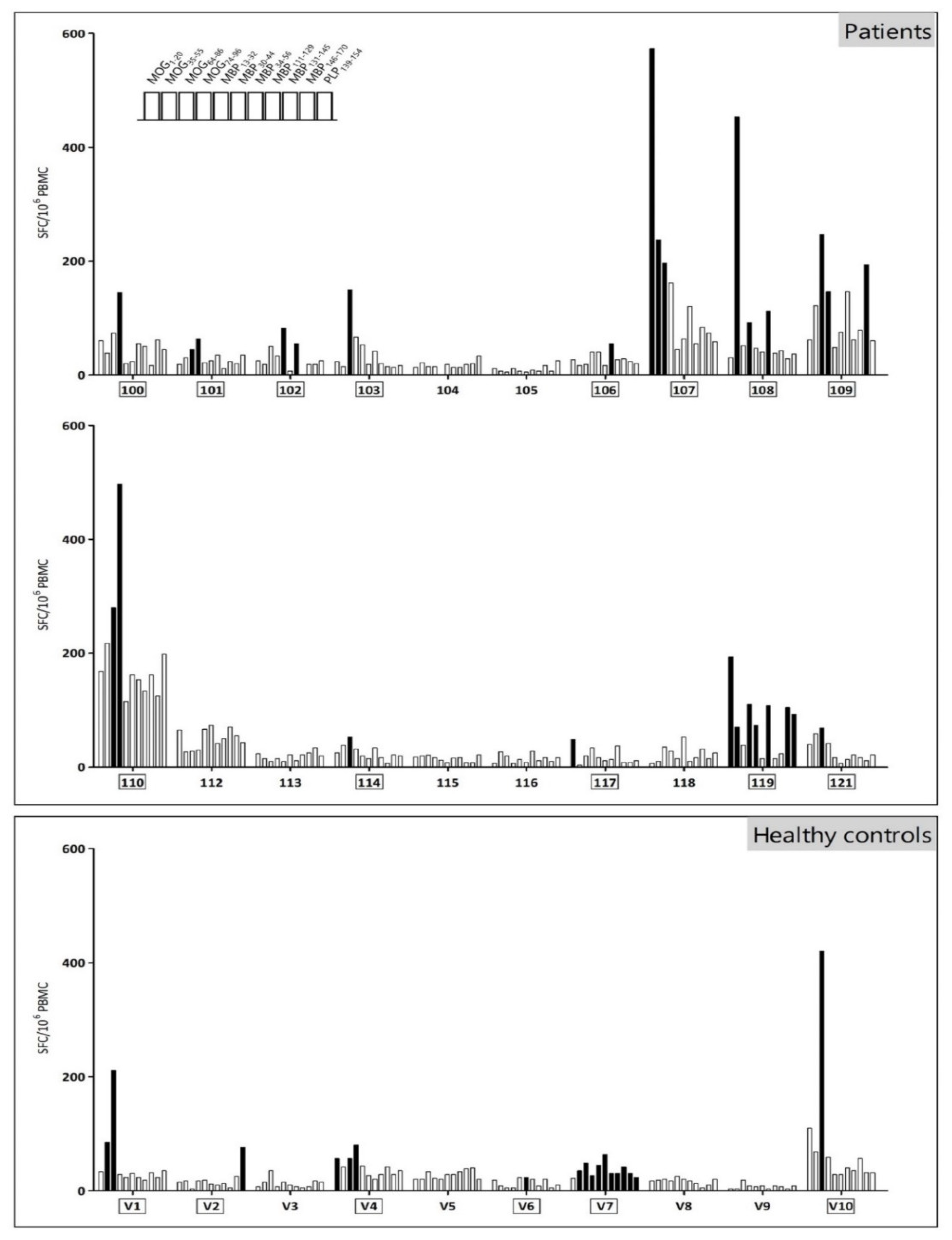
2.2. Myelin Reactivity Screening

Peripheral blood mononuclear cells (PBMC) were isolated by density gradient centrifugation (Ficoll-Plaque PLUS, GE Healthcare, Chalfont St. Giles, UK). Subsequently, PBMC were resuspended in Iscove’s Modified Dulbecco’s Medium (IMDM) supplemented with 5% human AB serum (hAB) at a concentration of 4 × 10⁶ cells/mL and were prestimulated with a peptide mix containing the following myelin peptides: MOG1–20, MOG35–55 (both Hemmo Pharmaceuticals Private Limited, Mumbai, India), MOG64–86, MOG74–96 (both Severn Biotech Ltd., Kidderminster, UK), MBP13–32, MBP34–56, MBP111–129, MBP146–170 (all Hemmo Pharmaceuticals), MBP30–44, MBP131–145 (both Pepscan, Lelystad, the Netherlands) and PLP139–154 (Hemmo Pharmaceuticals), at a concentration of 10 nM for each peptide. After 6 days, PBMC were tested for myelin reactivity using direct interferon (IFN)-γ enzyme-linked immune absorbent spot (ELISPOT) (Mabtech, NackaStrand, Sweden), according to the manufacturer’s instructions. For this, 2 × 10⁵ cells were restimulated per well with each peptide individually (10 nM) for 18 h at 37 °C in a 96-well plate (Millipore, Bedford, MA, USA). Experiments were performed in triplicate. Unstimulated PBMC served as a negative control. PBMC stimulated with 1 µg/mL phytohemagglutinin (PHA; Sigma-Aldrich, Bornem, Belgium) were used as a positive control. Frequencies of IFN-γ-secreting cells were calculated based on the number of spots counted using an automated AID ELISPOT Reader system (AID GmbH, Strassberg, Germany) and analyzed using AID ELISPOT software version 5.0. A peptide-specific response was defined as present when >50% of the peptide-restimulated wells were positive. A well was defined as positive when the number of spot-forming cells in the well was higher than [mean spot forming cells + 3 standard deviations (S.D.)] of the negative control wells. A subject displaying at least one peptide-specific response was defined as a responder.

2.3. HLA Class II Haplotyping

HLA typing was performed for the following loci: HLA-DRB1, -DRB3, -DRB4, -DRB5, -DQA1, -DQB1, -DPA1 and -DPB1. For this, samples were tested with an IMMUCOR® MIA FORA NGS HLA Typing kit (Immucor, Georgia, USA) on the Illumina MiSeq platform (Illumina, Inc., San Diego, CA, USA) and analyzed with MIA FORA IMMUCOR® MIA FORA NGS HLA Typing kit software v3.1.

2.4. Data Analysis

Data were analyzed using Graphpad Prism software version 5.01 (Graphpad, San Diego, CA, USA). For comparisons, Fisher’s exact test, unpaired students t-test, two-way analysis of variance (ANOVA) and Pearson’ correlation test were used when appropriate. Any p-value < 0.05 was considered as statistically significant.

3. Results

3.1. Study Subjects’ Characteristics

Twenty relapsing-remitting MS patients (12 female, 8 male) with a median age of 44 years (range: 24–69 years) and a median Expanded Disability Status Scale (EDSS) score of 2.0 (range: 0–6.5) were included (Table 1). Two patients had active MS, as defined by the presence of either clinical or radiological disease progression during the last 12 months. Fourteen patients were treated with first-line disease-modifying treatments, while six patients did not receive any treatment. Ten age- and gender-matched healthy controls (6 female, 4 male) with a median age of 42.5 years (range: 22–64) were included.

3.2. The Myelin Reactivity Profile of MS Patients and Healthy Controls Displays a High Inter- and Intramolecular Variability

Using IFN-γ ELISPOT, we analyzed the myelin reactivity pattern for MOG, MBP and PLP. Thirteen out of 20 patients (65.0%) and 6 out of 10 healthy controls (60.0%) showed reactivity to at least one of the myelin-derived peptides and were defined as myelin-specific responder (Figure 1 and Table 2). High inter- and intramolecular variability of myelin-specific responses were observed. The majority of myelin-specific responses was directed towards MOG epitopes both in MS patients (19 out of 28 positive responses) and in healthy volunteers (9 out of 18 positive responses), with MOG64–86 being the peptide recognized most in all study subjects. MBP-responders were scarcer (5 out of 20 patients and 2 out of 10 healthy controls), and only one PLP139–154 responder could be detected among the MS patients. No significant difference in the number of responses between MS patients and healthy controls could be observed (mean number of responses among MS patients 1.4; among healthy controls 1.8; p = 0.87), neither did the number of subjects responding to multiple myelin peptides differ between MS patients (7 out of 13 patients; 53.8%) and healthy controls (3 out of 6 healthy controls; 50.0%). Additionally, large variations in the frequency of IFN-γ-secreting myelin-reactive T cells were observed, ranging from 3 to 573 spot-forming cells/106 PBMC (Figure 1). Neither the number of myelin-specific responses nor the frequency of myelin-reactive T cells were influenced by disease duration (R2 0.00005, p = 0.98 for number of myelin-specific responses; R2 varying between 0.004 to 0.06 among the 11 peptides analyzed; all p > 0.05) or EDSS score (R2 0.003; p = 0.81 for number of myelin-specific responses; R2 varying between 0.0002 to 0.05 among the 11 peptides analyzed; all p > 0.05). No effect of gender could be demonstrated for the presence of any myelin response, nor on the total number of myelin responses.

3.3. The Majority of MS Patients Carries MS-Associated HLA Risk Alleles

Next, we analyzed the HLA class II genotype of the included subjects, focusing on HLA-DRB1, -DRB3, -DRB4, -DRB5, -DQA1, -DQB1, -DPA1 and -DPB1 (Table 3). Eight out of 20 patients (40.0%) carried the HLA-DR15 haplotype, consisting of HLA-DRB1*15:01, HLA-DQA1*01:02 and HLA-DQB1*06:02. In contrast, only 1 out of 10 healthy controls (10.0%; p = 0.20) carried the HLA-DR15 haplotype. This resulted in an odds ratio for HLA-DR15 carriers of 6.00 (95%-confidence interval [0.6315–57.0059]; p = 0.11). Moreover, 14 out of 20 patients (70.0%) and 2 out of 10 healthy controls (20.0%; p = 0.02) carried at least one of the MS-associated alleles.

3.4. The HLA Class II Genotype Does Not Affect Myelin-Specific Responsiveness

Since we demonstrated no differences between the myelin-specific reactivity profile or HLA class II genotype among MS patients and healthy controls, results from both subject groups were pooled to analyze the possible association between the myelin-specific reactivity profile and HLA class II genotype in our study population. Both carriers and non-carriers of the primordial MS-associated HLA risk alleles, i.e., HLA-DRB1*15:01, HLA-DQA1*01:02 and HLA-DQB1*06:02—either alone or combined in the HLA-DR15 haplotype—demonstrated myelin responsiveness to at least one of the tested peptides in the same extent (Table 4). Additionally, no differences in the presence of a myelin-specific response could be detected between carriers and non-carriers of the HLA-DRB3 allele (14 responders out of 22 carriers versus 5 responders out of 8 non-carriers; p = 1.0), the HLA-DRB4 allele (5 responders out of 9 carriers versus 14 responders out of 21 non-carriers; p = 1.0) or the HLA-DPB1*03:01 allele (2 responders out of 3 carriers versus 17 responders out of 27 non-carriers; p = 1.0). Equally, no difference in the number of responders to the individual peptides, nor in the frequency of myelin peptide-reactive precursor cells, could be detected between carriers and non-carriers of HLA-DRB1*15:01, HLA-DQA1*01:02 and HLA-DQB1*06:02, individually or combined as the HLA-DR15 haplotype (Table 4).

4. Discussion

Identifying patients that would benefit from treatment with a particular therapy may aid in future patient-tailored therapeutic management of the disease. Especially for antigen-specific therapies, predicting the myelin-specific reactivity profile would allow patient stratification depending on the risk for epitope spreading and choice of antigens for tolerance-inducing treatment strategies. In this context, HLA haplotype may be an interesting parameter for patient stratification, given that some myelin peptides are HLA-restricted [11,12], meaning that they are preferentially presented by specific HLA-molecules. Thus far, different studies investigated the myelin reactivity of a selected group of HLA-DR15-positive patients [25,26,27]. However, no association between the myelin reactivity of carriers and non-carriers of this risk haplotype could be made. Nonetheless, only a few myelin peptides have been demonstrated to be restricted to HLA-DR15, including MBP84–102 [27,28], whereas other myelin peptides, including MBP149–170 [28], show no specific HLA-DR15 restriction. Hence, additional HLA haplotypes may be of interest for the prediction of myelin-specific reactivity.
In this study, we aimed to characterize the myelin reactivity and HLA class II profile in a Belgian population of MS patients and to evaluate a possible association between precursor frequency of myelin-reactive T cells and HLA haplotype. Nonetheless, we were not able to demonstrate an association between myelin responsiveness and the primordial MS-associated risk alleles of the HLA-DR15 haplotype, consisting of HLA-DRB1*15:01, HLA-DQA1*01:02 and HLA-DQB1*06:02 [15]. This is in line with findings from Bronge et al., who equally did not detect a difference in T cell response towards MOG protein between HLA-DRB1*15:01 carriers and non-carriers [29]. Similarly, we did not find an association between additional MS-associated alleles HLA-DRB3, HLA-DRB4 and HLA-DPB1*03:01 and myelin-specific reactivity.
The complex etiology of MS comprises both genetic and environmental factors [30,31]. The genetic risk for MS is strongly associated with HLA class II alleles, of which the HLA-DR15 haplotype has been studied extensively as a genetic risk factor for MS. The strong association between this haplotype and MS has been consistently reported in many studies [13]. However, the prevalence of HLA risk alleles depends on the geographical location [15]. Recently, Lysandropoulos et al. have analyzed the frequencies of HLA class I and II alleles in a population of 119 Belgian MS patients and 124 healthy controls [32]. They found the HLA-DRB1*15 allele to be more frequent in MS patients, namely 45.4% of MS patients as compared to 24.2% of healthy controls (p = 0.01). Moreover, the DRB1*15-DQB1*06 haplotype was more frequently observed in MS patients as compared to healthy controls (44.5% versus 23.4%, p = 0.01). Similar frequencies were found in this Belgian cohort, i.e., 45.0% of the patients (9/20) carried the HLA-DRB1*15:01 allele, whereas 40.0% of the patients (8/20) carried the HLA-DRB1*15:01-DQB1*06:02 haplotype. In contrast, only 10.0% (1/10) of healthy controls was a carrier of the HLA-DRB1*15:01 allele and of the HLA-DRB1*15:01-DQB1*06:02 haplotype.
In addition, we characterized the HLA class II profile of patients in more depth by analyzing the HLA-DRB3, -DRB4, -DRB5, -DQA1, -DPA1 and -DPB1 loci. The DRB3, DRB4 and DRB5 loci are presumed to act as modulators of the DRB1-associated MS risk [33]. In particular, the strong linkage disequilibrium between HLA-DRB1*15:01 and HLA-DRB5 is well-described [34] and was also confirmed in our hands, as indicated by the fact that all HLA-DRB5*01:01 carriers were HLA-DRB1*15:01 carriers. It has been hypothesized that HLA-DRB5 acts as a modifier of progression, rather than a factor for MS susceptibility, since HLA-DRB5*null patients are at higher risk for the development of secondary progressive MS [35]. Since only RR-MS patients were included in this study, long-term follow-up of our patient cohort would be of interest to evaluate the HLA-DRB5*null genotype as a risk factor for secondary progressive MS. Furthermore, we report a prevalence of 65.0% (13 out of 20 patients, of which two homozygous) for the HLA-DRB3 allele. The HLA-DRB3 gene, expressed as DR52 on the cell membrane, is in linkage as a haplotype with DRB1*03:01 and DRB1*13:03 [13]. Previously, others found 25% of Brazilian MS patients carrying the HLA-DRB3 allele [36]. Whether the difference reflects geographical variations or is due to the detection technique used needs to be elucidated. The HLA-DRB4 gene, on the other hand, expresses the DR53 antigen and is in linkage as a haplotype with DRB1*04:05 [13]. Interestingly, the HLA-DR53 haplotype was previously shown to be modestly protective for MS in a Finnish population (OR = 0.5; MS patients versus healthy controls) [33], but could not be detected in our Belgian patient cohort.
Overall, a myelin-specific IFN-γ response could be detected in 13 out of 20 patients (65.0%). Previously, Grau-Lopez et al. detected a positive proliferative reaction to a mix of seven myelin-derived peptides (MBP13–32, MBP111–129, MBP146–170, MBP83–99, MOG1–20, MOG35–55 and PLP139–154) in 74% of MS patients [37]. In our hands, only 6 out of 20 (33.3%) of MS patients demonstrated a positive response to these peptides. Whether the observed differences in myelin responsiveness are due to the use of a different detection technique (IFN-γ response versus proliferation test), or as a reflection of the high variability of myelin response among patient cohorts, needs further investigation. In addition, we tested T cell reactivity towards the MBP-peptides, MBP30–44 and MBP131–145. These so-called naturally-processed peptides [38] are more likely to be involved in disease pathogenesis in comparison with non-naturally-processed or cryptic epitopes [39]. However, in our hands, responses towards these naturally-processed peptides could only be demonstrated in healthy controls and not in MS patients. Hence, the exact role of these peptides in MS pathogenesis needs further investigation. In this context, additional cytokine read-out could help to further elucidate the specific nature of immune responses towards these peptides. The presence of myelin-reactive T cells could be demonstrated in both MS patients and healthy controls. The fact that myelin-reactive T cells appear to be part of the normal T cell repertoire implies that the culprit for their pathophysiological role in MS is dependent on other factors, such as frequency or functional properties. However, regarding frequency, both similar [40,41] and higher [29,42,43,44,45,46] numbers of myelin-reactive T cells have been described in MS patients compared to healthy controls. This contrast in the obtained results may be caused by differences in detection technique (proliferation versus cytokine release assays) or by different antigen sources (e.g., full-length protein- versus peptide-based assays, naturally-obtained versus recombinantly-produced protein). Indeed, high-sensitivity assays with minimal risk of increase in background signal due to contamination of the antigen source are needed in the light of the anticipated low frequency of MOG-reactive T cells. In this context, Bronge et al. have recently detected a higher number of MOG-reactive T cells in MS patients versus healthy controls, using a sensitive cytokine release assay with bead bound-MOG [29]. Moreover, they demonstrated that MOG-reactive T cells are present in almost half of MS patients. Concerning functional properties, Cao et al. recently demonstrated that myelin-reactive T cells from MS patients tend to produce primarily proinflammatory cytokines (IFN-γ, IL-17 and GM-CSF) compared to T cells from healthy subjects, producing anti-inflammatory IL-10 [47]. This may point towards functional differences between antigen-specific T cells from MS patients and healthy controls as additional driving factor in disease development.
There are a few limitations to this study. Firstly, this study was a cohort study investigating the HLA haplotype and myelin responsiveness of MS patients and healthy controls at one moment in time. Longitudinal follow-up of myelin reactivity of this cohort could be of interest to further evaluate epitope spreading. Secondly, our study was powered to detect a large effect size of HLA class II genotype on the myelin reactivity profile. Confirmation of our findings in larger cohorts would allow statistical evaluation of smaller effect sizes. Thirdly, myelin-specific responsiveness was based on secretion of one cytokine upon restimulation with myelin peptides, i.e., IFN-γ, given the established importance of this cytokine in MS pathogenesis [48]. For future research, further characterization of the myelin-specific response, including GM-CSF, IL-17 and IL-10 secretion and myelin-specific proliferation, may provide more information regarding the potential pathogenic nature of specific myelin responses.
In conclusion, the results of this study show that the majority of MS patients carries MS risk alleles and displays responsiveness to multiple myelin-derived epitopes. However, no association between HLA haplotype and myelin reactivity could be demonstrated in our hands. These findings are of importance for the development of new antigen-specific therapeutic interventions, aiming to restore tolerance towards myelin-derived proteins. While these antigen-specific strategies are gaining interest as a potential treatment option for MS, the use of different myelin-derived peptides, of which some are HLA-restricted [49,50], may not be sufficient to fully address the wide variation in myelin-specific responses and HLA profiles in a heterogeneous group of MS patients. Therefore, next-generation antigen-specific treatment approaches may benefit from the use of other strategies for antigen administration, including nucleic acids encoding myelin proteins, either by direct vaccination or by administration of nucleic acid-transfected antigen-presenting cells. In doing so, induction of tolerance may be achieved by targeting myelin-reactive T cells in a multi-epitope and HLA-unrestricted manner.

Author Contributions

Conceptualization, J.D. and N.C.; Data curation, J.D., I.N., N.O., L.V.B., M.-P.E. and L.D.; Formal analysis, J.D., N.O., M.-P.E., L.D. and N.C.; Funding acquisition, Z.N.B. and N.C.; Investigation, J.D. and N.C.; Methodology, J.D. and N.C.; Resources, T.R., B.W., P.C. and N.C.; Supervision, B.W., P.C., Z.N.B. and N.C.; Writing—original draft, J.D.; Writing—review and editing, J.D., I.N., N.O., L.V.B., M.-P.E., L.D., T.R., B.W., P.C., Z.N.B. and N.C. All authors have read and agreed to the published version of the manuscript.

Funding

This study has received funding from the European Union’s Horizon 2020 research and innovation program under grant agreement. Further support was provided by the Methusalem Funding Program from the University of Antwerp, by an applied biomedical research project of the Institute for the Promotion of Innovation by Science and Technology in Flanders (IWT-TBM 140191) and by the Belgian Charcot Foundation. Furthermore, the authors received research funding from Roche Belgium. Judith Derdelinckx recently held a PhD fellowship from the Research Foundation Flanders (FWO). Barbara Willekens currently holds a clinical PhD fellowship from the Research Foundation Flanders (FWO 1701919N).

Acknowledgments

We thank all the patients and healthy controls that contributed to this preclinical research. Our gratitude goes to all members of the Histocompatibility and Immunogenetics Laboratory (HILA, Red Cross-Flanders) for their contribution to the next generation sequencing experiments.

Conflicts of Interest

The authors declare no conflict of interest.

References

  1. Hohlfeld, R.; Dornmair, K.; Meinl, E.; Wekerle, H. The search for the target antigens of multiple sclerosis, part 1: Autoreactive CD4+ T lymphocytes as pathogenic effectors and therapeutic targets. Lancet Neurol. 2015, 15, 198–209. [Google Scholar] [CrossRef]
  2. Sáez-Torres, I.; Brieva, L.; Espejo, C.; Barrau, M.A.; Montalban, X.; Martínez-Caceres, E.M. Specific proliferation towards myelin antigens in patients with multiple sclerosis during a relapse. Autoimmunity 2002, 35, 45–50. [Google Scholar] [CrossRef] [PubMed]
  3. Greer, J.M. Autoimmune T-cell reactivity to myelin proteolipids and glycolipids in multiple sclerosis. Mult. Scler. Int. 2013, 2013, 151427. [Google Scholar] [CrossRef] [PubMed] [Green Version]
  4. Goebels, N.; Hofstetter, H.; Schmidt, S.; Brunner, C.; Wekerle, H.; Hohlfeld, R. Repertoire dynamics of autoreactive T cells in multiple sclerosis patients and healthy subjects: Epitope spreading versus clonal persistence. Brain 2000, 123, 508–518. [Google Scholar] [CrossRef] [PubMed] [Green Version]
  5. Tuohy, V.K.; Kinkel, R.P. Epitope spreading: A mechanism for progression of autoimmune disease. Arch. Immunol. Ther. Exp. 2000, 48, 347–351. [Google Scholar]
  6. Tuohy, V.K.; Yu, M.; Yin, L.; Kawczak, J.A.; Johnson, J.M.; Mathisen, P.M.; Weinstock-Guttman, B.; Kinkel, R.P. The epitope spreading cascade during progression of experimental autoimmune encephalomyelitis and multiple sclerosis. Immunol. Rev. 1998, 164, 93–100. [Google Scholar] [CrossRef]
  7. Davies, S.; Nicholson, T.; Laura, M.; Giovannoni, G.; Altmann, D.M. Spread of T lymphocyte immune responses to myelin epitopes with duration of multiple sclerosis. J. Neuropathol. Exp. Neurol. 2005, 64, 371–377. [Google Scholar] [CrossRef] [Green Version]
  8. Hilkens, C.M.; Isaacs, J.D.; Thomson, A.W. Development of dendritic cell-based immunotherapy for autoimmunity. Int. Rev. Immunol. 2010, 29, 156–183. [Google Scholar] [CrossRef]
  9. Yu, M.; Johnson, J.M.; Tuohy, V.K. A predictable sequential determinant spreading cascade invariably accompanies progression of experimental autoimmune encephalomyelitis: A basis for peptide-specific therapy after onset of clinical disease. J. Exp. Med. 1996, 183, 1777–1788. [Google Scholar] [CrossRef] [Green Version]
  10. Derdelinckx, J.; Mansilla, M.J.; De Laere, M.; Lee, W.P.; Navarro-Barriuso, J.; Wens, I.; Nkansah, I.; Daans, J.; De Reu, H.; Jolanta Keliris, A.; et al. Clinical and immunological control of experimental autoimmune encephalomyelitis by tolerogenic dendritic cells loaded with MOG-encoding mRNA. J. Neuroinflamm. 2019, 16, 167. [Google Scholar] [CrossRef] [Green Version]
  11. Corthay, A. A three-cell model for activation of naive T helper cells. Scand. J. Immunol. 2006, 64, 93–96. [Google Scholar] [CrossRef] [PubMed]
  12. Fletcher, J.M.; Lalor, S.J.; Sweeney, C.M.; Tubridy, N.; Mills, K.H. T cells in multiple sclerosis and experimental autoimmune encephalomyelitis. Clin. Exp. Immunol. 2010, 162, 1–11. [Google Scholar] [CrossRef] [PubMed]
  13. Hollenbach, J.A.; Oksenberg, J.R. The immunogenetics of multiple sclerosis: A comprehensive review. J. Autoimmun. 2015, 64, 13–25. [Google Scholar] [CrossRef] [PubMed] [Green Version]
  14. Sawcer, S.; Hellenthal, G.; Pirinen, M.; Spencer, C.C.; Patsopoulos, N.A.; Moutsianas, L.; Dilthey, A.; Su, Z.; Freeman, C.; Hunt, S.E.; et al. Genetic risk and a primary role for cell-mediated immune mechanisms in multiple sclerosis. Nature 2011, 476, 214–219. [Google Scholar] [PubMed]
  15. Schmidt, H.; Williamson, D.; Ashley-Koch, A. HLA-DR15 haplotype and multiple sclerosis: A HuGE review. Am. J. Epidemiol. 2007, 165, 1097–1109. [Google Scholar] [CrossRef]
  16. Moutsianas, L.; Jostins, L.; Beecham, A.H.; Dilthey, A.T.; Xifara, D.K.; Ban, M.; Shah, T.S.; Patsopoulos, N.A.; Alfredsson, L.; Anderson, C.A.; et al. Class II HLA interactions modulate genetic risk for multiple sclerosis. Nat. Genet. 2015, 47, 1107–1113. [Google Scholar]
  17. Field, J.; Browning, S.R.; Johnson, L.J.; Danoy, P.; Varney, M.D.; Tait, B.D.; Gandhi, K.S.; Charlesworth, J.C.; Heard, R.N.; Australia and New Zealand Multiple Sclerosis Genetics Consortium; et al. A polymorphism in the HLA-DPB1 gene is associated with susceptibility to multiple sclerosis. PLoS ONE 2010, 5, e13454. [Google Scholar] [CrossRef] [Green Version]
  18. O’Sullivan, D.; Arrhenius, T.; Sidney, J.; Del Guercio, M.F.; Albertson, M.; Wall, M.; Oseroff, C.; Southwood, S.; Colón, S.M.; Gaeta, F.C.; et al. On the interaction of promiscuous antigenic peptides with different DR alleles. Identification of common structural motifs. J. Immunol. 1991, 147, 2663–2669. [Google Scholar]
  19. Wucherpfennig, K.W.; Strominger, J.L. Selective binding of self peptides to disease-associated major histocompatibility complex (MHC) molecules: A mechanism for MHC-linked susceptibility to human autoimmune diseases. J. Exp. Med. 1995, 181, 1597–1601. [Google Scholar] [CrossRef]
  20. Busch, R.; Strang, G.; Howland, K.; Rothbard, J.B. Degenerate binding of immunogenic peptides to HLA-DR proteins on B cell surfaces. Int. Immunol. 1990, 2, 443–451. [Google Scholar] [CrossRef]
  21. Valli, A.; Sette, A.; Kappos, L.; Oseroff, C.; Sidney, J.; Miescher, G.; Hochberger, M.; Albert, E.D.; Adorini, L. Binding of myelin basic protein peptides to human histocompatibility leukocyte antigen class II molecules and their recognition by T cells from multiple sclerosis patients. J. Clin. Investig. 1993, 91, 616–628. [Google Scholar] [CrossRef] [PubMed] [Green Version]
  22. Fridkis-Hareli, M.; Teitelbaum, D.; Gurevich, E.; Pecht, I.; Brautbar, C.; Kwon, O.J.; Brenner, T.; Arnon, R.; Sela, M. Direct binding of myelin basic protein and synthetic copolymer 1 to class II major histocompatibility complex molecules on living antigen-presenting cells—Specificity and promiscuity. Proc. Natl. Acad. Sci. USA 1994, 91, 4872–4876. [Google Scholar] [CrossRef] [PubMed] [Green Version]
  23. Weissert, R.; Kuhle, J.; de Graaf, K.L.; Wienhold, W.; Herrmann, M.M.; Müller, C.; Forsthuber, T.G.; Wiesmüller, K.H.; Melms, A. High immunogenicity of intracellular myelin oligodendrocyte glycoprotein epitopes. J. Immunol. 2002, 169, 548–556. [Google Scholar] [CrossRef] [PubMed] [Green Version]
  24. Polman, C.H.; Reingold, S.C.; Banwell, B.; Clanet, M.; Cohen, J.A.; Filippi, M.; Fujihara, K.; Havrdova, E.; Hutchinson, M.; Kappos, L.; et al. Diagnostic criteria for multiple sclerosis: 2010 revisions to the McDonald criteria. Ann. Neurol. 2011, 69, 292–302. [Google Scholar] [CrossRef] [Green Version]
  25. Vergelli, M.; Kalbus, M.; Rojo, S.C.; Hemmer, B.; Kalbacher, H.; Tranquill, L.; Beck, H.; McFarland, H.F.; De Mars, R.; Long, E.O.; et al. T cell response to myelin basic protein in the context of the multiple sclerosis-associated HLA-DR15 haplotype: Peptide binding, immunodominance and effector functions of T cells. J. Neuroimmunol. 1997, 77, 195–203. [Google Scholar] [CrossRef]
  26. Wallström, E.; Khademi, M.; Andersson, M.; Weissert, R.; Linington, C.; Olsson, T. Increased reactivity to myelin oligodendrocyte glycoprotein peptides and epitope mapping in HLA DR2(15)+ multiple sclerosis. Eur. J. Immunol. 1998, 28, 3329–3335. [Google Scholar] [CrossRef]
  27. Pette, M.; Fujita, K.; Wilkinson, D.; Altmann, D.M.; Trowsdale, J.; Giegerich, G.; Hinkkanen, A.; Epplen, J.T.; Kappos, L.; Wekerle, H. Myelin autoreactivity in multiple sclerosis: Recognition of myelin basic protein in the context of HLA-DR2 products by T lymphocytes of multiple-sclerosis patients and healthy donors. Proc. Natl. Acad. Sci. USA 1990, 87, 7968–7972. [Google Scholar] [CrossRef] [Green Version]
  28. Jingwu, Z.; Medaer, R.; Hashim, G.A.; Chin, Y.; van den Berg-Loonen, E.; Raus, J.C. Myelin basic protein-specific T lymphocytes in multiple sclerosis and controls: Precursor frequency, fine specificity, and cytotoxicity. Ann. Neurol. 1992, 32, 330–338. [Google Scholar] [CrossRef]
  29. Bronge, M.; Ruhrmann, S.; Carvalho-Queiroz, C.; Nilsson, O.B.; Kaiser, A.; Holmgren, E.; Macrini, C.; Winklmeier, S.; Meinl, E.; Brundin, L.; et al. Myelin oligodendrocyte glycoprotein revisited-sensitive detection of MOG-specific T-cells in multiple sclerosis. J. Autoimmun. 2019, 102, 38–49. [Google Scholar] [CrossRef]
  30. Olsson, T.; Barcellos, L.F.; Alfredsson, L. Interactions between genetic, lifestyle and environmental risk factors for multiple sclerosis. Nat. Rev. Neurol. 2017, 13, 25–36. [Google Scholar] [CrossRef]
  31. Waubant, E.; Lucas, R.; Mowry, E.; Graves, J.; Olsson, T.; Alfredsson, L.; Langer-Gould, A. Environmental and genetic risk factors for MS: An integrated review. Ann. Clin. Transl. Neurol. 2019, 6, 1905–1922. [Google Scholar] [CrossRef] [PubMed]
  32. Lysandropoulos, A.P.; Racapé, J.; Holovska, V.; Toungouz, M. Human leucocyte antigen (HLA) class I and II typing in Belgian multiple sclerosis patients. Acta Neurol. Belg. 2017, 117, 61–65. [Google Scholar] [CrossRef] [PubMed]
  33. Luomala, M.; Elovaara, I.; Ukkonen, M.; Koivula, T.; Lehtimäki, T. The combination of HLA-DR1 and HLA-DR53 protects against MS. Neurology 2001, 56, 383–385. [Google Scholar] [CrossRef] [PubMed]
  34. Creary, L.E.; Mallempati, K.C.; Gangavarapu, S.; Caillier, S.J.; Oksenberg, J.R.; Fernández-Viňa, M.A. Deconstruction of HLA-DRB1*04:01:01 and HLA-DRB1*15:01:01 class II haplotypes using next-generation sequencing in European-Americans with multiple sclerosis. Mult. Scler. 2018, 25, 772–782. [Google Scholar] [CrossRef] [PubMed]
  35. Caillier, S.J.; Briggs, F.; Cree, B.A.; Baranzini, S.E.; Fernandez-Viña, M.; Ramsay, P.P.; Khan, O.; Royal, W., 3rd; Hauser, S.L.; Barcellos, L.F.; et al. Uncoupling the roles of HLA-DRB1 and HLA-DRB5 genes in multiple sclerosis. J. Immunol. 2008, 181, 5473–5480. [Google Scholar] [CrossRef] [PubMed] [Green Version]
  36. Brum, D.G.; Barreira, A.A.; dos Santos, A.C.; Kaimen-Maciel, D.R.; Matiello, M.; Costa, R.M.; Deghaide, N.H.; Costa, L.S.; Louzada-Junior, P.; Diniz, P.R.; et al. HLA-DRB association in neuromyelitis optica is different from that observed in multiple sclerosis. Mult. Scler. 2010, 16, 21–29. [Google Scholar] [CrossRef] [PubMed]
  37. Grau-López, L.; Raïch, D.; Ramo-Tello, C.; Naranjo-Gómez, M.; Dávalos, A.; Pujol-Borrell, R.; Borràs, F.E.; Martínez-Cáceres, E. Specific T-cell proliferation to myelin peptides in relapsing-remitting multiple sclerosis. Eur. J. Neurol. 2011, 18, 1101–1104. [Google Scholar] [CrossRef]
  38. Streeter, H.B.; Rigden, R.; Martin, K.F.; Scolding, N.J.; Wraith, D.C. Preclinical development and first-in-human study of ATX-MS-1467 for immunotherapy of MS. Neurol. Neuroimmunol. NeuroInflamm. 2015, 2, e93. [Google Scholar] [CrossRef] [Green Version]
  39. Anderton, S.M.; Viner, N.J.; Matharu, P.; Lowrey, P.A.; Wraith, D.C. Influence of a dominant cryptic epitope on autoimmune T cell tolerance. Nat. Immunol. 2002, 3, 175–181. [Google Scholar] [CrossRef]
  40. Hellings, N.; Barée, M.; Verhoeven, C.; D’hooghe, M.B.; Medaer, R.; Bernard, C.C.; Raus, J.; Stinissen, P. T-cell reactivity to multiple myelin antigens in multiple sclerosis patients and healthy controls. J. Neurosci. Res. 2001, 63, 290–302. [Google Scholar] [CrossRef]
  41. Van der Aa, A.; Hellings, N.; Medaer, R.; Gelin, G.; Palmers, Y.; Raus, J.; Stinissen, P. T cell vaccination in multiple sclerosis patients with autologous CSF-derived activated T cells: Results from a pilot study. Clin. Exp. Immunol. 2003, 131, 155–168. [Google Scholar] [CrossRef] [PubMed]
  42. Sun, J.; Link, H.; Olsson, T.; Xiao, B.G.; Andersson, G.; Ekre, H.P.; Linington, C.; Diener, P. T and B cell responses to myelin-oligodendrocyte glycoprotein in multiple sclerosis. J. Immunol. 1991, 146, 1490–1495. [Google Scholar] [PubMed]
  43. Sun, J.B.; Rask, C.; Olsson, T.; Holmgren, J.; Czerkinsky, C. Treatment of experimental autoimmune encephalomyelitis by feeding myelin basic protein conjugated to cholera toxin B subunit. Proc. Natl. Acad. Sci. USA 1996, 93, 7196–7201. [Google Scholar] [CrossRef] [PubMed] [Green Version]
  44. Raddassi, K.; Kent, S.C.; Yang, J.; Bourcier, K.; Bradshaw, E.M.; Seyfert-Margolis, V.; Nepom, G.T.; Kwok, W.W.; Hafler, D.A. Increased frequencies of myelin oligodendrocyte glycoprotein/MHC class II-binding CD4 cells in patients with multiple sclerosis. J. Immunol. 2011, 187, 1039–1046. [Google Scholar] [CrossRef] [Green Version]
  45. Tejada-Simon, M.V.; Hong, J.; Rivera, V.M.; Zhang, J.Z. Reactivity pattern and cytokine profile of T cells primed by myelin peptides in multiple sclerosis and healthy individuals. Eur. J. Immunol. 2001, 31, 907–917. [Google Scholar] [CrossRef]
  46. Bielekova, B.; Sung, M.H.; Kadom, N.; Simon, R.; McFarland, H.; Martin, R. Expansion and functional relevance of high-avidity myelin-specific CD4+ T cells in multiple sclerosis. J. Immunol. 2004, 172, 3893–3904. [Google Scholar] [CrossRef] [Green Version]
  47. Cao, Y.; Goods, B.A.; Raddassi, K.; Nepom, G.T.; Kwok, W.W.; Love, J.C.; Hafler, D.A. Functional inflammatory profiles distinguish myelin-reactive T cells from patients with multiple sclerosis. Sci. Transl. Med. 2015, 7, 287ra74. [Google Scholar] [CrossRef] [Green Version]
  48. Lees, J.R.; Cross, A.H. A little stress is good: IFN-gamma, demyelination, and multiple sclerosis. J. Clin. Investig. 2007, 117, 297–299. [Google Scholar] [CrossRef] [Green Version]
  49. Berthelot, L.; Laplaud, D.A.; Pettré, S.; Ballet, C.; Michel, L.; Hillion, S.; Braudeau, C.; Connan, F.; Lefrère, F.; Wiertlewski, S.; et al. Blood CD8+ T cell responses against myelin determinants in multiple sclerosis and healthy individuals. Eur. J. Immunol. 2008, 38, 1889–1899. [Google Scholar] [CrossRef]
  50. Martin, R.; Jaraquemada, D.; Flerlage, M.; Richert, J.; Whitaker, J.; Long, E.O.; McFarlin, D.E.; McFarland, H.F. Fine specificity and HLA restriction of myelin basic protein-specific cytotoxic T cell lines from multiple sclerosis patients and healthy individuals. J. Immunol. 1990, 145, 540–548. [Google Scholar]
Figure 1. Myelin reactivity screening in 10 healthy controls and 20 MS patients, untreated or treated with first-line immunomodulatory therapy. Per patient or healthy control, each bar represents the IFN-γ response towards the different peptides in the following order: MOG1–20-MOG35–55-MOG64–86-MOG74–96-MBP13–32-MBP34–56-MBP111–129-MBP146–170-MBP30–44-MBP131–145-PLP139–154. Black bars indicate a positive peptide response, defined as present when >50% of the peptide-restimulated wells were positive. A well was defined as positive when the number of spot-forming cells in the well was higher than the [mean spot forming cells + 3 standard deviations (S.D.)] of the negative control wells. Responder subjects are denoted by a box around the subject code; a responder was defined as a subject demonstrating at least one positive myelin peptide response. Abbreviations used: SFC, spot forming cells; PBMC, peripheral blood mononuclear cells; MOG, myelin oligodendrocyte glycoprotein; MBP, myelin basic protein; PLP, proteolipid protein.
Cells 09 02703 g001
Table 1. Clinical details of the study subjects included into the study.
MS PatientsHealthy Controls
CodeGenderAgeDisease Duration (Years)EDSS ScoreCurrent Treatment (Treatment Duration in Years)Active MSCodeGenderAge
100male62256.5glatiramer acetate (16)noV1male23
101female69326.0-noV2male46
102female39121.5dimethyl fumarate (0.5)noV3female39
103female49143.0-yesV4male27
104female5242.5glatiramer acetate (0.5)noV5female50
105male31101.0interferon-β (9)noV6female56
106female66232.0glatiramer acetate (24)noV7female22
107female2732.0-yesV8female34
108female2450dimethyl fumarate (5)noV9female64
109male62192.5interferon-β (19)noV10male64
110male53163.0interferon-β (8)no
112male2422.5dimethyl fumarate (2)no
113female27112.0dimethyl fumarate (3)no
114male4612.0-no
115male5011.0dimethyl fumarate (1)no
116female3051.0dimethyl fumarate (3)no
117male53163.5-no
118female2810-no
119female4112.0dimethyl fumarate (1)no
121female4292.0glatiramer acetate (9)no
Median
(range)
44
(24–69)
9.5
(1–32)
2.0
(0–6.5)
42.5
(22–64)
Active multiple sclerosis (MS) was defined as the occurrence of at least one clinical relapse in the previous year and/or at least one new or enlarging T2 lesion on magnetic resonance imaging (MRI) in the previous year. Abbreviations used: RR-MS, relapsing-remitting multiple sclerosis; EDSS, Expanded Disability Status Scale.
Table 2. Myelin reactivity of study subjects.
Subject CodeMOG1–20MOG35–55MOG64–86MOG74–96MBP13–32MBP30–44MBP34–56MBP111–129MBP131–145MBP146–170PLP139–154
MS patients100---x-------
101--xx-------
102----x-x----
103--x--------
104-----------
105-----------
106------x----
107xxx--------
108-x-x--x----
109--xx-----x-
110--xx-------
112-----------
113-----------
114--x--------
115-----------
116-----------
117x----------
118-----------
119xx-xx-x--xx
121--x--------
Total n° of responses33762040021
Healthy controlsV1-xx--------
V2----------x
V3-----------
V4x-xx-------
V5-----------
V6-----x-----
V7-xxxxxxxxxx
V8-----------
V9-----------
V10--x--------
Total n° of responses12421211112
For 11 individual myelin peptides, presence of PBMC reactivity is indicated in grey. A peptide-specific response was defined as present when >50% of the peptide-restimulated wells were positive. A well was defined as positive when the number of spot-forming cells in the well was higher than the [mean spot forming cells + 3 standard deviations (S.D.)] of the negative control wells. A subject displaying at least one peptide-specific response was defined as a responder. Abbreviations used: PBMC, peripheral blood mononuclear cells; MOG, myelin oligodendrocyte glycoprotein; MBP, myelin basic protein; PLP, proteolipid protein.
Table 3. Human leukocyte antigen (HLA) class II genotyping of study subjects.
CodeDRB1DQA1DQB1DRB3DRB4DRB5DPA1DPB1
MS patients100*03:01*01:01*05:01*01:01*02:01*05:01*02:02---*01:03*01:03*04:01*02:01
101*13:02*08:03*01:02*06:01*06:09*03:01*03:01---*01:03-*02:01*16:01
102*03:01*01:02*05:01*01:01*02:01*05:01*01:01---*02:01*01:03*17:01*02:01
103*15:01*03:01*01:02*05:01*06:03*02:01*01:01--*01:01*02:01*01:03*09:01*04:01
104*11:01*15:01*05:09*01:02*03:01*06:02*02:02--*01:01*01:03*02:06*03:01*05:01
105*15:01*01:01*01:02*01:01*06:02*05:01---*01:01*01:03*02:01*04:02*14:01
106*15:01-*01:02-*06:02----*01:01*01:03-*04:01-
107*15:01*01:02*01:02*01:01*05:01*06:02---*01:01*01:03*01:03*04:02*02:01
108*15:01*08:XX*01:02*04:01*06:02*04:02---*01:01*01:03-*04:01-
109*11:01*01:01*05:05*01:01*03:01*05:01*02:02---*01:03*01:03*04:02*04:01
110*12:01*01:01*05:05*01:01*03:01*05:01*02:02---*01:03*01:03*02:01*03:01
112*11:02*10:01*05:05*01:05*03:19*05:01*02:02---*01:03*02:01*04:01*30:01
113*07:01*15:01*02:01*01:02*02:02*06:02--*01:01*01:01*01:03*02:01*04:01*11:01
114*13:02*03:01*01:02*05:01*06:04*02:01*03:01*01:01--*02:01*01:03*01:01*03:01
115*03:01*15:01*05:01*01:02*02:01*06:02*01:01--*01:01*02:01*01:03*01:01*04:01
116*15:01*08:01*01:02*04:01*06:02*04:02---*01:01*01:03*01:03*04:02-
117*04:01*03:01*03:01*05:01*03:02*02:01*01:01-*01:03-*01:03*01:03*04:01-
118*11:04*10:01*05:05*01:05*03:01*05:01*02:02---*01:03*01:03*02:01*04:02
119*07:01*01:01*02:01*01:02*03:03*05:04--*01:03:01:02N-*01:03*01:03*02:01*04:01
121*03:01*13:02*05:01*01:02*02:01*06:04*01:01*03:01--*01:03*02:02*04:01*01:01
Healthy controlsV1*07:01*03:01*02:01*05:01*02:02*02:01*01:01-*01:03-*02:01*01:03--
V2*07:01*12:01*02:01*05:05*03:03*03:01*02:02-*01:03:01:02N-*01:03*01:03--
V3*14:54*07:01*01:04*02:01*05:03*02:02*02:02-*01:01-*01:03*01:03--
V4*03:01*01:01*05:01*01:01*02:01*05:01*02:02---*01:03*01:03--
V5*07:01*03:01*02:01*05:01*02:02*02:01*01:01-*01:03:01:02N-*02:01*01:03*11:01*04:02
V6*16:01*04:01*01:02*03:03*05:02*03:02--*01:03*02:02*02:01*01:03*01:01*04:01
V7*13:01-*01:03-*06:03-*02:02*01:01--*01:03*02:02*04:02*05:01
V8*07:01*03:01*02:01*05:01*02:02*02:01*01:01-*01:03-*01:03-*04:01-
V9*03:01*15:01*05:01*01:02*02:01*06:02*01:01--*01:01*02:01*01:03--
V10*13:05*14:54*05:05*01:04*03:01*05:03*02:02*02:02--*01:03*02:01--
Patients with the HLA-DR15 haplotype (combination of the risk alleles HLA-DRB1*15:01, HLA-DQA1*01:02, HLA-DQB1*06:02) are highlighted in orange. HLA-DRB1*08:XX is a new variant, similar to DRB1*08:01, but with a point mutation in exon 3 (valine-methionin substitution). In the table, “-” refers to negative; an allele could not be detected.
Table 4. Overview of the myelin peptide-specific T cell responses among carriers and non-carriers of MS-associated HLA susceptibility alleles.
HLA-DR15HLA-DRB1*15:01HLA-DQA1*01:02HLA-DQB1*06:02
+
(9/30)
-
(21/30)
+
(10/30)
-
(20/30)
+
(15/30)
-
(15/30)
+
(9/30)
-
(21/30)
MOG
All MOGResponse21331269213
MOG1–20Response13132213
Frequency78.5 ± 185.849.2 ± 50.473.0 ± 176.050.5 ± 51.368.3 ± 147.047.7 ± 43.778.5 ± 185.849.2 ± 50.4
MOG35–55Response22233222
Frequency88.9 ± 155.145.5 ± 49.081.5 ± 148.147.0 ± 49.868.0 ± 121.149.0 ± 56.488.9 ± 155.145.5 ± 49.0
MOG64–86Response1102956110
Frequency39.5 ± 60.491.5 ± 108.850.6 ± 66.868.2 ± 93.347.7 ± 55.2104.1 ± 124.339.5 ± 60.491.5 ± 108.8
MOG74–96Response17173517
Frequency40.7 ± 52.745.8 ± 34.743.3 ± 50.349.3 ± 41.545.7 ± 45.642.9 ± 35.040.7 ± 52.745.8 ± 34.7
MBP
All MBPResponse25254325
MBP13–32Response03031203
Frequency20.1 ± 18.439.8 ± 32.123.4 ± 20.330.8 ± 20.425.9 ± 21.041.8 ± 35.620.1 ± 18.439.8 ± 32.1
MBP30–44Response02021102
Frequency21.1 ± 19.127.2 ± 19.320.8 ± 18.126.0 ±18.019.5 ± 15.131.2 ± 21.421.1 ± 19.127.2 ± 19.3
MBP34–56Response23233223
Frequency40.8 ± 45.230.6 ± 22.740.9 ± 42.632.5 ± 23.541.3 ± 39.826.1 ± 15.540.8 ± 45.230.6 ± 22.7
MBP111–129Response01010101
Frequency22.0 ± 15.924.4 ± 15.321.8 ±15.023.3 ± 15.219.4 ± 12.828.0 ± 16.622.0 ± 15.924.4 ± 15.3
MBP131–145Response01010101
Frequency27.4 ± 23.825.0 ± 17.226.1 ± 22.725.1 ±17.623.4 ± 19.028.0 ± 19.427.4 ± 23.825.0 ± 17.2
MBP146–170Response03031203
Frequency22.9 ± 21.628.7 ± 23.422.0 ± 20.630.4 ± 24.125.5 ± 28.028.3 ± 16.722.9 ± 21.628.7 ± 23.4
PLP
PLP139–154Response03031203
Frequency26.6 ± 14.630.6 ± 20.625.6 ± 14.133.3 ± 20.729.1 ± 21.729.8 ± 16.326.6 ± 14.630.6 ± 20.6
All peptides
Response to at least one peptide316415910316
The number of study subjects demonstrating a peptide-specific T cell response is indicated per MS susceptibility HLA allele. A peptide-specific response was defined as present when >50% of the peptide-restimulated wells were positive. A well was defined as positive when the number of spot-forming cells in the well was higher than the [mean spot forming cells + 3 standard deviations (S.D.)] of the negative control wells. The frequency of peptide-reactive T cells is expressed as mean ± standard deviation. No statistically significant difference in the presence of a peptide-specific T cell response or in the frequency of peptide-reactive T cells could be demonstrated between carriers and non-carriers of a particular susceptibility HLA allele, using Fisher’s exact test and a two-way ANOVA, respectively. Abbreviations used: MOG, myelin oligodendrocyte glycoprotein; MBP, myelin basic protein; PLP, proteolipid protein; PBMC, peripheral blood mononuclear cells; HLA, human leukocyte antigen.
Publisher’s Note: MDPI stays neutral with regard to jurisdictional claims in published maps and institutional affiliations.

