Next Issue
Volume 10, January
Previous Issue
Volume 9, November


Cells, Volume 9, Issue 12 (December 2020) – 196 articles

Cover Story (view full-size image): 
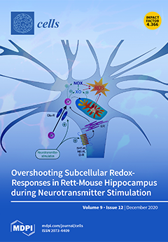
Rett syndrome (RTT) is a developmental disorder associated with impaired neuronal network function and disturbed redox homeostasis. In a mouse model of RTT, quantitative redox imaging revealed that neurotransmitters evoke intensified oxidizing responses in hippocampal neurons. Mitochondria, xanthine oxidase (XO), and NADPH oxidase (NOX) differently contribute to these cytosolic oxidations. Glutamate causes a massive Ca2+ influx, which then activates XO and NOX. In the case of dopamine, which mediates only moderate Ca2+ transients, mitochondria and NOX are the main mediators of the oxidizing response. As these exaggerated redox responses already arise in neonatal neurons, they may markedly contribute to the altered neuronal network performance and the disturbed neuronal signaling in RTT. View this paper
  • Issues are regarded as officially published after their release is announced to the table of contents alert mailing list.
  • You may sign up for e-mail alerts to receive table of contents of newly released issues.
  • PDF is the official format for papers published in both, html and pdf forms. To view the papers in pdf format, click on the "PDF Full-text" link, and use the free Adobe Reader to open them.
Order results
Result details
Section
Select all
Export citation of selected articles as:
18 pages, 683 KiB  
Review
Peripheral Neuropathic Pain: From Experimental Models to Potential Therapeutic Targets in Dorsal Root Ganglion Neurons
by Ti-Yen Yeh, I-Wei Luo, Yu-Lin Hsieh, To-Jung Tseng, Hao Chiang and Sung-Tsang Hsieh
Cells 2020, 9(12), 2725; https://doi.org/10.3390/cells9122725 - 21 Dec 2020
Cited by 19 | Viewed by 4219
Abstract
Neuropathic pain exerts a global burden caused by the lesions in the somatosensory nerve system, including the central and peripheral nervous systems. The mechanisms of nerve injury-induced neuropathic pain involve multiple mechanisms, various signaling pathways, and molecules. Currently, poor efficacy is the major [...] Read more.
Neuropathic pain exerts a global burden caused by the lesions in the somatosensory nerve system, including the central and peripheral nervous systems. The mechanisms of nerve injury-induced neuropathic pain involve multiple mechanisms, various signaling pathways, and molecules. Currently, poor efficacy is the major limitation of medications for treating neuropathic pain. Thus, understanding the detailed molecular mechanisms should shed light on the development of new therapeutic strategies for neuropathic pain. Several well-established in vivo pain models were used to investigate the detail mechanisms of peripheral neuropathic pain. Molecular mediators of pain are regulated differentially in various forms of neuropathic pain models; these regulators include purinergic receptors, transient receptor potential receptor channels, and voltage-gated sodium and calcium channels. Meanwhile, post-translational modification and transcriptional regulation are also altered in these pain models and have been reported to mediate several pain related molecules. In this review, we focus on molecular mechanisms and mediators of neuropathic pain with their corresponding transcriptional regulation and post-translational modification underlying peripheral sensitization in the dorsal root ganglia. Taken together, these molecular mediators and their modification and regulations provide excellent targets for neuropathic pain treatment. Full article
Show Figures

Figure 1

3 pages, 178 KiB  
Erratum
Erratum: Gruber, E.S.; et al. The Oncogene AF1Q is Associated with WNT and STAT Signaling and Offers a Novel Independent Prognostic Marker in Patients with Resectable Esophageal Cancer. Cells 2019, 8, 1357
by Elisabeth S. Gruber, Georg Oberhuber, Peter Birner, Michaela Schlederer, Michael Kenn, Wolfgang Schreiner, Gerd Jomrich, Sebastian F. Schoppmann, Michael Gnant, William Tse and Lukas Kenner
Cells 2020, 9(12), 2724; https://doi.org/10.3390/cells9122724 - 21 Dec 2020
Viewed by 1446
Abstract
The authors wish to make the following change to their paper [...] Full article
(This article belongs to the Special Issue Killing Cancer: Discovery and Selection of New Target Molecules)
18 pages, 2564 KiB  
Article
Endothelial Protease Activated Receptor 1 (PAR1) Signalling Is Required for Lymphocyte Transmigration across Brain Microvascular Endothelial Cells
by Silvia Dragoni, Anna Papageorgiou, Caroline Araiz, John Greenwood and Patric Turowski
Cells 2020, 9(12), 2723; https://doi.org/10.3390/cells9122723 - 21 Dec 2020
Cited by 6 | Viewed by 2817
Abstract
Lymphocyte transendothelial migration (TEM) relies on ICAM-1 engagement on the luminal surface of the endothelial cells (ECs). In blood–brain barrier (BBB) ECs, ICAM-1 triggers TEM signalling, including through JNK MAP kinase and AMP-activated protein kinase (AMPK), which lead to the phosphorylation and internalisation [...] Read more.
Lymphocyte transendothelial migration (TEM) relies on ICAM-1 engagement on the luminal surface of the endothelial cells (ECs). In blood–brain barrier (BBB) ECs, ICAM-1 triggers TEM signalling, including through JNK MAP kinase and AMP-activated protein kinase (AMPK), which lead to the phosphorylation and internalisation of the adherens junction protein VE-cadherin. In addition to ICAM-1, G protein-coupled receptors (GPCRs) are also required for lymphocytes TEM across BBB ECs. Here, we investigated the role of protease activated GPCRs (PARs) and found a specific role for PAR1 in support of lymphocyte TEM across BBB ECs in vitro. PAR1 requirement for TEM was confirmed using protease inhibitors, specific small molecule and peptide antagonists, function blocking antibodies and siRNA-mediated knockdown. In BBB ECs, PAR1 stimulation led to activation of signalling pathways essential to TEM; notably involving JNK and endothelial nitric oxide synthase (eNOS), with the latter downstream of AMPK. In turn, nitric oxide production through eNOS was essential for TEM by modulating VE-cadherin on Y731. Collectively, our data showed that non-canonical PAR1 activation by a lymphocyte-released serine protease is required for lymphocyte TEM across the BBB in vitro, and that this feeds into previously established ICAM-1-mediated endothelial TEM signalling pathways. Full article
(This article belongs to the Special Issue Vascular Signalling)
Show Figures

Graphical abstract

15 pages, 1457 KiB  
Article
Grapefruit-Derived Micro and Nanovesicles Show Distinct Metabolome Profiles and Anticancer Activities in the A375 Human Melanoma Cell Line
by Christopher Stanly, Mariaevelina Alfieri, Alfredo Ambrosone, Antonietta Leone, Immacolata Fiume and Gabriella Pocsfalvi
Cells 2020, 9(12), 2722; https://doi.org/10.3390/cells9122722 - 21 Dec 2020
Cited by 62 | Viewed by 5259
Abstract
Fruit juice is one of the most easily accessible resources for the isolation of plant-derived vesicles. Here we found that micro- and nano-sized vesicles (MVs and NVs) from four Citrus species, C. sinensis, C. limon, C. paradisi and C. aurantium, [...] Read more.
Fruit juice is one of the most easily accessible resources for the isolation of plant-derived vesicles. Here we found that micro- and nano-sized vesicles (MVs and NVs) from four Citrus species, C. sinensis, C. limon, C. paradisi and C. aurantium, specifically inhibit the proliferation of lung, skin and breast cancer cells, with no substantial effect on the growth of non-cancer cells. Cellular and molecular analyses demonstrate that grapefruit-derived vesicles cause cell cycle arrest at G2/M checkpoint associated with a reduced cyclins B1 and B2 expression levels and the upregulation of cell cycle inhibitor p21. Further data suggest the inhibition of Akt and ERK signalling, reduced intercellular cell adhesion molecule-1 and cathepsins expressions, and the presence of cleaved PARP-1, all associated with the observed changes at the cellular level. Gas chromatography-mass spectrometry-based metabolomics reveals distinct metabolite profiles for the juice and vesicle fractions. NVs exhibit a high relative amount of amino acids and organic acids whereas MVs and fruit juice are characterized by a high percentage of sugars and sugar derivatives. Grapefruit-derived NVs are in particular rich in alpha–hydroxy acids and leucine/isoleucine, myo-inositol and doconexent, while quininic acid was detected in MVs. Our findings reveal the metabolite signatures of grapefruit-derived vesicles and substantiate their potential use in new anticancer strategies. Full article
(This article belongs to the Special Issue Metabolomics in Plant Research)
Show Figures

Graphical abstract

26 pages, 3081 KiB  
Review
Targeting Chromatin Complexes in Myeloid Malignancies and Beyond: From Basic Mechanisms to Clinical Innovation
by Florian Perner and Scott A. Armstrong
Cells 2020, 9(12), 2721; https://doi.org/10.3390/cells9122721 - 21 Dec 2020
Cited by 12 | Viewed by 5535
Abstract
The aberrant function of chromatin regulatory networks (epigenetics) is a hallmark of cancer promoting oncogenic gene expression. A growing body of evidence suggests that the disruption of specific chromatin-associated protein complexes has therapeutic potential in malignant conditions, particularly those that are driven by [...] Read more.
The aberrant function of chromatin regulatory networks (epigenetics) is a hallmark of cancer promoting oncogenic gene expression. A growing body of evidence suggests that the disruption of specific chromatin-associated protein complexes has therapeutic potential in malignant conditions, particularly those that are driven by aberrant chromatin modifiers. Of note, a number of enzymatic inhibitors that block the catalytic function of histone modifying enzymes have been established and entered clinical trials. Unfortunately, many of these molecules do not have potent single-agent activity. One potential explanation for this phenomenon is the fact that those drugs do not profoundly disrupt the integrity of the aberrant network of multiprotein complexes on chromatin. Recent advances in drug development have led to the establishment of novel inhibitors of protein–protein interactions as well as targeted protein degraders that may provide inroads to longstanding effort to physically disrupt oncogenic multiprotein complexes on chromatin. In this review, we summarize some of the current concepts on the role epigenetic modifiers in malignant chromatin states with a specific focus on myeloid malignancies and recent advances in early-phase clinical trials. Full article
(This article belongs to the Special Issue Pathophysiology and Molecular Targets in Myeloid Neoplasia)
Show Figures

Figure 1

14 pages, 4783 KiB  
Article
Implication of the Association of Fibrinogen Citrullination and Osteoclastogenesis in Bone Destruction in Rheumatoid Arthritis
by Ji Soo Kim, Mikyung Choi, Ji Yong Choi, Joo Yeon Kim, Jeong Yeon Kim, Jin-Su Song, Lionel B. Ivashkiv and Eun Young Lee
Cells 2020, 9(12), 2720; https://doi.org/10.3390/cells9122720 - 20 Dec 2020
Cited by 14 | Viewed by 2653
Abstract
Immune complexes containing citrullinated fibrinogen are present in the sera and synovium of rheumatoid arthritis patients and potentially contribute to synovitis. However, fibrinogen can inhibit the osteoclastogenesis of precursor cells. We investigated the direct effect of citrullinated fibrinogen on osteoclastogenesis to understand the [...] Read more.
Immune complexes containing citrullinated fibrinogen are present in the sera and synovium of rheumatoid arthritis patients and potentially contribute to synovitis. However, fibrinogen can inhibit the osteoclastogenesis of precursor cells. We investigated the direct effect of citrullinated fibrinogen on osteoclastogenesis to understand the role of citrullination on bone erosion of rheumatoid arthritis patients. We evaluated the fibrinogen citrullination sites using mass spectrometry and quantified osteoclast-related protein and gene expression levels by Western blotting, microarray, and real-time polymerase chain reaction. Differences in spectral peaks were noted between fibrinogen and citrullinated fibrinogen at five sites in α-chains, two sites in β-chains, and one site in a γ-chain. Transcriptome changes induced by fibrinogen and citrullinated fibrinogen were identified and differentially expressed genes grouped into three distinctive modules. Fibrinogen was then citrullinated in vitro using peptidylarginine deiminase. When increasing doses of soluble fibrinogen and citrullinated fibrinogen were applied to human CD14+ monocytes, citrullination restored osteoclastogenesis-associated changes, including NF-ATc1 and ß3-integrin. Finally, citrullination rescued the number of osteoclasts by restoring fibrinogen-induced suppression of osteoclastogenesis. Taken together, the results indicate that the inhibitory function of fibrinogen on osteoclastogenesis is reversed by citrullination and suggest that citrullinated fibrinogen may contribute to erosive bone destruction in rheumatoid arthritis. Full article
(This article belongs to the Special Issue Molecular Basis of Osteoclast Differentiation and Activation)
Show Figures

Figure 1

18 pages, 5608 KiB  
Article
Annexin A1 Released in Extracellular Vesicles by Pancreatic Cancer Cells Activates Components of the Tumor Microenvironment, through Interaction with the Formyl-Peptide Receptors
by Nunzia Novizio, Raffaella Belvedere, Emanuela Pessolano, Alessandra Tosco, Amalia Porta, Mauro Perretti, Pietro Campiglia, Amelia Filippelli and Antonello Petrella
Cells 2020, 9(12), 2719; https://doi.org/10.3390/cells9122719 - 18 Dec 2020
Cited by 26 | Viewed by 3332
Abstract
Pancreatic cancer (PC) is one of the most aggressive cancers in the world. Several extracellular factors are involved in its development and metastasis to distant organs. In PC, the protein Annexin A1 (ANXA1) appears to be overexpressed and may be identified as an [...] Read more.
Pancreatic cancer (PC) is one of the most aggressive cancers in the world. Several extracellular factors are involved in its development and metastasis to distant organs. In PC, the protein Annexin A1 (ANXA1) appears to be overexpressed and may be identified as an oncogenic factor, also because it is a component in tumor-deriving extracellular vesicles (EVs). Indeed, these microvesicles are known to nourish the tumor microenvironment. Once we evaluated the autocrine role of ANXA1-containing EVs on PC MIA PaCa-2 cells and their pro-angiogenic action, we investigated the ANXA1 paracrine effect on stromal cells like fibroblasts and endothelial ones. Concerning the analysis of fibroblasts, cell migration/invasion, cytoskeleton remodeling, and the different expression of specific protein markers, all features of the cell switching into myofibroblasts, were assessed after administration of wild type more than ANXA1 Knock-Out EVs. Interestingly, we demonstrated a mechanism by which the ANXA1-EVs complex can stimulate the activation of formyl peptide receptors (FPRs), triggering mesenchymal switches and cell motility on both fibroblasts and endothelial cells. Therefore, we highlighted the importance of ANXA1/EVs-FPR axes in PC progression as a vehicle of intercommunication tumor cells-stroma, suggesting a specific potential prognostic/diagnostic role of ANXA1, whether in soluble form or even if EVs are captured in PC. Full article
(This article belongs to the Special Issue Role of Annexin A1 in the Tumor Microenvironment)
Show Figures

Figure 1

27 pages, 1636 KiB  
Review
The Functional Heterogeneity of Neutrophil-Derived Extracellular Vesicles Reflects the Status of the Parent Cell
by Ferenc Kolonics, Viktória Szeifert, Csaba I. Timár, Erzsébet Ligeti and Ákos M. Lőrincz
Cells 2020, 9(12), 2718; https://doi.org/10.3390/cells9122718 - 18 Dec 2020
Cited by 38 | Viewed by 4571
Abstract
Similar to other cell types, neutrophilic granulocytes also release extracellular vesicles (EVs), mainly medium-sized microvesicles/microparticles. According to published data, authors have reached a consensus on the physical parameters (size, density) and chemical composition (surface proteins, proteomics) of neutrophil-derived EVs. In contrast, there is [...] Read more.
Similar to other cell types, neutrophilic granulocytes also release extracellular vesicles (EVs), mainly medium-sized microvesicles/microparticles. According to published data, authors have reached a consensus on the physical parameters (size, density) and chemical composition (surface proteins, proteomics) of neutrophil-derived EVs. In contrast, there is large diversity and even controversy in the reported functional properties. Part of the discrepancy may be ascribed to differences in the viability of the starting cells, in eliciting factors, in separation techniques and in storage conditions. However, the most recent data from our laboratory prove that the same population of neutrophils is able to generate EVs with different functional properties, transmitting pro-inflammatory or anti-inflammatory effects on neighboring cells. Previously we have shown that Mac-1 integrin is a key factor that switches anti-inflammatory EV generation into pro-inflammatory and antibacterial EV production. This paper reviews current knowledge on the functional alterations initiated by neutrophil-derived EVs, listing their effects according to the triggering agents and target cells. We summarize the presence of neutrophil-derived EVs in pathological processes and their perspectives in diagnostics and therapy. Finally, the functional heterogeneity of differently triggered EVs indicates that neutrophils are capable of producing a broad spectrum of EVs, depending on the environmental conditions prevailing at the time of EV genesis. Full article
Show Figures

Figure 1

16 pages, 490 KiB  
Review
Fibronectin and Its Receptors in Hematopoiesis
by Franziska Wirth, Alexander Lubosch, Stefan Hamelmann and Inaam A. Nakchbandi
Cells 2020, 9(12), 2717; https://doi.org/10.3390/cells9122717 - 18 Dec 2020
Cited by 20 | Viewed by 4337
Abstract
Fibronectin is a ubiquitous extracellular matrix protein that is produced by many cell types in the bone marrow and distributed throughout it. Cells of the stem cell niche produce the various isoforms of this protein. Fibronectin not only provides the cells a scaffold [...] Read more.
Fibronectin is a ubiquitous extracellular matrix protein that is produced by many cell types in the bone marrow and distributed throughout it. Cells of the stem cell niche produce the various isoforms of this protein. Fibronectin not only provides the cells a scaffold to bind to, but it also modulates their behavior by binding to receptors on the adjacent hematopoietic stem cells and stromal cells. These receptors, which include integrins such as α4β1, α9β1, α4β7, α5β1, αvβ3, Toll-like receptor-4 (TLR-4), and CD44, are found on the hematopoietic stem cell. Because the knockout of fibronectin is lethal during embryonal development and because fibronectin is produced by almost all cell types in mammals, the study of its role in hematopoiesis is difficult. Nevertheless, strong and direct evidence exists for its stimulation of myelopoiesis and thrombopoiesis using in vivo models. Other reviewed effects can be deduced from the study of fibronectin receptors, which showed their activation modifies the behavior of hematopoietic stem cells. Erythropoiesis was only stimulated under hemolytic stress, and mostly late stages of lymphocytic differentiation were modulated. Because fibronectin is ubiquitously expressed, these interactions in health and disease need to be taken into account whenever any molecule is evaluated in hematopoiesis. Full article
Show Figures

Graphical abstract

22 pages, 1155 KiB  
Review
Histone Variant H3.3 Mutations in Defining the Chromatin Function in Mammals
by Matteo Trovato, Vibha Patil, Maja Gehre and Kyung Min Noh
Cells 2020, 9(12), 2716; https://doi.org/10.3390/cells9122716 - 18 Dec 2020
Cited by 8 | Viewed by 4511
Abstract
The systematic mutation of histone 3 (H3) genes in model organisms has proven to be a valuable tool to distinguish the functional role of histone residues. No system exists in mammalian cells to directly manipulate canonical histone H3 due to a large number [...] Read more.
The systematic mutation of histone 3 (H3) genes in model organisms has proven to be a valuable tool to distinguish the functional role of histone residues. No system exists in mammalian cells to directly manipulate canonical histone H3 due to a large number of clustered and multi-loci histone genes. Over the years, oncogenic histone mutations in a subset of H3 have been identified in humans, and have advanced our understanding of the function of histone residues in health and disease. The oncogenic mutations are often found in one allele of the histone variant H3.3 genes, but they prompt severe changes in the epigenetic landscape of cells, and contribute to cancer development. Therefore, mutation approaches using H3.3 genes could be relevant to the determination of the functional role of histone residues in mammalian development without the replacement of canonical H3 genes. In this review, we describe the key findings from the H3 mutation studies in model organisms wherein the genetic replacement of canonical H3 is possible. We then turn our attention to H3.3 mutations in human cancers, and discuss H3.3 substitutions in the N-terminus, which were generated in order to explore the specific residue or associated post-translational modification. Full article
(This article belongs to the Special Issue Histone Variants from Structure to Molecular Function)
Show Figures

Figure 1

17 pages, 3078 KiB  
Article
HCV-Associated Exosomes Upregulate RUNXOR and RUNX1 Expressions to Promote MDSC Expansion and Suppressive Functions through STAT3–miR124 Axis
by Bal Krishna Chand Thakuri, Jinyu Zhang, Juan Zhao, Lam N. Nguyen, Lam N. T. Nguyen, Madison Schank, Sushant Khanal, Xindi Dang, Dechao Cao, Zeyuan Lu, Xiao Y. Wu, Yong Jiang, Mohamed El Gazzar, Shunbin Ning, Ling Wang, Jonathan P. Moorman and Zhi Q. Yao
Cells 2020, 9(12), 2715; https://doi.org/10.3390/cells9122715 - 18 Dec 2020
Cited by 27 | Viewed by 3062
Abstract
RUNX1 overlapping RNA (RUNXOR) is a long non-coding RNA and plays a pivotal role in the differentiation of myeloid cells via targeting runt-related transcription factor 1 (RUNX1). We and others have previously reported that myeloid-derived suppressor cells (MDSCs) expand and inhibit host immune [...] Read more.
RUNX1 overlapping RNA (RUNXOR) is a long non-coding RNA and plays a pivotal role in the differentiation of myeloid cells via targeting runt-related transcription factor 1 (RUNX1). We and others have previously reported that myeloid-derived suppressor cells (MDSCs) expand and inhibit host immune responses during chronic viral infections; however, the mechanisms responsible for MDSC differentiation and suppressive functions, in particular the role of RUNXOR–RUNX1, remain unclear. Here, we demonstrated that RUNXOR and RUNX1 expressions are significantly upregulated and associated with elevated levels of immunosuppressive molecules, such as arginase 1 (Arg1), inducible nitric oxide synthase (iNOS), signal transducer and activator of transcription 3 (STAT3), and reactive oxygen species (ROS) in MDSCs during chronic hepatitis C virus (HCV) infection. Mechanistically, we discovered that HCV-associated exosomes (HCV-Exo) can induce the expressions of RUNXOR and RUNX1, which in turn regulates miR-124 expression via STAT3 signaling, thereby promoting MDSC differentiation and suppressive functions. Importantly, overexpression of RUNXOR in healthy CD33+ myeloid cells promoted differentiation and suppressive functions of MDSCs. Conversely, silencing RUNXOR or RUNX1 expression in HCV-derived CD33+ myeloid cells significantly inhibited their differentiation and expressions of suppressive molecules and improved the function of co-cultured autologous CD4 T cells. Taken together, these results indicate that the RUNXOR–RUNX1–STAT3–miR124 axis enhances the differentiation and suppressive functions of MDSCs and could be a potential target for immunomodulation in conjunction with antiviral therapy during chronic HCV infection. Full article
Show Figures

Graphical abstract

34 pages, 5070 KiB  
Review
Dark Matter of Primate Genomes: Satellite DNA Repeats and Their Evolutionary Dynamics
by Syed Farhan Ahmad, Worapong Singchat, Maryam Jehangir, Aorarat Suntronpong, Thitipong Panthum, Suchinda Malaivijitnond and Kornsorn Srikulnath
Cells 2020, 9(12), 2714; https://doi.org/10.3390/cells9122714 - 18 Dec 2020
Cited by 31 | Viewed by 5955
Abstract
A substantial portion of the primate genome is composed of non-coding regions, so-called “dark matter”, which includes an abundance of tandemly repeated sequences called satellite DNA. Collectively known as the satellitome, this genomic component offers exciting evolutionary insights into aspects of primate genome [...] Read more.
A substantial portion of the primate genome is composed of non-coding regions, so-called “dark matter”, which includes an abundance of tandemly repeated sequences called satellite DNA. Collectively known as the satellitome, this genomic component offers exciting evolutionary insights into aspects of primate genome biology that raise new questions and challenge existing paradigms. A complete human reference genome was recently reported with telomere-to-telomere human X chromosome assembly that resolved hundreds of dark regions, encompassing a 3.1 Mb centromeric satellite array that had not been identified previously. With the recent exponential increase in the availability of primate genomes, and the development of modern genomic and bioinformatics tools, extensive growth in our knowledge concerning the structure, function, and evolution of satellite elements is expected. The current state of knowledge on this topic is summarized, highlighting various types of primate-specific satellite repeats to compare their proportions across diverse lineages. Inter- and intraspecific variation of satellite repeats in the primate genome are reviewed. The functional significance of these sequences is discussed by describing how the transcriptional activity of satellite repeats can affect gene expression during different cellular processes. Sex-linked satellites are outlined, together with their respective genomic organization. Mechanisms are proposed whereby satellite repeats might have emerged as novel sequences during different evolutionary phases. Finally, the main challenges that hinder the detection of satellite DNA are outlined and an overview of the latest methodologies to address technological limitations is presented. Full article
(This article belongs to the Collection Non-human Chromosome Analysis)
Show Figures

Figure 1

22 pages, 793 KiB  
Review
Harnessing the Power of Mast Cells in unconventional Immunotherapy Strategies and Vaccine Adjuvants
by Steven Willows and Marianna Kulka
Cells 2020, 9(12), 2713; https://doi.org/10.3390/cells9122713 - 18 Dec 2020
Cited by 7 | Viewed by 3630
Abstract
Mast cells are long-lived, granular, myeloid-derived leukocytes that have significant protective and repair functions in tissues. Mast cells sense disruptions in the local microenvironment and are first responders to physical, chemical and biological insults. When activated, mast cells release growth factors, proteases, chemotactic [...] Read more.
Mast cells are long-lived, granular, myeloid-derived leukocytes that have significant protective and repair functions in tissues. Mast cells sense disruptions in the local microenvironment and are first responders to physical, chemical and biological insults. When activated, mast cells release growth factors, proteases, chemotactic proteins and cytokines thereby mobilizing and amplifying the reactions of the innate and adaptive immune system. Mast cells are therefore significant regulators of homeostatic functions and may be essential in microenvironmental changes during pathogen invasion and disease. During infection by helminths, bacteria and viruses, mast cells release antimicrobial factors to facilitate pathogen expulsion and eradication. Mast cell-derived proteases and growth factors protect tissues from insect/snake bites and exposure to ultraviolet radiation. Finally, mast cells release mediators that promote wound healing in the inflammatory, proliferative and remodelling stages. Since mast cells have such a powerful repertoire of functions, targeting mast cells may be an effective new strategy for immunotherapy of disease and design of novel vaccine adjuvants. In this review, we will examine how certain strategies that specifically target and activate mast cells can be used to treat and resolve infections, augment vaccines and heal wounds. Although these strategies may be protective in certain circumstances, mast cells activation may be deleterious if not carefully controlled and any therapeutic strategy using mast cell activators must be carefully explored. Full article
Show Figures

Figure 1

19 pages, 2725 KiB  
Article
Entacapone Treatment Modulates Hippocampal Proteins Related to Synaptic Vehicle Trafficking
by Dae Young Yoo, Hyo Young Jung, Woosuk Kim, Kyu Ri Hahn, Hyun Jung Kwon, Sung Min Nam, Jin Young Chung, Yeo Sung Yoon, Dae Won Kim and In Koo Hwang
Cells 2020, 9(12), 2712; https://doi.org/10.3390/cells9122712 - 18 Dec 2020
Viewed by 2448
Abstract
Entacapone, a reversible inhibitor of catechol-O-methyl transferase, is used for patients in Parkinson’s disease because it increases the bioavailability and effectiveness of levodopa. In the present study, we observed that entacapone increases novel object recognition and neuroblasts in the hippocampus. In the present [...] Read more.
Entacapone, a reversible inhibitor of catechol-O-methyl transferase, is used for patients in Parkinson’s disease because it increases the bioavailability and effectiveness of levodopa. In the present study, we observed that entacapone increases novel object recognition and neuroblasts in the hippocampus. In the present study, two-dimensional electrophoresis (2-DE) and matrix-assisted laser desorption/ionization time-of-flight (MALDI-TOF) mass spectrometry were performed to compare the abundance profiles of proteins expressed in the hippocampus after entacapone treatment in mice. Results of 2-DE, MALDI-TOF mass spectrometry, and subsequent proteomic analysis revealed an altered protein expression profile in the hippocampus after entacapone treatment. Based on proteomic analysis, 556 spots were paired during the image analysis of 2-DE gels and 76 proteins were significantly changed more than two-fold among identified proteins. Proteomic analysis indicated that treatment with entacapone induced expressional changes in proteins involved in synaptic transmission, cellular processes, cellular signaling, the regulation of cytoskeletal structure, energy metabolism, and various subcellular enzymatic reactions. In particular, entacapone significantly increased proteins related to synaptic trafficking and plasticity, such as dynamin 1, synapsin I, and Munc18-1. Immunohistochemical staining showed the localization of the proteins, and western blot confirmed the significant increases in dynamin I (203.5% of control) in the hippocampus as well as synapsin I (254.0% of control) and Munc18-1 (167.1% of control) in the synaptic vesicle fraction of hippocampus after entacapone treatment. These results suggest that entacapone can enhance hippocampal synaptic trafficking and plasticity against various neurological diseases related to hippocampal dysfunction. Full article
Show Figures

Figure 1

22 pages, 1627 KiB  
Review
Right Place at the Right Time: How Changes in Protocadherins Affect Synaptic Connections Contributing to the Etiology of Neurodevelopmental Disorders
by Maria Mancini, Silvia Bassani and Maria Passafaro
Cells 2020, 9(12), 2711; https://doi.org/10.3390/cells9122711 - 18 Dec 2020
Cited by 12 | Viewed by 4110
Abstract
During brain development, neurons need to form the correct connections with one another in order to give rise to a functional neuronal circuitry. Mistakes during this process, leading to the formation of improper neuronal connectivity, can result in a number of brain abnormalities [...] Read more.
During brain development, neurons need to form the correct connections with one another in order to give rise to a functional neuronal circuitry. Mistakes during this process, leading to the formation of improper neuronal connectivity, can result in a number of brain abnormalities and impairments collectively referred to as neurodevelopmental disorders. Cell adhesion molecules (CAMs), present on the cell surface, take part in the neurodevelopmental process regulating migration and recognition of specific cells to form functional neuronal assemblies. Among CAMs, the members of the protocadherin (PCDH) group stand out because they are involved in cell adhesion, neurite initiation and outgrowth, axon pathfinding and fasciculation, and synapse formation and stabilization. Given the critical role of these macromolecules in the major neurodevelopmental processes, it is not surprising that clinical and basic research in the past two decades has identified several PCDH genes as responsible for a large fraction of neurodevelopmental disorders. In the present article, we review these findings with a focus on the non-clustered PCDH sub-group, discussing the proteins implicated in the main neurodevelopmental disorders. Full article
(This article belongs to the Special Issue Synaptic Dysfunction in Health and Disease)
Show Figures

Figure 1

16 pages, 765 KiB  
Review
Multi-Level Regulatory Interactions between NF-κB and the Pluripotency Factor Lin28
by William T. Mills IV, Noor N. Nassar, Deepa Ravindra, Xinbei Li and Mollie K. Meffert
Cells 2020, 9(12), 2710; https://doi.org/10.3390/cells9122710 - 17 Dec 2020
Cited by 6 | Viewed by 4168
Abstract
An appreciation for the complex interactions between the NF-κB transcription factor and the Lin28 RNA binding protein/let-7 microRNA pathways has grown substantially over the past decade. Both the NF-κB and Lin28/let-7 pathways are master regulators impacting cell survival, growth and proliferation, and an [...] Read more.
An appreciation for the complex interactions between the NF-κB transcription factor and the Lin28 RNA binding protein/let-7 microRNA pathways has grown substantially over the past decade. Both the NF-κB and Lin28/let-7 pathways are master regulators impacting cell survival, growth and proliferation, and an understanding of how interfaces between these pathways participate in governing pluripotency, progenitor differentiation, and neuroplastic responses remains an emerging area of research. In this review, we provide a concise summary of the respective pathways and focus on the function of signaling interactions at both the transcriptional and post-transcriptional levels. Regulatory loops capable of providing both reinforcing and extinguishing feedback have been described. We highlight convergent findings in disparate biological systems and indicate future directions for investigation. Full article
Show Figures

Figure 1

17 pages, 1340 KiB  
Review
Regulated Necrotic Cell Death in Alternative Tumor Therapeutic Strategies
by Yunseo Woo, Hyo-Ji Lee, Young Mee Jung and Yu-Jin Jung
Cells 2020, 9(12), 2709; https://doi.org/10.3390/cells9122709 - 17 Dec 2020
Cited by 38 | Viewed by 8414
Abstract
The treatment of tumors requires the induction of cell death. Radiotherapy, chemotherapy, and immunotherapy are administered to kill cancer cells; however, some cancer cells are resistant to these therapies. Therefore, effective treatments require various strategies for the induction of cell death. Regulated cell [...] Read more.
The treatment of tumors requires the induction of cell death. Radiotherapy, chemotherapy, and immunotherapy are administered to kill cancer cells; however, some cancer cells are resistant to these therapies. Therefore, effective treatments require various strategies for the induction of cell death. Regulated cell death (RCD) is systematically controlled by intracellular signaling proteins. Apoptosis and autophagy are types of RCD that are morphologically different from necrosis, while necroptosis, pyroptosis, and ferroptosis are morphologically similar to necrosis. Unlike necrosis, regulated necrotic cell death (RNCD) is caused by disruption of the plasma membrane under the control of specific proteins and induces tissue inflammation. Various types of RNCD, such as necroptosis, pyroptosis, and ferroptosis, have been used as therapeutic strategies against various tumor types. In this review, the mechanisms of necroptosis, pyroptosis, and ferroptosis are described in detail, and a potential effective treatment strategy to increase the anticancer effects on apoptosis- or autophagy-resistant tumor types through the induction of RNCD is suggested. Full article
(This article belongs to the Section Cellular Immunology)
Show Figures

Figure 1

15 pages, 3057 KiB  
Article
Phosphate Groups in the Lipid A Moiety Determine the Effects of LPS on Hepatic Stellate Cells: A Role for LPS-Dephosphorylating Activity in Liver Fibrosis
by Marlies Schippers, Eduard Post, Ilse Eichhorn, Jitske Langeland, Leonie Beljaars, Madhu S. Malo, Richard A. Hodin, José Luis Millán, Yury Popov, Detlef Schuppan and Klaas Poelstra
Cells 2020, 9(12), 2708; https://doi.org/10.3390/cells9122708 - 17 Dec 2020
Cited by 9 | Viewed by 3327
Abstract
Alkaline phosphatase (AP) activity is highly upregulated in plasma during liver diseases. Previously, we demonstrated that AP is able to detoxify lipopolysaccharide (LPS) by dephosphorylating its lipid A moiety. Because a role of gut-derived LPS in liver fibrogenesis has become evident, we now [...] Read more.
Alkaline phosphatase (AP) activity is highly upregulated in plasma during liver diseases. Previously, we demonstrated that AP is able to detoxify lipopolysaccharide (LPS) by dephosphorylating its lipid A moiety. Because a role of gut-derived LPS in liver fibrogenesis has become evident, we now examined the relevance of phosphate groups in the lipid A moiety in this process. The effects of mono-phosphoryl and di-phosphoryl lipid A (MPLA and DPLA, respectively) were studied in vitro and LPS-dephosphorylating activity was studied in normal and fibrotic mouse and human livers. The effects of intestinal AP were studied in mice with CCL4-induced liver fibrosis. DPLA strongly stimulated fibrogenic and inflammatory activities in primary rat hepatic stellate cells (rHSCs) and RAW264.7 macrophages with similar potency as full length LPS. However, MPLA did not affect any of the parameters. LPS-dephosphorylating activity was found in mouse and human livers and was strongly increased during fibrogenesis. Treatment of fibrotic mice with intravenous intestinal-AP significantly attenuated intrahepatic desmin+− and αSMA+ −HSC and CD68+− macrophage accumulation. In conclusion, the lack of biological activity of MPLA, contrasting with the profound activities of DPLA, shows the relevance of LPS-dephosphorylating activity. The upregulation of LPS-dephosphorylating activity in fibrotic livers and the protective effects of exogenous AP during fibrogenesis indicate an important physiological role of intestinal-derived AP during liver fibrosis. Full article
Show Figures

Graphical abstract

22 pages, 10186 KiB  
Article
Long-Term Cd Exposure Alters the Metabolite Profile in Stem Tissue of Medicago sativa
by Annelie Gutsch, Sophie Hendrix, Gea Guerriero, Jenny Renaut, Stanley Lutts, Saleh Alseekh, Alisdair R. Fernie, Jean-Francois Hausman, Jaco Vangronsveld, Ann Cuypers and Kjell Sergeant
Cells 2020, 9(12), 2707; https://doi.org/10.3390/cells9122707 - 17 Dec 2020
Cited by 17 | Viewed by 2975
Abstract
As a common pollutant, cadmium (Cd) is one of the most toxic heavy metals accumulating in agricultural soils through anthropogenic activities. The uptake of Cd by plants is the main entry route into the human food chain, whilst in plants it elicits oxidative [...] Read more.
As a common pollutant, cadmium (Cd) is one of the most toxic heavy metals accumulating in agricultural soils through anthropogenic activities. The uptake of Cd by plants is the main entry route into the human food chain, whilst in plants it elicits oxidative stress by unbalancing the cellular redox status. Medicago sativa was subjected to chronic Cd stress for five months. Targeted and untargeted metabolic analyses were performed. Long-term Cd exposure altered the amino acid composition with levels of asparagine, histidine and proline decreasing in stems but increasing in leaves. This suggests tissue-specific metabolic stress responses, which are often not considered in environmental studies focused on leaves. In stem tissue, profiles of secondary metabolites were clearly separated between control and Cd-exposed plants. Fifty-one secondary metabolites were identified that changed significantly upon Cd exposure, of which the majority are (iso)flavonoid conjugates. Cadmium exposure stimulated the phenylpropanoid pathway that led to the accumulation of secondary metabolites in stems rather than cell wall lignification. Those metabolites are antioxidants mitigating oxidative stress and preventing cellular damage. By an adequate adjustment of its metabolic composition, M. sativa reaches a new steady state, which enables the plant to acclimate under chronic Cd stress. Full article
(This article belongs to the Special Issue Metabolomics in Plant Research)
Show Figures

Figure 1

25 pages, 15254 KiB  
Review
New Insights into X-Chromosome Reactivation during Reprogramming to Pluripotency
by Amitesh Panda, Jan J. Zylicz and Vincent Pasque
Cells 2020, 9(12), 2706; https://doi.org/10.3390/cells9122706 - 17 Dec 2020
Cited by 11 | Viewed by 6861
Abstract
Dosage compensation between the sexes results in one X chromosome being inactivated during female mammalian development. Chromosome-wide transcriptional silencing from the inactive X chromosome (Xi) in mammalian cells is erased in a process termed X-chromosome reactivation (XCR), which has emerged as a paradigm [...] Read more.
Dosage compensation between the sexes results in one X chromosome being inactivated during female mammalian development. Chromosome-wide transcriptional silencing from the inactive X chromosome (Xi) in mammalian cells is erased in a process termed X-chromosome reactivation (XCR), which has emerged as a paradigm for studying the reversal of chromatin silencing. XCR is linked with germline development and induction of naive pluripotency in the epiblast, and also takes place upon reprogramming somatic cells to induced pluripotency. XCR depends on silencing of the long non-coding RNA (lncRNA) X inactive specific transcript (Xist) and is linked with the erasure of chromatin silencing. Over the past years, the advent of transcriptomics and epigenomics has provided new insights into the transcriptional and chromatin dynamics with which XCR takes place. However, multiple questions remain unanswered about how chromatin and transcription related processes enable XCR. Here, we review recent work on establishing the transcriptional and chromatin kinetics of XCR, as well as discuss a model by which transcription factors mediate XCR not only via Xist repression, but also by direct targeting of X-linked genes. Full article
(This article belongs to the Special Issue X Chromosome Inactivation)
Show Figures

Graphical abstract

19 pages, 2152 KiB  
Article
Mgll Knockout Mouse Resistance to Diet-Induced Dysmetabolism Is Associated with Altered Gut Microbiota
by Niokhor Dione, Sébastien Lacroix, Ulrike Taschler, Thomas Deschênes, Armita Abolghasemi, Nadine Leblanc, Vincenzo Di Marzo and Cristoforo Silvestri
Cells 2020, 9(12), 2705; https://doi.org/10.3390/cells9122705 - 17 Dec 2020
Cited by 24 | Viewed by 3147
Abstract
Monoglyceride lipase (MGLL) regulates metabolism by catabolizing monoacylglycerols (MAGs), including the endocannabinoid 2-arachidonoyl glycerol (2-AG) and some of its bioactive congeners, to the corresponding free fatty acids. Mgll knockout mice (Mgll−/−) exhibit elevated tissue levels of MAGs in association with [...] Read more.
Monoglyceride lipase (MGLL) regulates metabolism by catabolizing monoacylglycerols (MAGs), including the endocannabinoid 2-arachidonoyl glycerol (2-AG) and some of its bioactive congeners, to the corresponding free fatty acids. Mgll knockout mice (Mgll−/−) exhibit elevated tissue levels of MAGs in association with resistance to the metabolic and cardiovascular perturbations induced by a high fat diet (HFD). The gut microbiome and its metabolic function are disrupted in obesity in a manner modulated by 2-arachidonoyl glycerol (2-AG’s) main receptors, the cannabinoid CB1 receptors. We therefore hypothesized that Mgll−/− mice have an altered microbiome, that responds differently to diet-induced obesity from that of wild-type (WT) mice. We subjected mice to HFD and assessed changes in the microbiomes after 8 and 22 weeks. As expected, Mgll−/− mice showed decreased adiposity, improved insulin sensitivity, and altered circulating incretin/adipokine levels in response to HFD. Mgll−/− mice on a chow diet exhibited significantly higher levels of Hydrogenoanaerobacterium, Roseburia, and Ruminococcus than WT mice. The relative abundance of the Lactobacillaceae and Coriobacteriaceae and of the Lactobacillus, Enterorhabdus, Clostridium_XlVa, and Falsiporphyromonas genera was significantly altered by HFD in WT but not Mgll−/− mice. Differently abundant families were also associated with changes in circulating adipokine and incretin levels in HFD-fed mice. Some gut microbiota family alterations could be reproduced by supplementing 2-AG or MAGs in culturomics experiments carried out with WT mouse fecal samples. We suggest that the altered microbiome of Mgll−/− mice contributes to their obesity resistant phenotype, and results in part from increased levels of 2-AG and MAGs. Full article
Show Figures

Figure 1

23 pages, 2466 KiB  
Article
Calcium Increase and Substance P Release Induced by the Neurotoxin Brevetoxin-1 in Sensory Neurons: Involvement of PAR2 Activation through Both Cathepsin S and Canonical Signaling
by Ophélie Pierre, Maxime Fouchard, Paul Buscaglia, Nelig Le Goux, Raphaël Leschiera, Olivier Mignen, Joachim W. Fluhr, Laurent Misery and Raphaële Le Garrec
Cells 2020, 9(12), 2704; https://doi.org/10.3390/cells9122704 - 17 Dec 2020
Cited by 3 | Viewed by 2546
Abstract
Red tides involving Karenia brevis expose humans to brevetoxins (PbTxs). Oral exposition triggers neurotoxic shellfish poisoning, whereas inhalation induces a respiratory syndrome and sensory disturbances. No curative treatment is available and the pathophysiology is not fully elucidated. Protease-activated receptor 2 (PAR2), cathepsin S [...] Read more.
Red tides involving Karenia brevis expose humans to brevetoxins (PbTxs). Oral exposition triggers neurotoxic shellfish poisoning, whereas inhalation induces a respiratory syndrome and sensory disturbances. No curative treatment is available and the pathophysiology is not fully elucidated. Protease-activated receptor 2 (PAR2), cathepsin S (Cat-S) and substance P (SP) release are crucial mediators of the sensory effects of ciguatoxins (CTXs) which are PbTx analogs. This work explored the role of PAR2 and Cat-S in PbTx-1-induced sensory effects and deciphered the signaling pathway involved. We performed calcium imaging, PAR2 immunolocalization and SP release experiments in monocultured sensory neurons or co-cultured with keratinocytes treated with PbTx-1 or P-CTX-2. We demonstrated that PbTx-1-induced calcium increase and SP release involved Cat-S, PAR2 and transient receptor potential vanilloid 4 (TRPV4). The PbTx-1-induced signaling pathway included protein kinase A (PKA) and TRPV4, which are compatible with the PAR2 biased signaling induced by Cat-S. Internalization of PAR2 and protein kinase C (PKC), inositol triphosphate receptor and TRPV4 activation evoked by PbTx-1 are compatible with the PAR2 canonical signaling. Our results suggest that PbTx-1-induced sensory disturbances involve the PAR2-TRPV4 pathway. We identified PAR2, Cat-S, PKA, and PKC that are involved in TRPV4 sensitization induced by PbTx-1 in sensory neurons. Full article
(This article belongs to the Special Issue Pain, Itch and Sensory Nerve Endings)
Show Figures

Figure 1

15 pages, 554 KiB  
Article
HLA Class II Genotype Does Not Affect the Myelin Responsiveness of Multiple Sclerosis Patients
by Judith Derdelinckx, Irene Nkansah, Naomi Ooms, Laura Van Bruggen, Marie-Paule Emonds, Liesbeth Daniëls, Tatjana Reynders, Barbara Willekens, Patrick Cras, Zwi N. Berneman and Nathalie Cools
Cells 2020, 9(12), 2703; https://doi.org/10.3390/cells9122703 - 17 Dec 2020
Viewed by 2508
Abstract
Background: When aiming to restore myelin tolerance using antigen-specific treatment approaches in MS, the wide variety of myelin-derived antigens towards which immune responses are targeted in multiple sclerosis (MS) patients needs to be taken into account. Uncertainty remains as to whether the myelin [...] Read more.
Background: When aiming to restore myelin tolerance using antigen-specific treatment approaches in MS, the wide variety of myelin-derived antigens towards which immune responses are targeted in multiple sclerosis (MS) patients needs to be taken into account. Uncertainty remains as to whether the myelin reactivity pattern of a specific MS patient can be predicted based upon the human leukocyte antigen (HLA) class II haplotype of the patient. Methods: In this study, we analyzed the reactivity towards myelin oligodendrocyte glycoprotein (MOG), myelin basic protein (MBP) and proteolipid protein (PLP) peptides using direct interferon (IFN)-γ enzyme-linked immune absorbent spot (ELISPOT). Next, the HLA class II haplotype profile was determined by next-generation sequencing. In doing so, we aimed to evaluate the possible association between the precursor frequency of myelin-reactive T cells and the HLA haplotype. Results: Reactivity towards any of the analyzed peptides could be demonstrated in 65.0% (13/20) of MS patients and in 60.0% (6/10) of healthy controls. At least one of the MS risk alleles HLA-DRB1*15:01, HLA-DQA1*01:02 and HLA-DQB1*06:02 was found in 70.0% (14/20) of patients and in 20.0% (2/10) of healthy controls. No difference in the presence of a myelin-specific response, nor in the frequency of myelin peptide-reactive precursor cells could be detected among carriers and non-carriers of these risk alleles. Conclusion: No association between HLA haplotype and myelin reactivity profile was present in our study population. This complicates the development of antigen-specific treatment approaches and implies the need for multi-epitope targeting in an HLA-unrestricted manner to fully address the wide variation in myelin responses and HLA profiles in a heterogeneous group of MS patients. Full article
(This article belongs to the Special Issue The Molecular and Cellular Basis for Multiple Sclerosis 2020)
Show Figures

Figure 1

22 pages, 3857 KiB  
Article
Kindlin-2 Mediates Mechanical Activation of Cardiac Myofibroblasts
by Elena Godbout, Dong Ok Son, Stephanie Hume, Stellar Boo, Vincent Sarrazy, Sophie Clément, Andras Kapus, Bernhard Wehrle-Haller, Leena Bruckner-Tuderman, Cristina Has and Boris Hinz
Cells 2020, 9(12), 2702; https://doi.org/10.3390/cells9122702 - 17 Dec 2020
Cited by 11 | Viewed by 3762
Abstract
We identify the focal adhesion protein kindlin-2 as player in a novel mechanotransduction pathway that controls profibrotic cardiac fibroblast to myofibroblast activation. Kindlin-2 is co-upregulated with the myofibroblast marker α-smooth muscle actin (α-SMA) in fibrotic rat hearts and in human cardiac fibroblasts exposed [...] Read more.
We identify the focal adhesion protein kindlin-2 as player in a novel mechanotransduction pathway that controls profibrotic cardiac fibroblast to myofibroblast activation. Kindlin-2 is co-upregulated with the myofibroblast marker α-smooth muscle actin (α-SMA) in fibrotic rat hearts and in human cardiac fibroblasts exposed to fibrosis-stiff culture substrates and pro-fibrotic TGF-β1. Stressing fibroblasts using ferromagnetic microbeads, stretchable silicone membranes, and cell contraction agonists all result in kindlin-2 translocation to the nucleus. Overexpression of full-length kindlin-2 but not of kindlin-2 missing a putative nuclear localization sequence (∆NLS kindlin-2) results in increased α-SMA promoter activity. Downregulating kindlin-2 with siRNA leads to decreased myofibroblast contraction and reduced α-SMA expression, which is dependent on CC(A/T)-rich GG(CArG) box elements in the α-SMA promoter. Lost myofibroblast features under kindlin-2 knockdown are rescued with wild-type but not ∆NLS kindlin-2, indicating that myofibroblast control by kindlin-2 requires its nuclear translocation. Because kindlin-2 can act as a mechanotransducer regulating the transcription of α-SMA, it is a potential target to interfere with myofibroblast activation in tissue fibrosis. Full article
(This article belongs to the Special Issue Biomechanical Signaling and Fibrosis)
Show Figures

Graphical abstract

24 pages, 3904 KiB  
Article
Diet Rich in Simple Sugars Promotes Pro-Inflammatory Response via Gut Microbiota Alteration and TLR4 Signaling
by Alena Fajstova, Natalie Galanova, Stepan Coufal, Jana Malkova, Martin Kostovcik, Martina Cermakova, Helena Pelantova, Marek Kuzma, Blanka Sediva, Tomas Hudcovic, Tomas Hrncir, Helena Tlaskalova-Hogenova, Miloslav Kverka and Klara Kostovcikova
Cells 2020, 9(12), 2701; https://doi.org/10.3390/cells9122701 - 16 Dec 2020
Cited by 40 | Viewed by 5900
Abstract
Diet is a strong modifier of microbiome and mucosal microenvironment in the gut. Recently, components of western-type diets have been associated with metabolic and immune diseases. Here, we studied how high-sugar diet (HSD) consumption influences gut mucosal barrier and immune response under steady [...] Read more.
Diet is a strong modifier of microbiome and mucosal microenvironment in the gut. Recently, components of western-type diets have been associated with metabolic and immune diseases. Here, we studied how high-sugar diet (HSD) consumption influences gut mucosal barrier and immune response under steady state conditions and in a mouse model of acute colitis. We found that HSD significantly increased gut permeability, spleen weight, and neutrophil levels in spleens of healthy mice. Subsequent dextran sodium sulfate administration led to severe colitis. In colon, HSD significantly promoted neutrophil infiltration and increased the levels of IL-6, IL-1β, and TNF-α. Moreover, HSD-fed mice had significantly higher abundance of pathobionts, such as Escherichia coli and Candida, in fecal samples. Although germ-free mice colonized with microbiota of conventionally reared mice that consumed different diets had equally severe colitis, mice colonized with HSD microbiota showed markedly increased infiltration of neutrophils to the gut. The induction of colitis in Toll-like receptor 4 (TLR4)-deficient HSD-fed mice led to significantly milder colitis than in wild-type mice. In conclusion, our results suggested a significant role of HSD in disruption of barrier integrity and balanced mucosal and systemic immune response. In addition, these processes seemed to be highly influenced by resident potentially pathogenic microbiota or metabolites via the TLR4 signaling pathway. Full article
(This article belongs to the Special Issue Gut Microbiota in Immunity and Inflammatory Diseases)
Show Figures

Graphical abstract

24 pages, 1547 KiB  
Article
Differentially Expressed Genes Shared by Two Distinct Cytoplasmic Male Sterility (CMS) Types of Silene vulgaris Suggest the Importance of Oxidative Stress in Pollen Abortion
by Manuela Krüger, Oushadee A. J. Abeyawardana, Claudia Krüger, Miloslav Juříček and Helena Štorchová
Cells 2020, 9(12), 2700; https://doi.org/10.3390/cells9122700 - 16 Dec 2020
Cited by 6 | Viewed by 2563
Abstract
Cytoplasmic male sterility (CMS), encoded by the interacting mitochondrial and nuclear genes, causes pollen abortion or non-viability. CMS is widely used in agriculture and extensively studied in crops. Much less is known about CMS in wild species. We performed a comparative transcriptomic analysis [...] Read more.
Cytoplasmic male sterility (CMS), encoded by the interacting mitochondrial and nuclear genes, causes pollen abortion or non-viability. CMS is widely used in agriculture and extensively studied in crops. Much less is known about CMS in wild species. We performed a comparative transcriptomic analysis of male sterile and fertile individuals of Silene vulgaris, a model plant for the study of gynodioecy, to reveal the genes responsible for pollen abortion in this species. We used RNA-seq datasets previously employed for the analysis of mitochondrial and plastid transcriptomes of female and hermaphrodite flower buds, making it possible to compare the transcriptomes derived from three genomes in the same RNA specimen. We assembled de novo transcriptomes for two haplotypes of S. vulgaris and identified differentially expressed genes between the females and hermaphrodites, associated with stress response or pollen development. The gene for alternative oxidase was downregulated in females. The genetic pathways controlling CMS in S. vulgaris are similar to those in crops. The high number of the differentially expressed nuclear genes contrasts with the uniformity of organellar transcriptomes across genders, which suggests these pathways are evolutionarily conserved and that selective mechanisms may shield organellar transcription against changes in the cytoplasmic transcriptome. Full article
(This article belongs to the Special Issue RNA Biology in Plant Organelles)
Show Figures

Figure 1

13 pages, 12092 KiB  
Review
Human Tissue-Resident Memory T Cells in the Maternal–Fetal Interface. Lost Soldiers or Special Forces?
by Caitlin S. DeJong, Nicholas J. Maurice, Stephen A. McCartney and Martin Prlic
Cells 2020, 9(12), 2699; https://doi.org/10.3390/cells9122699 - 16 Dec 2020
Cited by 4 | Viewed by 3286
Abstract
The immune system plays a critical role during pregnancy, but the specific mechanisms and immune cell function needed to support pregnancy remain incompletely understood. Despite decades of research efforts, it is still unclear how the immune system maintains tolerance of fetal-derived tissues, which [...] Read more.
The immune system plays a critical role during pregnancy, but the specific mechanisms and immune cell function needed to support pregnancy remain incompletely understood. Despite decades of research efforts, it is still unclear how the immune system maintains tolerance of fetal-derived tissues, which include most cells of the placenta and of course the fetus itself, without forfeiting the ability to protect against harmful infections. T cells recognize antigen in the context of major histocompatibility complex (MHC) encoded proteins, but classical MHC class I and II expression are diminished in fetal-derived cells. Can T cells present at the maternal–fetal interface (MFI) protect these cells from infection? Here we review what is known in regard to tissue-resident memory T (Trm) cells at the MFI. We mainly focus on how Trm cells can contribute to protection in the context of the unique features of the MFI, such as limited MHC expression as well as the temporary nature of the MFI, that are not found in other tissues. Full article
(This article belongs to the Special Issue Tissue-Resident Memory T Cells)
Show Figures

Figure 1

16 pages, 3330 KiB  
Review
Calpain-1 and Calpain-2 in the Brain: New Evidence for a Critical Role of Calpain-2 in Neuronal Death
by Yubin Wang, Yan Liu, Xiaoning Bi and Michel Baudry
Cells 2020, 9(12), 2698; https://doi.org/10.3390/cells9122698 - 16 Dec 2020
Cited by 28 | Viewed by 6001
Abstract
Calpains are a family of soluble calcium-dependent proteases that are involved in multiple regulatory pathways. Our laboratory has focused on the understanding of the functions of two ubiquitous calpain isoforms, calpain-1 and calpain-2, in the brain. Results obtained over the last 30 years [...] Read more.
Calpains are a family of soluble calcium-dependent proteases that are involved in multiple regulatory pathways. Our laboratory has focused on the understanding of the functions of two ubiquitous calpain isoforms, calpain-1 and calpain-2, in the brain. Results obtained over the last 30 years led to the remarkable conclusion that these two calpain isoforms exhibit opposite functions in the brain. Calpain-1 activation is required for certain forms of synaptic plasticity and corresponding types of learning and memory, while calpain-2 activation limits the extent of plasticity and learning. Calpain-1 is neuroprotective both during postnatal development and in adulthood, while calpain-2 is neurodegenerative. Several key protein targets participating in these opposite functions have been identified and linked to known pathways involved in synaptic plasticity and neuroprotection/neurodegeneration. We have proposed the hypothesis that the existence of different PDZ (PSD-95, DLG and ZO-1) binding domains in the C-terminal of calpain-1 and calpain-2 is responsible for their association with different signaling pathways and thereby their different functions. Results with calpain-2 knock-out mice or with mice treated with a selective calpain-2 inhibitor indicate that calpain-2 is a potential therapeutic target in various forms of neurodegeneration, including traumatic brain injury and repeated concussions. Full article
(This article belongs to the Special Issue Calpains in Health and Diseases)
Show Figures

Figure 1

20 pages, 2153 KiB  
Article
Analysis of Activity-Dependent Energy Metabolism in Mice Reveals Regulation of Mitochondrial Fission and Fusion mRNA by Voluntary Physical Exercise in Subcutaneous Fat from Male Marathon Mice (DUhTP)
by Julia Brenmoehl, Daniela Ohde, Christina Walz, Martina Langhammer, Julia Schultz and Andreas Hoeflich
Cells 2020, 9(12), 2697; https://doi.org/10.3390/cells9122697 - 16 Dec 2020
Cited by 7 | Viewed by 2170
Abstract
Physical inactivity is considered as one of the main causes of obesity in modern civilizations, and it has been demonstrated that resistance training programs can be used to reduce fat mass. The effects of voluntary exercise on energy metabolism are less clear in [...] Read more.
Physical inactivity is considered as one of the main causes of obesity in modern civilizations, and it has been demonstrated that resistance training programs can be used to reduce fat mass. The effects of voluntary exercise on energy metabolism are less clear in adipose tissue. Therefore, the effects of three different voluntary exercise programs on the control of energy metabolism in subcutaneous fat were tested in two different mouse lines. In a cross-over study design, male mice were kept for three or six weeks in the presence or absence of running wheels. For the experiment, mice with increased running capacity (DUhTP) were used and compared to controls (DUC). Body and organ weight, feed intake, and voluntary running wheel activity were recorded. In subcutaneous fat, gene expression of browning markers and mitochondrial energy metabolism were analyzed. Exercise increased heart weight in control mice (p < 0.05) but significantly decreased subcutaneous, epididymal, perinephric, and brown fat mass in both genetic groups (p < 0.05). Gene expression analysis revealed higher expression of browning markers and individual complex subunits present in the electron transport chain in subcutaneous fat of DUhTP mice compared to controls (DUC; p < 0.01), independent of physical activity. While in control mice, voluntary exercise had no effect on markers of mitochondrial fission or fusion, in DUhTP mice, reduced mitochondrial DNA, transcription factor Nrf1, fission- (Dnm1), and fusion-relevant transcripts (Mfn1 and 2) were observed in response to voluntary physical activity (p < 0.05). Our findings indicate that the superior running abilities in DUhTP mice, on one hand, are connected to elevated expression of genetic markers for browning and oxidative phosphorylation in subcutaneous fat. In subcutaneous fat from DUhTP but not in unselected control mice, we further demonstrate reduced expression of genes for mitochondrial fission and fusion in response to voluntary physical activity. Full article
(This article belongs to the Section Mitochondria)
Show Figures

Figure 1

16 pages, 1764 KiB  
Article
Continuing Effect of Cytokines and Toll-Like Receptor Agonists on Indoleamine-2,3-Dioxygenase-1 in Human Periodontal Ligament Stem/Stromal Cells
by Christian Behm, Alice Blufstein, Johannes Gahn, Barbara Kubin, Andreas Moritz, Xiaohui Rausch-Fan and Oleh Andrukhov
Cells 2020, 9(12), 2696; https://doi.org/10.3390/cells9122696 - 16 Dec 2020
Cited by 12 | Viewed by 2218
Abstract
Transplanted mesenchymal stem/stromal cells (MSCs) are a promising and innovative approach in regenerative medicine. Their regenerative potential is partly based upon their immunomodulatory activities. One of the most investigated immunomediators in MSCs, such as in periodontal ligament-derived MSCs (hPDLSCs), is indoleamine-2,3-dioxygenase-1 (IDO-1) which [...] Read more.
Transplanted mesenchymal stem/stromal cells (MSCs) are a promising and innovative approach in regenerative medicine. Their regenerative potential is partly based upon their immunomodulatory activities. One of the most investigated immunomediators in MSCs, such as in periodontal ligament-derived MSCs (hPDLSCs), is indoleamine-2,3-dioxygenase-1 (IDO-1) which is upregulated by inflammatory stimuli, like cytokines. However, there are no data concerning continuing IDO-1 expression in hPDLSCs after the removal of inflammatory stimuli, such as cytokines and toll-like receptor (TLR) agonist-2 and TLR-3. Hence, primary hPDLSCs were stimulated with interleukin (IL)-1β, tumor necrosis factor (TNF)-α, interferon (IFN)-γ, TLR-2 agonist Pam3CSK4 or TLR-3 agonist Poly I/C. IDO-1 gene and protein expression and its enzymatic activity were measured up to five days after removing any stimuli. IL-1β- and TNF-α-induced IDO-1 expression and enzymatic activity decreased in a time-dependent manner after cessation of stimulation. IFN-γ caused a long-lasting effect on IDO-1 up to five days after removing IFN-γ. Both, TLR-2 and TLR-3 agonists induced a significant increase in IDO-1 gene expression, but only TLR-3 agonist induced significantly higher IDO-1 protein expression and enzymatic activity in conditioned media (CM). IDO-1 activity of Poly I/C- and Pam3CSK4-treated hPDLSCs was higher at one day after removal of stimuli than immediately after stimulation and declined to basal levels after five days. Among all tested stimuli, only IFN-γ was able to induce long-lasting IDO-1 expression and activity in hPDLSCs. The high plasticity of IDO-1 expression and its enzymatic activity in hPDLSCs due to the variable cytokine and virulence factor milieu and the temporal-dependent responsiveness of hPDLSCs may cause a highly dynamic potential of hPDLSCs to modulate immune responses in periodontal tissues. Full article
(This article belongs to the Special Issue Oral Stem Cells in Tissue Engineering and Regenerative Medicine)
Show Figures

Figure 1

Previous Issue
Next Issue
Back to TopTop