Article

Potential Utility of Urinary Follistatin as a Non-Invasive Indicator of Acute Tubular Damage in Patients with Acute Kidney Injury

1
Department of Nephrology and Hypertension, Saitama Medical Center, Saitama Medical University, Kawagoe 350-8550, Japan
2
Division of Nephrology, Department of Internal Medicine, Jichi Medical University, Shimotsuke 329-0431, Japan
*
Author to whom correspondence should be addressed.
Cells 2024, 13(6), 525; https://doi.org/10.3390/cells13060525
Submission received: 21 January 2024 / Revised: 11 March 2024 / Accepted: 13 March 2024 / Published: 16 March 2024

Abstract

:
Activin A is known to impede tubular repair following renal ischemia, whereas exogenous follistatin, an activin A antagonist, has been shown to ameliorate kidney damage in rats. Despite these findings, the precise role of endogenous follistatin in the kidney has yet to be elucidated. In this study, we investigated the localization of follistatin in the normal human kidney and its potential utility as a marker for acute kidney injury (AKI). In a total of 118 AKI patients and 16 healthy adults, follistatin levels in serum and urine were quantified using ELISA, and correlations with clinical parameters were analyzed. Follistatin-producing cells were positive for Na-Cl co-transporter and uromodulin, but negative for aquaporin 1 and aquaporin 2. Unlike healthy adults, urinary follistatin significantly increased in AKI patients, correlating positively with AKI severity. Urinary follistatin levels were notably higher in patients needing renal replacement therapy. Significant correlations were observed with urinary protein, α1 microglobulin, and urinary NGAL, but not with urinary KIM-1, urinary L-FABP, urinary NAG, urinary β2 microglobulin, or serum creatinine. Interestingly, no correlation between urinary and serum follistatin levels was identified, indicating a renal origin for urinary follistatin. In conclusion, follistatin, produced by distal tubules, is detectable in the urine of AKI patients, suggesting its potential as a valuable marker for monitoring acute tubular damage severity in AKI.

Graphical Abstract

1. Introduction

Acute kidney injury (AKI) is a prevalent yet intricate medical condition that is linked to increased morbidity and mortality [1,2]. AKI is often associated with adverse outcomes if not promptly identified and managed. Factors that contribute to AKI include renal ischemia, sepsis, and nephrotoxic drugs, and the global annual incidence is rising. AKI poses a substantial risk for the development of chronic kidney disease (CKD) [3,4,5,6,7]. Even in mild instances, AKI adversely impacts not only renal but also overall prognosis [8,9]. Early recognition of AKI is crucial for initiating interventions to minimize renal damage and improve patient outcomes. Timely diagnosis allows for the implementation of targeted therapeutic strategies, such as fluid management, medication adjustments, and, in severe cases, renal replacement therapy. The current diagnostic criteria for AKI, which are reliant on serum creatinine levels and urine output, may exhibit limited sensitivity, leading to delayed intervention.
Traditionally, AKI diagnosis has relied on an increase in serum creatinine levels, yet its sensitivity and specificity for detecting acute declines in renal function are suboptimal. To address this challenge, several AKI biomarkers, including NGAL [10], KIM-1 [11], IL-18 [12], L-FABP [13], and TIMP-2*IGFBP-7 [14,15] have been identified as predictors of the onset of AKI, the severity of AKI, and renal prognosis of AKI in critically ill patients. However, the therapeutic options for AKI are limited, and suitable intervention biomarkers remain elusive.
Follistatin, a single-chain glycoprotein hormone expressed in various tissues, including the ovary, testis, uterus, liver, brain, and kidney, has garnered attention. Recognized for its ability to suppress follicle-stimulating hormone secretion, follistatin possesses distinct properties from inhibin [16] and functions as an endogenous antagonist of activin A [17], a multifunctional cytokine belonging to the transforming growth factor-β (TGF-β) superfamily, which regulates cell growth and differentiation in various organs. Activin A has been shown to negatively regulate tubular regeneration after renal ischemia [18]. Expression of mRNA for activin A was absent in normal kidneys but significantly increased after renal ischemia. Activin protein was found in tubular cells of the outer medulla in ischemic but not normal kidneys. Exogenous administration of recombinant follistatin prevented the histologic changes induced by renal ischemia, reduced apoptosis in tubular cells, and promoted tubular cell proliferation. Serum levels of creatinine and blood urea nitrogen were significantly lower in follistatin-treated rats [18]. The upregulation of activin A in tubular cells after renal ischemia in rats further suggests its role as a paracrine factor activating renal interstitial fibroblasts during kidney fibrotic processes [19]. Immunoreactive activin A was upregulated in tubular cells in the kidneys with unilateral ureteral obstruction, but not in normal and contralateral kidneys. Activin A induced cell growth, enhanced the expression of type I collagen, and induced the production of alpha-smooth muscle actin in a rat kidney fibroblast cell line (NRK-49F cells) as well as in primary cultured renal interstitial fibroblasts. Follistatin significantly reduced cell growth in NRK-49F cells. Activin A expression was induced by TGF-beta 1 or activin A itself. Induction of type I collagen expression by TGF-beta 1 was reduced by follistatin [19]. In addition, exogenous follistatin administration has demonstrated a reduction in renal damage in models of cisplatin nephropathy [20] and CKD models [21].
Although previous studies have reported that urinary activin is detected in mouse models with ischemia-reperfusion injury [22] and human AKI [23], correlating with renal injury severity, whether urinary follistatin is present in AKI patients remains uncertain. The present study aimed to elucidate the localization of follistatin in the normal human kidney and quantify urinary follistatin levels in AKI patients, thereby investigating the potential of urinary follistatin as a marker for AKI.

2. Materials and Methods

2.1. Setting and Patients

One hundred eighteen patients with AKI treated at Jichi Medical University Hospital and Saitama Medical Center from December 2018 to January 2023 were included in the present study. AKI was diagnosed and staged for severity according to the Kidney Disease Improving Global Outcomes (KDIGO) guidelines [24]. Patients with an estimated GFR < 45 mL/min/1.73 m2 before the onset of AKI and patients with anti-neutrophil cytoplasmic antibody-associated vasculitis were excluded. Serum and urine were obtained from living kidney donors before nephrectomy (n = 16), serving as healthy controls (HC). The unaffected parts of nephrectomized kidneys from patients with renal cell carcinoma were used as normal human kidney. The protocol of this study was approved by the ethics committee on human research of Jichi Medical University (approval number A18-081, A18-089, A18-213) and Saitama Medical Center, Saitama Medical University (approval number 2487). All experiments were conducted in accordance with the relevant guidelines and regulations. Written informed consent for study participation was obtained from each patient and control subject.

2.2. Sample and Data Collection

Urine and serum samples were obtained from AKI patients at the time of diagnosis and stored at −80 °C until analysis. In certain instances, urine and serum were collected sequentially until discharge. Clinical data at diagnosis, such as age, gender, complete blood count, serum and urine biochemical parameters, medications, complications, duration of renal replacement therapy (RRT), survival, and cause of death, were extracted from the medical records of each patient.

2.3. Immunohistochemical Analysis

Immunostaining was performed using the DAKO EnVision system (DAKO EnVision labelled polymer, peroxidase) as described previously [25]. Briefly, paraffin-embedded sections (3 μm thick) were deparaffinized and hydrated using standard methods. The sections were then treated with a protein block serum-free solution (Dako, Santa Clara, CA, USA) and incubated overnight at 4 °C with the primary antibody. After washing with Tris-buffered saline containing 0.1% Tween 20 (TBS-T), the sections were incubated with a peroxidase-conjugated secondary antibody, followed by visualization with diaminobenzidine and counterstaining with hematoxylin. Subsequently, the slides were examined and photographed under a light microscope BX-61 (Olympus Optical Co., Ltd., Tokyo, Japan), and the images were analyzed. Indirect fluorescent immunostaining was performed as follows. Briefly, sections were incubated with fluorescein-labeled secondary antibodies (Alexa; Molecular Probes, Eugene, OR, USA) and 4′,6′-diamidino-2′-phenylindole dihydrochloride (DAPI). Fluorescent images were recorded with the BX-61 fluorescence microscope (Olympus Optical Co., Ltd., Tokyo, Japan). For the immunostaining control, the primary antibody was replaced with PBS, which did not show positive staining, confirming specificity. Primary antibodies used in the present study were as follows: rabbit anti-follistatin antibody (1:200; LS-B 14665; Lifespan Biosciences, Seattle, WA, USA), mouse anti-aquaporin 1 antibody (1:2500; sc-25287; Santa Cruz Biotechnology, Santa Cruz, CA, USA), mouse anti-aquaporin 2 antibody (1:2500; sc-515770; Santa Cruz Biotechnology), rabbit anti-uromodulin antibody (1:7500; HPA043420; Sigma-Aldrich, St. Louis, MO, USA), and rabbit anti-NCC antibody (1:7500; ab3553; Abcam, Cambridge, UK).

2.4. ELISA

Urinary and serum human follistatin (DY669), urinary human KIM-1 (kidney injury molecule-1; DY1750B), urinary human NGAL (neutrophil gelatinase-associated lipocalin; DY1757), and urinary human L-FABP (liver-type fatty acid-binding protein; DY9465) (R&D Systems, Minneapolis, MN, USA) were measured by ELISA according to the manufacturer’s instructions.

2.5. Statistical Analysis

All statistical analyses were conducted using GraphPad Prism 8.3 (GraphPad Software Inc., San Diego, CA, USA). Two-group comparisons were performed using a two-sided t-test for normally distributed data and the Mann–Whitney test or Wilcoxon test for skewed data. For comparisons involving more than two variables, the Kruskal–Wallis test was used, followed by Dunn’s multiple comparison test to adjust the probability. Correlations were assessed using Spearman’s rank correlation coefficient. Normality was evaluated using the Shapiro–Wilk test. Results with a p-value less than 0.05 were considered statistically significant.

3. Results

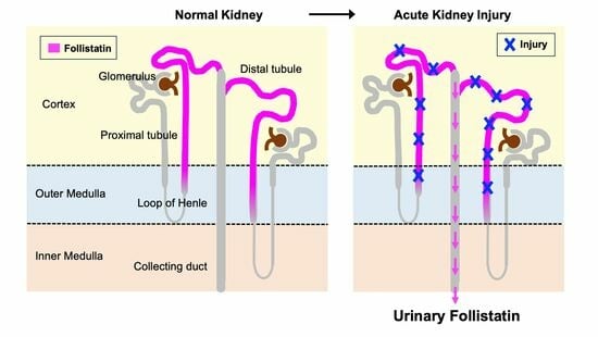
3.1. Localization of Follistatin in Normal Human Kidney

Immunostaining analyses of nephrectomized kidney specimens were conducted to elucidate the subcellular localization of follistatin protein in the normal human kidney. Follistatin was discerned within the renal tubules located in the cortex, outer medulla, and inner medulla (Figure 1A). Subsequent immunostaining using serial sections revealed that follistatin-positive tubules exhibited positivity for uromodulin and NCC (Figure 1B), while concurrently displaying negativity for aquaporin 1 and aquaporin 2 (Figure 1C). These observations suggest that follistatin is synthesized by distal tubules in the normal human kidney.

3.2. Baseline Characteristics of Patients

The baseline characteristics of the participants, including both patients and healthy controls, involved in this study are shown in Table 1. Among the 118 patients diagnosed with AKI, 80 were male, and the mean age was 62.2 ± 16.6 years (± standard error). Notably, no statistically significant differences between AKI patients and their healthy counterparts were observed in the prevalence of comorbidities, except for dyslipidemia. In comparison to the healthy controls, AKI patients exhibited significantly elevated levels of blood urea nitrogen (BUN), serum creatinine (Cr), and C-reactive protein (CRP). Conversely, AKI patients demonstrated significantly lower serum sodium (Na), estimated glomerular filtration rate (GFR), and hemoglobin.

3.3. Significant Increase in Follistatin in the Urine of AKI Patients

In the present study, ELISA was used to assess the concentration of follistatin in the urine of patients diagnosed with AKI. The analysis revealed the absence of detectable levels of urinary follistatin in the samples obtained from healthy controls. Conversely, a notable elevation in urinary follistatin was observed in individuals with AKI (Figure 2A). Notably, a statistically significant variance in urinary follistatin levels was discerned between patients with renal AKI and those with prerenal AKI (Figure 2B). Furthermore, when compared to the levels in healthy controls, a substantial increase in urinary follistatin was evident in cases of sepsis-associated AKI (n = 20, 1214 ± 783.4) and drug-induced nephropathy (n = 27, 514.9 ± 246.4), while no such elevation was observed in AKI attributed to hypotension (n = 11, 268.6 ± 207.8), CIN (n = 10, 179.6 ± 90.6) or surgery (n = 3, 402.1 ± 291.7) (Figure 2D).

3.4. Urinary Follistatin Is Associated with the Severity and Prognosis of AKI

Next, the association between urinary follistatin levels and the severity of AKI was investigated. A marked elevation in urinary follistatin was observed in AKI stage 3 when compared to stage 1 (Figure 2C). Additionally, we assessed the variation in urinary follistatin levels between individuals who required renal replacement therapy (RRT) and those who did not. Notably, urinary follistatin levels were significantly elevated in patients requiring RRT compared to AKI patients who did not require such an intervention (Figure 2E). Conversely, no statistically significant difference was identified between patients who progressed to irreversible end-stage renal disease (ESRD) and those who did not (Figure 2F).

3.5. Correlation between Urinary Follistatin and Clinical Parameters

We further investigated the association between urinary follistatin levels and various clinical parameters. Urinary follistatin exhibited a significant correlation with urinary protein levels (Figure 3A), while no such correlation was observed with serum creatinine (Figure 3H). Furthermore, a significant correlation was established between urinary follistatin and urinary α1 microglobulin (Figure 3F), whereas no correlation was evident with urinary NAG (Figure 3E) or urinary β2 microglobulin (Figure 3G). In a comparative analysis of urinary follistatin and other acute kidney injury (AKI) biomarkers, we observed a significant correlation between urinary follistatin and urinary NGAL (Figure 3B). However, no significant correlations were observed with urinary KIM-1 (Figure 3C) or L-FABP (Figure 3D). Intriguingly, no substantial correlation was observed between urinary follistatin and serum follistatin levels (Figure 3I), suggesting a renal origin for urinary follistatin.

3.6. Time Course of Changes in Urinary Follistatin and Other AKI Biomarkers in an AKI Patient

In the present study, we conducted a comparative analysis of the temporal dynamics of urinary follistatin levels and other AKI biomarkers in a patient experiencing drug-induced AKI. Upon admission, the patient presented with oliguria and required RRT on the following day. RRT was administered for a duration of 18 days, during which the urine volume gradually increased, and renal function subsequently recovered (Figure 4A). Notably, an elevated level of urinary NGAL was observed at the time of AKI diagnosis, which persisted until 7 days post-discontinuation of RRT. Concurrently, an increase in urinary KIM-1 was noted at the time of AKI diagnosis. The peak of urinary KIM-1 occurred at 7 days post-cessation of RRT, followed by a subsequent decline. In contrast, urinary follistatin levels demonstrated a significant elevation at the onset of AKI; however, distinct from NGAL and KIM-1, these levels rapidly diminished before the normalization of serum creatinine (Figure 4B).

3.7. Urinary Follistatin in Patients with Different Causes of AKI

We examined the temporal dynamics of alterations in serum creatinine, urinary follistatin, and other urinary biomarkers in patients with diverse etiologies of AKI. In the setting of cadaveric kidney transplantation, urinary follistatin levels exhibited an initial elevation immediately post-renal transplantation, followed by a gradual decline to undetectable levels thereafter (Figure 5A). For instances of drug-induced AKI (Figure 5B) or AKI associated with Burkitt lymphoma accompanied by tumor lysis syndrome (Figure 5C), urinary follistatin levels returned to baseline prior to the normalization of serum creatinine levels. Among patients experiencing chemotherapy-induced AKI necessitating RRT, urinary follistatin levels increased promptly after the initiation of chemotherapy, but subsequently exhibited a rapid decline (Figure 5D). Conversely, in cases of AKI progressing to irreversible ESRD due to contrast-induced nephropathy (Figure 5E) or cholesterol crystal embolism (Figure 5F), elevated levels of urinary follistatin persisted until discharge.

4. Discussion

As is consistent with previous reports demonstrating the localization of follistatin protein in the renal tubules of normal rats [25,26], we observed that follistatin is localized in the distal tubules of normal human kidneys (Figure 1A–C). Notably, we observed that follistatin is undetectable in the urine of healthy controls, but is markedly elevated in the urine of patients with AKI (Figure 2). Moreover, urinary follistatin levels exhibited a correlation with the severity of AKI (Figure 3). Importantly, no significant correlation was identified between serum follistatin and urinary follistatin (Figure 3I), which suggests that urinary follistatin originates from the kidneys rather than the bloodstream. Collectively, our findings propose urinary follistatin as a novel tubular injury marker with potential utility for monitoring AKI in clinical settings. Although serum follistatin levels have been extensively investigated in various diseases, including muscle physiology [27], metabolic disorders [28,29,30], cancer [31,32,33], and cardiovascular diseases [34,35], to the best of our knowledge, the present study represents the first demonstration that urinary follistatin is present in AKI patients, identifying it as a novel biomarker for detecting renal diseases. This discovery highlights the potential use of follistatin in the assessment of kidney health and the diagnosis of renal disorders.
In the present study, we conducted a comparative analysis of the time course changes in urinary follistatin with several biomarkers. Neutrophil gelatinase-associated lipocalin (NGAL), a highly sensitive early biomarker for kidney injury [10], displayed early elevation in conditions such as AKI, diabetic nephropathy, nephrotic syndrome, and obstructive nephropathy [36]. Urinary follistatin exhibited a significant correlation with urinary NGAL (Figure 3B) and displayed a similar trend to urinary NGAL in many cases (Figure 4 and Figure 5A–C,E), suggesting the potential utility of urinary follistatin as an early diagnostic marker for AKI. In humans, urinary NGAL increased not only in AKI, but also in chronic kidney disease (CKD) [37], and is a marker for delayed graft function following kidney transplantation [38], suggesting that the potential of urinary follistatin as a tubular injury marker is not limited to AKI.
Kidney injury molecule-1 (KIM-1) serves as a valuable biomarker for the definitive diagnosis of AKI [11]. A soluble form of human KIM-1 can be detected in the urine of patients with acute tubular necrosis and serves as a useful biomarker for renal proximal tubule injury facilitating the early diagnosis of the disease and serving as a diagnostic discriminator [11]. Our study revealed that urinary follistatin exhibited a generally analogous pattern to KIM-1, with heightened sensitivity in specific scenarios such as renal transplantation (Figure 5A) and tumor lysis syndrome (Figure 5C). While a strong correlation was observed between urinary follistatin and urinary KIM-1 in a rat model of ischemia-reperfusion injury [25], urinary follistatin did not correlate with KIM-1 in our human study. This discrepancy may arise from differences in the timing of urine collection after AKI, highlighting the importance of considering variations in sample collection timing.
The results of the present study must be viewed in light of several limitations. First, the analysis was conducted on a small number of subjects from a single center, potentially limiting the generalizability of our findings to the broader spectrum of AKI. The absence of consistent data on daily or hourly urine output trends necessitated the definition of AKI solely based on serum creatinine criteria. Secondly, the short follow-up period of AKI patients (median, 38 days) precludes a comprehensive assessment of the impact of urinary follistatin on renal prognosis. Additionally, the variable timing of sample collection after the onset of AKI introduced heterogeneity in our patient cohort, encompassing both the injury and recovery phases. There was significant variation in the number of cases among each etiological group. Furthermore, the lack of investigation into follistatin in the renal tissue of AKI patients leaves unanswered questions regarding its increase in human kidneys with AKI, as observed in rats [25]. The mechanism underlying the appearance of follistatin produced in distal tubules in the urine during AKI remains unclear in our study. To clarify the significance of urinary FST measurement in AKI, it is necessary to confirm the changes in the expression site and amount of FST in the renal tissue of cases with AKI. However, renal biopsy, an invasive examination, is not routinely performed in cases of AKI. In cases of AKI with unknown causes, a renal biopsy is performed to investigate the cause, but in many cases of AKI, the causative factor is known beforehand, so histological evaluation by renal biopsy is not necessary and treatment decisions can be made even without renal biopsy results. Therefore, the number of renal biopsy specimens from cases of AKI is very small. Furthermore, it is difficult to confirm the changes in the expression site and amount of FST associated with AKI by evaluation through renal biopsy. In this study, we observed that the expression site of FST in healthy kidneys is widely distributed from the cortex to the medulla. Since the tissue usually collected in a renal biopsy consists mostly of the cortex, it is not possible to observe the expression of FST in the medulla. One possibility is that the mechanism involves the breakdown of tubular cells, akin to NAG, causing intracellular follistatin to leak into the urine. Additionally, factors such as ischemia and hypoxia associated with AKI may increase the expression and production of follistatin in the tubules, leading to its leakage into the urine. Further study is required in order to address this issue.
The results of the present study highlight a significant increase in urinary follistatin levels in patients with AKI, with this elevation correlating with the severity and prognosis of AKI. The concurrent assessment of urinary follistatin along with other biomarkers holds the potential to facilitate the surveillance of AKI progression and aid in determining the optimal timing for interventions.

5. Conclusions

Urinary follistatin may serve as a valuable marker reflecting the severity of acute tubular damage in sepsis-associated AKI and drug-induced AKI.

Author Contributions

A.M. designed the study; I.N. and K.T. carried out the experiments; I.N. analyzed the data; I.N. and A.M. created the figures; I.N. and A.M. drafted and revised the paper; D.N. and H.H. supervised the project; all authors contributed to data analysis and interpretation and approved the final version of the manuscript. All authors have read and agreed to the published version of the manuscript.

Funding

This research was funded by Grants-in-Aid for Scientific Research (C) (Grant Number 17K09686) and Young Scientists (B) (Grant Number 40835344) from the Ministry of Education, Culture, Sports, Science and Technology of Japan (MEXT).

Institutional Review Board Statement

The study was conducted in accordance with the Declaration of Helsinki and approved by the ethics committee on human research of Jichi Medical University (approval number A18-099, 28 November 2019, A18-150, 21 November 2022, A18-195, 26 March 2022) and Saitama Medical Center, Saitama Medical University (approval number 2487, 8 February 2021).

Informed Consent Statement

Informed consent was obtained from all subjects involved in the study. Written informed consent has been obtained from the patients to publish this paper.

Data Availability Statement

The datasets used and analyzed in the current study are available from the corresponding author upon reasonable request.

Acknowledgments

We would like to thank Saori Nakahashi, Hitomi Hirate, Keiko Fukuda, Minami Watanabe, and Tomoko Tateno for their expert technical assistance. We also thank the lab members for their helpful comments.

Conflicts of Interest

The authors declare no conflicts of interest.

References

  1. See, E.J.; Jayasinghe, K.; Glassford, N.; Bailey, M.; Johnson, D.W.; Polkinghorne, K.R.; Toussaint, N.D.; Bellomo, R. Long-term risk of adverse outcomes after acute kidney injury: A systematic review and meta-analysis of cohort studies using consensus definitions of exposure. Kidney Int. 2019, 95, 160–172. [Google Scholar] [CrossRef] [PubMed]
  2. Ronco, C.; Bellomo, R.; Kellum, J.A. Acute kidney injury. Lancet 2019, 394, 1949–1964. [Google Scholar] [CrossRef] [PubMed]
  3. Amdur, R.L.; Chawla, L.S.; Amodeo, S.; Kimmel, P.L.; Palant, C.E. Outcomes following diagnosis of acute renal failure in US veterans: Focus on acute tubular necrosis. Kidney Int. 2009, 76, 1089–1097. [Google Scholar] [CrossRef]
  4. Ishani, A.; Xue, J.L.; Himmelfarb, J.; Eggers, P.W.; Kimmel, P.L.; Molitoris, B.A.; Collins, A.J. Acute kidney injury increases risk of ESRD among elderly. J. Am. Soc. Nephrol. 2009, 20, 223–228. [Google Scholar] [CrossRef]
  5. Lo, L.J.; Go, A.S.; Chertow, G.M.; McCulloch, C.E.; Fan, D.; Ordoñez, J.D.; Hsu, C.-y. Dialysis-requiring acute renal failure increases the risk of progressive chronic kidney disease. Kidney Int. 2009, 76, 893–899. [Google Scholar] [CrossRef]
  6. Wald, R.; Quinn, R.R.; Luo, J.; Li, P.; Scales, D.C.; Mamdani, M.M.; Ray, J.G.; Group, U.o.T.A.K.I.R. Chronic dialysis and death among survivors of acute kidney injury requiring dialysis. JAMA J. Am. Med. Assoc. 2009, 302, 1179–1185. [Google Scholar] [CrossRef]
  7. Chawla, L.S.; Amdur, R.L.; Amodeo, S.; Kimmel, P.L.; Palant, C.E. The severity of acute kidney injury predicts progression to chronic kidney disease. Kidney Int. 2011, 79, 1361–1369. [Google Scholar] [CrossRef] [PubMed]
  8. Parikh, C.R.; Coca, S.G.; Wang, Y.; Masoudi, F.A.; Krumholz, H.M. Long-term prognosis of acute kidney injury after acute myocardial infarction. Arch. Intern. Med. 2008, 168, 987–995. [Google Scholar] [CrossRef]
  9. Chawla, L.S. Acute kidney injury leading to chronic kidney disease and long-term outcomes of acute kidney injury: The best opportunity to mitigate acute kidney injury? Controv. Acute Kidney Inj. 2011, 174, 182–190. [Google Scholar]
  10. Mishra, J.; Dent, C.; Tarabishi, R.; Mitsnefes, M.M.; Ma, Q.; Kelly, C.; Ruff, S.M.; Zahedi, K.; Shao, M.; Bean, J. Neutrophil gelatinase-associated lipocalin (NGAL) as a biomarker for acute renal injury after cardiac surgery. Lancet 2005, 365, 1231–1238. [Google Scholar] [CrossRef]
  11. Han, W.K.; Bailly, V.; Abichandani, R.; Thadhani, R.; Bonventre, J.V. Kidney Injury Molecule-1 (KIM-1): A novel biomarker for human renal proximal tubule injury. Kidney Int. 2002, 62, 237–244. [Google Scholar] [CrossRef]
  12. Parikh, C.; Mishra, J.; Thiessen-Philbrook, H.; Dursun, B.; Ma, Q.; Kelly, C.; Dent, C.; Devarajan, P.; Edelstein, C. Urinary IL-18 is an early predictive biomarker of acute kidney injury after cardiac surgery. Kidney Int. 2006, 70, 199–203. [Google Scholar] [CrossRef]
  13. Portilla, D.; Dent, C.; Sugaya, T.; Nagothu, K.; Kundi, I.; Moore, P.; Noiri, E.; Devarajan, P. Liver fatty acid-binding protein as a biomarker of acute kidney injury after cardiac surgery. Kidney Int. 2008, 73, 465–472. [Google Scholar] [CrossRef] [PubMed]
  14. Kashani, K.; Al-Khafaji, A.; Ardiles, T.; Artigas, A.; Bagshaw, S.M.; Bell, M.; Bihorac, A.; Birkhahn, R.; Cely, C.M.; Chawla, L.S. Discovery and validation of cell cycle arrest biomarkers in human acute kidney injury. Crit. Care 2013, 17, R25. [Google Scholar] [CrossRef]
  15. Koyner, J.L.; Shaw, A.D.; Chawla, L.S.; Hoste, E.A.; Bihorac, A.; Kashani, K.; Haase, M.; Shi, J.; Kellum, J.A. Tissue inhibitor metalloproteinase-2 (TIMP-2)⋅ IGF-binding protein-7 (IGFBP7) levels are associated with adverse long-term outcomes in patients with AKI. J. Am. Soc. Nephrol. 2015, 26, 1747–1754. [Google Scholar] [CrossRef]
  16. Esch, F.S.; Shimasaki, S.; Mercado, M.; Cooksey, K.; Ling, N.; Ying, S.; Ueno, N.; Guillemin, R. Structural characterization of follistatin: A novel follicle-stimulating hormone release-inhibiting polypeptide from the gonad. Mol. Endocrinol. 1987, 1, 849–855. [Google Scholar] [CrossRef]
  17. Nakamura, T.; Takio, K.; Eto, Y.; Shibai, H.; Titani, K.; Sugino, H. Activin-binding protein from rat ovary is follistatin. Science 1990, 247, 836–838. [Google Scholar] [CrossRef]
  18. Maeshima, A.; Zhang, Y.-q.; Nojima, Y.; Naruse, T.; Kojima, I. Involvement of the activin-follistatin system in tubular regeneration after renal ischemia in rats. J. Am. Soc. Nephrol. 2001, 12, 1685–1695. [Google Scholar] [CrossRef]
  19. Yamashita, S.; Maeshima, A.; Kojima, I.; Nojima, Y. Activin A is a potent activator of renal interstitial fibroblasts. J. Am. Soc. Nephrol. 2004, 15, 91–101. [Google Scholar] [CrossRef] [PubMed]
  20. Koken, E.; Oyar, E.O.; Uyanikgil, Y.; Pazarlar, B.A.; Bilister, C.; Aksun, S.; Yigitturk, G.; Koken, E.C. Exogenous follistatin administration ameliorates cisplatin-induced acute kidney injury through anti-inflammation and anti-apoptosis effects. Bratisl. Lek. Listy 2020, 121, 143–150. [Google Scholar] [CrossRef] [PubMed]
  21. Mehta, N.; Gava, A.L.; Zhang, D.; Gao, B.; Krepinsky, J.C. Follistatin Protects Against Glomerular Mesangial Cell Apoptosis and Oxidative Stress to Ameliorate Chronic Kidney Disease. Antioxid. Redox Signal 2019, 31, 551–571. [Google Scholar] [CrossRef] [PubMed]
  22. Takahashi, S.; Nakasatomi, M.; Takei, Y.; Ikeuchi, H.; Sakairi, T.; Kaneko, Y.; Hiromura, K.; Nojima, Y.; Maeshima, A. Identification of Urinary Activin A as a Novel Biomarker Reflecting the Severity of Acute Kidney Injury. Sci. Rep. 2018, 8, 5176. [Google Scholar] [CrossRef] [PubMed]
  23. Nagayama, I.; Maeshima, A.; Nagata, D. Urinary Activin A: A Novel Biomarker for Human Acute Kidney Injury. Diagnostics 2022, 12, 661. [Google Scholar] [CrossRef]
  24. Palevsky, P.M.; Liu, K.D.; Brophy, P.D.; Chawla, L.S.; Parikh, C.R.; Thakar, C.V.; Tolwani, A.J.; Waikar, S.S.; Weisbord, S.D. KDOQI US commentary on the 2012 KDIGO clinical practice guideline for acute kidney injury. Am. J. Kidney Dis. 2013, 61, 649–672. [Google Scholar] [CrossRef] [PubMed]
  25. Nagayama, I.; Takayanagi, K.; Hasegawa, H.; Maeshima, A. Tubule-Derived Follistatin Is Increased in the Urine of Rats with Renal Ischemia and Reflects the Severity of Acute Tubular Damage. Cells 2023, 12, 801. [Google Scholar] [CrossRef] [PubMed]
  26. Kogawa, K.; Ogawa, K.; Hayashi, Y.; Nakamura, T.; Titani, K.; Sugino, H. Immunohistochemical localization of follistatin in rat tissues. Endocrinol. Jpn. 1991, 38, 383–391. [Google Scholar] [CrossRef]
  27. Miyamoto, T.; Carrero, J.J.; Qureshi, A.R.; Anderstam, B.; Heimbürger, O.; Bárány, P.; Lindholm, B.; Stenvinkel, P. Circulating follistatin in patients with chronic kidney disease: Implications for muscle strength, bone mineral density, inflammation, and survival. Clin. J. Am. Soc. Nephrol. 2011, 6, 1001–1008. [Google Scholar] [CrossRef]
  28. Hansen, J.S.; Plomgaard, P. Circulating follistatin in relation to energy metabolism. Mol. Cell Endocrinol. 2016, 433, 87–93. [Google Scholar] [CrossRef]
  29. Näf, S.; Escote, X.; Ballesteros, M.; Yañez, R.E.; Simon-Muela, I.; Gil, P.; Albaiges, G.; Vendrell, J.; Megia, A. Serum activin A and follistatin levels in gestational diabetes and the association of the Activin A-Follistatin system with anthropometric parameters in offspring. PLoS ONE 2014, 9, e92175. [Google Scholar] [CrossRef]
  30. Han, X.; Møller, L.L.V.; De Groote, E.; Bojsen-Møller, K.N.; Davey, J.; Henríquez-Olguin, C.; Li, Z.; Knudsen, J.R.; Jensen, T.E.; Madsbad, S. Mechanisms involved in follistatin-induced hypertrophy and increased insulin action in skeletal muscle. J. Cachexia Sarcopenia Muscle 2019, 10, 1241–1257. [Google Scholar] [CrossRef]
  31. Panagiotou, G.; Ghaly, W.; Upadhyay, J.; Pazaitou-Panayiotou, K.; Mantzoros, C.S. Serum follistatin is increased in thyroid cancer and is associated with Adverse tumor characteristics in humans. J. Clin. Endocrinol. Metab. 2021, 106, e2137–e2150. [Google Scholar] [CrossRef]
  32. Tomoda, T.; Nouso, K.; Miyahara, K.; Kobayashi, S.; Kinugasa, H.; Toyosawa, J.; Hagihara, H.; Kuwaki, K.; Onishi, H.; Nakamura, S. Prognotic impact of serum follistatin in patients with hepatocellular carcinoma. J. Gastroenterol. Hepatol. 2013, 28, 1391–1396. [Google Scholar] [CrossRef]
  33. Zhang, P.; Ruan, Y.; Xiao, J.; Chen, F.; Zhang, X. Association of serum follistatin levels with histological types and progression of tumor in human lung cancer. Cancer Cell Int. 2018, 18, 162. [Google Scholar] [CrossRef] [PubMed]
  34. Liu, M.; Mao, C.; Li, J.; Han, F.; Yang, P. Effects of the activin A–follistatin system on myocardial cell apoptosis through the endoplasmic reticulum stress pathway in heart failure. Int. J. Mol. Sci. 2017, 18, 374. [Google Scholar] [CrossRef]
  35. Chen, Y.; Rothnie, C.; Spring, D.; Verrier, E.; Venardos, K.; Kaye, D.; Phillips, D.J.; Hedger, M.P.; Smith, J.A. Regulation and actions of activin A and follistatin in myocardial ischaemia–reperfusion injury. Cytokine 2014, 69, 255–262. [Google Scholar] [CrossRef] [PubMed]
  36. Kuwabara, T.; Mori, K.; Mukoyama, M.; Kasahara, M.; Yokoi, H.; Saito, Y.; Yoshioka, T.; Ogawa, Y.; Imamaki, H.; Kusakabe, T. Urinary neutrophil gelatinase-associated lipocalin levels reflect damage to glomeruli, proximal tubules, and distal nephrons. Kidney Int. 2009, 75, 285–294. [Google Scholar] [CrossRef] [PubMed]
  37. Nickolas, T.L.; Forster, C.S.; Sise, M.E.; Barasch, N.; Solá-Del Valle, D.; Viltard, M.; Buchen, C.; Kupferman, S.; Carnevali, M.L.; Bennett, M. NGAL (Lcn2) monomer is associated with tubulointerstitial damage in chronic kidney disease. Kidney Int. 2012, 82, 718–722. [Google Scholar] [CrossRef]
  38. Parikh, C.; Jani, A.; Mishra, J.; Ma, Q.; Kelly, C.; Barasch, J.; Edelstein, C.; Devarajan, P. Urine NGAL and IL-18 are predictive biomarkers for delayed graft function following kidney transplantation. Am. J. Transplant. 2006, 6, 1639–1645. [Google Scholar] [CrossRef]
Figure 1. Localization of follistatin in normal human kidney.(A) Localization of follistatin in normal human kidney was examined by immunostaining. Follistatin (brown). NC, negative control. Magnification: ×100 (left panels) and ×400 (middle and right panels). (B) Identification of follistatin-positive tubules in normal human kidney using serial sections. Nonselective cation channels: NCC. Magnification: ×400. (C) Double staining of follistatin with aquaporin 1 (AQP1), aquaporin 2 (AQP2), or α-SMA in normal human kidney. Nucleus (blue). Magnification: ×400.
Cells 13 00525 g001
Figure 2. Urinary Follistatin Levels in AKI Patients. (A) Urinary follistatin levels in healthy controls (HC) (n = 16) and AKI patients (n = 118). * p < 0.05 vs. HC. (B) Urinary follistatin levels in patients with prerenal AKI (n = 18), renal AKI (n = 88), and postrenal AKI patients (n = 5). * p < 0.05 vs. prerenal AKI. (C) Urinary follistatin levels and severity of AKI according to KDIGO stage (stages 1–3). * p < 0.05. (D) Urinary follistatin levels for each cause of AKI (sepsis, 20; CIN, 10; hypotension, 11; drug-induced, 27; surgery, 3). CIN, contrast-induced nephropathy, N.D., not detected. ** p < 0.01, *** p < 0.001 vs. HC. (E) Urinary follistatin levels in AKI patients who did (+; n = 58) and did not (-; n = 60) require renal replacement therapy (RRT). *** p < 0.001 vs. RRT (-). (F) Urinary follistatin levels in AKI patients with (+; n = 16) and without (-; n = 42) progression to irreversible ESRD. n.s., not significant. The red line indicates the median.
Cells 13 00525 g002
Figure 3. Correlations between Urinary Follistatin and Clinical Parameters. Correlations between urinary follistatin and urinary protein level (A), NGAL (B), KIM-1 (C), L-FABP (D), NAG (E), α1 microglobulin (F), β2 microglobulin (G), serum creatinine (H), and serum follistatin (I) are shown.
Cells 13 00525 g003
Figure 4. Time Course Changes in Urinary Follistatin and Other AKI Biomarkers. Changes in serum creatinine and urine volume (A) and AKI biomarkers (B) in a patient with drug-induced AKI are shown. Triangle indicates the timing of hemodialysis (HD).
Cells 13 00525 g004
Figure 5. Time Course Changes in Urinary Follistatin in Patients with AKI. (AF): Changes in serum creatinine, urinary NGAL, urinary KIM-1, and urinary follistatin levels in patients with various causes of AKI, including cadaver kidney transplantation (A), drug-induced nephropathy (B), Burkitt lymphoma accompanied by tumor lysis syndrome (C), chemotherapy (D), contrast-induced nephropathy (E), and cholesterol crystal embolism (F), are shown. Triangle indicates the timing of hemodialysis (HD).
Cells 13 00525 g005
Table 1. Baseline Characteristics of Patients with Acute Kidney Injury (AKI) and Healthy Controls. Data were collected from healthy controls and patients with AKI at the initial visit. Abbreviations: AKI, acute kidney injury; BMI, body mass index; CKD, chronic kidney disease; eGFR, estimated glomerular filtration rate; WBC, white blood cell; CRP, C-reactive protein; NAG, N-acetyl-β-D-glucosaminidase; α1MG, α1 microglobulin; β2MG, β2 microglobulin, KIM-1, kidney injury molecule-1; NGAL, neutrophil gelatinase-associated lipocalin; L-FABP, liver-type fatty acid-binding protein; FENa, fractional excretion of sodium; FEurea, fractional excretion of urea.
AKIHealthy Control
n = 118n = 16P
  Age, years, mean ± S.E.62.2± 16.6 54.9 ± 2.9 0.379
  BMI, kg/m2, mean ± S.E.24.1± 5.2 23.4 ± 0.8 0.596
  Male gender, n (%)80 (67.8) 2 (12.5)<0.001
  Weight, kg, mean ± S.E.63.3± 15.8 60 ± 3.2 0.493
Complications, n (%)
  Diabetes37 (31.4) 1 (6.3)0.054
  Hypertension78 (66.1) 7 (43.8)0.294
  Dyslipidemia43 (36.4) 1 (6.3)0.037
  Old myocardial infarction11 (9.3) 0 (0.0)0.226
  Old cerebral infarction6 (5.1) 0 (0.0)0.401
  Angina pectoris12 (10.2) 0 (0.0)0.253
  Chronic obstructive pulmonary disease5 (4.2) 0 (0.0)0.401
  Liver disease4 (3.4) 0 (0.0)0.455
Hematological data, mean ± S.E.
  Na, mEq/L137 ± 5.5 142 ± 0.5 0.005
  K, mEq/L4.4 ± 0.9 4.2 ± 0.1 0.379
  Cl, mEq/L103 ± 6.3 106 ± 0.6 0.124
  BUN, mg/dL55 ± 33.2 14 ± 0.8 <0.001
  Creatinine, mg/dL4.52 ± 3.55 0.68 ± 0.04 <0.001
  eGFR, mL/min19.1 ± 16.7 79.9 ± 3.6 <0.001
  Hemoglobin, g/dL10.6 ± 2.9 13.3 ± 0.2 0.015
  Platelets, x104/μL74.8 ± 109.3 26.1 ± 1.7 0.195
  WBC, x103/μL6.9 ± 6.8 5.7 ± 0.4 0.462
  CRP, mg/dL7.93 ± 9.0 0.24 ± 0.1 0.015
Urinalysis, mean ± S.E.
  Urinary protein, mg/dL245.1 ± 494.6 - -
  NAG, IU/L33.6 ± 39.1 - -
  α1MG, mg/L55.4 ± 66.7 - -
  β2MG, mg/L12.5 ± 24.2 - -
  NGAL, ng/mL1206.7 ± 2750.9 - -
  KIM-1, ng/mL3.8 ± 7.5 - -
  L-FABP, ng/mL168.4 ± 480.5 - -
  FENa, %5.0 ± 7.4 - -
  FEUrea, %36.5 ± 22.6--
Disclaimer/Publisher’s Note: The statements, opinions and data contained in all publications are solely those of the individual author(s) and contributor(s) and not of MDPI and/or the editor(s). MDPI and/or the editor(s) disclaim responsibility for any injury to people or property resulting from any ideas, methods, instructions or products referred to in the content.

