
Expression of Stemness Markers in the Cervical Smear of Patients with Cervical Insufficiency
 , , , ,
Abstract
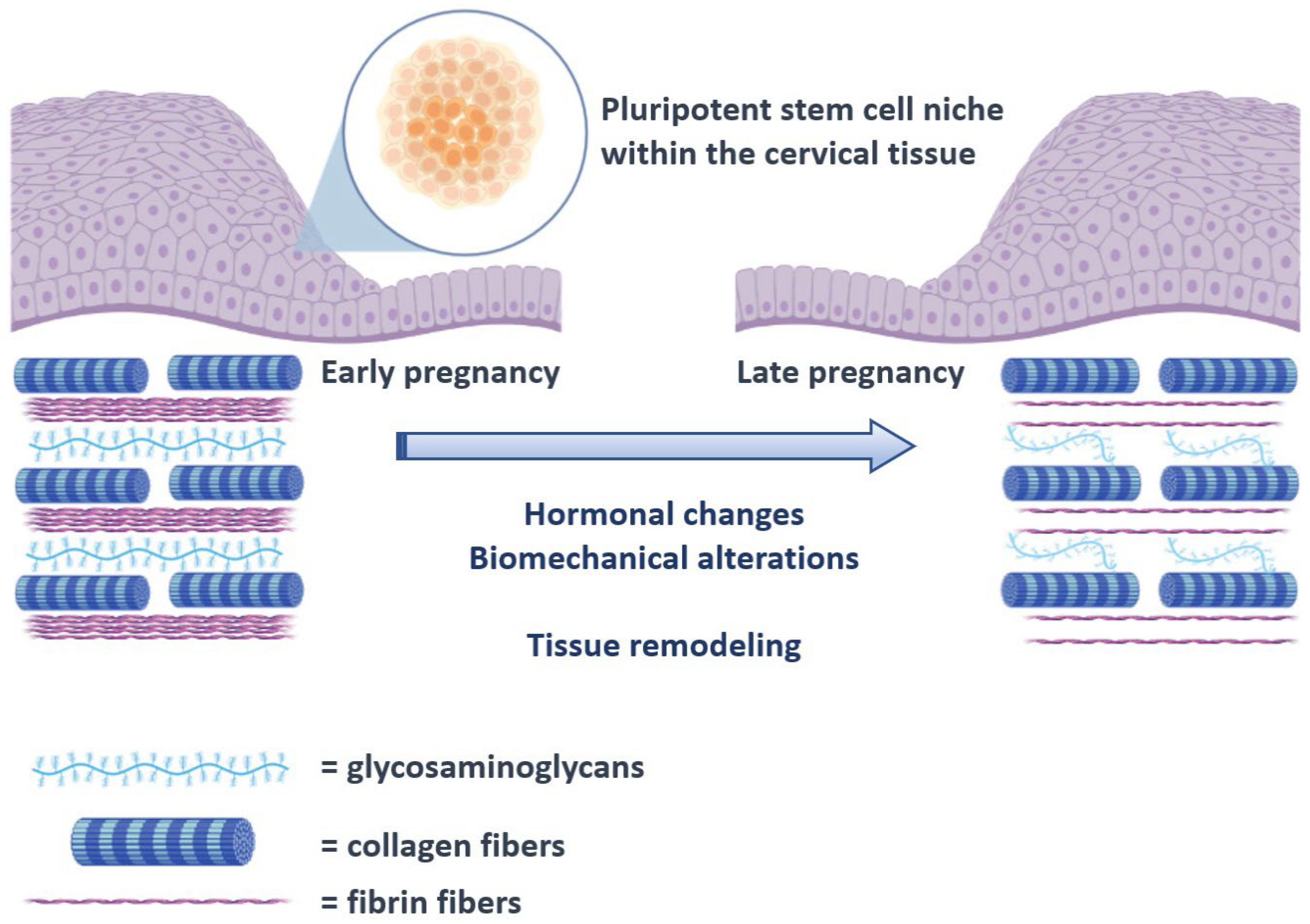
:1. Introduction
2. Materials and Methods
2.1. Patients, Ethical Approval, and Cervical Samples
2.2. RNA Extraction, Reverse Transcription, and Real-Time PCR
2.3. Statistical Analysis
3. Results
3.1. G6PD Expression
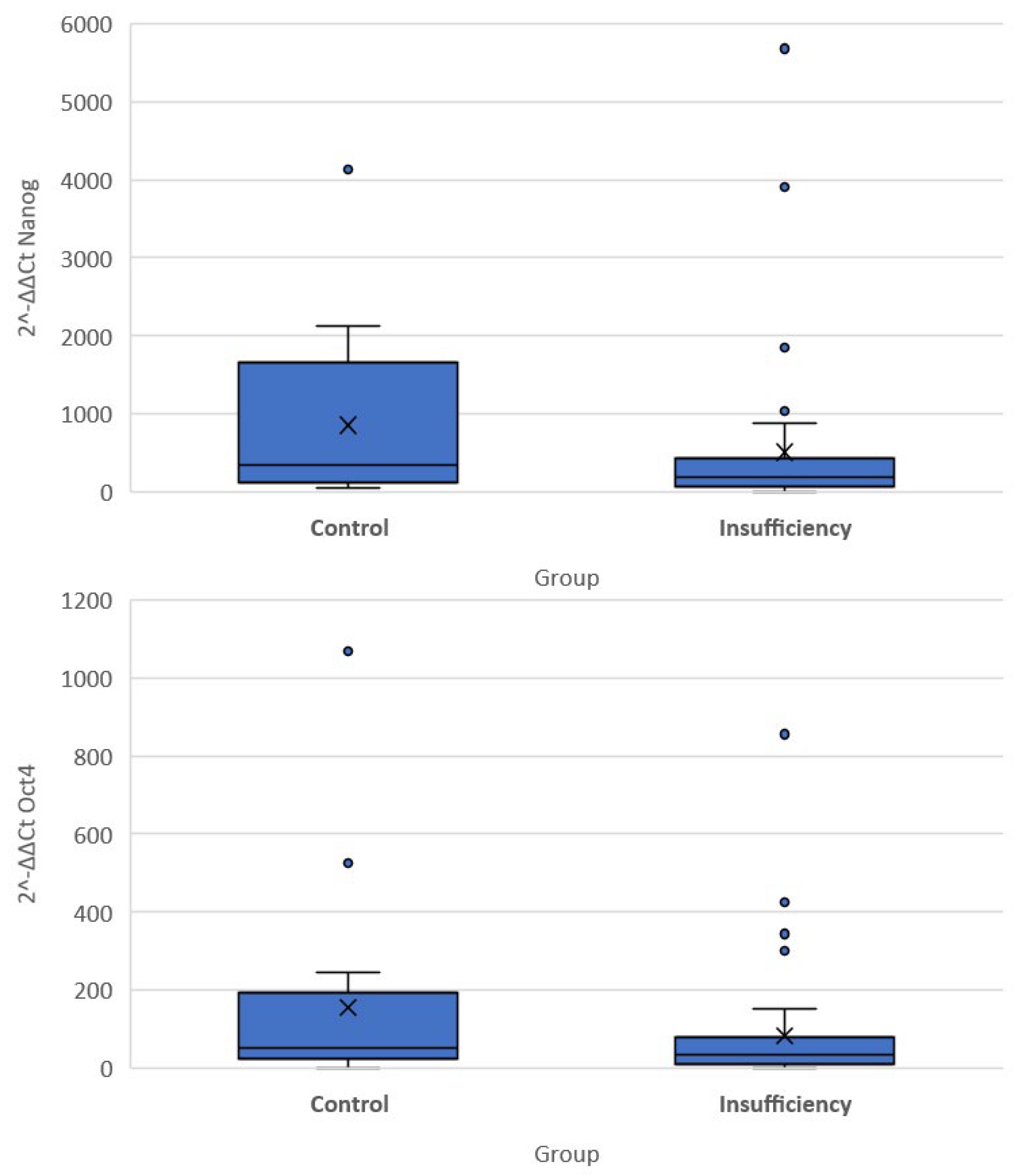
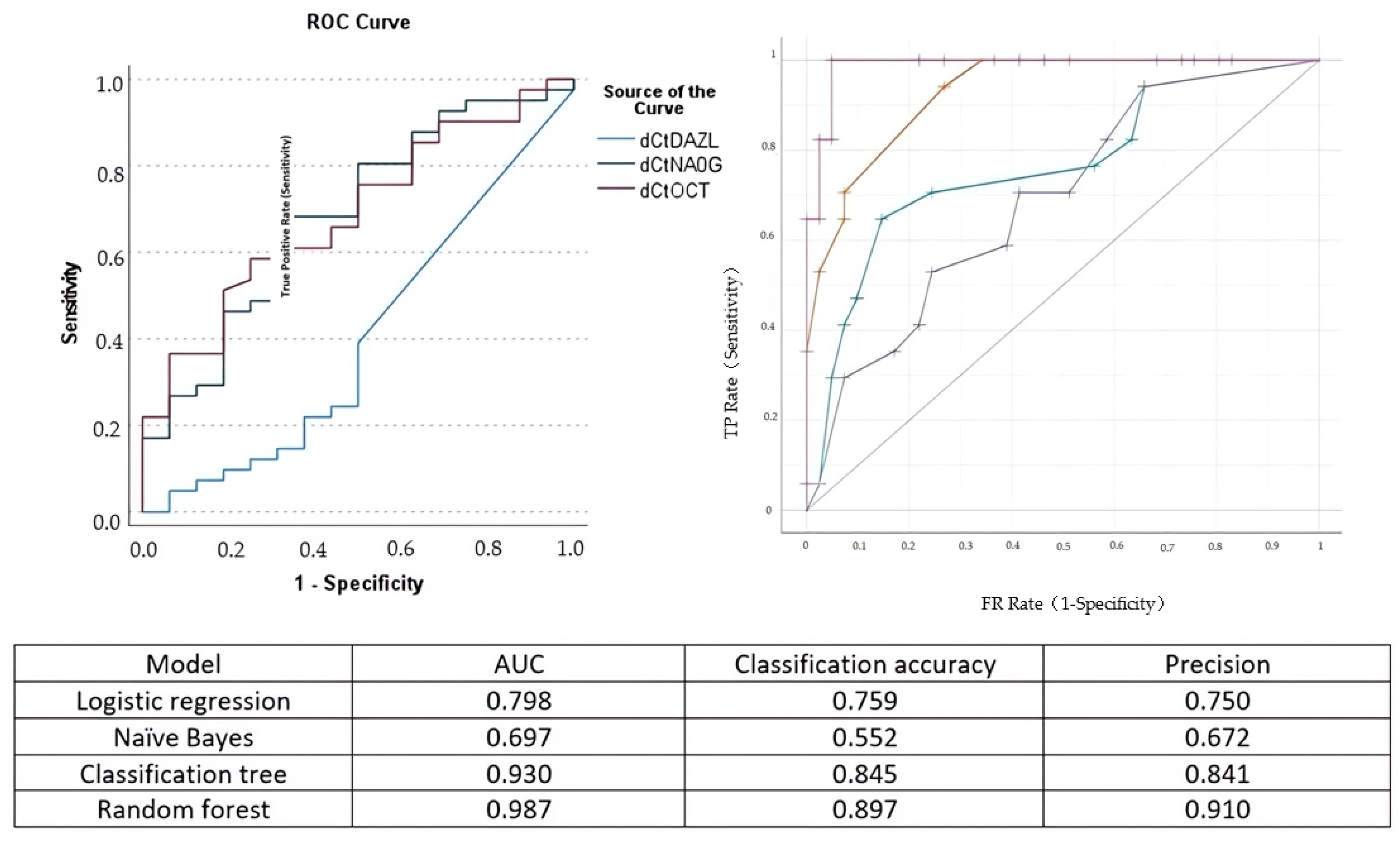
3.2. OCT-4, Nanog and DAZL Expression
4. Discussion
Study Limitations
5. Conclusions and Implications
Author Contributions
Funding
Institutional Review Board Statement
Informed Consent Statement
Data Availability Statement
Conflicts of Interest
References
- Daskalakis, G.; Goya, M.; Pergialiotis, V.; Cabero, L.; Kyvernitakis, I.; Antsaklis, A.; Arabin, B. Prevention of spontaneous preterm birth. Arch. Gynecol. Obstet. 2019, 299, 1261–1273. [Google Scholar] [CrossRef] [PubMed]
- Oxlund, B.S.; Ørtoft, G.; Brüel, A.; Danielsen, C.C.; Bor, P.; Oxlund, H.; Uldbjerg, N. Collagen concentration and biomechanical properties of samples from the lower uterine cervix in relation to age and parity in non-pregnant women. Reprod. Biol. Endocrinol. 2010, 8, 82. [Google Scholar] [CrossRef] [PubMed]
- Nallasamy, S.; Akins, M.; Tetreault, B.; Luby-Phelps, K.; Mahendroo, M. Distinct reorganization of collagen architecture in lipopolysaccharide-mediated premature cervical remodeling. Biol. Reprod. 2017, 98, 63–74. [Google Scholar] [CrossRef] [PubMed]
- Yellon, S.M. Immunobiology of Cervix Ripening. Front. Immunol. 2019, 10, 3156. [Google Scholar] [CrossRef] [PubMed]
- Tantengco, O.A.G.; Menon, R. Breaking Down the Barrier: The Role of Cervical Infection and Inflammation in Preterm Birth. Front. Glob. Women’s Health 2022, 2, 777643. [Google Scholar] [CrossRef]
- Warren, J.E.; Nelson, L.M.; Stoddard, G.J.; Esplin, M.S.; Varner, M.W.; Silver, R.M. Polymorphisms in the promoter region of the interleukin-10 (IL-10) gene in women with cervical insufficiency. Am. J. Obstet. Gynecol. 2009, 201, 372.e1–372.e5. [Google Scholar] [CrossRef]
- Brizzi, M.F.; Tarone, G.; Defilippi, P. Extracellular matrix, integrins, and growth factors as tailors of the stem cell niche. Curr. Opin. Cell Biol. 2012, 24, 645–651. [Google Scholar] [CrossRef]
- Xu, M.; Su, T.; Jin, X.; Li, Y.; Yao, Y.; Liu, K.; Chen, K.; Lu, F.; He, Y. Inflammation-mediated matrix remodeling of extracellular matrix-mimicking biomaterials in tissue engineering and regenerative medicine. Acta Biomater. 2022, 151, 106–117. [Google Scholar] [CrossRef]
- Xie, T.; Li, L. Stem cells and their niche: An inseparable relationship. Development. 2007, 134, 2001–2006. [Google Scholar] [CrossRef]
- Karine, R.; Marie-Ange, D.; Marisa, M.F.; Marina, A.G. Adhesion within the stem cell niches. Curr. Opin. Cell. Biol. 2009, 21, 623–629. [Google Scholar]
- Jariwala, N.; Ozols, M.; Bell, M.; Bradley, E.; Gilmore, A.; Debelle, L.; Sherratt, M.J. Matrikines as mediators of tissue remodelling. Adv. Drug Deliv. Rev. 2022, 185, 114240. [Google Scholar] [CrossRef]
- Swinehart, I.T.; Badylak, S.F. Extracellular matrix bioscaffolds in tissue remodeling and morphogenesis. Dev Dyn. 2016, 245, 351–360. [Google Scholar] [CrossRef] [PubMed]
- Stefanidis, K.; Pergialiotis, V.; Christakis, D.; Patta, J.; Stefanidi, D.; Loutradis, D. OCT-4 and DAZL expression in precancerous lesions of the human uterine cervix. J. Obstet. Gynaecol. Res. 2014, 41, 763–767. [Google Scholar] [CrossRef] [PubMed]
- Stefanidis, K.; Pappa, J.; Pergialiotis, V.; Stefanidi, D.; Loutradis, D. Imiquimod treatment effectively reduces the percentage of viable cells in a cervical carcinoma cell line but does not affect the expression of HLA-G or OCT-4. J. Stem Cells 2015, 10, 217–223. [Google Scholar] [PubMed]
- Stefanidis, K.; Loutradis, D.; Vassiliou, L.-V.; Anastasiadou, V.; Kiapekou, E.; Nikas, V.; Patris, G.; Vlachos, G.; Rodolakis, A.; Antsaklis, A. Nevirapine induces growth arrest and premature senescence in human cervical carcinoma cells. Gynecol. Oncol. 2008, 111, 344–349. [Google Scholar] [CrossRef] [PubMed]
- Kim, B.W.; Cho, H.; Choi, C.H.; Ylaya, K.; Chung, J.-Y.; Kim, J.-H.; Hewitt, S.M. Clinical significance of OCT4 and SOX2 protein expression in cervical cancer. BMC Cancer 2015, 15, 1–8. [Google Scholar] [CrossRef]
- Gu, T.-T.; Liu, S.-Y.; Zheng, P.-S. Cytoplasmic NANOG-Positive Stromal Cells Promote Human Cervical Cancer Progression. Am. J. Pathol. 2012, 181, 652–661. [Google Scholar] [CrossRef]
- Takeda, J.; Seino, S.; Bell, G.I. Human Oct3 gene family: cDNA sequences, alternative splicing, gene organization, chromosomal location, and expression at low levels in adult tissues. Nucleic Acids Res. 1992, 20, 4613–4620. [Google Scholar] [CrossRef]
- Peskova, L.; Cerna, K.; Oppelt, J.; Mraz, M.; Barta, T. Oct4-mediated reprogramming induces embryonic-like microRNA expression signatures in human fibroblasts. Sci. Rep. 2019, 9, 1–13. [Google Scholar] [CrossRef]
- Huang, X.; Lee, M.-R.; Cooper, S.; Hangoc, G.; Hong, K.-S.; Chung, H.-M.; Broxmeyer, H.E. Activation of OCT4 enhances ex vivo expansion of human cord blood hematopoietic stem and progenitor cells by regulating HOXB4 expression. Leukemia. 2015, 30, 144–153. [Google Scholar] [CrossRef]
- Ngan, K.W.; Jung, S.M.; Lee, L.Y.; Chuang, W.Y.; Yeh, C.J.; Hsieh, Y.Y. Immunohistochemical expression of OCT4 in primary central nervous system germ cell tumours. J. Clin. Neurosci. 2008, 15, 149–152. [Google Scholar] [CrossRef] [PubMed]
- Nichols, J.; Smith, A. Pluripotency in the Embryo and in Culture. Cold Spring Harb. Perspect. Biol. 2012, 4, a008128. [Google Scholar] [CrossRef] [PubMed]
- Yen, P.H.; Chai, N.N.; Salido, E. The human autosomal gene DAZLA: Testis specificity and a candidate for male infertility. Hum. Mol. Genet. 1996, 5, 2013–2017. [Google Scholar] [CrossRef]
- Li, H.; Liang, Z.; Yang, J.; Wang, D.; Wang, H.; Zhu, M.; Geng, B.; Xu, E.Y. DAZL is a master translational regulator of murine spermatogenesis. Natl. Sci. Rev. 2018, 6, 455–468. [Google Scholar] [CrossRef] [PubMed]
- Kato, Y.; Katsuki, T.; Kokubo, H.; Masuda, A.; Saga, Y. Dazl is a target RNA suppressed by mammalian NANOS2 in sexually differentiating male germ cells. Nat. Commun. 2016, 7, 11272. [Google Scholar] [CrossRef] [PubMed]
- Silva, J.; Nichols, J.; Theunissen, T.W.; Guo, G.; van Oosten, A.L.; Barrandon, O.; Wray, J.; Yamanaka, S.; Chambers, I.; Smith, A. Nanog Is the Gateway to the Pluripotent Ground State. Cell. 2009, 138, 722–737. [Google Scholar] [CrossRef] [PubMed]
- Zhang, J.; Guan, J.; Niu, X.; Shangchun, G.; Guo, S.; Li, Q.; Xie, Z.; Zhang, C.; Wang, Y. Exosomes released from human induced pluripotent stem cells-derived MSCs facilitate cutaneous wound healing by promoting collagen synthesis and angiogenesis. J. Transl. Med. 2015, 13, 49. [Google Scholar] [CrossRef]
- Kubosch, E.J.; Lang, G.; Furst, D.; Kubosch, D.; Izadpanah, K.; Rolauffs, B.; Sudkamp, N.P.; Schmal, H. The Potential for Synovium-derived Stem Cells in Cartilage Repair. Curr. Stem Cell Res. Ther. 2018, 13, 174–184. [Google Scholar] [CrossRef]
- Myers, K.M.; Feltovich, H.; Mazza, E.; Vink, J.; Bajka, M.; Wapner, R.J.; Hall, T.J.; House, M. The mechanical role of the cervix in pregnancy. J. Biomech. 2015, 48, 1511–1523. [Google Scholar] [CrossRef]
- Torres, J.; Faris, I.; Callejas, A. Histobiomechanical Remodeling of the Cervix during Pregnancy: Proposed Framework. Math. Probl. Eng. 2019, 2019, 1–11. [Google Scholar] [CrossRef]
- Yoshida, K.; Jayyosi, C.; Lee, N.; Mahendroo, M.; Myers, K.M. Mechanics of cervical remodelling: Insights from rodent models of pregnancy. Interface Focus. 2019, 9, 20190026. [Google Scholar] [CrossRef] [PubMed]
- Akins, M.L.; Luby-Phelps, K.; Bank, R.A.; Mahendroo, M. Cervical Softening During Pregnancy: Regulated Changes in Collagen Cross-Linking and Composition of Matricellular Proteins in the Mouse1. Biol. Reprod. 2011, 84, 1053–1062. [Google Scholar] [CrossRef] [PubMed]
- Yao, W.; Gan, Y.; Myers, K.M.; Vink, J.Y.; Wapner, R.J.; Hendon, C.P. Collagen Fiber Orientation and Dispersion in the Upper Cervix of Non-Pregnant and Pregnant Women. PLOS ONE. 2016, 11, e0166709. [Google Scholar] [CrossRef]
- Moghaddam, A.O.; Lin, Z.; Sivaguru, M.; Phillips, H.; McFarlin, B.; Toussaint, K.; Johnson, A.J.W. Heterogeneous microstructural changes of the cervix influence cervical funneling. Acta Biomater. 2022, 140, 434–445. [Google Scholar] [CrossRef]
- Jayyosi, C.; Lee, N.; Willcockson, A.; Nallasamy, S.; Mahendroo, M.; Myers, K. The mechanical response of the mouse cervix to tensile cyclic loading in term and preterm pregnancy. Acta Biomater. 2018, 78, 308–319. [Google Scholar] [CrossRef] [PubMed]
- Naqvi, S.M.; McNamara, L.M. Stem Cell Mechanobiology and the Role of Biomaterials in Governing Mechanotransduction and Matrix Production for Tissue Regeneration. Front. Bioeng. Biotechnol. 2020, 8, 597661. [Google Scholar] [CrossRef] [PubMed]
- Celso, C.L.; Scadden, D.T. The haematopoietic stem cell niche at a glance. J. Cell Sci. 2011, 124, 3529–3535. [Google Scholar] [CrossRef]
- Singh, P.; Schwarzbauer, J.E. Fibronectin and stem cell differentiation—Lessons from chondrogenesis. J. Cell Sci. 2012, 125, 3703–3712. [Google Scholar] [CrossRef]
- Maquart, F.-X.; Pasco, S.; Ramont, L.; Hornebeck, W.; Monboisse, J.-C. An introduction to matrikines: Extracellular matrix-derived peptides which regulate cell activity: Implication in tumor invasion. Crit. Rev. Oncol. 2004, 49, 199–202. [Google Scholar] [CrossRef]
- Maquart, F.; Bellon, G.; Pasco, S.; Monboisse, J. Matrikines in the regulation of extracellular matrix degradation. Biochimie 2005, 87, 353–360. [Google Scholar] [CrossRef]
- Rodrigues, M.; Yates, C.C.; Nuschke, A.; Griffith, L.; Wells, A. The Matrikine Tenascin-C Protects Multipotential Stromal Cells/Mesenchymal Stem Cells from Death Cytokines Such as FasL. Tissue Eng. Part A 2013, 19, 1972–1983. [Google Scholar] [CrossRef] [PubMed]
- Anderson, J.M.; Rodriguez, A.; Chang, D.T. Foreign body reaction to biomaterials. Semin. Immunol. 2008, 20, 86–100. [Google Scholar] [CrossRef] [PubMed]
- Allman, A.J.; McPherson, T.; Badylak, S.; Merrill, L.C.; Kallakury, B.; Sheehan, C.; Raeder, R.H.; Metzger, D. Xenogeneic extracellular matrix grafts elicit a th2-restricted immune response. Transplantation 2001, 71, 1631–1640. [Google Scholar] [CrossRef] [PubMed]
- Marangio, A.; Biccari, A.; D’Angelo, E.; Sensi, F.; Spolverato, G.; Pucciarelli, S.; Agostini, M. The Study of the Extracellular Matrix in Chronic Inflammation: A Way to Prevent Cancer Initiation? Cancers 2022, 14, 5903. [Google Scholar] [CrossRef]
- Marozzi, M.; Parnigoni, A.; Negri, A.; Viola, M.; Vigetti, D.; Passi, A.; Karousoum, E.; Rizzi, F. Inflammation, Extracellular Matrix Remodeling, and Proteostasis in Tumor Microenvironment. Int. J. Mol. Sci. 2021, 22, 8102. [Google Scholar] [CrossRef]
- Wong, S.W.; Lenzini, S.; Cooper, M.H.; Mooney, D.J.; Shin, J.-W. Soft extracellular matrix enhances inflammatory activation of mesenchymal stromal cells to induce monocyte production and trafficking. Sci. Adv. 2020, 6, eaaw0158. [Google Scholar] [CrossRef]
- Gerardo, H.; Lima, A.; Carvalho, J.; Ramos, J.R.D.; Couceiro, S.; Travasso, R.D.M.; das Neves, R.P.; Grãos, M. Soft culture substrates favor stem-like cellular phenotype and facilitate reprogramming of human mesenchymal stem/stromal cells (hMSCs) through mechanotransduction. Sci. Rep. 2019, 9, 1–18. [Google Scholar] [CrossRef]
- Yeo, G.C.; Weiss, A.S. Soluble matrix protein is a potent modulator of mesenchymal stem cell performance. Proc. Natl. Acad. Sci. USA 2019, 116, 2042–2051. [Google Scholar] [CrossRef]
- Bodle, J.C.; Loboa, E.G. Concise Review: Primary Cilia: Control Centers for Stem Cell Lineage Specification and Potential Targets for Cell-Based Therapies. Stem Cells 2016, 34, 1445–1454. [Google Scholar] [CrossRef]
- Argentati, C.; Morena, F.; Tortorella, I.; Bazzucchi, M.; Porcellati, S.; Emiliani, C.; Martino, S. Insight into Mechanobiology: How stem cells feel mechanical forces and orchestrate biobical functions. Int. J. Mol. Sci. 2019, 20, 5337. [Google Scholar] [CrossRef]


| Deleted in Azoospermia-Like (DAZL) Gene | |
|---|---|
| Primers | Sequence |
| DAZL S DAZL A | gCTATgTTgTACCTCCggTTA gCCCgACTTCTTCTAAAgATg |
| Probes | Sequence |
| DAZL FL | TTTCAgAgggTggAgTAgCTTCATg-FL |
| DAZL LC | ACTgAACATTCATTTggACAACTTCAgCT |
| OCT-4 | |
| Primers | Sequence |
| OCT-4 S | AAgCAgAAACCCTCgTg |
| OCT-4 A | ACTCggACCACATCCCT |
| Probes | Sequence |
| OCT-4 FL | AACAAATTCTCCAggTTgCCTC-FL |
| OCT-4 LC | CACTCggTTCTCgATACTggTTCgC |
| Nanog | |
| Primers | Sequence |
| Nanog S | AgATgCCTCACACggAgACT |
| Nanog A | CATCTgCTggAggCTgAggTA |
| Probes | Sequence |
| OCT-4 FL | AACAAATTCTCCAggTTgCCTC-FL |
| OCT-4 LC | CACTCggTTCTCgATACTggTTCgC |
| Cervical Insufficiency | Control | p-Value | |
|---|---|---|---|
| Maternal age | 29 (15–41) | 36 (23–46) | 0.001 |
| Gestational age at sampling | 23.1 (20.9–23.7) | 22.7 (16.7–23.5) | 0.760 |
| Primiparity | 0/17 | 4/41 | 0.310 |
| History of cervical insufficiency | 4/41 | 0/17 | 0.310 |
| History of preterm birth | 2/17 | 4/41 | 0.819 |
| Cervical conization | 1/17 | 5/41 | 0.660 |
| Cases | G6PD | OCT | NANOG | DAZL | dCtOCT | dCtNANOG | dCtDAZL |
|---|---|---|---|---|---|---|---|
| T1 | 27.53 | 26.77 | 24.73 | 0 | −0.76 | −2.8 | 0 |
| Τ2 | 29.09 | 22.45 | 20.8 | 0 | −6.64 | −8.29 | 0 |
| Τ3 | 28.03 | 29.89 | 28.31 | 0 | 1.86 | 0.28 | 0 |
| Τ4 | 30.05 | 25.08 | 23.07 | 0 | −4.97 | −6.98 | 0 |
| Τ5 | 28.3 | 32.29 | 30.76 | 0 | 3.99 | 2.46 | 0 |
| Τ6 | 26.14 | 20.8 | 22.65 | 0 | −5.34 | −3.49 | 0 |
| Τ7 | 30.18 | 35 | 32.1 | 0 | 4.82 | 1.92 | 0 |
| Τ8 | 27.45 | 23.92 | 20.45 | 0 | −3.53 | −7 | 0 |
| Τ9 | 31.77 | 27.19 | 23.96 | 0 | −3.98 | −7.21 | 0 |
| Τ10 | 29.56 | 25.8 | 22.9 | 0 | −3.76 | −6.66 | 0 |
| Τ11 | 27.46 | 22.29 | 19.74 | 0 | −5.17 | −7.72 | 0 |
| Τ12 | 28.14 | 21.73 | 19.02 | 0 | −6.41 | −9.12 | 0 |
| Τ13 | 25.91 | 19.33 | 16.69 | 0 | −6.58 | −9.22 | 0 |
| Τ14 | 26.74 | 19.5 | 17.15 | 0 | −7.24 | −9.59 | 0 |
| Τ15 | 29.83 | 23.97 | 21.52 | 0 | −5.86 | −8.31 | 0 |
| Τ16 | 29.06 | 20.33 | 17.13 | 0 | −8.73 | −11.93 | 0 |
| Τ17 | 27.84 | 21.93 | 19.04 | 0 | −5.91 | −8.8 | 0 |
| Τ18 | 29.17 | 24.73 | 22.19 | 34 | −4.44 | −6.98 | 4.83 |
| Τ19 | 28.24 | 24.47 | 23.19 | 34.52 | −3.77 | −5.05 | 6.28 |
| Τ20 | 27.71 | 20.49 | 17.69 | 35 | −7.22 | −10.02 | 7.29 |
| Τ21 | 29.27 | 25.59 | 22.93 | 26.95 | −3.68 | −6.34 | −2.32 |
| Τ22 | 27.31 | 22.28 | 19.84 | 35 | −5.03 | −7.47 | 7.69 |
| Τ23 | 28.68 | 22.09 | 18.89 | 0 | −6.59 | −9.79 | 0 |
| Τ24 | 29.43 | 25.53 | 22.54 | 35 | −3.9 | −6.89 | 5.57 |
| Τ25 | 27.67 | 24.76 | 21.94 | 35 | −2.91 | −5.73 | 7.33 |
| Τ26 | 28.33 | 22.54 | 19.63 | 0 | −5.79 | −8.7 | 0 |
| Τ27 | 28.84 | 25.06 | 20.88 | 33.66 | −3.78 | −7.96 | 4.82 |
| Τ28 | 29.84 | 23.8 | 21.65 | 0 | −6.04 | −8.19 | 0 |
| Τ29 | 28.03 | 23.51 | 19.41 | 35 | −4.52 | −8.62 | 6.97 |
| Τ30 | 28.32 | 19.89 | 17.47 | 30.32 | −8.43 | −10.85 | 2.04 |
| Τ31 | 28.91 | 22.77 | 20.95 | 0 | −6.14 | −7.96 | 0 |
| Τ32 | 28.28 | 23.96 | 21.45 | 35 | −4.32 | −6.83 | 6.72 |
| Τ33 | 28.4 | 23.47 | 20.46 | 35 | −4.93 | −7.94 | 6.6 |
| Τ34 | 30.08 | 24.36 | 21.24 | 35 | −5.72 | −8.84 | 4.92 |
| Τ35 | 30.1 | 20.36 | 17.63 | 35 | −9.74 | −12.47 | 4.9 |
| Τ36 | 30.46 | 28.78 | 25.86 | 34.28 | −1.68 | −4.6 | 3.82 |
| Τ37 | 26.2 | 21.01 | 19.73 | 34.28 | −5.19 | −6.47 | 8.08 |
| Τ38 | 29.57 | 24.32 | 22.58 | 0 | −5.25 | −6.99 | 0 |
| Τ39 | 27.89 | 24.67 | 21.69 | 0 | −3.22 | −6.2 | 0 |
| Τ40 | 30.88 | 22.64 | 22.11 | 0 | −8.24 | −8.77 | 0 |
| Τ41 | 29.06 | 27.52 | 23.17 | 35 | −1.54 | −5.89 | 5.94 |
| C1 | 29.4 | 21.9 | 18.78 | 35 | −7.5 | −10.62 | 5.6 |
| C2 | 28.12 | 22.31 | 19.68 | 35 | −5.81 | −8.44 | 6.88 |
| C3 | 26.98 | 22.84 | 20.61 | 0 | −4.14 | −6.37 | 0 |
| C4 | 27.16 | 21.01 | 18.66 | 35.4 | −6.15 | −8.5 | 8.24 |
| C5 | 28.45 | 23.14 | 21.21 | 0 | −5.31 | −7.24 | 0 |
| C6 | 27.55 | 21.86 | 19.17 | 0 | −5.69 | −8.38 | 0 |
| C7 | 26.6 | 18.76 | 15.71 | 34.08 | −7.84 | −10.89 | 7.48 |
| C8 | 29.2 | 19.14 | 17.19 | 0 | −10.06 | −12.01 | 0 |
| C9 | 28.32 | 23.15 | 18.56 | 35 | −5.17 | −9.76 | 6.68 |
| C10 | 29.45 | 22.54 | 20.56 | 35 | −6.91 | −8.89 | 5.55 |
| C11 | 26.32 | 22.78 | 19.77 | 0 | −3.54 | −6.55 | 0 |
| C12 | 27.7 | 19.75 | 16.78 | 35 | −7.95 | −10.92 | 7.3 |
| C13 | 29.6 | 20.56 | 18.54 | 0 | −9.04 | −11.06 | 0 |
| C14 | 28.72 | 23.18 | 19.65 | 0 | −5.54 | −9.07 | 0 |
| C15 | 26.98 | 22.56 | 21.35 | 0 | −4.14 | −5.63 | 0 |
| C16 | 27.6 | 21.34 | 20.57 | 34.68 | −6.26 | −7.03 | 7.08 |
| C17 | 26.75 | 22.89 | 18.89 | 0 | −4.86 | −7.86 | 0 |
Disclaimer/Publisher’s Note: The statements, opinions and data contained in all publications are solely those of the individual author(s) and contributor(s) and not of MDPI and/or the editor(s). MDPI and/or the editor(s) disclaim responsibility for any injury to people or property resulting from any ideas, methods, instructions or products referred to in the content. |
© 2023 by the authors. Licensee MDPI, Basel, Switzerland. This article is an open access article distributed under the terms and conditions of the Creative Commons Attribution (CC BY) license (https://creativecommons.org/licenses/by/4.0/).
Share and Cite
Pittokopitou, S.; Mavrogianni, D.; Pergialiotis, V.; Pappa, K.I.; Antsaklis, P.; Theodora, M.; Sindos, M.; Papapanagiotou, A.; Domali, A.; Stavros, S.; et al. Expression of Stemness Markers in the Cervical Smear of Patients with Cervical Insufficiency. Cells 2023, 12, 1183. https://doi.org/10.3390/cells12081183
Pittokopitou S, Mavrogianni D, Pergialiotis V, Pappa KI, Antsaklis P, Theodora M, Sindos M, Papapanagiotou A, Domali A, Stavros S, et al. Expression of Stemness Markers in the Cervical Smear of Patients with Cervical Insufficiency. Cells. 2023; 12(8):1183. https://doi.org/10.3390/cells12081183
Chicago/Turabian StylePittokopitou, Savvia, Despina Mavrogianni, Vasilios Pergialiotis, Kalliopi I. Pappa, Panagiotis Antsaklis, Marianna Theodora, Michail Sindos, Angeliki Papapanagiotou, Aikaterini Domali, Sofoklis Stavros, and et al. 2023. "Expression of Stemness Markers in the Cervical Smear of Patients with Cervical Insufficiency" Cells 12, no. 8: 1183. https://doi.org/10.3390/cells12081183