Next Issue
Volume 12, May-1
Previous Issue
Volume 12, April-1


Cells, Volume 12, Issue 8 (April-2 2023) – 111 articles

Cover Story (view full-size image): 
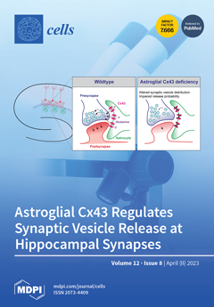
Connexin 43 (Cx43), an astroglial perisynaptic protein, is important for synaptic transmission and cognition via presynaptic regulation. However, whether it acts by modulating synaptic vesicle release is unknown. Using ultrastructural studies and live imaging of synaptic vesicle recycling, we found that a deficiency of astroglial Cx43 impairs synaptic vesicle distribution and release dynamics, despite the normal development of hippocampal neurons and synapses. Electrophysiological recordings further showed that synaptic vesicle release probability is also reduced and dependent on glutamine supply via the Cx43 hemichannel function. Taken together, we have uncovered a role for astroglial Cx43 in presynaptic function by controlling the rate and probability of synaptic vesicle release, further highlighting its significance in synaptic transmission and efficacy. View this paper
  • Issues are regarded as officially published after their release is announced to the table of contents alert mailing list.
  • You may sign up for e-mail alerts to receive table of contents of newly released issues.
  • PDF is the official format for papers published in both, html and pdf forms. To view the papers in pdf format, click on the "PDF Full-text" link, and use the free Adobe Reader to open them.
Order results
Result details
Section
Select all
Export citation of selected articles as:
23 pages, 2867 KiB  
Review
Essential Protein PHB2 and Its Regulatory Mechanisms in Cancer
by Amanda Qi, Lillie Lamont, Evelyn Liu, Sarina D. Murray, Xiangbing Meng and Shujie Yang
Cells 2023, 12(8), 1211; https://doi.org/10.3390/cells12081211 - 21 Apr 2023
Cited by 6 | Viewed by 2383
Abstract
Prohibitins (PHBs) are a highly conserved class of proteins and have an essential role in transcription, epigenetic regulation, nuclear signaling, mitochondrial structural integrity, cell division, and cellular membrane metabolism. Prohibitins form a heterodimeric complex, consisting of two proteins, prohibitin 1 (PHB1) and prohibitin [...] Read more.
Prohibitins (PHBs) are a highly conserved class of proteins and have an essential role in transcription, epigenetic regulation, nuclear signaling, mitochondrial structural integrity, cell division, and cellular membrane metabolism. Prohibitins form a heterodimeric complex, consisting of two proteins, prohibitin 1 (PHB1) and prohibitin 2 (PHB2). They have been discovered to have crucial roles in regulating cancer and other metabolic diseases, functioning both together and independently. As there have been many previously published reviews on PHB1, this review focuses on the lesser studied prohibitin, PHB2. The role of PHB2 in cancer is controversial. In most human cancers, overexpressed PHB2 enhances tumor progression, while in some cancers, it suppresses tumor progression. In this review, we focus on (1) the history, family, and structure of prohibitins, (2) the essential location-dependent functions of PHB2, (3) dysfunction in cancer, and (4) the promising modulators to target PHB2. At the end, we discuss future directions and the clinical significance of this common essential gene in cancer. Full article
Show Figures

Figure 1

17 pages, 777 KiB  
Review
Contribution of Axon Initial Segment Structure and Channels to Brain Pathology
by Juan José Garrido
Cells 2023, 12(8), 1210; https://doi.org/10.3390/cells12081210 - 21 Apr 2023
Cited by 3 | Viewed by 1908
Abstract
Brain channelopathies are a group of neurological disorders that result from genetic mutations affecting ion channels in the brain. Ion channels are specialized proteins that play a crucial role in the electrical activity of nerve cells by controlling the flow of ions such [...] Read more.
Brain channelopathies are a group of neurological disorders that result from genetic mutations affecting ion channels in the brain. Ion channels are specialized proteins that play a crucial role in the electrical activity of nerve cells by controlling the flow of ions such as sodium, potassium, and calcium. When these channels are not functioning properly, they can cause a wide range of neurological symptoms such as seizures, movement disorders, and cognitive impairment. In this context, the axon initial segment (AIS) is the site of action potential initiation in most neurons. This region is characterized by a high density of voltage-gated sodium channels (VGSCs), which are responsible for the rapid depolarization that occurs when the neuron is stimulated. The AIS is also enriched in other ion channels, such as potassium channels, that play a role in shaping the action potential waveform and determining the firing frequency of the neuron. In addition to ion channels, the AIS contains a complex cytoskeletal structure that helps to anchor the channels in place and regulate their function. Therefore, alterations in this complex structure of ion channels, scaffold proteins, and specialized cytoskeleton may also cause brain channelopathies not necessarily associated with ion channel mutations. This review will focus on how the AISs structure, plasticity, and composition alterations may generate changes in action potentials and neuronal dysfunction leading to brain diseases. AIS function alterations may be the consequence of voltage-gated ion channel mutations, but also may be due to ligand-activated channels and receptors and AIS structural and membrane proteins that support the function of voltage-gated ion channels. Full article
Show Figures

Figure 1

19 pages, 5600 KiB  
Article
Residual Foci of DNA Damage Response Proteins in Relation to Cellular Senescence and Autophagy in X-Ray Irradiated Fibroblasts
by Andrey Osipov, Anna Chigasova, Elizaveta Yashkina, Maxim Ignatov, Yuriy Fedotov, Daria Molodtsova, Natalia Vorobyeva and Andreyan N. Osipov
Cells 2023, 12(8), 1209; https://doi.org/10.3390/cells12081209 - 21 Apr 2023
Cited by 2 | Viewed by 1454
Abstract
DNA repair (DNA damage) foci observed 24 h and later after irradiation are called “residual” in the literature. They are believed to be the repair sites for complex, potentially lethal DNA double strand breaks. However, the features of their post-radiation dose-dependent quantitative changes [...] Read more.
DNA repair (DNA damage) foci observed 24 h and later after irradiation are called “residual” in the literature. They are believed to be the repair sites for complex, potentially lethal DNA double strand breaks. However, the features of their post-radiation dose-dependent quantitative changes and their role in the processes of cell death and senescence are still insufficiently studied. For the first time in one work, a simultaneous study of the association of changes in the number of residual foci of key DNA damage response (DDR) proteins (γH2AX, pATM, 53BP1, p-p53), the proportion of caspase-3 positive, LC-3 II autophagic and SA-β-gal senescent cells was carried out 24–72 h after fibroblast irradiation with X-rays at doses of 1–10 Gy. It was shown that with an increase in time after irradiation from 24 h to 72 h, the number of residual foci and the proportion of caspase-3 positive cells decrease, while the proportion of senescent cells, on the contrary, increases. The highest number of autophagic cells was noted 48 h after irradiation. In general, the results obtained provide important information for understanding the dynamics of the development of a dose-dependent cellular response in populations of irradiated fibroblasts. Full article
(This article belongs to the Special Issue DNA Damage and Senescence in Cellular Response to Cancer Therapies)
Show Figures

Figure 1

16 pages, 1703 KiB  
Systematic Review
Systematic Review of Roles of Arecoline and Arecoline N-Oxide in Oral Cancer and Strategies to Block Carcinogenesis
by Albert Min-Shan Ko, Hung-Pin Tu and Ying-Chin Ko
Cells 2023, 12(8), 1208; https://doi.org/10.3390/cells12081208 - 21 Apr 2023
Cited by 12 | Viewed by 2212
Abstract
Betel quid and areca nut are complex mixture carcinogens, but little is known about whether their derived single-agent arecoline or arecoline N-oxide (ANO) is carcinogenic, and the underlying mechanisms remain unclear. In this systematic review, we analyzed recent studies on the roles [...] Read more.
Betel quid and areca nut are complex mixture carcinogens, but little is known about whether their derived single-agent arecoline or arecoline N-oxide (ANO) is carcinogenic, and the underlying mechanisms remain unclear. In this systematic review, we analyzed recent studies on the roles of arecoline and ANO in cancer and strategies to block carcinogenesis. In the oral cavity, flavin-containing monooxygenase 3 oxidizes arecoline to ANO, and both alkaloids conjugate with N-acetylcysteine to form mercapturic acid compounds, which are excreted in urine, reducing arecoline and ANO toxicity. However, detoxification may not be complete. Arecoline and ANO upregulated protein expression in oral cancer tissue from areca nut users compared to expression levels in adjacent normal tissue, suggesting a causal relationship between these compounds and oral cancer. Sublingual fibrosis, hyperplasia, and oral leukoplakia were diagnosed in mice subjected to oral mucosal smearing of ANO. ANO is more cytotoxic and genotoxic than arecoline. During carcinogenesis and metastasis, these compounds increase the expression of epithelial–mesenchymal transition (EMT) inducers such as reactive oxygen species, transforming growth factor-β1, Notch receptor-1, and inflammatory cytokines, and they activate EMT-related proteins. Arecoline-induced epigenetic markers such as sirtuin-1 hypermethylation, low protein expression of miR-22, and miR-886-3-p accelerate oral cancer progression. Antioxidants and targeted inhibitors of the EMT inducers used reduce the risk of oral cancer development and progression. Our review findings substantiate the association of arecoline and ANO with oral cancer. Both of these single compounds are likely carcinogenic to humans, and their mechanisms and pathways of carcinogenesis are useful indicators for cancer therapy and prognosis. Full article
Show Figures

Graphical abstract

17 pages, 1500 KiB  
Review
The Functions and Phenotypes of Microglia in Alzheimer’s Disease
by Risako Fujikawa and Makoto Tsuda
Cells 2023, 12(8), 1207; https://doi.org/10.3390/cells12081207 - 21 Apr 2023
Cited by 4 | Viewed by 3349
Abstract
Alzheimer’s disease (AD) is the most prevalent neurodegenerative disease worldwide, but therapeutic strategies to slow down AD pathology and symptoms have not yet been successful. While attention has been focused on neurodegeneration in AD pathogenesis, recent decades have provided evidence of the importance [...] Read more.
Alzheimer’s disease (AD) is the most prevalent neurodegenerative disease worldwide, but therapeutic strategies to slow down AD pathology and symptoms have not yet been successful. While attention has been focused on neurodegeneration in AD pathogenesis, recent decades have provided evidence of the importance of microglia, and resident immune cells in the central nervous system. In addition, new technologies, including single-cell RNA sequencing, have revealed heterogeneous cell states of microglia in AD. In this review, we systematically summarize the microglial response to amyloid-β and tau tangles, and the risk factor genes expressed in microglia. Furthermore, we discuss the characteristics of protective microglia that appear during AD pathology and the relationship between AD and microglia-induced inflammation during chronic pain. Understanding the diverse roles of microglia will help identify new therapeutic strategies for AD. Full article
Show Figures

Graphical abstract

13 pages, 1859 KiB  
Article
Progesterone: A Neuroprotective Steroid of the Intestine
by Lennart Norman Stegemann, Paula Maria Neufeld, Ines Hecking, Matthias Vorgerd, Veronika Matschke, Sarah Stahlke and Carsten Theiss
Cells 2023, 12(8), 1206; https://doi.org/10.3390/cells12081206 - 21 Apr 2023
Cited by 2 | Viewed by 3444
Abstract
The enteric nervous system (ENS) is an intrinsic network of neuronal ganglia in the intestinal tube with about 100 million neurons located in the myenteric plexus and submucosal plexus. These neurons being affected in neurodegenerative diseases, such as Parkinson’s disease, before pathological changes [...] Read more.
The enteric nervous system (ENS) is an intrinsic network of neuronal ganglia in the intestinal tube with about 100 million neurons located in the myenteric plexus and submucosal plexus. These neurons being affected in neurodegenerative diseases, such as Parkinson’s disease, before pathological changes in the central nervous system (CNS) become detectable is currently a subject of discussion. Understanding how to protect these neurons is, therefore, particularly important. Since it has already been shown that the neurosteroid progesterone mediates neuroprotective effects in the CNS and PNS, it is now equally important to see whether progesterone has similar effects in the ENS. For this purpose, the RT-qPCR analyses of laser microdissected ENS neurons were performed, showing for the first time the expression of the different progesterone receptors (PR-A/B; mPRa, mPRb, PGRMC1) in rats at different developmental stages. This was also confirmed in ENS ganglia using immunofluorescence techniques and confocal laser scanning microscopy. To analyze the potential neuroprotective effects of progesterone in the ENS, we stressed dissociated ENS cells with rotenone to induce damage typical of Parkinson’s disease. The potential neuroprotective effects of progesterone were then analyzed in this system. Treatment of cultured ENS neurons with progesterone reduced cell death by 45%, underscoring the tremendous neuroprotective potential of progesterone in the ENS. The additional administration of the PGRMC1 antagonist AG205 abolished the observed effect, indicating the crucial role of PGRMC1 with regard to the neuroprotective effect of progesterone. Full article
(This article belongs to the Special Issue Progesterone Receptor Signaling)
Show Figures

Graphical abstract

19 pages, 1506 KiB  
Review
Peroxisome Proliferator-Activated Receptor-γ as a Target and Regulator of Epigenetic Mechanisms in Nonalcoholic Fatty Liver Disease
by Mohamed Zaiou
Cells 2023, 12(8), 1205; https://doi.org/10.3390/cells12081205 - 21 Apr 2023
Cited by 5 | Viewed by 2043
Abstract
Peroxisome proliferator-activated receptor-γ (PPARγ) belongs to the superfamily of nuclear receptors that control the transcription of multiple genes. Although it is found in many cells and tissues, PPARγ is mostly expressed in the liver and adipose tissue. Preclinical and clinical studies show that [...] Read more.
Peroxisome proliferator-activated receptor-γ (PPARγ) belongs to the superfamily of nuclear receptors that control the transcription of multiple genes. Although it is found in many cells and tissues, PPARγ is mostly expressed in the liver and adipose tissue. Preclinical and clinical studies show that PPARγ targets several genes implicated in various forms of chronic liver disease, including nonalcoholic fatty liver disease (NAFLD). Clinical trials are currently underway to investigate the beneficial effects of PPARγ agonists on NAFLD/nonalcoholic steatohepatitis. Understanding PPARγ regulators may therefore aid in unraveling the mechanisms governing the development and progression of NAFLD. Recent advances in high-throughput biology and genome sequencing have greatly facilitated the identification of epigenetic modifiers, including DNA methylation, histone modifiers, and non-coding RNAs as key factors that regulate PPARγ in NAFLD. In contrast, little is still known about the particular molecular mechanisms underlying the intricate relationships between these events. The paper that follows outlines our current understanding of the crosstalk between PPARγ and epigenetic regulators in NAFLD. Advances in this field are likely to aid in the development of early noninvasive diagnostics and future NAFLD treatment strategies based on PPARγ epigenetic circuit modification. Full article
(This article belongs to the Collection Functions of Nuclear Receptors)
Show Figures

Figure 1

26 pages, 1374 KiB  
Review
The Multifaceted Role of WNT Signaling in Alzheimer’s Disease Onset and Age-Related Progression
by William W. Kostes and David A. Brafman
Cells 2023, 12(8), 1204; https://doi.org/10.3390/cells12081204 - 21 Apr 2023
Cited by 1 | Viewed by 1896
Abstract
The evolutionary conserved WNT signaling pathway orchestrates numerous complex biological processes during development and is critical to the maintenance of tissue integrity and homeostasis in the adult. As it relates to the central nervous system, WNT signaling plays several roles as it relates [...] Read more.
The evolutionary conserved WNT signaling pathway orchestrates numerous complex biological processes during development and is critical to the maintenance of tissue integrity and homeostasis in the adult. As it relates to the central nervous system, WNT signaling plays several roles as it relates to neurogenesis, synaptic formation, memory, and learning. Thus, dysfunction of this pathway is associated with multiple diseases and disorders, including several neurodegenerative disorders. Alzheimer’s disease (AD) is characterized by several pathologies, synaptic dysfunction, and cognitive decline. In this review, we will discuss the various epidemiological, clinical, and animal studies that demonstrate a precise link between aberrant WNT signaling and AD-associated pathologies. In turn, we will discuss the manner in which WNT signaling influences multiple molecular, biochemical, and cellular pathways upstream of these end-point pathologies. Finally, we will discuss how merging tools and technologies can be used to generate next generation cellular models to dissect the relationship between WNT signaling and AD. Full article
(This article belongs to the Special Issue Wnt Signaling in Development and Aging)
Show Figures

Figure 1

17 pages, 2553 KiB  
Article
Overexpression of GREM1 Improves the Survival Capacity of Aged Cardiac Mesenchymal Progenitor Cells via Upregulation of the ERK/NRF2-Associated Antioxidant Signal Pathway
by Gurleen Kaur, Xiaoliang Wang, Xiuchun Li, Hannah Ong, Xiangfei He and Chuanxi Cai
Cells 2023, 12(8), 1203; https://doi.org/10.3390/cells12081203 - 21 Apr 2023
Viewed by 1964
Abstract
Ischemic heart disease is the leading cause of mortality in the United States. Progenitor cell therapy can restore myocardial structure and function. However, its efficacy is severely limited by cell aging and senescence. Gremlin-1 (GREM1), a member of the bone morphogenetic protein antagonist [...] Read more.
Ischemic heart disease is the leading cause of mortality in the United States. Progenitor cell therapy can restore myocardial structure and function. However, its efficacy is severely limited by cell aging and senescence. Gremlin-1 (GREM1), a member of the bone morphogenetic protein antagonist family, has been implicated in cell proliferation and survival. However, GREM1’s role in cell aging and senescence has never been investigated in human cardiac mesenchymal progenitor cells (hMPCs). Therefore, this study assessed the hypothesis that overexpression of GREM1 rejuvenates the cardiac regenerative potential of aging hMPCs to a youthful stage and therefore allows better capacity for myocardial repair. We recently reported that a subpopulation of hMPCs with low mitochondrial membrane potential can be sorted from right atrial appendage-derived cells in patients with cardiomyopathy and exhibit cardiac reparative capacity in a mouse model of myocardial infarction. In this study, lentiviral particles were used to overexpress GREM1 in these hMPCs. Protein and mRNA expression were assessed through Western blot and RT-qPCR. FACS analysis for Annexin V/PI staining and lactate dehydrogenase assay were used to assess cell survival. It was observed that cell aging and cell senescence led to a decrease in GREM1 expression. In addition, overexpression of GREM1 led to a decrease in expression of senescence genes. Overexpression of GREM1 led to no significant change in cell proliferation. However, GREM1 appeared to have an anti-apoptotic effect, with an increase in survival and decrease in cytotoxicity evident in GREM1-overexpressing hMPCs. Overexpressing GREM1 also induced cytoprotective properties by decreasing reactive oxidative species and mitochondrial membrane potential. This result was associated with increased expression of antioxidant proteins, such as SOD1 and catalase, and activation of the ERK/NRF2 survival signal pathway. Inhibition of ERK led to a decrease in GREM1-mediated rejuvenation in terms of cell survival, which suggests that an ERK-dependent pathway may be involved. Taken altogether, these results indicate that overexpression of GREM1 can allow aging hMPCs to adopt a more robust phenotype with improved survival capacity, which is associated with an activated ERK/NRF2 antioxidant signal pathway. Full article
(This article belongs to the Special Issue The Role of Oxidative Stress in Cardiovascular Diseases)
Show Figures

Figure 1

16 pages, 2370 KiB  
Article
Steatosis and Metabolic Disorders Associated with Synergistic Activation of the CAR/RXR Heterodimer by Pesticides
by Yannick Dauwe, Lucile Mary, Fabiana Oliviero, Marina Grimaldi, Patrick Balaguer, Véronique Gayrard and Laïla Mselli-Lakhal
Cells 2023, 12(8), 1201; https://doi.org/10.3390/cells12081201 - 21 Apr 2023
Cited by 2 | Viewed by 1634
Abstract
The nuclear receptor, constitutive androstane receptor (CAR), which forms a heterodimer with the retinoid X receptor (RXR), was initially reported as a transcription factor that regulates hepatic genes involved in detoxication and energy metabolism. Different studies have shown that CAR activation results in [...] Read more.
The nuclear receptor, constitutive androstane receptor (CAR), which forms a heterodimer with the retinoid X receptor (RXR), was initially reported as a transcription factor that regulates hepatic genes involved in detoxication and energy metabolism. Different studies have shown that CAR activation results in metabolic disorders, including non-alcoholic fatty liver disease, by activating lipogenesis in the liver. Our objective was to determine whether synergistic activations of the CAR/RXR heterodimer could occur in vivo as described in vitro by other authors, and to assess the metabolic consequences. For this purpose, six pesticides, ligands of CAR, were selected, and Tri-butyl-tin (TBT) was used as an RXR agonist. In mice, CAR’s synergic activation was induced by dieldrin associated with TBT, and combined effects were induced by propiconazole, bifenox, boscalid, and bupirimate. Moreover, a steatosis, characterized by increased triglycerides, was observed when TBT was combined with dieldrin, propiconazole, bifenox, boscalid, and bupirimate. Metabolic disruption appeared in the form of increased cholesterol and lowered free fatty acid plasma levels. An in-depth analysis revealed increased expression of genes involved in lipid synthesis and lipid import. These results contribute to the growing understanding of how environmental contaminants can influence nuclear receptor activity and associated health risks. Full article
(This article belongs to the Collection Functions of Nuclear Receptors)
Show Figures

Figure 1

19 pages, 2165 KiB  
Article
In Vitro Mineralisation of Tissue-Engineered Cartilage Reduces Endothelial Cell Migration, Proliferation and Tube Formation
by Encheng Ji, Lieke Leijsten, Janneke Witte-Bouma, Adelin Rouchon, Nunzia Di Maggio, Andrea Banfi, Gerjo J. V. M. van Osch, Eric Farrell and Andrea Lolli
Cells 2023, 12(8), 1202; https://doi.org/10.3390/cells12081202 - 20 Apr 2023
Viewed by 1460
Abstract
Tissue engineering bone via endochondral ossification requires the generation of a cartilage template which undergoes vascularisation and remodelling. While this is a promising route for bone repair, achieving effective cartilage vascularisation remains a challenge. Here, we investigated how mineralisation of tissue-engineered cartilage affects [...] Read more.
Tissue engineering bone via endochondral ossification requires the generation of a cartilage template which undergoes vascularisation and remodelling. While this is a promising route for bone repair, achieving effective cartilage vascularisation remains a challenge. Here, we investigated how mineralisation of tissue-engineered cartilage affects its pro-angiogenic potential. To generate in vitro mineralised cartilage, human mesenchymal stromal cell (hMSC)-derived chondrogenic pellets were treated with β-glycerophosphate (BGP). After optimising this approach, we characterised the changes in matrix components and pro-angiogenic factors by gene expression analysis, histology and ELISA. Human umbilical vein endothelial cells (HUVECs) were exposed to pellet-derived conditioned media, and migration, proliferation and tube formation were assessed. We established a reliable strategy to induce in vitro cartilage mineralisation, whereby hMSC pellets are chondrogenically primed with TGF-β for 2 weeks and BGP is added from week 2 of culture. Cartilage mineralisation determines loss of glycosaminoglycans, reduced expression but not protein abundance of collagen II and X, and decreased VEGFA production. Finally, the conditioned medium from mineralised pellets showed a reduced ability to stimulate endothelial cell migration, proliferation and tube formation. The pro-angiogenic potential of transient cartilage is thus stage-dependent, and this aspect must be carefully considered in the design of bone tissue engineering strategies. Full article
(This article belongs to the Special Issue Stem and Progenitor Cells in Bone Regeneration)
Show Figures

Figure 1

12 pages, 1783 KiB  
Article
The Antiepileptic Drug Oxcarbazepine Inhibits the Growth of Patient-Derived Isocitrate Dehydrogenase Mutant Glioma Stem-like Cells
by Philip Dao Trong, Gerhard Jungwirth, Andreas Unterberg, Christel Herold-Mende and Rolf Warta
Cells 2023, 12(8), 1200; https://doi.org/10.3390/cells12081200 - 20 Apr 2023
Viewed by 1640
Abstract
Patients diagnosed with isocitrate dehydrogenase mutant (IDHmut) gliomas suffer frequently from seizures. Although the clinical course is less aggressive than that of its IDH wildtype counterpart, recent discoveries have shown that epileptic activity can promote tumor proliferation. However, it is not [...] Read more.
Patients diagnosed with isocitrate dehydrogenase mutant (IDHmut) gliomas suffer frequently from seizures. Although the clinical course is less aggressive than that of its IDH wildtype counterpart, recent discoveries have shown that epileptic activity can promote tumor proliferation. However, it is not known if antiepileptic drugs confer additional value by inhibiting tumor growth. In this study, the antineoplastic properties of 20 FDA-approved antiepileptic drugs (AEDs) were tested in six patient-derived IDHmut glioma stem-like cells (GSCs). Cell proliferation was assessed using the CellTiterGlo-3D assay. Two of the screened drugs (oxcarbazepine and perampanel) demonstrated an antiproliferative effect. A subsequent eight-point dose–response curve proved the dose-dependent growth inhibition for both drugs, but only oxcarbazepine reached an IC50 value below 100 µM in 5/6 GSCs (mean 44.7 µM; range 17.4–98.0 µM), approximating the possible cmax for oxcarbazepine in patient serums. Furthermore, the treated GSC spheroids were 82% smaller (mean volume 1.6 nL vs. 8.7 nL; p = 0.01 (live/deadTM fluorescence staining)), and the apoptotic events increased by more than 50% (caspase-3/7 activity; p = 0.006). Taken together, this drug screen of a large series of antiepileptic drugs identified oxcarbazepine as a potent proapoptotic drug in IDHmut GSCs, which combines antiepileptic and antineoplastic properties to treat this seizure-prone patient population. Full article
(This article belongs to the Special Issue Cancer Stem-Like Cells and Cancer Therapeutic Strategy)
Show Figures

Figure 1

18 pages, 1357 KiB  
Systematic Review
Parsimonious Effect of Pentoxifylline on Angiogenesis: A Novel Pentoxifylline-Biased Adenosine G Protein-Coupled Receptor Signaling Platform
by William Khoury, Ryan Trus, Xingyu Chen, Leili Baghaie, Mira Clark, Myron R. Szewczuk and Mohammad El-Diasty
Cells 2023, 12(8), 1199; https://doi.org/10.3390/cells12081199 - 20 Apr 2023
Cited by 1 | Viewed by 1719
Abstract
Angiogenesis is the physiological process of developing new blood vessels to facilitate the delivery of oxygen and nutrients to meet the functional demands of growing tissues. It also plays a vital role in the development of neoplastic disorders. Pentoxifylline (PTX) is a vasoactive [...] Read more.
Angiogenesis is the physiological process of developing new blood vessels to facilitate the delivery of oxygen and nutrients to meet the functional demands of growing tissues. It also plays a vital role in the development of neoplastic disorders. Pentoxifylline (PTX) is a vasoactive synthetic methyl xanthine derivative used for decades to manage chronic occlusive vascular disorders. Recently, it has been proposed that PTX might have an inhibitory effect on the angiogenesis process. Here, we reviewed the modulatory effects of PTX on angiogenesis and its potential benefits in the clinical setting. Twenty-two studies met the inclusion and exclusion criteria. While sixteen studies demonstrated that pentoxifylline had an antiangiogenic effect, four suggested it had a proangiogenic effect, and two other studies showed it did not affect angiogenesis. All studies were either in vivo animal studies or in vitro animal and human cell models. Our findings suggest that pentoxifylline may affect the angiogenic process in experimental models. However, there is insufficient evidence to establish its role as an anti-angiogenesis agent in the clinical setting. These gaps in our knowledge regarding how pentoxifylline is implicated in host-biased metabolically taxing angiogenic switch may be via its adenosine A2BAR G protein-coupled receptor (GPCR) mechanism. GPCR receptors reinforce the importance of research to understand the mechanistic action of these drugs on the body as promising metabolic candidates. The specific mechanisms and details of the effects of pentoxifylline on host metabolism and energy homeostasis remain to be elucidated. Full article
(This article belongs to the Special Issue Emerging Topics in the Signaling Pathways of Angiogenesis)
Show Figures

Figure 1

23 pages, 25926 KiB  
Article
SARS-CoV-2 Enters Human Leydig Cells and Affects Testosterone Production In Vitro
by Lu Li, Chantal M. Sottas, Hsu-Yu Chen, Yuchang Li, Haoyi Cui, Jason S. Villano, Joseph L. Mankowski, Paula M. Cannon and Vassilios Papadopoulos
Cells 2023, 12(8), 1198; https://doi.org/10.3390/cells12081198 - 20 Apr 2023
Cited by 3 | Viewed by 2101
Abstract
Severe acute respiratory syndrome coronavirus 2 (SARS-CoV-2), a SARS-like coronavirus, continues to produce mounting infections and fatalities all over the world. Recent data point to SARS-CoV-2 viral infections in the human testis. As low testosterone levels are associated with SARS-CoV-2 viral infections in [...] Read more.
Severe acute respiratory syndrome coronavirus 2 (SARS-CoV-2), a SARS-like coronavirus, continues to produce mounting infections and fatalities all over the world. Recent data point to SARS-CoV-2 viral infections in the human testis. As low testosterone levels are associated with SARS-CoV-2 viral infections in males and human Leydig cells are the main source of testosterone, we hypothesized that SARS-CoV-2 could infect human Leydig cells and impair their function. We successfully detected SARS-CoV-2 nucleocapsid in testicular Leydig cells of SARS-CoV-2-infected hamsters, providing evidence that Leydig cells can be infected with SARS-CoV-2. We then employed human Leydig-like cells (hLLCs) to show that the SARS-CoV-2 receptor angiotensin-converting enzyme 2 is highly expressed in hLLCs. Using a cell binding assay and a SARS-CoV-2 spike-pseudotyped viral vector (SARS-CoV-2 spike pseudovector), we showed that SARS-CoV-2 could enter hLLCs and increase testosterone production by hLLCs. We further combined the SARS-CoV-2 spike pseudovector system with pseudovector-based inhibition assays to show that SARS-CoV-2 enters hLLCs through pathways distinct from those of monkey kidney Vero E6 cells, a typical model used to study SARS-CoV-2 entry mechanisms. We finally revealed that neuropilin-1 and cathepsin B/L are expressed in hLLCs and human testes, raising the possibility that SARS-CoV-2 may enter hLLCs through these receptors or proteases. In conclusion, our study shows that SARS-CoV-2 can enter hLLCs through a distinct pathway and alter testosterone production. Full article
(This article belongs to the Section Stem Cells)
Show Figures

Graphical abstract

14 pages, 2273 KiB  
Article
Fyn Phosphorylates Transglutaminase 2 (Tgm2) and Modulates Autophagy and p53 Expression in the Development of Diabetic Kidney Disease
by Ryota Uehara, Eijiro Yamada, Shuichi Okada, Claire C. Bastie, Akito Maeshima, Hidekazu Ikeuchi, Kazuhiko Horiguchi and Masanobu Yamada
Cells 2023, 12(8), 1197; https://doi.org/10.3390/cells12081197 - 20 Apr 2023
Viewed by 1578
Abstract
Autophagy is involved in the development of diabetic kidney disease (DKD), the leading cause of end-stage renal disease. The Fyn tyrosine kinase (Fyn) suppresses autophagy in the muscle. However, its role in kidney autophagic processes is unclear. Here, we examined the role of [...] Read more.
Autophagy is involved in the development of diabetic kidney disease (DKD), the leading cause of end-stage renal disease. The Fyn tyrosine kinase (Fyn) suppresses autophagy in the muscle. However, its role in kidney autophagic processes is unclear. Here, we examined the role of Fyn kinase in autophagy in proximal renal tubules both in vivo and in vitro. Phospho-proteomic analysis revealed that transglutaminase 2 (Tgm2), a protein involved in the degradation of p53 in the autophagosome, is phosphorylated on tyrosine 369 (Y369) by Fyn. Interestingly, we found that Fyn-dependent phosphorylation of Tgm2 regulates autophagy in proximal renal tubules in vitro, and that p53 expression is decreased upon autophagy in Tgm2-knockdown proximal renal tubule cell models. Using streptozocin (STZ)-induced hyperglycemic mice, we confirmed that Fyn regulated autophagy and mediated p53 expression via Tgm2. Taken together, these data provide a molecular basis for the role of the Fyn–Tgm2–p53 axis in the development of DKD. Full article
(This article belongs to the Special Issue Recent Advances in Development and Progression of Kidney Diseases)
Show Figures

Figure 1

28 pages, 2079 KiB  
Review
Perivascular Adipose Tissue and Vascular Smooth Muscle Tone: Friends or Foes?
by Amer Ahmed, Aasia Bibi, Massimo Valoti and Fabio Fusi
Cells 2023, 12(8), 1196; https://doi.org/10.3390/cells12081196 - 20 Apr 2023
Cited by 9 | Viewed by 2252
Abstract
Perivascular adipose tissue (PVAT) is a specialized type of adipose tissue that surrounds most mammalian blood vessels. PVAT is a metabolically active, endocrine organ capable of regulating blood vessel tone, endothelium function, vascular smooth muscle cell growth and proliferation, and contributing critically to [...] Read more.
Perivascular adipose tissue (PVAT) is a specialized type of adipose tissue that surrounds most mammalian blood vessels. PVAT is a metabolically active, endocrine organ capable of regulating blood vessel tone, endothelium function, vascular smooth muscle cell growth and proliferation, and contributing critically to cardiovascular disease onset and progression. In the context of vascular tone regulation, under physiological conditions, PVAT exerts a potent anticontractile effect by releasing a plethora of vasoactive substances, including NO, H2S, H2O2, prostacyclin, palmitic acid methyl ester, angiotensin 1-7, adiponectin, leptin, and omentin. However, under certain pathophysiological conditions, PVAT exerts pro-contractile effects by decreasing the production of anticontractile and increasing that of pro-contractile factors, including superoxide anion, angiotensin II, catecholamines, prostaglandins, chemerin, resistin, and visfatin. The present review discusses the regulatory effect of PVAT on vascular tone and the factors involved. In this scenario, dissecting the precise role of PVAT is a prerequisite to the development of PVAT-targeted therapies. Full article
Show Figures

Figure 1

16 pages, 2521 KiB  
Article
Inducible MLL-AF9 Expression Drives an AML Program during Human Pluripotent Stem Cell-Derived Hematopoietic Differentiation
by Branco M. H. Heuts, Saioa Arza-Apalategi, Sinne G. Alkema, Esther Tijchon, Laura Jussen, Saskia M. Bergevoet, Bert A. van der Reijden and Joost H. A. Martens
Cells 2023, 12(8), 1195; https://doi.org/10.3390/cells12081195 - 20 Apr 2023
Cited by 2 | Viewed by 2264
Abstract
A t(9;11)(p22;q23) translocation produces the MLL-AF9 fusion protein, which is found in up to 25% of de novo AML cases in children. Despite major advances, obtaining a comprehensive understanding of context-dependent MLL-AF9-mediated gene programs during early hematopoiesis is challenging. Here, we generated a [...] Read more.
A t(9;11)(p22;q23) translocation produces the MLL-AF9 fusion protein, which is found in up to 25% of de novo AML cases in children. Despite major advances, obtaining a comprehensive understanding of context-dependent MLL-AF9-mediated gene programs during early hematopoiesis is challenging. Here, we generated a human inducible pluripotent stem cell (hiPSC) model with a doxycycline dose-dependent MLL-AF9 expression. We exploited MLL-AF9 expression as an oncogenic hit to uncover epigenetic and transcriptomic effects on iPSC-derived hematopoietic development and the transformation into (pre-)leukemic states. In doing so, we observed a disruption in early myelomonocytic development. Accordingly, we identified gene profiles that were consistent with primary MLL-AF9 AML and uncovered high-confidence MLL-AF9-associated core genes that are faithfully represented in primary MLL-AF9 AML, including known and presently unknown factors. Using single-cell RNA-sequencing, we identified an increase of CD34 expressing early hematopoietic progenitor-like cell states as well as granulocyte-monocyte progenitor-like cells upon MLL-AF9 activation. Our system allows for careful chemically controlled and stepwise in vitro hiPSC-derived differentiation under serum-free and feeder-free conditions. For a disease that currently lacks effective precision medicine, our system provides a novel entry-point into exploring potential novel targets for personalized therapeutic strategies. Full article
(This article belongs to the Special Issue iPS Cells (iPSCs) for Modelling and Treatment of Human Diseases 2022)
Show Figures

Graphical abstract

19 pages, 3778 KiB  
Article
High-Fat Diet Modulates the Excitability of Neurons within the Brain–Liver Pathway
by Adrien J. R. Molinas, Lucie D. Desmoulins, Roslyn K. Davis, Hong Gao, Ryousuke Satou, Andrei V. Derbenev and Andrea Zsombok
Cells 2023, 12(8), 1194; https://doi.org/10.3390/cells12081194 - 20 Apr 2023
Cited by 3 | Viewed by 1362
Abstract
Stimulation of hepatic sympathetic nerves increases glucose production and glycogenolysis. Activity of pre-sympathetic neurons in the paraventricular nucleus (PVN) of the hypothalamus and in the ventrolateral and ventromedial medulla (VLM/VMM) largely influence the sympathetic output. Increased activity of the sympathetic nervous system (SNS) [...] Read more.
Stimulation of hepatic sympathetic nerves increases glucose production and glycogenolysis. Activity of pre-sympathetic neurons in the paraventricular nucleus (PVN) of the hypothalamus and in the ventrolateral and ventromedial medulla (VLM/VMM) largely influence the sympathetic output. Increased activity of the sympathetic nervous system (SNS) plays a role in the development and progression of metabolic diseases; however, despite the importance of the central circuits, the excitability of pre-sympathetic liver-related neurons remains to be determined. Here, we tested the hypothesis that the activity of liver-related neurons in the PVN and VLM/VMM is altered in diet-induced obese mice, as well as their response to insulin. Patch-clamp recordings were conducted from liver-related PVN neurons, VLM-projecting PVN neurons, and pre-sympathetic liver-related neurons in the ventral brainstem. Our data demonstrate that the excitability of liver-related PVN neurons increased in high-fat diet (HFD)-fed mice compared to mice fed with control diet. Insulin receptor expression was detected in a population of liver-related neurons, and insulin suppressed the firing activity of liver-related PVN and pre-sympathetic VLM/VMM neurons in HFD mice; however, it did not affect VLM-projecting liver-related PVN neurons. These findings further suggest that HFD alters the excitability of pre-autonomic neurons as well as their response to insulin. Full article
(This article belongs to the Special Issue Inter-organ Crosstalk in Energy Homeostasis)
Show Figures

Figure 1

25 pages, 1347 KiB  
Review
The Therapeutic Potential of Non-Invasive and Invasive Cerebellar Stimulation Techniques in Hereditary Ataxias
by Alberto Benussi, Giorgi Batsikadze, Carina França, Rubens G. Cury and Roderick P. P. W. M. Maas
Cells 2023, 12(8), 1193; https://doi.org/10.3390/cells12081193 - 20 Apr 2023
Cited by 5 | Viewed by 2964
Abstract
The degenerative ataxias comprise a heterogeneous group of inherited and acquired disorders that are characterized by a progressive cerebellar syndrome, frequently in combination with one or more extracerebellar signs. Specific disease-modifying interventions are currently not available for many of these rare conditions, which [...] Read more.
The degenerative ataxias comprise a heterogeneous group of inherited and acquired disorders that are characterized by a progressive cerebellar syndrome, frequently in combination with one or more extracerebellar signs. Specific disease-modifying interventions are currently not available for many of these rare conditions, which underscores the necessity of finding effective symptomatic therapies. During the past five to ten years, an increasing number of randomized controlled trials have been conducted examining the potential of different non-invasive brain stimulation techniques to induce symptomatic improvement. In addition, a few smaller studies have explored deep brain stimulation (DBS) of the dentate nucleus as an invasive means to directly modulate cerebellar output, thereby aiming to alleviate ataxia severity. In this paper, we comprehensively review the clinical and neurophysiological effects of transcranial direct current stimulation (tDCS), repetitive transcranial magnetic stimulation (rTMS), and dentate nucleus DBS in patients with hereditary ataxias, as well as the presumed underlying mechanisms at the cellular and network level and perspectives for future research. Full article
(This article belongs to the Special Issue Emerging Therapies for Hereditary Ataxia)
Show Figures

Figure 1

19 pages, 1241 KiB  
Review
Pluripotent Stem Cells as a Model for Human Embryogenesis
by Daniela Ávila-González, Mikel Ángel Gidi-Grenat, Guadalupe García-López, Alejandro Martínez-Juárez, Anayansi Molina-Hernández, Wendy Portillo, Néstor Emmanuel Díaz-Martínez and Néstor Fabián Díaz
Cells 2023, 12(8), 1192; https://doi.org/10.3390/cells12081192 - 20 Apr 2023
Viewed by 3657
Abstract
Pluripotent stem cells (PSCs; embryonic stem cells and induced pluripotent stem cells) can recapitulate critical aspects of the early stages of embryonic development; therefore, they became a powerful tool for the in vitro study of molecular mechanisms that underlie blastocyst formation, implantation, the [...] Read more.
Pluripotent stem cells (PSCs; embryonic stem cells and induced pluripotent stem cells) can recapitulate critical aspects of the early stages of embryonic development; therefore, they became a powerful tool for the in vitro study of molecular mechanisms that underlie blastocyst formation, implantation, the spectrum of pluripotency and the beginning of gastrulation, among other processes. Traditionally, PSCs were studied in 2D cultures or monolayers, without considering the spatial organization of a developing embryo. However, recent research demonstrated that PSCs can form 3D structures that simulate the blastocyst and gastrula stages and other events, such as amniotic cavity formation or somitogenesis. This breakthrough provides an unparalleled opportunity to study human embryogenesis by examining the interactions, cytoarchitecture and spatial organization among multiple cell lineages, which have long remained a mystery due to the limitations of studying in utero human embryos. In this review, we will provide an overview of how experimental embryology currently utilizes models such as blastoids, gastruloids and other 3D aggregates derived from PSCs to advance our understanding of the intricate processes involved in human embryo development. Full article
(This article belongs to the Special Issue Stem Cells in Organoid Technology)
Show Figures

Figure 1

33 pages, 2236 KiB  
Review
Experimental Validation and Prediction of Super-Enhancers: Advances and Challenges
by Ekaterina V. Kravchuk, German A. Ashniev, Marina G. Gladkova, Alexey V. Orlov, Anastasiia V. Vasileva, Anna V. Boldyreva, Alexandr G. Burenin, Artemiy M. Skirda, Petr I. Nikitin and Natalia N. Orlova
Cells 2023, 12(8), 1191; https://doi.org/10.3390/cells12081191 - 19 Apr 2023
Cited by 1 | Viewed by 2848
Abstract
Super-enhancers (SEs) are cis-regulatory elements of the human genome that have been widely discussed since the discovery and origin of the term. Super-enhancers have been shown to be strongly associated with the expression of genes crucial for cell differentiation, cell stability maintenance, and [...] Read more.
Super-enhancers (SEs) are cis-regulatory elements of the human genome that have been widely discussed since the discovery and origin of the term. Super-enhancers have been shown to be strongly associated with the expression of genes crucial for cell differentiation, cell stability maintenance, and tumorigenesis. Our goal was to systematize research studies dedicated to the investigation of structure and functions of super-enhancers as well as to define further perspectives of the field in various applications, such as drug development and clinical use. We overviewed the fundamental studies which provided experimental data on various pathologies and their associations with particular super-enhancers. The analysis of mainstream approaches for SE search and prediction allowed us to accumulate existing data and propose directions for further algorithmic improvements of SEs’ reliability levels and efficiency. Thus, here we provide the description of the most robust algorithms such as ROSE, imPROSE, and DEEPSEN and suggest their further use for various research and development tasks. The most promising research direction, which is based on topic and number of published studies, are cancer-associated super-enhancers and prospective SE-targeted therapy strategies, most of which are discussed in this review. Full article
(This article belongs to the Special Issue Epigenetics, Chromatin Structure and Transcription Regulation)
Show Figures

Figure 1

14 pages, 3089 KiB  
Article
Improving Schwann Cell Differentiation from Human Adipose Stem Cells with Metabolic Glycoengineering
by Jian Du, Zihui Wang, Xiao Liu, Cecilia Hu, Kevin J. Yarema and Xiaofeng Jia
Cells 2023, 12(8), 1190; https://doi.org/10.3390/cells12081190 - 19 Apr 2023
Cited by 2 | Viewed by 1548
Abstract
Schwann cells (SCs) are myelinating cells that promote peripheral nerve regeneration. When nerve lesions form, SCs are destroyed, ultimately hindering nerve repair. The difficulty in treating nerve repair is exacerbated due to SC’s limited and slow expansion capacity. Therapeutic use of adipose-derived stem [...] Read more.
Schwann cells (SCs) are myelinating cells that promote peripheral nerve regeneration. When nerve lesions form, SCs are destroyed, ultimately hindering nerve repair. The difficulty in treating nerve repair is exacerbated due to SC’s limited and slow expansion capacity. Therapeutic use of adipose-derived stem cells (ASCs) is emerging in combating peripheral nerve injury due to these cells’ SC differentiation capability and can be harvested easily in large numbers. Despite ASC’s therapeutic potential, their transdifferentiation period typically takes more than two weeks. In this study, we demonstrate that metabolic glycoengineering (MGE) technology enhances ASC differentiation into SCs. Specifically, the sugar analog Ac5ManNTProp (TProp), which modulates cell surface sialylation, significantly improved ASC differentiation with upregulated SC protein S100β and p75NGFR expression and elevated the neurotrophic factors nerve growth factor beta (NGFβ) and glial cell-line-derived neurotrophic factor (GDNF). TProp treatment remarkably reduced the SC transdifferentiation period from about two weeks to two days in vitro, which has the potential to improve neuronal regeneration and facilitate future use of ASCs in regenerative medicine. Full article
(This article belongs to the Collection Research on Adipose Stem Cells)
Show Figures

Figure 1

12 pages, 2068 KiB  
Article
A Preliminary Study of Mild Heat Stress on Inflammasome Activation in Murine Macrophages
by Simmie L. Foster, Abigail J. Dutton, Adina Yerzhan, Lindsay B. March, Katherine Barry, Corey R. Seehus, Xudong Huang, Sebastien Talbot and Clifford J. Woolf
Cells 2023, 12(8), 1189; https://doi.org/10.3390/cells12081189 - 19 Apr 2023
Cited by 1 | Viewed by 1324
Abstract
Inflammation and mitochondrial-dependent oxidative stress are interrelated processes implicated in multiple neuroinflammatory disorders, including Alzheimer’s disease (AD) and depression. Exposure to elevated temperature (hyperthermia) is proposed as a non-pharmacological, anti-inflammatory treatment for these disorders; however, the underlying mechanisms are not fully understood. Here [...] Read more.
Inflammation and mitochondrial-dependent oxidative stress are interrelated processes implicated in multiple neuroinflammatory disorders, including Alzheimer’s disease (AD) and depression. Exposure to elevated temperature (hyperthermia) is proposed as a non-pharmacological, anti-inflammatory treatment for these disorders; however, the underlying mechanisms are not fully understood. Here we asked if the inflammasome, a protein complex essential for orchestrating the inflammatory response and linked to mitochondrial stress, might be modulated by elevated temperatures. To test this, in preliminary studies, immortalized bone-marrow-derived murine macrophages (iBMM) were primed with inflammatory stimuli, exposed to a range of temperatures (37–41.5 °C), and examined for markers of inflammasome and mitochondrial activity. We found that exposure to mild heat stress (39 °C for 15 min) rapidly inhibited iBMM inflammasome activity. Furthermore, heat exposure led to decreased ASC speck formation and increased numbers of polarized mitochondria. These results suggest that mild hyperthermia inhibits inflammasome activity in the iBMM, limiting potentially harmful inflammation and mitigating mitochondrial stress. Our findings suggest an additional potential mechanism by which hyperthermia may exert its beneficial effects on inflammatory diseases. Full article
(This article belongs to the Special Issue Oxidative Stress in Aging and Neurodegenerative Diseases)
Show Figures

Figure 1

15 pages, 6959 KiB  
Review
Reversing Dysdynamism to Interrupt Mitochondrial Degeneration in Amyotrophic Lateral Sclerosis
by Gerald W. Dorn II
Cells 2023, 12(8), 1188; https://doi.org/10.3390/cells12081188 - 19 Apr 2023
Cited by 1 | Viewed by 1417
Abstract
Amyotrophic lateral sclerosis is one of several chronic neurodegenerative conditions in which mitochondrial abnormalities are posited to contribute to disease progression. Therapeutic options targeting mitochondria include enhancing metabolism, suppressing reactive oxygen production and disrupting mitochondria-mediated programmed cell death pathways. Herein is reviewed mechanistic [...] Read more.
Amyotrophic lateral sclerosis is one of several chronic neurodegenerative conditions in which mitochondrial abnormalities are posited to contribute to disease progression. Therapeutic options targeting mitochondria include enhancing metabolism, suppressing reactive oxygen production and disrupting mitochondria-mediated programmed cell death pathways. Herein is reviewed mechanistic evidence supporting a meaningful pathophysiological role for the constellation of abnormal mitochondrial fusion, fission and transport, collectively designated mitochondrial dysdynamism, in ALS. Following this is a discussion on preclinical studies in ALS mice that seemingly validate the idea that normalizing mitochondrial dynamism can delay ALS by interrupting a vicious cycle of mitochondrial degeneration, leading to neuronal die-back and death. Finally, the relative benefits of suppressing mitochondrial fusion vs. enhancing mitochondrial fusion in ALS are speculated upon, and the paper concludes with the prediction that the two approaches could be additive or synergistic, although a side-by-side comparative trial may be challenging to perform. Full article
Show Figures

Figure 1

20 pages, 1620 KiB  
Review
Mast Cells as a Target—A Comprehensive Review of Recent Therapeutic Approaches
by Joanna Baran, Anna Sobiepanek, Anna Mazurkiewicz-Pisarek, Marta Rogalska, Aleksander Gryciuk, Lukasz Kuryk, Soman N. Abraham and Monika Staniszewska
Cells 2023, 12(8), 1187; https://doi.org/10.3390/cells12081187 - 19 Apr 2023
Cited by 8 | Viewed by 4584
Abstract
Mast cells (MCs) are the immune cells distributed throughout nearly all tissues, mainly in the skin, near blood vessels and lymph vessels, nerves, lungs, and the intestines. Although MCs are essential to the healthy immune response, their overactivity and pathological states can lead [...] Read more.
Mast cells (MCs) are the immune cells distributed throughout nearly all tissues, mainly in the skin, near blood vessels and lymph vessels, nerves, lungs, and the intestines. Although MCs are essential to the healthy immune response, their overactivity and pathological states can lead to numerous health hazards. The side effect of mast cell activity is usually caused by degranulation. It can be triggered by immunological factors, such as immunoglobulins, lymphocytes, or antigen–antibody complexes, and non-immune factors, such as radiation and pathogens. An intensive reaction of mast cells can even lead to anaphylaxis, one of the most life-threatening allergic reactions. What is more, mast cells play a role in the tumor microenvironment by modulating various events of tumor biology, such as cell proliferation and survival, angiogenesis, invasiveness, and metastasis. The mechanisms of the mast cell actions are still poorly understood, making it difficult to develop therapies for their pathological condition. This review focuses on the possible therapies targeting mast cell degranulation, anaphylaxis, and MC-derived tumors. Full article
(This article belongs to the Special Issue Mast Cells in Immunity and Inflammation)
Show Figures

Figure 1

20 pages, 3112 KiB  
Article
Liver X Receptor Activation Attenuates Oxysterol-Induced Inflammatory Responses in Fetoplacental Endothelial Cells
by Meekha George, Magdalena Lang, Chaitanya Chakravarthi Gali, Joshua Adekunle Babalola, Carmen Tam-Amersdorfer, Anika Stracke, Herbert Strobl, Robert Zimmermann, Ute Panzenboeck and Christian Wadsack
Cells 2023, 12(8), 1186; https://doi.org/10.3390/cells12081186 - 19 Apr 2023
Cited by 2 | Viewed by 2015
Abstract
Oxysterols are oxidized cholesterol derivatives whose systemic levels are found elevated in pregnancy disorders such as gestational diabetes mellitus (GDM). Oxysterols act through various cellular receptors and serve as a key metabolic signal, coordinating inflammation. GDM is a condition of low-grade chronic inflammation [...] Read more.
Oxysterols are oxidized cholesterol derivatives whose systemic levels are found elevated in pregnancy disorders such as gestational diabetes mellitus (GDM). Oxysterols act through various cellular receptors and serve as a key metabolic signal, coordinating inflammation. GDM is a condition of low-grade chronic inflammation accompanied by altered inflammatory profiles in the mother, placenta and fetus. Higher levels of two oxysterols, namely 7-ketocholesterol (7-ketoC) and 7β-hydroxycholesterol (7β-OHC), were observed in fetoplacental endothelial cells (fpEC) and cord blood of GDM offspring. In this study, we tested the effects of 7-ketoC and 7β-OHC on inflammation and investigated the underlying mechanisms involved. Primary fpEC in culture treated with 7-ketoC or 7β-OHC, induced the activation of mitogen-activated protein kinase (MAPK) and nuclear factor kappa B (NFκB) signaling, which resulted in the expression of pro-inflammatory cytokines (IL-6, IL-8) and intercellular cell adhesion molecule-1 (ICAM-1). Liver-X receptor (LXR) activation is known to repress inflammation. Treatment with LXR synthetic agonist T0901317 dampened oxysterol-induced inflammatory responses. Probucol, an inhibitor of LXR target gene ATP-binding cassette transporter A-1 (ABCA-1), antagonized the protective effects of T0901317, suggesting a potential involvement of ABCA-1 in LXR-mediated repression of inflammatory signaling in fpEC. TLR-4 inhibitor Tak-242 attenuated pro-inflammatory signaling induced by oxysterols downstream of the TLR-4 inflammatory signaling cascade. Taken together, our findings suggest that 7-ketoC and 7β-OHC contribute to placental inflammation through the activation of TLR-4. Pharmacologic activation of LXR in fpEC decelerates its shift to a pro-inflammatory phenotype in the presence of oxysterols. Full article
Show Figures

Figure 1

18 pages, 18705 KiB  
Article
Aberrant APOBEC3B Expression in Breast Cancer Is Linked to Proliferation and Cell Cycle Phase
by Pieter A. Roelofs, Mieke A. M. Timmermans, Bojana Stefanovska, Myrthe A. den Boestert, Amber W. M. van den Borne, Hayri E. Balcioglu, Anita M. Trapman, Reuben S. Harris, John W. M. Martens and Paul N. Span
Cells 2023, 12(8), 1185; https://doi.org/10.3390/cells12081185 - 18 Apr 2023
Cited by 4 | Viewed by 1831
Abstract
APOBEC3B (A3B) is aberrantly overexpressed in a subset of breast cancers, where it associates with advanced disease, poor prognosis, and treatment resistance, yet the causes of A3B dysregulation in breast cancer remain unclear. Here, A3B mRNA and protein expression levels were quantified in [...] Read more.
APOBEC3B (A3B) is aberrantly overexpressed in a subset of breast cancers, where it associates with advanced disease, poor prognosis, and treatment resistance, yet the causes of A3B dysregulation in breast cancer remain unclear. Here, A3B mRNA and protein expression levels were quantified in different cell lines and breast tumors and related to cell cycle markers using RT-qPCR and multiplex immunofluorescence imaging. The inducibility of A3B expression during the cell cycle was additionally addressed after cell cycle synchronization with multiple methods. First, we found that A3B protein levels within cell lines and tumors are heterogeneous and associate strongly with the proliferation marker Cyclin B1 characteristic of the G2/M phase of the cell cycle. Second, in multiple breast cancer cell lines with high A3B, expression levels were observed to oscillate throughout the cell cycle and again associate with Cyclin B1. Third, induction of A3B expression is potently repressed throughout G0/early G1, likely by RB/E2F pathway effector proteins. Fourth, in cells with low A3B, induction of A3B through the PKC/ncNF-κB pathway occurs predominantly in actively proliferating cells and is largely absent in cells arrested in G0. Altogether, these results support a model in which dysregulated A3B overexpression in breast cancer is the cumulative result of proliferation-associated relief from repression with concomitant pathway activation during the G2/M phase of the cell cycle. Full article
(This article belongs to the Special Issue Advances in Cellular and Molecular Research in Breast Cancer)
Show Figures

Figure 1

16 pages, 3149 KiB  
Systematic Review
Utility of Blood-Based Tau Biomarkers for Mild Cognitive Impairment and Alzheimer’s Disease: Systematic Review and Meta-Analysis
by Alex Zabala-Findlay, Lewis K. Penny, Richard A. Lofthouse, Andrew J. Porter, Soumya Palliyil, Charles R. Harrington, Claude M. Wischik and Mohammad Arastoo
Cells 2023, 12(8), 1184; https://doi.org/10.3390/cells12081184 - 18 Apr 2023
Cited by 2 | Viewed by 2262
Abstract
Objectives: With the development of new technologies capable of detecting low concentrations of Alzheimer’s disease (AD) relevant biomarkers, the idea of a blood-based diagnosis of AD is nearing reality. This study aims to consider the evidence of total and phosphorylated tau as blood-based [...] Read more.
Objectives: With the development of new technologies capable of detecting low concentrations of Alzheimer’s disease (AD) relevant biomarkers, the idea of a blood-based diagnosis of AD is nearing reality. This study aims to consider the evidence of total and phosphorylated tau as blood-based biomarkers for mild cognitive impairment (MCI) and AD when compared to healthy controls. Methods: Studies published between 1 January 2012 and 1 May 2021 (Embase and MEDLINE databases) measuring plasma/serum levels of tau in AD, MCI, and control cohorts were screened for eligibility, including quality and bias assessment via a modified QUADAS. The meta-analyses comprised 48 studies assessing total tau (t-tau), tau phosphorylated at threonine 181 (p-tau181), and tau phosphorylated at threonine 217 (p-tau217), comparing the ratio of biomarker concentrations in MCI, AD, and cognitively unimpaired (CU) controls. Results: Plasma/serum p-tau181 (mean effect size, 95% CI, 2.02 (1.76–2.27)) and t-tau (mean effect size, 95% CI, 1.77 (1.49–2.04)) were elevated in AD study participants compared to controls. Plasma/serum p-tau181 (mean effect size, 95% CI, 1.34 (1.20–1.49)) and t-tau (mean effect size, 95% CI, 1.47 (1.26–1.67)) were also elevated with moderate effect size in MCI study participants compared to controls. p-tau217 was also assessed, albeit in a small number of eligible studies, for AD vs. CU (mean effect size, 95% CI, 1.89 (1.86–1.92)) and for MCI vs. CU groups (mean effect size, 95% CI, 4.16 (3.61–4.71)). Conclusions: This paper highlights the growing evidence that blood-based tau biomarkers have early diagnostic utility for Alzheimer’s disease. Registration: PROSPERO No. CRD42020209482. Full article
(This article belongs to the Special Issue Biomarker Diagnostics in Alzheimer’s Disease)
Show Figures

Figure 1

12 pages, 1163 KiB  
Communication
Expression of Stemness Markers in the Cervical Smear of Patients with Cervical Insufficiency
by Savvia Pittokopitou, Despina Mavrogianni, Vasilios Pergialiotis, Kalliopi I. Pappa, Panagiotis Antsaklis, Marianna Theodora, Michail Sindos, Angeliki Papapanagiotou, Aikaterini Domali, Sofoklis Stavros, Peter Drakakis and George Daskalakis
Cells 2023, 12(8), 1183; https://doi.org/10.3390/cells12081183 - 18 Apr 2023
Viewed by 1102
Abstract
The presence of stem cells has been previously described in human precancerous and malignant cervical cultures. Previous studies have shown a direct interplay of the stem cell niche, which is present in practically every tissue with the extracellular matrix. In the present study, [...] Read more.
The presence of stem cells has been previously described in human precancerous and malignant cervical cultures. Previous studies have shown a direct interplay of the stem cell niche, which is present in practically every tissue with the extracellular matrix. In the present study, we sought to determine the expression of stemness markers in cytological specimens collected from the ectocervix among women with cervical insufficiency during the second trimester of pregnancy and women with normal cervical length. A prospective cohort of 59 women was enrolled of whom 41 were diagnosed with cervical insufficiency. The expression of OCT-4 and NANOG was higher in the cervical insufficiency group compared to the control group (−5.03 (−6.27, −3.72) vs. −5.81 (−7.67, −5.02) p = 0.040 for OCT4) and (−7.47 (−8.78, −6.27) vs. −8.5 (−10.75, −7.14), p = 0.035 for NANOG. Differences in the DAZL gene were not significantly different (5.94 (4.82, 7.14) vs. 6.98 (5.87, 7.43) p = 0.097). Pearson correlation analysis indicated the existence of a moderate correlation of OCT-4 and Nanog with cervical length. Considering this information, the enhanced activity of stemness biomarkers among pregnant women diagnosed with cervical insufficiency may be predisposed to cervical insufficiency, and its predictive accuracy remains to be noted in larger population sizes. Full article
(This article belongs to the Special Issue Molecular and Cellular Mechanisms of Fetal Growth and Preterm Birth)
Show Figures

Figure 1

18 pages, 12535 KiB  
Communication
Single-Cell Transcriptome Analysis Revealed Heterogeneity and Identified Novel Therapeutic Targets for Breast Cancer Subtypes
by Radhakrishnan Vishnubalaji and Nehad M. Alajez
Cells 2023, 12(8), 1182; https://doi.org/10.3390/cells12081182 - 18 Apr 2023
Cited by 3 | Viewed by 2048
Abstract
Breast cancer (BC) is a heterogeneous disease, which is primarily classified according to hormone receptors and HER2 expression. Despite the many advances in BC diagnosis and management, the identification of novel actionable therapeutic targets expressed by cancerous cells has always been a daunting [...] Read more.
Breast cancer (BC) is a heterogeneous disease, which is primarily classified according to hormone receptors and HER2 expression. Despite the many advances in BC diagnosis and management, the identification of novel actionable therapeutic targets expressed by cancerous cells has always been a daunting task due to the large heterogeneity of the disease and the presence of non-cancerous cells (i.e., immune cells and stromal cells) within the tumor microenvironment. In the current study, we employed computational algorithms to decipher the cellular composition of estrogen receptor-positive (ER+), HER2+, ER+HER2+, and triple-negative BC (TNBC) subtypes from a total of 49,899 single cells’ publicly available transcriptomic data derived from 26 BC patients. Restricting the analysis to EPCAM+Lin tumor epithelial cells, we identified the enriched gene sets in each BC molecular subtype. Integration of single-cell transcriptomic with CRISPR-Cas9 functional screen data identified 13 potential therapeutic targets for ER+, 44 potential therapeutic targets for HER2+, and 29 potential therapeutic targets for TNBC. Interestingly, several of the identified therapeutic targets outperformed the current standard of care for each BC subtype. Given the aggressive nature and lack of targeted therapies for TNBC, elevated expression of ENO1, FDPS, CCT6A, TUBB2A, and PGK1 predicted worse relapse-free survival (RFS) in basal BC (n = 442), while elevated expression of ENO1, FDPS, CCT6A, and PGK1 was observed in the most aggressive BLIS TNBC subtype. Mechanistically, targeted depletion of ENO1 and FDPS halted TNBC cell proliferation, colony formation, and organoid tumor growth under 3-dimensional conditions and increased cell death, suggesting their potential use as novel therapeutic targets for TNBC. Differential expression and gene set enrichment analysis in TNBC revealed enrichment in the cycle and mitosis functional categories in FDPShigh, while ENO1high was associated with numerous functional categories, including cell cycle, glycolysis, and ATP metabolic processes. Taken together, our data are the first to unravel the unique gene signatures and to identify novel dependencies and therapeutic vulnerabilities for each BC molecular subtype, thus setting the foundation for the future development of more effective targeted therapies for BC. Full article
(This article belongs to the Special Issue Single-Cell Multi-Omics and Its Applications in Cancer Research)
Show Figures

Figure 1

Previous Issue
Next Issue
Back to TopTop