Article

Gbb Regulates Blood Cell Proliferation and Differentiation through JNK and EGFR Signaling Pathways in the Drosophila Lymph Gland

1
Department of Genetics, College of Life Sciences, Northeast Forestry University, Harbin 150040, China
2
Department of Basic Medical, Shenyang Medical College, Shenyang 110034, China
*
Authors to whom correspondence should be addressed.
Cells 2023, 12(4), 661; https://doi.org/10.3390/cells12040661
Submission received: 29 December 2022 / Revised: 8 February 2023 / Accepted: 17 February 2023 / Published: 19 February 2023
(This article belongs to the Section Cell Signaling)

Abstract

:
The Drosophila lymph gland is an ideal model for studying hematopoiesis, and unraveling the mechanisms of Drosophila hematopoiesis can improve our understanding of the pathogenesis of human hematopoietic malignancies. Bone morphogenetic protein (BMP) signaling is involved in a variety of biological processes and is highly conserved between Drosophila and mammals. Decapentaplegic (Dpp)/BMP signaling is known to limit posterior signaling center (PSC) cell proliferation by repressing the protooncogene dmyc. However, the role of two other TGF-β family ligands, Glass bottom boat (Gbb) and Screw (Scw), in Drosophila hematopoiesis is currently largely unknown. Here, we showed that the loss of Gbb in the cortical zone (CZ) induced lamellocyte differentiation by overactivation of the EGFR and JNK pathways and caused excessive differentiation of plasmatocytes, mainly by the hyperactivation of EGFR. Furthermore, we found that Gbb was also required for preventing the hyperproliferation of the lymph glands by inhibiting the overactivation of the Epidermal Growth Factor Receptor (EGFR) and c-Jun N-terminal Kinase (JNK) pathways. These results further advance our understanding of the roles of Gbb protein and the BMP signaling in Drosophila hematopoiesis and the regulatory relationship between the BMP, EGFR, and JNK pathways in the proliferation and differentiation of lymph gland hemocytes.

1. Introduction

Recent studies have revealed that many signaling pathways controlling hematopoiesis and innate immunity are highly conserved between mammals and Drosophila. The signaling pathways that regulate mammalian bone marrow hematopoiesis, such as the Dpp/BMP, JNK, Janus kinase/signal transducer and activator of transcription (JAK–STAT), and insulin signaling pathways, were also found to control the regeneration and differentiation of Drosophila lymph gland hematopoietic stem cells [1]. Moreover, some signaling pathways that cause leukemogenesis, such as the activation of JAK–STAT, can induce a leukemia-like phenotype in Drosophila, including the excessive growth of the lymph glands, an increased number of circulating hemocytes, and melanoma production [2,3]. In addition, the BCR–ABL, Tax-1, RUNX1, and NUP98–HOXA9 (NA9) fusion proteins associated with chronic granulocytic leukemia (CML) and acute myeloid or lymphoid leukemia (AML/ALL) have also been found to cause leukemia-like traits in Drosophila lymph glands [4,5,6,7]. As Drosophila is amenable to genetic manipulation and has a short growth cycle, it has increasingly become a genetic model for studying hematopoiesis, leukemia, and natural immunity [1].
Similar to that in vertebrates, hematopoiesis in Drosophila occurs in two stages of development. The first type of hematopoiesis is derived from the mesoderm of the embryonic head, and the second stage of hematopoiesis occurs in the lymph glands of the larvae [8,9]. The lymph gland is the hematopoietic organ of Drosophila and contains a pair of primary lobes and a series of posterior lobes. There are three distinct zones within the anterior lobe: the cortical zone (CZ), the medullary zone (MZ), and the posterior signaling center (PSC). The mature plasmatocytes and crystal cells are located in the CZ and can be defined by the expression of Nimrod1 (NimC1/P1) and Hindsight (Hnt), respectively [10]. The prohemocytes (stem cell-like precursor blood cells) within the MZ can be defined by markers such as Domeless (Dome), Patched (Ptc), and DE-cadherin (DE-cad), whose differentiation fate depends on intrinsic factors and extensive intercellular interactions in the microenvironment [10,11]. The PSC region serves as a hematopoietic niche to maintain the normal differentiation of prohemocytes [12]. The lamellocytes are also a type of mature blood cells; they are not present in normal lymph glands and are only induced by an immune response to an infection [13]. Moreover, the autonomic activation of immune signals, such as the JAK/STAT, JNK, Toll, and Ras/EGFR signaling pathways, can also induce the generation of lamellocytes [14,15,16,17].
Bone morphogenetic proteins (BMPs) are a functionally well-conserved class of growth factors belonging to the TGF-β superfamily. In Drosophila, there are three BMP ligands: Decapentaplegic (Dpp), which is the ortholog of vertebrate TGF-β superfamily ligands BMP 2/4; Screw (Scw), which appears to be a distantly related BMP with similarity to the activins; and Glass bottom boat (Gbb), which is the ortholog of the mammalian BMP 5/6/7/8 [18]. Dpp can bind to the BMP type I receptor Thickveins (Tkv), or Saxophone (Sax) and the type II receptor Punt, and it is involved in a variety of biological processes, including oogenesis, the patterning of the embryonic mesoderm, morphogenesis of the midgut, the imaginal disc and ventral head, and stem cell maintenance [19,20,21,22,23,24,25,26,27]. Of note, Dpp signaling also regulates hematopoiesis of the lymph gland via the limitation of PSC cell proliferation by antagonizing the activity of wingless (Wg)/Wnt signaling [28]. Similar to Dpp, Gbb has also been shown to regulate wing disc development, midgut formation, neuromuscular junctions, stem cell maintenance, and fat body metabolism [27,29,30,31,32,33]. However, Gbb is not required for PSC niche size control; the number of PSC cells was not changed in a gbb mutant [28]. Apart from this study, no other experiments have investigated the role of Gbb in the hematopoiesis of Drosophila.
In this study, we knocked down gbb in the hematopoietic system and determined the role of Gbb in the CZ. Our results showed that the loss of Gbb in CZ induced lamellocyte differentiation by the overactivation of the EGFR and JNK pathways and caused the excessive differentiation of plasmatocytes, mainly by the hyperactivation of the EGFR signaling pathway. Moreover, we found that Gbb was also required for preventing the hyperproliferation of the lymph glands by inhibiting the overactivation of the EGFR and JNK pathways. These results further advance our understanding of the roles of the Gbb protein and BMP signaling in hematopoiesis.

2. Materials and Methods

2.1. Fly Stocks and Culture

We used the following lines in this study: gbb RNAi#1(THU201501092), gbb RNAi#2 (THU1480), EGFR-DN, and Hml-Gal4, which were obtained from the Tsinghua Fly Center; Hml-Gal4; UAS-2xEGFP, which was a gift from Utpal Banerjee; UAS-puc, which was a gift from José Carlos Pastor-Pareja; gbb-GFP (BDSC:63056), w1118, Cg-Gal4, and Da-Gal4, which were obtained from the Bloomington Drosophila Stock Center (BDSC); Pxn-Gal4; UAS-GFP, which was a gift from Norbert Perrimon; dome-Gal4; and UAS-2xEGFP, which was a gift from Jiwon Shim. The crosses involving RNAi lines were reared at 29 °C, and the other strains and crosses were reared at 25 °C. All strains and crosses were cultured on standard cornmeal–yeast media.

2.2. Immunostaining

For antibody staining, the lymph glands and circulating hemocytes of third-instar larvae were fixed and stained as described previously [11,34]. Briefly, 5–6 third-instar larvae (96 h after egg laying) were opened via an incision at both the posterior and anterior ends in 20 μL of phosphate-buffered saline (PBS), and the circulating hemocytes were bled and allowed to attach to a glass slide for 30 min. The following primary antibodies were used: rabbit anti-GFP (1:100, Thermo Fisher Scientific, Waltham, MA, USA), mouse anti-L1 (1:50), and mouse anti-P1 (1:50), which were gifts from I. Ando; rabbit anti-p-Erk (1:50, Cell Signaling Technology, Danvers, MA, USA); and rabbit anti-p-JNK (1:200, Promega, Madison, WI, USA). Mouse anti-dorsal (1:50, Developmental Studies Hybridoma Bank, Iowa City, IA, USA); rat anti-Stat92E (made in our lab); rabbit anti-PPO1 (1:100, a gift from Erjun Ling); rabbit anti-PH3 (1:200, Millipore, Burlington, MA, USA); Alexa Fluor 488-, Alexa Fluor 568-, and Alexa Fluor 594-conjugated secondary antibodies (Thermo Fisher Scientific, USA) and Hoechst (1:500, Sigma-Aldrich, Burlington, MA, USA) were used. All staining was performed in at least three independent experiments.

2.3. Image Analysis and Quantification

All images used for quantification were captured with a Zeiss Axioplan 2 microscope, and all quantification analyses were performed as described previously [11,34]. The total intensity value of p-Erk and p-JNK in each ROI (region of interest) with an identical threshold was captured and measured with ImageJ 1.47v. The ROIs in the fluorescent images were captured using the freehand tool and then converted to 8-bit images. For the quantification of the area of GFP+ and P1+ cells, the images were converted to eight bits and adjusted to obtain an identical threshold using ImageJ 1.47v. The area with an identical threshold was measured as the fluorescence+ area. For the quantification of the crystal cells index, the number of PPO1+ cells per relative unit area (the total number of PPO1+ cells/anterior lobe area) was calculated. For the quantification of the numbers of PH3+ cells, the total number of PH3+ cells in each primary lobe was counted with ImageJ. The third-instar larvae (96 h after egg laying) were used in all image analysis and quantification except for the quantification of PH3+ cells. For each genotype in each independent experiment, at least 10 lymph glands or at least 10 images of hemocytes were analyzed.

2.4. Quantitative Real-Time PCR

Total RNA from third-instar larvae was isolated with TRIzol (Invitrogen, Waltham, MA, USA). The obtained total RNA was used to generate cDNA with M-MLV Reverse Transcriptase RNase H Minus Point Mutant (Promega, Madison, WI, USA). Real-time PCR amplification was performed using a LightCycler 480 real-time PCR system (Roche, NY, USA) with FastStart Universal SYBR Green Master Mix (ROX) (Roche, USA). The following primers were used: gbb: F-GAGTGGCTGGTCAAGTCGAA and R-GAAGCCGATCATGAAGGGCT; rp49: F-AGTCGGATCGATATGCTAAGCTGT and R-TAACCGATGTTGGGCATCAGATACT. The results were normalized to the level of rp49 mRNA in each sample. Three experiments per genotype were averaged.

2.5. Statistical Analysis

For the statistical analyses, the p-values were calculated with two-tailed unpaired Student’s t-tests or one-way ANOVAs using GraphPad Prism 6.0 software. The thresholds for statistical significance were established as * p < 0.05, ** p < 0.01, and *** p < 0.001, and a p-value > 0.05 indicated a nonsignificant difference. The data in all bar charts are shown as the means ± SD (error bar).

3. Results

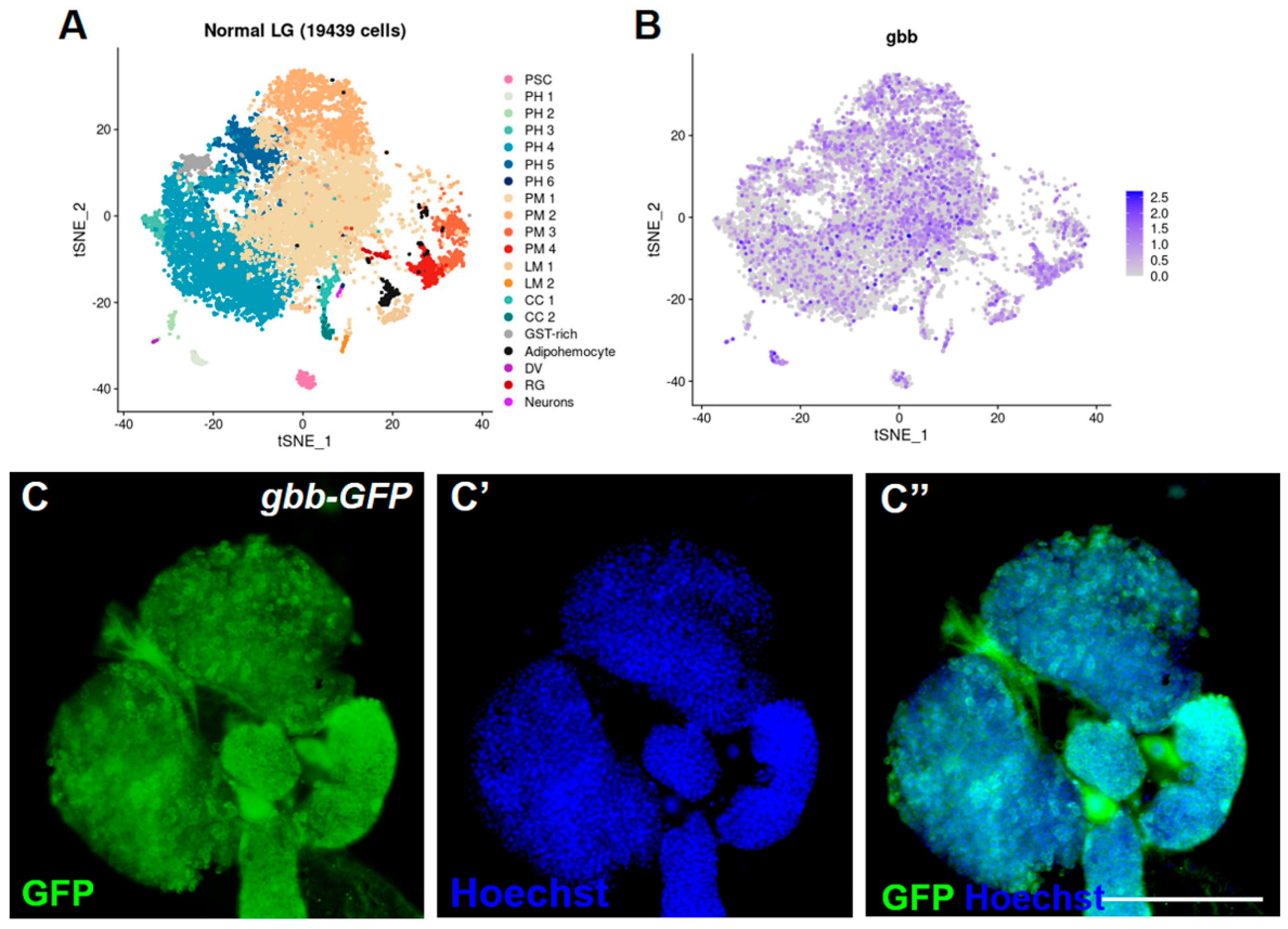
3.1. Gbb Is Widely Expressed in the Drosophila Lymph Gland

To investigate the role of Gbb in the Drosophila lymph gland, we first identified the expression of the gbb gene and Gbb protein in the lymph gland. Bumsik et al.’s previously processed datasets of single-cell RNA-seq (http://big.hanyang.ac.kr/flyscrna (accessed on 25 July 2020)) were used to search the expression pattern of the gbb gene at the transcriptional level in the whole lymph gland [35], and we found that the gbb gene was widely expressed in most cell types of the lymph gland (Figure 1A,B). The endogenous localization of Gbb was surveyed using Gbb-GFP. Similar to the expression at the transcriptional level, we found that the Gbb protein was also expressed in the entire lymph gland, especially the posterior lobes, and was mainly located in the cytoplasm of hemocytes (Figure 1C,C′).

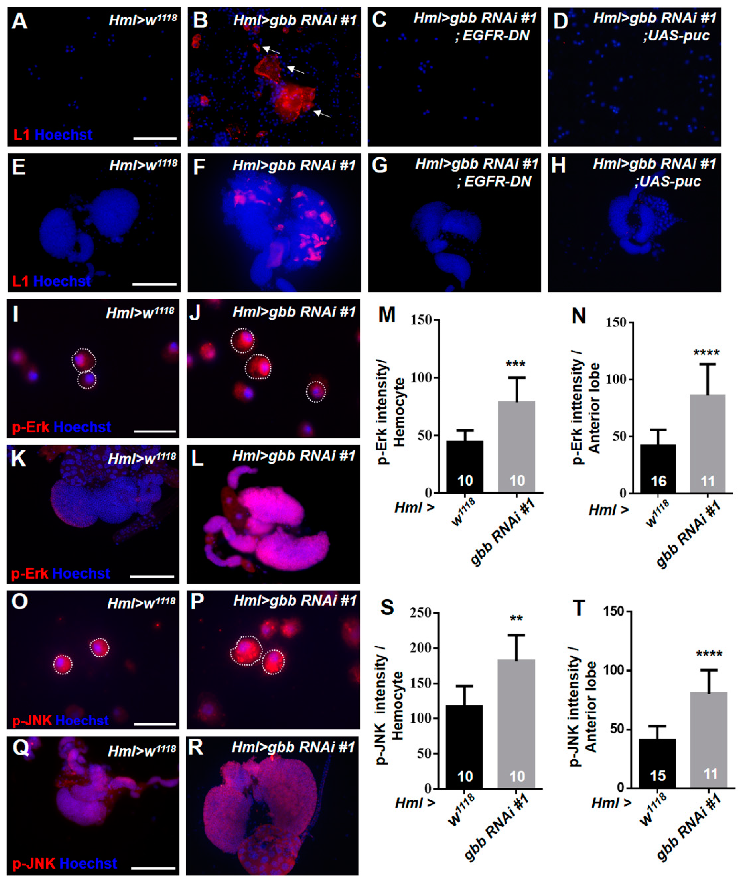
3.2. Knockdown of Gbb in the CZ Can Induce Lamellocyte Differentiation by Activating the EGFR and JNK Pathways

According to the expression pattern of Gbb in the lymph gland shown in the above results, Gbb is dramatically expressed in plasmatocytes, which are the major hemocyte type in the CZ. Thus, we first investigated the role of Gbb in the CZ. We knocked down gbb using the differentiated hemocyte-specific driver Hml-Gal4 in the CZ and evaluated the changes in hemocyte differentiation. The anti-L1 antibody was used to examine the lamellocytes. We found that large numbers of lamellocytes appeared in the circulating hemolymph and lymph glands of Hml > gbb RNAi#1 and Hml > gbb RNAi#2 larvae (Figure 2A,B,E,F and Figure S1A–D). Moreover, we used another differentiated hemocyte-specific driver, Cg-Gal4, to knock down gbb and found lamellocytes in the circulating hemolymph (Figure S1E–G). To further determine whether the lamellocyte differentiation shown in the larvae with gbb gene knockdown depends on the expression levels of gbb, the transcription level of the gbb gene in gbb knockdown larvae was quantified. We used the ubiquitous driver da-Gal4 to knock down gbb and found that the transcription level of gbb was reduced by nearly tenfold in the gbb knockdown larvae (Figure S2). These results confirmed that the loss of gbb can induce the generation of lamellocytes. Next, we further investigated the mechanism underlying lamellocyte differentiation after gbb knockdown. It is known that the excessive activation of some classical signaling pathways, such as Toll, JAK–STAT, JNK, and Ras/EGFR, can induce the generation of lamellocytes. Thus, we detected the activation of these pathways in the lymph glands and circulating hemolymph, respectively. The Toll signaling transcription factor Dorsal and the JAK–STAT transcription factor Stat92E were detected via antibody staining. We found that the localization and expression of Dorsal and Stat92E in Hml > gbb RNAi#1 were not changed compared with those found in the control (Figure S3A–D). Then, we focused on the Ras/EGFR and JNK pathways and stained lymph glands and circulating hemocytes with the anti-p-Erk antibody and anti-p-JNK antibody to detect the expression of the target of EGFR, p-Erk, and the target of the JNK pathway, p-JNK, respectively. Notably, both the p-Erk and p-JNK signals were significantly increased in the circulating hemocytes and lymph glands of Hml > gbb RNAi#1 (Figure 2I–T). To further determine whether the generation of lamellocytes in Hml > gbb RNAi#1 is caused by the overactivation of the EGFR and JNK pathways, the Hml > gbb RNAi#1 line was crossed with UAS-puc, which is the negative regulator of the JNK pathway, and UAS-EGFR-DN, which expresses a dominant-negative EGFR. We found that the inactivation of the EGFR or JNK pathway effectively inhibited the formation of lamellocytes in the circulating hemocytes and lymph glands of Hml > gbb RNAi#1 (Figure 2C,D,G,H). These results indicate that Gbb in the CZ restricts lamellocyte differentiation by preventing the hyperactivation of the EGFR and JNK pathways.

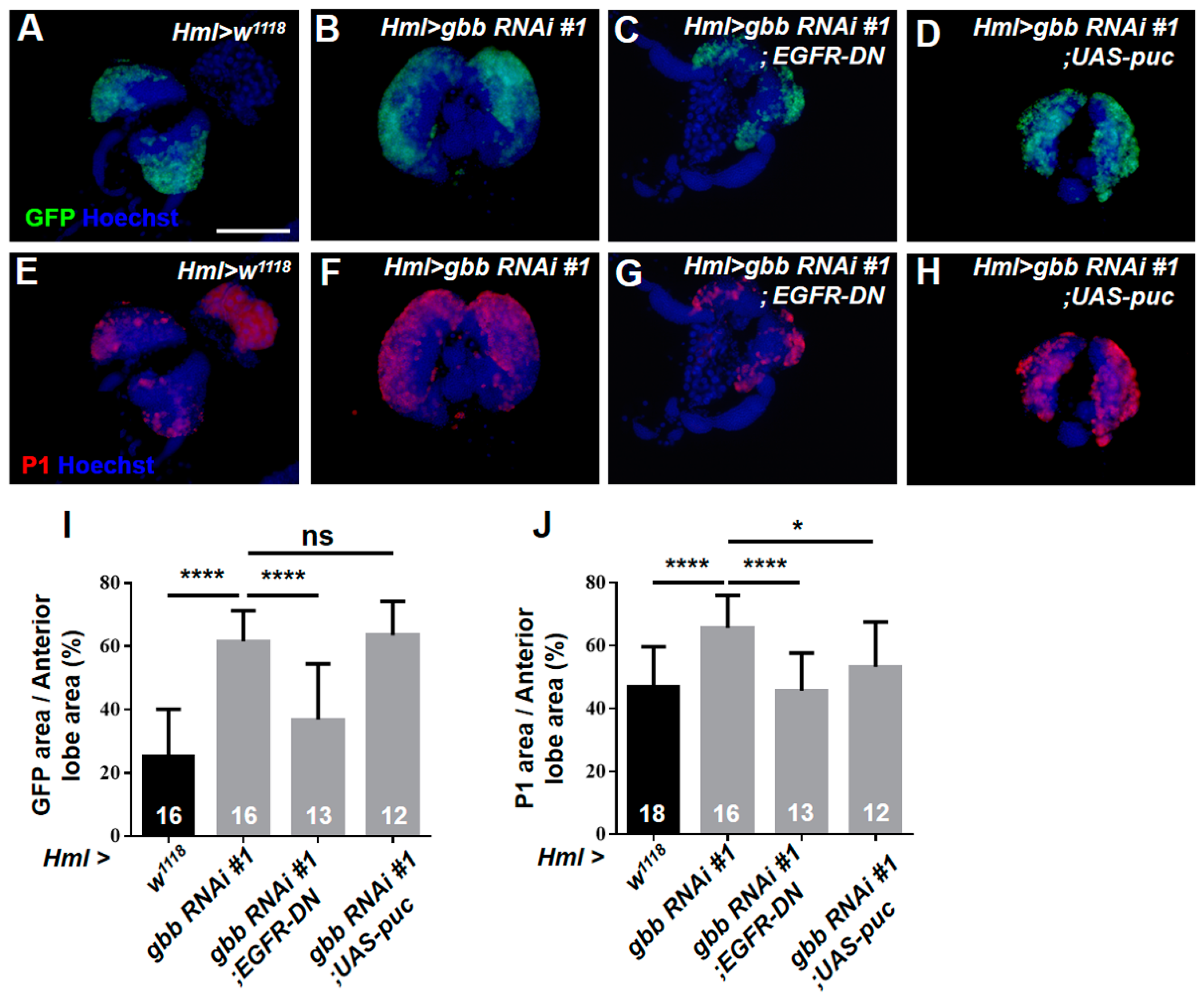
3.3. The Loss of Gbb in the CZ Can Induce Plasmatocyte Differentiation Primarily by Activating the EGFR Pathway but Not the JNK Pathway

Next, we examined the changes in the CZ upon the knockdown of Gbb using Hml > UAS-GFP, and the GFP-positive area represented the area of the CZ. The size of the GFP-positive area was larger in the lymph glands of Hml > gbb RNAi#1 than those in the control (Figure 3A,B,I). Subsequently, we used the antibody against the mature plasmatocyte marker P1 to detect plasmatocyte differentiation. Similar to the Hml-GFP-positive area, the expansion of P1-positive plasmatocytes was also shown in Hml > gbb RNAi#1 (Figure 3E,F,J). Moreover, we used another differentiated hemocyte-specific driver, Pxn-Gal4, to knock down gbb and found a similar expansion of Pxn-GFP-positive and P1-positive cells (Figure S4A–H). These results suggest that Gbb in the CZ cell-autonomously controls plasmatocyte differentiation. Next, we further investigated the regulatory mechanism of Gbb in plasmatocyte differentiation. The above results show that the knockdown of Gbb in the CZ can induce lamellocyte differentiation by activating the EGFR and JNK pathways. We therefore first asked whether Gbb also controls the differentiation of plasmatocytes via both signaling pathways. As expected, the inactivation of the Ras/EGFR pathway can effectively inhibit the massive differentiation of plasmatocytes in Hml > gbb RNAi#1 (Figure 3C,G,I,J). However, inhibiting the activation of the JNK pathways did not rescue the expansion of the Hml-GFP-positive area and only slightly prevented the expansion of the P1-positive area in Hml > gbb RNAi#1 (Figure 3D,H,I,J). Taken together, these results indicate that Gbb in the CZ cell-autonomously prevents the excessive differentiation of plasmatocytes primarily by inhibiting the hyperactivation of the EGFR pathway but not the JNK pathway.

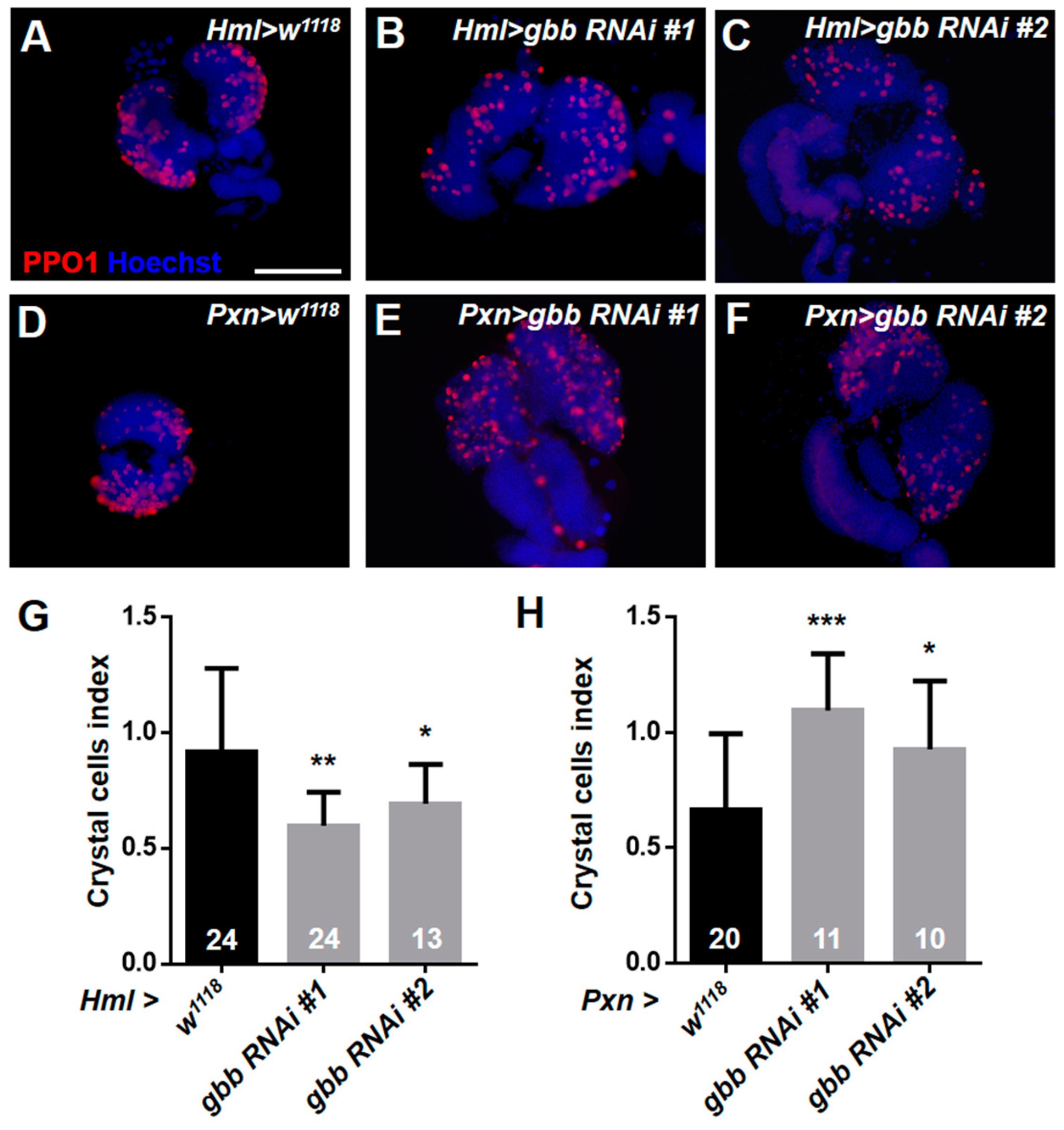
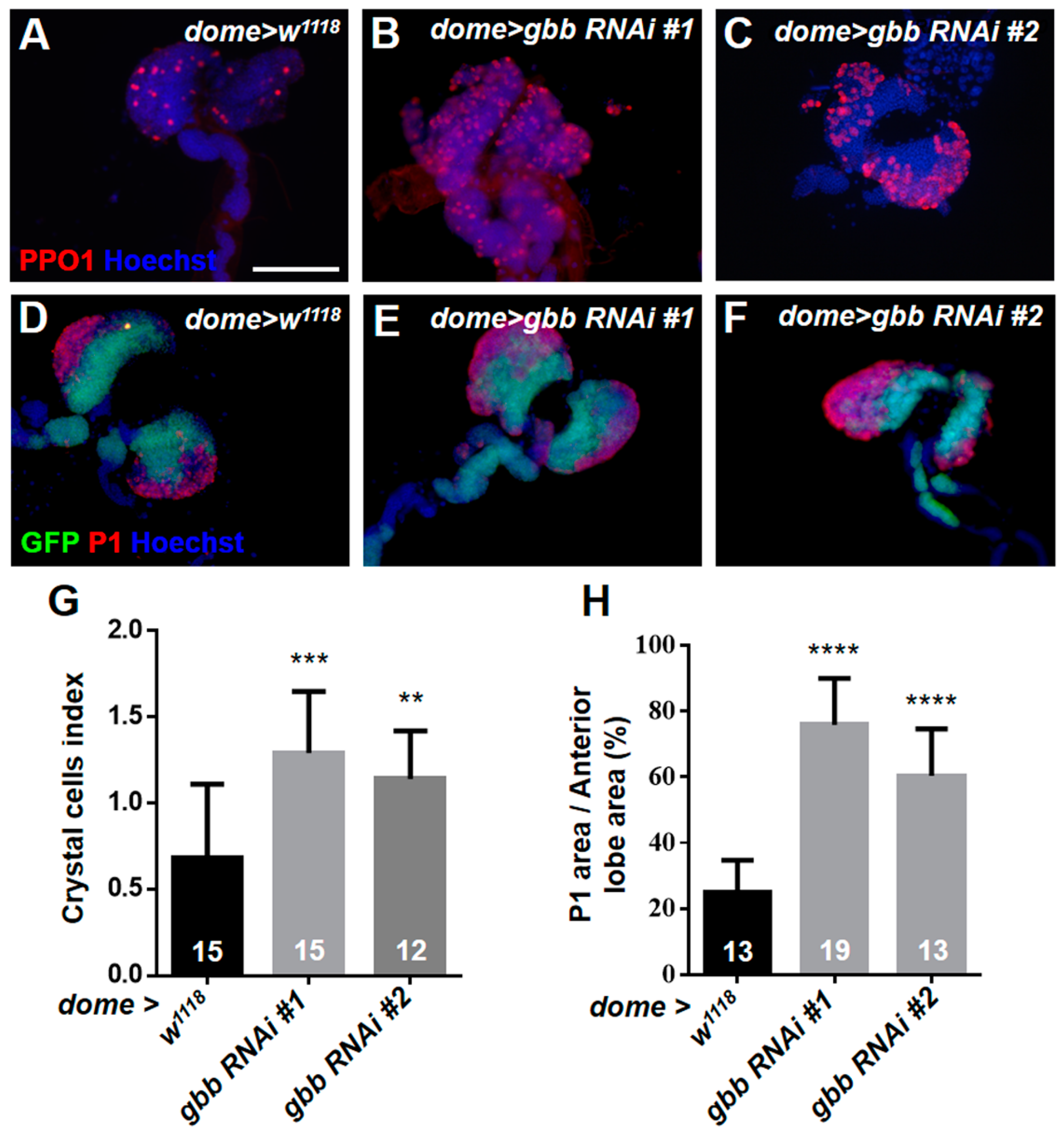
3.4. Knockdown of Gbb in Intermediate Progenitors and Progenitors Resulted in the Over-Differentiation of Crystal Cells and Plasmatocytes

We further detected the differentiation of crystal cells with the anti-PPO1 antibody. However, unlike the differentiation of plasmatocytes, the differentiation index of the crystal cells in Hml > gbb RNAi lymph glands was significantly reduced compared with that in the control (Figure 4A–C,G). Then, we used Pxn-Gal4 to knock down gbb and found that the number of crystal cells in Pxn > gbb RNAi lymph glands was significantly increased compared with that in the control (Figure 4D–F,H). These results indicate that the expression pattern of Hml-Gal4 and Pxn-Gal4 may not be exactly the same, although both of them are maturation markers of hemocytes. In addition to being expressed in mature hemocytes, Pxn is also expressed in intermediate progenitors, initially described as being in a “transition state”, as these cells are both Dome+ and Pxn+ [36,37]. We next used the MZ progenitor-specific driver dome-Gal4 to knock down gbb, and found that the loss of gbb in the MZ resulted in the massive differentiation of crystal cells and plasmatocytes; especially, the crystal cells were also observed in the posterior lobes of dome > gbb RNAi#1 lymph glands (Figure 5A–H). Taken together, these results suggest that the knockdown of gbb only in the CZ maturing hemocytes can prevent the differentiation of crystal cells, but the knockdown of gbb in intermediate progenitors and progenitors results in the over-differentiation of crystal cells and plasmatocytes.

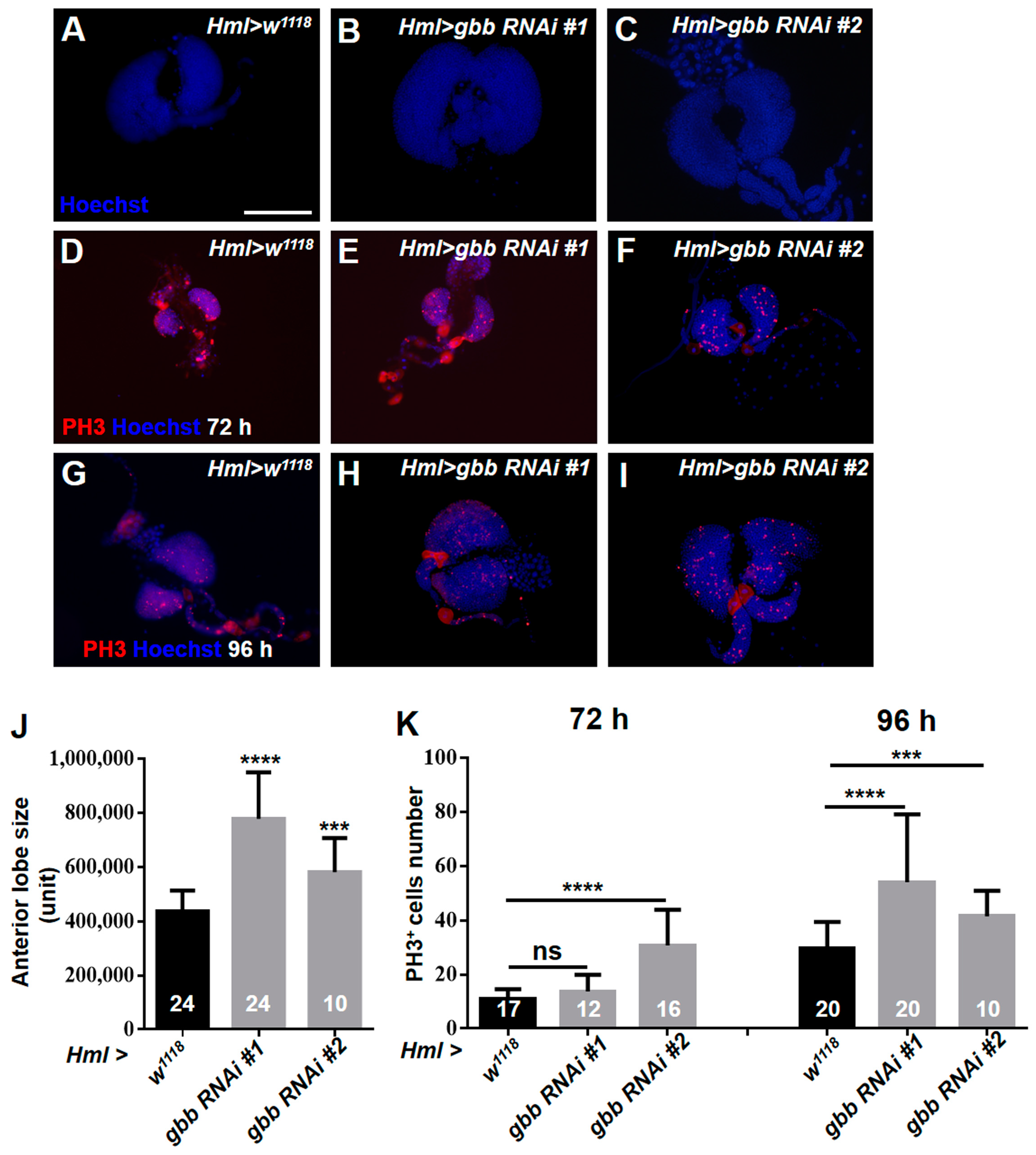
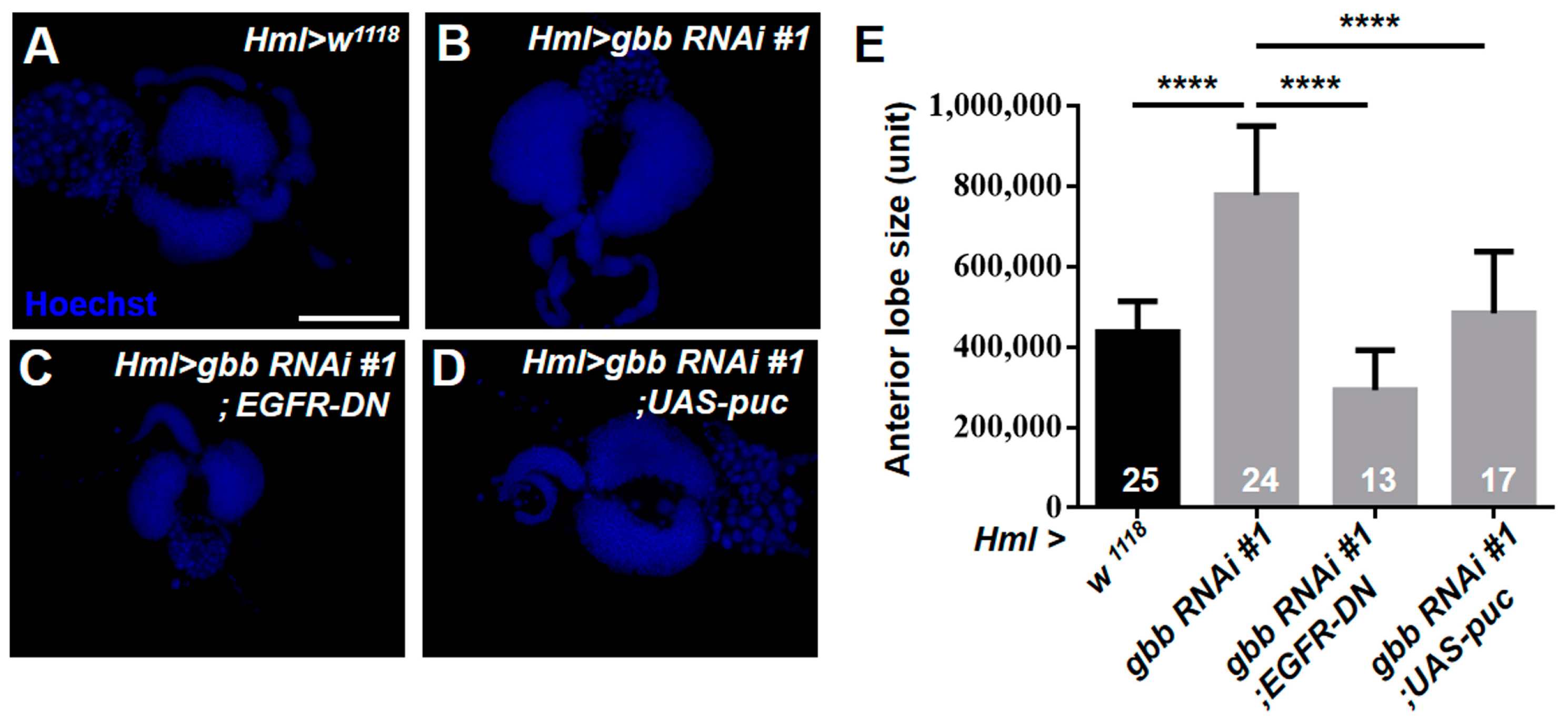
3.5. Gbb in the CZ Is Required for Preventing the Hyperproliferation of Lymph Glands by Inhibiting the Overactivation of EGFR and JNK Pathways

Of note, we observed that the loss of Gbb in the CZ resulted in a dramatic enlargement of the anterior lobes of the lymph glands (Figure 6A–C,J), suggesting that Gbb may regulate cell proliferation in the lymph gland. To confirm our speculation, the anti-PH3 antibody was used to examine the mitotic activity of the lymph glands at different larval stages. The increased PH3-positive cells were observed in the Hml > gbb RNAi#1 and Hml > gbb RNAi#2 lymph glands at 72 h and 96 h after egg laying (Figure 6D–I,K). The overactivation of the EGFR and JNK signaling pathways can promote excessive cell proliferation and tumorigenicity [16,38]. Thus, we asked whether Gbb regulates cell proliferation by inhibiting the activation of the EGFR and JNK pathways. Consistent with this hypothesis, the inactivation of the EGFR or JNK pathway dramatically reduced the overgrowth of the anterior lobes in the Hml > gbb RNAi#1 lymph glands (Figure 7A–E). These results demonstrate that Gbb expression in the CZ is required for preventing the hyperproliferation of the lymph glands by inhibiting the overactivation of the EGFR and JNK pathways.

4. Discussion

The BMP signaling pathway is highly conserved between Drosophila and mammals, and Drosophila has become a valuable system to study BMPs due to the high functional conservation of the pathway and the molecular genetic tools available. It has been shown that BMP signaling is well known for its role in controlling proliferation in imaginal tissues and maintaining germline stem cells in the ovaries [21,39,40,41,42]. Recent studies have indicated that BMP signaling modulates the Drosophila immune response following bacterial infection, wounding, and parasitic nematodes [43,44,45]. NF-κB transcription factors are required for the activation of the BMP signaling pathway in nematode-infected flies [46]. Gbb and its receptor Wishful Thinking (BMPRII) are necessary for injury-induced allodynia in Drosophila [47]. Furthermore, injury can stimulate the production of Dpp and Gbb, which drive an expansion of intestinal stem cells (ISCs) by promoting their symmetric self-renewing division in the adult Drosophila midgut [48]. The TGF-β signaling pathway is involved in inflammation and tissue repair in mammals, and the lack of TGF-β signaling can affect the function, proliferation, and differentiation of immune cells [49,50]. Moreover, previous studies have suggested that suppressing BMP receptor 1A in mouse bone marrow stroma can cause an increased osteoblast count [39], and BMP4 was shown to be expressed in and regulate the mouse HSC [51]. Similarly, the BMP signaling pathway also controls the size of the Drosophila hematopoietic niche PSC. Dpp antagonizes the activity of wingless (Wg)/Wnt signaling, which positively regulates the number of PSC cells via the control of Dmyc expression [28]. Furthermore, this study also suggests that Gbb is not required for the size control of the PSC of the lymph gland.
Here, we further determined the role of Gbb in Drosophila hematopoiesis. We showed that Gbb is mainly expressed in the cytoplasm of the lymph gland hemocytes and cell-autonomously regulates the differentiation of the lymph glands in the CZ via multiple regulatory mechanisms. Consistent with its subcellular localization in the lymph glands, the cytoplasmic localization of Gbb has been found in neurons and intestinal cells in previous studies [47,52]. We found that the knockdown of gbb in the CZ significantly induced the abnormal differentiation of lamellocytes and plasmatocytes. However, the crystal cell number was significantly reduced in the lymph glands of Hml > gbb RNAi, probably because the knockdown of gbb triggers lamellocyte differentiation at the expense of crystal cells. Similar to these results, our previous studies also have shown that the loss of jumu or ectopic expression of col favors lamellocyte differentiation at the expense of crystal cells [11]. Moreover, the knockdown of gbb in intermediate progenitors and progenitors also induced the over-differentiation of crystal cells and plasmatocytes. Consistent with these results, a recent study showed that knocking down Dlp in the progenitors, which regulate Dpp signaling by stabilizing Dpp at the cell membranes, also increased blood cell differentiation and decreased the progenitor pool [53]. The role of Dpp in the PSC and MZ has been evaluated [28,53]; however, the function of this ligand in CZ is still unknown. In Drosophila, two other characterized BMP family ligands, Gbb and Scw, can form heterodimers with Dpp to augment the level and increase the range of BMP signaling in different cells and tissues [54]. Whether Gbb regulates the differentiation of the lymph gland hemocytes by forming heterodimers with Dpp and the function of Dpp in the CZ remain to be addressed.
Previous studies have shown that the JNK and EGFR signaling pathways participate in lamellocyte formation [16,55]. Our recent study further elucidated the role of the JNK and Ras/EGFR pathways in the CZ of the lymph glands [17]. Using UAS-hepAct to activate the JNK signaling pathway in the CZ can induce the generation of lamellocytes. However, the overexpression of bsk or Rasv12 (an activated form of Ras) in the CZ failed to induce lamellocyte formation, but overexpressing both genes simultaneously could lead to the production of lamellocytes [17]. These results indicated that the Ras/EGFR pathway in the CZ can cooperate with the JNK pathway to regulate the differentiation of lamellocytes. Here, we found that the loss of Gbb in the CZ promoted the phosphorylation of Erk and JNK, and inhibiting EGFR or JNK activation can effectively rescue the lamellocyte formation caused by the loss of Gbb. These results suggest that the BMP pathway may control the differentiation of lamellocytes by preventing the hyperactivation of EGFR and JNK signaling. Moreover, our study also showed that Gbb expression in the CZ is required for preventing the hyperproliferation of the lymph glands by inhibiting the overactivation of the EGFR and JNK pathways. It has been demonstrated that EGFR and JNK signaling can promote growth and proliferation in many cell types, and genetic hyperactivation of both signaling pathways can drive tumor formation [56,57,58,59]. Consistent with our results, a recent study showed that the loss of BMP induced tumorigenesis and consequently led to the aberrant activation of JNK/Mmp2 signaling, followed by intestinal barrier dysfunction and commensal imbalance [60]. Our present results provide a better understanding of the regulatory mechanism of the BMP signaling pathway in Drosophila hematopoiesis and important insights into the regulatory relationships of the BMP, EGFR, and JNK pathways in human hematopoietic malignancies. Furthermore, future studies will address the role of other components of the BMP pathway in Drosophila hematopoiesis.

Supplementary Materials

The following supporting information can be downloaded at: https://www.mdpi.com/article/10.3390/cells12040661/s1, Figure S1: The knockdown of gbb in the maturing hemocytes can induce lamellocyte differentiation; Figure S2: Real-time PCR analysis of the gbb level in the third-instar larvae; Figure S3: The knockdown of gbb does not induce the activation of Toll and JAK–STAT pathways; Figure S4: The knockdown of gbb in the CZ induces plasmatocyte differentiation.

Author Contributions

Investigation, W.Z., D.W., J.S. and Y.H.; Methodology, W.Z., D.W., J.S. and L.H.J.; Supervision, L.H.J.; Writing—original draft, W.Z. and Y.H.; Writing—review and editing, L.H.J. and Y.H. All authors have read and agreed to the published version of the manuscript.

Funding

This work was supported by the National Natural Science Foundation of China (31900366 to Y.H.) and the Shenyang Medical College of Science and Technology fund project (20186064 to Y.H.).

Institutional Review Board Statement

Not applicable.

Informed Consent Statement

Not applicable.

Data Availability Statement

All data has been included in the manuscript.

Acknowledgments

We thank Utpal Banerjee: José Carlos Pastor-Pareja, Norbert Perrimon, and Jiwon Shim for providing the numerous fly strains used in this study. We acknowledge I. Ando and Erjun Ling for providing the antibodies.

Conflicts of Interest

The authors declare no conflict of interest.

References

  1. Boulet, M.; Miller, M.; Vandel, L.; Waltzer, L. From Drosophila Blood Cells to Human Leukemia. Adv. Exp. Med. Biol. 2018, 1076, 195–214. [Google Scholar]
  2. Harrison, D.A.; Binari, R.; Nahreini, T.S.; Gilman, M.; Perrimon, N. Activation of a Drosophila Janus kinase (JAK) causes hematopoietic neoplasia and developmental defects. EMBO J. 1995, 14, 2857–2865. [Google Scholar] [CrossRef] [PubMed]
  3. Sorrentino, R.P.; Melk, J.P.; Govind, S. Genetic analysis of contributions of dorsal group and JAK-Stat92E pathway genes to larval hemocyte concentration and the egg encapsulation response in Drosophila. Genetics 2004, 166, 1343–1356. [Google Scholar] [CrossRef] [PubMed]
  4. Mughal, T.I.; Radich, J.P.; Deininger, M.W.; Apperley, J.F.; Hughes, T.P.; Harrison, C.J.; Gambacorti-Passerini, C.; Saglio, G.; Cortes, J.; Daley, G.Q. Chronic myeloid leukemia: Reminiscences and dreams. Haematologica 2016, 101, 541–558. [Google Scholar] [CrossRef] [PubMed]
  5. Shirinian, M.; Kambris, Z.; Hamadeh, L.; Grabbe, C.; Journo, C.; Mahieux, R.; Bazarbachi, A. A Transgenic Drosophila melanogaster Model To Study Human T-Lymphotropic Virus Oncoprotein Tax-1-Driven Transformation In Vivo. J. Virol. 2015, 89, 8092–8095. [Google Scholar] [CrossRef]
  6. Sinenko, S.A.; Hung, T.; Moroz, T.; Tran, Q.M.; Sidhu, S.; Cheney, M.D.; Speck, N.A.; Banerjee, U. Genetic manipulation of AML1-ETO-induced expansion of hematopoietic precursors in a Drosophila model. Blood 2010, 116, 4612–4620. [Google Scholar] [CrossRef] [PubMed]
  7. Baril, C.; Gavory, G.; Bidla, G.; Knævelsrud, H.; Sauvageau, G.; Therrien, M. Human NUP98-HOXA9 promotes hyperplastic growth of hematopoietic tissues in Drosophila. Dev. Biol. 2017, 421, 16–26. [Google Scholar] [CrossRef] [PubMed]
  8. Holz, A.; Bossinger, B.; Strasser, T.; Janning, W.; Klapper, R. The two origins of hemocytes in Drosophila. Development 2003, 130, 4955–4962. [Google Scholar] [CrossRef] [PubMed]
  9. Lanot, R.; Zachary, D.; Holder, F.; Meister, M. Postembryonic hematopoiesis in Drosophila. Dev. Biol. 2001, 230, 243–257. [Google Scholar] [CrossRef]
  10. Yu, S.; Luo, F.; Jin, L.H. The Drosophila lymph gland is an ideal model for studying hematopoiesis. Dev. Comp. Immunol. 2018, 83, 60–69. [Google Scholar] [CrossRef]
  11. Hao, Y.; Jin, L.H. Dual role for Jumu in the control of hematopoietic progenitors in the Drosophila lymph gland. eLife 2017, 6, e25094. [Google Scholar] [CrossRef]
  12. Jung, S.H.; Evans, C.J.; Uemura, C.; Banerjee, U. The Drosophila lymph gland as a developmental model of hematopoiesis. Development 2005, 132, 2521–2533. [Google Scholar] [CrossRef] [PubMed]
  13. Crozatier, M.; Meister, M. Drosophila haematopoiesis. Cell. Microbiol. 2007, 9, 1117–1126. [Google Scholar] [CrossRef]
  14. Sorrentino, R.P.; Carton, Y.; Govind, S. Cellular immune response to parasite infection in the Drosophila lymph gland is developmentally regulated. Dev. Biol. 2002, 243, 65–80. [Google Scholar] [CrossRef] [PubMed]
  15. Louradour, I.; Sharma, A.; Morin-Poulard, I.; Letourneau, M.; Vincent, A.; Crozatier, M.; Vanzo, N. Reactive oxygen species-dependent Toll/NF-κB activation in the Drosophila hematopoietic niche confers resistance to wasp parasitism. eLife 2017, 6, e25496. [Google Scholar] [CrossRef] [PubMed]
  16. Zettervall, C.J.; Anderl, I.; Williams, M.J.; Palmer, R.; Kurucz, E.; Ando, I.; Hultmark, D. A directed screen for genes involved in Drosophila blood cell activation. Proc. Natl. Acad. Sci. USA 2004, 101, 14192–14197. [Google Scholar] [CrossRef] [PubMed]
  17. Yu, S.; Luo, F.; Jin, L.H. Rab5 and Rab11 maintain hematopoietic homeostasis by restricting multiple signaling pathways in Drosophila. eLife 2021, 10, e60870. [Google Scholar] [CrossRef]
  18. Stultz, B.G.; Hursh, D.A. Gene Regulation of BMP Ligands in Drosophila. Methods Mol. Biol. 2019, 1891, 75–89. [Google Scholar]
  19. Parker, L.; Stathakis, D.G.; Arora, K. Regulation of BMP and activin signaling in Drosophila. Prog. Mol. Subcell. Biol. 2004, 34, 73–101. [Google Scholar]
  20. Twombly, V.; Blackman, R.K.; Jin, H.; Graff, J.M.; Padgett, R.W.; Gelbart, W.M. The TGF-beta signaling pathway is essential for Drosophila oogenesis. Development 1996, 122, 1555–1565. [Google Scholar] [CrossRef] [PubMed]
  21. Xie, T.; Spradling, A.C. Decapentaplegic is essential for the maintenance and division of germline stem cells in the Drosophila ovary. Cell 1998, 94, 251–260. [Google Scholar] [CrossRef] [PubMed]
  22. Staehling-Hampton, K.; Hoffmann, F.M.; Baylies, M.K.; Rushton, E.; Bate, M. dpp induces mesodermal gene expression in Drosophila. Nature 1994, 372, 783–786. [Google Scholar] [CrossRef] [PubMed]
  23. Frasch, M. Induction of visceral and cardiac mesoderm by ectodermal Dpp in the early Drosophila embryo. Nature 1995, 374, 464–467. [Google Scholar] [CrossRef]
  24. Panganiban, G.E.; Reuter, R.; Scott, M.P.; Hoffmann, F.M. A Drosophila growth factor homolog, decapentaplegic, regulates homeotic gene expression within and across germ layers during midgut morphogenesis. Development 1990, 110, 1041–1050. [Google Scholar] [CrossRef] [PubMed]
  25. Spencer, F.A.; Hoffmann, F.M.; Gelbart, W.M. Decapentaplegic: A gene complex affecting morphogenesis in Drosophila melanogaster. Cell 1982, 28, 451–461. [Google Scholar] [CrossRef]
  26. Stultz, B.G.; Lee, H.; Ramon, K.; Hursh, D.A. Decapentaplegic head capsule mutations disrupt novel peripodial expression controlling the morphogenesis of the Drosophila ventral head. Dev. Biol. 2006, 296, 329–339. [Google Scholar] [CrossRef] [PubMed]
  27. Kawase, E.; Wong, M.D.; Ding, B.C.; Xie, T. Gbb/Bmp signaling is essential for maintaining germline stem cells and for repressing bam transcription in the Drosophila testis. Development 2004, 131, 1365–1375. [Google Scholar] [CrossRef]
  28. Pennetier, D.; Oyallon, J.; Morin-Poulard, I.; Dejean, S.; Vincent, A.; Crozatier, M. Size control of the Drosophila hematopoietic niche by bone morphogenetic protein signaling reveals parallels with mammals. Proc. Natl. Acad. Sci. USA 2012, 109, 3389–3394. [Google Scholar] [CrossRef] [PubMed]
  29. McCabe, B.D.; Marqués, G.; Haghighi, A.P.; Fetter, R.D.; Crotty, M.L.; Haerry, T.E.; Goodman, C.S.; O’Connor, M.B. The BMP homolog Gbb provides a retrograde signal that regulates synaptic growth at the Drosophila neuromuscular junction. Neuron 2003, 39, 241–254. [Google Scholar] [CrossRef] [PubMed]
  30. Khalsa, O.; Yoon, J.W.; Torres-Schumann, S.; Wharton, K.A. TGF-beta/BMP superfamily members, Gbb-60A and Dpp, cooperate to provide pattern information and establish cell identity in the Drosophila wing. Development 1998, 125, 2723–2734. [Google Scholar] [CrossRef]
  31. Wharton, K.A.; Cook, J.M.; Torres-Schumann, S.; de Castro, K.; Borod, E.; Phillips, D.A. Genetic analysis of the bone morphogenetic protein-related gene, gbb, identifies multiple requirements during Drosophila development. Genetics 1999, 152, 629–640. [Google Scholar] [CrossRef] [PubMed]
  32. Ballard, S.L.; Jarolimova, J.; Wharton, K.A. Gbb/BMP signaling is required to maintain energy homeostasis in Drosophila. Dev. Biol. 2010, 337, 375–385. [Google Scholar] [CrossRef]
  33. Tian, A.; Jiang, J. Intestinal epithelium-derived BMP controls stem cell self-renewal in Drosophila adult midgut. eLife 2014, 3, e01857. [Google Scholar] [CrossRef]
  34. Hao, Y.; Yu, S.; Luo, F.; Jin, L.H. Jumu is required for circulating hemocyte differentiation and phagocytosis in Drosophila. Cell Commun. Signal. 2018, 16, 95. [Google Scholar] [CrossRef]
  35. Cho, B.; Yoon, S.H.; Lee, D.; Koranteng, F.; Tattikota, S.G.; Cha, N.; Shin, M.; Do, H.; Hu, Y.; Oh, S.Y.; et al. Single-cell transcriptome maps of myeloid blood cell lineages in Drosophila. Nat. Commun. 2020, 11, 4483. [Google Scholar] [CrossRef]
  36. Sinenko, S.A.; Mandal, L.; Martinez-Agosto, J.A.; Banerjee, U. Dual role of wingless signaling in stem-like hematopoietic precursor maintenance in Drosophila. Dev. Cell 2009, 16, 756–763. [Google Scholar] [CrossRef] [PubMed]
  37. Krzemien, J.; Oyallon, J.; Crozatier, M.; Vincent, A. Hematopoietic progenitors and hemocyte lineages in the Drosophila lymph gland. Dev. Biol. 2010, 346, 310–319. [Google Scholar] [CrossRef] [PubMed]
  38. Pinal, N.; Martín, M.; Medina, I.; Morata, G. Short-term activation of the Jun N-terminal kinase pathway in apoptosis-deficient cells of Drosophila induces tumorigenesis. Nat. Commun. 2018, 9, 1541. [Google Scholar] [CrossRef]
  39. Zhang, J.; Niu, C.; Ye, L.; Huang, H.; He, X.; Tong, W.G.; Ross, J.; Haug, J.; Johnson, T.; Feng, J.Q.; et al. Identification of the haematopoietic stem cell niche and control of the niche size. Nature 2003, 425, 836–841. [Google Scholar] [CrossRef]
  40. Leatherman, J.L.; Dinardo, S. Germline self-renewal requires cyst stem cells and stat regulates niche adhesion in Drosophila testes. Nat. Cell Biol. 2010, 12, 806–811. [Google Scholar] [CrossRef]
  41. Song, X.; Wong, M.D.; Kawase, E.; Xi, R.; Ding, B.C.; McCarthy, J.J.; Xie, T. Bmp signals from niche cells directly repress transcription of a differentiation-promoting gene, bag of marbles, in germline stem cells in the Drosophila ovary. Development 2004, 131, 1353–1364. [Google Scholar] [CrossRef]
  42. Harris, R.E.; Pargett, M.; Sutcliffe, C.; Umulis, D.; Ashe, H.L. Brat promotes stem cell differentiation via control of a bistable switch that restricts BMP signaling. Dev. Cell 2011, 20, 72–83. [Google Scholar] [CrossRef] [PubMed]
  43. Clark, R.I.; Woodcock, K.J.; Geissmann, F.; Trouillet, C.; Dionne, M.S. Multiple TGF-β superfamily signals modulate the adult Drosophila immune response. Curr. Biol. 2011, 21, 1672–1677. [Google Scholar] [CrossRef] [PubMed]
  44. Patrnogic, J.; Heryanto, C.; Eleftherianos, I. Wounding-induced upregulation of the Bone Morphogenic Protein signaling pathway in Drosophila promotes survival against parasitic nematode infection. Gene 2018, 673, 112–118. [Google Scholar] [CrossRef]
  45. Patrnogic, J.; Heryanto, C.; Eleftherianos, I. Transcriptional up-regulation of the TGF-β intracellular signaling transducer Mad of Drosophila larvae in response to parasitic nematode infection. Innate Immun. 2018, 24, 349–356. [Google Scholar] [CrossRef]
  46. Patrnogic, J.; Heryanto, C.; Ozakman, Y.; Eleftherianos, I. Transcript analysis reveals the involvement of NF-κB transcription factors for the activation of TGF-β signaling in nematode-infected Drosophila. Immunogenetics 2019, 71, 501–510. [Google Scholar] [CrossRef] [PubMed]
  47. Gjelsvik, K.J.; Follansbee, T.L.; Ganter, G.K. Bone Morphogenetic Protein Glass Bottom Boat (BMP5/6/7/8) and its receptor Wishful Thinking (BMPRII) are required for injury-induced allodynia in Drosophila. Mol. Pain 2018, 14, 1744806918802703. [Google Scholar] [CrossRef] [PubMed]
  48. Tian, A.; Wang, B.; Jiang, J. Injury-stimulated and self-restrained BMP signaling dynamically regulates stem cell pool size during Drosophila midgut regeneration. Proc. Natl. Acad. Sci. USA 2017, 114, E2699–E2708. [Google Scholar] [CrossRef]
  49. Wan, Y.Y.; Flavell, R.A. TGF-beta and regulatory T cell in immunity and autoimmunity. J. Clin. Immunol. 2008, 28, 647–659. [Google Scholar] [CrossRef]
  50. Johnston, C.J.; Smyth, D.J.; Dresser, D.W.; Maizels, R.M. TGF-β in tolerance, development and regulation of immunity. Cell Immunol. 2016, 299, 14–22. [Google Scholar] [CrossRef] [PubMed]
  51. Goldman, D.C.; Bailey, A.S.; Pfaffle, D.L.; Al Masri, A.; Christian, J.L.; Fleming, W.H. BMP4 regulates the hematopoietic stem cell niche. Blood 2009, 114, 4393–4401. [Google Scholar] [CrossRef] [PubMed]
  52. Zhou, J.; Florescu, S.; Boettcher, A.L.; Luo, L.; Dutta, D.; Kerr, G.; Cai, Y.; Edgar, B.A.; Boutros, M. Dpp/Gbb signaling is required for normal intestinal regeneration during infection. Dev. Biol. 2015, 15, 189–203. [Google Scholar] [CrossRef]
  53. Khadilkar, R.J.; Ho, K.Y.L.; Venkatesh, B.; Tanentzapf, G. Integrins Modulate Extracellular Matrix Organization to Control Cell Signaling during Hematopoiesis. Curr. Biol. 2020, 30, 3316–3329. [Google Scholar] [CrossRef]
  54. Yang, S.; Wu, X.; Daoutidou, E.I.; Zhang, Y.; Shimell, M.; Chuang, K.H.; Peterson, A.J.; O’Connor, M.B.; Zheng, X. The NDNF-like factor Nord is a Hedgehog-induced extracellular BMP modulator that regulates Drosophila wing patterning and growth. eLife 2022, 11, e73357. [Google Scholar] [CrossRef]
  55. Tokusumi, T.; Sorrentino, R.P.; Russell, M.; Ferrarese, R.; Govind, S.; Schulz, R.A. Characterization of a lamellocyte transcriptional enhancer located within the misshapen gene of Drosophila melanogaster. PLoS ONE 2009, 4, e6429. [Google Scholar] [CrossRef] [PubMed]
  56. Zhang, C.; Jin, Y.; Marchetti, M.; Lewis, M.R.; Hammouda, O.T.; Edgar, B.A. EGFR signaling activates intestinal stem cells by promoting mitochondrial biogenesis and β-oxidation. Curr. Biol. 2022, 32, 3704–3719. [Google Scholar] [CrossRef] [PubMed]
  57. Richardson, H.E.; Portela, M. Modelling Cooperative Tumorigenesis in Drosophila. BioMed Res. Int. 2018, 2018, 4258387. [Google Scholar] [CrossRef] [PubMed]
  58. La Marca, J.E.; Richardson, H.E. Two-Faced: Roles of JNK Signalling During Tumourigenesis in the Drosophila Model. Front. Cell Dev. Biol. 2020, 8, 42. [Google Scholar] [CrossRef] [PubMed]
  59. Pinal, N.; Calleja, M.; Morata, G. Pro-apoptotic and pro-proliferation functions of the JNK pathway of Drosophila: Roles in cell competition, tumorigenesis and regeneration. Open Biol. 2019, 9, 180256. [Google Scholar] [CrossRef]
  60. Zhou, J.; Boutros, M. JNK-dependent intestinal barrier failure disrupts host-microbe homeostasis during tumorigenesis. Proc. Natl. Acad. Sci. USA 2020, 117, 9401–9412. [Google Scholar] [CrossRef] [PubMed]
Figure 1. Gbb is widely expressed in the Drosophila lymph gland. (A) The t−SNE plot shows the two−dimensional projection of major cell type subclusters identified in the scRNA−seq of normal 120 h AEL (after egg laying) lymph glands. (B) Relative expression level of gbb selected in scRNA−seq of normal 120 h AEL lymph gland dataset corresponding to the t−SNE plot in A. (CC″) Immunostaining against GFP shows that Gbb is expressed in the entire lymph gland. Scale bars: 100 μm (CC″).
Cells 12 00661 g001
Figure 2. The knockdown of gbb in the CZ induces the differentiation of lamellocytes by activating the EGFR and JNK pathways. (AH) Immunostaining against lamellocyte marker L1 in the circulating hemocytes (AD) and lymph glands (EH) of third-instar larvae. (IL) The activity of the EGFR pathway was detected with anti-p-Erk antibodies. Immunostaining of circulating hemocytes (I,J) and lymph glands (K,L) showed high p-Erk (red) signals upon gbb knockdown. (M,N) Quantification of p-Erk intensity in circulating hemocytes (M) and lymph glands (N). In this and similar subsequent analyses, numbers of images of circulating hemocytes and numbers of lymph glands analyzed are indicated on histograms. (OR) The activity of the JNK pathway was detected with anti-p-JNK antibodies. Immunostaining of circulating hemocytes (O,P) and lymph glands (Q,R) showed high p-JNK (red) signals upon gbb knockdown. (S,T) Quantification of p-JNK intensity in circulating hemocytes (S) and lymph glands (T). The arrows in B indicate the lamellocytes. Dashed white lines in (I,J,O,P) outline the edges of circulating hemocytes. For all quantifications: ** p < 0.01; *** p < 0.001; **** p < 0.0001 (Student’s t test). Scale bars: 50 μm (AD), 100 μm (EH,K,L,Q,R), 20 μm (I,J,O,P).
Cells 12 00661 g002
Figure 3. The knockdown of Gbb in the CZ induces plasmatocyte differentiation by activating the EGFR pathway but not the JNK pathway. (AD) The Hml-GFP-positive area represents the area of the CZ. (EH) Immunostaining for the plasmatocyte marker P1 showed that the P1-positive area was increased in the Hml > gbb RNAi#1 lymph gland (F), and the aberrant plasmatocyte differentiation was rescued in Hml > gbb RNAi #1 > EGFR-DN (G) and Hml > gbb RNAi #1 > UAS-puc lymph glands (H). (I,J) Quantification of the proportions of the anterior lobes occupied by the Hml-GFP + area (I) and P1+ area (J), respectively. For all quantifications: ns, not significant; * p < 0.05; **** p < 0.0001 (Student’s t test). Scale bars: 100 μm (AH).
Cells 12 00661 g003
Figure 4. The knockdown of gbb in the CZ using the Pxn-Gal4 driver causes the excessive differentiation of crystal cells. (AF) Immunostaining of the lymph gland with anti-PPO1 antibody showed that using Hml-Gal4 to knock down gbb reduced the differentiation of crystal cells (AC), but using Pxn-Gal4 to knock down gbb induced the excessive differentiation of crystal cells (DF). (G,H) Quantification of the number of crystal cells. For all quantifications: * p < 0.05; ** p < 0.01; *** p < 0.001 (Student’s t test). Scale bars: 100 μm (AF).
Cells 12 00661 g004
Figure 5. The knockdown of gbb in the MZ using the dome-Gal4 driver results in the massive differentiation of crystal cells and plasmatocytes. (AC) Immunostaining of lymph gland with anti-PPO1 antibody. (DF) Immunostaining of lymph gland with anti-P1 antibody. The GFP signal area represents the area of dome > GFP+ MZ. (G,H) Quantification of crystal cell number (G) and P1-positive area (H). For all quantifications: ** p < 0.01; *** p < 0.001; **** p < 0.0001 (Student’s t test). Scale bars: 100 μm (AF).
Cells 12 00661 g005
Figure 6. The loss of Gbb in the CZ induces the hyperproliferation of lymph glands. (AC) Immunostaining of lymph glands with Hoechst showed that the anterior lobes of Hml > gbb RNAi#1 and Hml > gbb RNAi#2 lymph glands were obviously enlarged. (DI) Immunostaining of lymph glands with anti-PH3 antibody showed increased PH3-positive cells in the Hml > gbb RNAi#1 and Hml > gbb RNAi#2 lymph glands at 72 h (DF) and 96 h (GI) after egg laying. (J) Quantification of anterior lobe size. (K) Quantification of PH3-positive cell number at 72 h (D–F) and 96 h (GI) after egg laying. For all quantifications: NS, not significant; *** p < 0.001; **** p < 0.0001 (Student’s t test). Scale bars: 100 μm (AI).
Cells 12 00661 g006
Figure 7. Inactivation of the EGFR or JNK pathway dramatically reduced the overgrowth of the anterior lobes in the Hml > gbb RNAi#1 lymph glands. (AD) Immunostaining of the lymph gland with Hoechst showed that the enlargement of the anterior lobes of Hml > gbb RNAi#1 lymph glands was obviously rescued by the inactivation of EGFR and JNK pathways. (E) Quantification of anterior lobe size. For all quantifications: **** p < 0.0001 (Student’s t test). Scale bars: 100 μm (AD).
Cells 12 00661 g007
Disclaimer/Publisher’s Note: The statements, opinions and data contained in all publications are solely those of the individual author(s) and contributor(s) and not of MDPI and/or the editor(s). MDPI and/or the editor(s) disclaim responsibility for any injury to people or property resulting from any ideas, methods, instructions or products referred to in the content.

