Article

Genetic and Chemical Controls of Sperm Fate and Spermatocyte Dedifferentiation via PUF-8 and MPK-1 in Caenorhabditis elegans

1
Division of Hematology/Oncology, Department of Internal Medicine, Brody School of Medicine at East Carolina University, Greenville, NC 27834, USA
2
Biological Resources Research Group, Bioenvironmental Science & Toxicology Division, Korea Institute of Toxicology, Jinju 52834, Gyeongsangnam-do, Republic of Korea
3
Department of Biology, East Carolina University, Greenville, NC 27858, USA
*
Author to whom correspondence should be addressed.
These authors contributed equally to this work.
Cells 2023, 12(3), 434; https://doi.org/10.3390/cells12030434
Submission received: 19 December 2022 / Revised: 21 January 2023 / Accepted: 24 January 2023 / Published: 28 January 2023
(This article belongs to the Topic Stem Cell Differentiation and Applications)

Abstract

:
Using the nematode C. elegans germline as a model system, we previously reported that PUF-8 (a PUF RNA-binding protein) and LIP-1 (a dual-specificity phosphatase) repress sperm fate at 20 °C and the dedifferentiation of spermatocytes into mitotic cells (termed “spermatocyte dedifferentiation”) at 25 °C. Thus, double mutants lacking both PUF-8 and LIP-1 produce excess sperm at 20 °C, and their spermatocytes return to mitotically dividing cells via dedifferentiation at 25 °C, resulting in germline tumors. To gain insight into the molecular competence for spermatocyte dedifferentiation, we compared the germline phenotypes of three mutant strains that produce excess sperm—fem-3(q20gf), puf-8(q725); fem-3(q20gf), and puf-8(q725); lip-1(zh15). Spermatocyte dedifferentiation was not observed in fem-3(q20gf) mutants, but it was more severe in puf-8(q725); lip-1(zh15) than in puf-8(q725); fem-3(q20gf) mutants. These results suggest that MPK-1 (the C. elegans ERK1/2 MAPK ortholog) activation in the absence of PUF-8 is required to promote spermatocyte dedifferentiation. This idea was confirmed using Resveratrol (RSV), a potential activator of MPK-1 and ERK1/2 in C. elegans and human cells, respectively. Notably, spermatocyte dedifferentiation was significantly enhanced by RSV treatment in the absence of PUF-8, and its effect was blocked by mpk-1 RNAi. We, therefore, conclude that PUF-8 and MPK-1 are essential regulators for spermatocyte dedifferentiation and tumorigenesis. Since these regulators are broadly conserved, we suggest that similar regulatory circuitry may control cellular dedifferentiation and tumorigenesis in other organisms, including humans.

1. Introduction

The nematode C. elegans is a multicellular organism that has become a popular model for biological and basic medical research. It has also been widely used as a model system to explore fundamental questions in multiple aspects of biology, including development, stem cell regulation, cell fate decision, tumorigenesis, and aging [1,2,3,4,5]. C. elegans has two sexes: male and hermaphrodite. In males and hermaphrodites, spermatogenesis begins in the L4 larval stage [6]. Spermatogenesis continues throughout the lifetime of a male, whereas it ceases and switches to oogenesis in the late L4 stage in a hermaphrodite [6] (Figure 1A).
Development of the C. elegans germline progresses by many of the same steps typical of other animal germlines [8]. The C. elegans germline is organized in a simple linear fashion that progresses from germline stem cells (GSCs) at one end to maturing gametes at the other (Figure 1A). Germ cells progress from GSCs at the distal end through meiotic prophase as they move proximally to become differentiated gametes at the proximal end [6]. C. elegans germline development is tightly regulated by conserved external signaling pathways, including GLP-1/Notch signaling, and intrinsic regulators, including RNA binding proteins and cell cycle regulators [9] (Figure 1B). The Notch signaling pathway and its core components in C. elegans are highly conserved. C. elegans has two Notch receptors, GLP-1 and LIN-12, which mediate cell-cell interaction during the development [10]. Specifically, GLP-1/Notch signaling in the C. elegans germline is critical for GSC maintenance and continued mitotic division through its direct target genes—sygl-1 and lst-1 [9,11] (Figure 1B). In addition to GLP-1/Notch signal pathways, a battery of RNA regulators, including PUF (Pumilio/FBF) RNA-binding proteins, play critical roles in GSC maintenance, differentiation, and cell fate specification in the C. elegans germline [7] (Figure 1B). In vertebrates, PUF proteins control various physiological processes such as stem cell proliferation [12,13], tumorigenesis [13], neurogenesis [14,15], germline development [16,17], mesenchymal cell fate decision [12], and mitochondrial dynamics/mitophagy [18] by interacting with the 3′ untranslated regions (UTRs) of specific mRNAs to repress the mRNA translation or stability (Figure 1C).
C. elegans has 11 PUF proteins that recognize a family of related sequence motifs in the target mRNAs (Figure 1D), yet individual PUF proteins have distinct biological functions [19]. Among them, the PUF-8 (mainly like the Drosophila and human PUFs) protein controls multiple cellular processes, including GSC proliferation, differentiation, dedifferentiation, and sperm-oocyte decision, depending on the genetic context [20] (Figure 1E). Most puf-8(q725 or ok302) single mutants make both sperm and oocytes, and they are self-fertile at the permissive temperature (20 °C) [21,22,23,24]. However, MPK-1 activation promotes sperm fate, resulting in masculinization of the germline (Mog) phenotype at 20 °C [25,26] and spermatocyte dedifferentiation, resulting in germline tumors at 25 °C in the absence of PUF-8 [21,23] (Figure 1E,F). Dedifferentiation is a cellular process by which cells from partially or terminally differentiated stages revert to a less differentiated stage. This cellular phenomenon has been implicated in regenerative medicine and tumorigenesis [27]. Although this cellular process is observed in vivo in many eukaryotes, its cellular mechanism remains poorly understood.
To better understand the mechanism of sperm fate specification and spermatocyte dedifferentiation, we have generated a puf-8(q725); fem-3(q20gf) double mutant. The puf-8(q725); fem-3(q20gf) mutants exhibit a similar germline phenotype (Mog phenotype) as seen in puf-8(q725); lip-1(zh15) mutant germlines at 20 °C, but they had significantly less dedifferentiation-mediated germline tumors than puf-8(q725); lip-1(zh15) at 25 °C. Our genetic and chemical analyses demonstrated that sperm fate and spermatocyte dedifferentiation require PUF-8 loss and MPK-1 activation in the C. elegans germline. Activation of MPK-1 by resveratrol treatment in the absence of PUF-8 significantly promotes sperm fate and spermatocyte dedifferentiation. Mammalian ERK1/2 MAPK has also been implicated in cellular dedifferentiation [28,29,30,31,32]. Therefore, our findings provide insights into cellular dedifferentiation and tumorigenesis in other organisms, including humans.

2. Materials and Methods

2.1. Worm Maintenance and Strains

C. elegans strains were maintained at 20 °C or 25 °C as previously described [1]. C. elegans strains were provided by Caenorhabditis Genetics Center (CGC) and Dr. Kimble’s lab (University of Wisconsin-Madison, Madison, WI, USA) or generated by us using a standard genetic method. Supplemental Table S1 lists strains used in this study.

2.2. RNA Interference (RNAi)

RNAi experiments were performed by feeding bacteria expressing double-strand RNAs (dsRNAs) corresponding to the gene of interest [33]. Briefly, synchronized L1 staged worms were plated onto RNAi plates and incubated at 25 °C. Germline phenotypes were determined by staining dissected gonads with specific markers and DAPI. For mpk-1b RNAi, the unique region (exon 1; 1–240 nt) of the mpk-1b gene was amplified by PCR from C. elegans genomic DNA and cloned into the pPD129.36 (L4440) vector containing two convergent T7 polymerase promoters in opposite orientations separated by a multi-cloning site [25,34]. Other RNAi bacteria were from the C. elegans RNAi feeding library (Source Bioscience LifeSciences) and C. elegans ORF-RNAi library (Open Biosystems).

2.3. Generation of puf-8(q725)/mln1[mIs14 dpy-10(e128)]; fem-3(q20gf) Mutants

Three adult puf-8(q725) homozygote male mutants were mated with five adult fem-3(q20) gain-of-function (gf) homozygote hermaphrodite mutants at 20 °C. Male progeny (predicted genotype: puf-8(q725)/+; fem-3(q20gf)/+) were mated with dumpy homozygote hermaphrodite mutants with an LGII mln1[mIs14 dpy-10(e128)] GFP balancer chromosome. Non-dumpy, pharyngeal GFP-positive hermaphrodites were selected and singled out. puf-8(q725)/mln1[mIs14 dpy-10(e128)]; fem-3(q20gf)/+ progeny were identified by PCR and phenotype analysis at 25 °C (~25% F1 progeny exhibited Mog phenotype). Finally, puf-8(q725)/mln1[mIs14 dpy-10(e128)]; fem-3(q20gf)/fem-3(q20gf) double hermaphrodite mutants were identified by PCR and phenotype analysis in the next generation.

2.4. Generation of lin-41(tn1541[GFP::tev::s::lin-41]); puf-8(q725)/mln1[mIs14 dpy-10(e128)]; fem-3(q20gf) Mutant

Three adult puf-8(q725)/mln1[mIs14 dpy-10(e128)]; fem-3(q20gf) homozygote male mutants were mated with five adult lin-41(tn1541[GFP::tev::s::lin-41]) homozygote hermaphrodites at 20 °C. Male progeny (predicted genotype: lin-41(tn1541[GFP::tev::s::lin-41])/+; puf-8(q725)/+; fem-3(q20gf)/+) were mated with dumpy homozygote hermaphrodite mutants with an LGII mln1[mIs14 dpy-10(e128)] GFP balancer chromosome. Next, non-dumpy, pharyngeal GFP-positive hermaphrodites were selected and singled out. lin-41(tn1541[GFP::tev::s::lin-41])/+; puf-8(q725)/mln1[mIs14 dpy-10(e128)]; fem-3(q20gf)/+ progeny were identified by PCR, oocyte GFP expression, and phenotype analysis at 25 °C (~25% F1 progeny exhibited Mog phenotype). Finally, lin-41(tn1541[GFP::tev::s::lin-41])/lin-41(tn1541[GFP::tev::s::lin-41]); puf-8(q725)/mln1[mIs14 dpy-10(e128)]; fem-3(q20gf)/fem-3(q20gf) hermaphrodite mutants were identified by PCR, oocyte GFP expression, and phenotype analysis in the next generation.

2.5. Germline Antibody Staining

For germline antibody staining, dissected gonads were fixed in 4% paraformaldehyde/1xPBS solution (VWR, Radnor, PA, #AAJ61899-AK) for 20 min at 25 °C followed by 100% cold methanol for 5 min at −20 °C as described in [35]. After blocking for 30 min with 0.5% Bovine Serum Albumin (BSA, Sigma-Aldrich, St. Louis, MO, USA, #A7030) in 1 × PBST (1 × PBS + 0.1% Tween 20), fixed gonads were incubated overnight at 4 °C with primary antibodies followed by 1 h at 25 °C with secondary antibodies. Supplemental Table S2 lists antibodies used in this study.

2.6. 5-Ethynyl-2′-deoxyuridine (EdU) Labeling

To label mitotically cycling cells, worms were incubated with rocking in 0.2 mL M9 buffer (3 g KH2PO4, 6 g Na2HPO4, 5 g NaCl, 1 mL 1M MgSO4, H2O to 1 L) containing 0.1% Tween 20 and 1 mM EdU for 30 min at 20 °C. Gonads were dissected and fixed in 3% paraformaldehyde/0.1M K2HPO4 (pH 7.2) solution for 20 min, followed by −20 °C methanol fixation for 10 min. Fixed gonads were blocked in 1 × PBST/0.5% BSA solution for 30 min at 20 °C. EdU labeling was performed using the Click-iT EdU Alexa Fluor 488 Imaging Kit (Thermo Fisher Scientific, Waltham, MA, USA, #C10337), according to the manufacturer’s instructions. For co-staining with antibodies, EdU-labeled gonads were incubated in the primary antibodies after washing three times and subsequently in the secondary antibodies as described above.

2.7. Resveratrol (RSV) Treatment

RSV (Sigma-Aldrich, St. Louis, MO, USA; Cat# R5010) was dissolved in ethanol (EtOH) to stock concentrations of 100 mM. RSV was directly added to the NGM media before pouring the solution into Petri dishes. The worms were transferred to the EtOH- or RSV-containing NGM agar plates. All worms tested were transferred to fresh plates every 2 days, and their germline phenotypes were determined by staining dissected gonads with cell type-specific antibodies or an EdU-labeling kit.

2.8. Cell Culture and RSV Treatment

MDA MB231 and WPMY-1 cells were grown in an appropriate growth culture medium (Dulbecco’s Modified Eagle’s Medium (DMEM) with sodium pyruvate) supplemented with 10% FBS (Thermo Fisher Scientific, Waltham, MA, USA, #10082147) and penicillin/streptomycin (10,000 U/mL). Cells were incubated with RSV for 24h after 80% confluence.

2.9. Western Blot Analysis

Cells were lysed as previously described [36]. Proteins were subjected to 10% SDS PAGE. Gels were transferred to the iblot transfer stack (Invitrogen, MA, USA, #IB4010 01) using a transfer apparatus (Invitrogen iBlot 2). Primary antibody incubations were performed in a blocking solution (5% BSA, 1 × TBS, 0.1% Tween20) overnight at 4 °C after blocking for 1 h in the blocking solution. Secondary antibody incubations were performed for 1 h at room temperature in a blocking solution. After washing three times, bands were visualized using Clarity Western ECL substrate (Bio-Rad, Hercules, CA, USA, #1705061) and calibrated by the Chemidoc Imaging System (Bio-Rad, Hercules, CA, USA). Supplemental Table S3 lists antibodies used in this study.

3. Results

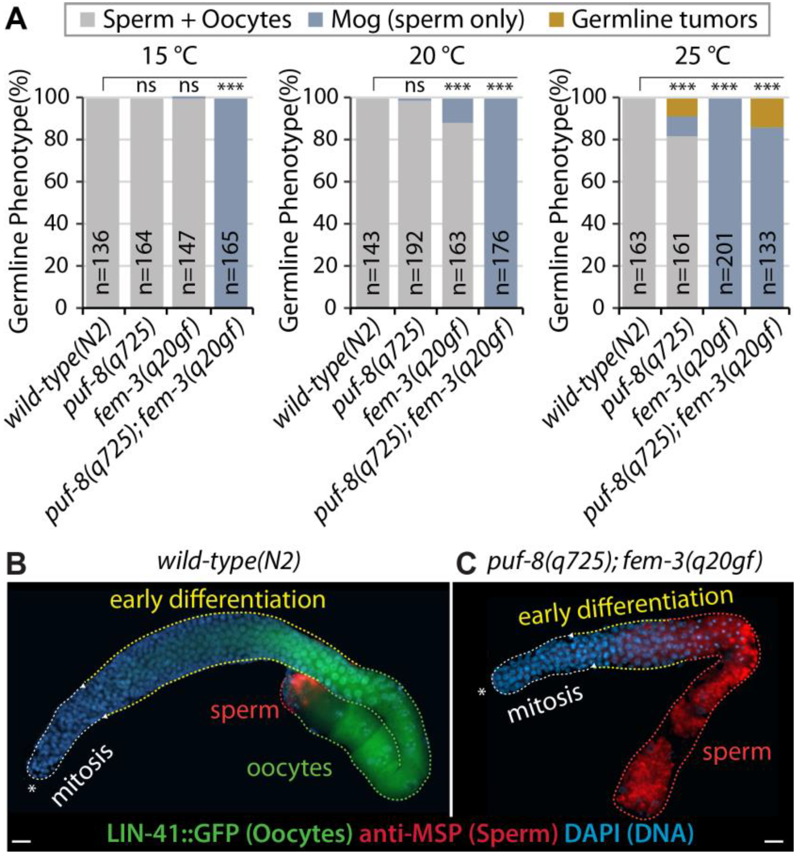
3.1. puf-8(q725) Mutation Enhances fem-3(q20gf) Mog Phenotype

Most puf-8(q725) single mutants (1–2 days old adult) are similar to wild-type worms at permissive temperatures (15–20 °C) [21,22,23,24,37]. However, at the restrictive temperature (~25 °C), puf-8(q725) mutant males have significantly more spermatocyte dedifferentiation-mediated germline tumors than hermaphrodites [23,24]. This finding led us to test whether excess sperm production in puf-8(q725) hermaphrodite germlines could enhance spermatocyte dedifferentiation. To this end, we employed a temperature-sensitive fem-3(q20) gain-of-function (gf) mutant (henceforth called fem-3(q20gf)). The C. elegans fem-3 gene is required for spermatogenesis in both hermaphrodite and male germlines [38]. Thus, fem-3(q20gf) mutants produce only sperm without switching into oogenesis, even in hermaphrodite germlines at 25 °C (Figure 2A). puf-8(q725); fem-3(q20gf) double mutants were generated by a standard genetic process (see Materials and Methods). The germline phenotypes of puf-8(q725) single, fem-3(q20gf) single, and puf-8(q725); fem-3(q20gf) double mutants were determined by staining dissected gonads with anti-MSP antibodies (a marker for sperm fate cells) [35,39] and DAPI (a marker for DNA). Most puf-8(q725) and fem-3(q20gf) single mutants produce both sperm and oocytes, and they are self-fertile at the permissive temperature (15–20 °C), but puf-8(q725); fem-3(q20gf) double mutants produce sperm continuously throughout adulthood (masculinization of the germline (Mog) phenotype) at the permissive temperatures (15–20 °C) (Figure 2A) such as puf-8(q725); lip-1(zh15) mutants (Figure 1F). To confirm this phenotype, we generated a lin-41(tn1541[GFP::tev::s::lin-41]); puf-8(q725); fem-3(q20gf) mutant. The lin-41(tn1541[GFP::tev::s::lin-41]) allele was used to visualize oogenic cells (Figure 2B,C) [40]. Most wild-type(N2) and puf-8(q725) mutant worms produced sperm (MSP-positive) and oocytes (GFP::LIN-41-positive) at 15, 20, and 25 °C (Figure 2B). However, most puf-8(q725); fem-3(q20gf) hermaphrodite mutants produced only sperm without switching to oogenesis at 15–20 °C (MSP-positive and GFP::LIN-41-negative) (Figure 2C). This result suggests that the puf-8(q725) mutation enhances the fem-3(q20gf) Mog phenotype.

3.2. RNAi of fog-1, fog-2, or fog-3 Rescues puf-8(q725); fem-3(q20gf) Mog Phenotype

In both hermaphrodites and males, sperm production requires fog-1, fog-3, and the three fem genes [41] (Figure 3A). Mutations in any of these genes cause all germ cells to differentiate as oocytes, the Fog (feminization of the germline) phenotype, and mutations in tra genes cause hermaphrodites to make sperm instead of oocytes, or the Mog (masculinization of germline) phenotype [9,41,42,43]. Moreover, the phosphorylation state of fog-3, probably by MPK-1, modulates the initiation and maintenance of the C. elegans sperm fate program [44]. A previous epistasis study showed that puf-8 and fbf-1 (C. elegans PUF protein family) are upstream of fog-2, a gene thought to be near the top of the germline sex determination pathway [37]. Since FBF-1 represses the expression of fem-3 mRNA [45], we also performed epistasis experiments. Specifically, genes required for sperm production were depleted by RNAi in lin-41(tn1541[GFP::tev::s::lin-41]); puf-8(q725); fem-3(q20gf) mutants at 20 °C. Our results showed that puf-8(q725); fem-3(q20gf) Mog phenotypes were completely suppressed by the depletion of fog-1, 2, 3, or fem-3 (Figure 3B,C). Interestingly, RNAi of fog-1, fog-2, or fog-3 dramatically rescued the Mog sterile phenotype and made them fertile (56.4 ± 8.9 %, n = 120). These results indicate that the puf-8 gene inhibits sperm fate at the top of the germline sex determination pathway.

3.3. MPK-1 Dependence of puf-8(q725); lip-1(zh15) and puf-8(q725); fem-3(q20gf) Mog Phenotype

The C. elegans mpk-1 gene encodes two major transcripts, mpk-1a and mpk-1b, which produce MPK-1A and MPK-1B proteins, respectively [34,46]. The mpk-1a mRNA is contained entirely within mpk-1b, but mpk-1b harbors a unique exon [34]. The mpk-1a isoform is predominantly expressed in somatic cells, but the mpk-1b isoform is abundantly expressed in germ cells [34,46]. The germline-specific MPK-1B isoform promotes germline differentiation but has no apparent role in GSC proliferation. However, the soma-specific MPK-1A isoform promotes GSC proliferation non-cell autonomously [47]. We previously reported that MPK-1B activity is required for the puf-8(q725); lip-1(zh15) Mog phenotype [25]. Either mpk-1b RNAi or germline-specific mpk-1(ga111) mutation dramatically rescued puf-8(q725); lip-1(zh15) Mog sterility [25] (Figure 4A). To test whether puf-8(q725); fem-3(q20gf) Mog sterility is also dependent on MPK-1 activity (Figure 4B), we performed mpk-1b RNAi in puf-8(q725); fem-3(q20gf) mutants and determined their germline phenotype by staining dissected gonads with the anti-MSP antibody and DAPI. As previously reported [25], mpk-1b RNAi completely rescued the puf-8(q725); lip-1(zh15) Mog phenotype at 20 °C (Figure 4C,E), but it partially rescued the puf-8(q725); fem-3(q20gf) Mog phenotypes (Figure 4D,F). These results indicate that the puf-8(q725); fem-3(q20gf) Mog phenotype depends less on the germline MPK-1B activity.
PUF-8 has been known to repress the expression of let-60 (a Ras homolog), an upstream activator of MPK-1 [48]. MPK-1 activity is absolutely required for puf-8(q725); lip-1(zh15) Mog phenotypes, but not for puf-8(q725); fem-3(q20gf) Mog phenotypes at 20 °C (Figure 4A–F) [25,26]. To ask whether puf-8(q725); lip-1(zh15) mutant germlines have more activated (phosphorylated) MPK-1 (pMPK-1) proteins than puf-8(q725); fem-3(q20gf) mutant germlines, we stained dissected gonads (4 days past L1) using an anti-DP-MAPK monoclonal antibody, which recognizes the active form of MPK-1 by its dual phosphorylation (DP) [34,46]. In the puf-8(q725); fem-3(q20gf) Mog germlines, pMPK-1 proteins were detected in the early differentiating cells (Figure 4G) as seen in wild-type male germlines [46]. Notably, pMPK-1 proteins were more in the puf-8(q725); lip-1(zh15) mutant germlines than in the puf-8(q725); fem-3(q20gf) mutant germlines (Figure 4G,H). We also quantitated levels with ImageJ software. The average intensity of pMPK-1 in the puf-8(q725); lip-1(zh15) mutant germlines (n= 16) was ~2.7 times higher than that in the puf-8(q725); fem-3(q20gf) mutant germlines (n = 15) (Figure 4I). These results indicate that puf-8(q725); lip-1(zh15) mutant germlines have a high level of pMPK-1 proteins compared to puf-8(q725); fem-3(q29gf) mutant germlines.

3.4. Competence for Spermatocyte Dedifferentiation in the Absence of PUF-8

We have previously reported that excess sperm does not necessarily lead to spermatocyte dedifferentiation [23]. Rather, the activation of MPK-1 in the puf-8 (q725) mutant may be critical for initiating spermatocyte dedifferentiation in the C. elegans germline [23]. Based on this finding, we tested the hypothesis that competence for spermatocyte dedifferentiation may require MPK-1 activation in the absence of PUF-8. To this end, we also employed wild-type(N2) and other mutant strains—fem-3(q20gf) mutant with both wild-type puf-8(+/+) and lip-1(+/+) genes, puf-8(q725); fem-3(q20gf) with the wild-type lip-1(+/+) gene, and puf-8(q725); lip-1(zh15) mutants. To score the percentage of worms with germline tumors via spermatocyte dedifferentiation, synchronized L1-staged mutants were placed on NGM plates seeded with OP50 E. coli bacteria food at 25 °C, and their germline phenotypes were determined daily (2.5, 3–6 days past L1) by staining dissected gonads with DAPI. Although the wild-type(N2) and fem-3(q20gf) mutant worms never formed germline tumors throughout adulthood at 25 °C (Figure 5A), most puf-8(q725); lip-1(zh15) mutants developed germline tumors even 3 days past L1 (Figure 5A). Notably, the percentage of puf-8(q725); fem-3(q20gf) hermaphrodite mutants with germline tumors increased gradually from 3 days past L1 (Figure 5A). Since puf-8(q725); lip-1(zh15) germline phenotypes largely depend on MPK-1 activity [25] (Figure 4), we suggest that MPK-1 activity may be required to induce the formation of germline tumors via spermatocyte dedifferentiation in the absence of PUF-8 at 25 °C.

3.5. Resveratrol Induces Germline Tumors by Activating MPK-1 in the Absence of PUF-8

Our previous study found that Resveratrol (RSV) maintains MPK-1 activity throughout the lifespan of C. elegans [49] (Figure 5B). To better understand the effect of RSV on ERK1/2, two human cell lines, MDA MB231 and WPMY-1, were treated with the different concentrations (0, 10, 20, 50 µM) of RSV for 24 h after 80% confluence. The expression levels of total ERK1/2 and phospho-ERK1/2 (pERK1/2) proteins were examined by Western blot using anti-ERK1/2 and anti-pERK1/2 antibodies (Figure 5C). RSV significantly increased the levels of pERK1/2 proteins dose-dependently up to 20 µM (Figure 5C). However, their levels significantly decreased at 50 µM RSV in both cell lines (Figure 5C). Similarly, C. elegans pMPK-1 levels were increased up to 3.3-fold by 100 µM RSV treatment, but their increasing levels were decreased up to 2.3-fold by 200 µM RSV treatment [49]. We also found that 50 µM RSV treatment significantly decreased cell viability in a dose-dependent manner (data not shown), as previously reported [50,51,52]. Thus, the reduction in pERK1/2 and pMPK levels in cells and worms exposed to high RSV concentration might be due to an increased cell death. This result indicates that the effects of RSV on the activation of MPK-1 and ERK1/2 are conserved in human cell lines and C. elegans.
Based on these findings, we tested whether 100 µM RSV could induce the formation of germline tumors via spermatocyte dedifferentiation by activating MPK-1 signaling in puf-8(q725); fem-3(q20gf) mutant germlines. Synchronized L1 staged puf-8(q725); fem-3(q20gf) mutant worms were cultured on NGM agar plates containing 100 µM RSV or 0.1% ethanol (EtOH) control at 25 °C. Their germline phenotypes were determined daily by staining dissected gonads with an EdU-labeling kit and DAPI. Notably, RSV significantly induced the formation of germline tumors via spermatocyte dedifferentiation from day 3 in puf-8(q725); fem-3(q20gf) mutant germlines (Figure 5D,E). This result suggests that RSV is a potential inducer of spermatocyte dedifferentiation in vivo in the absence of PUF-8. Next, to test whether RSV-induced spermatocyte dedifferentiation relies on MPK-1 activity, we depleted the expression of mpk-1 by RNAi from L1 staged puf-8(q725); fem-3(q20gf) mutants in the presence of 100 µM RSV. While vector RNAi control did not suppress the formation of germline tumors via spermatocyte dedifferentiation, mpk-1 RNAi significantly suppressed the formation of puf-8(q725); fem-3(q20gf) germline tumors even in the presence of 100 µM RSV (Figure 5F). This result was confirmed by staining dissected gonads with anti-HIM-3 antibodies which recognize meiotic differentiating cells (Figure 5G). mpk-1 RNAi inhibited the formation of germline tumors via spermatocyte dedifferentiation and induced HIM-3-postive meiotic cells (non-proliferative cells) in the puf-8(q725); fem-3(q20gf) mutant germline (Figure 5G). Therefore, we suggest that MPK-1 activation could chemically induce the formation of germline tumors via spermatocyte dedifferentiation in the absence of PUF-8 in vivo (Figure 6).

4. Discussion

RNA-binding proteins (RBPs) bind to either single-stranded or double-stranded RNA and play a role in the post-transcriptional control of RNAs, such as mRNA stabilization, localization, splicing, polyadenylation, and translation [53]. PUF family RBPs are highly conserved among most eukaryotic organisms [7]. PUF proteins are conserved RBPs that maintain GSCs in worms and flies and have also been implicated in this role in mammals [12,15,17,54,55,56,57]. PUF proteins bind specifically to PUF binding elements (PBE: UGUAnAUA) within the 3′ untranslated region (3′UTR) of their direct target mRNAs to repress their translation [7,19,20] (Figure 1C). PUF proteins also have diverse roles depending on the organism. For example, in Drosophila melanogaster, Pumilio is required for embryonic development through the regulation of Hunchback (necessary for the establishment of an anterior-posterior gradient) [58] and GSC maintenance [59]. The yeast PUF protein Mpt5, a broad RNA regulator in Saccharomyces cerevisiae, binds to more than 1,000 RNA targets [60]. The Mpt5 is required to promote G2/M cell cycle progress [61] and cell wall integrity [62]. Humans have two PUF proteins, PUM1 and PUM2. The PUM1 and PUM2 have high structural similarity and recognize the same RNA binding motif. Despite their similarities, PUM1 is critical for stem cell proliferation, and PUM2 is more important for stem cell differentiation and cell lineage specification [12]. Notably, PUF proteins repress mRNAs encoding MAPK enzymes in worms, flies, yeast, and humans [34,63,64]. However, the role of PUF proteins in limiting dedifferentiation remain poorly understood.
Cellular dedifferentiation counteracts the decline of stem cells during aging but has also been implicated in the formation of tumor-initiating cells [65]. Thus, a comprehensive examination of what causes stem cells to differentiate into desired cell types and how committed cells return to undifferentiated cells is a central question in stem cell biology, regenerative medicine, and tumorigenesis [66]. This cellular dedifferentiation can take many forms depending on the specific organism and tissue type. In zebrafish (Danio rerio), cellular dedifferentiation occurs in a controlled environment where cardiomyocytes partially dedifferentiate to repopulate lost ventricular tissue [67]. In fruit flies (Drosophila melanogaster), it has been shown that differentiating germ cells can revert into functional stem cells both in second instar larval ovaries and in adult fruit flies [68]. Drosophila has also been shown to induce dedifferentiation in spermatogonia cells as there is considerable plasticity due to Jak-STAT signaling [69]. Reduction in stem cell division in Drosophila has been shown due to an accumulation of GSCs with misoriented centrosomes that increases as the flies age [70]. In Mus musculus, dedifferentiated basal-like cells originating from luminal airway cells can function as stem cells in the repopulation of damaged airway epithelia [71]. Dedifferentiation in the mouse model has also been shown in the intestine, where tumorigenesis is initiated due to increased Wnt-activation allowing for polyp formation [72]. In these different examples of dedifferentiation, other signaling pathways are reverting differentiated cells into either stem cell-like or tumor-initiating cells.
In C. elegans, dedifferentiation in the germline can occur in oogenic [73] and spermatogenic [21,24] germlines. In a mutant lacking gld-1 (a KH-motif containing RNA-binding protein), germ cells destined for oogenesis early in the meiotic cell fate return to a mitotic cell cycle, which results in germline tumors [73]. In a double mutant lacking puf-8 and lip-1, spermatocytes do not undergo normal meiotic division but instead return to mitosis, resulting in germline tumors [21,23,24] (Figure 6). Notably, PUF-8 and GLD-1 inhibit MPK-1 signaling pathways at post-transcriptional levels [48,74,75,76,77], and the formation of germline tumors via dedifferentiation was significantly inhibited by the depletion of mpk-1 [21,23]. This result indicates that MPK-1 activity may be critical for the formation of germline tumors via dedifferentiation in the absence of PUF-8 (Figure 6) or GLD-1. Consistently, mammalian Ras-ERK MAPK signaling has similarly been implicated in the cellular dedifferentiation of the Sertoli cells [78], myoblasts [79], and islet cells [80]. In addition to MPK-1, our results using puf-8(q725); fem-3(q20gf) and fem-3(q20gf) mutants demonstrated that spermatocyte dedifferentiation also requires puf-8 loss. We also examined the germline phenotypes of other PUF mutants in the absence of LIP-1, such as fbf-1(ok91); lip-1(zh15) [23], fbf-2(q704); lip-1(zh15) [23], and puf-9(ok1136); lip-1(zh15) (Jones et al., unpublished result). None of them formed germline tumors, indicating that PUF-8 has a special function in inhibiting spermatocyte dedifferentiation. Therefore, the identification and characterization of PUF-8 target genes involved in spermatocyte dedifferentiation will be critical to understanding the mechanism of spermatocyte dedifferentiation.
During the testing of germline tumors with mpk-1 RNAi, we found that puf-8(q725); lip-1(zh15) mutants fed OP50 had a lower percentage of tumor formation than that fed HT115, an RNase III-deficient E. coli strain used for feeding RNAi in C. elegans [81,82] (see Figure 5D,F; Supplementary Figure S2). The HT115 E. coli provided a greater metabolic energy source due to the recycling of excess nucleotides by the bacteria being RNase III-deficient [83]. Due to this increase in energy boosting the formation of germline tumors, we decided to look at the levels of active MPK-1. Notably, the worms fed HT115 E. coli had increased pMPK-1 than those fed OP50 E. coli (unpublished result). These unpublished results suggest that the HT115 diet may increase spermatocyte dedifferentiation via activating MPK-1 signaling. Notably, increasing evidence suggests that metabolic changes alter cell fates by changing multiple signaling pathways. For example, starvation or starvation-induced quiescence maintains GSCs, independent of GLP-1/Notch signaling [84,85]. Furthermore, short-term starvation stress enhances the meiotic activity of germ cells to prevent age-related declines in sperm production [86]. Similarly, the dedifferentiation of primary hepatocytes is accompanied by the reorganization of lipid metabolism [87]. Therefore, non-genetic factors may play a vital role in cell fate reprogramming and tumorigenesis. Our findings reveal fundamental mechanisms of the differentiation/dedifferentiation decision in vivo and may provide a future platform for identifying therapeutic targets for dedifferentiation-mediated tumorigenesis.

Supplementary Materials

The following Supporting Information can be downloaded at: https://www.mdpi.com/article/10.3390/cells12030434/s1, Table S1: C. elegans strains used in this study; Table S2: Antibodies used in this study; Figure S1: The effect of bacteria foods on spermatocyte dedifferentiation-mediated tumorigenesis in puf-8(q725); fem-3(q20gf) mutants at 25 °C.

Author Contributions

Conceptualization, H.M.A. and M.H.L.; Funding acquisition, M.H.L.; Investigation, Y.P., M.G., M.H., M.E.J. and M.H.L.; Supervision, M.H.L.; Writing—original draft, Y.P., M.G. and M.H.L.; Writing—review & editing, M.H.L. All authors have read and agreed to the published version of the manuscript.

Funding

This work was supported by the National Research Foundation of Korea (NRF 2022R1F1A1069374) to MH, and the NIH (AG060373-01), NSF (IOS 2132286), and Lung Cancer Initiative (217045) to MHL. The Caenorhabditis Genetic Center (CGC) was supported by the National Institutes of Health–Office of Research Infrastructure Programs (P40 OD010440).

Institutional Review Board Statement

Not applicable.

Informed Consent Statement

Not applicable.

Data Availability Statement

Not applicable.

Acknowledgments

We are grateful to Judith Kimble (University of Wisconsin-Madison) for C. elegans strains and Eun Suk Kim (Brody School of Medicine at ECU) for critical reading.

Conflicts of Interest

The authors declare no conflict of interest.

References

  1. Brenner, S. The genetics of Caenorhabditis elegans. Genetics 1974, 77, 71–94. [Google Scholar] [CrossRef] [PubMed]
  2. Van Pelt, K.M.; Truttmann, M.C. Caenorhabditis elegans as a model system for studying aging-associated neurodegenerative diseases. Transl. Med. Aging 2020, 4, 60–72. [Google Scholar] [CrossRef] [PubMed]
  3. Markaki, M.; Tavernarakis, N. Caenorhabditis elegans as a model system for human diseases. Curr. Opin. Biotechnol. 2020, 63, 118–125. [Google Scholar] [CrossRef] [PubMed]
  4. Kobet, R.A.; Pan, X.; Zhang, B.; Pak, S.C.; Asch, A.S.; Lee, M.H. Caenorhabditis elegans: A Model System for Anti-Cancer Drug Discovery and Therapeutic Target Identification. Biomol. Ther. 2014, 22, 371–383. [Google Scholar] [CrossRef] [PubMed] [Green Version]
  5. Chang, C.; Sternberg, P.W. C. elegans vulval development as a model system to study the cancer biology of EGFR signaling. Cancer Metastasis Rev. 1999, 18, 203–213. [Google Scholar] [CrossRef] [PubMed]
  6. Hubbard, E.J.; Greenstein, D. Introduction to the germ line. WormBook Online Rev. C. Elegans Biol. 2005, 1–4. [Google Scholar] [CrossRef]
  7. Wickens, M.; Bernstein, D.S.; Kimble, J.; Parker, R. A PUF family portrait: 3’UTR regulation as a way of life. Trends Genet. TIG 2002, 18, 150–157. [Google Scholar] [CrossRef]
  8. Kimble, J.; Crittenden, S.L. Germline proliferation and its control. WormBook Online Rev. C. Elegans Biol. 2005, 1–14. [Google Scholar] [CrossRef]
  9. Kimble, J.; Crittenden, S.L. Controls of germline stem cells, entry into meiosis, and the sperm/oocyte decision in Caenorhabditis elegans. Annu. Rev. Cell Dev. Biol. 2007, 23, 405–433. [Google Scholar] [CrossRef] [Green Version]
  10. Greenwald, I. LIN-12/Notch signaling in C. elegans. WormBook Online Rev. C. Elegans Biol. 2005, 1–16. [Google Scholar] [CrossRef]
  11. Shin, H.; Haupt, K.A.; Kershner, A.M.; Kroll-Conner, P.; Wickens, M.; Kimble, J. SYGL-1 and LST-1 link niche signaling to PUF RNA repression for stem cell maintenance in Caenorhabditis elegans. PLoS Genet. 2017, 13, e1007121. [Google Scholar] [CrossRef] [Green Version]
  12. Lee, M.H.; Wu, X.; Zhu, Y. RNA-binding protein PUM2 regulates mesenchymal stem cell fate via repression of JAK2 and RUNX2 mRNAs. J. Cell. Physiol. 2020, 235, 3874–3885. [Google Scholar] [CrossRef] [PubMed]
  13. Uyhazi, K.E.; Yang, Y.; Liu, N.; Qi, H.; Huang, X.A.; Mak, W.; Weatherbee, S.D.; de Prisco, N.; Gennarino, V.A.; Song, X.; et al. Pumilio proteins utilize distinct regulatory mechanisms to achieve complementary functions required for pluripotency and embryogenesis. Proc. Natl. Acad. Sci. USA 2020, 117, 7851–7862. [Google Scholar] [CrossRef] [PubMed]
  14. Martinez, J.C.; Randolph, L.K.; Iascone, D.M.; Pernice, H.F.; Polleux, F.; Hengst, U. Pum2 Shapes the Transcriptome in Developing Axons through Retention of Target mRNAs in the Cell Body. Neuron 2019, 104, 931–946.e935. [Google Scholar] [CrossRef] [PubMed]
  15. Zhang, M.; Chen, D.; Xia, J.; Han, W.; Cui, X.; Neuenkirchen, N.; Hermes, G.; Sestan, N.; Lin, H. Post-transcriptional regulation of mouse neurogenesis by Pumilio proteins. Genes Dev. 2017, 31, 1354–1369. [Google Scholar] [CrossRef] [Green Version]
  16. Chen, D.; Zheng, W.; Lin, A.; Uyhazi, K.; Zhao, H.; Lin, H. Pumilio 1 suppresses multiple activators of p53 to safeguard spermatogenesis. Curr. Biol. CB 2012, 22, 420–425. [Google Scholar] [CrossRef] [Green Version]
  17. Mak, W.; Fang, C.; Holden, T.; Dratver, M.B.; Lin, H. An Important Role of Pumilio 1 in Regulating the Development of the Mammalian Female Germline. Biol. Reprod. 2016, 94, 134. [Google Scholar] [CrossRef]
  18. D’Amico, D.; Mottis, A.; Potenza, F.; Sorrentino, V.; Li, H.; Romani, M.; Lemos, V.; Schoonjans, K.; Zamboni, N.; Knott, G.; et al. The RNA-Binding Protein PUM2 Impairs Mitochondrial Dynamics and Mitophagy During Aging. Mol. Cell 2019, 73, 775–787 e710. [Google Scholar] [CrossRef] [PubMed] [Green Version]
  19. Wang, X.; Voronina, E. Diverse Roles of PUF Proteins in Germline Stem and Progenitor Cell Development in C. elegans. Front. Cell Dev. Biol. 2020, 8, 29. [Google Scholar] [CrossRef] [Green Version]
  20. Datla, U.S.; Scovill, N.C.; Brokamp, A.J.; Kim, E.; Asch, A.S.; Lee, M.H. Role of PUF-8/PUF protein in stem cell control, sperm-oocyte decision and cell fate reprogramming. J. Cell. Physiol. 2014, 229, 1306–1311. [Google Scholar] [CrossRef]
  21. Park, Y.; O’Rourke, S.; Taki, F.A.; Alfhili, M.A.; Lee, M.H. Dose-Dependent Effects of GLD-2 and GLD-1 on Germline Differentiation and Dedifferentiation in the Absence of PUF-8. Front. Cell Dev. Biol. 2020, 8, 5. [Google Scholar] [CrossRef] [Green Version]
  22. Ariz, M.; Mainpal, R.; Subramaniam, K.C. elegans RNA-binding proteins PUF-8 and MEX-3 function redundantly to promote germline stem cell mitosis. Dev. Biol. 2009, 326, 295–304. [Google Scholar] [CrossRef] [PubMed] [Green Version]
  23. Cha, D.S.; Datla, U.S.; Hollis, S.E.; Kimble, J.; Lee, M.H. The Ras-ERK MAPK regulatory network controls dedifferentiation in Caenorhabditis elegans germline. Biochim. Biophys. Acta 2012, 1823, 1847–1855. [Google Scholar] [CrossRef] [PubMed] [Green Version]
  24. Subramaniam, K.; Seydoux, G. Dedifferentiation of primary spermatocytes into germ cell tumors in C. elegans lacking the pumilio-like protein PUF-8. Curr. Biol. CB 2003, 13, 134–139. [Google Scholar] [CrossRef] [PubMed] [Green Version]
  25. Morgan, C.T.; Lee, M.H.; Kimble, J. Chemical reprogramming of Caenorhabditis elegans germ cell fate. Nat. Chem. Biol. 2010, 6, 102–104. [Google Scholar] [CrossRef] [PubMed] [Green Version]
  26. Benson, J.A.; Cummings, E.E.; O’Reilly, L.P.; Lee, M.H.; Pak, S.C. A high-content assay for identifying small molecules that reprogram C. elegans germ cell fate. Methods 2014, 68, 529–535. [Google Scholar] [CrossRef] [PubMed]
  27. Liu, H.X.; Hu, D.H.; Jia, C.Y.; Fu, X.B. Progress of cellular dedifferentiation research. Chin. J. Traumatol. Zhonghua Chuang Shang Za Zhi 2006, 9, 308–315. [Google Scholar] [PubMed]
  28. Tiong, Y.L.; Ng, K.Y.; Koh, R.Y.; Ponnudurai, G.; Chye, S.M. Melatonin promotes Schwann cell dedifferentiation and proliferation through the Ras/Raf/ERK and MAPK pathways, and glial cell-derived neurotrophic factor expression. Exp. Ther. Med. 2020, 20, 16. [Google Scholar] [CrossRef]
  29. Almairac, F.; Turchi, L.; Sakakini, N.; Debruyne, D.N.; Elkeurti, S.; Gjernes, E.; Polo, B.; Bianchini, L.; Fontaine, D.; Paquis, P.; et al. ERK-Mediated Loss of miR-199a-3p and Induction of EGR1 Act as a “Toggle Switch” of GBM Cell Dedifferentiation into NANOG- and OCT4-Positive Cells. Cancer Res. 2020, 80, 3236–3250. [Google Scholar] [CrossRef]
  30. Boerboom, A.; Reusch, C.; Pieltain, A.; Chariot, A.; Franzen, R. KIAA1199: A novel regulator of MEK/ERK-induced Schwann cell dedifferentiation. Glia 2017, 65, 1682–1696. [Google Scholar] [CrossRef]
  31. Chen, R.; Lee, W.Y.; Zhang, X.H.; Zhang, J.T.; Lin, S.; Xu, L.L.; Huang, B.; Yang, F.Y.; Liu, H.L.; Wang, B.; et al. Epigenetic Modification of the CCL5/CCR1/ERK Axis Enhances Glioma Targeting in Dedifferentiation-Reprogrammed BMSCs. Stem Cell Rep. 2017, 8, 743–757. [Google Scholar] [CrossRef] [PubMed] [Green Version]
  32. Harrisingh, M.C.; Perez-Nadales, E.; Parkinson, D.B.; Malcolm, D.S.; Mudge, A.W.; Lloyd, A.C. The Ras/Raf/ERK signalling pathway drives Schwann cell dedifferentiation. EMBO J. 2004, 23, 3061–3071. [Google Scholar] [CrossRef] [PubMed] [Green Version]
  33. Kamath, R.S.; Martinez-Campos, M.; Zipperlen, P.; Fraser, A.G.; Ahringer, J. Effectiveness of specific RNA-mediated interference through ingested double-stranded RNA in Caenorhabditis elegans. Genome Biol. 2001, 2, research0002.1. [Google Scholar] [CrossRef] [Green Version]
  34. Lee, M.H.; Hook, B.; Pan, G.; Kershner, A.M.; Merritt, C.; Seydoux, G.; Thomson, J.A.; Wickens, M.; Kimble, J. Conserved regulation of MAP kinase expression by PUF RNA-binding proteins. PLoS Genet. 2007, 3, e233. [Google Scholar] [CrossRef] [Green Version]
  35. Yoon, D.S.; Pendergrass, D.L.; Lee, M.H. A simple and rapid method for combining fluorescent in situ RNA hybridization (FISH) and immunofluorescence in the C. elegans germline. MethodsX 2016, 3, 378–385. [Google Scholar] [CrossRef]
  36. Meier, J.A.; Hyun, M.; Cantwell, M.; Raza, A.; Mertens, C.; Raje, V.; Sisler, J.; Tracy, E.; Torres-Odio, S.; Gispert, S.; et al. Stress-induced dynamic regulation of mitochondrial STAT3 and its association with cyclophilin D reduce mitochondrial ROS production. Sci. Signal. 2017, 10, eaag2588. [Google Scholar] [CrossRef] [Green Version]
  37. Bachorik, J.L.; Kimble, J. Redundant control of the Caenorhabditis elegans sperm/oocyte switch by PUF-8 and FBF-1, two distinct PUF RNA-binding proteins. Proc. Natl. Acad. Sci. USA 2005, 102, 10893–10897. [Google Scholar] [CrossRef] [Green Version]
  38. Ahringer, J.; Rosenquist, T.A.; Lawson, D.N.; Kimble, J. The Caenorhabditis elegans sex determining gene fem-3 is regulated post-transcriptionally. EMBO J. 1992, 11, 2303–2310. [Google Scholar] [CrossRef]
  39. Miller, M.A.; Nguyen, V.Q.; Lee, M.H.; Kosinski, M.; Schedl, T.; Caprioli, R.M.; Greenstein, D. A sperm cytoskeletal protein that signals oocyte meiotic maturation and ovulation. Science 2001, 291, 2144–2147. [Google Scholar] [CrossRef] [Green Version]
  40. Spike, C.A.; Tsukamoto, T.; Greenstein, D. Ubiquitin ligases and a processive proteasome facilitate protein clearance during the oocyte-to-embryo transition in Caenorhabditis elegans. Genetics 2022, 221, iyac051. [Google Scholar] [CrossRef]
  41. Ellis, R.E. Sex determination in the Caenorhabditis elegans germ line. Curr. Top. Dev. Biol. 2008, 83, 41–64. [Google Scholar] [CrossRef] [PubMed]
  42. Zanetti, S.; Puoti, A. Sex determination in the Caenorhabditis elegans germline. Adv. Exp. Med. Biol. 2013, 757, 41–69. [Google Scholar] [CrossRef]
  43. Ellis, R.; Schedl, T. Sex determination in the germ line. WormBook Online Rev. C. Elegans Biol. 2007, 1–13. [Google Scholar] [CrossRef]
  44. Lee, M.H.; Kim, K.W.; Morgan, C.T.; Morgan, D.E.; Kimble, J. Phosphorylation state of a Tob/BTG protein, FOG-3, regulates initiation and maintenance of the Caenorhabditis elegans sperm fate program. Proc. Natl. Acad. Sci. USA 2011, 108, 9125–9130. [Google Scholar] [CrossRef] [PubMed] [Green Version]
  45. Crittenden, S.L.; Bernstein, D.S.; Bachorik, J.L.; Thompson, B.E.; Gallegos, M.; Petcherski, A.G.; Moulder, G.; Barstead, R.; Wickens, M.; Kimble, J. A conserved RNA-binding protein controls germline stem cells in Caenorhabditis elegans. Nature 2002, 417, 660–663. [Google Scholar] [CrossRef] [PubMed]
  46. Lee, M.H.; Ohmachi, M.; Arur, S.; Nayak, S.; Francis, R.; Church, D.; Lambie, E.; Schedl, T. Multiple functions and dynamic activation of MPK-1 extracellular signal-regulated kinase signaling in Caenorhabditis elegans germline development. Genetics 2007, 177, 2039–2062. [Google Scholar] [CrossRef] [PubMed] [Green Version]
  47. Robinson-Thiewes, S.; Dufour, B.; Martel, P.O.; Lechasseur, X.; Brou, A.A.D.; Roy, V.; Chen, Y.; Kimble, J.; Narbonne, P. Non-autonomous regulation of germline stem cell proliferation by somatic MPK-1/MAPK activity in C. elegans. Cell Rep. 2021, 35, 109162. [Google Scholar] [CrossRef]
  48. Vaid, S.; Ariz, M.; Chaturbedi, A.; Kumar, G.A.; Subramaniam, K. PUF-8 negatively regulates RAS/MAPK signalling to promote differentiation of C. elegans germ cells. Development 2013, 140, 1645–1654. [Google Scholar] [CrossRef] [PubMed] [Green Version]
  49. Yoon, D.S.; Cha, D.S.; Choi, Y.; Lee, J.W.; Lee, M.H. MPK-1/ERK is required for the full activity of resveratrol in extended lifespan and reproduction. Aging Cell 2019, 18, e12867. [Google Scholar] [CrossRef] [Green Version]
  50. Wu, H.; Chen, L.; Zhu, F.; Han, X.; Sun, L.; Chen, K. The Cytotoxicity Effect of Resveratrol: Cell Cycle Arrest and Induced Apoptosis of Breast Cancer 4T1 Cells. Toxins 2019, 11, 731. [Google Scholar] [CrossRef]
  51. Shaito, A.; Posadino, A.M.; Younes, N.; Hasan, H.; Halabi, S.; Alhababi, D.; Al-Mohannadi, A.; Abdel-Rahman, W.M.; Eid, A.H.; Nasrallah, G.K.; et al. Potential Adverse Effects of Resveratrol: A Literature Review. Int. J. Mol. Sci. 2020, 21, 2084. [Google Scholar] [CrossRef] [PubMed] [Green Version]
  52. Takashina, M.; Inoue, S.; Tomihara, K.; Tomita, K.; Hattori, K.; Zhao, Q.L.; Suzuki, T.; Noguchi, M.; Ohashi, W.; Hattori, Y. Different effect of resveratrol to induction of apoptosis depending on the type of human cancer cells. Int. J. Oncol. 2017, 50, 787–797. [Google Scholar] [CrossRef] [PubMed] [Green Version]
  53. Hentze, M.W.; Castello, A.; Schwarzl, T.; Preiss, T. A brave new world of RNA-binding proteins. Nat. Rev. Mol. Cell Biol. 2018, 19, 327–341. [Google Scholar] [CrossRef] [PubMed]
  54. Shigunov, P.; Sotelo-Silveira, J.; Kuligovski, C.; de Aguiar, A.M.; Rebelatto, C.K.; Moutinho, J.A.; Brofman, P.S.; Krieger, M.A.; Goldenberg, S.; Munroe, D.; et al. PUMILIO-2 is involved in the positive regulation of cellular proliferation in human adipose-derived stem cells. Stem Cells Dev. 2012, 21, 217–227. [Google Scholar] [CrossRef] [Green Version]
  55. Xu, E.Y.; Chang, R.; Salmon, N.A.; Reijo Pera, R.A. A gene trap mutation of a murine homolog of the Drosophila stem cell factor Pumilio results in smaller testes but does not affect litter size or fertility. Mol. Reprod. Dev. 2007, 74, 912–921. [Google Scholar] [CrossRef]
  56. Moore, F.L.; Jaruzelska, J.; Fox, M.S.; Urano, J.; Firpo, M.T.; Turek, P.J.; Dorfman, D.M.; Pera, R.A. Human Pumilio-2 is expressed in embryonic stem cells and germ cells and interacts with DAZ (Deleted in AZoospermia) and DAZ-like proteins. Proc. Natl. Acad. Sci. USA 2003, 100, 538–543. [Google Scholar] [CrossRef] [Green Version]
  57. Naudin, C.; Hattabi, A.; Michelet, F.; Miri-Nezhad, A.; Benyoucef, A.; Pflumio, F.; Guillonneau, F.; Fichelson, S.; Vigon, I.; Dusanter-Fourt, I.; et al. PUMILIO/FOXP1 signaling drives expansion of hematopoietic stem/progenitor and leukemia cells. Blood 2017, 129, 2493–2506. [Google Scholar] [CrossRef] [Green Version]
  58. Murata, Y.; Wharton, R.P. Binding of pumilio to maternal hunchback mRNA is required for posterior patterning in Drosophila embryos. Cell 1995, 80, 747–756. [Google Scholar] [CrossRef] [Green Version]
  59. Lin, H.; Spradling, A.C. A novel group of pumilio mutations affects the asymmetric division of germline stem cells in the Drosophila ovary. Development 1997, 124, 2463–2476. [Google Scholar] [CrossRef]
  60. Porter, D.F.; Koh, Y.Y.; VanVeller, B.; Raines, R.T.; Wickens, M. Target selection by natural and redesigned PUF proteins. Proc. Natl. Acad. Sci. USA 2015, 112, 15868–15873. [Google Scholar] [CrossRef]
  61. Ohkuni, K.; Kikuchi, Y.; Hara, K.; Taneda, T.; Hayashi, N.; Kikuchi, A. Suppressor analysis of the mpt5/htr1/uth4/puf5 deletion in Saccharomyces cerevisiae. Mol. Genet. Genom. MGG 2006, 275, 81–88. [Google Scholar] [CrossRef] [PubMed]
  62. Kaeberlein, M.; Guarente, L. Saccharomyces cerevisiae MPT5 and SSD1 function in parallel pathways to promote cell wall integrity. Genetics 2002, 160, 83–95. [Google Scholar] [CrossRef] [PubMed]
  63. Kim, S.Y.; Kim, J.Y.; Malik, S.; Son, W.; Kwon, K.S.; Kim, C. Negative regulation of EGFR/MAPK pathway by Pumilio in Drosophila melanogaster. PloS ONE 2012, 7, e34016. [Google Scholar] [CrossRef] [PubMed] [Green Version]
  64. Takasu, N.; Yamada, T.; Takasu, M.; Komiya, I.; Nagasawa, Y.; Asawa, T.; Shinoda, T.; Aizawa, T.; Koizumi, Y. Disappearance of thyrotropin-blocking antibodies and spontaneous recovery from hypothyroidism in autoimmune thyroiditis. New Engl. J. Med. 1992, 326, 513–518. [Google Scholar] [CrossRef]
  65. Bansal, N.; Banerjee, D. Tumor initiating cells. Curr. Pharm. Biotechnol. 2009, 10, 192–196. [Google Scholar] [CrossRef]
  66. Daley, G.Q. Common themes of dedifferentiation in somatic cell reprogramming and cancer. Cold Spring Harb. Symp. Quant. Biol. 2008, 73, 171–174. [Google Scholar] [CrossRef] [Green Version]
  67. Jopling, C.; Sleep, E.; Raya, M.; Marti, M.; Raya, A.; Izpisua Belmonte, J.C. Zebrafish heart regeneration occurs by cardiomyocyte dedifferentiation and proliferation. Nature 2010, 464, 606–609. [Google Scholar] [CrossRef] [Green Version]
  68. Kai, T.; Spradling, A. Differentiating germ cells can revert into functional stem cells in Drosophila melanogaster ovaries. Nature 2004, 428, 564–569. [Google Scholar] [CrossRef]
  69. Brawley, C.; Matunis, E. Regeneration of male germline stem cells by spermatogonial dedifferentiation in vivo. Science 2004, 304, 1331–1334. [Google Scholar] [CrossRef] [Green Version]
  70. Cheng, J.; Turkel, N.; Hemati, N.; Fuller, M.T.; Hunt, A.J.; Yamashita, Y.M. Centrosome misorientation reduces stem cell division during ageing. Nature 2008, 456, 599–604. [Google Scholar] [CrossRef]
  71. Tata, P.R.; Mou, H.; Pardo-Saganta, A.; Zhao, R.; Prabhu, M.; Law, B.M.; Vinarsky, V.; Cho, J.L.; Breton, S.; Sahay, A.; et al. Dedifferentiation of committed epithelial cells into stem cells in vivo. Nature 2013, 503, 218–223. [Google Scholar] [CrossRef] [PubMed] [Green Version]
  72. Schwitalla, S.; Fingerle, A.A.; Cammareri, P.; Nebelsiek, T.; Goktuna, S.I.; Ziegler, P.K.; Canli, O.; Heijmans, J.; Huels, D.J.; Moreaux, G.; et al. Intestinal tumorigenesis initiated by dedifferentiation and acquisition of stem-cell-like properties. Cell 2013, 152, 25–38. [Google Scholar] [CrossRef] [PubMed] [Green Version]
  73. Jones, A.R.; Schedl, T. Mutations in gld-1, a female germ cell-specific tumor suppressor gene in Caenorhabditis elegans, affect a conserved domain also found in Src-associated protein Sam68. Genes Dev. 1995, 9, 1491–1504. [Google Scholar] [CrossRef] [PubMed] [Green Version]
  74. Akay, A.; Craig, A.; Lehrbach, N.; Larance, M.; Pourkarimi, E.; Wright, J.E.; Lamond, A.; Miska, E.; Gartner, A. RNA-binding protein GLD-1/quaking genetically interacts with the mir-35 and the let-7 miRNA pathways in Caenorhabditis elegans. Open Biol. 2013, 3, 130151. [Google Scholar] [CrossRef] [PubMed] [Green Version]
  75. Berset, T.; Hoier, E.F.; Battu, G.; Canevascini, S.; Hajnal, A. Notch inhibition of RAS signaling through MAP kinase phosphatase LIP-1 during C. elegans vulval development. Science 2001, 291, 1055–1058. [Google Scholar] [CrossRef] [PubMed]
  76. Hajnal, A.; Berset, T. The C.elegans MAPK phosphatase LIP-1 is required for the G(2)/M meiotic arrest of developing oocytes. EMBO J. 2002, 21, 4317–4326. [Google Scholar] [CrossRef] [PubMed] [Green Version]
  77. Lee, M.H.; Hook, B.; Lamont, L.B.; Wickens, M.; Kimble, J. LIP-1 phosphatase controls the extent of germline proliferation in Caenorhabditis elegans. EMBO J. 2006, 25, 88–96. [Google Scholar] [CrossRef] [Green Version]
  78. Zhang, X.S.; Zhang, Z.H.; Jin, X.; Wei, P.; Hu, X.Q.; Chen, M.; Lu, C.L.; Lue, Y.H.; Hu, Z.Y.; Sinha Hikim, A.P.; et al. Dedifferentiation of adult monkey Sertoli cells through activation of extracellularly regulated kinase 1/2 induced by heat treatment. Endocrinology 2006, 147, 1237–1245. [Google Scholar] [CrossRef]
  79. Chen, X.; Mao, Z.; Liu, S.; Liu, H.; Wang, X.; Wu, H.; Wu, Y.; Zhao, T.; Fan, W.; Li, Y.; et al. Dedifferentiation of adult human myoblasts induced by ciliary neurotrophic factor in vitro. Mol. Biol. Cell 2005, 16, 3140–3151. [Google Scholar] [CrossRef] [Green Version]
  80. Hanley, S.C.; Assouline-Thomas, B.; Makhlin, J.; Rosenberg, L. Epidermal growth factor induces adult human islet cell dedifferentiation. J. Endocrinol. 2011, 211, 231–239. [Google Scholar] [CrossRef]
  81. Asencio, C.; Rodriguez-Aguilera, J.C.; Ruiz-Ferrer, M.; Vela, J.; Navas, P. Silencing of ubiquinone biosynthesis genes extends life span in Caenorhabditis elegans. FASEB J. Off. Publ. Fed. Am. Soc. Exp. Biol. 2003, 17, 1135–1137. [Google Scholar] [CrossRef]
  82. Reinke, S.N.; Hu, X.; Sykes, B.D.; Lemire, B.D. Caenorhabditis elegans diet significantly affects metabolic profile, mitochondrial DNA levels, lifespan and brood size. Mol. Genet. Metab. 2010, 100, 274–282. [Google Scholar] [CrossRef] [PubMed]
  83. Chi, C.; Ronai, D.; Than, M.T.; Walker, C.J.; Sewell, A.K.; Han, M. Nucleotide levels regulate germline proliferation through modulating GLP-1/Notch signaling in C. elegans. Genes Dev. 2016, 30, 307–320. [Google Scholar] [CrossRef] [Green Version]
  84. Angelo, G.; Van Gilst, M.R. Starvation protects germline stem cells and extends reproductive longevity in C. elegans. Science 2009, 326, 954–958. [Google Scholar] [CrossRef] [PubMed]
  85. Seidel, H.S.; Kimble, J. Cell-cycle quiescence maintains Caenorhabditis elegans germline stem cells independent of GLP-1/Notch. eLife 2015, 4, e10832. [Google Scholar] [CrossRef] [PubMed]
  86. Chou, W.Y.; Lin, Y.C.; Lee, Y.H. Short-term starvation stress at young adult stages enhances meiotic activity of germ cells to maintain spermatogenesis in aged male Caenorhabditis elegans. Aging Cell 2019, 18, e12930. [Google Scholar] [CrossRef] [PubMed] [Green Version]
  87. Kiamehr, M.; Heiskanen, L.; Laufer, T.; Dusterloh, A.; Kahraman, M.; Kakela, R.; Laaksonen, R.; Aalto-Setala, K. Dedifferentiation of Primary Hepatocytes is Accompanied with Reorganization of Lipid Metabolism Indicated by Altered Molecular Lipid and miRNA Profiles. Int. J. Mol. Sci. 2019, 20, 2910. [Google Scholar] [CrossRef] [PubMed]
Figure 1. Introduction. (A) Schematics of adult C. elegans and its germline. Germ cells at the distal end of the germline, including GSCs, divide mitotically (yellow). As germ cells move proximally, they enter meiosis (green) and differentiate into either sperm (blue) or oocytes (pink). (B) GLP-1/Notch signaling and intrinsic regulators control GSC maintenance. (C) PUF as a translational repressor. (D) The PUF protein family is widespread throughout eukaryotes. The phylogenetic tree is adapted from Wickens’ 2002 review [7]. (E) PUF-8 and LIP-1 control many cellular processes. (F) Schematics of puf-8(q725); lip-1(zh15) germline phenotypes at 20 °C and 25 °C. Yellow circles in proximal gonads indicate dedifferentiated tumor cells.
Cells 12 00434 g001
Figure 2. Phenotype analysis of puf-8(q725); fem-3(q20gf) germlines. (A) Germline phenotypes were determined by staining dissected gonads with anti-MSP antibodies and DAPI. (B,C) Expression of LIN-41::GFP (oocyte marker) and MSP (sperm marker) in wild-type(N2) and puf-8(q725); fem-3(q20gf) mutant germlines. Scale bars are 10 μm. All experiments were performed in triplicate. The statistical significance was assessed by Student’s t-test without corrections for multiple comparisons. ***, p < 0.001; ns, not statistically significant.
Cells 12 00434 g002
Figure 3. Epistatic analysis. (A) A simplified version of the germline sex determination pathway. Red genes promote oocyte fate, and blue genes promote sperm fate. (B) Depletion of sperm-promoting genes by RNAi rescues puf-8(q725); fem-3(q20gf) Mog sterility. (C) The expression of LIN-41::GFP (oocyte marker). Scale bars are 10 μm. All experiments were performed in triplicate. The statistical significance was assessed by Student’s t-test without corrections for multiple comparisons. ***, p < 0.001.
Cells 12 00434 g003
Figure 4. MPK-1B dependence of puf-8(q725); lip-1(zh15) and puf-8(q725); fem-3(q20gf) Mog phenotypes. (A) MPK-1B dependence of puf-8(q725); lip-1(zh15) Mog sterility. (B) The potential role of mpk-1b in puf-8(q725); fem-3(q20gf) Mog sterility. (C,D) mpk-1b RNAi completely rescues puf-8(q725); lip-1(zh15) Mog sterility, but partially rescues puf-8(q725); fem-3(q20gf) Mog sterility. All experiments were performed in triplicate. The statistical significance was assessed by Student’s t-test without corrections for multiple comparisons. ***, p < 0.001; *, p < 0.05. (E,F) Germline staining with anti-HIM-3 and anti-MSP antibodies. Scale bars are 10 μm. (G,H) Germline staining with anti-DP-MAPK monoclonal antibody and DAPI. Germlines (4 days past L1) were treated identically, and images were taken with the same settings at the same magnification for comparison. Scale bars are 10 μm. (I) Quantitation of pMPK-1 proteins in puf-8(q725); fem-3(q20gf) (n = 15) and puf-8(q725); lip-1(zh15) (n = 15) mutant germlines. The intensity of pMPK-1 proteins was quantified using ImageJ software. The x-axis represents the distance from the distal end of the germline, and the y-axis is pixel intensity. Error bars are standard deviations.
Cells 12 00434 g004
Figure 5. The activation of MPK-1 induces the formation of germline tumors via spermatocyte dedifferentiation in the absence of PUF-8. (A) % germline tumors. (B) Chemical structure of Resveratrol and its effect on MPK-1 activation. (C) Western blot. (D) The percentage of germline tumors at 25 °C. The germline phenotypes were determined at 2.5, 3, 4, 5, and 6 days after the L1 stage. (E) Staining of dissected adult hermaphrodite germline with EdU-labeling kit, anti-MSP, and DAPI. Scale bars are 10 μm. (F) The percentage of germline tumors at 25 °C. The germline phenotypes were determined at 2.5, 3, 4, 5, and 6 days after the L1 stage. (G) Staining of dissected adult hermaphrodite germline with anti-HIM-3 and DAPI. Scale bars are 10 μm. All experiments were performed in triplicate. The statistical significance was assessed by Student’s t-test without corrections for multiple comparisons. ***, p < 0.001; *, p < 0.05; ns, not statistically significant.
Cells 12 00434 g005
Figure 6. A model for the spermatocyte differentiation/dedifferentiation decision. Top: The schematic of normal spermatogenesis in the C. elegans germline. Middle: Genetic and chemical regulation of the spermatocyte differentiation/dedifferentiation decision. Bottom: Schematic of spermatocyte dedifferentiation-mediated tumorigenesis via PUF-8 loss and activation of MPK-1 signaling.
Cells 12 00434 g006
Disclaimer/Publisher’s Note: The statements, opinions and data contained in all publications are solely those of the individual author(s) and contributor(s) and not of MDPI and/or the editor(s). MDPI and/or the editor(s) disclaim responsibility for any injury to people or property resulting from any ideas, methods, instructions or products referred to in the content.

