Article

Acute Stress Regulates Sex-Related Molecular Responses in the Human Jejunal Mucosa: Implications for Irritable Bowel Syndrome

by
Bruno K. Rodiño-Janeiro
1,2,†,‡,
Marc Pigrau
1,2,‡,
Eloísa Salvo-Romero
1,2,3,
Adoración Nieto
1,2,
Elba Expósito
1,3,
Ana M. González-Castro
1,2,3,
Carmen Galán
1,2,
Inés de Torres
4,5,
Teodora Pribic
1,2,
Laura Hernández
1,2,
Beatriz Lobo
1,2,4,
Marina Fortea
1,3,
Milagros Gallart
1,2,
Cristina Pardo-Camacho
1,3,
Danila Guagnozzi
1,3,4,6,
Javier Santos
1,2,4,6,* and
Carmen Alonso-Cotoner
1,2,4,6,*
1
Department of Gastroenterology, Vall d’Hebron Hospital Universitari, Vall d’Hebron Barcelona Hospital Campus, Passeig Vall d’Hebron 119-129, 08035 Barcelona, Spain
2
Laboratory of Neuro-Immuno-Gastroenterology, Digestive System Research Unit, Vall d’Hebron Institut de Recerca (VHIR), Vall d’Hebron Hospital Universitari, Vall d’Hebron Barcelona Hospital Campus, Passeig Vall d’Hebron 119-129, 08035 Barcelona, Spain
3
Laboratory of Translational Mucosal Immunology, Vall d’Hebron Institut de Recerca (VHIR), Vall d’Hebron Hospital Universitari, Vall d’Hebron Barcelona Hospital Campus, Passeig Vall d’Hebron 119-129, 08035 Barcelona, Spain
4
Facultad de Medicina, Universitat Autònoma de Barcelona, 08193 Barcelona, Spain
5
Department of Pathology, Vall d’Hebron Hospital Universitari, Vall d’Hebron Barcelona Hospital Campus, Passeig Vall d’Hebron 119-129, 08035 Barcelona, Spain
6
Centro de Investigación Biomédica en Red de Enfermedades Hepáticas y Digestivas (CIBERHED), Instituto de Salud Carlos III, 28029 Madrid, Spain
*
Authors to whom correspondence should be addressed.
Current address: BFlow, Edificio Emprendia, Campus Vida, Avenida do Mestre Mateo, 2, 15706 Santiago de Compostela, Spain.
There authors contributed equally to this work.
Cells 2023, 12(3), 423; https://doi.org/10.3390/cells12030423
Submission received: 14 November 2022 / Revised: 17 January 2023 / Accepted: 23 January 2023 / Published: 27 January 2023
(This article belongs to the Special Issue Neuro-Immune Crosstalk at Mucosal Interfaces in Health and Disease)

Abstract

:
Irritable bowel syndrome (IBS) is a prevalent gastrointestinal disorder linked to intestinal barrier dysfunction and life stress. We have previously reported that female sex per se determines an increased susceptibility to intestinal barrier dysfunction after cold pain stress (CPS). We aimed to identify sex-related molecular differences in response to CPS in healthy subjects to understand the origin of sex bias predominance in IBS. In 13 healthy males and 21 females, two consecutive jejunal biopsies were obtained using Watson’s capsule, at baseline, and ninety minutes after CPS. Total mucosal RNA and protein were isolated from jejunal biopsies. Expression of genes related to epithelial barrier (CLDN1, CLDN2, OCLN, ZO-1, and ZO-3), mast cell (MC) activation (TPSAB1, SERPINA1), and the glucocorticoid receptor (NR3C1) were analyzed using RT-qPCR. NR3C1, ZO-1 and OCLN protein expression were evaluated through immunohistochemistry and western blot, and mucosal inflammation through MC, lymphocyte, and eosinophil numbering. Autonomic, hormonal, and psychological responses to CPS were monitored. We found an increase in jejunal MCs, a reduced CLDN1 and OCLN expression, and an increased CLDN2 and SERPINA1 expression 90 min after CPS. We also found a significant decrease in ZO-1, OCLN, and NR3C1 gene expression, and a decrease in OCLN protein expression only in females, when compared to males. CPS induced a significant increase in blood pressure, plasma cortisol and ACTH, and subjective stress perception in all participants. Specific and independent sex-related molecular responses in epithelial barrier regulation are unraveled by acute stress in the jejunum of healthy subjects and may partially explain female predominance in IBS.

1. Introduction

The influence of life stress in the development and severity of clinical conditions characterized by chronic pain and behavioral and psychological abnormalities is undisputed [1,2,3,4]. Intriguingly, many of these conditions show an unexplained female predominance that, until recently, has not been properly addressed. Irritable bowel syndrome (IBS) is one of such chronic conditions displaying distorted brain–gut communication characterized by abdominal pain and a change in bowel habits in frequent association with psychological alterations [5]. IBS is one of the most prevalent gastrointestinal disorders, affecting 4.1% of the general population with a female predominance particularly in Western countries (1.7 odds ratio) [6]. Gastrointestinal infections, diet, and life stress are common etiopathogenic factors in IBS [7,8,9] along with genetic predisposition and gut microbiota alterations [10,11]. In the last years, different observations in IBS patients, with predominant diarrhea (IBS-D), have found intestinal barrier dysfunction, immune activation, and low-grade mucosal inflammation in both the small and large intestine [12,13,14,15,16,17,18]. Specifically, some IBS-D patients display decreased expression of zonula occludens-1 (ZO-1) and increased expression of claudin-2 (CLDN2), reduced occludin (OCLN) phosphorylation, enhanced myosin light chain phosphorylation and dilatation of paracellular spaces, and condensation of cytoskeleton structures in the cell junctions between enterocytes at the jejunal mucosal [19,20]. These molecular and structural changes in epithelial tight junctions (TJs) in the small intestine have been linked to mast cell (MC) activation [14,19,20] and also to enhanced intestinal permeability [13,21].
There is also converging evidence that stress can impair intestinal barrier regulation [22] and induce MC activation [13]. Both acute and chronic stress have been shown to increase ion and water secretion, and enhance transcellular and paracellular permeability of small and large molecules involving MCs and neural mechanisms [22]. In human studies, acute physical or psychological stress increased ion and water secretion in healthy subjects and patients with food allergy [23,24,25] through parasympathetic pathways involving corticotropin releasing factor (CRF) and MC activation [13,21,25]. Stress also activates the hypothalamic–pituitary–adrenal (HPA) axis, leading to the release of glucocorticoids, which in turn downregulate CRF expression through the glucocorticoid nuclear receptor, subfamily 3, group C, member 1 (NR3C1), under a negative feedback mechanism [22]. Interestingly, NR3C1 has recently been shown to regulate TJ protein Claudin-1 promoter in an animal model [26], suggesting that NR3C1 could play a role in stress-induced intestinal barrier dysfunction in humans.
We have previously described that female sex per se determines an increased intestinal permeability in response to acute experimental stress in healthy subjects when compared to males, a mechanism that could contribute to female susceptibility to IBS [27]. However, the underlying molecular mechanisms have not yet been elucidated. Hence, in the present work, we aim to identify the molecular changes involved in the differential sex-determined changes in human intestinal permeability in response to acute stress in healthy individuals, as an initial step to understanding the origin of IBS’s female predominance.

2. Materials and Methods

2.1. Participants

Healthy subjects (HS) ranging 18–50 years old were prospectively recruited through public advertising. Prior to entering the study, potential candidates were asked to fill out the modified social readjustment scale of Holmes–Rahe to evaluate significant life events in the last year [28], the perceived stress scale (PSS) of Cohen [29] to assess levels of stress in the last month, and the Beck’s inventory for depression to assess depression levels in the last week [30], and only subjects with low baseline stress levels (Holmes–Rahe score < 150) and without signs of depression (Beck’s score < 9) were included. Medical history and a physical examination were carried out in all candidates. Exclusion criteria included acute gastroenteritis in the last two years, active smoking, abnormal menstrual cycle, pregnancy, contraceptive use, and any chronic health disorders. Food allergies were ruled out using a battery of skin prick tests (Leti S.A., Madrid, Spain) for 22 common foodstuffs and 12 inhalants, with histamine and saline as positive and negative controls, respectively. Subjects were not allowed to take salicylates, nonsteroidal anti-inflammatory drugs, anticholinergic drugs, or opioids 15 days prior to the biopsy. None of the participants were taking probiotics in the two months prior to inclusion.
The study protocol was approved by the Ethics Committee at Hospital Vall d’Hebron (PR (IR) 248/2012) and conducted according to the revised Declaration of Helsinki. All subjects provided their written informed consent.

2.2. Cold Pain Stress (CPS)

Acute experimental stress was induced by the cold-water pressor test [31]. Briefly, participants immersed the non-dominant hand in iced water (4 °C) for periods of 45 s followed by 15 s withdrawal intervals to prevent adaptation to pain for a total time of 15 min.
To assess whether the changes observed could be related to the intubation itself, a sham protocol was performed using the same procedures but without CPS. Intestinal biopsies, blood samples and processing of samples were performed identically in both groups.

2.3. Jejunal Biopsy

After an overnight fast, the Watson’s capsule was orally inserted under fluoroscopic control, to the proximal jejunum, 5 cm distal to the Treitz’s angle. Two consecutive mucosal biopsies were obtained: the first, at baseline, and the second 90 min after completing a 15 min-period of CPS. Tissue samples were immediately split into two similar pieces each with a sterile scalpel. One fragment was fixed in formalin and embedded in paraffin for histology and immunohistochemistry. The other fragment was placed in RNA later (Ambion, Invitrogen), kept at 4 °C for two hours and stored at −80 °C until processed for RNA isolation for RT-qPCR and protein isolation for western blot (WB).

2.4. Systemic Response to CPS

2.4.1. Hand Pain Perception

The level of hand discomfort/pain was assessed using a visual analogue scale from 0 (no discomfort) to 10 (intolerable pain).

2.4.2. Autonomic Response

Autonomic response was evaluated by measuring blood pressure and heart rate with an automated sphygmomanometer (Omron M4-I, Omron Healthcare Europe B.V., Hoofddorp, The Netherlands).

2.4.3. Psychological Response

The level of acute stress experienced by participants was evaluated using the subjective stress rating scale (SSRS) [32].

2.4.4. Hormonal Response

HPA-axis activation was assessed through plasma adrenocorticotropic hormone (ACTH) levels, determined using sandwich chemiluminescence immunoassay (LaisonXL, DiaSorin S.p.A., Saluggia, Italy), and cortisol levels, measured by chemiluminescent immunoassay (ADVIA Centaur Cortisol assay, Siemens Healthcare Diagnostics, Munich, Germany). Blood samples were collected in plastic tubes (BD Vacutainer® Plus Plastic K2-EDTA Tubes, Franklin Lakes, NJ, USA), centrifuged, and aliquoted for hormonal determinations.

2.5. Intestinal Response to CPS

2.5.1. Molecular Response

Mucosal molecular response to CPS was assessed by measuring gene expression levels of the following genes using qPCR (defined in Table S1, described in detail in Supplementary Material and Methods): claudin 1 (CLDN1), claudin 2 (CLDN2), occludin (OCLN), zonula occludens 1 (ZO-1), and zonula occludens 3 (ZO-3) as markers of epithelial function; MC activation-related genes such as tryptase alpha/beta 1 (TPSAB1) and serpin family A member 1 (SERPINA1); and the glucocorticoid receptor nuclear receptor subfamily 3 group C member 1 (NR3C1) as a regulator of the stress response. To compare gene expression differences between groups, Ct values for each experimental group were determined, involving a target gene and a housekeeping gene (PPIA) that were subtracted to obtain the fold change 2 − ΔΔCt (ΔΔCt = ΔCtPost-stress − ΔCtpre-post = (Ct target genePost-stress − Ct PPIAPost-stress) − (Ct target genePre-stress − Ct PPIAPre-stress)). For the basal expression levels relative to reference, gene (PPIA) levels were calculated as 2 − ΔCt (ΔCt = (Ct target genePre-stress − Ct PPIAPre-stress)) and represented as log10(deltaCt).
In addition, ZO-1 and OCLN protein expression was analyzed using WB (described in detail in Supplementary Material and Methods). N3CR1 expression was assessed through immunohistochemistry (explained in detail in the Supplementary Material and Methods, anti-NR3C1 antibody described in Table S2).

2.5.2. Mucosal Inflammation and NR3C1 Expression

Jejunal low-grade inflammation was evaluated by numbering mucosal MC, eosinophils, and lymphocytes. Tissue sections fixed in formalin were stained with routine hematoxylin and eosin procedures and an expert pathologist blindly assessed epithelial morphology and eosinophilic infiltration. In addition, the number of intraepithelial lymphocytes and MCs was determined at ×400 magnification after immunohistochemical staining using anti-human CD3 and c-kit (CD117) antibodies (DAKO, Agilent, Santa Clara, CA, USA) (used antibodies described in Table S2), respectively, as previously described [12].

2.6. Experimental Design and Procedures (Figure 1)

Physical examination, allergy testing, and baseline stress and depression assessments were performed the week prior to the jejunal biopsy. After an overnight fast, participants were orally intubated at 8:00 h. A baseline biopsy was collected after adequate placing of the Watson’s capsule 5 cm distal to the Treitz’s angle. The capsule was withdrawn and, as it only allows one biopsy per procedure, subjects were again intubated, submitted to the CPS protocol and 90 min after finishing CPS, a second biopsy from the same location was taken.
Blood samples (20 mL) were collected before oral intubation (t−95), after baseline biopsy (t−45), immediately before initiating CPS (t0), 5, 10, and 15 min after stress initiation (t5, t10, t15) and 30 (t45) and 90 (t105) minutes after stress cessation. Autonomic and psychological responses were measured before oral intubation (t−95), before (t−65), and after baseline biopsy (t−45), immediately before initiating CPS (t0), 5, 10, 15, and 20 min after stress initiation (t5, t10, t15, t20) and 30 (t45) and 90 (t105) minutes after stress cessation. Hand pain perception was assessed every 5 min during the stress period, and 5 and 30 min after stress cessation.

2.7. Statistical Analysis

Data from systolic blood pressure (SBP), diastolic blood pressure (DBP), heart rate (HR), subjective stress rating scale (SRSS), hand pain, ACTH, cortisol, and immune cells are expressed as median with first and third quartiles [Q1–Q3] and compared using a two-way repeated-measure ANOVA where sex was considered the between-subjects factor, and changes throughout perfusion time the within-subject factor (time). Differences between the different time points were analyzed with paired t-test corrected by Bonferoni as post hoc analysis.
Baseline expression levels compared with the reference gene (PPIA) are expressed as median with first and third quartiles (Q1–Q3) and the differences by sex were parametric and analyzed with an unpaired t-test. Fold change expressions after and before stress corrected by the reference gene (PPIA) are expressed as median with first and third quartiles (Q1–Q3). Fold change effects by stress were analyzed using one sample t-test (compared with the value of 1). Fold change effects by stress and sex were analyzed using Mann–Whitney U test for ZO3 and tryptase (TRYP) (no parametric variables) or unpaired t-test for the rest of genes (parametric variables).
Data were scaled (“scale” command in R) for correlation plots and clustered by hierarchical clustering with the “hclust” command in R, and the method “complete”. Regression lines from the correlation dot plots of variables by pairs were calculated with the “geom_smooth” command in R, and with the “loess” method; the grey area represents the 95% confidence interval. p values < 0.05 were considered significant. In the figures, * represents p value < 0.05 and ** represents p value < 0.01.
All data were analyzed using commercial software (SPSS 22.0, IBM, Armonk, NY, USA) and RStudio (RStudio v 1.1.419, PBC, Boston, MA, USA). All authors had access to the study data and had reviewed and approved the final manuscript.

3. Results

3.1. Demographics, Baseline Stress, and Depression Levels

Thirty-seven HS were initially recruited and submitted to the experimental protocol (Figure 1). Three participants were excluded from the study (two were unable to end the protocol, another presented significant intraepithelial lymphocytosis, Figure 2). Six participants were excluded from RNA expression analysis due to low RNA quality. Protein expression experiments were only performed on 11 subjects due to limited sample availability (Figure 2).
No demographic or psychological (stress and depression levels) differences between male (M) and female (F) groups were observed in the remaining 34 eligible subjects (13 males, 21 females, Table 1).
A sham protocol was performed on seven participants (three males and four females). Demographic data of the participants included in the sham protocol are shown in Supplementary Table S3.

3.2. Systemic Response to CPS

3.2.1. Autonomic Response

Baseline biopsy significantly increased diastolic blood pressure (DBP) [F(2, 43) = 5.202; p = 0.009] with no differences between M and F but had no effect on systolic blood pressure (SBP) [F(2, 43) = 1.891; p = 0.163] and heart rate (HR) [F(2, 43) = 0.360; p = 0.700] (Supplementary Figure S1).
CPS significantly increased SBP [F(3, 95) = 7.602; p < 0.001] and DBP [F(3, 95) = 11.533; p < 0.0001], but not HR [F(3, 95) = 2.165; p = 0.0973] (Figure 3A) with no differences between M and F groups (SBP: [F(1, 31) = 2.288; p = 0.14], DBP: [F(1, 31) = 4.089; p = 0.0519], HR: [F(1, 31) = 2. 83; p = 0.103]).
In the sham stress protocol, baseline biopsy significantly increased DBP [F(2, 10) = 7.209; p= 0.0115] and HR [F(2, 10) = 5.187; p= 0.0285], but did not affect SBP [F(2, 10) = 1.968; p = 0.190] (Supplementary Figure S2). Sham stress decreased SBP [F(3, 15) = 4.777; p = 0.0157] and HR [F(3, 15) = 3.440; p = 0.0441] (Supplementary Figure S3).

3.2.2. Psychological Response

The levels of stress experienced by participants (SSRS) increased after baseline biopsy, although this increase did not reach statistical significance [F(2, 34) = 2.698; p = 0.0817] (Supplementary Figure S1).
CPS was associated with a significant enhancement in SSRS [F(3, 86) = 12.123; p < 0.0001] that was similar between M and F groups [F(1, 28) = 0.416; p = 0.524] (Figure 3A).
In the sham stress protocol, baseline biopsy did not significantly increase SSRS [F(2, 10) = 2.032; p = 0.182] (Supplementary Figure S2) and sham stress did not affect SRSS either [F(3, 15) = 0.947; p = 0.443] (Supplementary Figure S3).

3.2.3. Hand Pain Perception

CPS increased hand pain perception in both groups [F(3, 93) = 101.84; p < 0.0001], and this increase was higher in females when compared to males [F(1, 31) = 4.72; p = 0.038] (Figure 3B).

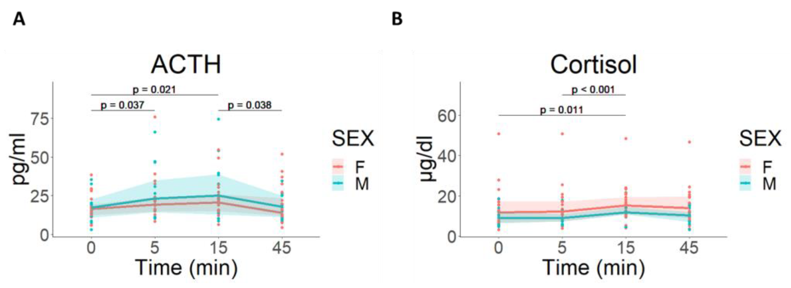
3.2.4. Hormonal Response

There were no differences in ACTH or cortisol values at baseline between M and F groups (U = 109, p = 0.669 for ACTH and U = 91, p = 0.259 for cortisol). Baseline biopsy was associated with a significant decrease in plasma ACTH [F(1, 30) = 5.973; p = 0.0206] and cortisol levels [F(1, 30) = 10.430; p = 0.003].
CPS induced a similar activation of HPA-axis in both groups in ACTH [F(3, 90) = 5.968; p < 0.001] and cortisol [F(3, 96) = 7,344; p < 0.001], with no differences between M and F groups ([F(1, 30) = 0,481; p = 0.493] and [F(1, 32) = 2,53; p = 0.122], respectively) (Figure 4).
In the sham protocol, baseline biopsy was associated with a significant increase in plasma ACTH [F(1, 5) = 21.06; p = 0.006] and cortisol [F(1, 5) = 7.984; p = 0.037] that decreased during sham stress period (ACTH: [F(3,1 5) = 8.50; p = 0.002]; cortisol: [F(3,1 5) = 20.412; p < 0.001]) (Supplementary Figure S5).

3.3. Intestinal Response to CPS

3.3.1. Mucosal Gene Expression

(a)
Gene expression levels in the jejunal mucosa at baseline
Baseline expression compared with the reference gene is shown in Figure S6A. When compared between sexes, baseline expression levels of NR3C1 were significantly higher in females compared to males (F: −1.23 (−1.40–−1.17); M: −1.49 (−1.55–−1.29), p = 0.006), as were OCLN (F: −0.97 (−1.09–−0.92); M: −1.17 (−1.33–−1.09), p = 0.009) and ZO-1 expression levels (F: −1.39 (−1.59–−1.34); M: −1.67 (−1.76–−1.46), p < 0.034) (Supplementary Figure S6B). There was an upward trend in baseline expression levels of TRYP in males (F: −1.50 (−1.57–−1.37); M: −1.25 (−1.32–−1.17), p = 0.067). No significant differences were found in the basal expression levels of CLDN1, CLDN2, ZO-3, and SERPINA1 between sexes. A clustering analysis of the correlations of the basal gene expression levels found that NR3C1 clustered together with OCLN and ZO-1 and, to a lesser degree, with CLDN1 and CLDN2 (Supplementary Figure S6C–F).
Baseline expression levels compared with the reference gene in the subjects submitted to the sham stress protocol is shown in Figure S7.
  • (b) Gene expression levels in the jejunal mucosa after CPS
Ninety minutes after CPS, we found a significant change in the expression levels of CLDN1, CLDN2, OCLN, and SERPINA1 genes (Figure 5A). The expression of CLDN1 and OCLN was significantly reduced (CLDN1: 0.81 (0.74–0.98), fold change, p = 0.008; OCLN: 0.87 (0.76–1), fold change, p = 0.016), while CLDN2 and SERPINA1 expression significantly increased (CLDN2: 1.33 (0.94–1.7), fold change, p = 0.002; SERPINA1: 1.24 (1.04–1.45), fold change, p < 0.001).
Interestingly, after CPS, a differential response in gene expression between male and female groups was observed, as NR3C1 expression was significantly reduced only in women, when compared to men (F: 0.88 (0.76–0.96); M: 1.05 (0.99–1.15), fold change, p = 0.007) (Figure 5B). Similarly, when we analyzed TJ gene expression, we found a significant decrease in OCLN gene expression, and this decrease was enhanced in women (F: 0.82 0.76–0.88); M: 0.99 (0.83–1.17), fold change, p = 0.022). In addition, we observed a differential regulation of ZO-1 gene expression, with a decrease in women and a slight increase in men 90 min after CPS (F: 0.83 (0.73–0.98); M: 1.04 (0.96–1.12), fold change, p = 0.046) (Figure 5B).
A clustering analysis of the correlations of gene expression changes after CPS showed that NR3C1 clustered together with ZO-1, OCLN, and SERPINA1 (Figure 5C–G).
In the sham stress protocol, a significant reduction in the expression of CLDN1 (CLDN1: 0.79 (0.77–0.82), fold change, p = 0.005) and an increase in the expression of SEPINA1 (SERPINA1: 1.19 (1.12–1.41), fold change, p = 0.021) was observed (Supplementary Figure S8).

3.3.2. Mucosal Inflammation and NR3C1 Protein Expression

At baseline, eosinophil (Eo) counts were higher in male when compared to female (Eo M: 3.2 (1.5–6.05) cells/hpf; Eo F: 1.30 (0.7–3.5) cells/hpf, p = 0.040) with no differences in MCs or CD3+ counts.
Ninety minutes after finishing CPS we found an increase in MCs counts [F(1, 31) = 6,79; p = 0.014], with no differences between male and females [F (1,32) = 0.86; p = 0.362]. In contrast, Eo counts [F(1, 32) = 0,127; p = 0.724] and CD3+ counts did not change [F(1, 32) = 1,19 p = 0.282] after CPS (Figure 6A–C).
NR3C1 mean intensity by IHC in jejunal mucosal samples was similar at baseline between male and female groups (M 119.1 ± 2.3; F: 121.7 ± 1.535 mean intensity AU, p = 0.357) and we did not find significant modification after CPS (M: 122.1 ± 3.303, F: 121.4 ± 2.167) (Supplementary Figure S9).
No significant changes in MCs, Eos, and CD3+ counts were observed in the sham stress protocol (Supplementary Figure S10).

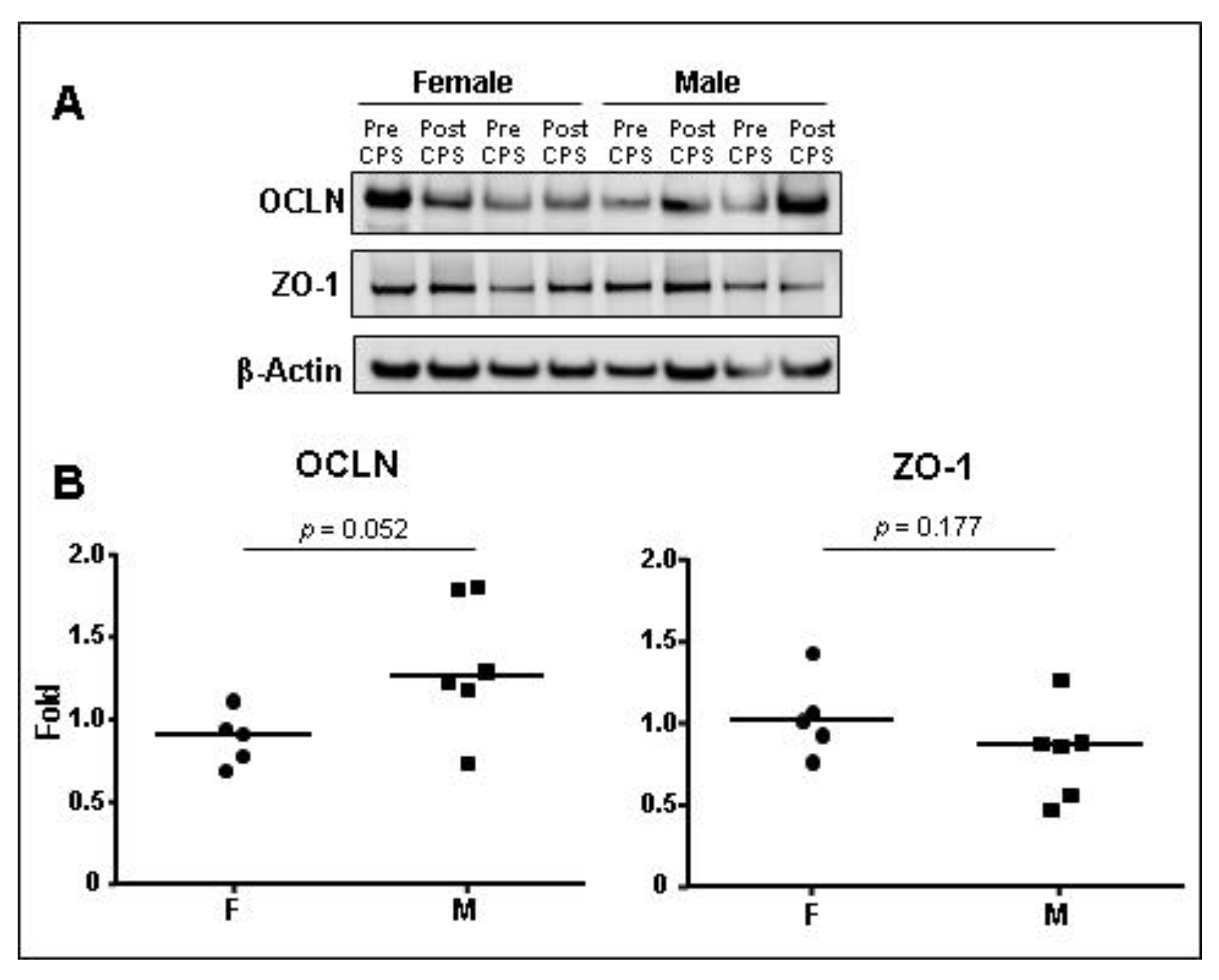
3.3.3. TJ Protein Expression

After CPS, we found an increase in OCLN protein expression only in the male group, while there was a decrease in OCLN expression in the female group (M: 1.266 (1.072–1.799); F: 0.917 (0.738–1.036), fold change, p = 0.052) after CPS (Figure 7). ZO-1 protein expression was not affected by CPS (M: 0.873 (0.543–0.979); F: 1.019 (0.848–1.247), p = 0.177).

4. Discussion

The present work shows that sex determines the expression of TJs and stress-related genes/proteins in the human jejunal mucosa in response to acute stress. Experimental acute stress induced a differential regulation of gene expression levels between sexes in the human jejunal mucosa and increased the number of MCs, mainly in males. Moreover, the expression of TJ-related genes in males seems to be more resistant to stress-induced changes compared to their female counterparts. Finally, although not novel, we confirm that CPS induces a strong autonomic, hormonal, and psychological stress response, as previously described [27,33].
CPS is a well-known acute experimental stress model in humans [31] that induces both physical and psychological responses. Our previous studies have shown that CPS as well as the stress mediator CRF affect the intestinal barrier function in healthy subjects (HS) [13,27,33]. However, the molecular basis of these changes has not yet been completely elucidated.
There is no doubt that biological sex plays a role in the prevalence and severity of a number of important stress-related gastrointestinal and immune-related diseases such as IBS [2,34,35]. However, the underlying molecular mechanisms contributing to sex differences remain poorly understood. Our results show that acute stress downregulates gene expression of CLDN1 and OCLN, while upregulating the expression of CLDN2. Interestingly, ZO-1 and OCLN expression after CPS was different between male and female groups, being downregulated only in females. Similar findings have been described in different animal models of stress showing that acute stress is associated with a decreased expression of Cldn1, Ocln, and Nr3c1 and an increased expression of Cldn2 along with intestinal permeability and visceromotor changes in the colon [26,36,37,38,39]. In line with these results, we also found a higher stability of TJ gene expression in males in response to CPS, similarly to the strengthening of TJs function after pilocarpine stimulation reported in salivary glands of male mice when compared to females [40]. Of note, very few studies have focused on the small intestine. One of such studies showed that the combination of water avoidance and restraint stress downregulated mRNA and protein levels of ZO-1 in the duodenal mucosa of male rats [41]. While permeability studies in rodents have been performed only in male, a porcine model of early weaning stress showed a decrease in the expression of OCLN, CLDN1, and ZO-1 in the jejunal mucosa in both sexes in association with impaired intestinal barrier function [42]. ZO-1 plays an essential role in the control of paracellular permeability of large molecules through the leak pathway, as shown in MDCK monolayers after its knockdown through zonula adheren and zonula occluden belt regulation [43]. Our results show that a coordinated genetic regulation of TJs genes may also be involved, as suggested by the strong correlation between the expression of ZO-1 and NR3C1 in the human jejunum. Conversely, the role of Ocln in the paracellular integrity is controverted, as Ocln seems not essential for the regulation of intestinal barrier function and epithelial cell polarization in OCLN-deficient mice [44,45], while other studies indicate that OCLN can regulate the paracellular pore pathway in polarized Caco-2 cells [46], along with the passage of monovalent cations and uncharged solutes through the leak pathway in Caco-2 cells [47]. These actions of OCLN involve its ability to modulate strand architecture and CLDN channels and may support its regulatory role of barrier function under stressed conditions. Last, but not least, CLDN2 is a pore-forming molecule that facilitates paracellular flux of water [48], and Na+ and small uncharged molecules [49], whereas CLDN1 seems to play the opposite role [50]. Altogether, our findings suggest that females may be more susceptible to develop stress-related molecular alterations in the intestinal barrier leading to enhanced intestinal permeability in response to acute stress. This is in line with our previous results [13,27,33], where we showed an increased albumin secretion to the intestinal lumen in females as a marker of altered intestinal permeability. Albumin is a macromolecule normally transcellularly transported from the intestinal lumen to the blood. However, a bidirectional paracellular flux between the intestinal lumen and the lamina propria may occur through the leak pathway after immune activation [47,51]. Our present results may be an explanation for the higher increase in albumin secretion that we found in women when compared to males in response to CPS.
A novel finding of our study is the ability of acute stress to downregulate the expression of NR3C1 in the human jejunum, only in females. Prior studies in male rats suggest a central role of glucocorticoids as mediators of the stress-induced enhancement of intestinal permeability as both adrenalectomy and the glucocorticoid receptor inhibitor RU-486 prevented the increase in permeability [52]. In addition, pretreatment with CRF increases the expression of CLDN2 in HT-29, T84, and MDCK cells [53]. HPA-axis response also seems essential for understanding sex-related differences in psychiatric disorders such as post-traumatic stress disorder and depression, but little is known about the role of the intestinal barrier in these disorders [54]. More studies are needed to ascertain the specific consequences of sex-dependent differential glucocorticoid receptor expression in the intestinal barrier function, their nuclear targets, and its clinical implication. Intriguingly, we also found that epithelial barrier genes correlate with NR3C1 both at baseline, and after CPS. This is interesting, as the expression of NR3C1-correlated genes (i.e., OCLN and ZO-1) was different between sexes. Chronic stress limits the binding of NR3C1 to CLDN1 and OCLN promoters in Caco-2 BBE cortisol-treated cells, but binding of HES1 is not affected, leading to a decreased expression of Cldn1 in rat colon [26]. It would be interesting to explore whether acute stress activates a common nuclear mechanism leading to a coordinated change of ZO-1, OCLN, and NR3C1 gene expression in the human jejunal mucosa and in both sexes.
Additionally, to the intestinal molecular response to acute stress, we also evaluated mucosal microinflammation and MC response, and found that stress increased MC counts and SERPINA1 expression, but not TRYP mRNA expression, with no differences between sexes. Stress has been shown to increase MC degranulation and activation in the rat intestinal mucosa [55,56,57,58,59] as well as in humans [13,21,25], and several reports indicate that MCs play a key role in the regulation of intestinal barrier in animals and humans [21,56,58,59,60,61,62]. Among the mechanisms by which MCs can induce intestinal dysfunction, proteases have been shown to increase intestinal permeability through PAR2-mediated TJ regulation [59,63,64]. In fact, IBS patients showed higher protease activity in feces that has been related to impaired intestinal barrier function [65,66], particularly TRYP and trypsin-3 [63,67] signaling through epithelial PAR2 cleavage and MC activation. Signaling through PAR2 leads to changes in localization and expression of TJ regulatory proteins [68]. SERPINA1 encodes alpha-1-antitrypsin, one of the main human antiproteases that inhibits trypsin, tryptase, and neutrophil elastase, and it is produced by gut epithelial cells [69], specifically by enterocytes [70]. Interestingly, corticosteroid binding globulin (encoded by SERPINA6), a major high-affinity transporter for cortisol in the blood, is inactivated via proteolytic cleavage by neutrophil elastase, which, as mentioned before, is in turn inhibited by alpha-1-antitrypsin [71]. SERPINA6 and SERPINA1 are closely located in chromosome 14 and both were related in a meta-analysis of genome-wide association studies with morning plasma cortisol levels [72]. The lack of correlation between SERPINA1 and TRYP gene expression levels and MCs count in our study raises the question on the meaning of the anti-protease gene upregulation in the control of barrier function. In this sense, SERPINA1 gene expression could be increased to counter-regulate other proteases related with the dysfunctional intestinal barrier function, or alternatively, MCs from both sexes may react differently to stress, as has been shown in distal ileum of mice submitted to restraint stress [73]. Our results suggest a differential expression profile of MCs between sexes in response to stress. Sexually dimorphic responses have been previously reported in bone marrow MCs from mice submitted to restraint stress, with greater intestinal permeability and serum histamine responses to restraint stress in female mice [73]. In this study, female MCs showed a markedly increased capacity for synthesis and storage of granule-associated immune mediators, indicative of a sex-related differential transcriptome in MCs resulting from stress exposure [73]. However, more studies are needed to understand the role of sexual dimorphism in MC-mediated intestinal function in humans.
We acknowledge that our work has several limitations mainly derived from the complexity of the methodology that limited the recruitment of participants, and adaptation of the time point for the second biopsy to technical feasibility that may explain the inability to explore genetic expression dynamics in more detail and lack of significant changes in protein expression, although the small sample size for protein expression experiments could also contribute to this point. Moreover, we cannot exclude that the effects on mucosal gene expression represent the combined effect of intubation, baseline biopsy, and CPS, as baseline biopsy was a stressful experience for participants by itself and we found changes in molecular expression of some genes in the sham protocol. Unfortunately, we could not prevent this response due to the complex experimental design. However, our study also has important strengths; notably, the performance of these in vivo experiments using a sophisticated and invasive procedure in human subjects that provides valuable information on the effect of acute stress in the intestinal mucosa in humans, although we acknowledge that this could be a limitation for reproducibility.
In conclusion, acute experimental stress uncovers a differential sex-related molecular response in the intestinal mucosa that may explain differences in stress-induced enhanced permeability between sexes. We speculate that these molecular alterations induced by acute stress could help to understand female susceptibility to develop IBS and possibly other gastrointestinal and systemic disorders related to life stress and brain–gut dysfunction.

Supplementary Materials

The following supporting information can be downloaded at: https://www.mdpi.com/article/10.3390/cells12030423/s1, Figure S1: Systemic response to baseline biopsy in subjects submitted to CPS; Figure S2: Systemic response to baseline biopsy in subjects submitted to sham stress.; Figure S3: Systemic response to sham stress; Figure S4: Hormonal response to baseline biopsy in subjects submitted to CPS and sham stress; Figure S5: Hormonal response to sham stress; Figure S6: Baseline expression levels of CLDN1, CLDN2, NR3C1, OCLN, SERPINA1, TJP1, TJP3 and TRYP in jejunal mucosa of subjects submitted to CPS; Figure S7: Baseline expression levels of CLDN1, CLDN2, NR3C1, OCLN, SERPINA1, TJP1, TJP3 and TRYP in jejunal mucosa of subjects submitted to sham stress; Figure S8: Gene expression levels changes of CLDN1, CLDN2, NR3C1, OCLN, SERPINA1, ZO-1, ZO-3 and TRYP in jejunal mucosa after sham stress; Figure S9: Protein expression levels of NR3C1 in jejunal mucosa after CPS; Figure S10: Mucosal inflammation in jejunal mucosa after sham stress; Table S1: List of taqman probes used for the RT-qPCR gene expression assays; Table S2: Primary Antibodies used for Western blot and IHC studies; Table S3. Clinical variables from subjects submitted to Sham stress.

Author Contributions

Conceptualization, M.P., B.L., J.S. and C.A.-C.; Data curation, M.P., A.N., C.G., T.P., L.H., B.L., M.G. and C.A.-C.; Formal analysis, B.K.R.-J., M.P. and C.A.-C.; Investigation, B.K.R.-J., M.P., E.S.-R., A.N., E.E., A.M.G.-C., C.G., I.d.T., T.P., L.H., B.L., M.F., M.G., C.P.-C., D.G. and C.A.-C.; Methodology, B.K.R.-J., M.P., E.S.-R., A.N., E.E., A.M.G.-C., C.G., I.d.T., T.P., L.H., B.L., M.F., M.G., C.P.-C., D.G. and C.A.-C.; Project administration, C.A.-C.; Resources, C.A.-C.; Supervision, J.S. and C.A.-C.; Visualization, C.A.-C.; Writing—original draft, B.K.R.-J., M.P. and J.S.; Writing—review and editing, B.K.R.-J., J.S. and C.A.-C. All authors have read and agreed to the published version of the manuscript.

Funding

Supported in part by Fondo Europeo de Desarrollo Regional (FEDER), Fondo de Investigación Sanitaria and Centro de Investigación Biomédica en Red de Enfermedades Hepáticas y Digestivas (CIBEREHD), Instituto de Salud Carlos III, Subdirección General de Investigación Sanitaria, Ministerio de Economiá y Competitividad: CM08/00229 (BL); CM10/00155 (MP); EII2011-0035, CD15/00010, and MV17-00043 (BKRJ.); FI12/00254 (ESR.), PI17/0190 (JS), PI12/00314 and PI15/00301 (CAC), CIBEREHD CB06/04/0021 (JS, CAC.); Vall d’Hebron Institut de Recerca, Programa de becas predoctorales Amics de Vall d’Hebron: PRED-VHIR-2014-018 (MF), PRED-VHIR-2016-53 34 (CPC.).

Institutional Review Board Statement

This study was conducted in accordance with the Declaration of Helsinki and approved by the Institutional Review Board (or Ethics Committee) of Vall d’Hebron Hospital (protocol code PR(IR)248/2012, date 26 February 2013).

Informed Consent Statement

Informed consent was obtained from all subjects involved in the study.

Data Availability Statement

All the data, analytic methods, and study materials are present in the article and the Supplementary Materials.

Acknowledgments

We thank all healthy volunteers for participating in this study.

Conflicts of Interest

C.A.-C. discloses past scientific collaborations with Norgine; J.S. serves as consultant for Noventure, Reckitt, Ipsen, and Ordesa and discloses present or past recent scientific collaborations with Salvat, Norgine, Alfa-Sigma, Cosmo, Adare, Devintecpharma, Pileje, and Danone.

References

  1. Nater, U.M.; Maloney, E.; Heim, C.; Reeves, W.C. Cumulative Life Stress in Chronic Fatigue Syndrome. Psychiatry Res. 2011, 189, 318–320. [Google Scholar] [CrossRef] [PubMed]
  2. Enck, P.; Aziz, Q.; Barbara, G.; Farmer, A.D.; Fukudo, S.; Mayer, E.A.; Niesler, B.; Quigley, E.M.M.; Rajilić-Stojanović, M.; Schemann, M.; et al. Irritable Bowel Syndrome. Nat. Rev. Dis. Prim. 2016, 2, 16014. [Google Scholar] [CrossRef] [PubMed] [Green Version]
  3. Galvez-Sánchez, C.M.; Duschek, S.; Reyes Del Paso, G.A. Psychological Impact of Fibromyalgia: Current Perspectives. Psychol. Res. Behav. Manag. 2019, 12, 117–127. [Google Scholar] [CrossRef] [PubMed] [Green Version]
  4. Gupta, A.; Bhatt, R.R.; Naliboff, B.D.; Kutch, J.J.; Labus, J.S.; Vora, P.P.; Alaverdyan, M.; Schrepf, A.; Lutgendorf, S.; Mayer, E.A.; et al. Impact of Early Adverse Life Events and Sex on Functional Brain Networks in Patients with Urological Chronic Pelvic Pain Syndrome (UCPPS): A MAPP Research Network Study. PLoS ONE 2019, 14, e0217610. [Google Scholar] [CrossRef] [Green Version]
  5. Lacy, B.E.; Mearin, F.; Chang, L.; Chey, W.D.; Lembo, A.J.; Simren, M.; Spiller, R. Bowel Disorders. Gastroenterology 2016, 150, 1393–1407.e5. [Google Scholar] [CrossRef] [Green Version]
  6. Sperber, A.D.; Bangdiwala, S.I.; Drossman, D.A.; Ghoshal, U.C.; Simren, M.; Tack, J.; Whitehead, W.E.; Dumitrascu, D.L.; Fang, X.; Fukudo, S.; et al. Worldwide Prevalence and Burden of Functional Gastrointestinal Disorders, Results of Rome Foundation Global Study. Gastroenterology 2021, 160, 99–114.e3. [Google Scholar] [CrossRef]
  7. Chey, W.D. Food: The Main Course to Wellness and Illness in Patients with Irritable Bowel Syndrome. Am. J. Gastroenterol. 2016, 111, 366–371. [Google Scholar] [CrossRef]
  8. Dinan, T.G.; Cryan, J.; Shanahan, F.; Keeling, P.W.N.; Quigley, E.M.M. IBS: An Epigenetic Perspective. Nat. Rev. Gastroenterol. Hepatol. 2010, 7, 465–471. [Google Scholar] [CrossRef]
  9. Henström, M.; D’Amato, M. Genetics of Irritable Bowel Syndrome. Mol. Cell. Pediatr. 2016, 3, 7. [Google Scholar] [CrossRef] [Green Version]
  10. Gazouli, M.; Wouters, M.M.; Kapur-Pojskić, L.; Bengtson, M.-B.; Friedman, E.; Nikčević, G.; Demetriou, C.A.; Mulak, A.; Santos, J.; Niesler, B. Lessons Learned—Resolving the Enigma of Genetic Factors in IBS. Nat. Rev. Gastroenterol. Hepatol. 2016, 13, 77–87. [Google Scholar] [CrossRef]
  11. Rodiño-Janeiro, B.K.; Pardo-Camacho, C.; Santos, J.; Martínez, C. Mucosal RNA and Protein Expression as the next Frontier in IBS: Abnormal Function despite Morphologically Intact Small Intestinal Mucosa. Am. J. Physiol. Gastrointest. Liver Physiol. 2019, 316, G701–G719. [Google Scholar] [CrossRef]
  12. Guilarte, M.; Santos, J.; de Torres, I.; Alonso, C.; Vicario, M.; Ramos, L.; Martínez, C.; Casellas, F.; Saperas, E.; Malagelada, J.R. Diarrhoea-Predominant IBS Patients Show Mast Cell Activation and Hyperplasia in the Jejunum. Gut 2007, 56, 203–209. [Google Scholar] [CrossRef] [Green Version]
  13. Guilarte, M.; Vicario, M.; Martínez, C.; de Torres, I.; Lobo, B.; Pigrau, M.; González-Castro, A.; Rodiño-Janeiro, B.K.; Salvo-Romero, E.; Fortea, M.; et al. Peripheral Corticotropin-Releasing Factor Triggers Jejunal Mast Cell Activation and Abdominal Pain in Patients With Diarrhea-Predominant Irritable Bowel Syndrome. Am. J. Gastroenterol. 2020, 115, 2047–2059. [Google Scholar] [CrossRef]
  14. Vicario, M.; González-Castro, A.M.; Martínez, C.; Lobo, B.; Pigrau, M.; Guilarte, M.; de Torres, I.; Mosquera, J.L.; Fortea, M.; Sevillano-Aguilera, C.; et al. Increased Humoral Immunity in the Jejunum of Diarrhoea-Predominant Irritable Bowel Syndrome Associated with Clinical Manifestations. Gut 2015, 64, 1379–1388. [Google Scholar] [CrossRef]
  15. Martin-Viñas, J.J.; Quigley, E.M.M. Immune Response in Irritable Bowel Syndrome: A Systematic Review of Systemic and Mucosal Inflammatory Mediators. J. Dig. Dis. 2016, 17, 572–581. [Google Scholar] [CrossRef]
  16. Martínez, C.; Rodiño-Janeiro, B.K.; Lobo, B.; Stanifer, M.L.; Klaus, B.; Granzow, M.; González-Castro, A.M.; Salvo-Romero, E.; Alonso-Cotoner, C.; Pigrau, M.; et al. MiR-16 and MiR-125b Are Involved in Barrier Function Dysregulation through the Modulation of Claudin-2 and Cingulin Expression in the Jejunum in IBS with Diarrhoea. Gut 2017, 66, 1537–1538. [Google Scholar] [CrossRef] [Green Version]
  17. Bashashati, M.; Moossavi, S.; Cremon, C.; Barbaro, M.R.; Moraveji, S.; Talmon, G.; Rezaei, N.; Hughes, P.A.; Bian, Z.X.; Choi, C.H.; et al. Colonic Immune Cells in Irritable Bowel Syndrome: A Systematic Review and Meta-Analysis. Neurogastroenterol. Motil. 2018, 30, e13192. [Google Scholar] [CrossRef]
  18. Mavrangelos, C.; Campaniello, M.A.; Andrews, J.M.; Bampton, P.A.; Hughes, P.A. Longitudinal Analysis Indicates Symptom Severity Influences Immune Profile in Irritable Bowel Syndrome. Gut 2018, 67, 398–399. [Google Scholar] [CrossRef]
  19. Martínez, C.; González-Castro, A.; Vicario, M.; Santos, J. Cellular and Molecular Basis of Intestinal Barrier Dysfunction in the Irritable Bowel Syndrome. Gut Liver 2012, 6, 305–315. [Google Scholar] [CrossRef] [Green Version]
  20. Martínez, C.; Lobo, B.; Pigrau, M.; Ramos, L.; González-Castro, A.M.; Alonso, C.; Guilarte, M.; Guilá, M.; de Torres, I.; Azpiroz, F.; et al. Diarrhoea-Predominant Irritable Bowel Syndrome: An Organic Disorder with Structural Abnormalities in the Jejunal Epithelial Barrier. Gut 2013, 62, 1160–1168. [Google Scholar] [CrossRef]
  21. Vanuytsel, T.; van Wanrooy, S.; Vanheel, H.; Vanormelingen, C.; Verschueren, S.; Houben, E.; Salim Rasoel, S.; Tόth, J.; Holvoet, L.; Farré, R.; et al. Psychological Stress and Corticotropin-Releasing Hormone Increase Intestinal Permeability in Humans by a Mast Cell-Dependent Mechanism. Gut 2014, 63, 1293–1299. [Google Scholar] [CrossRef] [PubMed]
  22. Rodiño-Janeiro, B.K.; Alonso-Cotoner, C.; Pigrau, M.; Lobo, B.; Vicario, M.; Santos, J. Role of Corticotropin-Releasing Factor in Gastrointestinal Permeability. J. Neurogastroenterol. Motil. 2015, 21, 33–50. [Google Scholar] [CrossRef] [PubMed] [Green Version]
  23. Barclay, G.R.; Turnberg, L.A. Effect of Psychological Stress on Salt and Water Transport in the Human Jejunum. Gastroenterology 1987, 93, 91–97. [Google Scholar] [CrossRef] [PubMed]
  24. Barclay, G.R.; Turnberg, L.A. Effect of Cold-Induced Pain on Salt and Water Transport in the Human Jejunum. Gastroenterology 1988, 94, 994–998. [Google Scholar] [CrossRef] [PubMed]
  25. Santos, J.; Saperas, E.; Nogueiras, C.; Mourelle, M.; Antolín, M.; Cadahia, A.; Malagelada, J.R. Release of Mast Cell Mediators into the Jejunum by Cold Pain Stress in Humans. Gastroenterology 1998, 114, 640–648. [Google Scholar] [CrossRef]
  26. Zheng, G.; Victor Fon, G.; Meixner, W.; Creekmore, A.; Zong, Y.; KDame, M.; Colacino, J.; Dedhia, P.H.; Hong, S.; Wiley, J.W. Chronic Stress and Intestinal Barrier Dysfunction: Glucocorticoid Receptor and Transcription Repressor HES1 Regulate Tight Junction Protein Claudin-1 Promoter. Sci. Rep. 2017, 7, 4502. [Google Scholar] [CrossRef] [Green Version]
  27. Alonso, C.; Guilarte, M.; Vicario, M.; Ramos, L.; Rezzi, S.; Martínez, C.; Lobo, B.; Martin, F.-P.; Pigrau, M.; González-Castro, A.M.; et al. Acute Experimental Stress Evokes a Differential Gender-Determined Increase in Human Intestinal Macromolecular Permeability. Neurogastroenterol. Motil. 2012, 24, 740–746, e348–e349. [Google Scholar] [CrossRef]
  28. Holmes, T.H.; Rahe, R.H. The Social Readjustment Rating Scale. J. Psychosom. Res. 1967, 11, 213–218. [Google Scholar] [CrossRef]
  29. Cohen, S.; Kamarck, T.; Mermelstein, R. A Global Measure of Perceived Stress. J. Health Soc. Behav. 1983, 24, 385–396. [Google Scholar] [CrossRef]
  30. Beck, A.T.; Ward, C.H.; Mendelson, M.; Mock, J.; Erbaugh, J. An Inventory for Measuring Depression. Arch. Gen. Psychiatry 1961, 4, 561–571. [Google Scholar] [CrossRef]
  31. Lovallo, W. The Cold Pressor Test and Autonomic Function: A Review and Integration. Psychophysiology 1975, 12, 268–282. [Google Scholar] [CrossRef]
  32. Naliboff, B.D.; Benton, D.; Solomon, G.F.; Morley, J.E.; Fahey, J.L.; Bloom, E.T.; Makinodan, T.; Gilmore, S.L. Immunological Changes in Young and Old Adults during Brief Laboratory Stress. Psychosom. Med. 1991, 53, 121–132. [Google Scholar] [CrossRef]
  33. Alonso, C.; Guilarte, M.; Vicario, M.; Ramos, L.; Ramadan, Z.; Antolín, M.; Martínez, C.; Rezzi, S.; Saperas, E.; Kochhar, S.; et al. Maladaptive Intestinal Epithelial Responses to Life Stress May Predispose Healthy Women to Gut Mucosal Inflammation. Gastroenterology 2008, 135, 163–172.e1. [Google Scholar] [CrossRef]
  34. Bekhbat, M.; Neigh, G.N. Sex Differences in the Neuro-Immune Consequences of Stress: Focus on Depression and Anxiety. Brain Behav. Immun. 2018, 67, 1–12. [Google Scholar] [CrossRef]
  35. Meester, I.; Rivera-Silva, G.F.; González-Salazar, F. Immune System Sex Differences May Bridge the Gap Between Sex and Gender in Fibromyalgia. Front. Neurosci. 2019, 13, 1414. [Google Scholar] [CrossRef]
  36. Zheng, G.; Wu, S.-P.; Hu, Y.; Smith, D.E.; Wiley, J.W.; Hong, S. Corticosterone Mediates Stress-Related Increased Intestinal Permeability in a Region-Specific Manner. Neurogastroenterol. Motil. 2013, 25, e127–e139. [Google Scholar] [CrossRef]
  37. Matsuo, K.; Zhang, X.; Ono, Y.; Nagatomi, R. Acute Stress-Induced Colonic Tissue HSP70 Expression Requires Commensal Bacterial Components and Intrinsic Glucocorticoid. Brain Behav. Immun. 2009, 23, 108–115. [Google Scholar] [CrossRef]
  38. Creekmore, A.L.; Hong, S.; Zhu, S.; Xue, J.; Wiley, J.W. Chronic Stress-Associated Visceral Hyperalgesia Correlates with Severity of Intestinal Barrier Dysfunction. Pain 2018, 159, 1777–1789. [Google Scholar] [CrossRef]
  39. Zong, Y.; Zhu, S.; Zhang, S.; Zheng, G.; Wiley, J.W.; Hong, S. Chronic Stress and Intestinal Permeability: Lubiprostone Regulates Glucocorticoid Receptor-Mediated Changes in Colon Epithelial Tight Junction Proteins, Barrier Function, and Visceral Pain in the Rodent and Human. Neurogastroenterol. Motil. 2018, 31, e13477. [Google Scholar] [CrossRef]
  40. Kawedia, J.D.; Nieman, M.L.; Boivin, G.P.; Melvin, J.E.; Kikuchi, K.-I.; Hand, A.R.; Lorenz, J.N.; Menon, A.G. Interaction between Transcellular and Paracellular Water Transport Pathways through Aquaporin 5 and the Tight Junction Complex. Proc. Natl. Acad. Sci. USA 2007, 104, 3621–3626. [Google Scholar] [CrossRef]
  41. Lee, H.S.; Kim, D.-K.; Kim, Y.B.; Lee, K.J. Effect of Acute Stress on Immune Cell Counts and the Expression of Tight Junction Proteins in the Duodenal Mucosa of Rats. Gut Liver 2013, 7, 190–196. [Google Scholar] [CrossRef] [PubMed] [Green Version]
  42. Hu, C.H.; Xiao, K.; Luan, Z.S.; Song, J. Early Weaning Increases Intestinal Permeability, Alters Expression of Cytokine and Tight Junction Proteins, and Activates Mitogen-Activated Protein Kinases in Pigs. J. Anim. Sci. 2013, 91, 1094–1101. [Google Scholar] [CrossRef] [PubMed] [Green Version]
  43. Van Itallie, C.M.; Fanning, A.S.; Bridges, A.; Anderson, J.M. ZO-1 Stabilizes the Tight Junction Solute Barrier through Coupling to the Perijunctional Cytoskeleton. Mol. Biol. Cell 2009, 20, 3930–3940. [Google Scholar] [CrossRef] [PubMed] [Green Version]
  44. Schulzke, J.D.; Gitter, A.H.; Mankertz, J.; Spiegel, S.; Seidler, U.; Amasheh, S.; Saitou, M.; Tsukita, S.; Fromm, M. Epithelial Transport and Barrier Function in Occludin-Deficient Mice. Biochim. Biophys. Acta Biomembr. 2005, 1669, 34–42. [Google Scholar] [CrossRef] [Green Version]
  45. Saitou, M.; Fujimoto, K.; Doi, Y.; Itoh, M.; Fujimoto, T.; Furuse, M.; Takano, H.; Noda, T.; Tsukita, S. Occludin-Deficient Embryonic Stem Cells Can Differentiate into Polarized Epithelial Cells Bearing Tight Junctions. J. Cell Biol. 1998, 141, 397–408. [Google Scholar] [CrossRef]
  46. Raleigh, D.R.; Boe, D.M.; Yu, D.; Weber, C.R.; Marchiando, A.M.; Bradford, E.M.; Wang, Y.; Wu, L.; Schneeberger, E.E.; Shen, L.; et al. Occludin S408 Phosphorylation Regulates Tight Junction Protein Interactions and Barrier Function. J. Cell Biol. 2011, 193, 565–582. [Google Scholar] [CrossRef]
  47. Buschmann, M.M.; Shen, L.; Rajapakse, H.; Raleigh, D.R.; Wang, Y.; Wang, Y.; Lingaraju, A.; Zha, J.; Abbott, E.; McAuley, E.M.; et al. Occludin OCEL-Domain Interactions Are Required for Maintenance and Regulation of the Tight Junction Barrier to Macromolecular Flux. MBoC 2013, 24, 3056–3068. [Google Scholar] [CrossRef]
  48. Tamura, A.; Hayashi, H.; Imasato, M.; Yamazaki, Y.; Hagiwara, A.; Wada, M.; Noda, T.; Watanabe, M.; Suzuki, Y.; Tsukita, S. Loss of Claudin-15, but Not Claudin-2, Causes Na+ Deficiency and Glucose Malabsorption in Mouse Small Intestine. Gastroenterology 2011, 140, 913–923. [Google Scholar] [CrossRef]
  49. Van Itallie, C.M.; Holmes, J.; Bridges, A.; Gookin, J.L.; Coccaro, M.R.; Proctor, W.; Colegio, O.R.; Anderson, J.M. The Density of Small Tight Junction Pores Varies among Cell Types and Is Increased by Expression of Claudin-2. J. Cell. Sci. 2008, 121, 298–305. [Google Scholar] [CrossRef] [Green Version]
  50. Inai, T.; Kobayashi, J.; Shibata, Y. Claudin-1 Contributes to the Epithelial Barrier Function in MDCK Cells. Eur. J. Cell Biol. 1999, 78, 849–855. [Google Scholar] [CrossRef]
  51. Zuo, L.; Kuo, W.-T.; Turner, J.R. Tight Junctions as Targets and Effectors of Mucosal Immune Homeostasis. Cell. Mol. Gastroenterol. Hepatol. 2020, 10, 327–340. [Google Scholar] [CrossRef]
  52. Meddings, J.B.; Swain, M.G. Environmental Stress-Induced Gastrointestinal Permeability Is Mediated by Endogenous Glucocorticoids in the Rat. Gastroenterology 2000, 119, 1019–1028. [Google Scholar] [CrossRef]
  53. Yu, Y.; Liu, Z.-Q.; Liu, X.-Y.; Yang, L.; Geng, X.-R.; Yang, G.; Liu, Z.-G.; Zheng, P.-Y.; Yang, P.-C. Stress-Derived Corticotropin Releasing Factor Breaches Epithelial Endotoxin Tolerance. PLoS ONE 2013, 8, e65760. [Google Scholar] [CrossRef] [Green Version]
  54. Bangasser, D.A.; Valentino, R.J. Sex Differences in Stress-Related Psychiatric Disorders: Neurobiological Perspectives. Front. Neuroendocrinol. 2014, 35, 303–319. [Google Scholar] [CrossRef] [Green Version]
  55. Vicario, M.; Guilarte, M.; Alonso, C.; Yang, P.; Martínez, C.; Ramos, L.; Lobo, B.; González, A.; Guilà, M.; Pigrau, M.; et al. Chronological Assessment of Mast Cell-Mediated Gut Dysfunction and Mucosal Inflammation in a Rat Model of Chronic Psychosocial Stress. Brain Behav. Immun. 2010, 24, 1166–1175. [Google Scholar] [CrossRef]
  56. Söderholm, J.D.; Yang, P.-C.; Ceponis, P.; Vohra, A.; Riddell, R.; Sherman, P.M.; Perdue, M.H. Chronic Stress Induces Mast Cell-Dependent Bacterial Adherence and Initiates Mucosal Inflammation in Rat Intestine. Gastroenterology 2002, 123, 1099–1108. [Google Scholar] [CrossRef]
  57. Kaushik, S.; Kaur, J. Effect of Chronic Cold Stress on Intestinal Epithelial Cell Proliferation and Inflammation in Rats. Stress 2005, 8, 191–197. [Google Scholar] [CrossRef]
  58. Santos, J.; Yang, P.C.; Söderholm, J.D.; Benjamin, M.; Perdue, M.H. Role of Mast Cells in Chronic Stress Induced Colonic Epithelial Barrier Dysfunction in the Rat. Gut 2001, 48, 630–636. [Google Scholar] [CrossRef] [Green Version]
  59. Smith, F.; Clark, J.E.; Overman, B.L.; Tozel, C.C.; Huang, J.H.; Rivier, J.E.F.; Blikslager, A.T.; Moeser, A.J. Early Weaning Stress Impairs Development of Mucosal Barrier Function in the Porcine Intestine. Am. J. Physiol. Gastrointest. Liver Physiol. 2010, 298, G352–G363. [Google Scholar] [CrossRef] [Green Version]
  60. Santos, J.; Yates, D.; Guilarte, M.; Vicario, M.; Alonso, C.; Perdue, M.H. Stress Neuropeptides Evoke Epithelial Responses via Mast Cell Activation in the Rat Colon. Psychoneuroendocrinology 2008, 33, 1248–1256. [Google Scholar] [CrossRef]
  61. Santos, J.; Benjamin, M.; Yang, P.C.; Prior, T.; Perdue, M.H. Chronic Stress Impairs Rat Growth and Jejunal Epithelial Barrier Function: Role of Mast Cells. Am. J. Physiol. Gastrointest. Liver Physiol. 2000, 278, G847–G854. [Google Scholar]
  62. Wallon, C.; Yang, P.-C.; Keita, A.V.; Ericson, A.-C.; McKay, D.M.; Sherman, P.M.; Perdue, M.H.; Söderholm, J.D. Corticotropin-Releasing Hormone (CRH) Regulates Macromolecular Permeability via Mast Cells in Normal Human Colonic Biopsies in Vitro. Gut 2008, 57, 50–58. [Google Scholar] [CrossRef]
  63. Jacob, C.; Yang, P.-C.; Darmoul, D.; Amadesi, S.; Saito, T.; Cottrell, G.S.; Coelho, A.-M.; Singh, P.; Grady, E.F.; Perdue, M.; et al. Mast Cell Tryptase Controls Paracellular Permeability of the Intestine. Role of Protease-Activated Receptor 2 and Beta-Arrestins. J. Biol. Chem. 2005, 280, 31936–31948. [Google Scholar] [CrossRef] [PubMed] [Green Version]
  64. Goldblum, S.E.; Rai, U.; Tripathi, A.; Thakar, M.; De Leo, L.; Di Toro, N.; Not, T.; Ramachandran, R.; Puche, A.C.; Hollenberg, M.D.; et al. The Active Zot Domain (Aa 288-293) Increases ZO-1 and Myosin 1C Serine/Threonine Phosphorylation, Alters Interaction between ZO-1 and Its Binding Partners, and Induces Tight Junction Disassembly through Proteinase Activated Receptor 2 Activation. FASEB J. 2011, 25, 144–158. [Google Scholar] [CrossRef] [PubMed] [Green Version]
  65. Vergnolle, N. Protease Inhibition as New Therapeutic Strategy for GI Diseases. Gut 2016, 65, 1215–1224. [Google Scholar] [CrossRef] [Green Version]
  66. Ray, K. IBS: The Power of Protease Activity in IBS. Nat. Rev. Gastroenterol. Hepatol. 2017, 14, 139. [Google Scholar] [CrossRef]
  67. Rolland-Fourcade, C.; Denadai-Souza, A.; Cirillo, C.; Lopez, C.; Jaramillo, J.O.; Desormeaux, C.; Cenac, N.; Motta, J.-P.; Larauche, M.; Taché, Y.; et al. Epithelial Expression and Function of Trypsin-3 in Irritable Bowel Syndrome. Gut 2017, 66, 1767–1778. [Google Scholar] [CrossRef]
  68. Lau, C.; Lytle, C.; Straus, D.S.; DeFea, K.A. Apical and Basolateral Pools of Proteinase-Activated Receptor-2 Direct Distinct Signaling Events in the Intestinal Epithelium. Am. J. Physiol. Cell Physiol. 2011, 300, C113–C123. [Google Scholar] [CrossRef]
  69. Greene, C.M.; Marciniak, S.J.; Teckman, J.; Ferrarotti, I.; Brantly, M.L.; Lomas, D.A.; Stoller, J.K.; McElvaney, N.G. A1-Antitrypsin Deficiency. Nat. Rev. Dis. Primers 2016, 2, 16051. [Google Scholar] [CrossRef]
  70. Geboes, K.; Ray, M.B.; Rutgeerts, P.; Callea, F.; Desmet, V.J.; Vantrappen, G. Morphological Identification of Alpha-I-Antitrypsin in the Human Small Intestine. Histopathology 1982, 6, 55–60. [Google Scholar] [CrossRef]
  71. Henley, D.E.; Lightman, S.L. New Insights into Corticosteroid-Binding Globulin and Glucocorticoid Delivery. Neuroscience 2011, 180, 1–8. [Google Scholar] [CrossRef]
  72. Bolton, J.L.; Hayward, C.; Direk, N.; Lewis, J.G.; Hammond, G.L.; Hill, L.A.; Anderson, A.; Huffman, J.; Wilson, J.F.; Campbell, H.; et al. Genome Wide Association Identifies Common Variants at the SERPINA6/SERPINA1 Locus Influencing Plasma Cortisol and Corticosteroid Binding Globulin. PLoS Genet. 2014, 10, e1004474. [Google Scholar] [CrossRef] [Green Version]
  73. Mackey, E.; Ayyadurai, S.; Pohl, C.S.; D’Costa, S.; Li, Y.; Moeser, A.J. Sexual Dimorphism in the Mast Cell Transcriptome and the Pathophysiological Responses to Immunological and Psychological Stress. Biol. Sex Differ. 2016, 7, 60. [Google Scholar] [CrossRef]
Figure 1. Experimental design and procedures. SSRS: subjective stress rating scale; Min: minutes.
Cells 12 00423 g001
Figure 2. Flow chart of subjects’ inclusion and analysis.
Cells 12 00423 g002
Figure 3. Systemic response to cold pain stress (CPS). The response to stress (change from baseline) was significantly different between males (M: blue circles) and females (F: pink circles). (A) A two-way ANOVA revealed significant differences for time and for sex group both in systolic and diastolic blood pressure. A two-way ANOVA revealed significant differences for time but not for sex in heart rate. CPS increased subjective stress rating scale (SSRS), with no differences between M and F groups. (B) A two-way ANOVA revealed significant differences for time and for sex group both in hand pain perception. Lines represent the median for each time point and the area represents the 25 and 75 percentiles. F: females; M: males; min: minutes; SSRS: subjective stress rating scale.
Cells 12 00423 g003
Figure 4. Hormonal response to CPS. (A) ACTH: A two-way ANOVA revealed significant differences for time, but not for sex groups. (B) Cortisol: A two-way ANOVA revealed significant differences for time, but not for sex groups. Males and females are represented with blue circles and pink circles, respectively. Lines represent the median for each time point and the area represents the 25 and 75 percentiles. ACTH: adrenocorticotropic hormone; CPS: cold pain stress; F: females; M: males; min: minutes.
Cells 12 00423 g004
Figure 5. Gene expression levels of CLDN1, CLDN2, NR3C1, OCLN, SERPINA1, ZO-1, ZO-3, and TRYP in jejunal mucosa after CPS. (A) Fold change of gene expression levels induced by stress in jejunal mucosa. The red line indicates the median. (B) Fold change of gene expression levels induced by stress in jejunal mucosa by sex. The red line indicates the median. (C) Correlation graph with hierarchical clustering of the fold change in the gene expression levels and the number of MCs induced by stress. The dots size and color of the dots represent the degree of correlation (Spearman’s rho). (CG) represent the correlation distribution of the fold change expression levels induced by stress among NR3C1, ZO-1, SERPINA1, and MC cell numbers. The blue line indicates the Loess regression and the gray area the confidence interval of 95%. Males and females are represented with back circles and white circles, respectively. F: females; M: males; CLDN1: claudin 1; CLDN2: claudin 2; OCLN: occludin; ZO-1: zonula occludens 1; ZO-3: zonula occludens 3; TRYP: tryptase; SERPINA1: serpin family A member 1; NR3C1: glucocorticoid receptor nuclear receptor subfamily 3 group C member 1.
Cells 12 00423 g005
Figure 6. Mucosal inflammation in jejunal mucosa after CPS. (A) Mast cell counts before (Baseline) and after stress (Post Stress) expressed per HPF (400×). A two-way ANOVA revealed differences by stress, but not for sex. The red line indicates the median. Indicates p value = 0.013. (B) Eosinophil counts before (Baseline) and after stress (Post Stress) expressed per HPF. (C) Intraepithelial lymphocyte counts before (Baseline) and after stress (Post Stress) expressed per 100 intestinal epithelial cells. CD117: cluster of differentiation 117; HPF: high-power field; Eosin: eosinophils; CD3: cluster of differentiation 3; IEC: intestinal epithelial cells; F: females; M: males.
Cells 12 00423 g006
Figure 7. Protein expression levels OCLN and ZO-1 in jejunal mucosa after CPS. (A) Representative Western blot image showing expression of OCLN and ZO-1 in the jejunal mucosa of two representative females and two representative males before and after cold pain stress (CPS). Normalization was performed using β-actin as loading control. (B) Bands were quantified, and results are expressed as fold–change, each dot represents the fold change of each individual (five females and six males) before (Pre) and after CPS (Post). Intergroup comparisons were performed using the Mann–Whitney U test. p values are indicated. F: females; M: males; OCLN: occludin, ZO-1: zonula occludens 1.
Cells 12 00423 g007
Table 1. Demographic characteristics of participants submitted to CPS protocol.
Sex
VariableM (Q1–Q3)F (Q1–Q3)p Value
Age (years)22.9 (22.1–29.2)22.6 (21.7–25.2)0.462
Holmes–Rahe scale (score)76.0 (39.5–98.5)89.0 (63.8–131.3)0.420
Cohen’s scale (score)14.0 (11.0–23.5)19.0 (12.3–24.0)0.529
Beck’s inventory (score)0.0 (0.0–0.5)0.5 (0.0–2.8)0.737
Holmes–Rahe scale (to assess the level of stress over the last year); reference values: low stress <150), moderate 150–299, high ≥300. Cohen’s scale (to assess the level of stress over the last month); reference values: no perceived stress <19, mild perceived stress 19–28, moderate perceived stress 29–38, severe perceived stress >38. Beck’s depression inventory, to evaluate the level of depression during the last week; reference values: no depression <10, mild 10–18, moderate 19–29, severe depression 30–63. Data are expressed as median with first and third quartiles Q1–Q3). A Mann–Whitney U test was used for comparisons of continuous variables between groups.
Disclaimer/Publisher’s Note: The statements, opinions and data contained in all publications are solely those of the individual author(s) and contributor(s) and not of MDPI and/or the editor(s). MDPI and/or the editor(s) disclaim responsibility for any injury to people or property resulting from any ideas, methods, instructions or products referred to in the content.

