Commentary

The Evolution of Current Concept of the Reconstructive Ladder in Plastic Surgery: The Emerging Role of Translational Medicine

by
Francesco De Francesco
1,*,
Nicola Zingaretti
2,
Pier Camillo Parodi
2 and
Michele Riccio
1
1
Department of Reconstructive Surgery and Hand Surgery, University Hospital (AOU Ospedali Riuniti di Ancona), Via Conca 71, Torrette di Ancona, 60123 Ancona, Italy
2
Department of Medical Area (DAME), Clinic of Plastic and Reconstructive Surgery, Academic Hospital of Udine, University of Udine, 33100 Udine, Italy
*
Author to whom correspondence should be addressed.
Cells 2023, 12(21), 2567; https://doi.org/10.3390/cells12212567
Submission received: 2 October 2023 / Revised: 30 October 2023 / Accepted: 1 November 2023 / Published: 3 November 2023
(This article belongs to the Special Issue New Insights into Adipose-Derived Stem Cells (ADSCs))

Abstract

:
Plastic surgeons have used the reconstructive ladder for many decades as a standard directory for complex trauma reconstruction with the goal of repairing body structures and restoring functionality. This consists of different surgical maneuvers, such as secondary intention and direct tissue closure, as well as more complex methods such as local tissue transfer and free flap. The reconstructive ladder represents widely known options achievable for tissue reconstruction and wound closure that puts at the bottom rung the simplest methods of reconstruction and strengthens the complexity by moving upward. Regenerative medicine and surgery constitute a quickly spreading area of translational research that can be employed by minimally invasive surgical strategies, with the aim of regenerating cells and tissues in vivo in order to reestablish normal function through the intrinsic potential of cells, in combination with biomaterials and appropriate biochemical stimuli. These translational procedures have the aim of creating an appropriate microenvironment capable of supporting the physiological cellular function to generate the desired cells or tissues and to generate parenchymal, stromal, and vascular components on demand, and above all to produce intelligent materials capable of determining the fate of cells. Smart technologies have been grown that give extra “rungs” on the classic reconstructive ladder to integrate a more holistic, patient-based approach with improved outcomes. This commentary presents the evolution of the traditional concept of the reconstructive ladder in the field of plastic surgery into a new course with the aim of achieving excellent results for soft tissue reconstruction by applying innovative technologies and biologically active molecules for a wide range of surgical diseases.

Graphical Abstract

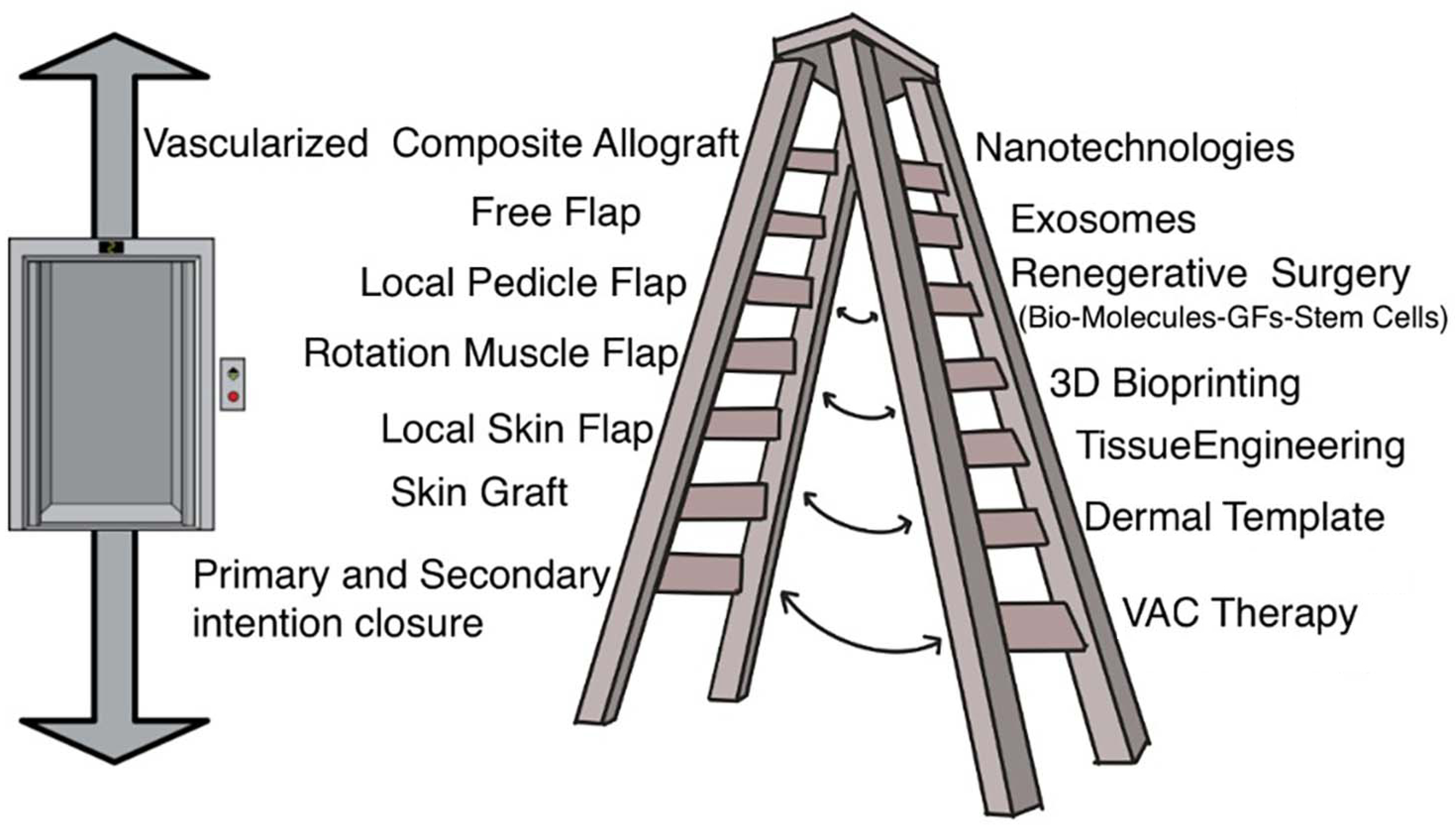
1. Reconstructive Ladder

Many surgeries carry out a demolitive and reconstructive activity but those that are truly “reconstructive” are distinguished based on their ability to provide not only repair of the defect but also on their ability to provide a restoration of the function of the damaged organ. Historically, the term reconstructive is above all the prerogative of that surgical specialty that is able to reconstruct a defect through the use of new parts or a local reorganization of the defect itself [1]. In particular, the “reconstructive ladder” represents the guideline of all surgeons in this discipline [2,3]. A guiding principle establishes that wound repair should begin with a simple method, keeping in mind that, in certain clinical situations, more advanced techniques will be necessary. In this regard, the reconstructive ladder represents the wide spectrum of reconstructive possibilities available to the plastic surgeon [4], from the simplest to the most complex option. The strategy of choosing the best option (grafts, flaps, and microsurgery) for substance loss requires a deep understanding of tissue biology, wound healing physiology, and anatomy. Despite this, the surgeon’s clinical intuition regarding not only the factors that affect wound healing; his own surgical skill is also important. The principal aim of reconstruction consists of “wound coverage”, “infection control”, “anatomical replacement”, “functional maintenance”, and “aesthetic amelioration”. The first two are fundamental for the patient’s problem resolution. The last two are important for the improvement of the patient’s life quality. Moreover, anatomical replacement is related to functional and aesthetic outcomes for the restoration of the patient’s defect. Obviously, based on the site defect, functional loss, and reconstructive difficulty, the surgeon chooses the best reconstructive strategy, based on the reconstructive ladder and on their skill. In recent years, the principles of the reconstructive ladder had become a truly universal dogma, whereby the simplest technique was taken into consideration without considering the final functional result and the transition to the subsequent rungs was used only when there was no other solution [5]. This dogma has obviously been overcome by reconstructive surgeons, by introducing the concept of the “reconstructive elevator” [6], as it allows you to jump from one step to another in a creative way and above all according to the patient’s needs in order to achieve the desired result. This school of thought was born, principally, with the advent of microsurgery [7]. This school also has its disadvantages, in that the principles may be attractive for wound healing, but the lift leads the reconstructive surgeon to use, inevitably, more complex techniques such as free flaps. It is not the technique that solves the problem, but the problem that calls for a particular type of technique depending on the site, function, aesthetic considerations, and morbidity. In this regard, in recent years different opinions have developed regarding the reconstructive possibilities [8,9,10]. This is certainly due to the implementation of surgical techniques becoming more and more sophisticated and therefore the authors have attempted to improve the different “ladders” by adding rungs for an application more suitable for different scenarios. Even if the reconstructive ladder sustained moderate modification over time, the fundamental view of reconstructive procedures graded by difficulty has been maintained and been spread in different theories. The ever-increasing complexity of injuries has led to greater interaction between basic sciences and surgery [11,12,13] to create new and innovative techniques for tissue regeneration. Farid and colleagues [14] performed a critical review of the concept of the reconstructive stair and elevator opening the doors for the use of new technologies. Translational medicine is made up of a multidisciplinary team [15,16] that applies modern regenerative-medicine techniques to complex traumas with the aim of improving the results expected by the patient.

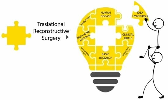
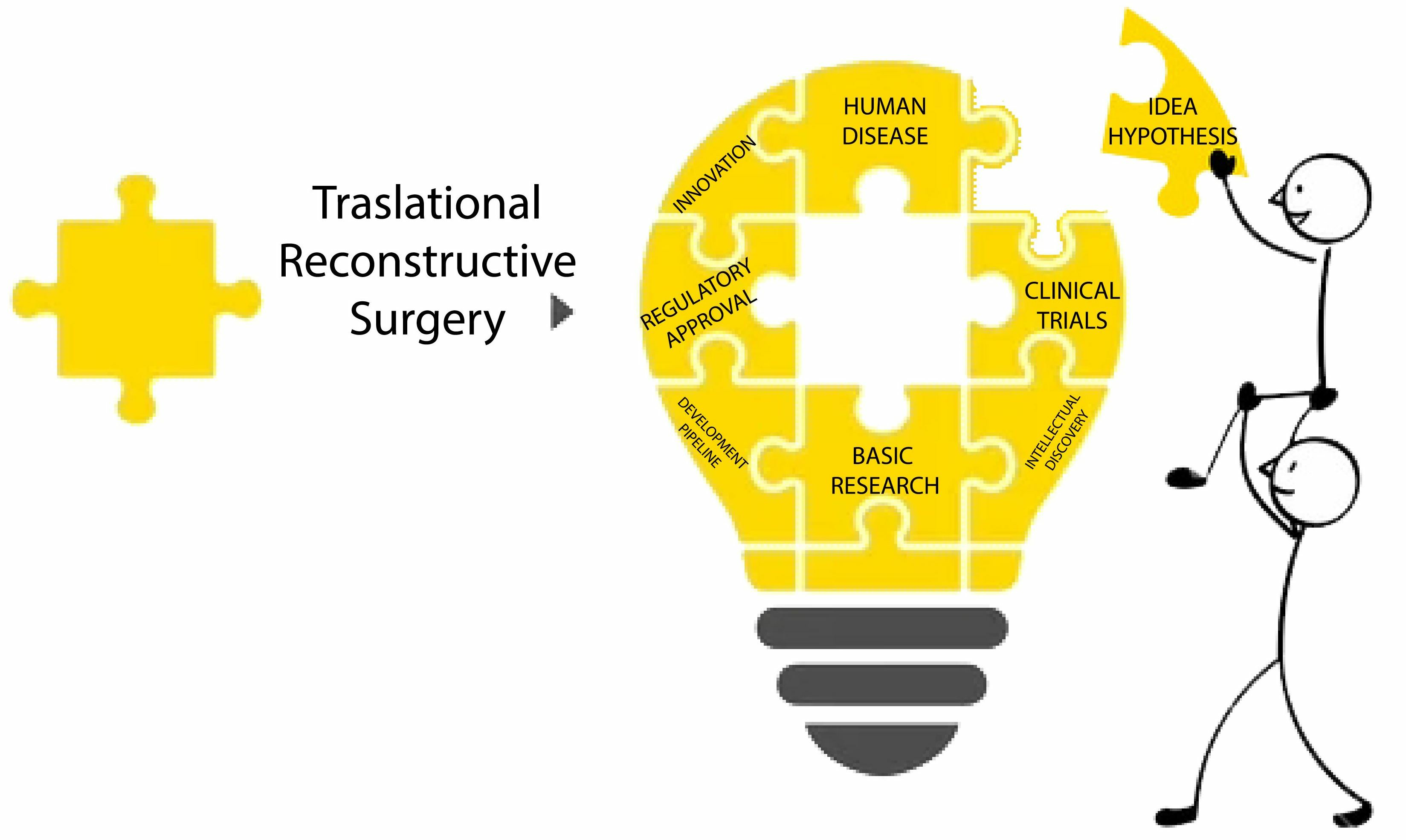
2. Translational Reconstructive Ladder

Translational medicine (TM) can be described as the interdisciplinary use of biomedical investigation for the enhancement of different disease conditions; in particular, the European Society for Translational Medicine (ESTM) has established that translational medicine is supported by three fundamental pillars (bench, bedside and community) with the aim of improving the health of society through the development of new therapies [17]. Despite encouraging preclinical and clinical results, the high degree of heterogeneity in MSC stemness and differentiation potential represents a challenge for reproducibility and therapeutic standardization. MSC heterogeneity can occur at multiple levels, depending on the donor, tissue source, isolated cell subset, and manipulation techniques [18]. A promising route to improving the efficiency of MSCs is to select and isolate specific sub-populations, using cell sorting based on specific markers. Likewise, there is no scaffold that is suitable for every type of tissue to regenerate. Today, scaffold designs are controlled at the nano- or micro-scale and are based on the property of the extracellular matrix (ECM). The ECM provides a variety of physical, chemical, and biological signals that influence cell growth and proliferation. At the same time, the design of smart constructs must take into account the specific characteristics of the tissue and must be “physiologically relevant”, considering the specific anatomical and functional properties of the tissue [19,20]. To improve the “physiological relevance” of engineered constructs, it is important to understand the biological context, such as ECM, vasculature, and cell type, as well as the different chemical, physical, mechanical, and spatial signals; however, timescales should also be considered [21]. In the last decade, the increasingly strong interaction between translational medicine and clinical practice has become the cornerstone of the new biomedicine, ushering in a real revolution in modern science. This integration, which embraces all medical specialties but finds its maximum application in reconstructive surgery, allows the new technologies to offer new opportunities in the treatment of diseases to the point of currently having a prominent role in the community [22,23]. The translation of regenerative approaches into clinical practice is limited by the strict legal regulation of in vitro expanded cells and the risks associated with substantial manipulations. Therefore, micrografts created directly in the operating room (OR) with minimal cell manipulation appear extremely promising, and has demonstrated its efficacy in recent clinical trials [24,25,26].
Smart technologies have been developed that provide extra rungs on the reconstructive ladder to integrate the principles of translational medicine into a global approach, in which the center is not only represented by the patient but also by the community. In this regard, the traditional reconstructive scale, although providing excellent outcomes, does not take into account the economic and healthcare problems linked to the treatment process [27,28]. The “translational reconstructive ladder” paradigm represents a conjunction of regenerative-medicine therapies with traditional reconstructive approaches (Figure 1). This original paradigm can be applied: (i) to large wounds with extensive soft tissue losses, to obtain a better and definitive closure; (ii) pain therapy; (iii) inflammatory and chronic pathologies; (iv) vascular pathologies; (v) post-cancer reconstruction; and many others. Moreover, translational medicine involves the use of cell- and tissue-engineering scaffolds, decellularized extracellular matrix, wearable medical sensors, micro- and nano-medicine, 3D bioprinting, biologically inspired engineering, organ chips, and bioelectronics. The advent of microsurgery has brought important improvements in autologous reconstructive options [29,30,31,32]; however, the morbidity of the donor site and the scarring outcomes still remain prevalent [33,34]. Reconstructive plastic surgery aims to provide vital tissue and, above all, replacing like tissue with like tissue, respecting anatomical zones, to restore a wide range of defects. Plastic surgeons, in this regard, are in a favorable position due to the versatility deriving from the use of all human tissues including the skin, fat, nerves, muscles, bones and cartilage, but above all for being intrinsically involved in the research and development of engineered tissues in the laboratory and in their clinical use [35]. Thanks to the combination of microsurgery, composite vascularized allografts [36,37] and nanotechnologies [38], biomaterials [39], and 3D printing [40], the weapons in the plastic surgeon’s baggage are notable both for the reconstruction of large defects and for the reduction of scarring in donor sites, with consequent reduction of the associated hospital care (Figure 1).

2.1. Autologous Adipose Tissue Grafting and Adipose Stem Cells (ASCs)

The most classic clinical approach, through translational medicine, is the use of tissue-derived cells seeded on scaffolds [41]. The preferred choice is the use of autologous cells, to avoid immunogenicity problems. The cells used can be of adult origin, and therefore completely differentiated [42], or multipotent progenitor cells with low proliferative potential and their differentiation commitment, and finally stem cells capable of proliferating through multiple generations and differentiating into a variety of cell types [43]. Adult stem cells have been isolated from various tissues, such as bone marrow [44,45], epithelial tissue [46], umbilical cord tissue [47,48], and adipose tissue [49,50,51,52]. Plastic surgeons are very familiar with the use of adipose tissue to restore subcutaneous tissue associated with scar contractures or body contour deformities. This procedure is called lipofilling or fat grafting [53]. There are currently numerous procedures to try to obtain the best purified fat through various mechanical systems for adipose tissue concentration [54,55]. The aim is to preserve the components of the lipoaspirate, such as adipocytes, pre-adipocytes, and stroma. Furthermore, within the adipose tissue it was possible to isolate a portion of stem cells that was called the stromal vascular fraction [56]. The stromal vascular fraction (SVF) is represented by diversified cell populations, such as stem cells, preadipocytes, endothelial cells, pericytes, T cells, and M2 macrophages [57], and capable of multipotency differentiation [58,59,60]. Adipose tissue solutions contain many sources of growth factors, cytokines, adipokines, and transcriptional factors, which together produce secretomes. These acellular secretomes have extensive biological activity [61]. In this regard, the therapeutic capacity of MSCs can be increased by gene modification to force the expression of paracrine activity. In a recent review [62], the authors showed how therapies based on engineered MSCs have been used for the treatment of acute graft-versus-host, limb ischemia, and perianal fistulas in Crohn’s disease. In these studies, genetically modified or engineered MSCs were used in which the production of specific paracrine factors, necessary for a particular pathological condition, was forced. By exploiting the paracrine potential of ASCs secretomes, they act directly at the site of inoculation, activating a clinical response with an enormously increased regenerative potential [63]. Among all the products derived from adipose tissue, currently, through modern regenerative-medicine techniques and based on the current legislation, it is possible to use above all these biological peptides, such as exosomes [64,65]. Exosomes are an innovative boundary of intercellular connection managing the cells’ biological action such as immunomodulation and anti-inflammatory properties [66]. Exosomes derived from adipose stem cells are a significant unit liberating by SVF and have many biological activeness [67]. There is evidence of regenerative potential in dermatological disorders such as scars [68], wrinkles [69], pigmentation [70]; in plastic surgery as chronic wounds [71,72], in the orthopedic field such as small joints [73,74,75], and tendons [76,77]. Exosomes have acquired much consideration because they are substantial paracrine mediators providing tissue regeneration. Furthermore, exosomes derived from adipose tissue have the ability to encapsulate various types of bioactive molecules and therefore have great application potential in tissue regeneration [78,79]. Although various findings highlight the significant role of adipose tissue-derived exosomes for tissue regeneration in the field of reconstructive plastic surgery, adequate application in clinical practice is currently lacking. In this regard, the link provided by translational medicine, and in particular by the translational scale, can provide an increasingly important role for these mediators to maximize the therapeutic effect in a large variety of pathologies.

2.2. ECM Scaffold and Dermal Regeneration Template (DRT)

A suitable scaffold is essential to any tissue-engineering strategy. The scaffold supplies a structure for the cell growth, permitting cells to attach, proliferate, and differentiate, at the same time as performing cellular running into a workable 3D network [80]. Moreover, the preferable scaffold should be biomimetic, biodegradable, biocompatible, and non-immunogenic [81] but also should have appropriate mechanical strength and optimal micropores [82]. Finally, the scaffold should be suitable for clinical grade sterilization and industrial production. A cell-scaffold construct can be attempted in vitro in a bioreactor or in vivo by implanting scaffold with autologous cells into the body. Research on biomaterials aims to design “functionalized” or “smart” scaffolds that, on the one hand, incorporate cells on the surface in order to create a new tissue and on the other hand release biomolecules, growth factors, or antibiotics over time capable of prolonging their activities [83]. The dermal regeneration template (DRT) is a three-dimensional bioactive scaffold that actually constitutes the regenerative-medicine applications in the reconstructive fields [84,85]. These DRT materials are capable of supporting tissue regeneration and remodeling with the development of acceptable scars and, above all, they avoid scars in the donor areas, such as grafts. However, the main obstacles to the application of dermal substitutes include the slow vascularization of the substitute and the risk of bacterial infection. Also in this case, various bioactive factors, such as exosomes, can play an important role in the angiogenesis process, just as they themselves can release antibiotics at the site of use with maintenance of the right bacterial biofilm. Nanomaterials have great potential for tissue engineering. The effective delivery of bioactive factors (including growth factors, peptides, and nucleic acids) by nanomaterials is of growing research interest [86]. Growth factors such as vascular endothelial growth factor, platelet-derived growth factor, and angiopontin are able to promote vascularization by promoting the formation of new blood vessels. However, these factors, in addition to being expensive, are very unstable and therefore must be preloaded to ensure an effective, continuous, and gradual release. Currently, sponge-like or film-like scaffolds composed of nanoparticles and nanospheres [87] have been developed, which are loaded with growth factors and therefore these are released into the wound site to promote neoangiogenesis. In view of the difficulty of using commercial growth factors, recent studies envisage a “personalized” application using the patient’s platelet-rich plasma in combination with different biomaterials in order to improve their properties to stimulate wound healing and regeneration of tissues [88]. Interestingly, the combination of polymers and platelet-rich plasma provides controlled spatiotemporal local release. Similarly, scaffolds composed of bioactive materials such as hydrotalcite [89] have been designed that are capable of loading antibiotics that can be released gradually and over time in the wound site, ensuring “sterility” against colonization [90,91]. The combination of biomaterials suitable for wound management (such as dermal substitutes) with fibroblasts, or dermal stem cells is the result of advances in tissue bioengineering. Historically, dermal substitutes can be used in combination with skin grafts or with skin micrografts. In this regard, the Meek technique offers an alternative method of covering large areas in the absence of adequate donor sites [92,93]. The aim of this technology is to mechanically disintegrate epidermal–dermal tissue, gathering autologous micrografts enriched in progenitor cells, growth factor, and extracellular matrix [94,95]. When combined with collagen sponges, micrografts can form a viable and proliferative bio-complex, enhancing their regenerative potential [96].

2.3. D Bioprinting Applications

The replacement and embellishment of patients’ tissues has largely been a dilemma that reconstructive surgeons have scuffled, but traditional surgical therapies frequently have restricted abilities. Conventional biomaterials, not infrequently, may outcome in postoperative complications such as contraction, infection, or rejection. The main complication of biomaterials, in current use, includes infection, which is emerging as a serious threat as bacteria often adhere to the surface of biomaterials (especially porous ones), are difficult to remove, and show high resistance to bactericides [97]. On the other hand, autologous tissue transplantation may develop the question of donor-site impairment. 3D bioprinting technology permits the planning of customized implants, introducing an elevated level of accuracy and shape. Moreover, implantations arranged with printed biomaterials and individual patient’s cells have greater biocompatibility and minor immunogenicity than traditional biomaterials. The current definition of 3D Bioprinting is based on the three-dimensional printing of cells or biomaterials on a specific substrate according to the requirements of the cellular morphology and microenvironment, tissue functionality, forming biologically functional three-dimensional constructs [98]. Three steps are involved in the 3D bioprinting work: pre-processing, processing and post-processing [99]. The use of 3D bioprinting generally incorporates many areas of tissue engineering, such as skin, bone, cartilage, and, in this regard, many areas of reconstructive surgery [100,101]. In this aspect, it must be taken into consideration that tissues contain different cells and different extracellular matrix, and therefore a universal printing material cannot be used for these tissues; therefore, the right material must be chosen for different tissues, on the features of biocompatibility and mechanical peculiarities [102]. Today, the principal bioprinting materials are: (i) inorganic materials, such as metals (titanium), bioceramics, clay, hydroxyapatite, graphene, carbon nanotubes [103,104] that have high strength, low elasticity, osteoconductivity, and corrosion resistance; (ii) synthetic polymers, such as polycaprolactone (PCL), polylactide (PLA) and polyurethane (PU) [105] that have variable degradation rate, good stiffness, high elasticity, and malleability; (iii) natural biopolymers, such as alginate, gelatin, collagen, fibrin, decellularized ECM, and hyaluronic acid [106,107] that have low cost, easy extraction, biodegradability, high biocompatibility, antibacterial properties, non-immune response, and provide a cell-specific microenvironment. In addition, the ingredients of the bioink can be “added” by including extracellular vesicles, exosomes, and growth factors that make the bioprinting functionalized material [108,109]. In plastic reconstructive surgery, these 3D bioprinting materials can be used in patients with severe burns, diabetic ulcers, tumors, or traumatic severe skin tissue defects, which currently can only be treated with alloplastic grafts or pedunculated or free autologous flaps. To avoid scarring in the donor site or discomfort for the patient, this technology can be a new tool available to the plastic surgeon. Some authors have already used this technology for the regeneration of skin defects, and have combined 3D bioprinting materials with mesenchymal stem cells [110,111,112]. This research has highlighted how this technology is able to heal wounds in vivo by generating collagen and improving cell proliferation. Since the microenvironment in which 3D bioprinted MSCs were used was nutritionally deficient, various bioactive substances were applied to improve the capabilities of MSCs, such as angiogenesis [113]. Such regenerative “smart dressing” provided an appropriate microenvironment to enhance MSC proliferation and differentiation but also accelerated anti-inflammatory activities to promote wound healing by increasing the expression of wound healing factors. 3D bioprinting technology, with or without bioactive substances, permits a personalized and tailored patient-needs and prevents the surgical complications and adverse reactions of traditional surgery [114]. When designing a scaffold or biomaterial, physiologically relevant design decisions must be made, depending on the target site and the function needed. The 3D bioprinting technology considers various characteristics, such as biological, physical, mechanical, and structural, for optimal scaffold design and manufacturing. The customization of scaffold characteristics is conceptualized based on the target tissue and required purpose. Biological issues are related to biocompatibility, biodegradability, and non-toxic properties; furthermore, structural and physical issues are related to porosity, mechanical behavior, pore size, and surface topography. Finally, chemical issues are related to capacity to include growth factors, proteins, drugs, and antibiotics in the biomaterial [115]. In addition, biomaterials, as well as being customizable and functionalized, can be sensitive to the stimuli of the microenvironment (such as temperature, pH, and infections) in which they are used. These biomaterials exploit nanotechnological properties as vehicles for the controlled release of drugs or growth factors that respond spatiotemporally to specific endogenous stimuli, promoting controlled tissue regeneration or the controlled management of infections [116]. The 3D bioprinting field is a rapidly expanding area of research, albeit with questions related to the correct mixture of cells, an adequate printable scaffold, and the ideal microenvironment to mimic native tissue. 3D bioprinting is still an embryonic technology, evidenced by the fact that most current studies are only in vitro proof of concept [117].

2.4. Exosomes

Exosomes (EVs) are extracellular vesicles involved in intercellular communication and in the transfer of cargo incorporating proteins, lipids, and nucleic acid [118,119]. Exosomes have been discovered in many bodily fluids [120], emphasizing their role in intercellular communication in both physiological and pathological proceeding. Currently, a correlation between exosomes and chronic pain has been demonstrated [121]. In reconstructive and plastic surgery, in particular in hand surgery, there are many situations, such as neuropathic pain from peripheral neuropathy, from nerve injury, from complex pain regional syndrome (CPRS), or chronic inflammatory pain from osteoarthritis in which there is currently no consolidated therapy. In this context, exosomes appear to be a possible therapeutic strategy by transferring substances that improve pain and have immunoprotective and anti-inflammatory potential [122,123]. Neuropathic pain is a type of chronic pain that occurs following an injury or disease in the peripheral nervous system [124], and appears to be related to neuroinflammation [125,126]. Various clinical studies have shown that exosomes can improve neuropathic pain by reducing proinflammatory cytokines, promoting vascular regeneration, promoting neuronal proliferation and function by axonal regrowth and Schwann cell activation [127,128], which contribute to providing a favorable microenvironment for peripheral nerve regeneration. Another pathology responsible for chronic inflammatory pain is osteoarthritis. In this pathology, the pain is classically considered to be nociceptive, resulting from the abnormal load of a damaged joint. Changes in joint biomechanics interact on nociceptive nerve endings by opening ion channels and generate this specific type of pain [129]. Exosomes from different sources, such as synovial fibroblasts, chondrocytes but also adipose tissue, plays a role in reducing excessive chondrocyte death by inhibiting apoptosis process [130]. Moreover, exosomes inhibit extracellular matrix degradation, resulting in alleviation of synovial inflammation. Degeneration of cartilage tissue during osteoarthritis is caused by the presence of chronic inflammation. ASCs have the potential to attenuate degenerative and inflammatory processes in OA, through EVs isolated from human ASCs exerting chondroprotective functions through multiple mechanisms, such as reducing the production of inflammatory mediators, decreasing the release of metalloproteinase activity, and improving the production of the anti-inflammatory cytokine [131,132].

3. Conclusions and Perspectives

Translational medicine has brought an evolution to classical research in the field of cell biology. Here, the models are based on conditions that, although on the one hand managing to explain biological events and processes, on the other hand failing to explain and translate such biological knowledge into clinical practice. Advances in biology and cellular engineering have led to the development of applied translational medicine, which allows us to examine biological processes and their interactions in a more biomimetic context and therefore allows us to understand how they function in a real microenvironment. Many medical disciplines are similar to this new translational model, and reconstructive plastic surgery is certainly one of them. Modern translational research has new technological approaches available, which can certainly be applied to the “reconstructive ladder”, making it translational, based on more sophisticated tissue-engineering models. Adipose tissue today has a wide application in the field of soft tissue reconstruction, but has limitations that can be resolved with the use of biomaterials that provide additional reinforcement or support, providing critical signals for regeneration. Further success in the use of these biomaterials will require a deeper understanding of the components of the ECM in regeneration and the role of extracellular vesicles in immunomodulatory processes. Defining the physical, mechanical, and biomechanical properties of biomaterials and their integration with the host, as well as defining degradation properties, is also an area under development. There is a continuing need for further interdisciplinary research to understand biological processes in order to create safe and durable translational technologies in the clinic. The “translational reconstructive ladder” is not just an algorithm that allows the plastic surgeon to correctly perform surgery. Today, reconstruction capabilities have been increased a thousand times with incredible resources and cutting-edge technological devices. All of this, if inserted within this “translational reconstructive scale”, allows us to act in a more precise, rapid, and also safer way, to better satisfy reconstructive and aesthetic needs. No one, alone, will be able to include within their own cultural experience all the tools available in the “translational reconstructive scale”, but the best reconstructive surgeon will somehow guarantee the best alternative.
The other side of the coin is understanding how the success of translational research from the laboratory to the bedside depends on the creation of a multidisciplinary and interdisciplinary scientific team capable of collaborating and communicating bidirectionally, so that all actors involved (academia, non-profit foundations, pharmaceutical and biotechnology companies, hospitals) synchronize efforts toward a common good. There is currently consensus that there is a rift involving the translatability of basic science into bedside applications. This gap, defined by many authors as the “valley of death” [133], has caused a slowdown in the development of translational medicine fundamentally linked to the high costs of production and the healthcare system, reproducibility, clinical relevance, and regulatory. Considering that reconstructive surgery is a procedure that restores the “form and above all function” of a tissue after damage or disease, plastic surgeons will certainly be the major users of the translational techniques, through an integrated approach between technologies coming from engineering, biomaterials science, cell biology, and reconstructive microsurgery.

Author Contributions

Conceptualization, F.D.F.; validation, M.R. and P.C.P.; resources, N.Z.; data curation, N.Z.; writing—original draft preparation, F.D.F.; writing—review and editing, F.D.F. and M.R.; visualization, P.C.P.; supervision, M.R. All authors have read and agreed to the published version of the manuscript.

Funding

This research received no external funding.

Institutional Review Board Statement

Not applicable.

Informed Consent Statement

Not applicable.

Data Availability Statement

No new data were created or analyzed in this study. Data sharing is not applicable to this article.

Conflicts of Interest

The authors declare no conflict of interest.

References

  1. Tamai, S. History of Microsurgery-from the beginning until the end of the 1970s. Microsurgery 1993, 14, 6–13. [Google Scholar] [CrossRef]
  2. Simman, R. Wound closure and the reconstructive ladder in plastic surgery. J. Am. Coll. Certif. Wound Spec. 2009, 1, 6–11. [Google Scholar] [CrossRef]
  3. Scott Levin, L. The reconstructive ladder. An orthoplastic approach. Orthop. Clin. 1993, 24, 393–409. [Google Scholar]
  4. Mathes, S.J.; Nahai, F. Clinical Applications for Muscle and Musculocutaneous Flaps, 2nd ed.; C.V. Mosby Company: St. Louis, MO, USA, 2010. [Google Scholar]
  5. Mardini, S.; Wei, F.C.; Salgado, C.J.; Chen, H.C. Reconstruction of the reconstructive ladder. Plast. Reconstr. Surg. 2005, 115, 2174. [Google Scholar] [CrossRef]
  6. Gottlieb, L.J.; Krieger, L.M. From the reconstructive ladder to the reconstructive elevator. Plast. Reconstr. Surg. 1994, 93, 1503–1504. [Google Scholar] [CrossRef]
  7. Tamai, S. History of microsurgery. Plast. Reconstr. Surg. 2009, 124, e282–e294. [Google Scholar] [CrossRef]
  8. Erba, P.; Ogawa, R.; Vyas, R.; Orgill, D.P. The reconstructive matrix: A new paradigm in reconstructive plastic surgery. Plast. Reconstr. Surg. 2010, 126, 492–498. [Google Scholar] [CrossRef]
  9. Knobloch, K.; Vogt, P.M. The reconstructive clockwork of the twenty-first century: An extension of the concept of the reconstructive ladder and reconstructive elevator. Plast. Reconstr. Surg. 2010, 126, 220e–222e. [Google Scholar] [CrossRef]
  10. Sandhir, R.K. Learn to climb the simple reconstructive ladder properly for optimum results. Indian J. Plast. Surg. 2018, 51, 331–332. [Google Scholar] [CrossRef]
  11. Carrel, T. The relationship between surgeon and basic scientist. Transpl. Immunol. 2002, 9, 331–337. [Google Scholar] [CrossRef]
  12. Tetteh, E.S.; Bajaj, S.; Ghodadra, N.S. Basic science and surgical treatment options for articular cartilage injuries of the knee. J. Orthop. Sports Phys. Ther. 2012, 42, 243–253. [Google Scholar] [CrossRef]
  13. Keswani, S.G.; Moles, C.M.; Morowitz, M.; Zeh, H.; Kuo, J.S.; Levine, M.H.; Cheng, L.S.; Hackam, D.J.; Ahuja, N.; Goldstein, A.M. Basic Science Committee of the Society of University Surgeons. The future of basic science in academic surgery: Identifying barriers to success for surgeon-scientists. Ann. Surg. 2017, 265, 1053–1059. [Google Scholar] [CrossRef] [PubMed]
  14. Farid, M.; Friebel, T.; Nikkhah, D. Decision-Making in Flap Surgery: Reconstructive Ladder versus Elevator. In Core Techniques in Flap Reconstructive Microsurgery; Nikkhah, D., Rawlins, J., Pafitanis, G., Eds.; Springer: Cham, Switzerland, 2023. [Google Scholar]
  15. Shahzad, A.; Gilden, D.; Cohrs, R.J. Translational medicine and varicella zoster visrus: Need for disease modeling. New Horiz. Transl. Med. 2015, 2, 89–91. [Google Scholar] [PubMed]
  16. Gohar, F.; Gohar, A.; Hulskamp, G.; Debus, O. The translational medicine professional: A bridge between bench and bedside? Front. Med. 2018, 5, 294. [Google Scholar] [CrossRef] [PubMed]
  17. Cohrs, R.J.; Ghahramani, P.; Bidaut, L.; Higgins, P.J.; Shahzad, A. Translational medicine definition by the European Society for Translational Medicine. New Horiz. Transl. Med. 2015, 2, 86–88. [Google Scholar] [CrossRef]
  18. Goh, D.; Yang, Y.; Lee, E.H.; Hui, J.H.P.; Yang, Z. Managing the heterogeneity of mesenchymal stem cells for cartilage regenerative therapy: A review. Bioengineering 2023, 10, 355. [Google Scholar] [CrossRef] [PubMed]
  19. Wang, W.; Shi, Y.; Lin, G.; Tang, B.; Li, X.; Zhang, J.; Ding, X.; Zhou, G. Advances in mechanical properties of hydrogels for cartilage tissue defect repair. Macromol. Biosci. 2023, 23, e2200539. [Google Scholar] [CrossRef]
  20. Klabukov, I.; Tenchurin, T.; Shepelev, A.; Baranovskii, D.; Mamagulashvili, V.; Dyuzheva, T.; Krasilnikova, O.; Balyasin, M.; Lyundup, A.; Krasheninnikov, M.; et al. Biomechanical behaviors and degradation properties of multilayered polymer scaffolds: The phase space method for bile duct design and bioengineering. Biomedicine 2023, 11, 745. [Google Scholar] [CrossRef]
  21. Abbott, R.D.; Kaplan, D.L. Strategies for improving the physiological relevance of human engineered tissues. Trends Biotechnol. 2015, 33, 401–407. [Google Scholar] [CrossRef]
  22. Waldman, S.A.; Terzic, A. Translational medicine in the era of health care reform. Clin. Transl. Sci. 2009, 2, 96–97. [Google Scholar] [CrossRef]
  23. Hamburg, M.A.; Collins, F.S. The path of personalized medicine. N. Engl. J. Med. 2010, 363, 301–304. [Google Scholar] [CrossRef] [PubMed]
  24. Krasilnikova, O.A.; Baranovskii, D.S.; Yakimova, A.O.; Arguchinskaya, N.; Kisel, A.; Sosin, D.; Sulina, Y.; Ivanov, S.A.; Shegay, P.V.; Kaprin, A.D.; et al. Intraoperative creation of tissue-engineered grafts with minimally manipulated cells: New concept of bone tissue engineering in situ. Bioengineering 2022, 9, 704. [Google Scholar] [CrossRef] [PubMed]
  25. Ceccarelli, G.; Gentile, P.; Marcarelli, M.; Balli, M.; Ronzoni, F.L.; Benedetti, L.; Cusella De Angelis, M.G. In vitro and in vivo studies of alar-nasal cartilage using autologous micro-grafts: The use of the rigenera protocol on the treatment of an osteochondral lesion of the nose. Pharmaceuticals 2017, 10, 53. [Google Scholar] [CrossRef]
  26. Palumbo Piccionello, A.; Riccio, V.; Senesi, L.; Volta, A.; Pennasilico, L.; Botto, R.; Rossi, G.; Tambella, A.M.; Galosi, L.; Marini, C.; et al. Adipose micro-grafts enhance tendinopathy healing in ovine model: An in vivo experimental perspective study. Stem Cells Transl. Med. 2021, 10, 1544–1560. [Google Scholar] [CrossRef] [PubMed]
  27. Sandberg, L.J. The plastic surgery compass: Navigating the reconstructive ladder in the personalized health care era. Plast. Reconstr. Surg. Glob. Open 2016, 4, e1035. [Google Scholar] [CrossRef]
  28. Borrelli, M.R. What is the role of plastic surgery in global health? A review. World J. Plast. Surg. 2018, 7, 275–282. [Google Scholar] [CrossRef]
  29. Giesen, T.; Politikou, O.; Tami, I.; Calcagni, M. Retrograde free venous flaps for extremity reconstruction: A roadmap. Medicina 2022, 58, 1065. [Google Scholar] [CrossRef]
  30. Marchesini, A.; Senesi, L.; De Francesco, F.; Pangrazi, P.P.; Campodonico, A.; Politano, R.; Riccio, M. Efficacy of the arteriovenous loop for free flap reconstruction in patients complex limb trauma: Case series and literature review. Medicina 2020, 56, 632. [Google Scholar] [CrossRef]
  31. Anolik, R.A.; Sacks, J.M. Advances and innovations in Breast Microsurgery. Mo. Med. 2021, 118, 153–155. [Google Scholar]
  32. Gravina, P.; De Francesco, F.; Pangrazi, P.P.; Marchesini, A.; Neuendorf, A.D.; Campodonico, A.; Gigante, A.; Riccio, M. A case report of upper limb loss of substance: Use of functional gracilis free flap, brachioradialis transposition and bioglass for bone regeneration. Trauma Case Rep. 2022, 38, 100609. [Google Scholar] [CrossRef]
  33. Augustin, A.; Pulzl, P.; Morandi, E.M.; Winkelmann, S.; Schoberleitner, I.; Brunner, C.; Ritter, M.; Bauer, T.; Wachter, T.; Wolfram, D. Donor-site morbidity and quality of life after autologous breast reconstruction with PAP versus TMG flap. Curr. Oncol. 2022, 29, 5682–5697. [Google Scholar] [CrossRef] [PubMed]
  34. Rotatori, R.M.; Starr, B.; Peake, M.; Fowler, L.; James, L.; Nelson, J.; Dale, E.L. Prevalence and risk factor for hypertrophic scarring of split thickness autograft donor sites in a pediatric burn population. Burns 2019, 45, 1066–1074. [Google Scholar] [CrossRef] [PubMed]
  35. Al-Himdani, S.; Jessop, Z.M.; Al-Sabah, A.; Combellack, E.; Ibrahim, A.; Doak, S.H.; Hart, A.M.; Archer, C.W.; Thornton, C.A.; Whitaker, I.S. Tissue-Engineered solutions in plastic and reconstructive surgery: Principles and practice. Front. Surg. 2017, 4, 4. [Google Scholar] [CrossRef]
  36. Shores, J.T.; Brandacher, G.; Lee, W.P.A. Hand and upper extremity transplantation: An update of outcomes in the worldwide experience. Plast. Reconstr. Surg. 2015, 135, 351e–360e. [Google Scholar] [CrossRef]
  37. Matar, A.J.; Crepeau, R.L.; Mundinger, G.S.; Cetrulo Jr, C.L.; Ttorabi, R. Large animal models of vascularized composite allotransplantation: A review of immune strategies to improve allograft outcomes. Front. Immunol. 2020, 12, 664577. [Google Scholar] [CrossRef]
  38. Tan, A.; Chawla, r.; Natasha, G.; Mahdibeiraghdar, S.; Jeyaraj, R.; Rajadas, J.; Hamblin, M.R.; Seifalian, A.M. Nanotechnology and regenerative therapeutics in plastic surgery: The next frontier. J. Plast. Reconstr. Aesthet. Surg. 2016, 69, 1–13. [Google Scholar] [CrossRef]
  39. Peng, W.; Peng, Z.; Tang, P.; Sun, H.; Lei, H.; Li, Z.; Hui, D.; Du, C.; Zhou, C.; Wang, Y. Review of plastic surgery biomaterials and current progress in their 3D manufacturing technology. Materials 2020, 13, 4108. [Google Scholar] [CrossRef] [PubMed]
  40. Lynn, A.Q.; Pflibsen, L.R.; Smith, A.A.; Rebecca, A.M.; Teven, C.M. Three-dimensional printing in plastic surgery: Current applications, future directions, and ethical implications. Plast. Reconstr. Surg. Glob. Open 2021, 9, e3465. [Google Scholar] [CrossRef] [PubMed]
  41. Webber, M.J.; Khan, O.F.; Sydlik, S.A.; Tang, B.C.; Langer, R. A perspective on the clinical translation of scaffolds for tissue engineering. Ann. Biomed. Eng. 2015, 43, 641–656. [Google Scholar] [CrossRef]
  42. Graf, T.; Stadtfeld, M. heterogeneity of embryonic and adult stem cells. Cell Stem Cell 2008, 3, 480–483. [Google Scholar] [CrossRef]
  43. Baksh, D.; Song, L.; Tuan, R.S. Adult mesenchymal stem cells: Characterization, differentiation, and application in cell and gene therapy. J. Cell Mol. Med. 2004, 8, 301–316. [Google Scholar] [CrossRef]
  44. Bianco, P.; Robey, P.G. Marrow stromal stem cells. J. Clin. Investig. 2000, 105, 1663–1668. [Google Scholar] [CrossRef] [PubMed]
  45. Mendez-Ferrer, S.; Michurina, T.V.; Ferraro, F.; Mazloom, A.R.; Macarthur, B.D.; Lira, S.A.; Scadden, D.T.; Ma’ayan, A.; Enikolopov, G.N.; Frenette, P.S. Mesenchymal and haematopoietic stem cells form a unique bone marrow niche. Nature 2010, 466, 829–834. [Google Scholar] [CrossRef]
  46. Ferraces-Riegas, P.; Galbraith, A.C.; Doupè, D.P. Epithelial Stem Cells: Making, shaping and creaking the niche. Adv. Exp. Med. Biol. 2022, 1387, 1–12. [Google Scholar] [PubMed]
  47. Shaikh, M.S.; Shahzad, Z.; Tash, E.A.; Janjua, O.S.; Khan, M.I.; Zafar, M.S. Human Umbelical cord mesenchymal stem cells: Current literature and role in periodontal regeneration. Cells 2022, 11, 1168. [Google Scholar] [CrossRef] [PubMed]
  48. Lim, S.; Lyu, H.Z.; Lee, J.R.; Han, S.H.; Lee, J.H.; Kim, B.S. Umbelical cord mesenchymal stem cell-derived nanovesicles potentiate the bone-formation efficacy of bone morphogenetic protein 2. Int. J. Mol. Sci. 2020, 21, 6425. [Google Scholar] [CrossRef] [PubMed]
  49. Wankhade, U.D.; Shen, M.; Kolhe, R.; Fulzele, S. Advances in adipose-derived stem cells isolation, characterization, and application in regenerative tissue engineering. Stem Cells Int. 2016, 2016, 3206807. [Google Scholar] [CrossRef]
  50. Ferraro, G.A.; De Francesco, F.; Nicoletti, G.; Paino, F.; Desiderio, V.; Tirino, V.; D’Andrea, F. Human adipose Cd34+ CD90+ stem cells and collagen scaffold constructs grafted in vivo fabricate loose connective and adipose tissues. J. Cell Biochem. 2013, 114, 1039–1049. [Google Scholar] [CrossRef]
  51. De Francesco, F.; Ricci, G.; D’Andrea, F.; Nicoletti, G.F.; Ferraro, G.A. Human adipose stem cells: From bench to bedside. Tissue Eng. Part B Rev. 2015, 21, 572–584. [Google Scholar] [CrossRef]
  52. Ferroni, L.; De Francesco, F.; Pinton, P.; Gardin, C.; Zavan, B. Methods to isolate adipose tissue-derived stem cells. Methods Cell Biol. 2022, 171, 215–228. [Google Scholar]
  53. Bellini, E.; Grieco, M.P.; Raposio, E. The science behind autologous fat grafting. Ann. Med. Surg. 2017, 24, 65–73. [Google Scholar] [CrossRef] [PubMed]
  54. Gir, P.; Brown, S.A.; Oni, G.; Kashefi, N.; Mojallal, A.; Rohrich, R.J. Fat grafting: Evidence-based review on autologous fat harvesting, processing, reinjection, and storage. Plast. Reconstr. Surg. 2012, 130, 249–258. [Google Scholar] [CrossRef] [PubMed]
  55. Xue, E.Y.; Narvaez, L.; Chu, C.K.; Hanson, S.E. Fat processing techniques. Semin. Plast. Surg. 2020, 34, 11–16. [Google Scholar] [CrossRef] [PubMed]
  56. Nguyen, A.; Guo, J.; Banyard, D.A.; Fadavi, D.; Toranto, J.D.; Wirth, G.A.; Paydar, K.Z.; Evans, G.R.D.; Widgerow, A.D. Stromal vascular fraction: A regenerative reality? Part 1: Current concepts and review of the literature. J. Plast. Reconstr. Aesthet. Surg. 2016, 69, 170–179. [Google Scholar] [CrossRef]
  57. Han, S.; Sun, H.M.; Hwang, K.C.; Kim, S.W. Adipose-derived stromal vascular fraction cells: Update on clinical utility and efficacy. Crit. Rev. Eukaryot. Gene Expr. 2015, 25, 145–152. [Google Scholar] [CrossRef]
  58. Shridhar, A.; Amsden, B.G.; Gillies, E.R.; Flynn, L.E. Investigating the effects of tissue-specific extracellular matrix on the adipogenic and osteogenic differentiation of human adipose-derived stromal cells within composite hydrogel scaffolds. Front. Bioeng. Biotechnol. 2019, 7, 402. [Google Scholar] [CrossRef]
  59. Nicoletti, G.F.; De Francesco, F.; D’Andrea, F.; Ferraro, G.A. Methods and procedures in adipose stem cells: State of the art and perspective for translation medicine. J. Cell Physiol. 2015, 230, 489–495. [Google Scholar] [CrossRef]
  60. Romano, I.R.; D’Angeli, F.; Vicario, N.; Russo, C.; Genovese, C.; Lo Furno, D.; Mannino, G.; Tamburino, S.; Parenti, R.; Giuffrida, R. Adipose-derived mesenchymal stromal cells: A tool for bone and cartilage repair. Biomedicines 2023, 11, 1781. [Google Scholar] [CrossRef]
  61. Silva, K.R.; Baptista, L.S. Adipose-derived stromal/stem cells from different adipose depots in obesity development. World J. Stem Cells 2019, 11, 147–166. [Google Scholar] [CrossRef]
  62. Pawitan, J.A.; Bui, T.A.; Mubarok, W.; Antarianto, R.D.; Nurhayati, R.W.; Dilogo, I.H.; Oceandy, D. Enhancement of the therapeutic capacity of mesenchymal stem cells by genetic modification: A systematic review. Front. Cell Dev. Biol. 2020, 8, 587776. [Google Scholar] [CrossRef]
  63. Melief, S.M.; Zwaginga, J.J.; Fibbe, W.E.; Roelofs, H. Adipose tissue-derived multipotent stromal cells have a higher immunomodulatory capacity than their bone marrow-derived counterparts. Stem Cells Transl. Med. 2013, 2, 455–463. [Google Scholar] [CrossRef] [PubMed]
  64. Long, C.; Wang, J.; Gan, W.; Qin, X.; Yang, R.; Chen, X. Therapeutic potential of exosomes from adipose-derived stem cells in chronic wound healing. Front. Surg. 2022, 9, 1030288. [Google Scholar] [CrossRef] [PubMed]
  65. Quesenberry, P.; Goldberg, L.R. A new stem cell biology: Transplantation and baseline, cell cycles and exosomes. Adv. Exp. Med. Biol. 2018, 1056, 3–9. [Google Scholar] [PubMed]
  66. Munes, G.; Sipos, F. Mesenchymal stem cell-derived secretome: A potential therapeutic option for autoiummune and immune-medianted inflammatory diseases. Cells 2022, 11, 2300. [Google Scholar]
  67. Xiong, M.; Zhang, Q.; Hu, W.; Zhao, C.; Lv, W.; Yi, Y.; Wu, Y.; Wu, M. Exosomes from adipose-derived stem cells: The emerging roles and applications in tissue regeneration of plastic and cosmetic surgery. Front. Cell Dev. Biol. 2020, 8, 574223. [Google Scholar] [CrossRef]
  68. Chen, H.; Hou, K.; Wu, Y.; Liu, Z. Use of adipose stem cells against hypertrophic scarring or keloid. Front. Cell Dev. Biol. 2022, 9, 823694. [Google Scholar] [CrossRef]
  69. Surowiecka, A.; Struzyma, J. Adipose-derived stem cells for facial rejuvenation. J. Pers. Med. 2022, 12, 117. [Google Scholar] [CrossRef]
  70. Chen, S.; He, Z.; Xu, J. Application of adipose-derived stem cells in photoaging: Basic science and literature review. Stem Cell Res. Ther. 2020, 11, 491. [Google Scholar] [CrossRef]
  71. Brembilla, N.C.; Vuagnat, H.; Boehncke, W.H.; Krause, K.H.; Preynat-Seauve, O. Adipose-derived stromal cells for chronic wounds: Scientific evidence and roadmap toward clinical practice. Stem Cells Transl. Med. 2023, 12, 17–25. [Google Scholar] [CrossRef]
  72. Hao, Z.; Qi, W.; Sun, J.; Zhou, M.; Guo, N. Review: Research progress of adipose-derived stem cells in the treatment of chronic wounds. Front. Chem. 2023, 11, 1094693. [Google Scholar] [CrossRef]
  73. Karagergou, E.; Ligomenou, T.; Chalidis, B.; Kitridis, D.; Papadopoulou, S.; Givissis, P. Evaluation of adipose cell-based therapies for the treatment of thumb carpometacarpal joinjtg osteoarthritis. Biomolecules 2022, 12, 473. [Google Scholar] [CrossRef] [PubMed]
  74. Yan, B.; Lv, S.; Tong, P.; Yan, L.; Chen, Z.; Zhou, L.; Yuan, Q.; Guo, L.; Shan, L. Intra-articular injection of adipose-derived stem cells ameliorates pain and cartilage anabolism/catabolism in osteoarthritis: Preclinical and clinical evidences. Front. Pharmacol. 2022, 13, 854025. [Google Scholar] [CrossRef] [PubMed]
  75. De Francesco, F.; Gravina, P.; Busato, A.; Farinelli, L.; Soranzo, C.; Vidal, L.; Zingaretti, N.; Zavan, B.; Sbarbati, A.; Riccio, M.; et al. Stem Cells in autologous microfragmented adipose tissue: Current perspectives in osteoarthritis disease. Int. J. Mol. Sci. 2021, 22, 10197. [Google Scholar] [CrossRef] [PubMed]
  76. Senesi, L.; De Francesco, F.; Marchesini, A.; Pangrazi, P.P.; Bertolini, M.; Riccio, V.; Riccio, M. Efficacy of adipose-derived mesenchymal stem cells and stromal vascular fraction alone and combined to biomaterials in tendinopathy or tendon injury: Systematic review of current concepts. Medicina 2023, 59, 273. [Google Scholar] [CrossRef] [PubMed]
  77. Kokubu, S.; Inaki, R.; Hoshi, K.; Hikita, A. Adipose-derived stem cells improve tendon repair and prevent ectopic ossification in tendinopathy by inhibiting inflammation and inducing neovascularization in the early stage of tendon healing. Regen. Ther. 2020, 14, 103–110. [Google Scholar] [CrossRef]
  78. Zhang, Z.; Yang, X.; Cao, X.; Qin, A.; Zhao, J. Current applications of adipose-derived mesenchymal stem cells in bone repair and regeneration: A review of cell experiments, animal models, and clinical trials. Front. Bioeng. Biotechnol. 2022, 10, 942128. [Google Scholar] [CrossRef]
  79. Storti, G.; Scioli, M.G.; Kim, B.S.; Orlandi, A.; Cervelli, V. Adipose-derived stem cells in bone tissue engineering: Useful tools with new application. Stem Cells Int. 2019, 2019, 3673857. [Google Scholar] [CrossRef]
  80. Chan, B.P.; Leong, K.W. Scaffolding in tissue engineering: General approaches and tissue-specific considerations. Eur. Spine J. 2008, 17, 467–479. [Google Scholar] [CrossRef]
  81. Krishani, M.; Shin, W.Y.; Suhaimi, H.; Sambudi, N.S. Development of scaffolds from bio-based natural materials for tissue regeneration applications: A review. Gels 2023, 9, 100. [Google Scholar] [CrossRef]
  82. Suamte, L.; Tirkey, A.; Babu, P.J. Design of 3D smart scaffolds using natural, synthetic and hybrid derived polymers for skin regenerative applications. Smart Mater. Med. 2023, 4, 243–256. [Google Scholar] [CrossRef]
  83. Holzapfel, B.M.; Reichert, J.C.; Schantz, J.T.; Gbureck, U.; Rackwitz, L.; Noth, U.; Jakob, F.; Rudert, M.; Groll, J.; Hutmacher, D.W. How smart do biomaterials need to be? A translational science and clinical point of view. Adv. Drug Deliv. Rev. 2013, 65, 581–603. [Google Scholar] [CrossRef] [PubMed]
  84. Cottone, G.; Amendola, F.; Strada, C.; Bagnato, M.C.; Brambilla, R.; De Francesco, F.; Vaienti, L. Comparison of efficacy among three dermal substitutes in the management of critical lower-limb wounds: The largest biases-reduced single-center retrospective cohort study in literature. Medicina 2021, 57, 1367. [Google Scholar] [CrossRef] [PubMed]
  85. De Francesco, F.; Busato, A.; Mannucci, S.; Zingaretti, N.; Cottone, G.; Amendola, F.; De Francesco, M.; Merigo, F.; Riccio, V.; Vaienti, L.; et al. Artificial dermal substitutes for tissue regeneration: Comparison of the clinical outcomes and histological findings of two templates. J. Int. Med. Res. 2020, 48, 3000605520945508. [Google Scholar] [CrossRef] [PubMed]
  86. Weng, T.; Wang, J.; Yang, M.; Zhang, W.; Wu, P.; You, C.; Han, C.; Wang, X. Nanomaterials for the delivery of bioactive factors to enhance angiogenesis of dermal substitutes during wound healing. Burns Trauma 2022, 10, tkab049. [Google Scholar] [CrossRef] [PubMed]
  87. Gainza, G.; Villullas, S.; Pedraz, J.L.; Hernandez, R.M.; Igartua, M. Advances in drug delivery systems (DDSs) to release growth factors for wound healing and skin regeneration. Nanomedicine 2015, 11, 1551–1573. [Google Scholar] [CrossRef]
  88. Anitua, E.; Tejero, R.; Alkhraisat, M.H.; Orive, G. Platelet-Rich plasma to improve the bio-functionality of biomaterials. Biodrugs 2013, 27, 97–111. [Google Scholar] [CrossRef]
  89. Cavani, F.; Trifirò, F.; Vaccari, A. Hydrotalcite.type anionic clays: Preparation, properties and applications. Catal. Today 1991, 11, 173–301. [Google Scholar] [CrossRef]
  90. Ray, S.S.; Mosangi, D.; Pillai, S. Layered double hydroxide-based functional nanohybrids as controlled release carriers of pharmaceutically active ingredients. Chem. Rec. 2018, 18, 913–927. [Google Scholar] [CrossRef]
  91. Munhoz, D.R.; Bernardo, M.P.; Malafatti, J.O.; Moreira, F.K.V.; Mattoso, L.H.C. Alginate films functionalized with silver sulfadiazine-loaded [Mg-Al] layered double hydroxide as antimicrobial wound dressing. Int. J. Biol. Macromol. 2019, 141, 504–510. [Google Scholar] [CrossRef]
  92. Meek, C.P. Successful microdermagrafting using the Meel-Wall microdermatome. Am. J. Surg. 1958, 96, 557–558. [Google Scholar] [CrossRef]
  93. Astarita, C.; Arora, C.L.; Trovato, L. Tissue regeneration: An overview from stem cells to micrografts. J. Int. Med. Res. 2020, 48, 300060520914794. [Google Scholar] [CrossRef] [PubMed]
  94. Purpura, V.; Bondioli, E.; Graziano, A.; Trovato, L.; Melandri, D.; Ghetti, M.; Marchesini, A.; Cusella De Angelis, M.G.; Benedetti, L.; Ceccarelli, G.; et al. Tissue characterization after a new disaggregation method for skin micro-grafts generation. J. Vis. Exp. 2016, 109, e53579. [Google Scholar]
  95. Riccio, M.; Bondioli, E.; Senesi, L.; Zingaretti, N.; Gargiulo, P.; De Francesco, F.; Parodi, P.C.; Zavan, B. Fragmented dermo-epidermal units (FdeU) as an emerging strategy to improve wound healing process: An in vitro evaluation and a pilot clinical study. J. Clin. Med. 2023, 12, 6165. [Google Scholar] [CrossRef] [PubMed]
  96. Riccio, M.; Marchesini, A.; Zingaretti, N.; Carella, S.; Senesi, L.; Onesti, M.G.; Parodi, P.C.; Ribuffo, D.; Vaienti, L.; De Francesco, F. A multicentre study: The use of micrografts in the reconstruction of full-thickness posttraumatic skin defects of the limbs-a whole innovative concept in regenerativer surgery. Stem Cells Int. 2019, 2019, 5043518. [Google Scholar] [CrossRef]
  97. Davidson, D.J.; Spratt, D.; Liddle, A.D. Implant materials and prosthetic joint infection: The battle with the biofilm. EFORT Open Rev. 2019, 4, 633–639. [Google Scholar] [CrossRef]
  98. Murphy, S.V.; Atala, A. 3D bioprinting of tissues and organs. Nat. Biotechnol. 2014, 32, 773–785. [Google Scholar] [CrossRef]
  99. Yan, W.C.; Davoodi, P.; Vijayavenkataraman, S.; Tian, Y.; Ng, W.C.; Fuh, J.Y.H.; Robinson, K.S.; Wang, C.H. 3D bioprinting of skin tissue: From pre-processing to final product evaluation. Adv. Drug Deliv. Rev. 2018, 132, 270–295. [Google Scholar] [CrossRef]
  100. Irvine, S.A.; Venkatraman, S.S. Bioprinting and differentiation of stem cells. Molecules 2016, 21, 1188. [Google Scholar] [CrossRef]
  101. Xie, Z.; Gao, M.; Lobo, A.O.; Webster, T.J. 3D bioprinting in tissue engineering for medical applications: The classic and the hybrid. Polymers 2020, 12, 1717. [Google Scholar] [CrossRef]
  102. Groll, J.; Burdick, J.A.; Cho, D.W.; Derby, B.; Gelinsky, M.; Heilshorn, S.C.; Jungst, T.; Malda, J.; Mironov, V.A.; Nakayama, K.; et al. A definition of bioinks and their distinction from biomaterial inks. Biofabrication 2018, 11, 013001. [Google Scholar] [CrossRef]
  103. Bianchi, E.; Vigani, B.; Viseras, C.; Ferrari, F.; Rossi, S.; Sandri, G. Inorganic nanomaterials in tissue engineering. Pharmaceutics 2022, 14, 1127. [Google Scholar] [CrossRef] [PubMed]
  104. Vaiani, L.; Boccaccio, A.; Uva, A.E.; Palimbo, G.; Piccinini, A.; Guglielmi, P.; Cantore, S.; Santacroce, L.; Charitos, I.A.; Ballini, A. Ceramic materials for biomedical applications: An overview on properties and fabrication processes. J. Funct. Biomater. 2023, 14, 146. [Google Scholar] [CrossRef] [PubMed]
  105. Liu, F.; Wang, X. Synthetic polymers for organ 3D printing. Polymers 2020, 12, 1765. [Google Scholar] [CrossRef] [PubMed]
  106. Liu, J.; Sun, L.; Xu, W.; Wang, Q.; Yu, S.; Sun, J. Current advances and future perspectives of 3D printing natural-derived biopolymers. Carbohydr. Polym. 2019, 1, 207–316. [Google Scholar] [CrossRef]
  107. Petta, D.; D’Amora, U.; Ambrosio, L.; Grijpma, D.W.; Eglin, D.; D’Este, M. Hyaluronic acid as a bioink for extrusion-based 3D printing. Biofabrication 2020, 12, 032001. [Google Scholar] [CrossRef]
  108. Fang, H.; Xu, J.; Ma, H.; Liu, J.; Xing, E.; Cheng, Y.Y.; Wang, H.; Nie, Y.; Pan, B.; Song, K. Functional materials of 3D bioprinting for wound dressings and skin tissue engineering applications: A review. Int. J. Bioprint. 2023, 9, 757. [Google Scholar] [CrossRef]
  109. Theus, A.S.; Ning, L.; Jin, l.; Roeder, R.K.; Zhang, J.; Serpooshan, V. Nanomaterials for bioprinting: Functionalization of tissue-specific bioinks. Essays Biochem. 2021, 65, 429–439. [Google Scholar]
  110. Roshangar, L.; Rad, J.S.; Kheirjou, R.; Khosroshahi, A.F. Using 3D-bioprinting scaffold loaded with adipose-derived stem cells to burns wound healing. J. Tissue Eng. Regen. Med. 2021, 15, 546–555. [Google Scholar] [CrossRef]
  111. Luo, Y.; Xu, X.; Ye, Z.; Xu, Q.; Li, J.; Liu, N.; Du, Y. 3D bioprinted mesenchymal stromal cells in skin wound repair. Front. Surg. 2022, 9, 988843. [Google Scholar] [CrossRef]
  112. Yasti, A.C.; Akgun, A.E.; Surel, A.A.; Kim, J.; Akin, M. Graft of 3D bioprinted autologous minimally manipulated homologous adipose tissue for the treatment of diabetic foot ulcer. Wounds 2023, 35, E22–E28. [Google Scholar] [CrossRef]
  113. Turner, P.R.; McConnell, M.; Young, S.L.; Cabral, J.D. 3D living dressing improves healing and modulates immune response in a thermal injury model. Tissue Eng. Part C Methods 2022, 28, 431–439. [Google Scholar] [CrossRef] [PubMed]
  114. Yang, P.; Ju, Y.; Hu, Y.; Xie, X.; Fang, B.; Lei, L. Emerging 3D bioprinting applications in plastic surgery. Biomater. Res. 2023, 27, 1. [Google Scholar] [CrossRef] [PubMed]
  115. Adel, I.M.; ElMeligy, M.F.; Elkasabgy, N.A. Conventional and Recent trends of scaffolds fabrication: A superior mode for tissue engineering. Pharmaceutics 2022, 14, 306. [Google Scholar] [CrossRef] [PubMed]
  116. Municoy, S.; Echazù, M.I.A.; Antezana, P.E.; Galdoporpora, J.M.; Olivetti, C.; Mebert, A.M.; Foglia, M.L.; Tuttolomondo, M.V.; Alvarez, G.S.; Hardy, J.G.; et al. Stimuli-responsive materials for tissue engineering and drug delivery. Int. J. Mol. Sci. 2020, 21, 4724. [Google Scholar] [CrossRef] [PubMed]
  117. Jessop, Z.M.; Al-Sabah, A.; Gardiner, M.D.; Combellack, E.; Hawkins, K.; Whitaker, I.S. 3D bioprinting for reconstructive surgery: Principles, applications and challenges. J. Plast. Reconstr. Aesth. Surg. 2017, 70, 1155–1170. [Google Scholar] [CrossRef]
  118. Thery, C.; Zitvogel, L.; Amigorena, S. Exosomes: Composition, biogenesis and function. Nat. Rev. Immunol. 2002, 2, 569–579. [Google Scholar] [CrossRef] [PubMed]
  119. Vlassov, A.V.; Magdaleno, S.; Setterquist, R.; Conrad, R. Exosomes: Current knowledge of their composition, biological functions, and diagnostic and therapeutic potentials. Biochim. Biophys. Acta 2012, 1820, 940–948. [Google Scholar] [CrossRef]
  120. Boukouris, S.; Mathivanan, S. Exosomes in body fluids are a highly stable resource of disease biomarkers. Proteom. Clin. Appl. 2015, 9, 358–367. [Google Scholar] [CrossRef] [PubMed]
  121. D’Agneli, S.; Gerra, M.C.; Bignami, E.; Arendt-Nielsen, L. Exosomes as a new pain biomarker opportunity. Mol. Pain. 2020, 16, 1744806920957800. [Google Scholar] [CrossRef]
  122. Suh, J.H.; Joo, H.S.; Hong, E.B.; Lee, H.J.; Lee, J.M. Therapeutic application of exosomes in inflammatory diseases. Int. J. Mol. Sci. 2021, 22, 1144. [Google Scholar] [CrossRef]
  123. Namini, M.S.; Daneshimehr, F.; Beheshtizadeh, N.; Mansouri, V.; Ai, J.; Jahromi, H.K.; Ebrahimi-Barough, S. Cell-free therapy based on extracellular vesicles: A promising therapeutic strategy for peripheral nerve injury. Stem Cell Res. Ther. 2023, 14, 254. [Google Scholar] [CrossRef] [PubMed]
  124. Costigan, M.; Scholz, J.; Woolf, C.J. Neuropathic pain: A maladaptive response of the nervous system to damage. Annu. Rev. Neurosci. 2009, 32, 1–32. [Google Scholar] [CrossRef] [PubMed]
  125. Myers, R.R.; Campana, W.M.; Shubayev, V.I. The role of neuroinflammation in neuropathic pain: Mechanisms and therapeutic targets. Drug Discov. Today 2006, 11, 8–20. [Google Scholar] [CrossRef]
  126. Ellis, A.; Bennett, D.L.H. Neuroinflammation and the generation of neuropathic pain. Br. J. Anaesth. 2013, 111, 26–37. [Google Scholar] [CrossRef]
  127. Yu, T.; Xu, Y.; Ahmad, M.A.; Javed, R.; Hagiwara, H.; Tian, X. Exosomes as a promising therapeutic strategy for peripheral nerve injury. Curr. Neuropharmacol. 2021, 19, 2141–2151. [Google Scholar] [CrossRef] [PubMed]
  128. Zhang, K.; Li, P.; Jia, Y.; Liu, M.; Jiang, J. Concise review: Current understanding of extracellular vesicles to treat neuropathic pain. Front. Aging Neurosci. 2023, 15, 1131536. [Google Scholar] [CrossRef]
  129. Thakur, M.; Dickenson, A.H.; Baron, R. Osteoarthritis pain: Nociceptive or neuropathic? Nat. Rev. Rheumatol. 2014, 10, 374–380. [Google Scholar] [CrossRef]
  130. Wu, C.; He, Y.; Yao, Y.; Yang, H.; Lu, F. Exosomes treating osteoarthritis: Hope with challenge. Helyon 2023, 9, e13152. [Google Scholar] [CrossRef]
  131. Mianehsaz, E.; Mirzaei, H.R.; Mahjoubin-Tehran, M.; Rezaee, A.; Sahebnasagh, R.; Pourhanifeh, M.H.; Mirzaei, H.; Hamblin, M.R. Mesenchymal stem cell-derived exosomes: A new therapeutic approach to osteoarthritis? Stem Cell Res. Ther. 2019, 10, 340. [Google Scholar] [CrossRef]
  132. Yin, H.; Li, M.; Tian, G.; Ma, Y.; Ning, C.; Yan, Z.; Wu, J.; Ge, Q.; Sui, X.; Liu, S.; et al. The role of extracellular vesicles in osteoarthritis treatment via microenvironment regulation. Biomater. Res. 2022, 26, 52. [Google Scholar] [CrossRef]
  133. Seyhan, A.A. Lost in translation: The valley of death across preclinical and clinical divide-identification of problems and overcoming obstacles. Transl. Med. Commun. 2019, 4, 18. [Google Scholar] [CrossRef]
Figure 1. The figure shows the “translational reconstructive ladder”, incorporating the classic reconstructive ladder (left) and the translational clinical procedures (right) as a booklet scale. The reconstructive elevator can be applied to both sections of the ladder and, as well, the surgeon can switch from one side to the other of the two sections depending on the patient’s clinical condition.
Cells 12 02567 g001
Disclaimer/Publisher’s Note: The statements, opinions and data contained in all publications are solely those of the individual author(s) and contributor(s) and not of MDPI and/or the editor(s). MDPI and/or the editor(s) disclaim responsibility for any injury to people or property resulting from any ideas, methods, instructions or products referred to in the content.

