
Biogenic Silver Nanoparticles for Targeted Cancer Therapy and Enhancing Photodynamic Therapy


Abstract
:1. Introduction
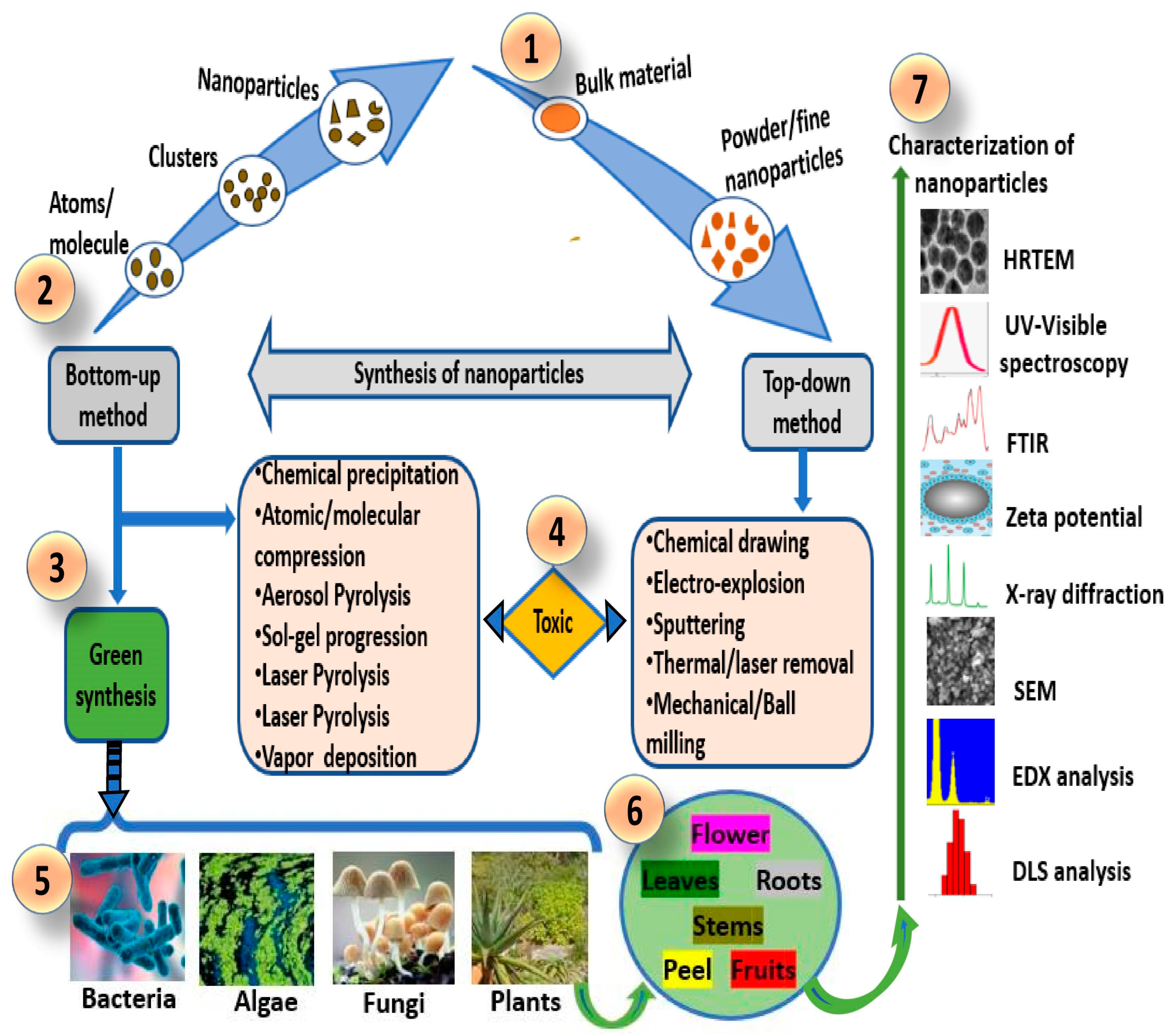
2. Nanotechnology
3. Biogenic AgNPs Synthesis
4. Anticancer Efficacy of Biogenic AgNPs
5. Photodynamic Therapy (PDT)
Optical Property of AgNPs for Cancer PDT
6. Mechanisms of Biogenic AgNPs and in Combination with PDT
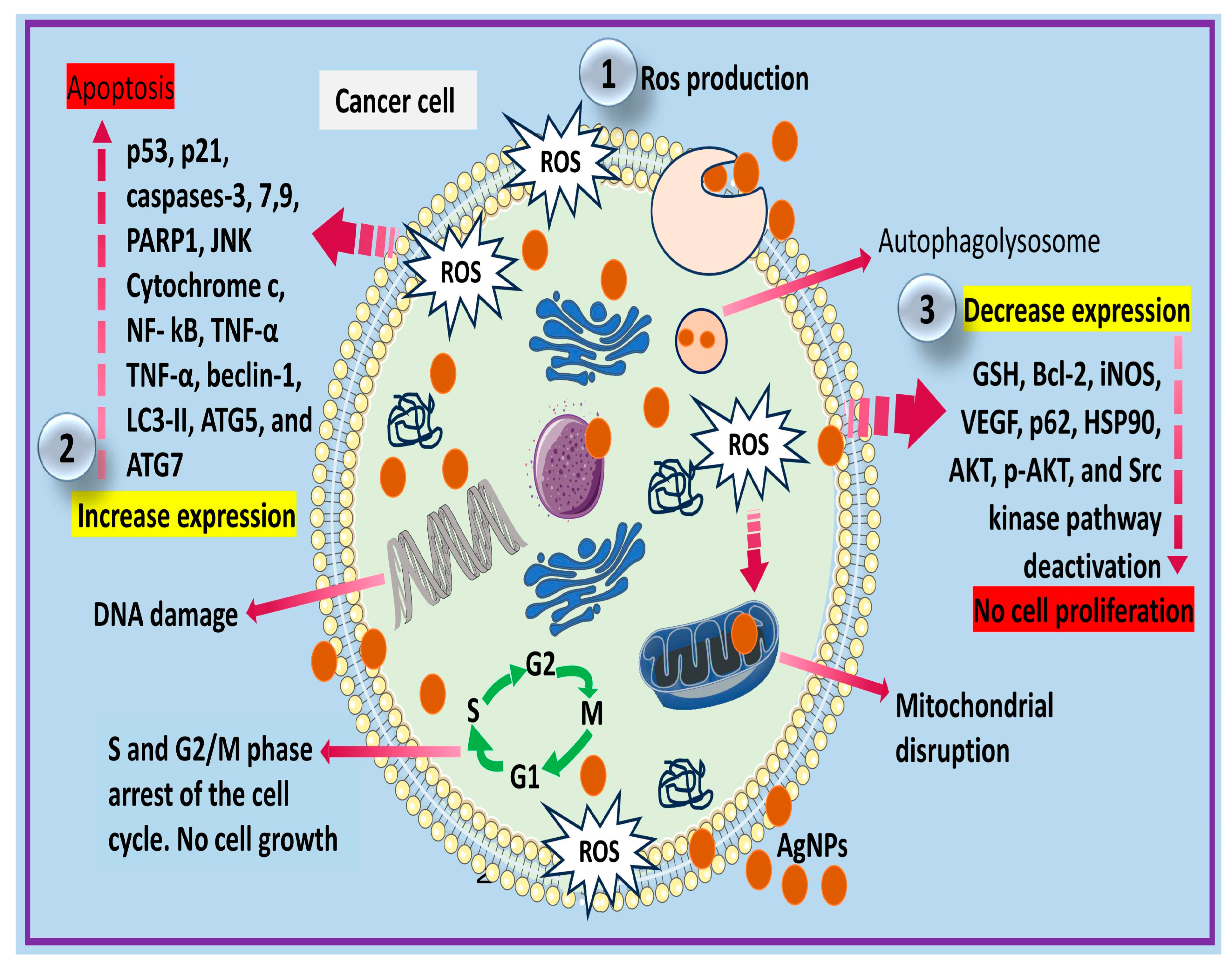
6.1. Mechanisms of Biogenic AgNPs as Lone Molecules for Cancer
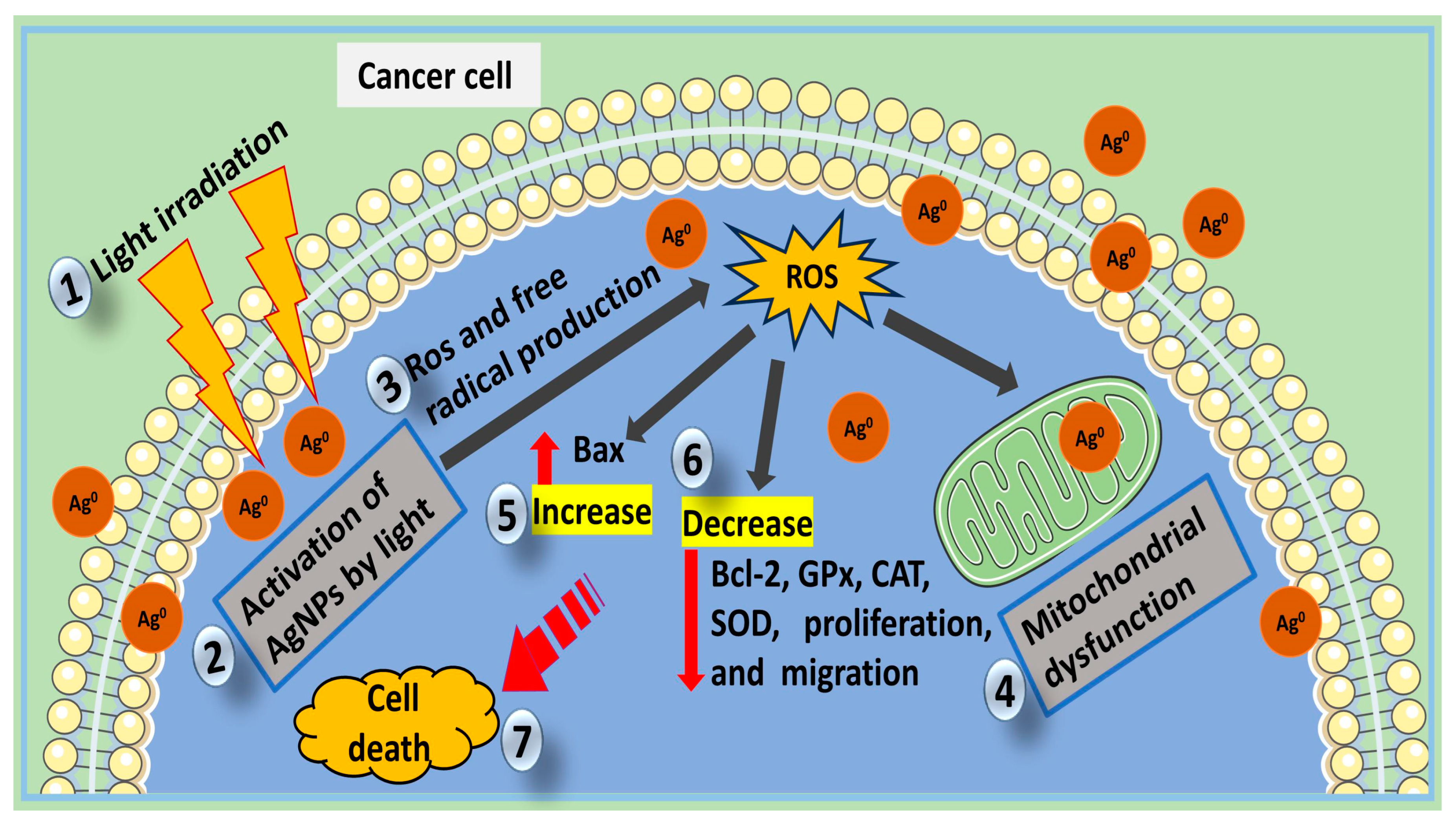
6.2. Mechanism of Biogenic AgNPs in Combination with PDT
7. Biogenic AgNPs in Cancer-Targeted Therapy
8. Toxicity of Biogenic AgNPs
9. Future Prospects
10. Conclusions
Author Contributions
Funding
Institutional Review Board Statement
Informed Consent Statement
Data Availability Statement
Acknowledgments
Conflicts of Interest
References
- Mfouo-Tynga, I.; Hussein, A.E.; Abdel-Harith, M.; Abrahamse, H. Photodynamic Ability of Silver Nanoparticles in Inducing Cytotoxic Effects in Breast and Lung Cancer Cell Lines. Int. J. Nanomed. 2014, 9, 3771–3781. [Google Scholar]
- Javed, B.; Nadhman, A.; Mashwani, Z.-R. Optimization, Characterization and Antimicrobial Activity of Silver Nanoparticles against Plant Bacterial Pathogens Phyto-Synthesized by Mentha Longifolia. Mater. Res. Express 2020, 7, 085406. [Google Scholar] [CrossRef]
- Sung, H.; Ferlay, J.; Siegel, R.L.; Laversanne, M.; Soerjomataram, I.; Jemal, A.; Bray, F. Global Cancer Statistics 2020: GLOBOCAN Estimates of Incidence and Mortality Worldwide for 36 Cancers in 185 Countries. CA Cancer J. Clin. 2021, 71, 209–249. [Google Scholar] [CrossRef]
- Mosleh-Shirazi, S.; Abbasi, M.; Moaddeli, M.R.; Vaez, A.; Shafiee, M.; Kasaee, S.R.; Amani, A.M.; Hatam, S. Nanotechnology Advances in the Detection and Treatment of Cancer: An Overview. Nanotheranostics 2022, 6, 400–423. [Google Scholar] [CrossRef] [PubMed]
- Mansoori, B.; Mohammadi, A.; Davudian, S.; Shirjang, S.; Baradaran, B. The Different Mechanisms of Cancer Drug Resistance: A Brief Review. Adv. Pharm. Bull. 2017, 7, 339–348. [Google Scholar] [CrossRef] [PubMed]
- Sutradhar, K.B.; Amin, M.L. Nanotechnology in Cancer Drug Delivery and Selective Targeting. Int. Sch. Res. Not. 2014, 2014, e939378. [Google Scholar] [CrossRef] [Green Version]
- Dhilip Kumar, S.S.; Abrahamse, H. Biocompatible Nanocarriers for Enhanced Cancer Photodynamic Therapy Applications. Pharmaceutics 2021, 13, 1933. [Google Scholar] [CrossRef]
- Han, H.J.; Ekweremadu, C.; Patel, N. Advanced Drug Delivery System with Nanomaterials for Personalised Medicine to Treat Breast Cancer. J. Drug Deliv. Sci. Technol. 2019, 52, 1051–1060. [Google Scholar] [CrossRef]
- He, Y.; Li, X.; Wang, J.; Yang, Q.; Yao, B.; Zhao, Y.; Zhao, A.; Sun, W.; Zhang, Q. Synthesis, Characterization and Evaluation Cytotoxic Activity of Silver Nanoparticles Synthesized by Chinese Herbal Cornus Officinalis via Environment Friendly Approach. Env. Toxicol. Pharmacol. 2017, 56, 56–60. [Google Scholar] [CrossRef]
- Zhang, X.-F.; Liu, Z.-G.; Shen, W.; Gurunathan, S. Silver Nanoparticles: Synthesis, Characterization, Properties, Applications, and Therapeutic Approaches. Int. J. Mol. Sci. 2016, 17, 1534. [Google Scholar] [CrossRef]
- Ovais, M.; Khalil, A.T.; Raza, A.; Khan, M.A.; Ahmad, I.; Islam, N.U.; Saravanan, M.; Ubaid, M.F.; Ali, M.; Shinwari, Z.K. Green Synthesis of Silver Nanoparticles via Plant Extracts: Beginning a New Era in Cancer Theranostics. Nanomedicine 2016, 11, 3157–3177. [Google Scholar] [CrossRef] [PubMed]
- Yeşilot, Ş.; Aydın Acar, Ç. Silver Nanoparticles; a New Hope in Cancer Therapy? East. J. Med. 2019, 24, 111–116. [Google Scholar] [CrossRef]
- Kah, G.; Njobeh, P. Biosynthesis and Characterization of Silver Nanoparticles. Available online: https://www.researchsquare.com (accessed on 29 May 2023).
- Wei, L.; Lu, J.; Xu, H.; Patel, A.; Chen, Z.-S.; Chen, G. Silver Nanoparticles: Synthesis, Properties, and Therapeutic Applications. Drug Discov. Today 2015, 20, 595–601. [Google Scholar] [CrossRef] [PubMed] [Green Version]
- Karuppaiya, P.; Satheeshkumar, E.; Tsay, H.S. Biogenic Synthesis of Silver Nanoparticles Using Rhizome Extract of Dysosma Pleiantha and Its Antiproliferative Effect against Breast and Human Gastric Cancer Cells. Mol. Biol. Rep. 2019, 46, 4725–4734. [Google Scholar] [CrossRef] [PubMed]
- Erdogan, O.; Abbak, M.; Demirbolat, G.M.; Birtekocak, F.; Aksel, M.; Pasa, S.; Cevik, O. Green Synthesis of Silver Nanoparticles via Cynara Scolymus Leaf Extracts: The Characterization, Anticancer Potential with Photodynamic Therapy in MCF7 Cells. PLoS ONE 2019, 14, e0216496. [Google Scholar] [CrossRef] [Green Version]
- ISO ISO/TR 18401:2017(En), Nanotechnologies—Plain Language Explanation of Selected Terms from the ISO/IEC 80004 Series. Available online: https://www.iso.org/obp/ui/#iso:std:iso:tr:18401:ed-1:v1:en:en.%20Accessed%20date%2023%20May%202023 (accessed on 29 May 2023).
- Zahoor, M.; Nazir, N.; Iftikhar, M.; Naz, S.; Zekker, I.; Burlakovs, J.; Uddin, F.; Kamran, A.W.; Kallistova, A.; Pimenov, N.; et al. A Review on Silver Nanoparticles: Classification, Various Methods of Synthesis, and Their Potential Roles in Biomedical Applications and Water Treatment. Water 2021, 13, 2216. [Google Scholar] [CrossRef]
- Das, D.; Roy, A. Synthesis of Diameter Controlled Multiwall Carbon Nanotubes by Microwave Plasma-CVD on Low-Temperature and Chemically Processed Fe Nanoparticle Catalysts. Appl. Surf. Sci. 2020, 515, 146043. [Google Scholar] [CrossRef]
- Zhu, B.; Li, Y.; Lin, Z.; Zhao, M.; Xu, T.; Wang, C.; Deng, N. Silver Nanoparticles Induce HePG-2 Cells Apoptosis Through ROS-Mediated Signaling Pathways. Nanoscale Res. Lett. 2016, 11, 198. [Google Scholar] [CrossRef] [Green Version]
- Pulit-Prociak, J.; Banach, M. Silver Nanoparticles–A Material of the Future…? Open Chem. 2016, 14, 76–91. [Google Scholar] [CrossRef]
- Ghobashy, M.M.; Elkodous, M.A.; Shabaka, S.H.; Younis, S.A.; Alshangiti, D.M.; Madani, M.; Al-Gahtany, S.A.; Elkhatib, W.F.; Noreddin, A.M.; Nady, N.; et al. An Overview of Methods for Production and Detection of Silver Nanoparticles, with Emphasis on Their Fate and Toxicological Effects on Human, Soil, and Aquatic Environment. Nanotechnol. Rev. 2021, 10, 954–977. [Google Scholar] [CrossRef]
- Heinemann, M.G.; Rosa, C.H.; Rosa, G.R.; Dias, D. Biogenic Synthesis of Gold and Silver Nanoparticles Used in Environmental Applications: A Review. Trends Environ. Anal. Chem. 2021, 30, e00129. [Google Scholar] [CrossRef]
- Dhayalan, M.; Denison, M.I.J.; Ayyar, M.; Gandhi, N.N.; Krishnan, K.; Abdulhadi, B. Biogenic Synthesis, Characterization of Gold and Silver Nanoparticles from Coleus Forskohlii and Their Clinical Importance. J. Photochem. Photobiol. B Biol. 2018, 183, 251–257. [Google Scholar] [CrossRef]
- Sánchez-López, E.; Gomes, D.; Esteruelas, G.; Bonilla, L.; Lopez-Machado, A.L.; Galindo, R.; Cano, A.; Espina, M.; Ettcheto, M.; Camins, A.; et al. Metal-Based Nanoparticles as Antimicrobial Agents: An Overview. Nanomaterials 2020, 10, 292. [Google Scholar] [CrossRef] [Green Version]
- Husain, S.; Nandi, A.; Simnani, F.Z.; Saha, U.; Ghosh, A.; Sinha, A.; Sahay, A.; Samal, S.K.; Panda, P.K.; Verma, S.K. Emerging Trends in Advanced Translational Applications of Silver Nanoparticles: A Progressing Dawn of Nanotechnology. J. Funct. Biomater. 2023, 14, 47. [Google Scholar] [CrossRef]
- Inshakova, E.; Inshakov, O. World Market for Nanomaterials: Structure and Trends. MATEC Web Conf. 2017, 129, 02013. [Google Scholar] [CrossRef] [Green Version]
- Iravani, S.; Korbekandi, H.; Mirmohammadi, S.V.; Zolfaghari, B. Synthesis of Silver Nanoparticles: Chemical, Physical and Biological Methods. Res. Pharm. Sci. 2014, 9, 385–406. [Google Scholar]
- Keat, C.L.; Aziz, A.; Eid, A.M.; Elmarzugi, N.A. Biosynthesis of Nanoparticles and Silver Nanoparticles. Bioresour. Bioprocess. 2015, 2, 47. [Google Scholar] [CrossRef] [Green Version]
- Ahmed, S.; Ahmad, M.; Swami, B.L.; Ikram, S. A Review on Plants Extract Mediated Synthesis of Silver Nanoparticles for Antimicrobial Applications: A Green Expertise. J. Adv. Res. 2016, 7, 17–28. [Google Scholar] [CrossRef]
- Adebayo, I.A.; Arsad, H.; Gagman, H.A.; Ismail, N.Z.; Samian, M.R. Inhibitory Effect of Eco-Friendly Naturally Synthesized Silver Nanoparticles from the Leaf Extract of Medicinal Detarium Microcarpum Plant on Pancreatic and Cervical Cancer Cells. Asian Pac. J. Cancer Prev. 2020, 21, 1247–1252. [Google Scholar] [CrossRef]
- Jalani, N.S.; Zati-Hanani, S.; Teoh, Y.P.; Abdullah, R. Short Review: The Effect of Reaction Conditions on Plant-Mediated Synthesis of Silver Nanoparticles. Mater. Sci. Forum 2018, 917, 145–151. [Google Scholar] [CrossRef]
- Malik, P.; Shankar, R.; Malik, V.; Sharma, N.; Mukherjee, T.K. Green Chemistry Based Benign Routes for Nanoparticle Synthesis. J. Nanoparticles 2014, 2014, 302429. [Google Scholar] [CrossRef] [Green Version]
- Duan, H.; Wang, D.; Li, Y. Green Chemistry for Nanoparticle Synthesis. Chem. Soc. Rev. 2015, 44, 5778–5792. [Google Scholar] [CrossRef]
- Noga, M.; Milan, J.; Frydrych, A.; Jurowski, K. Toxicological Aspects, Safety Assessment, and Green Toxicology of Silver Nanoparticles (AgNPs)—Critical Review: State of the Art. Int. J. Mol. Sci. 2023, 24, 5133. [Google Scholar] [CrossRef]
- Hembram, K.C.; Kumar, R.; Kandha, L.; Parhi, P.K.; Kundu, C.N.; Bindhani, B.K. Therapeutic Prospective of Plant-Induced Silver Nanoparticles: Application as Antimicrobial and Anticancer Agent. Artif. Cells Nanomed. Biotechnol. 2018, 46, S38–S51. [Google Scholar] [CrossRef] [Green Version]
- Ashraf, J.M.; Ansari, M.A.; Khan, H.M.; Alzohairy, M.A.; Choi, I. Green Synthesis of Silver Nanoparticles and Characterization of Their Inhibitory Effects on AGEs Formation Using Biophysical Techniques. Sci. Rep. 2016, 6, 20414. [Google Scholar] [CrossRef] [Green Version]
- Sreekanth, T.V.M.; Pandurangan, M.; Kim, D.H.; Lee, Y.R. Green Synthesis: In-Vitro Anticancer Activity of Silver Nanoparticles on Human Cervical Cancer Cells. J. Clust. Sci. 2016, 27, 671–681. [Google Scholar] [CrossRef]
- Salehi, S.; Shandiz, S.A.S.; Ghanbar, F.; Darvish, M.R.; Ardestani, M.S.; Mirzaie, A.; Jafari, M. Phytosynthesis of Silver Nanoparticles Using Artemisia Marschalliana Sprengel Aerial Part Extract and Assessment of Their Antioxidant, Anticancer, and Antibacterial Properties. Int. J. Nanomed. 2016, 11, 1835–1846. [Google Scholar] [CrossRef] [Green Version]
- Banerjee, P.P.; Bandyopadhyay, A.; Harsha, S.N.; Policegoudra, R.S.; Bhattacharya, S.; Karak, N.; Chattopadhyay, A. Mentha Arvensis (Linn.)-Mediated Green Silver Nanoparticles Trigger Caspase 9-Dependent Cell Death in MCF7 and MDA-MB-231 Cells. Breast Cancer 2017, 9, 265–278. [Google Scholar] [CrossRef] [Green Version]
- Mokhtar, F.A.; Selim, N.M.; Elhawary, S.S.; Abd El Hadi, S.R.; Hetta, M.H.; Albalawi, M.A.; Shati, A.A.; Alfaifi, M.Y.; Elbehairi, S.E.I.; Fahmy, L.I.; et al. Green Biosynthesis of Silver Nanoparticles Using Annona Glabra and Annona Squamosa Extracts with Antimicrobial, Anticancer, Apoptosis Potentials, Assisted by In Silico Modeling, and Metabolic Profiling. Pharmaceuticals 2022, 15, 1354. [Google Scholar] [CrossRef]
- Baharara, J.; Namvar, F.; Ramezani, T.; Mousavi, M.; Mohamad, R. Silver Nanoparticles Biosynthesized Using Achillea Biebersteinii Flower Extract: Apoptosis Induction in MCF-7 Cells via Caspase Activation and Regulation of Bax and Bcl-2 Gene Expression. Molecules 2015, 20, 2693–2706. [Google Scholar] [CrossRef] [Green Version]
- Lee, Y.J.; Song, K.; Cha, S.-H.; Cho, S.; Kim, Y.S.; Park, Y. Sesquiterpenoids from Tussilago Farfara Flower Bud Extract for the Eco-Friendly Synthesis of Silver and Gold Nanoparticles Possessing Antibacterial and Anticancer Activities. Nanomaterials 2019, 9, 819. [Google Scholar] [CrossRef] [Green Version]
- Lakshmanan, G.; Sathiyaseelan, A.; Kalaichelvan, P.T.; Murugesan, K. Plant-Mediated Synthesis of Silver Nanoparticles Using Fruit Extract of Cleome Viscosa L.: Assessment of Their Antibacterial and Anticancer Activity. Karbala Int. J. Mod. Sci. 2018, 4, 61–68. [Google Scholar] [CrossRef]
- Mittal, A.K.; Tripathy, D.; Choudhary, A.; Aili, P.K.; Chatterjee, A.; Singh, I.P.; Banerjee, U.C. Bio-Synthesis of Silver Nanoparticles Using Potentilla Fulgens Wall. Ex Hook. and Its Therapeutic Evaluation as Anticancer and Antimicrobial Agent. Mater. Sci. Eng. C Mater. Biol. Appl. 2015, 53, 120–127. [Google Scholar] [CrossRef]
- AlSalhi, M.S.; Elangovan, K.; Ranjitsingh, A.J.A.; Murali, P.; Devanesan, S. Synthesis of Silver Nanoparticles Using Plant Derived 4-N-Methyl Benzoic Acid and Evaluation of Antimicrobial, Antioxidant and Antitumor Activity. Saudi J. Biol. Sci. 2019, 26, 970–978. [Google Scholar] [CrossRef]
- Firdhouse, M.J.; Lalitha, P. Biosynthesis of Silver Nanoparticles Using the Extract of Alternanthera Sessilis—Antiproliferative Effect against Prostate Cancer Cells. Cancer Nano 2013, 4, 137–143. [Google Scholar] [CrossRef] [Green Version]
- Gorbe, M.; Bhat, R.; Aznar, E.; Sancenón, F.; Marcos, M.D.; Herraiz, F.J.; Prohens, J.; Venkataraman, A.; Martínez-Máñez, R. Rapid Biosynthesis of Silver Nanoparticles Using Pepino (Solanum Muricatum) Leaf Extract and Their Cytotoxicity on HeLa Cells. Materials 2016, 9, 325. [Google Scholar] [CrossRef] [Green Version]
- Chanthini, A.B.; Balasubramani, G.; Ramkumar, R.; Sowmiya, R.; Balakumaran, M.D.; Kalaichelvan, P.T.; Perumal, P. Structural Characterization, Antioxidant and in Vitro Cytotoxic Properties of Seagrass, Cymodocea Serrulata (R.Br.) Asch. & Magnus Mediated Silver Nanoparticles. J. Photochem. Photobiol. B 2015, 153, 145–152. [Google Scholar] [CrossRef]
- Bharadwaj, K.K.; Rabha, B.; Pati, S.; Choudhury, B.K.; Sarkar, T.; Gogoi, S.K.; Kakati, N.; Baishya, D.; Kari, Z.A.; Edinur, H.A. Green Synthesis of Silver Nanoparticles Using Diospyros Malabarica Fruit Extract and Assessments of Their Antimicrobial, Anticancer and Catalytic Reduction of 4-Nitrophenol (4-NP). Nanomaterials 2021, 11, 1999. [Google Scholar] [CrossRef]
- Elemike, E.E.; Onwudiwe, D.C.; Nundkumar, N.; Singh, M.; Iyekowa, O. Green Synthesis of Ag, Au and Ag-Au Bimetallic Nanoparticles Using Stigmaphyllon Ovatum Leaf Extract and Their in Vitro Anticancer Potential. Mater. Lett. 2019, 243, 148–152. [Google Scholar] [CrossRef]
- Bhat, M.P.; Kumar, R.S.; Rudrappa, M.; Basavarajappa, D.S.; Swamy, P.S.; Almansour, A.I.; Perumal, K.; Nayaka, S. Bio-Inspired Silver Nanoparticles from Artocarpus Lakoocha Fruit Extract and Evaluation of Their Antibacterial Activity and Anticancer Activity on Human Prostate Cancer Cell Line. Appl. Nanosci. 2023, 13, 3041–3051. [Google Scholar] [CrossRef]
- Nagaraja, S.K.; Kumar, R.S.; Chakraborty, B.; Hiremath, H.; Almansour, A.I.; Perumal, K.; Gunagambhire, P.V.; Nayaka, S. Biomimetic Synthesis of Silver Nanoparticles Using Cucumis Sativus Var. Hardwickii Fruit Extract and Their Characterizations, Anticancer Potential and Apoptosis Studies against Pa-1 (Human Ovarian Teratocarcinoma) Cell Line via Flow Cytometry. Appl. Nanosci. 2023, 13, 3073–3084. [Google Scholar] [CrossRef]
- Shiripoure Ganjineh Ketab, R.; Tafvizi, F.; Khodarahmi, P. Biosynthesis and Chemical Characterization of Silver Nanoparticles Using Satureja Rechingeri Jamzad and Their Apoptotic Effects on AGS Gastric Cancer Cells. J. Clust. Sci. 2021, 32, 1389–1399. [Google Scholar] [CrossRef]
- Sarkar, S.; Kotteeswaran, V. Green Synthesis of Silver Nanoparticles from Aqueous Leaf Extract of Pomegranate (Punica Granatum) and Their Anticancer Activity on Human Cervical Cancer Cells. Adv. Nat. Sci Nanosci. Nanotechnol. 2018, 9, 025014. [Google Scholar] [CrossRef]
- Khan, A.A.; Alanazi, A.M.; Alsaif, N.; Wani, T.A.; Bhat, M.A. Pomegranate Peel Induced Biogenic Synthesis of Silver Nanoparticles and Their Multifaceted Potential against Intracellular Pathogen and Cancer. Saudi J. Biol. Sci. 2021, 28, 4191–4200. [Google Scholar] [CrossRef]
- Venugopal, K.; Ahmad, H.; Manikandan, E.; Thanigai Arul, K.; Kavitha, K.; Moodley, M.K.; Rajagopal, K.; Balabhaskar, R.; Bhaskar, M. The Impact of Anticancer Activity upon Beta Vulgaris Extract Mediated Biosynthesized Silver Nanoparticles (Ag-NPs) against Human Breast (MCF-7), Lung (A549) and Pharynx (Hep-2) Cancer Cell Lines. J. Photochem. Photobiol. B 2017, 173, 99–107. [Google Scholar] [CrossRef] [PubMed]
- Hemlata; Meena, P.R.; Singh, A.P.; Tejavath, K.K. Biosynthesis of Silver Nanoparticles Using Cucumis Prophetarum Aqueous Leaf Extract and Their Antibacterial and Antiproliferative Activity against Cancer Cell Lines. ACS Omega 2020, 5, 5520–5528. [Google Scholar] [CrossRef] [PubMed] [Green Version]
- Hublikar, L.V.; Ganachari, S.V.; Patil, V.B.; Nandi, S.; Honnad, A. Anticancer Potential of Biologically Synthesized Silver Nanoparticles Using Lantana Camara Leaf Extract. Prog. Biomater. 2023, 12, 155–169. [Google Scholar] [CrossRef]
- Reddy, N.V.; Li, H.; Hou, T.; Bethu, M.S.; Ren, Z.; Zhang, Z. Phytosynthesis of Silver Nanoparticles Using Perilla Frutescens Leaf Extract: Characterization and Evaluation of Antibacterial, Antioxidant, and Anticancer Activities. Int. J. Nanomed. 2021, 16, 15–29. [Google Scholar] [CrossRef]
- Xu, Z.; Feng, Q.; Wang, M.; Zhao, H.; Lin, Y.; Zhou, S. Green Biosynthesized Silver Nanoparticles with Aqueous Extracts of Ginkgo Biloba Induce Apoptosis via Mitochondrial Pathway in Cervical Cancer Cells. Front. Oncol. 2020, 10, 575415. [Google Scholar] [CrossRef]
- Cyril, N.; George, J.B.; Joseph, L.; Raghavamenon, A.C.; Sylas, V.P. Assessment of Antioxidant, Antibacterial and Anti-Proliferative (Lung Cancer Cell Line A549) Activities of Green Synthesized Silver Nanoparticles from Derris Trifoliata. Toxicol. Res. 2019, 8, 297–308. [Google Scholar] [CrossRef] [Green Version]
- Shinde, A.; Mendhulkar, V.D. Anti-proliferative Activity of Elephantopus scaber Mediated Silver Nanoparticles against MCF-7, A-549, SCC-40 and COLO-205 Human Cancer Cell Lines. Asian J. Pharm. Clin. Res. 2019, 13, 163–167. [Google Scholar] [CrossRef] [Green Version]
- Atmaca, H.; Çamlı Pulat, Ç.; Ilhan, S. Synthesis of Silver Nanoparticles Using Alpinia Officinarum Rhizome Extract Induces Apoptosis through Down-Regulating Bcl-2 in Human Cancer Cells. Biol. Futur. 2022, 73, 327–334. [Google Scholar] [CrossRef] [PubMed]
- Kah, G.; Chandran, R.; Abrahamse, H. Curcumin a Natural Phenol and Its Therapeutic Role in Cancer and Photodynamic Therapy: A Review. Pharmaceutics 2023, 15, 639. [Google Scholar] [CrossRef] [PubMed]
- Lin, L.; Xiong, L.; Wen, Y.; Lei, S.; Deng, X.; Liu, Z.; Chen, W.; Miao, X. Active Targeting of Nano-Photosensitizer Delivery Systems for Photodynamic Therapy of Cancer Stem Cells. J. Biomed. Nanotechnol. 2015, 11, 531–554. [Google Scholar] [CrossRef]
- Mkhobongo, B.; Chandran, R.; Abrahamse, H. The Role of Melanoma Cell-Derived Exosomes (MTEX) and Photodynamic Therapy (PDT) within a Tumor Microenvironment. Int. J. Mol. Sci. 2021, 22, 9726. [Google Scholar] [CrossRef]
- Yi, G.; Hong, S.H.; Son, J.; Yoo, J.; Park, C.; Choi, Y.; Koo, H. Recent Advances in Nanoparticle Carriers for Photodynamic Therapy. Quant. Imaging Med. Surg. 2018, 8, 433–443. [Google Scholar] [CrossRef]
- Shang, L.; Zhou, X.; Zhang, J.; Shi, Y.; Zhong, L. Metal Nanoparticles for Photodynamic Therapy: A Potential Treatment for Breast Cancer. Molecules 2021, 26, 6532. [Google Scholar] [CrossRef]
- Hou, Y.; Yang, X.; Liu, R.; Zhao, D.; Guo, C.; Zhu, A.; Wen, M.; Liu, Z.; Qu, G.; Meng, H. Pathological Mechanism of Photodynamic Therapy and Photothermal Therapy Based on Nanoparticles. Int. J. Nanomed. 2020, 15, 6827–6838. [Google Scholar] [CrossRef]
- Chen, H.; Tian, J.; He, W.; Guo, Z. H2O2-Activatable and O2-Evolving Nanoparticles for Highly Efficient and Selective Photodynamic Therapy against Hypoxic Tumor Cells. J. Am. Chem. Soc. 2015, 137, 1539–1547. [Google Scholar] [CrossRef]
- Jiang, W.; Liang, M.; Lei, Q.; Li, G.; Wu, S. The Current Status of Photodynamic Therapy in Cancer Treatment. Cancers 2023, 15, 585. [Google Scholar] [CrossRef]
- Kataoka, H.; Nishie, H.; Hayashi, N.; Tanaka, M.; Nomoto, A.; Yano, S.; Joh, T. New Photodynamic Therapy with Next-Generation Photosensitizers. Ann. Transl. Med. 2017, 5, 183. [Google Scholar] [CrossRef] [PubMed] [Green Version]
- Svenskaya, Y.; Parakhonskiy, B.; Haase, A.; Atkin, V.; Lukyanets, E.; Gorin, D.; Antolini, R. Anticancer Drug Delivery System Based on Calcium Carbonate Particles Loaded with a Photosensitizer. Biophys. Chem. 2013, 182, 11–15. [Google Scholar] [CrossRef] [PubMed] [Green Version]
- Mokoena, D.R.; George, B.P.; Abrahamse, H. Enhancing Breast Cancer Treatment Using a Combination of Cannabidiol and Gold Nanoparticles for Photodynamic Therapy. Int. J. Mol. Sci. 2019, 20, 4771. [Google Scholar] [CrossRef] [Green Version]
- Yoon, I.; Li, J.Z.; Shim, Y.K. Advance in Photosensitizers and Light Delivery for Photodynamic Therapy. Clin. Endosc. 2013, 46, 7–23. [Google Scholar] [CrossRef] [PubMed]
- Zhou, T.-J.; Xing, L.; Fan, Y.-T.; Cui, P.-F.; Jiang, H.-L. Inhibition of Breast Cancer Proliferation and Metastasis by Strengthening Host Immunity with a Prolonged Oxygen-Generating Phototherapy Hydrogel. J. Control Release 2019, 309, 82–93. [Google Scholar] [CrossRef]
- George, B.P.; Chota, A.; Sarbadhikary, P.; Abrahamse, H. Fundamentals and Applications of Metal Nanoparticle- Enhanced Singlet Oxygen Generation for Improved Cancer Photodynamic Therapy. Front. Chem. 2022, 10, 964674. [Google Scholar] [CrossRef]
- Sun, J.; Kormakov, S.; Liu, Y.; Huang, Y.; Wu, D.; Yang, Z. Recent Progress in Metal-Based Nanoparticles Mediated Photodynamic Therapy. Molecules 2018, 23, 1704. [Google Scholar] [CrossRef] [Green Version]
- Cole, A.J.; Yang, V.C.; David, A.E. Cancer Theranostics: The Rise of Targeted Magnetic Nanoparticles. Trends Biotechnol. 2011, 29, 323–332. [Google Scholar] [CrossRef] [Green Version]
- Kreibig, U.; Vollmer, M. (Eds.) Introduction. In Optical Properties of Metal Clusters; Springer Series in Materials Science; Springer: Berlin/Heidelberg, Germany, 1995; pp. 1–12. ISBN 978-3-662-09109-8. [Google Scholar]
- Basavegowda, N.; Baek, K.-H. Advances in Functional Biopolymer-Based Nanocomposites for Active Food Packaging Applications. Polymers 2021, 13, 4198. [Google Scholar] [CrossRef]
- Rycenga, M.; Cobley, C.M.; Zeng, J.; Li, W.; Moran, C.H.; Zhang, Q.; Qin, D.; Xia, Y. Controlling the Synthesis and Assembly of Silver Nanostructures for Plasmonic Applications. Chem. Rev. 2011, 111, 3669–3712. [Google Scholar] [CrossRef] [Green Version]
- Ratan, Z.A.; Haidere, M.F.; Nurunnabi, M.; Shahriar, S.M.; Ahammad, A.J.S.; Shim, Y.Y.; Reaney, M.J.T.; Cho, J.Y. Green Chemistry Synthesis of Silver Nanoparticles and Their Potential Anticancer Effects. Cancers 2020, 12, 855. [Google Scholar] [CrossRef] [Green Version]
- Kajani, A.A.; Bordbar, A.-K.; Esfahani, S.H.Z.; Khosropour, A.R.; Razmjou, A. Green Synthesis of Anisotropic Silver Nanoparticles with Potent Anticancer Activity Using Taxus Baccata Extract. RSC Adv. 2014, 4, 61394–61403. [Google Scholar] [CrossRef]
- Sun, Y.; Xia, Y. Gold and silver nanoparticles: A class of chromophores with colors tunable in the range from 400 to 750 nm. Analyst 2003, 128, 686–691. [Google Scholar] [CrossRef]
- Jabeen, S.; Qureshi, R.; Munazir, M.; Maqsood, M.; Munir, M.; Shah, S.S.H.; Rahim, B.Z. Application of Green Synthesized Silver Nanoparticles in Cancer Treatment—A Critical Review. Mater. Res. Express 2021, 8, 092001. [Google Scholar] [CrossRef]
- Piao, M.J.; Kim, K.C.; Choi, J.-Y.; Choi, J.; Hyun, J.W. Silver Nanoparticles Down-Regulate Nrf2-Mediated 8-Oxoguanine DNA Glycosylase 1 through Inactivation of Extracellular Regulated Kinase and Protein Kinase B in Human Chang Liver Cells. Toxicol. Lett. 2011, 207, 143–148. [Google Scholar] [CrossRef] [PubMed]
- Piao, M.J.; Kang, K.A.; Lee, I.K.; Kim, H.S.; Kim, S.; Choi, J.Y.; Choi, J.; Hyun, J.W. Silver Nanoparticles Induce Oxidative Cell Damage in Human Liver Cells through Inhibition of Reduced Glutathione and Induction of Mitochondria-Involved Apoptosis. Toxicol. Lett. 2011, 201, 92–100. [Google Scholar] [CrossRef]
- Eom, H.-J.; Choi, J. P38 MAPK Activation, DNA Damage, Cell Cycle Arrest and Apoptosis as Mechanisms of Toxicity of Silver Nanoparticles in Jurkat T Cells. Env. Sci. Technol. 2010, 44, 8337–8342. [Google Scholar] [CrossRef]
- Lu, W.; Senapati, D.; Wang, S.; Tovmachenko, O.; Singh, A.K.; Yu, H.; Ray, P.C. Effect of Surface Coating on the Toxicity of Silver Nanomaterials on Human Skin Keratinocytes. Chem. Phys. Lett. 2010, 487, 92–96. [Google Scholar] [CrossRef] [PubMed] [Green Version]
- McShan, D.; Ray, P.C.; Yu, H. Molecular Toxicity Mechanism of Nanosilver. J Food Drug Anal 2014, 22, 116–127. [Google Scholar] [CrossRef] [Green Version]
- Asharani, P.V.; Hande, M.P.; Valiyaveettil, S. Anti-Proliferative Activity of Silver Nanoparticles. BMC Cell Biol. 2009, 10, 65. [Google Scholar] [CrossRef] [Green Version]
- Greulich, C.; Diendorf, J.; Simon, T.; Eggeler, G.; Epple, M.; Köller, M. Uptake and Intracellular Distribution of Silver Nanoparticles in Human Mesenchymal Stem Cells. Acta Biomater. 2011, 7, 347–354. [Google Scholar] [CrossRef] [PubMed]
- Kalishwaralal, K.; Banumathi, E.; Pandian, S.R.K.; Deepak, V.; Muniyandi, J.; Eom, S.H.; Gurunathan, S. Silver Nanoparticles Inhibit VEGF Induced Cell Proliferation and Migration in Bovine Retinal Endothelial Cells. Colloids Surf. B Biointerfaces 2009, 73, 51–57. [Google Scholar] [CrossRef]
- Sriram, M.I.; Kanth, S.B.M.; Kalishwaralal, K.; Gurunathan, S. Antitumor Activity of Silver Nanoparticles in Dalton’s Lymphoma Ascites Tumor Model. Int. J. Nanomed. 2010, 5, 753–762. [Google Scholar] [CrossRef] [Green Version]
- Kitimu, S.R.; Kirira, P.; Abdille, A.A.; Sokei, J.; Ochwang’i, D.; Mwitari, P.; Makanya, A.; Maina, N. Anti-Angiogenic and Anti-Metastatic Effects of Biogenic Silver Nanoparticles Synthesized Using azadirachta indica. Adv. Biosci. Biotechnol. 2022, 13, 188–206. [Google Scholar] [CrossRef]
- Kargozar, S.; Baino, F.; Hamzehlou, S.; Hamblin, M.R.; Mozafari, M. Nanotechnology for Angiogenesis: Opportunities and Challenges. Chem. Soc. Rev. 2020, 49, 5008–5057. [Google Scholar] [CrossRef]
- Singh, R.P.; Ramarao, P. Cellular Uptake, Intracellular Trafficking and Cytotoxicity of Silver Nanoparticles. Toxicol. Lett. 2012, 213, 249–259. [Google Scholar] [CrossRef]
- Talarska, P.; Boruczkowski, M.; Żurawski, J. Current Knowledge of Silver and Gold Nanoparticles in Laboratory Research—Application, Toxicity, Cellular Uptake. Nanomaterials 2021, 11, 2454. [Google Scholar] [CrossRef] [PubMed]
- Zhang, T.; Wang, L.; Chen, Q.; Chen, C. Cytotoxic Potential of Silver Nanoparticles. Yonsei Med. J. 2014, 55, 283–291. [Google Scholar] [CrossRef] [PubMed] [Green Version]
- He, W.; Zhou, Y.-T.; Wamer, W.G.; Boudreau, M.D.; Yin, J.-J. Mechanisms of the PH Dependent Generation of Hydroxyl Radicals and Oxygen Induced by Ag Nanoparticles. Biomaterials 2012, 33, 7547–7555. [Google Scholar] [CrossRef]
- Yin, M.; Xu, X.; Han, H.; Dai, J.; Sun, R.; Yang, L.; Xie, J.; Wang, Y. Preparation of Triangular Silver Nanoparticles and Their Biological Effects in the Treatment of Ovarian Cancer. J. Ovarian Res. 2022, 15, 121. [Google Scholar] [CrossRef]
- Ding, L.; Cao, J.; Lin, W.; Chen, H.; Xiong, X.; Ao, H.; Yu, M.; Lin, J.; Cui, Q. The Roles of Cyclin-Dependent Kinases in Cell-Cycle Progression and Therapeutic Strategies in Human Breast Cancer. Int. J. Mol. Sci. 2020, 21, 1960. [Google Scholar] [CrossRef] [PubMed] [Green Version]
- Chota, A.; George, B.P.; Abrahamse, H. Interactions of Multidomain Pro-Apoptotic and Anti-Apoptotic Proteins in Cancer Cell Death. Oncotarget 2021, 12, 1615–1626. [Google Scholar] [CrossRef]
- Wang, L.-H.; Wu, C.-F.; Rajasekaran, N.; Shin, Y.K. Loss of Tumor Suppressor Gene Function in Human Cancer: An Overview. Cell Physiol. Biochem. 2018, 51, 2647–2693. [Google Scholar] [CrossRef] [PubMed]
- Reisman, D.; Takahashi, P.; Polson, A.; Boggs, K. Transcriptional Regulation of the P53 Tumor Suppressor Gene in S-Phase of the Cell-Cycle and the Cellular Response to DNA Damage. Biochem. Res. Int. 2012, 2012, 808934. [Google Scholar] [CrossRef] [PubMed] [Green Version]
- Jia, M.; Zhang, W.; He, T.; Shu, M.; Deng, J.; Wang, J.; Li, W.; Bai, J.; Lin, Q.; Luo, F.; et al. Evaluation of the Genotoxic and Oxidative Damage Potential of Silver Nanoparticles in Human NCM460 and HCT116 Cells. Int. J. Mol. Sci. 2020, 21, 1618. [Google Scholar] [CrossRef] [PubMed] [Green Version]
- Avalos, A.; Haza, A.I.; Mateo, D.; Morales, P. Interactions of Manufactured Silver Nanoparticles of Different Sizes with Normal Human Dermal Fibroblasts. Int. Wound J. 2014, 13, 101–109. [Google Scholar] [CrossRef]
- Nishanth, R.P.; Jyotsna, R.G.; Schlager, J.J.; Hussain, S.M.; Reddanna, P. Inflammatory Responses of RAW 264.7 Macrophages upon Exposure to Nanoparticles: Role of ROS-NFκB Signaling Pathway. Nanotoxicology 2011, 5, 502–516. [Google Scholar] [CrossRef]
- Gurunathan, S.; Jeong, J.-K.; Han, J.W.; Zhang, X.-F.; Park, J.H.; Kim, J.-H. Multidimensional Effects of Biologically Synthesized Silver Nanoparticles in Helicobacter Pylori, Helicobacter Felis, and Human Lung (L132) and Lung Carcinoma A549 Cells. Nanoscale Res. Lett. 2015, 10, 35. [Google Scholar] [CrossRef] [Green Version]
- Halawani, E.M.; Hassan, A.M.; Gad El-Rab, S.M.F. Nanoformulation of Biogenic Cefotaxime-Conjugated-Silver Nanoparticles for Enhanced Antibacterial Efficacy Against Multidrug-Resistant Bacteria and Anticancer Studies. Int. J. Nanomed. 2020, 15, 1889–1901. [Google Scholar] [CrossRef] [Green Version]
- Pei, J.; Fu, B.; Jiang, L.; Sun, T. Biosynthesis, Characterization, and Anticancer Effect of Plant-Mediated Silver Nanoparticles Using Coptis Chinensis. Int. J. Nanomed. 2019, 14, 1969–1978. [Google Scholar] [CrossRef] [Green Version]
- Jain, N.; Jain, P.; Rajput, D.; Patil, U.K. Green Synthesized Plant-Based Silver Nanoparticles: Therapeutic Prospective for Anticancer and Antiviral Activity. Micro Nano Syst. Lett. 2021, 9, 5. [Google Scholar] [CrossRef]
- He, Y.; Du, Z.; Ma, S.; Liu, Y.; Li, D.; Huang, H.; Jiang, S.; Cheng, S.; Wu, W.; Zhang, K.; et al. Effects of Green-Synthesized Silver Nanoparticles on Lung Cancer Cells in Vitro and Grown as Xenograft Tumors in Vivo. Int. J. Nanomed. 2016, 11, 1879–1887. [Google Scholar] [CrossRef] [Green Version]
- Wan, J.; Liu, T.; Mei, L.; Li, J.; Gong, K.; Yu, C.; Li, W. Synergistic Antitumour Activity of Sorafenib in Combination with Tetrandrine Is Mediated by Reactive Oxygen Species (ROS)/Akt Signaling. Br. J. Cancer 2013, 109, 342–350. [Google Scholar] [CrossRef]
- Plackal Adimuriyil George, B.; Kumar, N.; Abrahamse, H.; Ray, S.S. Apoptotic Efficacy of Multifaceted Biosynthesized Silver Nanoparticles on Human Adenocarcinoma Cells. Sci. Rep. 2018, 8, 14368. [Google Scholar] [CrossRef] [Green Version]
- Manikandan, R.; Manikandan, B.; Raman, T.; Arunagirinathan, K.; Prabhu, N.M.; Jothi Basu, M.; Perumal, M.; Palanisamy, S.; Munusamy, A. Biosynthesis of Silver Nanoparticles Using Ethanolic Petals Extract of Rosa Indica and Characterization of Its Antibacterial, Anticancer and Anti-Inflammatory Activities. Spectrochim. Acta A Mol. Biomol. Spectrosc. 2015, 138, 120–129. [Google Scholar] [CrossRef]
- Balkrishna, A.; Sharma, V.K.; Das, S.K.; Mishra, N.; Bisht, L.; Joshi, A.; Sharma, N. Characterization and Anti-Cancerous Effect of Putranjiva Roxburghii Seed Extract Mediated Silver Nanoparticles on Human Colon (HCT-116), Pancreatic (PANC-1) and Breast (MDA-MB 231) Cancer Cell Lines: A Comparative Study. Int. J. Nanomed. 2020, 15, 573–585. [Google Scholar] [CrossRef] [Green Version]
- Wang, J.; Zhou, G.; Chen, C.; Yu, H.; Wang, T.; Ma, Y.; Jia, G.; Gao, Y.; Li, B.; Sun, J. Acute Toxicity and Biodistribution of Different Sized Titanium Dioxide Particles in Mice after Oral Administration. Toxicol. Lett. 2007, 168, 176–185. [Google Scholar] [CrossRef]
- Al-Sheddi, E.S.; Farshori, N.N.; Al-Oqail, M.M.; Al-Massarani, S.M.; Saquib, Q.; Wahab, R.; Musarrat, J.; Al-Khedhairy, A.A.; Siddiqui, M.A. Anticancer Potential of Green Synthesized Silver Nanoparticles Using Extract of Nepeta Deflersiana against Human Cervical Cancer Cells (HeLA). Bioinorg. Chem. Appl. 2018, 2018, 9390784. [Google Scholar] [CrossRef] [Green Version]
- Al Sufyani, N.M.; Hussien, N.A.; Hawsawi, Y.M. Characterization and Anticancer Potential of Silver Nanoparticles Biosynthesized from Olea Chrysophylla and Lavandula Dentata Leaf Extracts on HCT116 Colon Cancer Cells. J. Nanomater. 2019, 2019, e7361695. [Google Scholar] [CrossRef] [Green Version]
- Panzarini, E.; Mariano, S.; Vergallo, C.; Carata, E.; Fimia, G.M.; Mura, F.; Rossi, M.; Vergaro, V.; Ciccarella, G.; Corazzari, M.; et al. Glucose Capped Silver Nanoparticles Induce Cell Cycle Arrest in HeLa Cells. Toxicol. Vitr. 2017, 41, 64–74. [Google Scholar] [CrossRef]
- Sheikpranbabu, S.; Kalishwaralal, K.; Venkataraman, D.; Eom, S.H.; Park, J.; Gurunathan, S. Silver Nanoparticles Inhibit VEGF-and IL-1β-Induced Vascular Permeability via Src Dependent Pathway in Porcine Retinal Endothelial Cells. J. Nanobiotechnology 2009, 7, 8. [Google Scholar] [CrossRef] [Green Version]
- Singh, R.P.; Agarwal, R. Inducible Nitric Oxide Synthase-Vascular Endothelial Growth Factor Axis: A Potential Target to Inhibit Tumor Angiogenesis by Dietary Agents. Curr. Cancer Drug Targets 2007, 7, 475–483. [Google Scholar] [CrossRef]
- Jayson, G.C.; Kerbel, R.; Ellis, L.M.; Harris, A.L. Antiangiogenic Therapy in Oncology: Current Status and Future Directions. Lancet 2016, 388, 518–529. [Google Scholar] [CrossRef]
- Fageria, L.; Pareek, V.; Dilip, R.V.; Bhargava, A.; Pasha, S.S.; Laskar, I.R.; Saini, H.; Dash, S.; Chowdhury, R.; Panwar, J. Biosynthesized Protein-Capped Silver Nanoparticles Induce ROS-Dependent Proapoptotic Signals and Prosurvival Autophagy in Cancer Cells. ACS Omega 2017, 2, 1489–1504. [Google Scholar] [CrossRef]
- Cordani, M.; Somoza, Á. Targeting Autophagy Using Metallic Nanoparticles: A Promising Strategy for Cancer Treatment. Cell Mol. Life Sci. 2019, 76, 1215–1242. [Google Scholar] [CrossRef] [Green Version]
- Buttacavoli, M.; Albanese, N.N.; Di Cara, G.; Alduina, R.; Faleri, C.; Gallo, M.; Pizzolanti, G.; Gallo, G.; Feo, S.; Baldi, F.; et al. Anticancer Activity of Biogenerated Silver Nanoparticles: An Integrated Proteomic Investigation. Oncotarget 2018, 9, 9685–9705. [Google Scholar] [CrossRef] [Green Version]
- Mahajan, P.G.; Dige, N.C.; Vanjare, B.D.; Eo, S.-H.; Seo, S.-Y.; Kim, S.J.; Hong, S.-K.; Choi, C.-S.; Lee, K.H. A Potential Mediator for Photodynamic Therapy Based on Silver Nanoparticles Functionalized with Porphyrin. J. Photochem. Photobiol. A Chem. 2019, 377, 26–35. [Google Scholar] [CrossRef]
- Natesan, S.; Krishnaswami, V.; Ponnusamy, C.; Madiyalakan, M.; Woo, T.; Palanisamy, R. Hypocrellin B and Nano Silver Loaded Polymeric Nanoparticles: Enhanced Generation of Singlet Oxygen for Improved Photodynamic Therapy. Mater. Sci. Eng. C Mater. Biol. Appl. 2017, 77, 935–946. [Google Scholar] [CrossRef]
- de Freitas, C.F.; Kimura, E.; Rubira, A.F.; Muniz, E.C. Curcumin and Silver Nanoparticles Carried out from Polysaccharide-Based Hydrogels Improved the Photodynamic Properties of Curcumin through Metal-Enhanced Singlet Oxygen Effect. Mater. Sci. Eng. C Mater. Biol. Appl. 2020, 112, 110853. [Google Scholar] [CrossRef]
- Jesus, V.P.S.; Raniero, L.; Lemes, G.M.; Bhattacharjee, T.T.; Caetano Júnior, P.C.; Castilho, M.L. Nanoparticles of Methylene Blue Enhance Photodynamic Therapy. Photodiagnosis Photodyn. Ther. 2018, 23, 212–217. [Google Scholar] [CrossRef]
- Ferrín, G.; Linares, C.I.; Muntané, J. Mitochondrial Drug Targets in Cell Death and Cancer. Curr. Pharm. Des. 2011, 17, 2002–2016. [Google Scholar] [CrossRef]
- Abel, F.; Sjöberg, R.-M.; Nilsson, S.; Kogner, P.; Martinsson, T. Imbalance of the Mitochondrial Pro- and Anti-Apoptotic Mediators in Neuroblastoma Tumours with Unfavourable Biology. Eur. J. Cancer 2005, 41, 635–646. [Google Scholar] [CrossRef]
- Kajani, A.A.; Zarkesh-Esfahani, S.H.; Bordbar, A.-K.; Khosropour, A.R.; Razmjou, A.; Kardi, M. Anticancer Effects of Silver Nanoparticles Encapsulated by Taxus Baccata Extracts. J. Mol. Liq. 2016, 223, 549–556. [Google Scholar] [CrossRef]
- Ghiuță, I.; Cristea, D. Silver Nanoparticles for Delivery Purposes. Nanoeng. Biomater. Adv. Drug Deliv. In Nanoengineered Biomaterials for Advanced Drug Delivery; Elsevier: Amsterdam, The Netherlands, 2020; pp. 347–371. [Google Scholar] [CrossRef]
- Hussein, H.A.; Abdullah, M.A. Novel Drug Delivery Systems Based on Silver Nanoparticles, Hyaluronic Acid, Lipid Nanoparticles and Liposomes for Cancer Treatment. Appl. Nanosci. 2022, 12, 3071–3096. [Google Scholar] [CrossRef]
- Gomes, H.I.O.; Martins, C.S.M.; Prior, J.A.V. Silver Nanoparticles as Carriers of Anticancer Drugs for Efficient Target Treatment of Cancer Cells. Nanomaterials 2021, 11, 964. [Google Scholar] [CrossRef]
- Lok, C.-N.; Zou, T.; Zhang, J.-J.; Lin, I.W.-S.; Che, C.-M. Controlled-Release Systems for Metal-Based Nanomedicine: Encapsulated/Self-Assembled Nanoparticles of Anticancer Gold(III)/Platinum(II) Complexes and Antimicrobial Silver Nanoparticles. Adv. Mater. 2014, 26, 5550–5557. [Google Scholar] [CrossRef]
- Locatelli, E.; Naddaka, M.; Uboldi, C.; Loudos, G.; Fragogeorgi, E.; Molinari, V.; Pucci, A.; Tsotakos, T.; Psimadas, D.; Ponti, J.; et al. Targeted Delivery of Silver Nanoparticles and Alisertib: In Vitro and in Vivo Synergistic Effect against Glioblastoma. Nanomedicine 2014, 9, 839–849. [Google Scholar] [CrossRef] [Green Version]
- Abdel-Hameed, M.E.; Farrag, N.S.; Aglan, H.; Amin, A.M.; Mahdy, M.A. Improving the Tumor Targeting Efficiency of Epirubicin via Conjugation with Radioiodinated Poly (Vinyl Alcohol)-Coated Silver Nanoparticles. J. Drug Deliv. Sci. Technol. 2022, 76, 103781. [Google Scholar] [CrossRef]
- Naz, M.; Nasiri, N.; Ikram, M.; Nafees, M.; Qureshi, M.Z.; Ali, S.; Tricoli, A. Eco-Friendly Biosynthesis, Anticancer Drug Loading and Cytotoxic Effect of Capped Ag-Nanoparticles against Breast Cancer. Appl. Nanosci. 2017, 7, 793–802. [Google Scholar] [CrossRef] [Green Version]
- Gurunathan, S.; Raman, J.; Malek, S.N.A.; John, P.A.; Vikineswary, S. Green Synthesis of Silver Nanoparticles Using Ganoderma Neo-Japonicum Imazeki: A Potential Cytotoxic Agent against Breast Cancer Cells. Int. J. Nanomed. 2013, 8, 4399–4413. [Google Scholar] [CrossRef] [Green Version]
- Mulenos, M.R.; Lujan, H.; Pitts, L.R.; Sayes, C.M. Silver Nanoparticles Agglomerate Intracellularly Depending on the Stabilizing Agent: Implications for Nanomedicine Efficacy. Nanomaterials 2020, 10, 1953. [Google Scholar] [CrossRef] [PubMed]
- Chouaib, H.G.-M.; Racha (Eds.) Nanoparticle Drug Delivery Systems for Cancer Treatment; Jenny Stanford Publishing: New York, NY, USA, 2020; ISBN 978-0-429-34125-0. [Google Scholar]
- Ivanova, N.; Gugleva, V.; Dobreva, M.; Pehlivanov, I.; Stefanov, S.; Andonova, V.; Ivanova, N.; Gugleva, V.; Dobreva, M.; Pehlivanov, I.; et al. Silver Nanoparticles as Multi-Functional Drug Delivery Systems. In Nanomedicines; IntechOpen: London, UK, 2018; ISBN 978-1-78985-284-4. [Google Scholar]
- Rozalen, M.; Sánchez-Polo, M.; Fernández-Perales, M.; Widmann, T.J.; Rivera-Utrilla, J. Synthesis of Controlled-Size Silver Nanoparticles for the Administration of Methotrexate Drug and Its Activity in Colon and Lung Cancer Cells. RSC Adv. 2020, 10, 10646–10660. [Google Scholar] [CrossRef] [PubMed]
- Wang, Y.; Newell, B.B.; Irudayaraj, J. Folic Acid Protected Silver Nanocarriers for Targeted Drug Delivery. J. Biomed. Nanotechnol. 2012, 8, 751–759. [Google Scholar] [CrossRef]
- Benyettou, F.; Rezgui, R.; Ravaux, F.; Jaber, T.; Blumer, K.; Jouiad, M.; Motte, L.; Olsen, J.-C.; Platas-Iglesias, C.; Magzoub, M.; et al. Synthesis of Silver Nanoparticles for the Dual Delivery of Doxorubicin and Alendronate to Cancer Cells. J. Mater. Chem. B 2015, 3, 7237–7245. [Google Scholar] [CrossRef]
- Ding, J.; Chen, G.; Chen, G.; Guo, M. One-Pot Synthesis of Epirubicin-Capped Silver Nanoparticles and Their Anticancer Activity against Hep G2 Cells. Pharmaceutics 2019, 11, 123. [Google Scholar] [CrossRef] [Green Version]
- Polyethylenimine-Functionalized Silver Nanoparticle-Based Co-Delivery of Paclitaxel to Induce HepG2 Cell Apoptosis-PMC. Available online: https://www.ncbi.nlm.nih.gov/pmc/articles/PMC5154725/ (accessed on 31 July 2023).
- Karuppaiah, A.; Siram, K.; Selvaraj, D.; Ramasamy, M.; Babu, D.; Sankar, V. Synergistic and Enhanced Anticancer Effect of a Facile Surface Modified Non-Cytotoxic Silver Nanoparticle Conjugated with Gemcitabine in Metastatic Breast Cancer Cells. Mater. Today Commun. 2020, 23, 100884. [Google Scholar] [CrossRef]
- Khalid, S.; Hanif, R. Green Biosynthesis of Silver Nanoparticles Conjugated to Gefitinib as Delivery Vehicle. Int. J. Adv. Sci. Eng. Technol. 2017, 5, 59–63. Available online: https://www.iraj.in/journal/journal_file/journal_pdf/6-380-150408670559-63.pdf (accessed on 26 July 2023).
- Palai, P.K.; Mondal, A.; Chakraborti, C.K.; Banerjee, I.; Pal, K. Green Synthesized Amino-PEGylated Silver Decorated Graphene Nanoplatform as a Tumor-Targeted Controlled Drug Delivery System. SN Appl. Sci. 2019, 1, 269. [Google Scholar] [CrossRef] [Green Version]
- Sadat Shandiz, S.A.; Shafiee Ardestani, M.; Shahbazzadeh, D.; Assadi, A.; Ahangari Cohan, R.; Asgary, V.; Salehi, S. Novel Imatinib-Loaded Silver Nanoparticles for Enhanced Apoptosis of Human Breast Cancer MCF-7 Cells. Artif. Cells Nanomed. Biotechnol. 2017, 45, 1082–1091. [Google Scholar] [CrossRef]
- Hsiao, I.-L.; Hsieh, Y.-K.; Wang, C.-F.; Chen, I.-C.; Huang, Y.-J. Trojan-Horse Mechanism in the Cellular Uptake of Silver Nanoparticles Verified by Direct Intra- and Extracellular Silver Speciation Analysis. Environ. Sci. Technol. 2015, 49, 3813–3821. [Google Scholar] [CrossRef]
- Quadros, M.E.; Marr, L.C. Environmental and Human Health Risks of Aerosolized Silver Nanoparticles. J. Air Waste Manag. Assoc. 2010, 60, 770–781. [Google Scholar] [CrossRef] [Green Version]
- De Matteis, V. Exposure to Inorganic Nanoparticles: Routes of Entry, Immune Response, Biodistribution and In Vitro/In Vivo Toxicity Evaluation. Toxics 2017, 5, 29. [Google Scholar] [CrossRef] [PubMed] [Green Version]
- Kokura, S.; Handa, O.; Takagi, T.; Ishikawa, T.; Naito, Y.; Yoshikawa, T. Silver Nanoparticles as a Safe Preservative for Use in Cosmetics. Nanomed. Nanotechnol. Biol. Med. 2010, 6, 570–574. [Google Scholar] [CrossRef] [PubMed]
- Larese, F.F.; D’Agostin, F.; Crosera, M.; Adami, G.; Renzi, N.; Bovenzi, M.; Maina, G. Human Skin Penetration of Silver Nanoparticles through Intact and Damaged Skin. Toxicology 2009, 255, 33–37. [Google Scholar] [CrossRef] [PubMed]
- Qiao, H.; Liu, W.; Gu, H.; Wang, D.; Wang, Y. The Transport and Deposition of Nanoparticles in Respiratory System by Inhalation. J. Nanomater. 2015, 2015, 394507. [Google Scholar] [CrossRef] [Green Version]
- Deng, Q.; Deng, L.; Miao, Y.; Guo, X.; Li, Y. Particle Deposition in the Human Lung: Health Implications of Particulate Matter from Different Sources. Env. Res. 2019, 169, 237–245. [Google Scholar] [CrossRef]
- Theodorou, I.G.; Ryan, M.P.; Tetley, T.D.; Porter, A.E. Inhalation of Silver Nanomaterials—Seeing the Risks. Int. J. Mol. Sci. 2014, 15, 23936–23974. [Google Scholar] [CrossRef] [Green Version]
- Axson, J.L.; Stark, D.I.; Bondy, A.L.; Capracotta, S.S.; Maynard, A.D.; Philbert, M.A.; Bergin, I.L.; Ault, A.P. Rapid Kinetics of Size and PH-Dependent Dissolution and Aggregation of Silver Nanoparticles in Simulated Gastric Fluid. J. Phys. Chem. C Nanomater. Interfaces 2015, 119, 20632–20641. [Google Scholar] [CrossRef] [Green Version]
- Ferdous, Z.; Nemmar, A. Health Impact of Silver Nanoparticles: A Review of the Biodistribution and Toxicity Following Various Routes of Exposure. Int. J. Mol. Sci. 2020, 21, 2375. [Google Scholar] [CrossRef] [Green Version]
- Park, E.-J.; Bae, E.; Yi, J.; Kim, Y.; Choi, K.; Lee, S.H.; Yoon, J.; Lee, B.C.; Park, K. Repeated-Dose Toxicity and Inflammatory Responses in Mice by Oral Administration of Silver Nanoparticles. Env. Toxicol. Pharmacol. 2010, 30, 162–168. [Google Scholar] [CrossRef]
- Cho, Y.-M.; Mizuta, Y.; Akagi, J.-I.; Toyoda, T.; Sone, M.; Ogawa, K. Size-Dependent Acute Toxicity of Silver Nanoparticles in Mice. J. Toxicol. Pathol. 2018, 31, 73–80. [Google Scholar] [CrossRef] [PubMed] [Green Version]
- Scherer, M.D.; Sposito, J.C.V.; Falco, W.F.; Grisolia, A.B.; Andrade, L.H.C.; Lima, S.M.; Machado, G.; Nascimento, V.A.; Gonçalves, D.A.; Wender, H.; et al. Cytotoxic and Genotoxic Effects of Silver Nanoparticles on Meristematic Cells of Allium Cepa Roots: A Close Analysis of Particle Size Dependence. Sci. Total Environ. 2019, 660, 459–467. [Google Scholar] [CrossRef] [PubMed]
- Tiwari, R.; Singh, R.D.; Khan, H.; Gangopadhyay, S.; Mittal, S.; Singh, V.; Arjaria, N.; Shankar, J.; Roy, S.K.; Singh, D.; et al. Oral Subchronic Exposure to Silver Nanoparticles Causes Renal Damage through Apoptotic Impairment and Necrotic Cell Death. Nanotoxicology 2017, 11, 671–686. [Google Scholar] [CrossRef] [PubMed]
- Aalapati, S.; Ganapathy, S.; Manapuram, S.; Anumolu, G.; Prakya, B.M. Toxicity and Bio-Accumulation of Inhaled Cerium Oxide Nanoparticles in CD1 Mice. Nanotoxicology 2014, 8, 786–798. [Google Scholar] [CrossRef]
- Triboulet, S.; Aude-Garcia, C.; Armand, L.; Collin-Faure, V.; Chevallet, M.; Diemer, H.; Gerdil, A.; Proamer, F.; Strub, J.-M.; Habert, A.; et al. Comparative Proteomic Analysis of the Molecular Responses of Mouse Macrophages to Titanium Dioxide and Copper Oxide Nanoparticles Unravels Some Toxic Mechanisms for Copper Oxide Nanoparticles in Macrophages. PLoS ONE 2015, 10, e0124496. [Google Scholar] [CrossRef] [PubMed]
- van Aerle, R.; Lange, A.; Moorhouse, A.; Paszkiewicz, K.; Ball, K.; Johnston, B.D.; de-Bastos, E.; Booth, T.; Tyler, C.R.; Santos, E.M. Molecular Mechanisms of Toxicity of Silver Nanoparticles in Zebrafish Embryos. Environ. Sci. Technol. 2013, 47, 8005–8014. [Google Scholar] [CrossRef] [Green Version]
- de Lima, R.; Seabra, A.B.; Durán, N. Silver Nanoparticles: A Brief Review of Cytotoxicity and Genotoxicity of Chemically and Biogenically Synthesized Nanoparticles. J. Appl. Toxicol. 2012, 32, 867–879. [Google Scholar] [CrossRef]
- Tarbali, S.; Karami Mehrian, S.; Khezri, S. Toxicity Effects Evaluation of Green Synthesized Silver Nanoparticles on Intraperitoneally Exposed Male Wistar Rats. Toxicol. Mech. Methods 2022, 32, 488–500. [Google Scholar] [CrossRef]
- Tareq, M.; Khadrawy, Y.A.; Rageh, M.M.; Mohammed, H.S. Dose-Dependent Biological Toxicity of Green Synthesized Silver Nanoparticles in Rat’s Brain. Sci. Rep. 2022, 12, 22642. [Google Scholar] [CrossRef] [PubMed]
- Jagiello, K.; Ciura, K. In Vitro to In Vivo Extrapolation to Support the Development of the next Generation Risk Assessment (NGRA) Strategy for Nanomaterials. Nanoscale 2022, 14, 6735–6742. [Google Scholar] [CrossRef]


| Plant | Part Used | Human Cancer Cell Lines | IC50 Values | AgNPs Size (nm) and Shape | Possible Reducing and Capping Agents | Reference |
|---|---|---|---|---|---|---|
| Dysosma pleiantha | Rhizomes | AGS cells, MDA-MB-231, and breast cancer cells (MDA-MB-453) | 7.14 µM (for AGS), 33.521 µM (for MDA-MB-231), and 36.25 µM (for MDA-MB-453) | 76 (spherical) | Carbohydrates, amino acids, and reducing sugars | [15] |
| Detarium microcarpum | Leaves | Cervical cancer cells (HeLa) and PANC-1 cells | 84 µg/mL (for PANC-1) and 31.5 µg/mL (for HeLa) | 84 (spherical) | Polyphenols, alcohol, carbonyl, and aromatic compounds | [31] |
| Artemisia marschalliana | Aerial parts | Gastric adenocarcinoma (AGS) | 21.05 µg/mL | 5–50 (spherical) | Phenolic acids and flavonoids | [39] |
| Mentha arvensis | Leaves | Breast cancer cells (MCF-7 and MDA-MB-231) | 6.25 μg/mL | 4–9 (spherical) | Alcohol, proteins, polyols, aliphatic amine, and alkyl halide | [40] |
| Annona squmosa L. | Fruit | Prostate adenocarcinoma (PC-3) | 1.7 ± 0.4 µg/mL | 6.63 (spherical) | Phenolic acids, flavonoids, and aromatic compounds | [41] |
| Annona Glabra L. | Fruit | PC-3, ovary adenocarcinoma (SKOV3) | 2.4 ± 0.3 (for PC3) and 2.8 ± 0.23 µg/mL (for SKOV3) | 7.11 (spherical) | Polyphenols | [41] |
| Achillea biebersteinii | Flowers | MCF-7 cells | 20 µg/mL | 10–40 (spherical and pentagonal) | Protein and phenolic compounds | [42] |
| Tussilago farfara | Sesquiterpenoids in flower bud | Pancreas ductal adenocarcinoma (PANC-1) cells, AGS, and colorectal adenocarcinoma (HT-29) cells | 338.0 μM (for AGS), 275.3 μM (for HT-29), and 166.1 μM (for PANC-1) | 13.57 ± 3.26 (spherical) | [43] | |
| Cleome viscosa L. | Fruit | Lung adenocarcinoma (A549) and ovarian teratocarcinoma (PA-1) cell lines | 28 mg/mL (for A549) and 30 mg/mL (for PA-1) | 5–30 (spherical and irregular) | Phenolic compounds, alkaloids, amino acids, tannins, and carbohydrates | [44] |
| Potentilla fulgens | Roots | MCF-7 and human glioblastoma cancer (U-87) | 4.91 mg/mL (for MCF-7) and 8.23 mg/mL (for U-87) | 10–15 (spherical) | Amino acids, phenolic, flavonoid, and terpenoids | [45] |
| Memecylon umbellatum Burm F. | 4-N-methyl benzoic acid (plant derivative) | MCF-7 | 42.19 mg/mL | 7–22 (spherical) | Phenolic derivative (4-N-methyl benzoic acid) | [46] |
| Alternanthera sessilis | Leaves | PC-3 cells | 6.85 μg/mL | 30–50 (spherical) | Proteins | [47] |
| Solanum muricatum | Leaves | HeLa cells | 37.5 µg/mL | 20–80 (irregularly) | Flavonoids | [48] |
| Cymodocea serrulata | Leaves | HeLa cells | 34.5 μg/mL | 17–29 (spherical) | Alcohols, phenols, proteins, alkenes, alkyl halides, ketones, isothiocyanates, and isocyanates | [49] |
| Diospyros malabarica | Fruit | Human primary glioblastoma (U87-MG) cell line | 58.63 ± 5.74 μg/mL. | 8–28 (spherical) | Polyphenols, proteins, amino acids, peptides, and alkynes | [50] |
| Stigmaphyllon ovatum | Leaves | HeLa cells | 9.1 × 10⁻⁹ µM | 24 (spherical) | [51] | |
| Artocarpus lakoocha | Fruit | PC-3 | 30.62 µg/mL | 6.6–25 (spherical) | Phenolic, flavonoids, terpenoids, polysaccharides, enzymes, alkaloids, amino acids, alcoholic, and protein compounds | [52] |
| Cucumis sativus | Fruit | PA-1 cells | 49.71 μg/mL. | 11.12–39 (spherical) | phenolic, and proteins | [53] |
| Satureja Rechingeri Jamzad | Leaves | AGS cells | 4.84 μg/mL | 62 ± 1 (spherical) | Phenolic, alcohols, and proteins | [54] |
| Punica granatum | Leaves | HeLa cells | 100 μg/mL | 41.69–69.61 (spherical) | Polyphenols, and flavonoids | [55] |
| Punica granatum | Pell | MDA-MB-231 cells | 72.314 µg/mL. | 15–30 (spheroidal) | [56] | |
| Beta vulgaris | Roots | MCF7, A549, and Hep-2 cell line (pharynx Hep-2) | 47.6 μg/mL (for MCF), 48.2 μg/mL (A549) and 47.1 μg/mL (for Hep-2) | 5–20 (spherical) | Alcohol, phenols, amine, and aromatic compounds | [57] |
| Cucumis prophetarum | Leaves | A549, MDA-MB-231, HepG-2, and MCF-7 | 105.8 μg/mL (for A549), 81.1 μg/mL (for MDA-MB-231), 94.2 μg/mL (for HepG-2), and 65.6 μg/mL (for MCF-7) | 30–50 (polymorphic shapes; with some ellipsoidal and irregularly granulated) | Tannins, alkaloids, triterpenoids, saponins, phenols, and steroids | [58] |
| Lantana camara | Leaves | A549 and MCF-7 cell lines | 49.52 g/mL (for A549) and 46.67 g/mL (for MCF-7) | 10–50 (irregular) | Hydroxyl and carbonyl compounds | [59] |
| Perilla frutescens | Leaves | Prostate adenocarcinoma (LNCaP) and colon carcinoma (COLO-205) | 24.33 μg/mL (for LNCaP) and 39.28 μg/mL (for COLO-205) | 20–50, various shapes (spherical, rod, rhombic, and triangle) | Flavonoids, phenolic triterpenoids, and glycosides components | [60] |
| Ginkgo biloba | Leaves | Cervical carcinoma cell lines (HeLa and SiHa cells) | 3 μg/mL for both cell lines | 40 (spherical) | [61] | |
| Derris trifoliata | Seeds | A549 cells | 100 μg/mL | 16.92 ± 7 (spherical) | Flavonoids, phenolic, saponins, and proteins | [62] |
| Elephantopus scaber | Leaves | MCF-7, A549, oral squamous cell carcinoma (SCC-40), and colon carcinoma (COLO-205) cell lines | GI50 < 10 µg/mL for all the cell lines | 59 (spherical) | Phenolic and amino acids | [63] |
| Alpinia officinarum | Rhizome | MCF-7, human small cell lung cancer (H69AR), and Human prostate cancer (DU-145) cells lines | 52.4 ± 0.6 μg/mL (for MCF-7), 44.11 ± 1.2μg/mL (for H69AR) and 36.1 ± 2.2 μg/mL (for DU-145) | 2.5 and 45.3 (spherical) | [64] |
Disclaimer/Publisher’s Note: The statements, opinions and data contained in all publications are solely those of the individual author(s) and contributor(s) and not of MDPI and/or the editor(s). MDPI and/or the editor(s) disclaim responsibility for any injury to people or property resulting from any ideas, methods, instructions or products referred to in the content. |
© 2023 by the authors. Licensee MDPI, Basel, Switzerland. This article is an open access article distributed under the terms and conditions of the Creative Commons Attribution (CC BY) license (https://creativecommons.org/licenses/by/4.0/).
Share and Cite
Kah, G.; Chandran, R.; Abrahamse, H. Biogenic Silver Nanoparticles for Targeted Cancer Therapy and Enhancing Photodynamic Therapy. Cells 2023, 12, 2012. https://doi.org/10.3390/cells12152012
Kah G, Chandran R, Abrahamse H. Biogenic Silver Nanoparticles for Targeted Cancer Therapy and Enhancing Photodynamic Therapy. Cells. 2023; 12(15):2012. https://doi.org/10.3390/cells12152012
Chicago/Turabian StyleKah, Glory, Rahul Chandran, and Heidi Abrahamse. 2023. "Biogenic Silver Nanoparticles for Targeted Cancer Therapy and Enhancing Photodynamic Therapy" Cells 12, no. 15: 2012. https://doi.org/10.3390/cells12152012