Article

IL-18 Signaling in the Rat Central Amygdala Is Disrupted in a Comorbid Model of Post-Traumatic Stress and Alcohol Use Disorder

1
Department of Molecular Medicine, The Scripps Research Institute, La Jolla, CA 92073, USA
2
Department of Pharmaceutical Sciences, University of Vienna, Josef-Holaubek-Platz 2, 1090 Vienna, Austria
3
Department of Pharmacology, Addiction Science, and Toxicology, The University of Tennessee Health Science Center, Memphis, TN 38163, USA
*
Author to whom correspondence should be addressed.
These authors contributed equally to this work.
Cells 2023, 12(15), 1943; https://doi.org/10.3390/cells12151943
Submission received: 30 June 2023 / Revised: 22 July 2023 / Accepted: 25 July 2023 / Published: 27 July 2023
(This article belongs to the Special Issue Alcohol and Neuroimmunology)

Abstract

:
Alcohol use disorder (AUD) and anxiety disorders are frequently comorbid and share dysregulated neuroimmune-related pathways. Here, we used our established rat model of comorbid post-traumatic stress disorder (PTSD)/AUD to characterize the interleukin 18 (IL-18) system in the central amygdala (CeA). Male and female rats underwent novel (NOV) and familiar (FAM) shock stress, or no stress (unstressed controls; CTL) followed by voluntary alcohol drinking and PTSD-related behaviors, then all received renewed alcohol access prior to the experiments. In situ hybridization revealed that the number of CeA positive cells for Il18 mRNA increased, while for Il18bp decreased in both male and female FAM stressed rats versus CTL. No changes were observed in Il18r1 expression across groups. Ex vivo electrophysiology showed that IL-18 reduced GABAA-mediated miniature inhibitory postsynaptic currents (mIPSCs) frequencies in CTL, suggesting reduced CeA GABA release, regardless of sex. Notably, this presynaptic effect of IL-18 was lost in both NOV and FAM males, while it persisted in NOV and FAM females. IL-18 decreased mIPSC amplitude in CTL female rats, suggesting postsynaptic effects. Overall, our results suggest that stress in rats with alcohol access impacts CeA IL-18-system expression and, in sex-related fashion, IL-18′s modulatory function at GABA synapses.

1. Introduction

Alcohol use disorder (AUD) is a stress surfeit disorder [1], and stress is a risk factor for alcohol dependence and relapse [2,3,4]. Comorbid anxiety disorders predict more severe alcohol withdrawal symptoms [5] and increased relapse risk [6]. Animal models have helped to improve understanding of the mechanistic effects of stress on drinking [4,7,8]. Several footshock studies that model fear/anxiety [9,10] employ Pavlovian approaches in which the animal forms an association between the footshock and an otherwise neutral cue [7,11]. We recently developed an inhibitory avoidance-based shock model of PTSD that elicits increased alcohol drinking and evokes sleep disturbances, hyperarousal, fear context generalization, and irritability in rats that persist at least 9–13 weeks post-stress [12,13]. This model bolsters translational significance by using a procedure that encompasses not only Pavlovian and non-associative sensitization to fear, but also instrumental aspects related to negative reinforcement [13,14]. Models that leverage non-Pavlovian elements may demonstrate advantages such as improved construct validity when compared to those that use pure classical conditioning [15]. The model’s heightened alcohol drinking and anxiety-like phenotypes also translationally resemble psychogenic symptoms of comorbid PTSD/AUD. We reported that shock stress in either novel (NOV) or familiar (FAM) contexts increased alcohol preference in both males and females [12]. In addition, this model encompasses sex differences wherein stressed females display increased hyperarousal and decreased sleep maintenance during alcohol abstinence versus stressed males [12]. We also showed that stress and ethanol increased CeA GABAergic transmission [12]. Specifically, FAM males showed increased GABAA receptor function, while FAM females showed increased GABA release. We speculated that the heightened ethanol intake of stressed subjects may contribute to the altered CeA GABA signaling.
Neuroinflammation is a common stress/alcohol pathway, and alcohol-induced disturbances in both pro- and anti-inflammatory pathways in the brain and blood support the “neuroimmune hypothesis” of alcohol addiction [16,17,18,19,20]. Prolonged alcohol consumption induced brain increases in proinflammatory cytokines and region-dependent increases in microglia [21,22,23,24]. Interleukin-18 (IL-18) is a member of the IL-1 family of cytokines, and it is widely expressed in brain regions involved in emotional regulation [25,26,27,28,29]. The activity of IL-18 depends on binding to IL-18 receptors, a heterodimer of the IL-18 receptor 1 ligand binding subunit (IL18R1) complex with IL-18 receptor accessory protein (IL18RAP), the subunit key for receptor signaling (formerly known as IL-18Ra and IL-18Rb subunits, respectively). Signaling is controlled by an endogenous IL-18 binding protein (IL-18BP), which inhibits excessive IL-18 activation [30,31]. An imbalance between them in the brain leads to an increase in the neuroinflammation state, which is also seen in patients suffering from generalized anxiety disorder, PTSD, and AUD [26]. There is evidence that alcohol stimulates microglial production of proinflammatory cytokines (e.g., IL-18 [24] and IL-1 β [32]) and chemokines [33,34,35,36,37]. Furthermore, a meta-analysis (human and rat) found that IL-18 system genes are the neuroimmune pathway most strongly upregulated by alcohol consumption [18,19]. Dysregulated levels of IL-18 [38,39] are also associated with major depressive episodes following stressful life events and pain [24,40,41,42,43]. Interestingly, chronic mild stress induced remarkable hippocampal microglial activation, NLRP3 inflammasome activation, and upregulation of inflammatory mediators, such as IL-1β and IL-18 [44]. Similarly, prenatal restraint stress increases the level of IL-18 in the hippocampus of male and female rat offspring, as well as levels of anxiety-related and depression-related behavior, impaired recognition memory, and diminished exploration of novel objects compared to control rats [27,29,45].
The amygdala is an anatomical hub that integrates stress and alcohol experiences [46,47]. The primarily GABAergic central nucleus of the amygdala (CeA) is the main output region of the amygdala complex, which plays a key role in stress and anxiety [46,48,49,50]. Chronic alcohol increases GABA transmission in the CeA across species, including rats [51,52,53,54,55,56,57], mice [58,59], macaques [60,61], and humans [62]. Furthermore, CeA GABA transmission also increased in our model of PTSD/AUD rats compared with controls [12,63]. Notably, IL-18Rα/IL-18Rβ receptor complexes are constitutively expressed on neurons in the amygdala, hypothalamus, and BNST [25,27,28,64,65,66], regulating release/activity of neuropeptides implicated in anxiety and alcohol intake [67,68]. Chronic restraint stress or footshock increases IL-18 expression [27]. Recent human studies found that (1) IL-18R gene expression is differentially associated with distinct PTSD subtypes [69], (2) a single nucleotide polymorphism (SNP) in the IL-18 gene (rs1946518) is associated with AUD in a highly traumatized civilian cohort comorbid for PTSD [70], and (3) this same SNP is also associated with increased IL-18 expression and greater amygdala reactivity in anxiety [71,72]. Thus, we hypothesized that while IL-18 is key in adaptive homeostatic regulation of normal neuronal functions in amygdala, it also contributes to the development of maladaptive stress-induced AUDs and anxiety.
Since the involvement of IL-18 in the neurobiology of PTSD and AUD remains unknown, this work aims to study IL-18 systems in the CeA of our model of PTSD and AUD comorbidity [12]. We used in situ hybridization (ISH) [53,73,74,75,76] to investigate potential alteration in the number of cells expressing gene of the IL-18 system including Il18r1 (encodes the ligand binding subunit of the IL-18 receptor), Il18 (encodes IL-18), and Il18bp (encodes for IL-18 binding protein) in the CeA of rats (both sexes) after shock stress in either novel (NOV) or familiar (FAM) contexts and alcohol drinking. Moreover, to gain insight into the functional effects of IL-18 on CeA activity, we employed ex vivo slice electrophysiology to study the effects of an acute application of IL-18 on spontaneous action-potential-independent GABAergic transmission in the CeA of these animals.

2. Materials and Methods

2.1. Animals

We used a total of 56 adult male and female Wistar rats. A main cohort of male and female Wistar rats (total n = 48, 24/sex, weighing 330–400 g and 180–225 g) was purchased from Charles River (Raleigh, NC, USA). Rats (12:12 L/D cycle, food and water ad libitum) were pair-housed, separated by a perforated clear plexiglass divider to permit individual 2-bottle choice (2BC) ethanol drinking while reducing isolation stress [12]. All the 48 rats received 2BC ethanol access for 4 weeks as previously described [12]. A separate cohort of age-matched adult male Wistar rats (n = 8) that did not receive any 2BC ethanol access or any stress (defined as naïve) was used for electrophysiological recording to assess the effective concentration of IL-18 on inhibitory synaptic transmission in ex vivo CeA slices. All procedures followed the National Institutes of Health Guide for the Care and Use of Laboratory Animals (8th edition) and were approved by The Scripps Research Institute Institutional Animal Care and Use Committee.

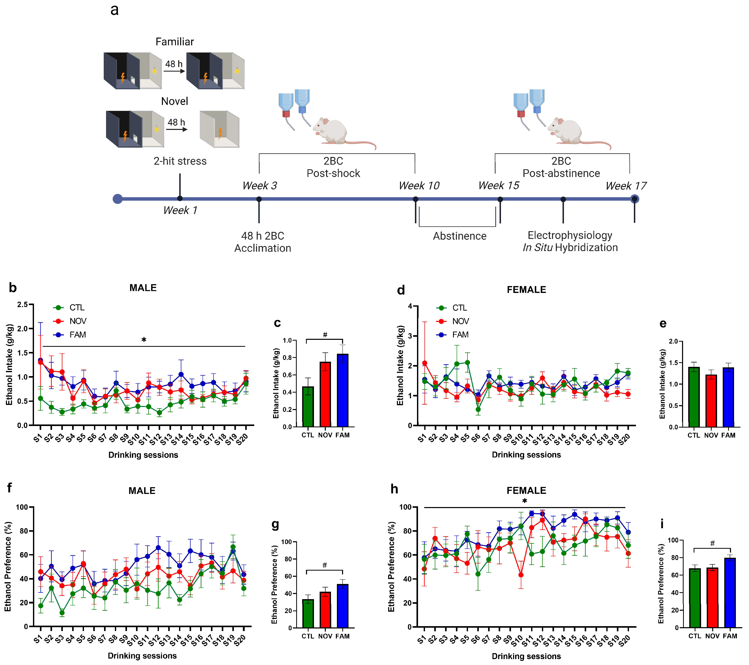
2.2. PTSD/AUD Model Procedures

Rats were generated using an established comorbid model of PTSD and AUD (see Figure 1a for experimental timeline). These procedures utilized an inhibitory avoidance (IA) shuttle box that was modified from a Habitest box (30.5 cm × 25 cm × 28 cm, Coulbourn, Holliston MA, USA) with a shocker as previously described [68]. The “2-hit’ shock stress was elicited using two distinguished contextual approaches. The first shock (a single 3 mA footshock administered for 2 s) was given after the rat crossed from the illuminated to the dark chamber of the IA shuttle box [12]. The second footshock occurred 48 h later, whereby half of the stressed rats received the second footstock in the same familiar apparatus as the first (termed FAM) [12]. The other half of the stressed rats received the second footshock in a novel, environmentally distinct, single-chambered apparatus (termed NOV) [12]. Controls (CTL) were handled by experimenters and were naïve to shock stress [12]. Beginning 2 weeks after the first footshock, rats received 48 h acclimation to ethanol (20% v/v) followed by chronic, intermittent (Mondays, Wednesdays, Fridays), limited 2BC access (2 h) to ethanol at scotophase onset [12]. Beginning 2 weeks after the first footshock, rats received 48 h acclimation to ethanol (20% v/v) followed by chronic, intermittent (Mondays, Wednesdays, Fridays), limited 2BC access (2 h) to ethanol at scotophase onset [12]. Two weeks later, all rats were acclimated to an initial 2-bottle choice (2BC) ethanol access (20% v/v) that lasted 48 h in their home cage. Specifically, all rats continued a 6-week regimen of 2BC intermittent ethanol access sessions. Ethanol and water consumption were calculated from bottle weights at the end of each 2 h session. Whenever ethanol was removed for nonaccess days, a second water bottle replaced it. Ethanol and water bottle positions were alternated each 2BC session [12]. FAM male rats showed significantly higher mean ethanol intake (~0.8 g/kg/2 h) (Figure 1b,c) and preference (Figure 1f,g) than CTL males (~0.4 g/kg/2 h). NOV and FAM female rats showed similar ethanol intake (~1.2–1.4 g/kg/2 h) as compared to CTL (~1.4 g/kg/2 h) (Figure 1d,e). However, FAM females showed higher preference for alcohol when compared to CTL rats (Figure 1h,i). Rats then entered a 5-week abstinence period followed by renewed post-abstinence 2BC drinking access over 3 sessions, 24 h apart, staggered across 2 weeks. The first 2 sessions were 2 h long and the final session was 24 h with drinking measured at 2 h and 24 h [12]. All rats were euthanized 24 h after the last 2BC alcohol session (therefore, at 24 h of ethanol withdrawal) for assessment of cell-type gene expression and synaptic function within the CeA. At the time of euthanasia, female rats were lavaged to determine their estrous cycle phase.

2.3. RNAscope In Situ Hybridization

A subset of rats (n = 13; n = 3–4/sex/treatment, 2 CeA sections from each rat) were deeply anesthetized with 3–5% isoflurane and perfused with cold PBS/Z-fix (Anathec LTD, 6480, Battle Creek, MI, USA). Brains were rapidly removed, and immersion-fixed in Z-fix for 24 h at 4 °C, cryoprotected in sterile 30% sucrose in PBS for 24–48 h at 4 °C, flash-frozen in prechilled isopentane on dry ice, and stored at −80 °C. Brains were then sliced on a cryostat in 20 μm thick sections, mounted on SuperFrost Plus slides (Fisher Scientific, 1255015, Portsmouth, NH, USA), and stored at −80 °C. In situ hybridization was performed using RNAscope fluorescent multiplex kit (ACD, 320850) in RNase-free conditions [53,73,74,75,76]. To perform the RNAscope in situ hybridization, a target retrieval protocol was performed as suggested by the manufacturer (ACD, 320535). Slides were then submerged in target retrieval buffer at 95–98 °C for 10 min, immediately washed in distilled water and then dehydrated with 100% ethanol, and lastly digested with Protease IV for 20 min at 40 °C. Following this pretreatment, the RNAscope Fluorescent Multiplex Detection kit User Manual (ACD, doc.no. 320293) was followed and slides were mounted with DAPI-containing Vectashield (Vector Laboratories, H100, CA, USA). A negative control (ACD, 320871) was run in tandem. The probes used from ACD Biotechne were as follows: Il18 (547731-C1), Il18r1 (547741-C2) and Il18bp (547751-C3).

2.4. Imaging and Analysis

We took multiple (6) images from each medial subdivision of CeA at similar bregma points to control for variation using a Zeiss LSM 780 laser scanning confocal microscope (40× oil immersion, 1024 × 1024, tile scanning of CeA 5-μm z-stacks) and kept all microscope settings the same between the experiments during image acquisition. Analysis and quantification were performed as previously described [53,73,74]. Quantification was performed using Fiji, by identifying nuclei based on DAPI staining in the CeA. Nuclei were considered positive (+) for probe if corresponding fluorescent signal was present after background (negative control) subtraction. The percentage of positive nuclei for labeled cells was calculated and then normalized to CTL group. The number of Il18bp+ nuclei that expressed Il18+ was determined by dividing the number of co-labeled nuclei by the total of Il18bp+ nuclei. For calculating the number of Il18+ nuclei that co-expressed Il18bp+, we divided the number of co-labeled nuclei by the total of Il18+. Brightness/contrast and pixel dilation are the same for all representative images.

2.5. Ex Vivo Slice Electrophysiology

A subset of 6–8 rats per group were used for electrophysiology. Rats were anesthetized with 3–5% isoflurane and sacrificed at the beginning of the dark cycle 24 h after the last 2BC session [12]. Brains were quickly isolated and placed in ice-cold oxygenated (95% O2 and 5% CO2) high-sucrose cutting solution (in mM: sucrose, 206; KCl, 2.5; CaCl2, 0.5; MgCl2, 7; NaH2PO4, 1.2; NaHCO3, 26; glucose, 5; HEPES, 5) [12,53,63,74]. Coronal slices (300 µm) containing the CeA were cut on a Leica 1200S vibratome (Leica Microsystems, Buffalo Grove, IL, USA) and then incubated in artificial cerebrospinal fluid (in mM: NaCl, 130; KCl, 3.5; NaH2PO4, 1.25; MgSO4•7H2O, 1.5; CaCl2, 2.0; NaHCO3, 24; glucose, 10) at 37 °C for 30 min and then at room temperature for a minimum of 30 min. We recorded from a total of 100 neurons in the medial subdivision of the CeA, that were visualized with infrared differential interference contrast optics and CCD cameras (QImaging, Surrey, BC, Canada). Recordings were performed in gap-free acquisition mode with a sampling rate per signal of 20 kHz and low-pass filtered at 10 kHz, using a Multiclamp 700 B amplifiers, Digidata 1440 A digitizer and pClamp 10 software (Molecular Devices, Sunnyvale, CA, USA). We filled borosilicate glass patch pipettes (3–6 MΩ; Warner Instruments, Hamden, CT, USA) with internal solution (in mM: 145.0 KCl; 5.0 EGTA; 0.5 MgCl2; 10.0 HEPES; 2.0 Mg-ATP; 0.2 Na-GTP). Spontaneous miniature GABAA–mediated inhibitory postsynaptic currents (mIPSCs) were pharmacologically isolated with 6,7-dinitroquinoxaline-2,3-dione (DNQX, 20 µM), DL-2-amino-5-phosphonovalerate (DL AP-5, 30 µM) and CGP55845A (1 µM) and 0.5 µM tetrodotoxin (TTX). Neurons were clamped at −60 mV and experiments with a series resistance > 25 MΩ or a >20% change in series resistance, as monitored with frequent 10 mV pulses, were excluded. All mIPSC frequency, amplitude, and kinetics data were analyzed using Mini Analysis (Synaptosoft Inc., Fort Lee, NJ, USA) and visually confirmed. Only mIPSCs > 5 pA were accepted for analysis, and mIPSC characteristics were binned based on 3–5 min of recording.

2.6. Statistical Analyses

Analysis of variance models (ANOVA) were used to analyze the data with stress condition (CTL versus NOV versus FAM) as a between-subjects factor. Drinking behavior was analyzed using repeated-measure ANOVA with session (1–20) as within-subjects factor and stress as between-subjects factor. Regarding RNAscope results, each probe was normalized to the naïve-stress group (CTL) and analyzed as one-way ANOVA. Electrophysiology data were analyzed with one-way ANOVA with stress condition as a between-subjects factor. In instances where ANOVA effects were significant, follow up post hoc analyses were conducted using Tukey’s HSD test, and alpha level was set at p < 0.05. All data graphing and statistical analysis were conducted in GraphPad Prism Version 8.

3. Results

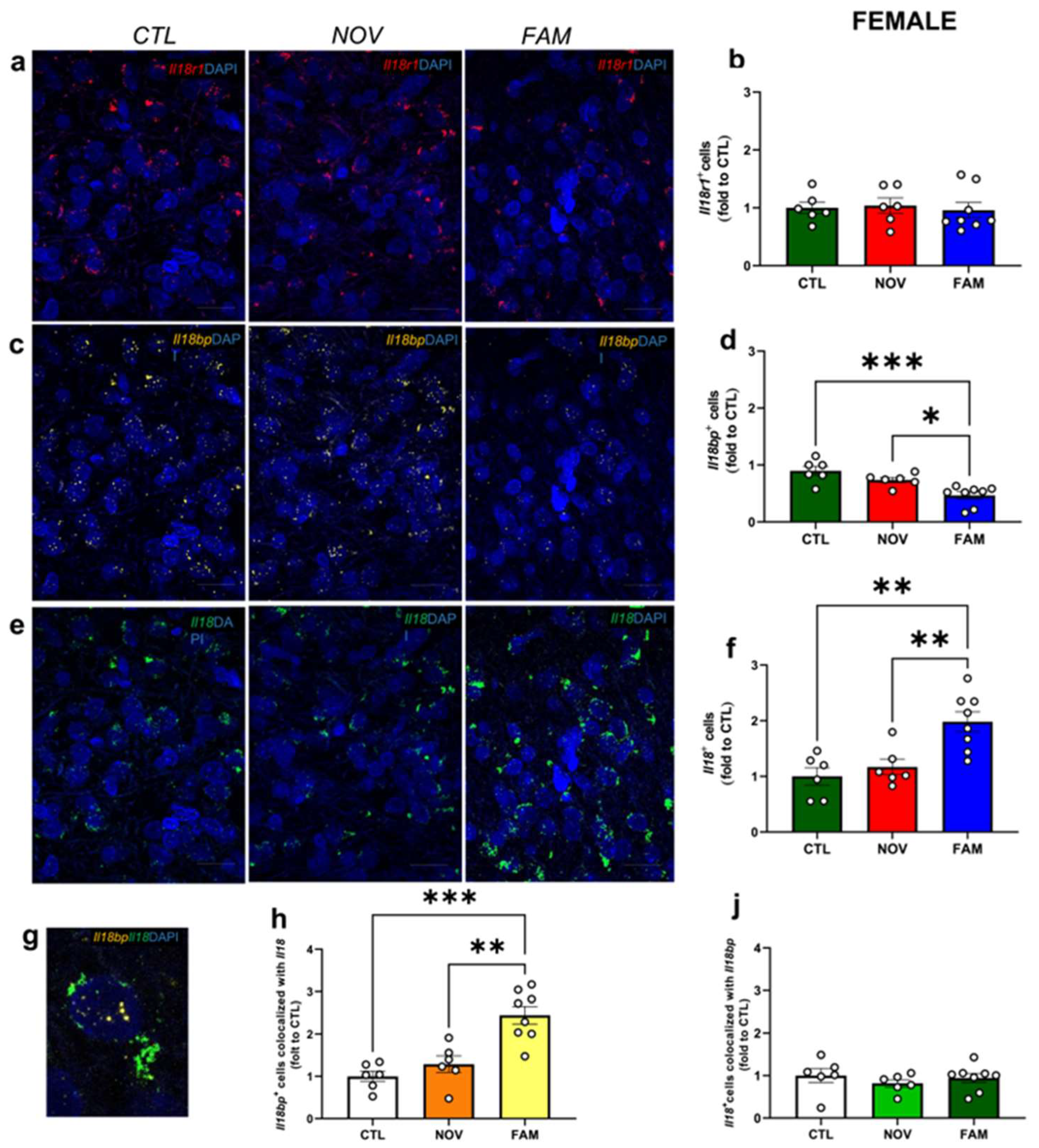
3.1. Expression of Il18r1, Il18 and Il18bp in the CeA of Male and Female PTSD/AUD Rats

To understand how the IL-18 system in the CeA is affected by comorbid PTSD/AUD, we used ISH/RNAscope to quantify the percent of cells expressing Il18r1 (encodes the ligand binding subunit of the IL-18 receptor), Il18 (encodes IL-18), and Il18bp (encodes IL-18 binding protein) in male and female rats that experienced shock stress to a novel context (NOV) or familiar context (FAM) followed by alcohol drinking [12]. Unstressed control (CTL) drinking rats were naïve to shock stress exposure but received 2BC alcohol access [12]. We found no significant differences in CeA Il18r1 expression across male groups (Figure 2a,b); however, although it did not reach statistical significance, we observed a slight increase in Il18r1 expression in NOV male rats. Importantly, we observed a significant increase (Figure 2e,f; F2,15 = 7.850, p < 0.05) of Il18 expression in the FAM group versus CTL and NOV groups. Notably, in male FAM groups we found a significant decrease (Figure 2c,d; F2,15 = 7.984, p < 0.05) in the expression of Il18bp as compared to the CTL and NOV rats. No significant differences were observed in Il18 and Il18bp expression between CTL and NOV groups.
Lastly, we identified an effect of stress to increase the co-localization of Il18bp and Il18+, whereby the number of Il18bp+ cells co-expressing Il18+ was significantly higher (Figure 2g,h; F2,15 = 13.17, p < 0.05) in the FAM compared to the CTL and NOV rats. Interestingly, there was also a marginal difference in the FAM stress group that showed a trend toward an increase in the number of Il18+ cells that co-expressed Il18bp+ (Figure 2J) as compared to CTL and NOV. This finding suggests that increased Il18bp co-localization may be a compensatory mechanism to counteract increased IL-18 expression.
Similarly to males, we did not observe significant differences in Il18r1 expression (Figure 3 a,b) between CTL, NOV, and FAM groups in female rats. However, female FAM stress rats displayed a significant (Figure 3e,f; F2,17 = 7.467, p < 0.05) increase in Il18+ cells compared to CTL and NOV female rats. Interestingly, FAM female rats showed a significant reduction (Figure 3c,d; F2,17 = 11.80, p < 0.05) of Il18bp+ cells versus CTL and NOV groups. Moreover, the number of Il18bp+ cells that also expressed Il18 was significantly higher (Figure 3g,h; F2,17 = 17.54, p < 0.05) than in CTL and NOV groups. Similar co-localization was found for the number of Il18+ cells that co-expressed Il18bp+ (Figure 3J) between CTL, NOV, and FAM groups. Overall, these findings support a critical interplay of IL-18BP and IL-18 in the CeA after stress in rats with a history of voluntary alcohol access, particularly in the FAM contextual environment.

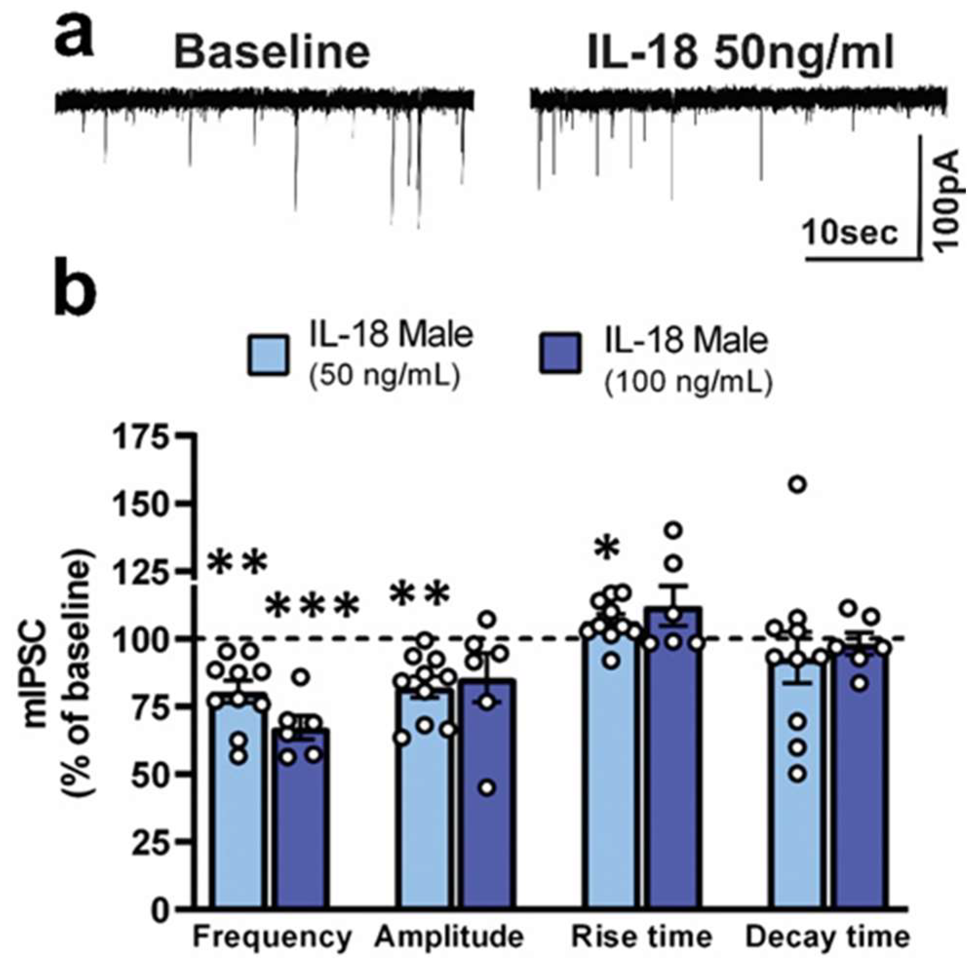
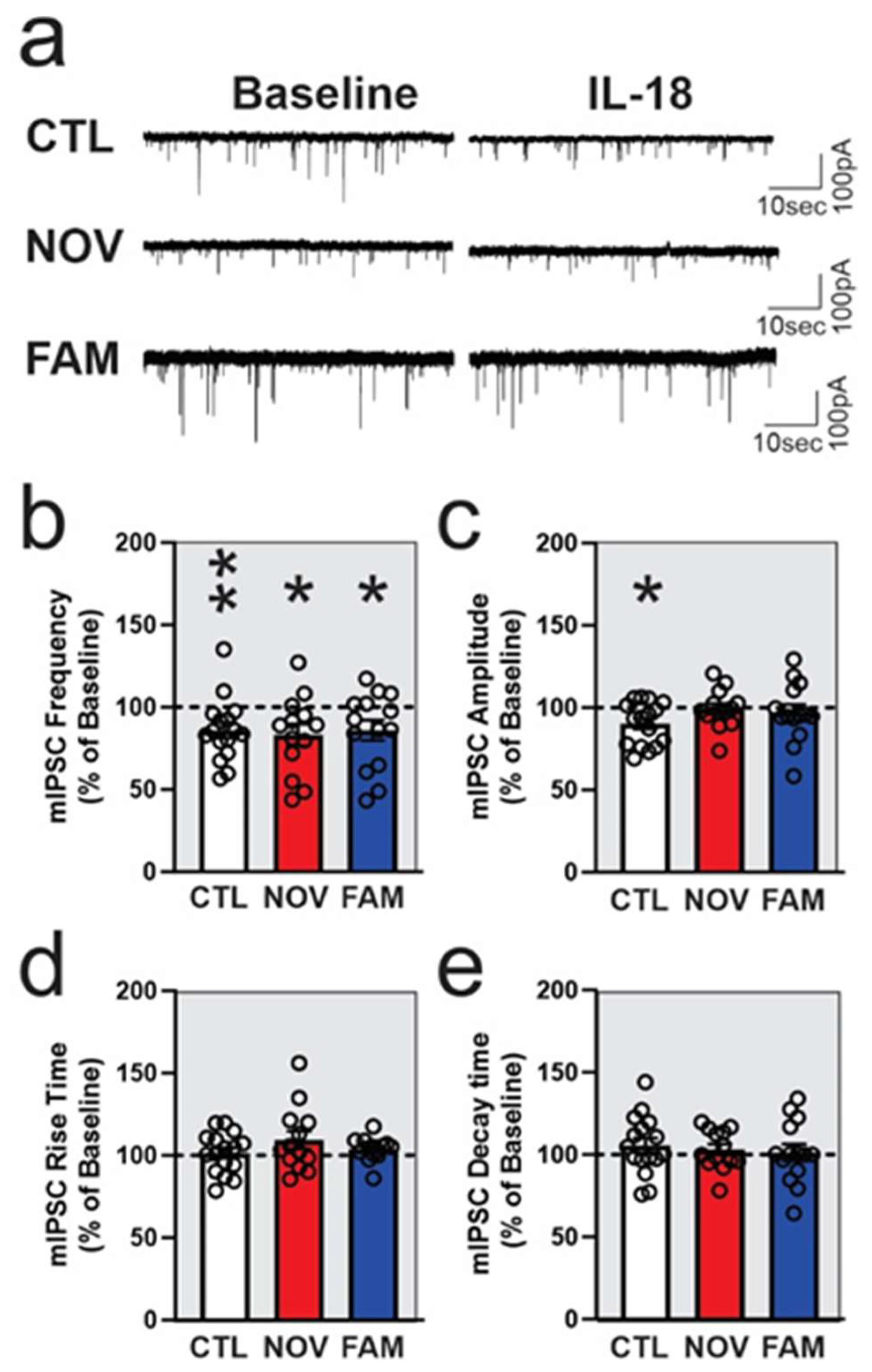
3.2. IL-18 Reduces Vesicular GABA Release in Ex Vivo CeA Slices of Ethanol- and Stress-Naïve Male Rats

To determine the potential functional role of IL-18 on synaptic transmission, we performed whole-cell patch clamp recordings of pharmacological-isolated GABAA-mediated miniature inhibitory postsynaptic currents (mIPSCs) in the medial subdivision of the CeA from unstressed Wistar males with no alcohol history (naïve control). Based on previous slice physiology studies [77,78,79,80] and brain homogenates IL-18 physiological concentrations [81,82], we tested 50 and 100 ng/mL of recombinant rat IL-18 on CeA mIPSCs from naïve control. We found that 50 and 100 ng/mL IL-18 applied for 10–15 min significantly (p < 0.05, one sample t-test) decreased mIPSC frequency by 20 ± 4% and by 34 ± 4% in the CeA of naïve rats (Figure 4a,b), respectively. These data suggest that IL-18 significantly decreases vesicular GABA release. IL-18 at 50 ng/ml, but not at 100 ng/ml, also significantly decreased mIPSC amplitude (without affecting rise and decay times) by 20 ± 4% (Figure 4a,b), suggesting effects on GABAA function at postsynaptic level. Overall, these data indicate that 50 ng/mL IL-18 is an effective concentration that modulates basal GABA transmission in the CeA in ethanol and stress-naïve controls. We used this concentration for the rest of the electrophysiological studies.

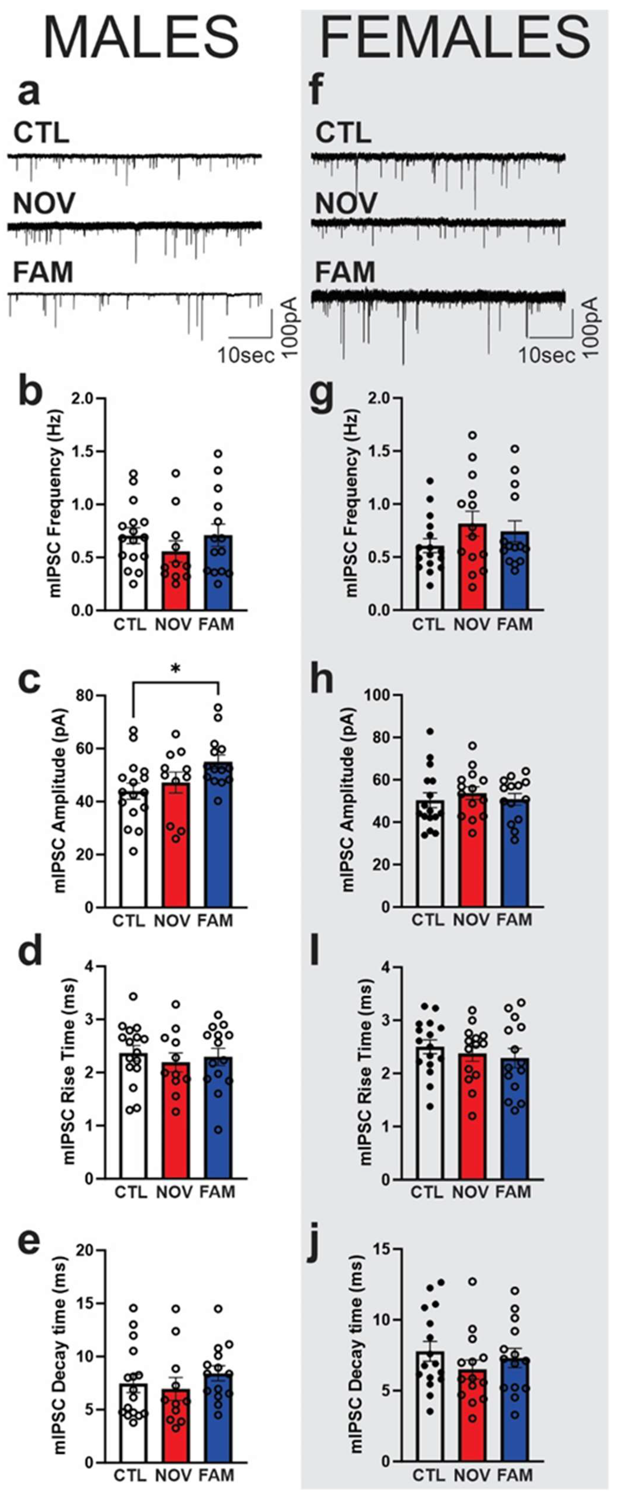
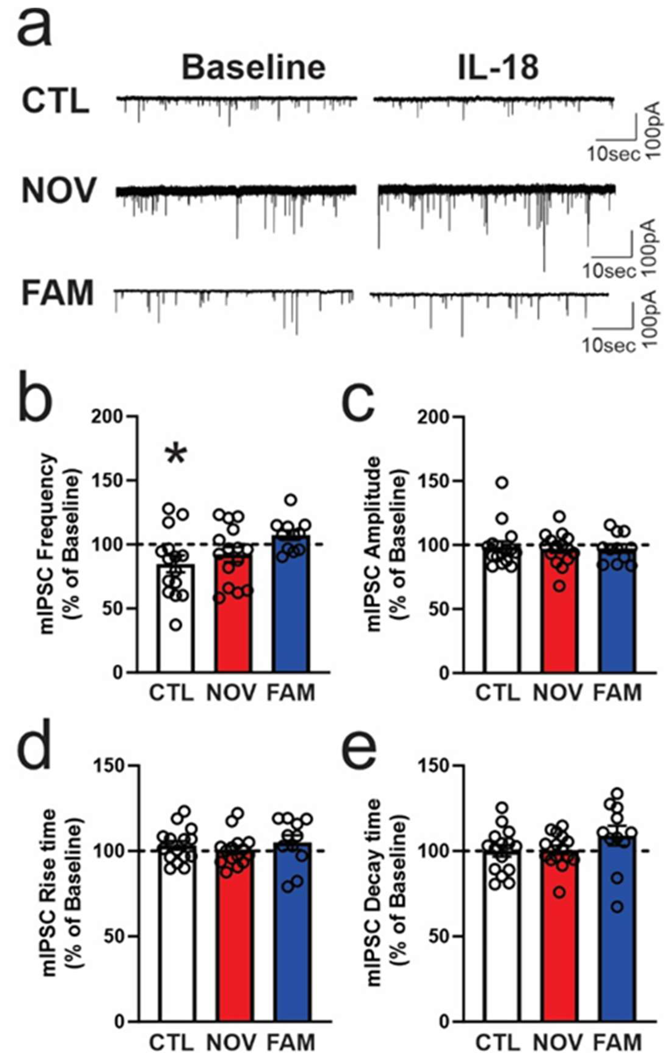
3.3. Stress Alters the Effects of IL-18 on Spontaneous Vesicular GABAergic Transmission in a Sex-Dependent Manner

We previously showed that elevated GABAA-receptor mediated synaptic transmission in the CeA represents a hallmark of alcohol dependence across species [46,54,61], and our 2-hit model also increased action-potential-independent mIPSCs from CeA neurons [12]. Thus, here, we used a random subset of rats (6–7) from each of three experimental groups (CTL, NOV, and FAM) for electrophysiological study (see Figure 1) to determine 1) potential baseline differences in CeA mIPSCs and 2) the synaptic effects of IL-18 on mIPSCs. Notably, CeA neurons from FAM males displayed significantly (p < 0.05) larger basal mIPSC amplitudes (52 ± 4 pA) compared to CTL males (44 ± 5 pA; Figure 5a,c), suggesting increased postsynaptic GABAA function. Frequency (Figure 5b) and kinetics (Figure 5d,e) of mIPSCs were not different among the three groups. In females, we did not observe significant differences between groups in any mIPSC properties (Figure 5f–k), although a trend towards increased mIPSC frequency (0.95 ± 0.2 Hz; 0.74 ± 0.10 Hz; Figure 5g) was seen in NOV and FAM compared to CTL (0.61 ± 0.1 Hz) rats.
We next investigated the effects of acute IL-18 application (12–15 min) at 50 ng/ml on CeA mIPSCs of both sexes in all treatment groups (Figure 6 and Figure 7). In CeA neurons of CTL males, we found that IL-18 significantly (p < 0.05) decreased (84.8 ± 6.6% of baseline, n = 15) the frequency of mIPSCs, suggesting decreased presynaptic GABA release. Interestingly, this IL-18-induced decrease in GABA release was not observed in CeA of NOV and FAM males (Figure 6a,b). Amplitude (Figure 6c) and rise and decay times (Figure 6d,e) of mIPSCs were not altered by IL-18 application in any of the three groups.
In CTL females, acute application of 50 ng/ml IL-18 significantly decreased (86.0 ± 4.8%; p < 05; n = 16) the frequency of mIPSCs, suggesting that it decreased presynaptic GABA release (Figure 7a,b). Notably, IL-18 also significantly (p < 0.05) decreased (90.2 ± 4.3% of baseline) the amplitude of mIPSCs (Figure 7a,c), suggesting that it decreased postsynaptic GABAA function in CTL females. In contrast to males, IL-18 also significantly decreased CeA mIPSC frequency in NOV (to 85.9 ± 6.2% of baseline; p < 0.05; n = 14) and FAM (83.1 ± 6.6% of baseline; p < 0.05; n = 13) females. However, in both NOV and FAM females, IL-18 did not alter the amplitude of mIPSCs (Figure 7a,c) as it does in CTL, suggesting loss of postsynaptic effects in these groups. No changes were observed in the kinetics of mIPSCs (Figure 7d–e) upon IL-18 application in any of the female groups.

4. Discussion

There is extensive research on the effects of stress on both alcohol intake and anxiety in rodent models. Our laboratory established a two-hit model of traumatic stress to study comorbid drinking-PTSD phenotypes, elicited by past stress in familiar (FAM) or novel contexts (NOV). We previously reported that FAM and NOV stress increased alcohol preference in males and females, respectively, along with fear overgeneralization (in males) and hyperarousal and sleep disturbances (in females) [12]. This model also alters CeA inhibitory GABAergic baseline transmission in the stress-context-specific manner [12]. Here, we examined potential effects of this model of comorbid PTSD/AUD on gene expression of components of IL-18 signaling and on the functional impact of IL-18 on inhibitory CeA transmission. Our study revealed that the number of CeA expressing ll18 was increased while Il18bp was decreased in FAM stressed rats of both sexes versus CTLs. FAM stressed rats also displayed higher co-expression of Il18bp positive cells with Il18 as compared to CTL or NOV rats. Furthermore, exogenous IL-18 application reduced mIPSC frequencies in the CeA of CTL rats, an effect lost in both NOV and FAM males but that persisted in NOV and FAM females. IL-18 also decreased mIPSC amplitude in female CTL rats, suggesting postsynaptic effects. Our results imply that stress in rats with alcohol access differentially impacts CeA IL-18 expression and its modulatory function at the GABA synapses.
We speculated that CeA IL-18 signaling undergoes numerous changes from gene expression to proteins in our model of stress with alcohol history. This was evident by our finding that CeA ll18 gene expression was increased after FAM stress and alcohol-drinking history. In a consistent manner, Il18bp, which functions to inhibit IL18 activity, was decreased in FAM stressed rats versus CTLs, suggesting a regulatory role of IL-18 signaling and highlighting a dysregulation of the IL-18 pathway in both sexes. Indeed, IL-18 acts via a receptor complex that closely resembles that of IL-1, consisting of a ligand binding protein, IL-18R1 (formerly IL-18Ra) and an accessory protein, IL-18RAP (formerly IL-18Rβ) [83]. The activity of IL-18 is controlled by endogenous soluble IL-18 binding protein (IL-18-BP), which inhibits IL18 excessive activation [83]. This action is consistent with our finding that FAM stress increases CeA IL18 while simultaneously decreasing IL18bp.
Our gene expression data revealed no significant differences in number of CeA expressing Il18r1 across all the experimental male and female groups. We also observed that no significant changes occurred in Il18 and Il18bp expression in NOV group of rats of either sex. Interestingly, we found a significant co-localization between Il18bp and Il18, in which the percentage of Il18bp+ cells co-expressing with Il18+ were significantly higher in the FAM stress compared to CTL or NOV group of both sexes. Note that in the FAM group the number of cells expressing Il18bp is significantly lower. We speculate that the increases in this Il18bp-Il18 co-localization may represent a compensatory mechanism in response to elevated Il18 expression. We postulate that traumatic stress and/or its resulting increases in alcohol drinking are accompanied by increases in IL-18 inflammatory action that likely leads to increases in comorbid-like behavioral phenotypes of PTSD/AUD. Future studies are needed to confirm the selective and functional role of IL-18 and receptor components in our established model of comorbid PTSD/AUD.
IL-18 is constitutively expressed in the brain under normal and pathological conditions [24], where it influences homeostasis and behavior. We hypothesized that an imbalance in IL-18 system molecules in the brain leads to an increase in the neuroinflammatory state, which we predicted would be observed in comorbid PTSD/AUD animals. Here, at the synaptic level, we did not find significant changes in baseline action-potential-independent GABAergic transmission at presynaptic levels across both FAM and NOV stress groups compared to control in both sexes. However, a significant increase in postsynaptic GABAA function was observed in FAM males, as previously reported [12]. In this cohort we did not observe increased basal vesicular GABA release in FAM compared to CTL females [12], but it is important to consider that in the present study the ethanol intake in CTL females was considerably high, and both FAM and NOV showed increased drinking, in contrast to our previous study [12]. Although voluntary intake without dependence is insufficient to alter GABAergic transmission in the CeA [84,85], we anticipated that the increased alcohol intake observed in stressed rats would contribute to the altered CeA GABA signaling. We hypothesized that the stress and alcohol history synergistically increase CeA GABA synaptic transmission, which is observed in other conditions (e.g., heightened anxiety and alcohol intake, such as alcohol dependence [46,86,87].
Interestingly, IL-18 application decreases vesicular GABA release in male CTL, an effect that was lost in FAM and NOV rats. In contrast, in females, the magnitude of the IL-18 presynaptic effects on vesicular GABA release is similar across CTL, FAM, and NOV groups. Furthermore, as discussed above, basal vesicular GABA release across groups was comparable, likely reflecting the comparable amount of alcohol intake between CTL and stressed females [12]. Of note, unlike the previous study [12], the current cohort of CTL females displayed high alcohol intake, reaching the drinking levels of the stressed groups. Notably, in female controls, IL-18 significantly decreased GABAA postsynaptic functions (as reflected by the IL-18 decrease in mIPSC amplitude) as it did in ethanol-naïve males in our initial study. However, this post-synaptic action of IL-18 was absent in male CTLs (who had a 2BC drinking history) and was blunted in both FAM and NOV female groups. Future studies will be conducted to understand the co-localization of IL-18 system elements with GABAergic cell types in the CeA under naïve vs. PTSD and AUD-like conditions.
A limitation of the current study is the lack of an alcohol-naïve group. We previously determined the impact of alcohol 2BC access in PTSD-like phenotypes, using a cohort of stressed rats that never had access to alcohol (they instead received 2BC access to water) [12]. We found that traumatic stress alone elicited signs of fear overgeneralization and impaired sleep maintenance, but post-stress alcohol access influenced the expression of other behaviors, exacerbating social avoidance and modifying startle and defensive behavioral responding [12]. Furthermore, at a synaptic level, FAM stress males (without history of alcohol intake) displayed significantly elevated vesicular GABA release compared to CTL males, while CeA GABA transmission did not differ between the other groups [12]. In the present study, our main goal was to study IL-18 signaling in AUD and PTSD comorbidity, and, thus, all groups were exposed to ethanol by 2BC paradigm. However, to determine the efficacy of exogenous application of IL-18 on the mIPSCs without an interference of the ethanol exposure, we used ethanol-naïve males. Indeed, application of IL-18 decreased the amplitude of mIPSCs in ethanol-naïve rats, suggesting postsynaptic effects elicited by IL-18, but not in the 2BC alcohol-exposed (CTL), rats. We speculate that the lack of IL-18-induced postsynaptic effects on GABAergic function derives from alcohol-induced changes at the GABA receptor level (e.g., receptor sensitivity and localization), which will need further investigation.
IL-18 is a compelling target in AUD and anxiety disorders, as it modulates biological functions during stress [26] and recent studies reported greater circulating IL-18 levels in men than women during states associated with inflammation [88,89]. In addition to stress-induced expression in stress-related brain regions [27,28], IL-18 also is present in the adrenal and pituitary gland. Its expression pattern during stress suggests that peripheral IL-18 also modulates the HPA-axis [90]. IL-18 is elevated during stress through the HPA-axis, and in paracrine fashion by CRF [43], and is downregulated through the parasympathetic nervous system activation in a tissue- and mechanism-specific fashion (IL-18BP and IL-18R) [26]. Our work points to the possibility that IL-18 serves as key regulator to the sensitivity of stress- and AUD-mediated CeA GABAergic synaptic function and its potential contribution to CeA dysregulation. This work represents a first step towards understanding the alterations induced by stress and alcohol on IL-18 signaling.
Several caveats are important to contextualize the lack of change in CeA Il18r1 ligand binding subunit mRNA expression across stress groups. First, protein levels of IL-18R1 were not assessed. Second, our study did not assess CeA Il18RAP expression, which encodes the signaling subunit of IL-18′s heterodimer receptor. Third, splice variants of both receptor IL-18R subunits with distinct actions have been identified in the CNS but were not distinguished here. For example, a shortened IL-18R1 isoform that lacks a Toll/IL-1 receptor (TIR) domain has been proposed to be an “IL-18Rα type II” decoy receptor, analogous to the type II IL-1R [25]. Similarly, a truncated splice variant of IL-18RAP encodes a soluble protein that consists of only the first immunoglobulin-like domain (EMBL/Genbank accession number AJ550893); both long and truncated forms of IL-18RAP were reported in rat brain and liver, as well as pure cultures of microglia, astrocytes, and neurons [75]. IL-18 binding to the IL-18R1 ligand binding subunit stabilizes its interaction with the signaling IL-18RAP subunit and the adaptor protein MyD88 through the TIR domain. Thus, decreases in expression of the signaling IL18RAP subunit or in relative levels of the active vs. type II decoy/inhibitory isoforms of each receptor subunit might contribute to the stress-induced blunting that we saw in IL-18′s functional effects. Finally, the level of IL-37, another endogenous ligand for the IL-18R1 receptor and IL-18BP that can influence IL-18 action [91], also was not studied. Future study of these molecular candidates may clarify the basis of adaptations in IL-18′s electrophysiologic actions.

5. Conclusions

In summary, we revealed that CeA IL-18 signaling is disrupted at multiple levels in the complex IL-18 signaling cascade, particularly following traumatic stress in familiar contexts. CeA ll18 expression was increased and the Il18bp component was decreased in rats with joint stress-alcohol drinking history. IL-18 normally affects CeA GABA synapses by decreasing inhibitory signaling in the CeA of stress-naïve rats, an effect that was absent in stressed males. IL-18 only decreased the postsynaptic inhibitory signaling of stress-naïve female and of stress- and alcohol-naïve male rats, suggesting that stress and/or alcohol history impairs IL-18′s postsynaptic effects. Our novel findings identify adaptations in neuroimmune IL-18 signaling within CeA GABA microcircuits in a model of PTSD/AUD comorbidity. Further work will elucidate the specific mechanisms and direct contribution of stress, drinking, or a combination of both on amygdala GABA transmission, as well as probe the IL-18 system as a candidate therapeutic target to reduce symptoms of PTSD/AUD comorbidity in both sexes.

Supplementary Materials

The following supporting information can be downloaded at: https://www.mdpi.com/article/10.3390/cells12151943/s1, Figure S1: Representative negative control images in the CeA of male and female indicate negligible fluorescence intensity for all the channels.

Author Contributions

Conceptualization, M.R., D.K., E.P.Z., V.B., B.C. and V.V.; methodology, V.B., M.B., M.Q.S., M.R., S.K., R.V., S.D. and C.S.O.; validation, D.K., V.B., B.C., V.V., M.Q.S., M.R., S.K., R.V., M.B. and C.S.O.; formal analysis, D.K., V.B., M.R., S.K., R.V. and M.B.; investigation, D.K., V.B., M.Q.S., M.R., S.K., R.V., M.B. and R.B.; resources, D.K., M.R. and E.P.Z.; data curation, V.B., M.R., S.K., M.B., V.V. and B.C.; writing—original draft preparation, V.B., M.R., V.V. and B.C.; writing—review and editing, V.B., M.R., S.K., M.B., V.V., B.C., D.K., M.Q.S., R.V., M.B., C.S.O., R.B. and S.D.; visualization, V.B., B.C., V.V., M.R., S.K. and M.B.; supervision, M.R. and E.P.Z.; project administration, M.R.; funding acquisition, D.K., M.R. and E.P.Z. All authors have read and agreed to the published version of the manuscript.

Funding

Support for this study was provided by the National Institute on Alcohol Abuse and Alcoholism grants AA027700, AA013498, P60 AA006420, AA017447, AA021491, AA029841, AA028879, AA029498, K99 AA026638, K99 AA030609, and T32 AA007456, as well as the Schimmel Family Chair and Pearson Center for Alcoholism and Addiction Research.

Institutional Review Board Statement

The animal study protocol was approved by the Institutional Animal Care and Use Committee of The Scripps Research Institute (IACUC 09-0006; approved 20 August 2020).

Informed Consent Statement

Not applicable.

Data Availability Statement

The data that support the findings of this study are available from the corresponding author upon reasonable request.

Acknowledgments

This is manuscript number 30237 from The Scripps Research Institute.

Conflicts of Interest

The authors declare no conflict of interest.

References

  1. Koob, G.F. Addiction is a Reward Deficit and Stress Surfeit Disorder. Front. Psychiatry 2013, 4, 72. [Google Scholar] [CrossRef] [PubMed] [Green Version]
  2. Sinha, R. Chronic stress, drug use, and vulnerability to addiction. Ann. N. Y. Acad. Sci. 2008, 1141, 105–130. [Google Scholar] [CrossRef] [PubMed] [Green Version]
  3. Logrip, M.L.; Zorrilla, E.P. Stress history increases alcohol intake in relapse: Relation to phosphodiesterase 10A. Addict. Biol. 2012, 17, 920–933. [Google Scholar] [CrossRef] [PubMed]
  4. Logrip, M.L.; Zorrilla, E.P.; Koob, G.F. Stress modulation of drug self-administration: Implications for addiction comorbidity with post-traumatic stress disorder. Neuropharmacology 2012, 62, 552–564. [Google Scholar] [CrossRef] [Green Version]
  5. Johnston, A.L.; Thevos, A.K.; Randall, C.L.; Anton, R.F. Increased severity of alcohol withdrawal in in-patient alcoholics with a co-existing anxiety diagnosis. Br. J. Addict. 1991, 86, 719–725. [Google Scholar] [CrossRef]
  6. Schellekens, A.F.; de Jong, C.A.; Buitelaar, J.K.; Verkes, R.J. Co-morbid anxiety disorders predict early relapse after inpatient alcohol treatment. Eur. Psychiatry J. Assoc. Eur. Psychiatr. 2015, 30, 128–136. [Google Scholar] [CrossRef]
  7. Becker, H.C.; Lopez, M.F.; Doremus-Fitzwater, T.L. Effects of stress on alcohol drinking: A review of animal studies. Psychopharmacology 2011, 218, 131–156. [Google Scholar] [CrossRef] [Green Version]
  8. Gilpin, N.W.; Weiner, J.L. Neurobiology of comorbid post-traumatic stress disorder and alcohol-use disorder. Genes Brain Behav. 2017, 16, 15–43. [Google Scholar] [CrossRef] [Green Version]
  9. Rorick, L.M.; Finn, P.R.; Steinmetz, J.E. High-alcohol-drinking rats exhibit persistent freezing responses to discrete cues following Pavlovian fear conditioning. Pharmacol. Biochem. Behav. 2003, 76, 223–230. [Google Scholar] [CrossRef]
  10. Liu, X.; Weiss, F. Stimulus conditioned to foot-shock stress reinstates alcohol-seeking behavior in an animal model of relapse. Psychopharmacology 2003, 168, 184–191. [Google Scholar] [CrossRef]
  11. Meyer, E.M.; Long, V.; Fanselow, M.S.; Spigelman, I. Stress increases voluntary alcohol intake, but does not alter established drinking habits in a rat model of posttraumatic stress disorder. Alcohol. Clin. Exp. Res. 2013, 37, 566–574. [Google Scholar] [CrossRef] [Green Version]
  12. Steinman, M.Q.; Kirson, D.; Wolfe, S.A.; Khom, S.; D’Ambrosio, S.R.; Spierling Bagsic, S.R.; Bajo, M.; Vlkolinsky, R.; Hoang, N.K.; Singhal, A.; et al. Importance of sex and trauma context on circulating cytokines and amygdalar GABAergic signaling in a comorbid model of posttraumatic stress and alcohol use disorders. Mol. Psychiatry 2021, 26, 3093–3107. [Google Scholar] [CrossRef]
  13. Cruz, B.; Vozella, V.; Carper, B.A.; Xu, J.C.; Kirson, D.; Hirsch, S.; Nolen, T.; Bradley, L.; Fain, K.; Crawford, M.; et al. FKBP5 inhibitors modulate alcohol drinking and trauma-related behaviors in a model of comorbid post-traumatic stress and alcohol use disorder. Neuropsychopharmacology 2023, 48, 1144–1154. [Google Scholar] [CrossRef] [PubMed]
  14. Bouton, M.E.; Mineka, S.; Barlow, D.H. A modern learning theory perspective on the etiology of panic disorder. Psychol. Rev. 2001, 108, 4–32. [Google Scholar] [CrossRef] [PubMed]
  15. Tinsley, M.R.; Quinn, J.J.; Fanselow, M.S. The role of muscarinic and nicotinic cholinergic neurotransmission in aversive conditioning: Comparing pavlovian fear conditioning and inhibitory avoidance. Learn. Mem. 2004, 11, 35–42. [Google Scholar] [CrossRef] [Green Version]
  16. Mayfield, J.; Ferguson, L.; Harris, R.A. Neuroimmune signaling: A key component of alcohol abuse. Curr. Opin. Neurobiol. 2013, 23, 513–520. [Google Scholar] [CrossRef] [PubMed] [Green Version]
  17. Crews, F.T.; Bechara, R.; Brown, L.A.; Guidot, D.M.; Mandrekar, P.; Oak, S.; Qin, L.; Szabo, G.; Wheeler, M.; Zou, J. Cytokines and alcohol. Alcohol. Clin. Exp. Res. 2006, 30, 720–730. [Google Scholar] [CrossRef]
  18. Kimpel, M.W.; Strother, W.N.; McClintick, J.N.; Carr, L.G.; Liang, T.; Edenberg, H.J.; McBride, W.J. Functional gene expression differences between inbred alcohol-preferring and -non-preferring rats in five brain regions. Alcohol 2007, 41, 95–132. [Google Scholar] [CrossRef] [Green Version]
  19. Ponomarev, I.; Wang, S.; Zhang, L.; Harris, R.A.; Mayfield, R.D. Gene co-expression networks in human brain identify epigenetic modifications in alcohol dependence. J. Neurosci. 2012, 32, 1884–1897. [Google Scholar] [CrossRef] [Green Version]
  20. Mulligan, M.K.; Ponomarev, I.; Hitzemann, R.J.; Belknap, J.K.; Tabakoff, B.; Harris, R.A.; Crabbe, J.C.; Blednov, Y.A.; Grahame, N.J.; Phillips, T.J.; et al. Toward understanding the genetics of alcohol drinking through transcriptome meta-analysis. Proc. Natl. Acad. Sci. USA 2006, 103, 6368–6373. [Google Scholar] [CrossRef]
  21. He, J.; Crews, F.T. Increased MCP-1 and microglia in various regions of the human alcoholic brain. Exp. Neurol. 2008, 210, 349–358. [Google Scholar] [CrossRef] [Green Version]
  22. Warden, A.S.; Wolfe, S.A.; Khom, S.; Varodayan, F.P.; Patel, R.R.; Steinman, M.Q.; Bajo, M.; Montgomery, S.; Vlkolinsky, R.; Nadav, T.; et al. Microglia control escalation of drinking in alcohol dependent mice: Genomic and synaptic drivers. Biol. Psychiatry 2020, 88, 910–921. [Google Scholar] [CrossRef] [PubMed]
  23. Nixon, K.; Kim, D.H.; Potts, E.N.; He, J.; Crews, F.T. Distinct cell proliferation events during abstinence after alcohol dependence: Microglia proliferation precedes neurogenesis. Neurobiol. Dis. 2008, 31, 218–229. [Google Scholar] [CrossRef] [PubMed] [Green Version]
  24. Dinarello, C.A.; Novick, D.; Kim, S.; Kaplanski, G. Interleukin-18 and IL-18 binding protein. Front. Immunol. 2013, 4, 289. [Google Scholar] [CrossRef] [PubMed] [Green Version]
  25. Alboni, S.; Cervia, D.; Sugama, S.; Conti, B. Interleukin 18 in the CNS. J. Neuroinflammation 2010, 7, 9. [Google Scholar] [CrossRef] [Green Version]
  26. Sugama, S.; Conti, B. Interleukin-18 and stress. Brain Res. Rev. 2008, 58, 85–95. [Google Scholar] [CrossRef]
  27. Kim, T.K.; Kim, J.E.; Choi, J.; Park, J.Y.; Lee, J.E.; Lee, E.H.; Lee, Y.; Kim, B.Y.; Oh, Y.J.; Han, P.L. Local Interleukin-18 System in the Basolateral Amygdala Regulates Susceptibility to Chronic Stress. Mol. Neurobiol. 2017, 54, 5347–5358. [Google Scholar] [CrossRef]
  28. Kim, T.K.; Kim, J.E.; Choi, J.; Park, J.Y.; Lee, J.E.; Lee, E.H.; Lee, Y.; Kim, B.Y.; Oh, Y.J.; Han, P.L. Erratum to: Local Interleukin-18 System in the Basolateral Amygdala Regulates Susceptibility to Chronic Stress. Mol. Neurobiol. 2017, 54, 5359–5360. [Google Scholar] [CrossRef] [Green Version]
  29. Lisboa, S.F.; Issy, A.C.; Biojone, C.; Montezuma, K.; Fattori, V.; Del-Bel, E.A.; Guimaraes, F.S.; Cunha, F.Q.; Verri, W.A.; Joca, S.R.L. Mice lacking interleukin-18 gene display behavioral changes in animal models of psychiatric disorders: Possible involvement of immunological mechanisms. J. Neuroimmunol. 2018, 314, 58–66. [Google Scholar] [CrossRef] [Green Version]
  30. Novick, D.; Kim, S.H.; Fantuzzi, G.; Reznikov, L.L.; Dinarello, C.A.; Rubinstein, M. Interleukin-18 binding protein: A novel modulator of the Th1 cytokine response. Immunity 1999, 10, 127–136. [Google Scholar] [CrossRef] [Green Version]
  31. Aizawa, Y.; Akita, K.; Taniai, M.; Torigoe, K.; Mori, T.; Nishida, Y.; Ushio, S.; Nukada, Y.; Tanimoto, T.; Ikegami, H.; et al. Cloning and expression of interleukin-18 binding protein. FEBS Lett. 1999, 445, 338–342. [Google Scholar] [CrossRef] [Green Version]
  32. Blednov, Y.A.; Benavidez, J.M.; Black, M.; Mayfield, J.; Harris, R.A. Role of interleukin-1 receptor signaling in the behavioral effects of ethanol and benzodiazepines. Neuropharmacology 2015, 95, 309–320. [Google Scholar] [CrossRef] [Green Version]
  33. Fernandez-Lizarbe, S.; Pascual, M.; Guerri, C. Critical role of TLR4 response in the activation of microglia induced by ethanol. J. Immunol. 2009, 183, 4733–4744. [Google Scholar] [CrossRef] [PubMed] [Green Version]
  34. Pascual, M.; Balino, P.; Aragon, C.M.; Guerri, C. Cytokines and chemokines as biomarkers of ethanol-induced neuroinflammation and anxiety-related behavior: Role of TLR4 and TLR2. Neuropharmacology 2015, 89, 352–359. [Google Scholar] [CrossRef]
  35. Mayeux, J.P.; Teng, S.X.; Katz, P.S.; Gilpin, N.W.; Molina, P.E. Traumatic brain injury induces neuroinflammation and neuronal degeneration that is associated with escalated alcohol self-administration in rats. Behav. Brain Res. 2015, 279, 22–30. [Google Scholar] [CrossRef]
  36. Varodayan, F.P.; Pahng, A.R.; Davis, T.D.; Gandhi, P.; Bajo, M.; Steinman, M.Q.; Kiosses, W.B.; Blednov, Y.A.; Burkart, M.D.; Edwards, S.; et al. Chronic ethanol induces a pro-inflammatory switch in interleukin-1beta regulation of GABAergic signaling in the medial prefrontal cortex of male mice. Brain Behav. Immun. 2023, 110, 125–139. [Google Scholar] [CrossRef]
  37. Patel, R.R.; Khom, S.; Steinman, M.Q.; Varodayan, F.P.; Kiosses, W.B.; Hedges, D.M.; Vlkolinsky, R.; Nadav, T.; Polis, I.; Bajo, M.; et al. IL-1beta expression is increased and regulates GABA transmission following chronic ethanol in mouse central amygdala. Brain Behav. Immun. 2019, 75, 208–219. [Google Scholar] [CrossRef] [PubMed]
  38. Dinarello, C.A.; Novick, D.; Puren, A.J.; Fantuzzi, G.; Shapiro, L.; Muhl, H.; Yoon, D.Y.; Reznikov, L.L.; Kim, S.H.; Rubinstein, M. Overview of interleukin-18: More than an interferon-gamma inducing factor. J. Leukoc. Biol. 1998, 63, 658–664. [Google Scholar] [CrossRef] [PubMed]
  39. Gracie, J.A.; Forsey, R.J.; Chan, W.L.; Gilmour, A.; Leung, B.P.; Greer, M.R.; Kennedy, K.; Carter, R.; Wei, X.Q.; Xu, D.; et al. A proinflammatory role for IL-18 in rheumatoid arthritis. J. Clin. Investig. 1999, 104, 1393–1401. [Google Scholar] [CrossRef] [Green Version]
  40. Prossin, A.R.; Koch, A.E.; Campbell, P.L.; McInnis, M.G.; Zalcman, S.S.; Zubieta, J.K. Association of plasma interleukin-18 levels with emotion regulation and mu-opioid neurotransmitter function in major depression and healthy volunteers. Biol. Psychiatry 2011, 69, 808–812. [Google Scholar] [CrossRef]
  41. Novick, D.; Kim, S.; Kaplanski, G.; Dinarello, C.A. Interleukin-18, more than a Th1 cytokine. Semin. Immunol. 2013, 25, 439–448. [Google Scholar] [CrossRef] [PubMed]
  42. Ciaramella, A.; Della Vedova, C.; Salani, F.; Viganotti, M.; D’Ippolito, M.; Caltagirone, C.; Formisano, R.; Sabatini, U.; Bossu, P. Increased levels of serum IL-18 are associated with the long-term outcome of severe traumatic brain injury. Neuroimmunomodulation 2014, 21, 8–12. [Google Scholar] [CrossRef] [PubMed]
  43. Yang, Y.; Hahm, E.; Kim, Y.; Kang, J.; Lee, W.; Han, I.; Myung, P.; Kang, H.; Park, H.; Cho, D. Regulation of IL-18 expression by CRH in mouse microglial cells. Immunol. Lett. 2005, 98, 291–296. [Google Scholar] [CrossRef] [PubMed]
  44. Wang, Y.L.; Han, Q.Q.; Gong, W.Q.; Pan, D.H.; Wang, L.Z.; Hu, W.; Yang, M.; Li, B.; Yu, J.; Liu, Q. Microglial activation mediates chronic mild stress-induced depressive- and anxiety-like behavior in adult rats. J. Neuroinflammation 2018, 15, 21. [Google Scholar] [CrossRef] [Green Version]
  45. Chen, M.X.; Liu, Q.; Cheng, S.; Lei, L.; Lin, A.J.; Wei, R.; Hui, T.C.; Li, Q.; Ao, L.J.; Sham, P.C. Interleukin-18 levels in the hippocampus and behavior of adult rat offspring exposed to prenatal restraint stress during early and late pregnancy. Neural Regen. Res. 2020, 15, 1748–1756. [Google Scholar] [CrossRef] [PubMed]
  46. Gilpin, N.W.; Herman, M.A.; Roberto, M. The central amygdala as an integrative hub for anxiety and alcohol use disorders. Biol. Psychiatry 2015, 77, 859–869. [Google Scholar] [CrossRef] [Green Version]
  47. Roberto, M.; Kirson, D.; Khom, S. The Role of the Central Amygdala in Alcohol Dependence. Cold Spring Harb. Perspect. Med. 2021, 11, a039339. [Google Scholar] [CrossRef] [Green Version]
  48. Davis, M.; Shi, C. The extended amygdala: Are the central nucleus of the amygdala and the bed nucleus of the stria terminalis differentially involved in fear versus anxiety? Ann. N. Y. Acad. Sci. 1999, 877, 281–291. [Google Scholar] [CrossRef]
  49. Janak, P.H.; Tye, K.M. From circuits to behaviour in the amygdala. Nature 2015, 517, 284–292. [Google Scholar] [CrossRef] [Green Version]
  50. Tasan, R.O.; Bukovac, A.; Peterschmitt, Y.N.; Sartori, S.B.; Landgraf, R.; Singewald, N.; Sperk, G. Altered GABA transmission in a mouse model of increased trait anxiety. Neuroscience 2011, 183, 71–80. [Google Scholar] [CrossRef] [Green Version]
  51. Khom, S.; Rodriguez, L.; Gandhi, P.; Kirson, D.; Bajo, M.; Oleata, C.S.; Vendruscolo, L.F.; Mason, B.J.; Roberto, M. Alcohol dependence and withdrawal increase sensitivity of central amygdalar GABAergic synapses to the glucocorticoid receptor antagonist mifepristone in male rats. Neurobiol. Dis. 2022, 164, 105610. [Google Scholar] [CrossRef]
  52. Khom, S.; Steinkellner, T.; Hnasko, T.S.; Roberto, M. Alcohol dependence potentiates substance P/neurokinin-1 receptor signaling in the rat central nucleus of amygdala. Sci. Adv. 2020, 6, eaaz1050. [Google Scholar] [CrossRef] [PubMed]
  53. Khom, S.; Wolfe, S.A.; Patel, R.R.; Kirson, D.; Hedges, D.M.; Varodayan, F.P.; Bajo, M.; Roberto, M. Alcohol Dependence and Withdrawal Impair Serotonergic Regulation of GABA Transmission in the Rat Central Nucleus of the Amygdala. J. Neurosci. 2020, 40, 6842–6853. [Google Scholar] [CrossRef]
  54. Roberto, M.; Cruz, M.T.; Gilpin, N.W.; Sabino, V.; Schweitzer, P.; Bajo, M.; Cottone, P.; Madamba, S.G.; Stouffer, D.G.; Zorrilla, E.P.; et al. Corticotropin Releasing Factor-Induced Amygdala Gamma-Aminobutyric Acid Release Plays a Key Role in Alcohol Dependence. Biol. Psychiatry 2010, 67, 831–839. [Google Scholar] [CrossRef] [PubMed] [Green Version]
  55. Roberto, M.; Madamba, S.G.; Stouffer, D.G.; Parsons, L.H.; Siggins, G.R. Increased GABA release in the central amygdala of ethanol-dependent rats. J. Neurosci. 2004, 24, 10159–10166. [Google Scholar] [CrossRef] [Green Version]
  56. Roberto, M.; Madamba, S.G.; Moore, S.D.; Tallent, M.K.; Siggins, G.R. Ethanol increases GABAergic transmission at both pre- and postsynaptic sites in rat central amygdala neurons. Proc. Natl. Acad. Sci. USA 2003, 100, 2053–2058. [Google Scholar] [CrossRef] [PubMed]
  57. Varodayan, F.P.; Correia, D.; Kirson, D.; Khom, S.; Oleata, C.S.; Luu, G.; Schweitzer, P.; Roberto, M. CRF modulates glutamate transmission in the central amygdala of naive and ethanol-dependent rats. Neuropharmacology 2017, 125, 418–428. [Google Scholar] [CrossRef]
  58. Herman, M.A.; Contet, C.; Roberto, M. A Functional Switch in Tonic GABA Currents Alters the Output of Central Amygdala Corticotropin Releasing Factor Receptor-1 Neurons Following Chronic Ethanol Exposure. J. Neurosci. 2016, 36, 10729–10741. [Google Scholar] [CrossRef] [Green Version]
  59. Patel, R.R.; Wolfe, S.A.; Bajo, M.; Abeynaike, S.; Pahng, A.; Borgonetti, V.; D’Ambrosio, S.; Nikzad, R.; Edwards, S.; Paust, S.; et al. IL-10 normalizes aberrant amygdala GABA transmission and reverses anxiety-like behavior and dependence-induced escalation of alcohol intake. Prog. Neurobiol. 2021, 199, 101952. [Google Scholar] [CrossRef]
  60. Patel, R.R.; Varodayan, F.P.; Herman, M.A.; Jimenez, V.; Agnore, R.; Gao, L.; Bajo, M.; Cuzon Carlson, V.C.; Walter, N.A.; Fei, S.S.; et al. Synaptic effects of IL-1beta and CRF in the central amygdala after protracted alcohol abstinence in male rhesus macaques. Neuropsychopharmacology 2022, 47, 847–856. [Google Scholar] [CrossRef]
  61. Jimenez, V.A.; Herman, M.A.; Cuzon Carlson, V.C.; Walter, N.A.; Grant, K.A.; Roberto, M. Synaptic adaptations in the central amygdala and hypothalamic paraventricular nucleus associated with protracted ethanol abstinence in male rhesus monkeys. Neuropsychopharmacology 2019, 44, 982–993. [Google Scholar] [CrossRef] [PubMed]
  62. Augier, E.; Barbier, E.; Dulman, R.S.; Licheri, V.; Augier, G.; Domi, E.; Barchiesi, R.; Farris, S.; Natt, D.; Mayfield, R.D.; et al. A molecular mechanism for choosing alcohol over an alternative reward. Science 2018, 360, 1321–1326. [Google Scholar] [CrossRef] [Green Version]
  63. Kirson, D.; Steinman, M.Q.; Wolfe, S.A.; Spierling Bagsic, S.R.; Bajo, M.; Sureshchandra, S.; Oleata, C.S.; Messaoudi, I.; Zorrilla, E.P.; Roberto, M. Sex and context differences in the effects of trauma on comorbid alcohol use and post-traumatic stress phenotypes in actively drinking rats. J. Neurosci. Res. 2021, 99, 3354–3372. [Google Scholar] [CrossRef]
  64. Wheeler, R.D.; Culhane, A.C.; Hall, M.D.; Pickering-Brown, S.; Rothwell, N.J.; Luheshi, G.N. Detection of the interleukin 18 family in rat brain by RT-PCR. Brain Res. Mol. Brain Res. 2000, 77, 290–293. [Google Scholar] [CrossRef] [PubMed]
  65. Alboni, S.; Cervia, D.; Ross, B.; Montanari, C.; Gonzalez, A.S.; Sanchez-Alavez, M.; Marcondes, M.C.; De Vries, D.; Sugama, S.; Brunello, N.; et al. Mapping of the full length and the truncated interleukin-18 receptor alpha in the mouse brain. J. Neuroimmunol. 2009, 214, 43–54. [Google Scholar] [CrossRef] [PubMed] [Green Version]
  66. Alboni, S.; Montanari, C.; Benatti, C.; Blom, J.M.; Simone, M.L.; Brunello, N.; Caggia, F.; Guidotti, G.; Marcondes, M.C.; Sanchez-Alavez, M.; et al. Constitutive and LPS-regulated expression of interleukin-18 receptor beta variants in the mouse brain. Brain Behav. Immun. 2011, 25, 483–493. [Google Scholar] [CrossRef]
  67. Yamamoto, Y.; Tanahashi, T.; Katsuura, S.; Kurokawa, K.; Nishida, K.; Kuwano, Y.; Kawai, T.; Teshima-Kondo, S.; Chikahisa, S.; Tsuruo, Y.; et al. Interleukin-18 deficiency reduces neuropeptide gene expressions in the mouse amygdala related with behavioral change. J. Neuroimmunol. 2010, 229, 129–139. [Google Scholar] [CrossRef]
  68. Zorrilla, E.P.; Sanchez-Alavez, M.; Sugama, S.; Brennan, M.; Fernandez, R.; Bartfai, T.; Conti, B. Interleukin-18 controls energy homeostasis by suppressing appetite and feed efficiency. Proc. Natl. Acad. Sci. USA 2007, 104, 11097–11102. [Google Scholar] [CrossRef]
  69. Mehta, D.; Gonik, M.; Klengel, T.; Rex-Haffner, M.; Menke, A.; Rubel, J.; Mercer, K.B.; Putz, B.; Bradley, B.; Holsboer, F.; et al. Using polymorphisms in FKBP5 to define biologically distinct subtypes of posttraumatic stress disorder: Evidence from endocrine and gene expression studies. Arch. Gen. Psychiatry 2011, 68, 901–910. [Google Scholar] [CrossRef] [Green Version]
  70. Almli, L.M.; Lori, A.; Meyers, J.L.; Shin, J.; Fani, N.; Maihofer, A.X.; Nievergelt, C.M.; Smith, A.K.; Mercer, K.B.; Kerley, K.; et al. Problematic alcohol use associates with sodium channel and clathrin linker 1 (SCLT1) in trauma-exposed populations. Addict. Biol. 2018, 23, 1145–1159. [Google Scholar] [CrossRef] [Green Version]
  71. Swartz, J.R.; Prather, A.A.; Di Iorio, C.R.; Bogdan, R.; Hariri, A.R. A Functional Interleukin-18 Haplotype Predicts Depression and Anxiety through Increased Threat-Related Amygdala Reactivity in Women but Not Men. Neuropsychopharmacology 2017, 42, 419–426. [Google Scholar] [CrossRef] [Green Version]
  72. Duncan, L.E.; Ratanatharathorn, A.; Aiello, A.E.; Almli, L.M.; Amstadter, A.B.; Ashley-Koch, A.E.; Baker, D.G.; Beckham, J.C.; Bierut, L.J.; Bisson, J.; et al. Largest GWAS of PTSD (N = 20070) yields genetic overlap with schizophrenia and sex differences in heritability. Mol. Psychiatry 2018, 23, 666–673. [Google Scholar] [CrossRef] [Green Version]
  73. Wolfe, S.A.; Sidhu, H.; Patel, R.R.; Kreifeldt, M.; D’Ambrosio, S.R.; Contet, C.; Roberto, M. Molecular, Morphological, and Functional Characterization of Corticotropin-Releasing Factor Receptor 1-Expressing Neurons in the Central Nucleus of the Amygdala. eNeuro 2019, 6, 1–16. [Google Scholar] [CrossRef] [Green Version]
  74. Varodayan, F.P.; Patel, R.R.; Matzeu, A.; Wolfe, S.A.; Curley, D.E.; Khom, S.; Gandhi, P.J.; Rodriguez, L.; Bajo, M.; D’Ambrosio, S.; et al. The Amygdala Noradrenergic System Is Compromised With Alcohol Use Disorder. Biol. Psychiatry 2022, 91, 1008–1018. [Google Scholar] [CrossRef] [PubMed]
  75. Rodriguez, L.; Kirson, D.; Wolfe, S.A.; Patel, R.R.; Varodayan, F.P.; Snyder, A.E.; Gandhi, P.J.; Khom, S.; Vlkolinsky, R.; Bajo, M.; et al. Alcohol Dependence Induces CRF Sensitivity in Female Central Amygdala GABA Synapses. Int. J. Mol. Sci. 2022, 23, 7842. [Google Scholar] [CrossRef] [PubMed]
  76. Khom, S.; Borgonetti, V.; Vozella, V.; Kirson, D.; Rodriguez, L.; Gandhi, P.; Bianchi, P.C.; Snyder, A.; Vlkolinsky, R.; Bajo, M.; et al. Glucocorticoid receptors regulate central amygdala GABAergic synapses in Marchigian-Sardinian alcohol-preferring rats. Neurobiol. Stress 2023, 25, 100547. [Google Scholar] [CrossRef]
  77. Francesconi, W.; Sanchez-Alavez, M.; Berton, F.; Alboni, S.; Benatti, C.; Mori, S.; Nguyen, W.; Zorrilla, E.; Moroncini, G.; Tascedda, F.; et al. The Proinflammatory Cytokine Interleukin 18 Regulates Feeding by Acting on the Bed Nucleus of the Stria Terminalis. J. Neurosci. 2016, 36, 5170–5180. [Google Scholar] [CrossRef] [Green Version]
  78. Cumiskey, D.; Pickering, M.; O’Connor, J.J. Interleukin-18 mediated inhibition of LTP in the rat dentate gyrus is attenuated in the presence of mGluR antagonists. Neurosci. Lett. 2007, 412, 206–210. [Google Scholar] [CrossRef]
  79. Curran, B.; O’Connor, J.J. The pro-inflammatory cytokine interleukin-18 impairs long-term potentiation and NMDA receptor-mediated transmission in the rat hippocampus in vitro. Neuroscience 2001, 108, 83–90. [Google Scholar] [CrossRef] [PubMed]
  80. Kanno, T.; Nagata, T.; Yamamoto, S.; Okamura, H.; Nishizaki, T. Interleukin-18 stimulates synaptically released glutamate and enhances postsynaptic AMPA receptor responses in the CA1 region of mouse hippocampal slices. Brain Res. 2004, 1012, 190–193. [Google Scholar] [CrossRef]
  81. Yatsiv, I.; Morganti-Kossmann, M.C.; Perez, D.; Dinarello, C.A.; Novick, D.; Rubinstein, M.; Otto, V.I.; Rancan, M.; Kossmann, T.; Redaelli, C.A.; et al. Elevated intracranial IL-18 in humans and mice after traumatic brain injury and evidence of neuroprotective effects of IL-18-binding protein after experimental closed head injury. J. Cereb. Blood FlowMetab. Off. J. Int. Soc. Cereb. Blood Flow Metab. 2002, 22, 971–978. [Google Scholar] [CrossRef] [PubMed] [Green Version]
  82. Schmidt, O.I.; Morganti-Kossmann, M.C.; Heyde, C.E.; Perez, D.; Yatsiv, I.; Shohami, E.; Ertel, W.; Stahel, P.F. Tumor necrosis factor-mediated inhibition of interleukin-18 in the brain: A clinical and experimental study in head-injured patients and in a murine model of closed head injury. J. Neuroinflammation 2004, 1, 13. [Google Scholar] [CrossRef] [PubMed] [Green Version]
  83. Andre, R.; Wheeler, R.D.; Collins, P.D.; Luheshi, G.N.; Pickering-Brown, S.; Kimber, I.; Rothwell, N.J.; Pinteaux, E. Identification of a truncated IL-18R beta mRNA: A putative regulator of IL-18 expressed in rat brain. J. Neuroimmunol. 2003, 145, 40–45. [Google Scholar] [CrossRef] [PubMed]
  84. Varodayan, F.P.; Soni, N.; Bajo, M.; Luu, G.; Madamba, S.G.; Schweitzer, P.; Parsons, L.H.; Roberto, M. Chronic ethanol exposure decreases CB1 receptor function at GABAergic synapses in the rat central amygdala. Addict. Biol. 2016, 21, 788–801. [Google Scholar] [CrossRef] [Green Version]
  85. Sparrow, A.M.; Lowery-Gionta, E.G.; Pleil, K.E.; Li, C.; Sprow, G.M.; Cox, B.R.; Rinker, J.A.; Jijon, A.M.; Pena, J.; Navarro, M.; et al. Central neuropeptide Y modulates binge-like ethanol drinking in C57BL/6J mice via Y1 and Y2 receptors. Neuropsychopharmacology 2012, 37, 1409–1421. [Google Scholar] [CrossRef] [Green Version]
  86. Crews, F.T.; Lawrimore, C.J.; Walter, T.J.; Coleman, L.G., Jr. The role of neuroimmune signaling in alcoholism. Neuropharmacology 2017, 122, 56–73. [Google Scholar] [CrossRef] [PubMed]
  87. Natividad, L.A.; Buczynski, M.W.; Herman, M.A.; Kirson, D.; Oleata, C.S.; Irimia, C.; Polis, I.; Ciccocioppo, R.; Roberto, M.; Parsons, L.H. Constitutive Increases in Amygdalar Corticotropin-Releasing Factor and Fatty Acid Amide Hydrolase Drive an Anxious Phenotype. Biol. Psychiatry 2017, 82, 500–510. [Google Scholar] [CrossRef]
  88. Takahashi, T.; Iwasaki, A. Sex differences in immune responses. Science 2021, 371, 347–348. [Google Scholar] [CrossRef] [PubMed]
  89. Lucas, C.; Wong, P.; Klein, J.; Castro, T.B.R.; Silva, J.; Sundaram, M.; Ellingson, M.K.; Mao, T.; Oh, J.E.; Israelow, B.; et al. Longitudinal analyses reveal immunological misfiring in severe COVID-19. Nature 2020, 584, 463–469. [Google Scholar] [CrossRef]
  90. Turnbull, A.V.; Rivier, C.L. Regulation of the hypothalamic-pituitary-adrenal axis by cytokines: Actions and mechanisms of action. Physiol. Rev. 1999, 79, 1–71. [Google Scholar]
  91. Vecchie, A.; Bonaventura, A.; Toldo, S.; Dagna, L.; Dinarello, C.A.; Abbate, A. IL-18 and infections: Is there a role for targeted therapies? J. Cell Physiol. 2021, 236, 1638–1657. [Google Scholar] [CrossRef] [PubMed]
Figure 1. (a) Schematic of experimental timeline and drinking behavior. Male and female rats received either no shock (CTL) or familiar shock (FAM) or novel context shock (NOV). Two weeks later, all rats were acclimated to an initial 2-bottle choice (2BC) ethanol access (20% v/v) that lasted 48 h in their home cage. The following week all rats continued a 6-week regimen of 2BC intermittent ethanol access sessions (20%; 2 h; MWF). The rats then received a 5-week alcohol abstinence period and finally received renewed access to 2BC drinking over 3 sessions, 24 h apart, staggered across 2 weeks. The first 2 sessions were 2 h long, and the final session was 24 h with drinking measured at 2 h and 24 h later. Rats were sacrificed 24 h after their last drinking session, and tissue was processed for RNAscope in situ hybridization or ex vivo slice electrophysiology to measure IL-18 signaling in the CeA. Line graphs show daily ethanol intake (g/kg) and ethanol preference across 20 sessions in male (b,f) and female (d,h) rats. Ethanol intake and preference for each sex were analyzed by repeated measure (RM) two-way ANOVAs with stress as a between-subject factor (CTL, FAM, NOV) and daily session (S1–S20) as a within-subject factor. * p < 0.05, main effect of stress. Male ethanol preference (e) showed a marginal difference in stress, p = 0.079. Bar graphs reflect total mean collapsed across the 20 sessions for male (c,g) and female (e,i) intake and preference, respectively. # p < 0.05, pairwise comparisons between groups. Data for 16 of 1960 (0.8%) observations missing due to technical issues (e.g., bottle leakage) were estimated as the average of 10 multiple imputations (SPSS 22). All data are expressed as mean ± SEM (n = 8–9 rats/group). Created with Biorender.com.
Cells 12 01943 g001
Figure 2. CeA gene expression of Il18r1, Il18, and Il18bp in males after NOV or FAM stress with alcohol history. Representative images of (a,b) Il18r1 (red), (c,d) Il18bp (yellow), (e,f) Il18 (green) and merged with the DAPI (blue) for CTL, NOV, and FAM rats. Scale bar = 10 µm. Summary graphs indicate the change in the positive nuclei expressing (b) Il18r1, (d) Il18 (green), and (f) Il18bp (yellow). Representative image for (g) co-localization between Il18 and Il18bp. Graphs reporting the analysis (h) for Il18bp+ nuclei co-expressing Il18+ and for (j) Il18+ cells that co-expressed Il18bp+. All data are presented as mean ± SEM * p < 0.05, ** p < 0.01, *** p < 0.001 by Tukey’s post hoc test one-way ANOVA analysis of variance; two sections of CeA from 3 rats/group (n = 11–12 images). Notably, as control, we also ran representative negative control images (Supplementary Figure S1).
Cells 12 01943 g002
Figure 3. CeA gene expression of Il18r1, Il18, and Il18bp in females after NOV or FAM stress with alcohol history. Representative images of (a,b) Il18r1 (red), (c,d) Il18bp (yellow), (e,f) Il18 (green) and merged with the DAPI (blue) for CTL, NOV, and FAM rats. Scale bar = 10 m. Summary graphs indicate the change in the positive nuclei expressing (b) Il18r1, (d) Il18bp (yellow), and (f) Il18 (green). Representative image for (g) co-localization between Il18 and Il18bp. Graphs reporting the analysis (h) for Il18bp+ nuclei co-expressing Il18+ and for (j) Il18+ nuclei that co-expressed Il18bp+. All data are presented as mean ± SEM. * p < 0.05, ** p < 0.01, *** p < 0.001 by Tukey’s post hoc test one-way ANOVA analysis of variance; two sections of CeA from 3–4 rats/group (n = 11–12 images). Notably, as control, we also ran representative negative control images (Supplementary Figure S1).
Cells 12 01943 g003
Figure 4. IL-18 decreases action-potential-independent GABAergic transmission in CeA of naïve control male rats. (a) Representative mIPSC before and during IL-18 application at 50 ng/mL in CeA neurons of stress- and ethanol-naïve control male rats. (b) Both 50 and 100 ng/mL IL-18 significantly (p < 0.05) decreased the frequency of mIPSCs. 50 ng/mL IL-18 also significantly (p < 0.05) decreased the mIPSC amplitude. Bars represent mean ± SEM; n = 6–10 neurons from 8 rats. * p < 0.05, ** p < 0.01, *** p < 0.001 by one-sample t-test.
Cells 12 01943 g004
Figure 5. Baseline action-potential-independent GABAergic transmission in CeA all groups. Representative baseline miniature inhibitory postsynaptic currents (mIPSC) in the CeA neurons were recorded from male (a) and female (f) stress-naïve controls (CTL) and rats stressed in the novel (NOV) and familiar (FAM) environments. (be) All rats shared 2BC ethanol access histories as per Figure 1. Baseline mIPSC frequencies, amplitudes, and kinetics (rise and decay time) in males (n = 11–16 neurons). The FAM male rats showed a significant (p < 0.05) increase in the mean mIPSC amplitude compared to the stress-naïve control rats. (gj) There were no significant differences in the baseline mIPSCs between the stress-naïve CTL vs. stressed female rats (n = 15–16 neurons). Bars represent mean ± SEM, and a significance of * p < 0.05 was calculated by post hoc Tukey’s multiple comparisons test.
Cells 12 01943 g005
Figure 6. IL-18 decreases action-potential-independent GABAergic transmission in CeA of male rats. (a) Representative recordings of the mIPSCs from the CeA neurons before (baseline; the same traces were shown in Figure 5a) and during application of the IL-18 (50 ng/ml). The IL-18 significantly decreased the mIPSC frequencies in the stress-naïve control (p < 0.05; n = 15 neurons) but not in the stressed NOV (n = 15 neurons) or FAM (n = 11 neurons) rats (b). All rats shared 2BC ethanol access histories as per Figure 1. The mIPSC amplitudes and kinetics were not significantly altered by the IL-18 in the CeA neurons from all the treatment groups (ce). Bars represent mean ± SEM, and a significance of * p < 0.05 was calculated by one-sample t-test.
Cells 12 01943 g006
Figure 7. IL-18 decreases action-potential-independent GABAergic transmission in CeA of female rats. Representative mIPSCs were recorded from the CeA neurons before (baseline; the same traces were shown in Figure 5f) and during the application of the IL-18 (50 ng/mL) (a). The mIPSC frequencies were significantly decreased by IL-18 (50 ng/ml) in stress-naïve control (p < 0.01; n = 16 neurons) and stressed NOV (p < 0.05; n = 13) and FAM (p < 0.05; n = 11) female rats (b). The IL-18 significantly decreased the mIPSC amplitudes in CTL females (p < 0.01) but not in stressed female rats (c). There were no significant differences in the mIPSC kinetics elicited by IL-18 in any group (d,e). All rats shared 2BC ethanol access histories as per Figure 1. Bars represent mean ± SEM, and significances ** p ≤ 0.01 and * p < 0.05 were calculated by one-sample t-test.
Cells 12 01943 g007
Disclaimer/Publisher’s Note: The statements, opinions and data contained in all publications are solely those of the individual author(s) and contributor(s) and not of MDPI and/or the editor(s). MDPI and/or the editor(s) disclaim responsibility for any injury to people or property resulting from any ideas, methods, instructions or products referred to in the content.

