Review

Sex and Brain: The Role of Sex Chromosomes and Hormones in Brain Development and Parkinson’s Disease

by
Francesca Terrin
,
Annachiara Tesoriere
,
Nicoletta Plotegher
* and
Luisa Dalla Valle
*
Department of Biology, University of Padova, 35131 Padova, Italy
*
Authors to whom correspondence should be addressed.
These authors contributed equally to this work.
Cells 2023, 12(11), 1486; https://doi.org/10.3390/cells12111486
Submission received: 7 April 2023 / Revised: 22 May 2023 / Accepted: 24 May 2023 / Published: 27 May 2023
(This article belongs to the Collection Feature Papers in Cell Nuclei: Function, Transport and Receptors)

Abstract

:
Sex hormones and genes on the sex chromosomes are not only key factors in the regulation of sexual differentiation and reproduction but they are also deeply involved in brain homeostasis. Their action is crucial for the development of the brain, which presents different characteristics depending on the sex of individuals. The role of these players in the brain is fundamental in the maintenance of brain function during adulthood as well, thus being important also with respect to age-related neurodegenerative diseases. In this review, we explore the role of biological sex in the development of the brain and analyze its impact on the predisposition toward and the progression of neurodegenerative diseases. In particular, we focus on Parkinson’s disease, a neurodegenerative disorder that has a higher incidence in the male population. We report how sex hormones and genes encoded by the sex chromosomes could protect from the disease or alternatively predispose toward its development. We finally underline the importance of considering sex when studying brain physiology and pathology in cellular and animal models in order to better understand disease etiology and develop novel tailored therapeutic strategies.

1. Sex Hormones and Sex Chromosomes as Players in the Sexual Differentiation of the Mammalian Brain

Sex differences in the human brain, both at the morphological and physiological levels, have always been an interesting and crucial research theme for the scientific community, not only for their correlation with cognition, learning capacity, and reproductive behavior but also for the implications that these differences could have in relation to pharmacological effects of drugs and disease susceptibility, including their relevance in the differential occurrence of neurodegenerative pathologies.
In recent years, more and more research has been conducted to shed light on the mechanisms that regulate brain sexual differentiation, a process that, in mammals, occurs starting from the early development of the embryo and extends through critical prenatal and perinatal periods. Sexual differentiation affects brain morphology and physiology, and the resulting sex differences are then maintained and reinforced through puberty and adult life.
The main forces that drive brain sexual differentiation comprehend the hormonal “milieu”, which is built up by early gonadal steroid hormone secretion [1], genetic contribution of genes encoded by the sex chromosomes [2,3], and long-term and short-term epigenetic regulation of gene expression modulated by gonadal hormones and their metabolites [4,5].

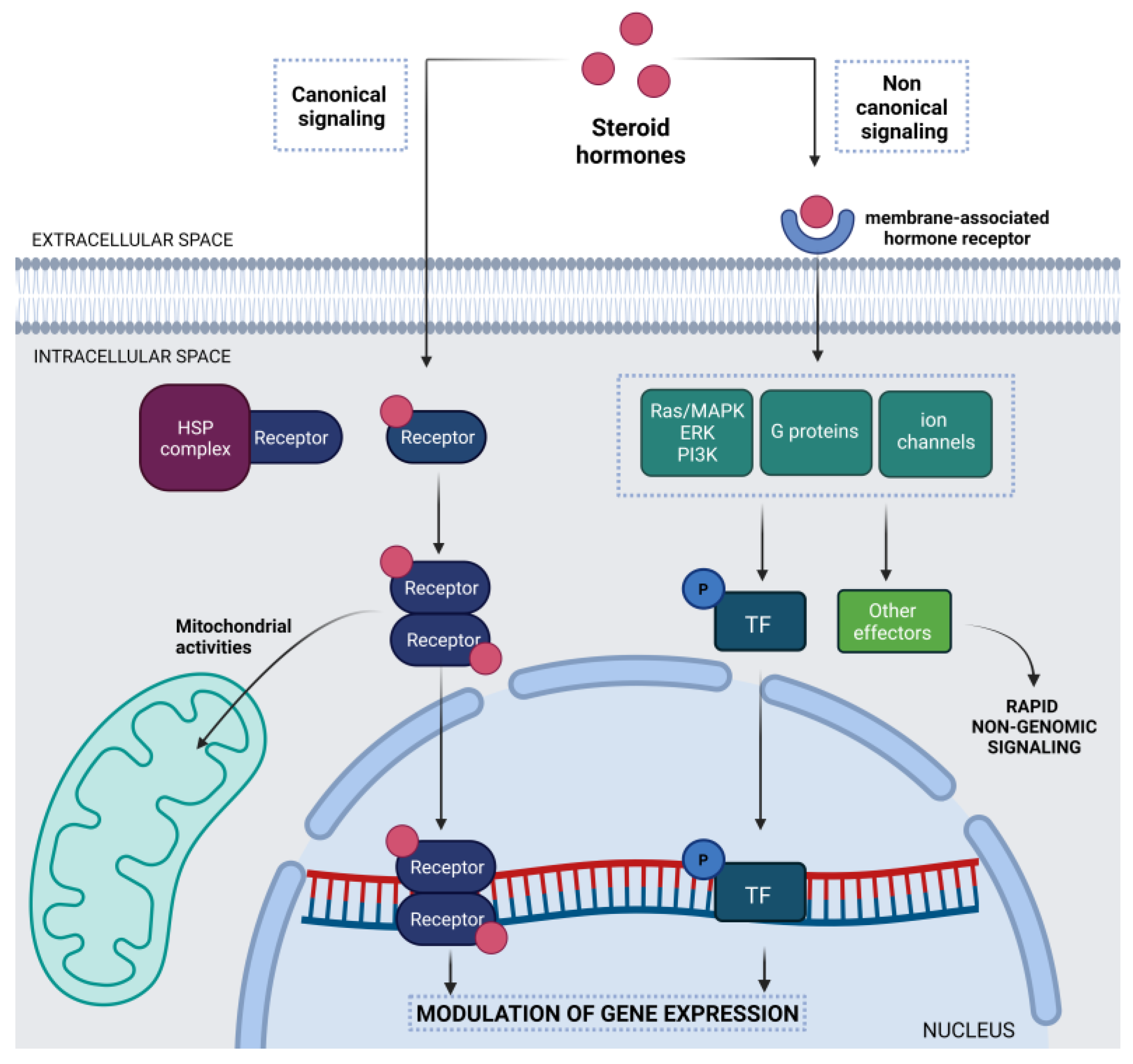
1.1. Steroid Hormones in the Central Nervous System

The central nervous system (CNS) has long been recognized as one of the main targets of the action of steroid hormones. In the brain, steroid hormones exert pleiotropic effects since they regulate many functions, including neurogenesis and neuroprotection [6,7].
Steroid hormones are produced by steroidogenic glands (gonads, adrenal glands, and placenta) and transported, bound to their partner transport proteins, through the circulatory system to the brain. Steroids can easily cross the blood–brain barrier to reach their target sites in nervous tissues. In addition, different steroid hormones can be synthesized in the central and peripheral nervous systems starting from cholesterol or steroid precursors generated in peripheral sources. These locally synthesized hormones can act through paracrine, autocrine, and intracrine communication mechanisms. The seminal discovery of the brain’s steroidogenic potential and the term “neurosteroids” to describe hormones produced therein were first reported in 1981 by the Baulieu and Robel group. They demonstrated how some steroids (such as pregnenolone, dehydroepiandrosterone (DHEA), and their sulfate esters) are present at higher concentrations in the brain than in the plasma of adult male rats, and persist after surgical excision of the classical steroidogenic glands (reviewed in [8]). The nervous system is then provided with the steroidogenic enzymes required to synthesize all classes of steroids and, thus, constitutes a target for hormonal steroids as well as neurosteroids produced by neurons and glial cells.
While the term “neuroactive steroids” comprises all steroids active in the brain, including those coming from the circulation, neurosteroids are limited to those directly synthesized or activated in the brain from peripheral sterol precursors (reviewed in [9]). Neurosteroids can work as endogenous regulators of neuronal functions and excitability, mainly via rapid non-genomic mechanisms [10]. Based on their structural characteristics, neurosteroids can be classified as pregnane, androstane, and sulfated neurosteroids [11]. Generally, neurosteroids modulate brain excitability primarily by interacting with neuronal membrane receptors and ion channels, principally GABA-A receptors [12].
The effects of neuroactive steroids, neurosteroids, and sex hormones working in the brain are mediated not only by classical nuclear signaling (genomic action through intracellular steroid receptors) but also by non-classical rapid signaling via binding to membrane-associated receptors or transmembrane receptors (Figure 1).
The canonical/classical mechanism of action involves intracellular binding of steroids to specific receptor proteins located in the cytoplasm or the nucleus, which belong to the nuclear receptor superfamily and act as transcription factors. Circulating steroids diffuse through the plasma membrane and bind to their cognate receptors. This process induces the release of the receptors from heat shock proteins and their conformational change and activation. Once activated, the complex of steroid hormone/receptor translocates into the nucleus, where it can bind hormone-responsive elements (HREs) in the promoter regions of target genes, thereby regulating gene expression [13]. The nuclear activity of steroid receptors can induce or repress the transcription of target genes involved in neural differentiation or neuronal functions [14]. These hormonal effects can also influence animal and human behaviors during development and adulthood, particularly sexual behavior [15] (Table 1).
Steroid hormonal effects are also finely modulated through the action of co-activators in the nucleus [16] or by the activation of rapid signaling pathways [17]. In this case, steroid hormones act through membrane-associated receptors in different ways based on the type of steroids (Table 2).
Other non-canonical mechanisms of action of steroid hormones could be identified in the mitochondria of neuronal cells of the nervous system. The presence of putative HREs in the mitochondrial genome suggests a possible role in regulating the transcription of mitochondrial genes [18]. In general, both the direct and non-direct effects of steroid hormones on mitochondrial metabolism have been found to be involved in their neuroprotective effects [19].
Table 1. Canonical signaling of steroid hormones in the CNS.
Steroid HormoneActivated ReceptorFunctionsTargetsReferences
EstrogenERα, ERβNeuroprotectionAntiapoptotic genes[20,21,22,23]
Proliferative genes
ProgesteronePR-A, PR-BNeuroprotectionPromyelinization genes[24,25]
Anti-inflammatory genes
TestosteroneARNeuroprotectionRemyelination process by oligodendrocytes[26,27,28]
Astrocyte recruitment
Sex phenotypeDevelopment and maintenance of male phenotype in CNS[29,30,31]
ERα: estrogen receptor alpha, ERβ: estrogen receptor beta, PR-A: progesterone receptor A, PR-B: progesterone receptor B, AR: androgen receptor.
Table 2. Non-canonical signaling of steroid hormones in the CNS.
Steroid HormoneActivated ReceptorFunctionsReferences
EstrogenERα, ERβ, mERs,
GPER1
Oligodendrocyte differentiation, survival, and function[32]
Spine plasticity[33,34,35,36]
Behavior
ProgesteronePR-A, PR-B, mPRs,
PGRMC1
Female reproductive behavior[37,38,39,40]
Cerebellum cortical formation
Neuroprotection
TestosteroneAR, GPCRsSexual differentiation[41,42,43,44,45,46]
Reproductive and aggressive behaviors
Neuroendocrine response
mERs: membrane-associated estrogen receptors, GPER1: G protein-coupled estrogen receptor 1, mPRs: membrane-associated progesterone receptors, PGRMC1: progesterone receptor component 1, GPCRs: G protein-coupled estrogen receptor.

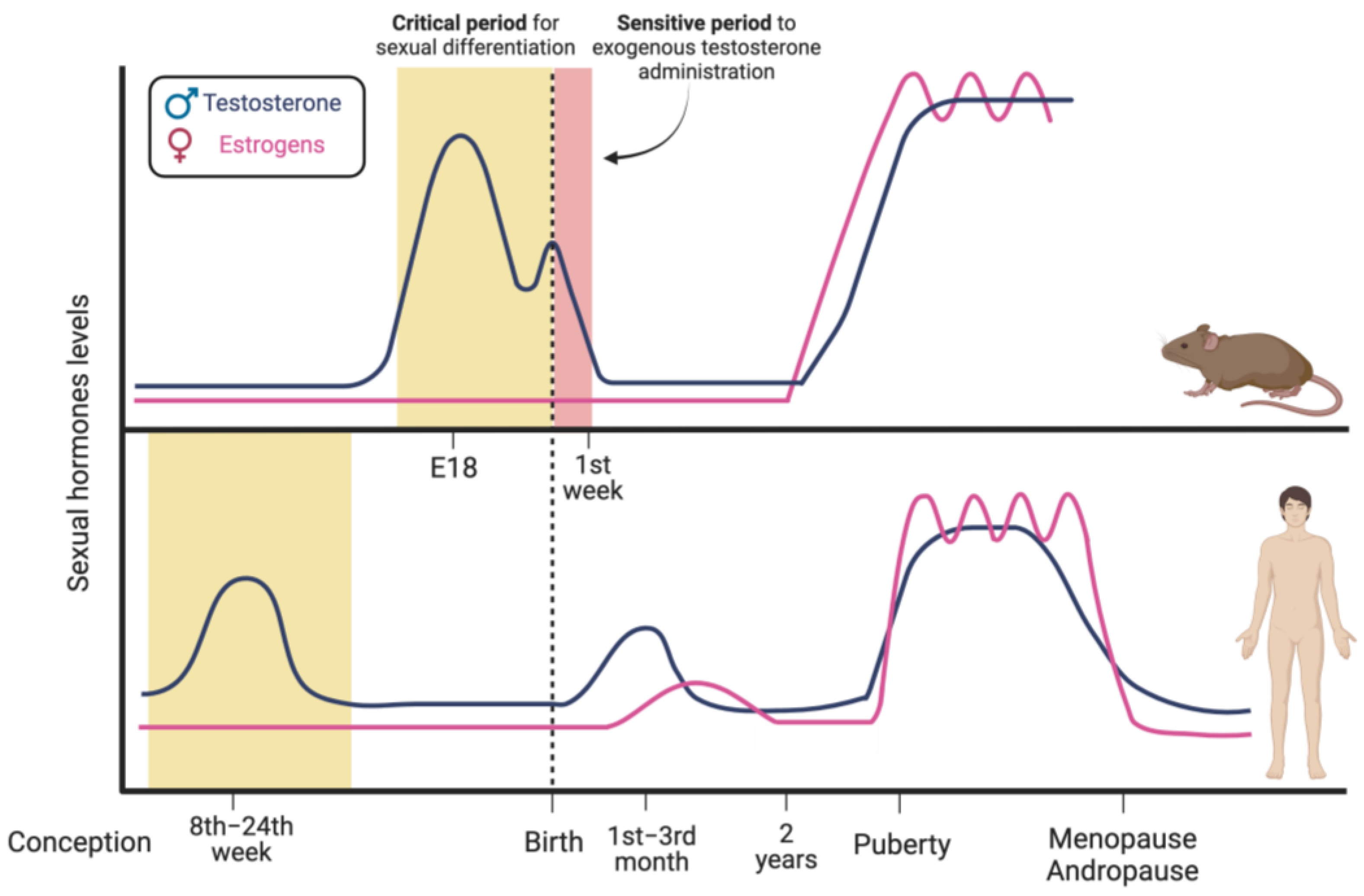
1.2. Sex Hormones’ Modulation of Brain Sexual Differentiation

In mammals, gonadal sex determination is directed by the presence or absence of the Y chromosome containing the Sex-determining Region Y gene (SRY) that encodes the “testis determining factor”. This is a transcription factor working as a molecular switch to activate Sertoli cell differentiation and the development of the testis. In the absence of this protein, the bipotential and undifferentiated gonad gives rise to an ovary [47,48].
The testis, then, starts to produce testosterone, which regulates the development of male sex organs, reproductive tract, and secondary sexual characteristics. In contrast to the steroidogenic activity already present in fetal Leydig cells of the testis, no hormones are produced by the developing ovaries, and the female sex organs progress without signals. The molecular mechanisms that control mammalian gonadal development are beyond the focus of this review but are fully described in many previous review papers [49,50,51].
Sex-related processes underlying brain sexual differentiation have been mainly studied via exploiting rodents as animal models. In male rodents, testosterone is produced by fetal Leydig cells and first peaks during late gestation, which is around embryonic days 17–19, while a second lower peak occurs within a few hours after birth (Figure 2, upper panel) [52,53].
In accordance with the “Organization and Activation Hypothesis” [54], sex differences in brain physiology and functions are initially defined by exposure of the brain to testosterone secreted during the so-called “critical period”, a precise temporal window of development in which the brain is particularly sensitive to estrogens produced by the neural aromatization of testosterone [1,54,55,56]. The aromatase enzyme, which is responsible for this conversion, is expressed by neurons of specific brain regions, such as the hypothalamus, the preoptic area, and the limbic system, which control not only the reproductive axis, but also the processing of sensory information, affective behavior, and the modulation of learning and memory [57,58,59].
The “critical period” represents a constant in the process of brain differentiation in mammalian species examined so far, even if its temporal positioning during development is variable [56]. The extraglandular synthesis of “neurosteroids” has also been reported in some regions of the developing brain, thus resulting in variable steroid content depending on the brain area and sex [60].
Therefore, the normal development of male rodent brain undergoes the process of masculinization, in which increased perinatal levels of estradiol orchestrate the formation of neuronal circuits, the synthesis of neurotransmitters, and the number, architecture, and connectivity of nerve cells, ultimately defining the morphological and size differences that exist among specific regions of the brain in males and females [4,56]. At the same time, male rodent brain undergoes the steroid-induced process of defeminization, which consists in the loss of the ability to display female-typical behaviors [61].
Female rodents, on the contrary, are protected from brain masculinization by the plasma glycoprotein α-fetoprotein, which binds circulating estradiol of maternal or placental origin, thus preventing it from entering the brain [62].
In male rodents, the critical period extends to the first two postnatal days, while females are sensitive during the first week after birth. From an experimental point of view, this larger postnatal window allows the easy execution of experiments to validate the hypothesis of the fundamental role of hormones in brain development. For example, if testosterone is administered to newborn female rats, thus mimicking the hormonal stimulation by fetal testis that occurs in males, the brain is masculinized. Consequently, these animals exhibit male-typical sexual behaviors, such as mounting behavior, despite the presence of the ovary and XX genotype. On the other hand, stimulation with androgen outside this sensitive time interval does not affect sexual brain differentiation [63].
Females are not exposed to endogenous steroids since the fetal ovaries are functionally quiescent. This lack of steroidogenic activity suggests the possibility that the brain acquires female morpho-functional characteristics in the absence of specific hormones, following a “default program”. However, previous experiments performed with aromatase knockout female mice and post-natal treatments with estradiol suggest that this hormone may contribute to the development of female sexual behaviors [64,65] and that brain plasticity and sensitivity to the organizational effects of steroid hormones persist longer than previously thought.
Hormones produced by the fetal testis act on the developing brain during the so-called “organizational” phase, in which modifications of the cerebral transcriptional profile determine the development of neural circuits responsible for sex-specific behaviors in subsequent periods of life [66].
After puberty, hormones produced by the gonads will instead carry out what are defined as “activation effects” on the brain circuits that are previously set in a sex-specific sense [67].
In humans, testosterone starts to be produced by the testis at the 8th week after conception, it peaks between weeks 11 and 15 and then declines to reach low levels at the 24th week [68,69]. Consequently, the period that is sensitive to the hormonal environment and sexual differentiation of the brain precedes birth [70].
Male newborns show a second, lower, and time-restricted peak around three months of life, while females show an increase in estradiol that then declines very slowly during the first two years of life. This hormonal situation is known as “mini-puberty” [71] and could contribute to the sexual organization of the brain in humans. Testosterone levels rise again at puberty and are maintained during the reproductive adult age (activational period). They finally tend to decrease with aging (Figure 2, lower panel) [72].
Unlike what has been shown in rodents, the masculinizing agent in humans is represented by androgens, as also demonstrated by males in which the aromatase enzyme or the ERα are not functional. These subjects identify themselves as males, suggesting that the differentiation likely depends on androgens [73,74,75].
To date, knowledge of the mechanisms that lead to brain differentiation in humans comes from the study of the so-called differences (or disorders) of sex development (DSD), which include congenital conditions linked with atypical progress of internal and external genital structures. As an example, in the Androgen Insensitivity Syndrome (AIS), the testis present in XY individuals produces testosterone but cannot transduce its signal due to the lack of a functional AR. These individuals not only have a female phenotype but also recognize themselves as females and exhibit female behaviors and brain morphology, in accordance with the fundamental role of androgens on the sexual differentiation of the brain [76,77].

1.3. Sex Hormone-Dependent Brain Sexual Dimorphism

The role of steroid hormones in the process of brain sexual differentiation and in the determination of structural dimorphism of some specific regions and nuclei has been known for a long time. As a main example, the medial preoptic area (MPOA) of the hypothalamus that controls male sexual behaviors and gonadotropin release is 3- to 5-fold larger in rodent males than females [66], and its neurons contain twice the number of dendritic spines than the neurons in the female brain [78]. This dimorphism seems to be related to testosterone regulation of the apoptotic process occurring in this area [79].
Moreover, the posterior bed nucleus of the stria terminalis (BNSTp) is a sexually dimorphic area that appears larger in males than females because of its male-biased gene expression and neuronal development driven by estrogens, which promote neuron survival in this area [80]. On the other hand, the anteroventral periventricular nucleus (AVPV), which is involved in the control of ovulation [66] and gonadotropin secretion [81], is larger in females than in males [63,66,82]. These structural differences relate to the rates of neurogenesis, cell migration and differentiation, axon guidance, synaptogenesis, and cell death [83], which can be regulated by estrogens binding to both nuclear and membrane receptors. Estrogens induce a higher rate of cell survival in male MPOA and an increase in apoptosis in the same region of the female developing brain. On the contrary, estradiol promotes cell death in male AVPV, leading to the aforementioned sex-dimorphic difference in this region. These opposite effects of estrogen indicate that cellular responses to the same gonadal hormone are sex-specific and differ among specific areas of the CNS [66].
Estrogen receptors exist in two isoforms, ERα and ERβ, which are encoded by the ESR1 and ESR2 genes and are widely expressed in the mammalian brain. They show a different pattern of distribution [84] and time- and sex-specific expression and functions in the brain [84,85,86]. While ERα is mainly associated with the masculinization effect of estrogens on neuronal circuits and behavior, ERβ seems to be involved in the defeminization process of the male brain [61,66]. Consequently, a loss of ERα in males results in reduced sexual behavior, whereas knockout of ERβ alone has no effect on sexual behavior. However, when both ERs are dysfunctional, male sexual behavior is completely lost [87,88].
ERs have been reported to play a fundamental role in orchestrating cell movements in the developing brain. Estradiol can rapidly affect nerve cells’ migration via non-genomic transduction of its signal. Indeed, both ERα and ERβ can be expressed as membrane-bound proteins that, upon interaction with their ligand, can activate signal cascades involving classical second messengers, such as protein kinases and phosphatases, the release of cyclic amines, and calcium signaling [89]. Estrogens target GABAergic neurons, which play a critical role in hypothalamic development [83,90], interfere with BDNF (brain-derived neurotrophic factor) release in specific cell types [81], and are involved in a complex interplay with CREB (cyclic AMP response element-binding protein) that, in turn, can have an impact on neuron migration during brain development [83]. Moreover, estradiol can activate PI3 kinase and promote presynaptic glutamate release, thus affecting postsynaptic AMPA and NMDA receptors [91,92]. Interestingly, estrogen-mediated presynaptic glutamate and GABA release can permanently alter the synaptic profile of postsynaptic neurons and the morphology of neighboring astrocytes, respectively, even if these cells appear devoid of ERs. Thus, sex steroids interfere with brain cell organization and differentiation either locally or by exploiting cell–cell interactions in a domino fashion [66].
Of note, the alteration in the levels of BDNF and GABA, which are involved in neuronal development and signaling, and the following impairment of synaptic transmission have been recognized as a pathogenic mechanism of neurodegeneration [93].
Although estrogens appear to be a driving force shaping male rodent brains, androgens also contribute to sex-dependent dimorphism. Testosterone reaching the brain during the late gestation and the early postnatal period can either act directly or after being metabolized into its biologically more active form, dihydrotestosterone (DHT), by the isoform 2 of the enzyme 5α-reductase. The expression of this enzyme increases upon the surges in testosterone in the critical perinatal period, and, thus, it has higher levels in male brains than in females. Its localization and the timing of its expression suggest a role for DHT in brain sexual differentiation [94]. Nevertheless, it is expressed not only in late fetal and early postnatal brains but also in regions of adult rodent and human brains that are mainly involved in the regulation of behaviors [95].
Progesterone is another steroid hormone that is involved in brain development. It can be either synthesized in adrenal glands in males and females and in ovaries and placenta in females, but it can also be directly synthesized in the brain by neurons and glial cells [96], thus being considered a “neurosteroid” [97]. It interacts with intracellular PR, which acts as a nuclear transcription factor, with the putative membrane-associated PGRMC1, and with mPR. These interactions lead to the activation of JAK/STAT, Src pathways, protein kinase G, MAPK, and G protein cascades or to the modulation of GABAA receptors through progesterone bioconversion into allopregnanolone [97]. Progesterone levels in the brain show subtle sex-related differences, but the levels can vary substantially according to the cycle phase [98]. Several studies have indicated that the prenatal testosterone surge in males and its further conversion to estradiol indirectly induce the upregulation of PR expression in rodent sex-dimorphic brain regions, thus contributing to the masculinization of neuronal functions and sexual behaviors [99].
Although sex hormones are the main inducers of sexual dimorphism during the development of the brain, there is also evidence of the role played by other steroid hormones, in particular glucocorticoids, in the shaping of brain functions. These are mainly related to the correct development of the hypothalamic–pituitary–adrenal (HPA) axis that regulates, among others, behaviors and stress response [100].

1.4. Sex Chromosomes’ Impact on Brain Sexual Differentiation

The arousal of sex differences in the brain has been historically described as a hormonal-dependent brain organization that relates to the gonadal secretion of sex steroid hormones [83]. However, some sex differences cannot be explained by the activity of gonadal steroids. Other players in this process are the sex chromosomes, which are differentially inherited by male and female cells, resulting in differential gene expression and cell-autonomous effects [2,3].
The critical role played by the Sry gene in male sex determination has already been briefly described. However, besides its expression and function in the gonads, this gene also exerts many different functions in the brain. The work by Reinius and Jazin (2009), indeed, demonstrated the expression of Y-chromosome genes in many regions of the prenatal brain, suggesting their involvement in the sexual dimorphism of this organ [101].
For example, the Sry gene is expressed in the hypothalamus and midbrain of adult mice [102], as well as in the hypothalamus and frontal and temporal cortex of adult men, as demonstrated by an analysis of samples derived from autopsies [103], thus possibly exerting gonad-independent actions. Interestingly, Sry is also expressed in tyrosine hydroxylase (TH)-positive neurons within the substantia nigra (SN), where it can act as a positive regulator of dopamine biosynthesis and functions [66,104,105,106].
Differences linked to the sex chromosomes can also be associated with the differential imprinting of the genes deriving from maternal or paternal origin (reviewed in [107]), as well as by the dosage of the X-linked genes.
The female genome, indeed, contains two copies of the X chromosome, one of which will be inactivated into heterochromatin to re-balance the different genetic loads, a process called X-chromosome inactivation (XCI). Due to the presence of the two copies of this chromosome and the random inactivation process in each cell, females result in a mosaic of paternal and maternal active X chromosomes. This occurrence protects females from disorders due to mutation in the X-linked genes [108]. A fraction of the X-linked genes (15% in humans [109]) escapes inactivation and is expressed, albeit to a lesser extent, not only by the active X chromosome but also by the silent X chromosome. This could lead to higher expression levels of these genes in XX individuals compared to XY individuals, likely resulting in sex differences in female cells [108].
The influence of the sex chromosomes and their contribution to brain sexual differentiation have been experimentally verified by using four core genotypes (the FCG model), which permit the comparison between mice with similar gonads but different sex chromosomes [110,111]. The significant number of studies performed with these models allowed researchers to identify XX versus XY differences in behaviors, in gene expression, and in the predisposition to diseases (reviewed in [54]), including neurodegenerative diseases.
In humans, the effects of the sex chromosomes on brain anatomy have been studied on sex chromosome aneuploidies, such as the Turner syndrome (45X, female lacking one X chromosome) or the Klinefelter syndrome (47XXY, male with an additional X chromosome). Both syndromes present variations in the copy number of the sex chromosomes, allowing the analysis of possible gene dosage effects [112,113].

1.5. Epigenetic Regulation of Brain Sexual Differentiation

As previously described, steroid hormones are responsible for long-lasting organizational effects that sculpt the structure and functions of the developing brain and that, in many cases, are maintained throughout adulthood. These long-lasting effects can be mediated by the possible impact of steroid hormones on epigenetic mechanisms that lead to sustained changes in gene expression [4]. Epigenetic mechanisms, including posttranscriptional regulation by noncoding RNAs, histone modifications, and DNA methylation, can affect gene expression without altering the genetic code. Gonadal hormones, and estrogen in particular, may indeed regulate DNA methyltransferase (DNMT) expression and activity in the brain, thus exerting an important function in tuning chromatin accessibility [4].
As recently demonstrated by Gegenhuber and collaborators, ERα activation determined by the perinatal hormonal surge controls a sustained male-biased gene expression program in the developing brain, at least in the sexually dimorphic BNSTp region. Interestingly, chromatin is maintained in an active state by estradiol in both sexes, while testosterone can promote chromatin activation or repression in males through the action of ARs on AREs (Androgen Responsive Elements). However, only a small percentage of neonatal estradiol-regulated sex-biased gene expression is maintained during adulthood (about 10% of neonatal estradiol-regulated regions). Most of them undergo reprogramming and further regulation after puberty-induced hormonal changes, demonstrating how the genome maintains its plasticity and responsiveness to sequential variations in the hormonal milieu [80].

2. Sex-Related Differences in Neurodegeneration

In the previous section, we have largely discussed how sex hormones and sexual genes (on X and Y chromosomes) can regulate brain development, thereby producing a clear dimorphism. Recent research on the role of sex in brain development and physiology has generated an increasing interest in the possibility of having a sex-related predisposition or vulnerability to certain brain diseases, in particular neurodegenerative diseases. This hypothesis has largely been investigated, and it is actually strongly supported by the literature. In brief, biological sex seems to be connected to the incidence of neurodegenerative diseases.
Starting from these considerations, in this second section, we analyze sex-related differences in neuroinflammation and in neurodegeneration, focusing our attention on Parkinson’s disease (PD).

2.1. Focus on Parkinson’s Disease

PD is the second most common neurodegenerative disease (following Alzheimer’s disease), affecting about 3% of the population by the age of 65 and more than 5% of people over 85, and it is caused by the preferential loss of dopaminergic (DA) neurons in the substantia nigra pars compacta (SNpc) [114,115]. Degeneration of DA neurons leads to the onset of classic motor symptoms, including tremors at rest, bradykinesia, rigidity, and gait disturbances. Other non-motor symptoms may occur either in the early stages of the pathology, such as gastrointestinal disorders and sleep disturbances, or in the later stages when dementia and psychosis may appear [114].
The typical hallmark of this disease is the accumulation of intracellular proteinaceous inclusions in surviving neurons, which are named Lewy bodies and Lewy neurites, and are mainly constituted by the presynaptic protein α-synuclein (α-syn) [116]. The accumulation of α-syn aggregates can hamper autophagic flux and impact on various intracellular processes critical for neuronal functions, such as synaptic vesicle recycling and docking, finally leading to neuron death [117].
Another characteristic of this disease is sustained microglial activation that determines chronic neuroinflammation, thus contributing to neuronal loss [118].
Astrocytes have also been found to be activated in PD brains [119] and, together with microglia, are known to be involved in α-syn aggregate removal [120]. Despite neurons being the most affected cells in PD, alterations in glial cells are known to contribute extensively to PD etiology. Microglial function presents various sex-related features, which may be relevant for the sex-differences observed in PD that will be discussed below. PD is characterized as being sporadic (of unknown etiology) in about 90% of the cases, while it is associated with gene mutations in about 10% of patients. Among its non-genetic forms, PD has been shown to be associated with exposure to pesticides, such as paraquat and rotenone, or to other environmental pollutants [121,122], which are known to impact mitochondrial function and to induce oxidative stress, and that may increase the risk of developing PD.
The causative genes associated with PD or parkinsonism encode proteins involved in several pathways that are keys for the maintenance of brain homeostasis and functions. These genes are involved in the functioning of mitochondria, of the ubiquitin–proteasome and autophagic–lysosomal systems, in the regulation of oxidative stress and inflammation, as well as in the control of the homeostasis of synapses and synaptic vesicles [123,124,125] (see Table 3 for a summary of these genes).
The impairment of these molecular mechanisms can lead to dyshomeostasis of neuronal cells and ultimately cause neuron death.
Among the different genes linked to PD, mutations or gene duplication and triplication in the SNCA gene, which encodes the protein α-syn, are of particular interest. SNCA mutations typically lead to an increased α-syn propensity to aggregate or to form oligomers. Moreover, mutations can alter α-syn function at the presynaptic terminal by impairing its ability to bind lipid membranes and other interactors [126], which are crucial for its role in the regulation of synaptic vesicle recycling. Finally, this protein, likely in its aggregated forms, is involved in the spreading of the pathology across the body and different brain regions according to the Braak hypothesis [127], which would explain disease progression. Despite being still under debate, it is clear that monomeric and aggregated α-syn can be transferred between cells through extracellular vesicles and between organs, i.e., from the gut to the brain, thus contributing to inflammatory processes and neuronal damage as determined by different toxicity mechanisms. Besides causative mutations in specific genes, a large set of other mutations have been identified as risk factors for PD. These mutations are located on genes mainly involved in autophagic–lysosomal function and mitochondrial activity, but also in many other intracellular processes [128].
According to existing information, several animal models have been developed (reviewed in [129]) to study the molecular mechanisms associated with this disease and to test novel therapeutic strategies. In particular, most of the studies have relied on the use of either rat or mouse models, which carry mutations in the genes mostly relevant to genetic PD forms, or neurotoxin-based models, which are obtained by administering to the animals toxins that have been linked to PD in environmental studies.
Interestingly, most of the mutant mouse models do not recapitulate very well the disease phenotype, especially in terms of symptoms, neurodegeneration of specific brain areas, and the formation of α-syn inclusions, making them not always suitable for preclinical studies. On the other hand, neurotoxin-based models present the typical issues of acute disease models, with limited possibilities for the study of the progression of the disease. Experimental approaches based on the administration of different toxins to mice, i.e., rotenone or the neurotoxin 1-methyl-4-phenyl-1,2,3,6-tetrahydropyridine (MPTP), have been extensively used to study the role of sex hormones in PD, as discussed below, and their key features are summarized in Table 4.

2.2. Sex-Related Differences in Microglia and Their Implication on Neuroinflammation in PD

Neuroinflammatory processes are considered critical in the onset and development of neurodegenerative diseases. The molecular mechanisms of neuroinflammation have largely been characterized in recent years in relation to PD [118,130]. Their role in PD etiology and progression has been strongly validated based on post-mortem PD brain analyses and data from model organisms for this disease [131,132]. A description of these neuroinflammatory mechanisms (recently reviewed in [133,134]) is beyond the aim of this review; thus, we focus this section on sex differences in microglia, and the outcomes in the development of neuroinflammation in PD are described in the next section.
The innate and adaptive immune responses display sex differences, which development is regulated by genes, sex hormones, age, and reproductive state [135]. These differences in the immune responses could also explain, at least in part, the different susceptibility to neuroinflammation and sex prevalence in neurodegenerative diseases.
Microglia are considered the macrophages of the CNS: they can release reparative factors and adopt both pro-inflammatory and immunosuppressive phenotypes. Moreover, these cells regulate neural proliferation in early development by secreting pro-proliferative cytokines and further pruning superfluous cells by inducing apoptosis and targeted phagocytosis [78]. During adult neurogenesis, microglia control the plastic remodeling of neuronal circuits [136].
As reviewed by Chowen and Garcia-Segura in 2021, macro- and microglia show marked sexual dimorphism in their physiology and in their ability to react to brain pathology. For this reason, glial cells are recognized for being able to contribute in different ways to brain homeostasis and to the innate immune response which protects the brain against insults [137].
Microglia show different immunoreactive properties between the two sexes, which are initially established during sexual differentiation. The perinatal surge in testosterone and the hormonal milieu shape the morphology of different brain areas, including the microglial phenotype, which is further maintained during adulthood [78,138]. Moreover, the X chromosome contains a large number of genes related to the immune system, thus strongly contributing to the sex-related differences in immune response and inflammation (reviewed in [139]).
Male microglia have a higher density and higher phagocytic capacity, and they have been associated with more neuroinflammatory action, while female microglia are more supportive of neuronal functions, are neuroprotective, promote repair, and inhibit inflammatory response (reviewed in [140]). Interestingly, these abilities are also retained in the adult brain, as demonstrated by Villa and colleagues in 2018, who observed sex-related differences in the gene expression, in the morphology, and in the competence of female microglia to reduce ischemic damage even when transplanted in the male brain [138]. Adult microglial cells show sex-specific transcriptomes and are differentially subjected to the action of sex steroids depending on the hormone receptors they express (reviewed in [141]).

2.3. Sex Bias in Parkinson’s Disease and the Role of Sex Hormones

As in other neurodegenerative diseases, PD primarily affects men, with a ratio of 1,5:1, and post-menopausal women with a similar incidence. Female patients experience later emergence of motor symptoms, with a more tremorigenic phenotype and a slower disease progression compared to men, suggesting that male PD and female PD patients may present two disorders with very different features [115]. This evidence suggests that either sex hormones or genetic sex, or both, can contribute to the predisposition to and the development and progression of the disease by impacting those cellular pathways that are known to play a role in PD etiology.

2.3.1. Estrogens

The sex difference in PD incidence points toward the possible protective effects of female steroid hormones, particularly estrogens. This possibility is also suggested by the reduced PD risk in women with longer exposure to endogenous estrogens due to early menarche and/or late menopause [142,143,144]. On the contrary, surgical or premature menopause, especially when occurring at an early age, is associated with an increased risk of PD [145,146]. However, besides these correlations, other studies have found no evidence of a positive effect of endogenous estrogens on the risk of PD [147,148], suggesting that the matter is likely more complicated.
Apart from epidemiological studies on PD incidence and its correlation with sex and female reproductive status, the neuroprotective and anti-inflammatory roles of estrogens have also been explored using model organisms of the disease.
Male rodents treated with the neurotoxin MPTP [149] show a higher depletion of striatal DA neurons and, consequently, of dopamine in the SN compared to females, thus mimicking the sex differences observed in human PD [150,151]. Similar results have recently been obtained using a rotenone animal model of PD [152].
The neuroprotective action of estrogens can also be due to the activation of anti-inflammatory pathways. In fact, estrogens have been found to modulate the response of microglia and astrocytes against oxidative and inflammatory injury in an MPTP rodent model, resulting in DA neuron protection [153]. Moreover, Tripanichkul and co-workers demonstrated that estrogen treatment prevents dopamine reduction, neuronal loss, and glial activation in the striatum of MPTP-treated animals and determines a decrease in the secretion of pro-inflammatory modulators [154]. A similar outcome was obtained with estradiol benzoate treatment in a MPP+ rat model of PD [155]. The authors also detected an upregulation of the expression of paraoxonase-2 (PON2), a mitochondrial protein working as an antioxidant. Interestingly, this protein is transcriptionally regulated by estrogens and presents higher expression levels in the brain of female mice, thus making female brain cells less sensitive to oxidative stress [156].
In a previous study, the anti-inflammatory action of estrogens was shown to also occur by reducing microglial reactivity in rodents treated with 6-hydroxydopamine (6-OHDA) [157]. In the same model, an induction of the autophagic process was found to be activated by estrogens to prevent degeneration of DA neurons [158].
Finally, by binding to the ERβ isoform, estrogens are known to decrease microglial activation [159]. Therefore, it is unsurprising that ovariectomy in mice leads to an increase in neuroinflammation [160].
The neuroprotective role of estrogens was confirmed using knockout mice for the Cyp19a1 gene, which encodes the aromatase enzyme. These mice, that are unable to synthesize these hormones, were found to show dopaminergic neuron impairment and enhanced vulnerability to MPTP-induced nigrostriatal damage, when compared with both treated WT and even their ovariectomized counterparts. The latter result suggests that estrogens produced locally by the brain may fulfill important neuroprotective functions [161].
Estradiol also exerts its protective role by upregulating BDNF expression in the SN, as demonstrated in a 6-OHDA male rat model of PD [162].
Evidence shows that circulating estradiol upregulates the expression of TH, an enzyme responsible for dopamine synthesis [163]. Estradiol treatment, indeed, increases the fiber density of DA neurons in pharmacologically induced PD mouse models. This neuroprotective action seems to be specifically mediated by ERα [164], as confirmed by the higher vulnerability to MPTP in mice in which ERα has been silenced, compared to WT and ERβ KO mice [165,166]. This protective effect of estrogen through ERα in DA neurons requires the activation of membrane-bound G protein-coupled estrogen receptor (GPER) [167].
mERα can also exert a neuroprotective role by activating rapid non-genomic signaling cascades aimed at counteracting oxidative imbalance and mitochondrial dysfunction, thus promoting cell survival [168].
Finally, estradiol, besides being involved in neuroprotection as previously described, is thought to contribute to the maintaining of lipid rafts present in cell membranes, which provide a proper environment for optimal protein stability and molecular interactions on the cell surface. Aging and menopause-associated reduction in estrogen levels could lead to the loss of essential lipid components of membranes, finally causing neuronal dysfunction [169].

2.3.2. Progesterone

Progesterone has been found to exert a protective effect on experimental models of PD. This hormone contrasts the DA neuron depletion induced by MPTP in male mice [170] and exerts neuroprotective effects on the striatal neurotransmission systems in a male rat PD model [171]. Interestingly, the myenteric plexus of the enteric nervous system in MPTP-treated mice benefits from progesterone, thus suggesting a role for this hormone in gut myenteric plexus defense and in the prevention of gastrointestinal alterations, which are one of the main non-motor features of PD [172].
Finally, the treatment with the progesterone metabolite, allopregnanolone, has been found to improve motor coordination and increase the expression of TH protein and TH cell number in a MPTP mice PD model [173].

2.3.3. Androgens

The role of androgens in neuroinflammation and PD is less clear. PD patients have been reported to present reduced testosterone levels [174], thus suggesting a possible protective role of this steroid. Moreover, testosterone replacement therapy has been found to ameliorate motor symptoms of PD patients [175].
However, this hormone has recently been found to increase neuroinflammation in N27 dopaminergic cells under oxidative stress. This is shown to occur through a putative membrane-associated androgen receptor via the activation of inflammatory pathways (nuclear translocation of NF-κB and activation of COX2 signaling), which induces the apoptosis of DA neurons [176] as well as exerts a suppressive role in the midbrain dopaminergic pathways [177].
In good agreement with these findings, testosterone and DHT are unable to counteract MPTP-induced dopaminergic toxicity [178,179].
In contrast with the results described above, the positive effects of chronic treatment with testosterone propionate, such as improved motor problems and reduced dopamine depletion, have recently been reported in a reserpine-induced progressive rat model of PD [180].
Unfortunately, we were unable to find studies reporting the outcome of sex hormone treatment in PD genetic animal models, which would be of great interest to understand the possible role of hormone-based therapies in different disease models. This could be ascribed to the limited ability of genetic PD animal models to decently recapitulate the disease phenotype, so this piece of information remains unavailable.

2.4. Effects of Estrogen-Based Pharmacological Treatments on Parkinson’s Disease

Exogenous sex steroid hormone therapies (oral contraceptives, supplemented estrogens and progestogens, and synthetic anabolic steroids) are largely used to treat different conditions, such as testosterone deficiency (hypogonadism) [181], contraception or postmenopausal symptoms [182].
Due to the pleiotropic functions that sex steroid hormones exert on the brain during both development and adult life, the therapeutic use of these steroids can affect the brain in different ways. Therefore, it is not surprising that hormone replacement therapies (HRT) can impact cellular processes and pathologies involving this organ.
Regarding postmenopausal hormone therapies, their use has been reported to lead to a reduced risk of developing PD or a diminished disease severity together with an improvement in DA activity and dopamine transporter density [145,183]. Still, the results reported in different studies regarding PD incidence and the possible positive effects of HRT on this disease remain controversial, likely because of the different factors (dosage, duration, and composition of the replacement therapies used) that have been considered in each study [184,185,186].
In different neurotoxin animal models of PD, protective effects have been obtained when treating the animals with estrogens, as well as ER modulators (SERMs) or ER agonists (reviewed in [187]), thus suggesting that a potential repositioning of steroids, particularly estrogens, may improve the life quality of PD patients [186]. Unfortunately, estrogen replacement therapies can also be associated with detrimental peripheral side effects, such as uterine and breast stimulation, and consequent increased risk of cancer in these organs, stroke, coronary heart disease, and vascular problems [188]. These complications can be overcome by using estrogens designed to act only in the brain, where they can positively affect neurological diseases and menopausal symptoms, such as hot flushes, depression, and cognitive impairment, while sparing the adverse peripheral side effects.
Recently, Prokai and co-workers [189] synthesized and characterized a small-molecule bio-precursor prodrug, the “10b,17b-dihydroxyestra-1,4-dien-3-one”, named DHED. After its systemic administration, DHED is rapidly converted to 17β-estradiol, preferentially in the rat brain, where it stimulates the same gene expression and neuroprotective effects obtained with 17β-estradiol treatment [190].
In vitro metabolic studies have demonstrated that DHED conversion to active estrogens does not occur in estrogen-sensitive peripheral tissues. Interestingly, this molecule reduces symptoms associated with the loss of brain estrogens and results in neuroprotective effects in rats [189,190]. In addition, this molecule cannot bind to ERs and, due to its physicochemical characteristics (increased water solubility and reduced binding to plasma proteins compared to 17β-estradiol), it crosses the blood–brain barrier more easily, thus showing a better uptake in the brain [190].
This prodrug has been tested in a double-transgenic mouse model of Alzheimer’s disease in a side-by-side comparison with 17β-estradiol [191,192]. The authors reported that the DHED therapy achieved the same positive neuro-biochemical effects and behavioral improvement obtained with the 17β-estradiol treatment.
The potential of this prodrug has also been analyzed in PD models. In a symptomatic α-syn mouse model carrying the E46K human PD mutation in the SNCA gene, the brain increase in estrogens after DHED administration reduced PD-like neuropathy and improved behavioral effects in both female and male mice [193]. In a MPTP-induced PD mouse model, DHED treatments reduced the behavioral impairment and degeneration of DA neurons in the striatum and in the SN. Moreover, the authors measured a decrease in α-syn monomer accumulation and aggregation and a reduction in the levels of oxidative and inflammatory markers [194].
Although requiring more investigations, the lack of peripheral side effects and its brain-specific estrogenic activity suggest that this prodrug could represent an attractive potential therapeutic approach for neuroprotection and treatment of neurodegenerative disorders, including PD.

2.5. Contribution of Genes Located on the Sex Chromosomes to Parkinson’s Disease Etiology

Other possible sex-related contributors to the etiology of PD, which may explain the prevalence of male PD patients, could be the genes expressed on the X and Y chromosomes.
Besides the obvious absence of Y-chromosome genes in XX individuals, it is well-established that the expression levels of X-linked genes can vary between males and females due to epigenetic marks and because some of these genes can be expressed from both gene copies in XX cells, leading to a higher genetic load compared to the XY genotype [195]. In fact, the XCI can occur, but it may not always be able to level the differences. Interestingly, the X chromosome contains a six-fold greater number of genes involved in neurodevelopmental and neurophysiological processes than autosomes [195]. Moreover, in XY individuals, the maternal inheritance of the X chromosome leads to a single possible imprinting of X-linked genes, which cannot be balanced as it happens for females. This evidence further suggests that the differential expression of some of the X- or Y-linked genes can occur. Finally, mutations in X-linked genes have been associated with different disorders that are often characterized by intellectual disability, such as the Rett syndrome, the fragile X syndrome, and the Börjeson–Forssman–Lehmann syndrome [195].
In the PD framework, X-linked forms of parkinsonism have been reported and are associated with mutations in different genes (see Table 3). One example is the RAB39B gene, which encodes the small GTPase RAB39B and is located on the X chromosome. A variety of RAB39B mutations have been found, which mainly cause protein truncation, destabilization, or mislocalization, and are linked to early onset PD with intellectual disability [196,197]. This is believed to be associated with defects in the autophagic pathway, which were found to impact on synapse formation and function in Rab39B knockout mice [198].
Along the same line, it has been shown that about 39% of the male carriers of the expanded CGG repeats of the fragile X mental retardation gene (FMR1) present fragile X-associated tremor/ataxia syndrome (FXTAS) [199]. FXTAS patients often present PD or parkinsonism, suggesting a possible link between the two diseases [200]. The occurrence of PD pathological hallmarks, such as the occurrence of protein inclusions in the brain, in FXTAS patients suggests that FMR1 can actually be considered a PD-associated gene [201].
Mutations in the TAF1 gene, which is located on the X chromosome, have been associated with X-linked dystonia-parkinsonism (XDP) [202]. TAF1 protein is involved in the regulation of the initiation of transcription by RNA polymerase II, and it is still unclear how it participates in the development of XDP. Nevertheless, XDP is primarily observed in males [203], while no association with TAF1 variants and female PD patients has been identified, which may be caused by the pattern of XCI previously mentioned [202].
Another interesting example is represented by a rare polymorphism in the X-linked gene, GLUD2, which encodes a mitochondrial glutamate dehydrogenase specifically expressed in the brain. Gain-of-function mutations in this gene result in an earlier age of onset in PD, which does not occur in heterozygous female patients with PD, likely because estrogens are known to suppress its enzymatic activity thus compensating for the aberrant enzymatic activity caused by the mutations [204]. This estrogen effect on GLUD2 activity would further suggest that the interplay between genetic sex and sex hormones also needs to be taken into account when investigating molecular mechanisms associated with PD in males and females.
In all existing cases, limited information is available to understand how X-linked mutations in parkinsonism-associated genes may differentially contribute to the molecular mechanisms underlying the etiology of the disease. In an attempt to further verify the specific contribution of mutations on genes located on the X chromosome, X-chromosome-wide association studies (XWAS) would be recommended. Nevertheless, even if the X chromosome accounts for about 5% of the human genome and would provide interesting information on the matter discussed in this part of the review, it is usually excluded from most GWAS studies because of technical difficulties, thus hampering the access to this information [205]. The first report providing a XWAS on PD patients was obtained through a meta-analysis that included all available PD cohorts with data on the X chromosome, with the goal of identifying possible sex-specific risk factors that were not observed before [205]. In this study, two novel genome-wide significant loci were found, rs7066890 and rs28602900, with the latter linking the RPL10 gene to PD, whereas it was formerly only associated with complex forms of intellectual disability with epilepsy [206].
To further examine the possible contribution of genes present on the sex chromosomes to PD, we considered a subset of genes located on the X or Y chromosome known to be involved independently in the brain sexual differentiation regardless the levels of sexual hormones [195]. Among these genes, some are associated with alterations in the functioning of the dopaminergic system and/or with altered proteostasis of α-syn, which are two of the key hallmarks of PD. This suggests that they may play a role in the predisposition to PD or contribute to disease progression or severity when their expression is altered.
We identified some X-linked and Y-linked genes that deserve to be examined thoroughly, but others may need attention in the future. For example, one interesting candidate is the X-linked MECP2 gene, which encodes Methyl-CpG-binding protein 2 (MeCP2) and is involved in the regulation of gene expression through various mechanisms. Interestingly, MECP2 is typically associated with a neurodevelopmental disorder called the Rett syndrome, which affects almost exclusively females and can present motor symptoms very similar to those observed in PD patients. In this framework, it was shown that the loss of the Mecp2 gene in a Rett syndrome mouse model led to the impairment of the nigrostriatal pathways and motor deficits, which include reduced dopamine release after striatal stimulation and defects in the D2 dopamine receptor-dependent electrophysiological activity of DA neurons [207]. A recent transcriptome analysis performed on Mecp2 knockout mice showed that they present alterations not only in development-related pathways but also in additional pathways that are more generally associated with other neurological diseases, such as defects in neuronal transmission and immune reactivity [208], both of which are important in PD.
When considering other X-linked genes within this subgroup, two additional genes reveal interesting hints for their possible link with PD development, i.e., USP9X encoding the protein ubiquitin-specific peptidase 9 X-linked and OGT encoding OGlcNAc transferase, both of which have been associated with intellectual disabilities [209,210].
In the PD framework, these proteins are particularly interesting because both ubiquitination and O-GlcNAcylation, together with phosphorylation and oxidation, are key posttranslational modifications of α-syn that allow the maintenance of its balance and homeostasis in cells [126,211]. USP9X was shown to deubiquitinate α-syn, but USP9X levels were significantly lower in the cytosolic fractions of the SN of PD patients compared to controls [212]. According to the authors, this may contribute to the accumulation of monoubiquitinated α-syn in Lewy bodies, which also occur in vitro when USP9X is downregulated.
In females, USP9X expression could be higher compared to males because USP9X is a gene known to escape X inactivation [213], providing a possible protective effect that reduces α-syn aggregation and, thus, the PD incidence in the female population.
Something similar may occur for the OGT gene: a higher expression in females may increase O-GlcNAcylation levels of α-syn. Since α-syn O-GlcNAcylation is shown to have site-specific inhibitory effects on α-syn aggregation and toxicity in vitro and in cell models [214] and in adenovirus α-syn mouse models for PD [215], OGT expression levels may be protective in XX individuals compared to XY individuals. It is worth noting that O-GlcNAcylation is generally crucial in neuronal development and signaling. Specifically, increased O-GlcNAcylation improves synaptic function in DA neurons [215], likely by reducing α-syn toxicity. However, other effects supporting DA neuronal function cannot be excluded.
Of note, male-specific genetics could also contribute to males’ higher susceptibility to PD. For example, the SRY gene is expressed in male nigrostriatal dopaminergic neurons, which are more numerous in males than females, and they are known to be involved in dopamine synthesis and metabolism [216]. It has been reported that misregulation of this protein could contribute to PD development. Czech and colleagues in 2012 and 2014 demonstrated an increase in SRY expression in response to toxin-induced insults in male DA neurons and in a male neuroblastoma cell line [106,217], whereas Lee et al. (2019) correlated the reduction in SRY expression in toxin-induced rat models for PD (6-ODHA and rotenone models) with a decreased loss of DA neurons and with ameliorated motor defects. This occurred through an improvement in mitochondrial defects and inflammation induced by the toxin administration and, thus, suggested that SRY inhibition might be a male-specific therapeutic strategy in PD. According to the authors, these seemingly contrasting results can be explained by the in vitro system that lacks the cellular network between DA neurons and microglia and by the different time course of SRY upregulation [218].
Finally, the Y-linked NLGN4Y gene is also reported to be a key for sexual brain development and relevant for the discussion. This gene encodes a membrane protein that is part of the neuroligin family, which includes cell adhesion proteins localized at the postsynaptic side of synapses and are crucial for synapse formation [219]. As previously discussed, synaptic defects are one of the molecular mechanisms associated with PD etiology [220].
Interestingly, a similar X-linked NLGN4X gene, which encodes a protein with 97% of sequence identity with the neuroligin encoded by NLGN4Y, has been suggested to have the same function. NLGN4X variants, which have been associated with autism [221], display defects in protein trafficking and in induced synaptogenesis similarly to NLGN4Y protein. This may suggest that in male PD patients, the expression of a less functional form of neuroligin may predispose these patients to the formation of less stable or less functional synapses, contributing to PD etiology or to some of the manifested symptoms.

2.6. Sex Differences in Immortalized and Primary Cells Relevant to Parkinson’s Disease Research

In light of what we have reported in this review, it appears clear the importance of taking into account the biological sex of the models used in scientific research, both when using animal models but also when considering cell lines. As shown in different reports, the sex of the animals used is often omitted, and a strong bias is observed toward the use of males. The genetic sex of cells is stated only in a minority of the published journal articles [222,223]. This situation has started changing in recent years, with an increase in the percentage of papers that comment on the sex of the model used. Kim and collaborators in 2018 reported that, among relevant manuscripts published in AJP-Cell Physiology, 50% indicated the sex of the cells used: a promising trend if compared with what was observed by Shah and collaborators in 2013, when the same journal could not boast more than 25% of papers assessing the sex of the cells used. However, between 2021 and 2022, this percentage dropped again (as reviewed in [224]). Investigators analyzing other neuroscience journals found less than satisfying rates of sex reporting (for a more detailed description of this phenomenon refer to [222,223,224,225,226]), and most of the existing studies failed to consider sex as a relevant biological variable even when both male and female models were used. This approach leads to an underestimation of possible sex differences due to different responses to drug treatments or genetic manipulations [227,228]. Interestingly, studies dealing with primary human cells were less precise on their model sex compared with those on non-human animal cells, and among the latter studies, cells isolated by researchers were better described than the purchased ones [222].
Cells do have biological sex that is intrinsically determined by the genetic complement of genes encoded by the X and Y sexual chromosomes and by the regulation of epigenetic mechanisms, all independent from the action of hormones, as evidenced, for example, by the morphological and functional differences observed in DA neurons derived and cultured from rats at embryonic day 14 (E14), while gonadal release of testosterone only starts from E15 onward [229].
Sex-related differences have been reported in cells of almost all tissues, including the brain [78,137,223]. Other than gene expression, male- and female-derived neuronal cells differ in morphology; growth rate and maturation; neurotransmitter metabolism; and response to insults, such as starvation, oxidative stress, and inflammation [137,223,230].
This is again of pivotal importance when dealing with the study of diseases characterized by a sex bias, as it happens in many neurodegenerative diseases, including PD.
The sex of cells and cell lines could be determined based on an analysis of the expression profile of the amelogenin gene, which is encoded by both X and Y chromosomes (AMELX and AMELY, respectively) and can be easily distinguished by using PCR because they present different lengths [231]. However, the sex of cells may change over time or after several passages in culture, even in established cell lines [224]. Cells derived from female donors may present chromosome Y fragments, while male-derived cells can completely lose their Y chromosome; thus, they can no longer be univocally assessed for their sex [223,232]. Moreover, different cell stocks, laboratory protocols, and culture conditions may contribute to the genetic variability and diversification among cell lines [224].
Beyond the intrinsic, chromosome-dependent sex, it is important to consider the hormonal environment where cells are grown. Cell culture media can indeed influence the hormonal milieu with estrogen-like compounds, which may differentially influence male and female cells. Serum contains all biomolecules and growth factors necessary for cell growth, cell metabolism, and cell attachment and proliferation, but it is usually rich also in hormones, including sex steroids. Phenol red, which is a common pH indicator present in most commercially available culture media, has been shown to bind and activate ERs, leading to estrogen-like effects in different cell types, including neurons, and even plastic materials could have weak estrogenic outcomes from leached phenolic compounds [226,232].
Therefore, the sex of cells is a matter that deserves attention and must be considered to provide as much complete information as possible, thus taking a first step toward considering and including sex as a biological variable. This is especially important from the perspective of tailored medicine that can address the differences observed between male and female patients.
As PD is one of the most frequent neurodegenerative diseases that affects the elderly population worldwide, with substantial human and social costs, it is necessary to address the molecular causes underlying its occurrence and the mechanisms of its progression to develop new strategies to counteract the neuronal degeneration and introduce new therapies. To do so, researchers are prompted to develop the most suitable models that better recapitulate the many features characterizing this complex disorder. As previously reported, PD shows a strong sex bias, affecting primarily males. Sex contribution, therefore, should be considered in the model used and monitored as a fundamental variable of disease outcome and progression.
The main cellular lines used to study PD are the well-established human neuroblastoma cell line SH-SY5Y and the lesser-characterized BE(2)-M17 [233].
The first one is a subline of the SK-N-SH cell line derived from the bone marrow biopsy of a metastatic neuroblastoma in a 4-year-old female. It was cultured in 1970 and underwent three rounds of clonal selection [234]. BE(2)-M17 cells, instead, were derived from a SK-N-BE(2) neuroblastoma cell line established in 1972 from the bone marrow tumor of a 2-year-old male patient.
They can both differentiate into neuron-like cells upon differentiation protocols with the administration of retinoic acid (RA). However, it has been reported that while RA is suited for obtaining valuable BE(2)-M17 neuronal morphology and functionality, in the SH-SY5Y cell line the most evident effects are induced after treatment with staurosporine [233,235].
It has been observed that these two cell lines show different features at the catecholaminergic level, with the BE(2)-M17 cell line having a more prominent dopaminergic phenotype, compared with the SH-SY5Y cell line, which expresses a more noradrenergic phenotype [233,235].
BE(2)-M17 cells show a more pronounced expression of TH and other markers associated with dopamine metabolism, such as DAT, a specific dopamine transporter, and VMAT2, a monoamine transporter that loads dopamine, serotonin, and other neurotransmitters into synaptic vesicles. Upon differentiation, all of these dopaminergic markers increase their expression. SH-SY5Y cells instead mildly express TH only when differentiated with staurosporine and present an increase in dopamine-ß-hydroxylase [236], which converts dopamine to noradrenaline and is specific of noradrenergic neurons [235,236]. Moreover, differentiation induces an increase in VMAT2 and SERT, a specific serotonin transporter, as it would be expected by serotonergic neurons [233]. Therefore, as it has been reported in different papers [233,235,236], the use of the BE(2)-M17 cell line could be more suitable to characterize the molecular mechanisms of PD that directly involve dopamine metabolism.
The genetic sex of these cells could provide other valuable information on the disease. Czech and collaborators verified that male BE(2)-M17 cells treated with toxin 6-OHDA, a product of dopamine metabolism that is involved in the generation of ROS and consequent DNA damage, present an increase in the expression of SRY mRNA and SRY protein transcription. This upregulation occurs rapidly and has a protective role in reducing reactive oxidative species and apoptosis [217]. However, SRY expression is reduced upon prolonged treatment and chronic neuronal injury, and its defensive role is, thus, ablated. Intriguingly, knockdown of endogenous SRY expression in BE(2)-M17 cells leads to increased cell damage and oxidative stress, whereas ectopic over-expression of SRY in treated feminine SH-SY5Y cells appears to be protective [217]. Human immortalized cell lines are valuable tools to dissect the molecular mechanisms that characterize PD, but they bring disadvantages, such as their oncogenic origin, which prevents them from fully recapitulating some key functional neuronal properties [237].
Other in vitro models are widely used, such as primary rodent cultures derived from different brain regions. Cultured DA neurons maintain morphological and physiological properties and produce the same receptors and transmitters as in vivo [237]. Interestingly, primary cultures display sex differences regarding growth rate, proliferation, and gene expression [227,238]; therefore, the sex of the animals used should be considered. Among the possible drawbacks of this model, as reviewed by Ferrari et al. (2020), there is the heterogeneity of the obtained cell population, with the simultaneous presence of different cellular types from the glia, and the fact that these cells can only divide a finite number of times. Moreover, they lack human genetic background [237].
The generation of human-induced pluripotent stem cells (iPSCs), after the discovery of this powerful technology by Takanshi and Yamanaka in 2006 [239], and the recent possibility of obtaining midbrain organoids overcome some of these issues.
iPSCs show a mature morphological and physiological phenotype, and they can also be derived from patients with known biological sex bearing different forms of familial or idiopathic PD. iPSC-derived DA neurons can be obtained [240], presenting the limitations of 2D cultures. More recently, 3D midbrain organoids have also been developed. These human-derived systems can model existing neuronal circuits and allow further analyses to better understand the interactions between neurons and glia in a mature physiological environment, thus providing a precious tool to study such a multifaceted and complex disease [237].

3. Conclusions

Altogether, the studies discussed in this review suggest that not only sex hormones but also genetic sex and epigenetic factors are key elements in brain development and physiology. The resulting sexual dimorphism seems to be connected to the predisposition toward and etiology of different neurodegenerative diseases.
Even if aging is a major known risk factor for neurodegenerative disorders, several pieces of evidence suggest that the contributions to their development may come from processes that occur earlier in life [241]. Thus, it is tempting to speculate that sex-related differences in brain development described in the first part of this review may already play a role in the etiology of all neurodegenerative diseases that occur not earlier than midlife.
In this paper, we have focused our attention on PD, one of the most frequent neurodegenerative pathologies that shows strong sex-related prevalence.
X-linked forms of parkinsonism are reported, and genes located on the sex chromosomes and involved in brain development may also have a key role in certain molecular mechanisms relevant to PD progression. At the same time, sex hormone levels seem to be connected to the incidence of PD, and estrogens have even been proposed as a possible alternative therapeutic strategy.
Based on the literature reviewed in this work, the study on PD etiology, progression, severity, and symptoms requires two major improvements in the future: (i) pre-clinical studies should clearly take into account the sex of the model in use, not only at the level of animal models, but also when using immortalized cell lines and primary cell cultures; (ii) therapeutic strategies specifically addressing male and female patients should be proposed, and clinical trials for biomarker evaluation and drug testing should be designed accordingly.
The topic we addressed in this review is wide and multifaceted, spanning from the role of biological sex in brain development to its contribution to the outcomes of neurodegenerative diseases. The interplay between hormones and genetic background is complex. Moreover, not only PD but also many other neurodegenerative disorders display sex prevalence. Thus, we could not cover all the aspects that may be relevant in this large framework. However, with this work, we aim to increase awareness on the importance of considering sex as a fundamental variable in the study of the molecular mechanisms defining brain physiology, pathophysiology, and neurodegenerative diseases.

Author Contributions

Conceptualization, N.P. and L.D.V.; all authors contributed to the writing-original draft preparation and to the writing-review and editing of the final version; tables and figure preparations using Biorender, F.T., A.T. and N.P. All authors have read and agreed to the published version of the manuscript.

Funding

This work was supported by University of Padova [RFO-ex 60%]. F.T. and A.T. are supported by a Ph.D. fellowship from the Ministero dell’Università e della Ricerca Scientifica e Tecnologica.

Institutional Review Board Statement

Not applicable.

Informed Consent Statement

Not applicable.

Data Availability Statement

Not applicable.

Conflicts of Interest

The authors declare no conflict of interest.

References

  1. Naftolin, F.; Ryan, K.J.; Davies, I.J. The Formation of Estrogens by Central Neuroendocrine Tissues. Recent Prog. Horm. Res. 1975, 31, 295–319. [Google Scholar] [CrossRef]
  2. Arnold, A.P. Sex Chromosomes and Brain Gender. Nat. Rev. Neurosci. 2004, 5, 701–708. [Google Scholar] [CrossRef]
  3. Arnold, A.P.; Xu, J.; Grisham, W.; Chen, X.; Kim, Y.H.; Itoh, Y. Minireview: Sex Chromosomes and Brain Sexual Differentiation. Endocrinology 2004, 145, 1057–1062. [Google Scholar] [CrossRef]
  4. Menger, Y.; Bettscheider, M.; Murgatroyd, C.; Spengler, D. Sex Differences in Brain Epigenetics. Epigenomics 2010, 2, 807–821. [Google Scholar] [CrossRef]
  5. Mccarthy, M.M.; Nugent, B.M. Epigenetic Contributions to Hormonally-Mediated Sexual Differentiation of the Brain. J. Neuroendocrinol. 2013, 25, 1133–1140. [Google Scholar] [CrossRef] [PubMed]
  6. Yilmaz, C.; Karali, K.; Fodelianaki, G.; Gravanis, A.; Chavakis, T.; Charalampopoulos, I.; Alexaki, V.I. Neurosteroids as Regulators of Neuroinflammation. Front. Neuroendocrinol. 2019, 55, 788. [Google Scholar] [CrossRef]
  7. Lloyd-Evans, E.; Waller-Evans, H. Biosynthesis and Signalling Functions of Central and Peripheral Nervous System Neurosteroids in Health and Disease. Essays Biochem. 2020, 64, 591–606. [Google Scholar] [CrossRef] [PubMed]
  8. Baulieu, E.E. Neurosteroids: A Novel Function of the Brain. Psychoneuroendocrinology 1998, 23, 963–987. [Google Scholar] [CrossRef]
  9. Do Rego, J.L.; Seong, J.Y.; Burel, D.; Leprince, J.; Luu-The, V.; Tsutsui, K.; Tonon, M.C.; Pelletier, G.; Vaudry, H. Neurosteroid Biosynthesis: Enzymatic Pathways and Neuroendocrine Regulation by Neurotransmitters and Neuropeptides. Front. Neuroendocrinol. 2009, 30, 259–301. [Google Scholar] [CrossRef]
  10. Diotel, N.; Charlier, T.D.; Lefebvre d’Hellencourt, C.; Couret, D.; Trudeau, V.L.; Nicolau, J.C.; Meilhac, O.; Kah, O.; Pellegrini, E. Steroid Transport, Local Synthesis, and Signaling within the Brain: Roles in Neurogenesis, Neuroprotection, and Sexual Behaviors. Front. Neurosci. 2018, 12, 84. [Google Scholar] [CrossRef] [PubMed]
  11. Reddy, D.S. Neurosteroids. Endogenous Role in the Human Brain and Therapeutic Potentials. Prog. Brain Res. 2010, 186, 113–137. [Google Scholar] [CrossRef]
  12. Akk, G.; Covey, D.F.; Evers, A.S.; Steinbach, J.H.; Zorumski, C.F.; Mennerick, S. The Influence of the Membrane on Neurosteroid Actions at GABAA Receptors. Psychoneuroendocrinology 2009, 34, 20. [Google Scholar] [CrossRef]
  13. Kawata, M. Roles of Steroid Hormones and Their Receptors in Structural Organization in the Nervous System. Neurosci. Res. 1995, 24, 1–46. [Google Scholar] [CrossRef]
  14. Panzica, G.C.; Melcangi, R.C. The Endocrine Nervous System: Source and Target for Neuroactive Steroids. Brain Res. Rev. 2008, 57, 271–276. [Google Scholar] [CrossRef] [PubMed]
  15. Pfaff, D. Hormone-Driven Mechanisms in the Central Nervous System Facilitate the Analysis of Mammalian Behaviours. J. Endocrinol. 2005, 184, 447–453. [Google Scholar] [CrossRef]
  16. Tetel, M.J. Nuclear Receptor Coactivators: Essential Players for Steroid Hormone Action in the Brain and in Behaviour. J. Neuroendocrinol. 2009, 21, 229–237. [Google Scholar] [CrossRef] [PubMed]
  17. Sakamoto, H.; Takahashi, H.; Matsuda, K.; Nishi, M.; Takanami, K.; Ogoshi, M.; Sakamoto, T.; Kawata, M. Rapid Signaling of Steroid Hormones in the Vertebrate Nervous System. Front. Biosci. 2012, 17, 996–1010. [Google Scholar] [CrossRef] [PubMed]
  18. Psarra, A.M.G.; Solakidi, S.; Sekeris, C.E. The Mitochondrion as a Primary Site of Action of Steroid and Thyroid Hormones: Presence and Action of Steroid and Thyroid Hormone Receptors in Mitochondria of Animal Cells. Mol. Cell. Endocrinol. 2006, 246, 21–33. [Google Scholar] [CrossRef] [PubMed]
  19. Gaignard, P.; Liere, P.; Thérond, P.; Schumacher, M.; Slama, A.; Guennoun, R. Role of Sex Hormones on Brain Mitochondrial Function, with Special Reference to Aging and Neurodegenerative Diseases. Front. Aging Neurosci. 2017, 9, 406. [Google Scholar] [CrossRef]
  20. Vegeto, E.; Villa, A.; Della Torre, S.; Crippa, V.; Rusmini, P.; Cristofani, R.; Galbiati, M.; Maggi, A.; Poletti, A. The Role of Sex and Sex Hormones in Neurodegenerative Diseases. Endocr. Rev. 2020, 41, 273–319. [Google Scholar] [CrossRef]
  21. Meda, C.; Vegeto, E.; Pollio, G.; Ciana, P.; Patrone, C.; Pellicciari, C.; Maggi, A. Oestrogen Prevention of Neural Cell Death Correlates with Decreased Expression of MRNA for the Pro-Apoptotic Protein Nip-2. J. Neuroendocrinol. 2000, 12, 1051–1059. [Google Scholar] [CrossRef]
  22. Brusadelli, A.; Sialino, H.; Piepoli, T.; Pollio, G.; Maggi, A. Expression of the Estrogen-Regulated Gene Nip2 during Rat Brain Maturation. Int. J. Dev. Neurosci. 2000, 18, 317–320. [Google Scholar] [CrossRef]
  23. Maggi, A.; Ciana, P.; Belcredito, S.; Vegeto, E. Estrogens in the Nervous System: Mechanisms and Nonreproductive Functions. Annu. Rev. Physiol. 2004, 66, 291–313. [Google Scholar] [CrossRef]
  24. De Nicola, A.F.; Labombarda, F.; Deniselle, M.C.G.; Gonzalez, S.L.; Garay, L.; Meyer, M.; Gargiulo, G.; Guennoun, R.; Schumacher, M. Progesterone Neuroprotection in Traumatic CNS Injury and Motoneuron Degeneration. Front. Neuroendocrinol. 2009, 30, 173–187. [Google Scholar] [CrossRef] [PubMed]
  25. González, S.L.; Coronel, M.F.; Raggio, M.C.; Labombarda, F. Progesterone Receptor-Mediated Actions and the Treatment of Central Nervous System Disorders: An up-Date of the Known and the Challenge of the Unknown. Steroids 2020, 153, 108525. [Google Scholar] [CrossRef] [PubMed]
  26. Fanaei, H.; Karimian, S.M.; Sadeghipour, H.R.; Hassanzade, G.; Kasaeian, A.; Attari, F.; Khayat, S.; Ramezani, V.; Javadimehr, M. Testosterone Enhances Functional Recovery after Stroke through Promotion of Antioxidant Defenses, BDNF Levels and Neurogenesis in Male Rats. Brain Res. 2014, 1558, 74–83. [Google Scholar] [CrossRef] [PubMed]
  27. Bielecki, B.; Mattern, C.; Ghoumari, A.M.; Javaid, S.; Smietanka, K.; Ghanem, C.A.; Mhaouty-Kodja, S.; Ghandour, M.S.; Baulieu, E.E.; Franklin, R.J.M.; et al. Unexpected Central Role of the Androgen Receptor in the Spontaneous Regeneration of Myelin. Proc. Natl. Acad. Sci. USA 2016, 113, 14829–14834. [Google Scholar] [CrossRef]
  28. Hussain, R.; Ghoumari, A.M.; Bielecki, B.; Steibel, J.; Boehm, N.; Liere, P.; MacKlin, W.B.; Kumar, N.; Habert, R.; Mhaouty-Kodja, S.; et al. The Neural Androgen Receptor: A Therapeutic Target for Myelin Repair in Chronic Demyelination. Brain 2013, 136, 132–146. [Google Scholar] [CrossRef] [PubMed]
  29. Zorrilla Veloz, R.I.; McKenzie, T.; Palacios, B.E.; Hu, J. Nuclear Hormone Receptors in Demyelinating Diseases. J. Neuroendocrinol. 2022, 34, 1–15. [Google Scholar] [CrossRef]
  30. Sato, T.; Matsumoto, T.; Kawano, H.; Watanabe, T.; Uematsu, Y.; Sekine, K.; Fukuda, T.; Aihara, K.I.; Krust, A.; Yamada, T.; et al. Brain Masculinization Requires Androgen Receptor Function. Proc. Natl. Acad. Sci. USA 2004, 101, 1673–1678. [Google Scholar] [CrossRef]
  31. Perrin, J.S.; Hervé, P.Y.; Leonard, G.; Perron, M.; Pike, G.B.; Pitiot, A.; Richer, L.; Veillette, S.; Pausova, Z.; Paus, T. Growth of White Matter in the Adolescent Brain: Role of Testosterone and Androgen Receptor. J. Neurosci. 2008, 28, 9519–9524. [Google Scholar] [CrossRef] [PubMed]
  32. Hirahara, Y.; Matsuda, K.I.; Gao, W.; Arvanitis, D.N.; Kawata, M.; Boggs, J.M. The Localization and Non-Genomic Function of the Membrane-Associated Estrogen Receptor in Oligodendrocytes. Glia 2009, 57, 153–165. [Google Scholar] [CrossRef]
  33. Mermelstein, P.G.; Micevych, P.E. Nervous System Physiology Regulated by Membrane Estrogen Receptors. Rev. Neurosci. 2008, 19, 413–424. [Google Scholar] [CrossRef] [PubMed]
  34. Vasudevan, N.; Pfaff, D.W. Non-Genomic Actions of Estrogens and Their Interaction with Genomic Actions in the Brain. Front. Neuroendocrinol. 2008, 29, 238–257. [Google Scholar] [CrossRef] [PubMed]
  35. McEwen, B.; Akama, K.; Alves, S.; Brake, W.G.; Bulloch, K.; Lee, S.; Li, C.; Yuen, G.; Milner, T.A. Tracking the Estrogen Receptor in Neurons: Implications for Estrogen-Induced Synapse Formation. Proc. Natl. Acad. Sci. USA 2001, 98, 7093–7100. [Google Scholar] [CrossRef] [PubMed]
  36. Qiu, J.; Rønnekleiv, O.K.; Kelly, M.J. Modulation of Hypothalamic Neuronal Activity through a Novel G-Protein-Coupled Estrogen Membrane Receptor. Steroids 2008, 73, 985–991. [Google Scholar] [CrossRef] [PubMed]
  37. Singh, M.; Su, C.; Ng, S. Non-Genomic Mechanisms of Progesterone Action in the Brain. Front. Neurosci. 2013, 7, 1–7. [Google Scholar] [CrossRef] [PubMed]
  38. Krebs, C.J.; Jarvis, E.D.; Chan, J.; Lydon, J.P.; Ogawa, S.; Pfaff, D.W. A Membrane-Associated Progesterone-Binding Protein, 25-Dx, Is Regulated by Progesterone in Brain Regions Involved in Female Reproductive Behaviors. Proc. Natl. Acad. Sci. USA 2000, 97, 12816–12821. [Google Scholar] [CrossRef]
  39. Sakamoto, H.; Ukena, K.; Takemori, H.; Okamoto, M.; Kawata, M.; Tsutsui, K. Expression and Localization of 25-Dx, a Membrane-Associated Putative Progesterone-Binding Protein, in the Developing Purkinje Cell. Neuroscience 2004, 126, 325–334. [Google Scholar] [CrossRef]
  40. Labombarda, F.; Meffre, D.; Delespierre, B.; Krivokapic-Blondiaux, S.; Chastre, A.; Thomas, P.; Pang, Y.; Lydon, J.P.; Gonzalez, S.L.; De Nicola, A.F.; et al. Membrane Progesterone Receptors Localization in the Mouse Spinal Cord. Neuroscience 2010, 166, 94–106. [Google Scholar] [CrossRef]
  41. Deng, Q.; Zhang, Z.; Wu, Y.; Yu, W.Y.; Zhang, J.; Jiang, Z.M.; Zhang, Y.; Liang, H.; Gui, Y.T. Non-Genomic Action of Androgens Is Mediated by Rapid Phosphorylation and Regulation of Androgen Receptor Trafficking. Cell. Physiol. Biochem. 2017, 43, 223–236. [Google Scholar] [CrossRef]
  42. Michels, G.; Hoppe, U.C. Rapid Actions of Androgens. Front. Neuroendocrinol. 2008, 29, 182–198. [Google Scholar] [CrossRef] [PubMed]
  43. Morris, J.A.; Jordan, C.L.; Breedlove, S.M. Sexual Differentiation of the Vertebrate Nervous System. Nat. Neurosci. 2004, 7, 1034–1039. [Google Scholar] [CrossRef] [PubMed]
  44. Matsuda, K.; Sakamoto, H.; Kawata, M. Androgen Action in the Brain and Spinal Cord for the Regulation of Male Sexual Behaviors. Curr. Opin. Pharmacol. 2008, 8, 747–751. [Google Scholar] [CrossRef] [PubMed]
  45. Breedlove, S.M.; Arnold, A.P. Hormonal Control of a Developing Neuromuscular System. I. Complete Demasculinization of the Male Rat Spinal Nucleus of the Bulbocavernosus Using the Anti-Androgen Flutamide. J. Neurosci. 1983, 3, 417–423. [Google Scholar] [CrossRef]
  46. Breedlove, S.M.; Arnold, A.P. Hormonal Control of a Developing Neuromuscular System. II. Sensitive Periods for the Androgen-Induced Masculinization of the Rat Spinal Nucleus of the Bulbocavernosus. J. Neurosci. 1983, 3, 424–432. [Google Scholar] [CrossRef]
  47. Tanaka, S.S.; Nishinakamura, R. Regulation of Male Sex Determination: Genital Ridge Formation and Sry Activation in Mice. Cell. Mol. Life Sci. 2014, 71, 4781–4802. [Google Scholar] [CrossRef]
  48. She, Z.Y.; Yang, W.X. Sry and SoxE Genes: How They Participate in Mammalian Sex Determination and Gonadal Development? Semin. Cell Dev. Biol. 2017, 63, 13–22. [Google Scholar] [CrossRef]
  49. Spiller, C.; Koopman, P.; Bowles, J. Sex Determination in the Mammalian Germline. Annu. Rev. Genet. 2017, 51, 265–287. [Google Scholar] [CrossRef]
  50. Stévant, I.; Nef, S. Genetic Control of Gonadal Sex Determination and Development. Trends Genet. 2019, 35, 346–358. [Google Scholar] [CrossRef]
  51. Lundgaard Riis, M.; Jørgensen, A. Deciphering Sex-Specific Differentiation of Human Fetal Gonads: Insight From Experimental Models. Front. Cell Dev. Biol. 2022, 10, 1–11. [Google Scholar] [CrossRef] [PubMed]
  52. Weisz, J.; Ward, I.L. Plasma Testosterone and Progesterone Titers of Pregnant Rats, Their Male and Female Fetuses, and Neonatal Offspring. Endocrinology 1980, 106, 306–316. [Google Scholar] [CrossRef] [PubMed]
  53. Corbier, P.; Edwards, D.A.; Roffi, J. The Neonatal Testosterone Surge: A Comparative Study. Arch. Physiol. Biochem. 1992, 100, 127–131. [Google Scholar] [CrossRef]
  54. Arnold, A.P.; Chen, X. What Does the “Four Core Genotypes” Mouse Model Tell Us about Sex Differences in the Brain and Other Tissues? Front. Neuroendocrinol. 2009, 30, 1–9. [Google Scholar] [CrossRef] [PubMed]
  55. McEwen, B.S.; Lieberburg, I.; Chaptal, C.; Krey, L.C. Aromatization: Important for Sexual Differentiation of the Neonatal Rat Brain. Horm. Behav. 1977, 9, 249–263. [Google Scholar] [CrossRef]
  56. Lenz, K.M.; Nugent, B.M.; McCarthy, M.M. Sexual Differentiation of the Rodent Brain: Dogma and Beyond. Front. Neurosci. 2012, 6, 26. [Google Scholar] [CrossRef]
  57. Negri-Cesi, P.; Poletti, A.; Celotti, F. Metabolism of Steroids in the Brain: A New Insight into the Role of 5α-Reductase and Aromatase in Brain Differentiation and Functions. J. Steroid Biochem. Mol. Biol. 1996, 58, 455–466. [Google Scholar] [CrossRef]
  58. Celotti, F.; Negri-Cesi, P.; Poletti, A. Steroid Metabolism in the Mammalian Brain: 5Alpha-Reduction and Aromatization. Brain Res. Bull. 1997, 44, 365–375. [Google Scholar] [CrossRef]
  59. Azcoitia, I.; Mendez, P.; Garcia-Segura, L.M. Aromatase in the Human Brain. Androgens 2021, 2, 189–202. [Google Scholar] [CrossRef]
  60. Tsutsui, K. Neurosteroid Biosynthesis and Action during Cerebellar Development. Cerebellum 2012, 11, 414–415. [Google Scholar] [CrossRef]
  61. Kudwa, A.E.; Bodo, C.; Gustafsson, J.Å.; Rissman, E.F. A Previously Uncharacterized Role for Estrogen Receptor β: Defeminization of Male Brain and Behavior. Proc. Natl. Acad. Sci. USA 2005, 102, 4608–4612. [Google Scholar] [CrossRef]
  62. Bakker, J.; De Mees, C.; Douhard, Q.; Balthazart, J.; Gabant, P.; Szpirer, J.; Szpirer, C. Alpha-Fetoprotein Protects the Developing Female Mouse Brain from Masculinization and Defeminization by Estrogens. Nat. Neurosci. 2006, 9, 220–226. [Google Scholar] [CrossRef] [PubMed]
  63. Schwarz, J.M.; McCarthy, M.M. Steroid-Induced Sexual Differentiation of the Developing Brain: Multiple Pathways, One Goal. J. Neurochem. 2008, 105, 1561–1572. [Google Scholar] [CrossRef] [PubMed]
  64. Bakker, J.; Honda, S.I.; Harada, N.; Balthazart, J. The Aromatase Knock-out Mouse Provides New Evidence That Estradiol Is Required during Development in the Female for the Expression of Sociosexual Behaviors in Adulthood. J. Neurosci. 2002, 22, 9104–9112. [Google Scholar] [CrossRef] [PubMed]
  65. Brock, O.; Baum, M.J.; Bakker, J. The Development of Female Sexual Behavior Requires Prepubertal Estradiol. J. Neurosci. 2011, 31, 5574–5578. [Google Scholar] [CrossRef] [PubMed]
  66. McCarthy, M.M.; Arnold, A.P. Reframing Sexual Differentiation of the Brain. Nat. Neurosci. 2011, 14, 677–683. [Google Scholar] [CrossRef] [PubMed]
  67. Sisk, C.L.; Zehr, J.L. Pubertal Hormones Organize the Adolescent Brain and Behavior. Front. Neuroendocrinol. 2005, 26, 163–174. [Google Scholar] [CrossRef] [PubMed]
  68. Reyes, F.I.; Boroditsky, R.S.; Winter, J.S.D.; Faiman, C. Studies on Human Sexual Development. II. Fetal and Maternal Serum Gonadotropin and Sex Steroid Concentrations. J. Clin. Endocrinol. Metab. 1974, 38, 612–617. [Google Scholar] [CrossRef] [PubMed]
  69. Word, R.A.; George, F.W.; Wilson, J.D.; Carr, B.R. Testosterone Synthesis and Adenylate Cyclase Activity in the Early Human Fetal Testis Appear to Be Independent of Human Chorionic Gonadotropin Control. J. Clin. Endocrinol. Metab. 1989, 69, 204–208. [Google Scholar] [CrossRef] [PubMed]
  70. Bao, A.M.; Swaab, D.F. Sexual Differentiation of the Human Brain: Relation to Gender Identity, Sexual Orientation and Neuropsychiatric Disorders. Front. Neuroendocrinol. 2011, 32, 214–226. [Google Scholar] [CrossRef]
  71. Kuiri-Hänninen, T.; Sankilampi, U.; Dunkel, L. Activation of the Hypothalamic-Pituitary-Gonadal Axis in Infancy: Minipuberty. Horm. Res. Paediatr. 2014, 82, 73–80. [Google Scholar] [CrossRef]
  72. Muller, M.; den Tonkelaar, I.; Thijssen, J.H.H.; Grobbee, D.E.; van der Schouw, Y.T. Endogenous Sex Hormones in Men Aged 40-80 Years. Eur. J. Endocrinol. 2003, 149, 583–589. [Google Scholar] [CrossRef]
  73. Morishima, A.; Grumbach, M.M.; Simpson, E.R.; Fisher, C.; Qin, K. Aromatase Deficiency in Male and Female Siblings Caused by a Novel Mutation and the Physiological Role of Estrogens. J. Clin. Endocrinol. Metab. 1995, 80, 3689–3698. [Google Scholar] [CrossRef] [PubMed]
  74. Carani, C.; Rochira, V.; Faustini-Fustini, M.; Balestrieri, A.; Granata, A.R.M. Role of Oestrogen in Male Sexual Behaviour: Insights from the Natural Model of Aromatase Deficiency. Clin. Endocrinol. 1999, 51, 517–524. [Google Scholar] [CrossRef] [PubMed]
  75. Smith, P.E.; Boyd, J. Estrogen Resistance Caused by a Mutation in the Estrogen-Receptor Gene in a Man. N. Engl. J. Med. 1994, 331, 1500–1507. [Google Scholar] [CrossRef] [PubMed]
  76. Wisniewski, A.B.; Migeon, C.J.; Meyer-Bahlburg, H.F.L.; Gearhart, J.P.; Berkovitz, G.D.; Brown, T.R.; Money, J. Complete Androgen Insensitivity Syndrome: Long-Term Medical, Surgical, and Psychosexual Outcome. J. Clin. Endocrinol. Metab. 2000, 85, 2664–2669. [Google Scholar] [CrossRef]
  77. Hines, M.; Ahmed, S.F.; Hughes, I.A. Psychological Outcomes and Gender-Related Development in Complete Androgen Insensitivity Syndrome. Arch. Sex. Behav. 2003, 32, 93–101. [Google Scholar] [CrossRef]
  78. VanRyzin, J.W.; Marquardt, A.E.; Pickett, L.A.; McCarthy, M.M. Microglia and Sexual Differentiation of the Developing Brain: A Focus on Extrinsic Factors. Glia 2019, 68, 1100–1113. [Google Scholar] [CrossRef]
  79. Davis, E.C.; Popper, P.; Gorski, R.A. The Role of Apoptosis in Sexual Differentiation of the Rat Sexually Dimorphic Nucleus of the Preoptic Area. Brain Res. 1996, 734, 10–18. [Google Scholar] [CrossRef]
  80. Gegenhuber, B.; Wu, M.V.; Bronstein, R.; Tollkuhn, J. Gene Regulation by Gonadal Hormone Receptors Underlies Brain Sex Differences. Nature 2022, 606, 153–159. [Google Scholar] [CrossRef]
  81. Simerly, R.B. Wired for Reproduction: Organization and Development of Sexually Dimorphic Circuits in the Mammalian Forebrain. Annu. Rev. Neurosci. 2002, 25, 507–536. [Google Scholar] [CrossRef] [PubMed]
  82. Davis, E.C.; Shryne, J.E.; Gorski, R.A. Structural Sexual Dimorphisms in the Anteroventral Periventricular Nucleus of the Rat Hypothalamus Are Sensitive to Gonadal Steroids Perinatally, but Develop Peripubertally. Neuroendocrinology 1996, 63, 142–148. [Google Scholar] [CrossRef] [PubMed]
  83. Tobet, S.; Knoll, J.G.; Hartshorn, C.; Aurand, E.; Stratton, M.; Kumar, P.; Searcy, B.; McClellan, K. Brain Sex Differences and Hormone Influences: A Moving Experience? J. Neuroendocrinol. 2009, 21, 387–392. [Google Scholar] [CrossRef] [PubMed]
  84. Pérez, S.E.; Chen, E.Y.; Mufson, E.J. Distribution of Estrogen Receptor Alpha and Beta Immunoreactive Profiles in the Postnatal Rat Brain. Dev. Brain Res. 2003, 145, 117–139. [Google Scholar] [CrossRef]
  85. Wilson, M.E.; Rosewell, K.L.; Kashon, M.L.; Shughrue, P.J.; Merchenthaler, I.; Wise, P.M. Age Differentially Influences Estrogen Receptor-α (ERα) and Estrogen Receptor-β (ERβ) Gene Expression in Specific Regions of the Rat Brain. Mech. Ageing Dev. 2002, 123, 593–601. [Google Scholar] [CrossRef]
  86. Gillies, G.E.; McArthur, S. Estrogen Actions in the Brain and the Basis for Differential Action in Men and Women: A Case for Sex-Specific Medicines. Pharmacol. Rev. 2010, 62, 155–198. [Google Scholar] [CrossRef]
  87. Ogawa, S.; Eng, V.; Taylor, J.; Lubahn, D.B.; Korach, K.S.; Pfaff, D.W. Roles of Estrogen Receptor-α Gene Expression in Reproduction-Related Behaviors in Female Mice. Endocrinology 1998, 139, 5070–5081. [Google Scholar] [CrossRef]
  88. Ogawa, S.; Chester, A.E.; Hewitt, S.C.; Walker, V.R.; Gustafsson, J.Å.; Smithies, O.; Korach, K.S.; Pfaff, D.W. Abolition of Male Sexual Behaviors in Mice Lacking Estrogen Receptors α and β (AβERKO). Proc. Natl. Acad. Sci. USA 2000, 97, 14737–14741. [Google Scholar] [CrossRef]
  89. Hammes, S.R.; Levin, E.R. Extranuclear Steroid Receptors: Nature and Actions. Endocr. Rev. 2007, 28, 726–741. [Google Scholar] [CrossRef]
  90. Mong, J.A.; Nuñez, J.L.; McCarthy, M.M. GABA Mediates Steroid-Induced Astrocyte Differentiation in the Neonatal Rat Hypothalamus. J. Neuroendocrinol. 2002, 14, 45–55. [Google Scholar] [CrossRef]
  91. Schwarz, J.M.; Liang, S.L.; Thompson, S.M.; McCarthy, M.M. Estradiol Induces Dendritic Spines by Enhancing Glutamate Release Independent of Transcription: A Mechanism for Organizational Sex Differences. Neuron 2008, 58, 584–598. [Google Scholar] [CrossRef]
  92. Schwarz, J.M.; McCarthy, M.M. The Role of Neonatal NMDA Receptor Activation in Defeminization and Masculinization of Sex Behavior in the Rat. Horm. Behav. 2008, 54, 662–668. [Google Scholar] [CrossRef]
  93. Kim, J.; Lee, S.; Kang, S.; Kim, S.H.; Kim, J.C.; Yang, M.; Moon, C. Brain-Derived Neurotropic Factor and GABAergic Transmission in Neurodegeneration and Neuroregeneration. Neural Regen. Res. 2017, 12, 1733–1741. [Google Scholar] [CrossRef]
  94. Melcangi, R.C.; Poletti, A.; Cavarretta, I.; Celotti, F.; Colciago, A.; Magnaghi, V.; Motta, M.; Negri-Cesi, P.; Martini, L. The 5α-Reductase in the Central Nervous System: Expression and Modes of Control. J. Steroid Biochem. Mol. Biol. 1998, 65, 295–299. [Google Scholar] [CrossRef] [PubMed]
  95. Castelli, M.P.; Casti, A.; Casu, A.; Frau, R.; Bortolato, M.; Spiga, S.; Ennas, M.G. Regional Distribution of 5α-Reductase Type 2 in the Adult Rat Brain: An Immunohistochemical Analysis. Psychoneuroendocrinology 2013, 38, 281–293. [Google Scholar] [CrossRef] [PubMed]
  96. Schumacher, M.; Guennoun, R.; Robert, F.; Carelli, C.; Gago, N.; Ghoumari, A.; Gonzalez Deniselle, M.C.; Gonzalez, S.L.; Ibanez, C.; Labombarda, F.; et al. Local Synthesis and Dual Actions of Progesterone in the Nervous System: Neuroprotection and Myelination. Growth Horm. IGF Res. 2004, 14, 18–33. [Google Scholar] [CrossRef] [PubMed]
  97. Guennoun, R. Progesterone in the Brain: Hormone, Neurosteroid and Neuroprotectant. Int. J. Mol. Sci. 2020, 21, 5271. [Google Scholar] [CrossRef]
  98. Gaignard, P.; Savouroux, S.; Liere, P.; Pianos, A.; Thérond, P.; Schumacher, M.; Slama, A.; Guennoun, R. Effect of Sex Differences on Brain Mitochondrial Function and Its Suppression by Ovariectomy and in Aged Mice. Endocrinology 2015, 156, 2893–2904. [Google Scholar] [CrossRef] [PubMed]
  99. González-Orozco, J.C.; Camacho-Arroyo, I. Progesterone Actions during Central Nervous System Development. Front. Neurosci. 2019, 13, 503. [Google Scholar] [CrossRef] [PubMed]
  100. Owen, D.; Matthews, S.G. Glucocorticoids and Sex-Dependent Development of Brain Glucocorticoid and Mineralocorticoid Receptors. Endocrinology 2003, 144, 2775–2784. [Google Scholar] [CrossRef]
  101. Reinius, B.; Jazin, E. Prenatal Sex Differences in the Human Brain. Mol. Psychiatry 2009, 14, 988–991. [Google Scholar] [CrossRef] [PubMed]
  102. Lahr, G.; Maxson, S.C.; Mayer, A.; Just, W.; Pilgrim, C.; Reisert, I. Transcription of the Y Chromosomal Gene, Sry, in Adult Mouse Brain. Mol. Brain Res. 1995, 33, 179–182. [Google Scholar] [CrossRef] [PubMed]
  103. Mayer, A.; Lahr, G.; Swaab, D.F.; Pilgrim, C.; Reisert, I. The Y-Chromosomal Genes SRY and ZFY Are Transcribed in Adult Human Brain. Neurogenetics 1998, 1, 281–288. [Google Scholar] [CrossRef] [PubMed]
  104. Dewing, P.; Chiang, C.W.K.; Sinchak, K.; Sim, H.; Fernagut, P.O.; Kelly, S.; Chesselet, M.F.; Micevych, P.E.; Albrecht, K.H.; Harley, V.R.; et al. Direct Regulation of Adult Brain Function by the Male-Specific Factor SRY. Curr. Biol. 2006, 16, 415–420. [Google Scholar] [CrossRef]
  105. Wu, J.B.; Chen, K.; Li, Y.; Lau, Y.F.C.; Shih, J.C. Regulation of Monoamine Oxidase A by the SRY Gene on the Y Chromosome. FASEB J. 2009, 23, 4029–4038. [Google Scholar] [CrossRef]
  106. Czech, D.P.; Lee, J.; Sim, H.; Parish, C.L.; Vilain, E.; Harley, V.R. The Human Testis-Determining Factor SRY Localizes in Midbrain Dopamine Neurons and Regulates Multiple Components of Catecholamine Synthesis and Metabolism. J. Neurochem. 2012, 122, 260–271. [Google Scholar] [CrossRef]
  107. Voskuhl, R.; Itoh, Y. Sex Differences Series The X Factor in Neurodegeneration. J. Exp. Med. 2022, 219, 1–12. [Google Scholar] [CrossRef] [PubMed]
  108. Migeon, B.R. X-Linked Diseases: Susceptible Females. Genet. Med. 2020, 22, 1156–1174. [Google Scholar] [CrossRef] [PubMed]
  109. Berletch, J.B.; Disteche, C.M. Genes the Escape X Inactivation. Hum. Genet. 2012, 130, 237–245. [Google Scholar] [CrossRef]
  110. De Vries, G.J.; Rissman, E.F.; Simerly, R.B.; Yang, L.Y.; Scordalakes, E.M.; Auger, C.J.; Swain, A.; Lovell-Badge, R.; Burgoyne, P.S.; Arnold, A.P. A Model System for Study of Sex Chromosome Effects on Sexually Dimorphic Neural and Behavioral Traits. J. Neurosci. 2002, 22, 9005–9014. [Google Scholar] [CrossRef] [PubMed]
  111. Burgoyne, P.S.; Arnold, A.P. A Primer on the Use of Mouse Models for Identifying Direct Sex Chromosome Effects That Cause Sex Differences in Non-Gonadal Tissues. Biol. Sex Differ. 2016, 7, 1–21. [Google Scholar] [CrossRef]
  112. Hong, D.S.; Hoeft, F.; Marzelli, M.J.; Lepage, J.F.; Roeltgen, D.; Ross, J.; Reiss, A.L. Influence of the X-Chromosome on Neuroanatomy: Evidence from Turner and Klinefelter Syndromes. J. Neurosci. 2014, 34, 3509–3516. [Google Scholar] [CrossRef]
  113. Zhang, X.; Hong, D.; Ma, S.; Ward, T.; Ho, M.; Pattni, R.; Duren, Z.; Stankov, A.; Shrestha, S.B.; Hallmayer, J.; et al. Integrated Functional Genomic Analyses of Klinefelter and Turner Syndromes Reveal Global Network Effects of Altered X Chromosome Dosage. Proc. Natl. Acad. Sci. USA 2020, 117, 4864–4873. [Google Scholar] [CrossRef]
  114. Kalia, L.V.; Lang, A.E. Parkinson ’ s Disease. Lancet 2015, 386, 896–912. [Google Scholar] [CrossRef] [PubMed]
  115. Cerri, S.; Mus, L.; Blandini, F. Parkinson’s Disease in Women and Men: What’s the Difference? J. Parkinsons. Dis. 2019, 9, 501–515. [Google Scholar] [CrossRef] [PubMed]
  116. Spillantini, M.G.; Shmidt, M.L.; Lee, M.-Y.V.; Trojanowski, J.; Jakes, R.; Goedert, M. A -Synuclein in Lewy Bodies. Nature 1997, 388, 839–840. [Google Scholar] [CrossRef] [PubMed]
  117. Plotegher, N.; Greggio, E.; Bisaglia, M.; Bubacco, L. Biophysical Groundwork as a Hinge to Unravel the Biology of α-Synuclein Aggregation and Toxicity. Q. Rev. Biophys. 2014, 47, 1–48. [Google Scholar] [CrossRef] [PubMed]
  118. Badanjak, K.; Fixemer, S.; Smajić, S.; Skupin, A.; Grünewald, A. The Contribution of Microglia to Neuroinflammation in Parkinson’s Disease. Int. J. Mol. Sci. 2021, 22, 4676. [Google Scholar] [CrossRef]
  119. Booth, H.D.E.; Hirst, W.D.; Wade-Martins, R. The Role of Astrocyte Dysfunction in Parkinson’s Disease Pathogenesis. Trends Neurosci. 2017, 40, 358–370. [Google Scholar] [CrossRef]
  120. Streubel-Gallasch, L.; Giusti, V.; Sandre, M.; Tessari, I.; Plotegher, N.; Giusto, E.; Masato, A.; Iovino, L.; Battisti, I.; Arrigoni, G.; et al. Parkinson’s Disease–Associated LRRK2 Interferes with Astrocyte-Mediated Alpha-Synuclein Clearance. Mol. Neurobiol. 2021, 58, 3119–3140. [Google Scholar] [CrossRef]
  121. Semchuk, K.M.; Love, E.J.; Lee, R.G. Parkinson’s Disease and Exposure to Agricultural Work and Pesticide Chemicals. Neurology 1992, 42, 1328–1335. [Google Scholar] [CrossRef] [PubMed]
  122. Seidler, A.; Hellenbrand, W.; Robra, B.P.; Vieregge, P.; Nischan, P.; Joerg, J.; Oertel, W.H.; Ulm, G.; Schneider, E. Possible Environmental, Occupational, and Other Etiologic Factors for Parkinson’s Disease: A Case-Control Study in Germany. Neurology 1996, 46, 1275–1284. [Google Scholar] [CrossRef] [PubMed]
  123. Verstraeten, A.; Theuns, J.; Van Broeckhoven, C. Progress in Unraveling the Genetic Etiology of Parkinson Disease in a Genomic Era. Trends Genet. 2015, 31, 140–149. [Google Scholar] [CrossRef]
  124. Jia, F.; Fellner, A.; Kumar, K.R. Monogenic Parkinson’s Disease: Genotype, Phenotype, Pathophysiology, and Genetic Testing. Genes 2022, 13, 471. [Google Scholar] [CrossRef] [PubMed]
  125. Tran, J.; Anastacio, H.; Bardy, C. Genetic Predispositions of Parkinson’s Disease Revealed in Patient-Derived Brain Cells. NPJ Park. Dis. 2020, 6, 8. [Google Scholar] [CrossRef]
  126. Oliveira, L.M.A.; Gasser, T.; Edwards, R.; Zweckstetter, M.; Melki, R.; Stefanis, L.; Lashuel, H.A.; Sulzer, D.; Vekrellis, K.; Halliday, G.M.; et al. Alpha-Synuclein Research: Defining Strategic Moves in the Battle against Parkinson’s Disease. NPJ Park. Dis. 2021, 7, 39. [Google Scholar] [CrossRef]
  127. Rietdijk, C.D.; Perez-Pardo, P.; Garssen, J.; van Wezel, R.J.A.; Kraneveld, A.D. Exploring Braak’s Hypothesis of Parkinson’s Disease. Front. Neurol. 2017, 8, 37. [Google Scholar] [CrossRef]
  128. Nalls, M.A.; Blauwendraat, C.; Vallerga, C.L.; Heilbron, K.; Bandres-Ciga, S.; Chang, D.; Tan, M.; Kia, D.A.; Noyce, A.J.; Xue, A.; et al. Identification of Novel Risk Loci, Causal Insights, and Heritable Risk for Parkinson’s Disease: A Meta-Analysis of Genome-Wide Association Studies. Lancet Neurol. 2019, 18, 1091–1102. [Google Scholar] [CrossRef]
  129. Konnova, E.A.; Swanberg, M. Animal Models of Parkinson’s Disease. In Parkinson’s Disease: Pathogenesis and Clinical Aspects; Stoker, T.B., Greenland, J.C., Eds.; Codon Publications: Brisbane, Australia, 2018; ISBN 978-0-9944381-6-4. [Google Scholar]
  130. Isik, S.; Yeman Kiyak, B.; Akbayir, R.; Seyhali, R.; Arpaci, T. Microglia Mediated Neuroinflammation in Parkinson’s Disease. Cells 2023, 12, 1012. [Google Scholar] [CrossRef]
  131. Hirsch, E.C.; Vyas, S.; Hunot, S. Neuroinflammation in Parkinson’s Disease. Park. Relat. Disord. 2012, 18, 210–212. [Google Scholar] [CrossRef]
  132. Castillo-Rangel, C.; Marin, G.; Hernández-Contreras, K.A.; Vichi-Ramírez, M.M.; Zarate-Calderon, C.; Torres-Pineda, O.; Diaz-Chiguer, D.L.; De la Mora González, D.; Gómez Apo, E.; Teco-Cortes, J.A.; et al. Neuroinflammation in Parkinson’s Disease: From Gene to Clinic: A Systematic Review. Int. J. Mol. Sci. 2023, 24, 5792. [Google Scholar] [CrossRef]
  133. Mishra, A.; Bandopadhyay, R.; Singh, P.K.; Mishra, P.S.; Sharma, N.; Khurana, N. Neuroinflammation in Neurological Disorders: Pharmacotherapeutic Targets from Bench to Bedside; Springer: New York, NY, USA, 2021; Volume 36, ISBN 0123456789. [Google Scholar]
  134. Teleanu, D.M.; Niculescu, A.G.; Lungu, I.I.; Radu, C.I.; Vladâcenco, O.; Roza, E.; Costăchescu, B.; Grumezescu, A.M.; Teleanu, R.I. An Overview of Oxidative Stress, Neuroinflammation and Neurodegenerative Diseases. Int. J. Mol. Sci. 2022, 23, 5938. [Google Scholar] [CrossRef]
  135. Klein, S.L.; Flanagan, K.L. Sex Differences in Immune Responses. Nat. Rev. Immunol. 2016, 16, 626–638. [Google Scholar] [CrossRef] [PubMed]
  136. Diaz-Aparicio, I.; Paris, I.; Sierra-Torre, V.; Plaza-Zabala, A.; Rodríguez-Iglesias, N.; Márquez-Ropero, M.; Beccari, S.; Huguet, P.; Abiega, O.; Alberdi, E.; et al. Microglia Actively Remodel Adult Hippocampal Neurogenesis through the Phagocytosis Secretome. J. Neurosci. 2020, 40, 1453–1482. [Google Scholar] [CrossRef]
  137. Chowen, J.A.; Garcia-Segura, L.M. Role of Glial Cells in the Generation of Sex Differences in Neurodegenerative Diseases and Brain Aging. Mech. Ageing Dev. 2021, 196, 111473. [Google Scholar] [CrossRef] [PubMed]
  138. Villa, A.; Gelosa, P.; Castiglioni, L.; Cimino, M.; Rizzi, N.; Pepe, G.; Lolli, F.; Marcello, E.; Sironi, L.; Vegeto, E.; et al. Sex-Specific Features of Microglia from Adult Mice. Cell Rep. 2018, 23, 3501–3511. [Google Scholar] [CrossRef] [PubMed]
  139. Bordt, E.A.; Ceasrine, A.M.; Bilbo, S.D. Microglia and Sexual Differentiation of the Developing Brain: A Focus on Ontogeny and Intrinsic Factors. Glia 2020, 68, 1085–1099. [Google Scholar] [CrossRef]
  140. Yanguas-Casás, N. Physiological Sex Differences in Microglia and Their Relevance in Neurological Disorders. Neuroimmunol. Neuroinflammation 2020, 7, 13–22. [Google Scholar] [CrossRef]
  141. Kodama, L.; Gan, L. Do Microglial Sex Differences Contribute to Sex Differences in Neurodegenerative Diseases? Trends Mol. Med. 2019, 25, 741–749. [Google Scholar] [CrossRef]
  142. Gatto, N.M.; Deapen, D.; Stoyanoff, S.; Pinder, R.; Narayan, S.; Bordelon, Y.; Ritz, B. Lifetime Exposure to Estrogens and Parkinson’s Disease in California Teachers. Park. Relat. Disord. 2014, 20, 1149–1156. [Google Scholar] [CrossRef]
  143. Kusters, C.D.J.; Paul, K.C.; Duarte Folle, A.; Keener, A.M.; Bronstein, J.M.; Bertram, L.; Hansen, J.; Horvath, S.; Sinsheimer, J.S.; Lill, C.M.; et al. Increased Menopausal Age Reduces the Risk of Parkinson’s Disease: A Mendelian Randomization Approach. Mov. Disord. 2021, 36, 2264–2272. [Google Scholar] [CrossRef]
  144. Ou, R.; Wei, Q.; Hou, Y.; Zhang, L.; Liu, K.; Lin, J.; Yang, T.; Yang, J.; Jiang, Z.; Song, W.; et al. Reproductive Lifespan and Motor Progression of Parkinson’s Disease. J. Clin. Med. 2022, 11, 6163. [Google Scholar] [CrossRef]
  145. Pesce, G.; Artaud, F.; Roze, E.; Degaey, I.; Portugal, B.; Nguyen, T.T.H.; Fournier, A.; Boutron-Ruault, M.-C.; Severi, G.; Elbaz, A.; et al. Reproductive Characteristics, Use of Exogenous Hormones and Parkinson Disease in Women from the E3N Study. Brain 2022, 2022, awac440. [Google Scholar] [CrossRef]
  146. Canonico, M.; Pesce, G.; Bonaventure, A.; Le Noan-Lainé, M.; Benatru, I.; Ranoux, D.; Moisan, F.; Elbaz, A. Increased Risk of Parkinson’s Disease in Women after Bilateral Oophorectomy. Mov. Disord. 2021, 36, 1696–1700. [Google Scholar] [CrossRef] [PubMed]
  147. Simon, K.C.; Chen, H.; Gao, X.; Schwarszchild, M.A.; Ascherio, A. Reproductive Factors, Exogenous Estrogen Use, and Risk of Parkinson’s Disease. Mov. Disord. 2009, 24, 1359–1365. [Google Scholar] [CrossRef]
  148. Lv, M.; Zhang, Y.; Chen, G.C.; Li, G.; Rui, Y.; Qin, L.; Wan, Z. Reproductive Factors and Risk of Parkinson’s Disease in Women: A Meta-Analysis of Observational Studies. Behav. Brain Res. 2017, 335, 103–110. [Google Scholar] [CrossRef]
  149. Langston, W.J.; Ballard, P.; Tetrud, W.J.; Irwin, I. Chronic Parkinsonism in Humans Due to a Product of Meperidine-Analog Synthesis. Science 1983, 219, 979–980. [Google Scholar] [CrossRef] [PubMed]
  150. Joniec, I.; Ciesielska, A.; Kurkowska-Jastrzebska, I.; Przybylkowski, A.; Czlonkowska, A.; Czlonkowski, A. Age- and Sex-Differences in the Nitric Oxide Synthase Expression and Dopamine Concentration in the Murine Model of Parkinson’s Disease Induced by 1-Methyl-4-Phenyl-1,2,3,6-Tetrahydropyridine. Brain Res. 2009, 1261, 7–19. [Google Scholar] [CrossRef] [PubMed]
  151. Chen, Y.H.; Wang, V.; Huang, E.Y.K.; Chou, Y.C.; Kuo, T.T.; Olson, L.; Hoffer, B.J. Delayed Dopamine Dysfunction and Motor Deficits in Female Parkinson Model Mice. Int. J. Mol. Sci. 2019, 20, 6251. [Google Scholar] [CrossRef]
  152. Makav, M.; Eroğlu, H.A. Recuperative Effect of Estrogen on Rotenone-Induced Experimental Model of Parkinson’s Disease in Rats. Environ. Sci. Pollut. Res. 2021, 28, 21266–21275. [Google Scholar] [CrossRef]
  153. Morale, M.C.; Serra, P.A.; L’Episcopo, F.; Tirolo, C.; Caniglia, S.; Testa, N.; Gennuso, F.; Giaquinta, G.; Rocchitta, G.; Desole, M.S.; et al. Estrogen, Neuroinflammation and Neuroprotection in Parkinson’s Disease: Glia Dictates Resistance versus Vulnerability to Neurodegeneration. Neuroscience 2006, 138, 869–878. [Google Scholar] [CrossRef] [PubMed]
  154. Tripanichkul, W.; Sripanichkulchai, K.; Finkelstein, D.I. Estrogen Down-Regulates Glial Activation in Male Mice Following 1-Methyl-4-Phenyl-1,2,3,6-Tetrahydropyridine Intoxication. Brain Res. 2006, 1084, 28–37. [Google Scholar] [CrossRef]
  155. Aguirre-Vidal, Y.; Morales-Montor, J.; Gómez de León, C.T.; Ostoa-Saloma, P.; Díaz-Zaragoza, M.; Montes, S.; Arteaga-Silva, M.; Monroy-Noyola, A. Protection Induced by Estradiol Benzoate in the MPP+ Rat Model of Parkinson’s Disease Is Associated with the Regulation of the Inflammatory Cytokine Profile in the Nigro Striatum. J. Neuroimmunol. 2020, 349, 577426. [Google Scholar] [CrossRef] [PubMed]
  156. Giordano, G.; Tait, L.; Furlong, C.E.; Cole, T.B.; Kavanagh, T.J.; Costa, L.G. Gender Differences in Brain Susceptibility to Oxidative Stress Are Mediated by Levels of Paraoxonase-2 Expression. Free Radic. Biol. Med. 2013, 58, 98–108. [Google Scholar] [CrossRef]
  157. Siani, F.; Greco, R.; Levandis, G.; Ghezzi, C.; Daviddi, F.; Demartini, C.; Vegeto, E.; Fuzzati-Armentero, M.T.; Blandini, F. Influence of Estrogen Modulation on Glia Activation in a Murine Model of Parkinson’s Disease. Front. Neurosci. 2017, 11, 306. [Google Scholar] [CrossRef] [PubMed]
  158. Varmazyar, R.; Noori-Zadeh, A.; Rajaei, F.; Darabi, S.; Bakhtiyari, S. 17 β-Estradiol Oxidative Stress Attenuation and Autophagy-Induced Dopaminergic Neuroprotection. Cell J. 2019, 21, 1–6. [Google Scholar] [CrossRef]
  159. Baker, A.E.; Brautigam, V.M.; Watters, J.J. Estrogen Modulates Microglial Inflammatory Mediator Production via Interactions with Estrogen Receptor β. Endocrinology 2004, 145, 5021–5032. [Google Scholar] [CrossRef] [PubMed]
  160. Benedusi, V.; Meda, C.; Della Torre, S.; Monteleone, G.; Vegeto, E.; Maggi, A. A Lack of Ovarian Function Increases Neuroinflammation in Aged Mice. Endocrinology 2012, 153, 2777–2788. [Google Scholar] [CrossRef]
  161. Morale, M.C.; L’Episcopo, F.; Tirolo, C.; Giaquinta, G.; Caniglia, S.; Testa, N.; Arcieri, P.; Serra, P.A.; Lupo, G.; Alberghina, M.; et al. Loss of Aromatase Cytochrome P450 Function as a Risk Factor for Parkinson’s Disease? Brain Res. Rev. 2008, 57, 431–443. [Google Scholar] [CrossRef] [PubMed]
  162. Campos, F.L.; Cristovão, A.C.; Rocha, S.M.; Fonseca, C.P.; Baltazar, G. GDNF Contributes to Oestrogen-Mediated Protection of Midbrain Dopaminergic Neurones. J. Neuroendocrinol. 2012, 24, 1386–1397. [Google Scholar] [CrossRef]
  163. Küppers, E.; Ivanova, T.; Karolczak, M.; Beyer, C. Estrogen: A Multifunctional Messenger to Nigrostriatal Dopaminergic Neurons. J. Neurocytol. 2000, 29, 375–385. [Google Scholar] [CrossRef] [PubMed]
  164. D’Astous, M.; Morissette, M.; Di Paolo, T. Effect of Estrogen Receptor Agonists Treatment in MPTP Mice: Evidence of Neuroprotection by an ERα Agonist. Neuropharmacology 2004, 47, 1180–1188. [Google Scholar] [CrossRef] [PubMed]
  165. Morissette, M.; Jourdain, S.; Al Sweidi, S.; Menniti, F.S.; Ramirez, A.D.; Di Paolo, T. Role of Estrogen Receptors in Neuroprotection by Estradiol against MPTP Toxicity. Neuropharmacology 2007, 52, 1509–1520. [Google Scholar] [CrossRef] [PubMed]
  166. Al Sweidi, S.; Sánchez, M.G.; Bourque, M.; Morissette, M.; Dluzen, D.; di Paolo, T. Oestrogen Receptors and Signalling Pathways: Implications for Neuroprotective Effects of Sex Steroids in Parkinson’s Disease. J. Neuroendocrinol. 2012, 24, 48–61. [Google Scholar] [CrossRef]
  167. Bourque, M.; Morissette, M.; Di Paolo, T. Neuroprotection in Parkinsonian-Treated Mice via Estrogen Receptor α Activation Requires G Protein-Coupled Estrogen Receptor 1. Neuropharmacology 2015, 95, 343–352. [Google Scholar] [CrossRef]
  168. Barbati, A.; Corona, P.; Mattioli, W.; Quatrini, A. Biomassa Forestale per La Produzione Di Energia: Un Modello Di Analisi per l’Alta Valle Dell’Aniene. Italia For. Mont. 2012, 67, 329–336. [Google Scholar] [CrossRef]
  169. Marin, R.; Diaz, M. Estrogen Interactions with Lipid Rafts Related to Neuroprotection. Impact of Brain Ageing and Menopause. Front. Neurosci. 2018, 12, 128. [Google Scholar] [CrossRef]
  170. Litim, N.; Morissette, M.; Di Paolo, T. Effects of Progesterone Administered after MPTP on Dopaminergic Neurons of Male Mice. Neuropharmacology 2017, 117, 209–218. [Google Scholar] [CrossRef]
  171. Casas, S.; Giuliani, F.; Cremaschi, F.; Yunes, R.; Cabrera, R. Neuromodulatory Effect of Progesterone on the Dopaminergic, Glutamatergic, and GABAergic Activities in a Male Rat Model of Parkinson’s Disease. Neurol. Res. 2013, 35, 719–725. [Google Scholar] [CrossRef]
  172. Jarras, H.; Bourque, M.; Poirier, A.A.; Morissette, M.; Coulombe, K.; Di Paolo, T.; Soulet, D. Neuroprotection and Immunomodulation of Progesterone in the Gut of a Mouse Model of Parkinson’s Disease. J. Neuroendocrinol. 2019, 32, e12782. [Google Scholar] [CrossRef]
  173. Adeosun, S.O.; Hou, X.; Jiao, Y.; Zheng, B.; Henry, S.; Hill, R.; He, Z.; Pani, A.; Kyle, P.; Ou, X.; et al. Allopregnanolone Reinstates Tyrosine Hydroxylase Immunoreactive Neurons and Motor Performance in an MPTP-Lesioned Mouse Model of Parkinson’s Disease. PLoS ONE 2012, 7, 40. [Google Scholar] [CrossRef] [PubMed]
  174. Okun, M.S.; Crucian, G.P.; Fischer, L.; Walter, B.L.; Testa, C.M.; Vitek, J.L.; DeLong, M.R.; Hanfelt, J.; Huang, X. Testosterone Deficiency in a Parkinson’ s Disease Clinic: Results of a Survey LBP-1c / CP2 / LSF Gene Polymorphism and Risk of Sporadic Alzheimer’ s Disease. J. Neurol. Neurosurg. Psychiatry 2004, 288, 165–166. [Google Scholar]
  175. Mitchell, E.; Thomas, D.; Burnet, R. Testosterone Improves Motor Function in Parkinson’s Disease. J. Clin. Neurosci. Off. J. Neurosurg. Soc. Australas. 2006, 13, 133–136. [Google Scholar] [CrossRef] [PubMed]
  176. Holmes, S.; Abbassi, B.; Su, C.; Singh, M.; Cunningham, R.L. Oxidative Stress Defines the Neuroprotective or Neurotoxic Properties of Androgens in Immortalized Female Rat Dopaminergic Neuronal Cells. Endocrinology 2013, 154, 4281–4292. [Google Scholar] [CrossRef] [PubMed]
  177. Johnson, M.L.; Day, A.E.; Ho, C.C.; Walker, Q.D.; Francis, R.; Kuhn, C.M. Androgen Decreases Dopamine Neurone Survival in Rat Midbrain. J. Neuroendocrinol. 2010, 22, 238–247. [Google Scholar] [CrossRef]
  178. Dluzen, D.E. Effects of Testosterone upon MPTP-Induced Neurotoxicity of the Nigrostriatal Dopaminergic System of C57/B1 Mice. Brain Res. 1996, 715, 113–118. [Google Scholar] [CrossRef]
  179. Ekue, A.; Boulanger, J.F.; Morissette, M.; Di Paolo, T. Lack of Effect of Testosterone and Dihydrotestosterone Compared to 17β-Oestradiol in 1-Methyl-4-Phenyl-1,2,3,6, Tetrahydropyridine-Mice. J. Neuroendocrinol. 2002, 14, 731–736. [Google Scholar] [CrossRef]
  180. Bispo, J.M.M.; Melo, J.E.C.; Gois, A.M.; Medeiros, K.A.A.L.; Silva, R.S.; Leal, P.C.; Franco, H.S.; Souza, M.F.; Lins, L.C.R.F.; Ribeiro, A.M.; et al. Testosterone Propionate Improves Motor Alterations and Dopaminergic Damage in the Reserpine-Induced Progressive Model of Parkinson’s Disease. Brain Res. Bull. 2022, 187, 162–168. [Google Scholar] [CrossRef]
  181. Khodamoradi, K.; Khosravizadeh, Z.; Parmar, M.; Kuchakulla, M.; Ramasamy, R.; Arora, H. Exogenous Testosterone Replacement Therapy versus Raising Endogenous Testosterone Levels: Current and Future Prospects. Physiol. Behav. 2021, 2, 32–42. [Google Scholar] [CrossRef]
  182. Lundberg, G.; Wu, P.; Wenger, N. Menopausal Hormone Therapy: A Comprehensive Review. Curr. Atheroscler. Rep. 2020, 22, 1–9. [Google Scholar] [CrossRef]
  183. Song, Y.; Li, S.; Li, X.; Chen, X.; Wei, Z.; Liu, Q.; Cheng, Y. The Effect of Estrogen Replacement Therapy on Alzheimer’s Disease and Parkinson’s Disease in Postmenopausal Women: A Meta-Analysis. Front. Neurosci. 2020, 14, 157. [Google Scholar] [CrossRef] [PubMed]
  184. Unda, S.R.; Marciano, S.; Milner, T.A.; Marongiu, R. State-of-the-Art Review of the Clinical Research on Menopause and Hormone Replacement Therapy Association with Parkinson’s Disease: What Meta-Analysis Studies Cannot Tell Us. Front. Aging Neurosci. 2022, 14. [Google Scholar] [CrossRef]
  185. Bourque, M.; Di Paolo, T. Neuroactive Steroids and Parkinson’s Disease. Curr. Opin. Endocr. Metab. Res. 2022, 22, 100312. [Google Scholar] [CrossRef]
  186. Bourque, M.; Morissette, M.; Di Paolo, T. Repurposing Sex Steroids and Related Drugs as Potential Treatment for Parkinson’s Disease. Neuropharmacology 2018, 147, 37–54. [Google Scholar] [CrossRef]
  187. Vaidya, B.; Dhamija, K.; Guru, P.; Sharma, S.S. Parkinson’s Disease in Women: Mechanisms Underlying Sex Differences. Eur. J. Pharmacol. 2021, 895, 173862. [Google Scholar] [CrossRef]
  188. Farkas, S.; Szabó, A.; Hegyi, A.E.; Török, B.; Fazekas, C.L.; Ernszt, D.; Kovács, T.; Zelena, D. Estradiol and Estrogen-like Alternative Therapies in Use: The Importance of the Selective and Non-Classical Actions. Biomedicines 2022, 10, 861. [Google Scholar] [CrossRef] [PubMed]
  189. Prokai-tatrai, K.; Prokai, L. A Novel Prodrug Approach for Central Nervous System-Selective Estrogen Therapy. Molecules 2019, 24, 4197. [Google Scholar] [CrossRef]
  190. Prokai, L.; Nguyen, V.; Szarka, S.; Garg, P.; Sabnis, G.; Bimonte-Nelson, H.A.; McLaughlin, K.J.; Talboom, J.S.; Conrad, C.D.; Shughrue, P.J.; et al. The Prodrug DHED Selectively Delivers 17β-Estradiol to the Brain for Treating Estrogen-Responsive Disorders. Sci. Transl. Med. 2015, 7, 297ra113. [Google Scholar] [CrossRef]
  191. Tschiffely, A.E.; Schuh, R.A.; Prokai-Tatrai, K.; Prokai, L.; Ottinger, M.A. A Comparative Evaluation of Treatments with 17β-Estradiol and Its Brain-Selective Prodrug in a Double-Transgenic Mouse Model of Alzheimer’s Disease. Horm. Behav. 2016, 83, 39–44. [Google Scholar] [CrossRef]
  192. Tschiffely, A.E.; Schuh, R.A.; Prokai-Tatrai, K.; Ottinger, M.A.; Prokai, L. An Exploratory Investigation of Brain-Selective Estrogen Treatment in Males Using a Mouse Model of Alzheimer’s Disease. Horm. Behav. 2018, 98, 16–21. [Google Scholar] [CrossRef]
  193. Rajsombath, M.M.; Nam, A.Y.; Ericsson, M.; Nuber, S. Female Sex and Brain-Selective Estrogen Benefit α-Synuclein Tetramerization and the PD-like Motor Syndrome in 3k Transgenic Mice. J. Neurosci. 2019, 39, 7628–7640. [Google Scholar] [CrossRef] [PubMed]
  194. Thadathil, N.; Xiao, J.; Hori, R.; Alway, S.E.; Khan, M.M. Brain Selective Estrogen Treatment Protects Dopaminergic Neurons and Preserves Behavioral Function in MPTP-Induced Mouse Model of Parkinson’s Disease. J. Neuroimmune Pharmacol. 2021, 16, 667–678. [Google Scholar] [CrossRef] [PubMed]
  195. Cabrera Zapata, L.E.; Garcia-Segura, L.M.; Cambiasso, M.J.; Arevalo, M.A. Genetics and Epigenetics of the X and Y Chromosomes in the Sexual Differentiation of the Brain. Int. J. Mol. Sci. 2022, 23, 2288. [Google Scholar] [CrossRef] [PubMed]
  196. Shi, C.H.; Zhang, S.Y.; Yang, Z.H.; Yang, J.; Shang, D.D.; Mao, C.Y.; Liu, H.; Hou, H.M.; Shi, M.M.; Wu, J.; et al. A Novel RAB39B Gene Mutation in X-Linked Juvenile Parkinsonism with Basal Ganglia Calcification. Mov. Disord. 2016, 31, 1905–1909. [Google Scholar] [CrossRef]
  197. Mata, I.F.; Jang, Y.; Kim, C.H.; Hanna, D.S.; Dorschner, M.O.; Samii, A.; Agarwal, P.; Roberts, J.W.; Klepitskaya, O.; Shprecher, D.R.; et al. The RAB39B p.G192R Mutation Causes X-Linked Dominant Parkinson’s Disease. Mol. Neurodegener. 2015, 10, 4–11. [Google Scholar] [CrossRef]
  198. Niu, M.; Zheng, N.; Wang, Z.; Gao, Y.; Luo, X.; Chen, Z.; Fu, X.; Wang, Y.; Wang, T.; Liu, M.; et al. RAB39B Deficiency Impairs Learning and Memory Partially Through Compromising Autophagy. Front. Cell Dev. Biol. 2020, 8, 598622. [Google Scholar] [CrossRef] [PubMed]
  199. Huber, K.M.; Gallagher, S.M.; Warren, S.T.; Bear, M.F. Altered Synaptic Plasticity in a Mouse Model of Fragile X Mental Retardation. Proc. Natl. Acad. Sci. USA 2002, 99, 7746–7750. [Google Scholar] [CrossRef]
  200. Kurz, M.W.; Melissa Schlitter, A.; Klenk, Y.; Mueller, T.; Larsen, J.P.; Aarsland, D.; Dekomien, G. FMR1 Alleles in Parkinson’s Disease: Relation to Cognitive Decline and Hallucinations, a Longitudinal Study. J. Geriatr. Psychiatry Neurol. 2007, 20, 89–92. [Google Scholar] [CrossRef]
  201. Salcedo-Arellano, M.J.; Wolf-Ochoa, M.W.; Hong, T.; Amina, S.; Tassone, F.; Lechpammer, M.; Hagerman, R.; Martínez-Cerdeño, V. Parkinsonism Versus Concomitant Parkinson’s Disease in Fragile X–Associated Tremor/Ataxia Syndrome. Mov. Disord. Clin. Pract. 2020, 7, 413–418. [Google Scholar] [CrossRef]
  202. Zeng, Q.; Pan, H.; Zhao, Y.; Wang, Y.; Xu, Q.; Tan, J.; Yan, X.; Li, J.; Tang, B.; Guo, J. Association Study of TAF1 Variants in Parkinson’s Disease. Front. Neurosci. 2022, 16, 1–6. [Google Scholar] [CrossRef]
  203. O’Rawe, J.A.; Wu, Y.; Dörfel, M.J.; Rope, A.F.; Au, P.Y.B.; Parboosingh, J.S.; Moon, S.; Kousi, M.; Kosma, K.; Smith, C.S.; et al. TAF1 Variants Are Associated with Dysmorphic Features, Intellectual Disability, and Neurological Manifestations. Am. J. Hum. Genet. 2015, 97, 922–932. [Google Scholar] [CrossRef]
  204. Plaitakis, A.; Latsoudis, H.; Kanavouras, K.; Ritz, B.; Bronstein, J.M.; Skoula, I.; Mastorodemos, V.; Papapetropoulos, S.; Borompokas, N.; Zaganas, I.; et al. Gain-of-Function Variant in GLUD2 Glutamate Dehydrogenase Modifies Parkinson’s Disease Onset. Eur. J. Hum. Genet. 2010, 18, 336–341. [Google Scholar] [CrossRef] [PubMed]
  205. Le Guen, Y.; Napolioni, V.; Belloy, M.E.; Yu, E.; Krohn, L.; Ruskey, J.A.; Gan-Or, Z.; Kennedy, G.; Eger, S.J.; Greicius, M.D. Common X-Chromosome Variants Are Associated with Parkinson Disease Risk. Ann. Neurol. 2021, 90, 22–34. [Google Scholar] [CrossRef]
  206. Bourque, D.K.; Hartley, T.; Nikkel, S.M.; Pohl, D.; Tétreault, M.; Kernohan, K.D.; Dyment, D.A. A de Novo Mutation in RPL10 Causes a Rare X-Linked Ribosomopathy Characterized by Syndromic Intellectual Disability and Epilepsy: A New Case and Review of the Literature. Eur. J. Med. Genet. 2018, 61, 89–93. [Google Scholar] [CrossRef] [PubMed]
  207. Gantz, S.C.; Ford, C.P.; Neve, K.A.; Williams, J.T. Loss of Mecp2 in Substantia Nigra Dopamine Neurons Compromises the Nigrostriatal Pathway. J. Neurosci. 2011, 31, 12629–12637. [Google Scholar] [CrossRef] [PubMed]
  208. Bach, S.; Ryan, N.M.; Guasoni, P.; Corvin, A.P.; El-Nemr, R.A.; Khan, D.; Sanfeliu, A.; Tropea, D. Methyl-CpG-Binding Protein 2 Mediates Overlapping Mechanisms across Brain Disorders. Sci. Rep. 2020, 10, 1–13. [Google Scholar] [CrossRef] [PubMed]
  209. Meira, J.G.C.; Magalhães, B.S.; Ferreira, I.B.B.; Tavares, D.F.; Kobayashi, G.S.; Leão, E.K.E.A. Novel USP9X Variant Associated with Syndromic Intellectual Disability in a Female: A Case Study and Review. Am. J. Med. Genet. Part A 2021, 185, 1569–1574. [Google Scholar] [CrossRef]
  210. Pravata, V.M.; Gundogdu, M.; Bartual, S.G.; Ferenbach, A.T.; Stavridis, M.; Õunap, K.; Pajusalu, S.; Žordania, R.; Wojcik, M.H.; van Aalten, D.M.F. A Missense Mutation in the Catalytic Domain of O-GlcNAc Transferase Links Perturbations in Protein O-GlcNAcylation to X-Linked Intellectual Disability. FEBS Lett. 2020, 594, 717–727. [Google Scholar] [CrossRef] [PubMed]
  211. Stefanis, L.; Emmanouilidou, E.; Pantazopoulou, M.; Kirik, D.; Vekrellis, K.; Tofaris, G.K. How Is Alpha-Synuclein Cleared from the Cell? J. Neurochem. 2019, 150, 577–590. [Google Scholar] [CrossRef]
  212. Rott, R.; Szargel, R.; Haskin, J.; Bandopadhyay, R.; Lees, A.J.; Shani, V.; Engelender, S. α-Synuclein Fate Is Determined by USP9X-Regulated Monoubiquitination. Proc. Natl. Acad. Sci. USA 2011, 108, 18666–18671. [Google Scholar] [CrossRef]
  213. Deng, X.; Berletch, J.B.; Nguyen, D.K.; Disteche, C.M. X Chromosome Regulation: Diverse Patterns in Development, Tissues and Disease. Nat. Rev. Genet. 2014, 15, 367–378. [Google Scholar] [CrossRef]
  214. Levine, P.M.; Galesic, A.; Balana, A.T.; Mahul-Mellier, A.L.; Navarro, M.X.; De Leon, C.A.; Lashuel, H.A.; Pratt, M.R. α-Synuclein O-GlcNAcylation Alters Aggregation and Toxicity, Revealing Certain Residues as Potential Inhibitors of Parkinson’s Disease. Proc. Natl. Acad. Sci. USA 2019, 116, 1511–1519. [Google Scholar] [CrossRef]
  215. Lee, B.E.; Kim, H.Y.; Kim, H.J.; Jeong, H.; Kim, B.G.; Lee, H.E.; Lee, J.; Kim, H.B.; Lee, S.E.; Yang, Y.R.; et al. O-GlcNAcylation Regulates Dopamine Neuron Function, Survival and Degeneration in Parkinson Disease. Brain 2020, 143, 3699–3716. [Google Scholar] [CrossRef] [PubMed]
  216. Milsted, A.; Serova, L.; Sabban, E.L.; Dunphy, G.; Turner, M.E.; Ely, D.L. Regulation of Tyrosine Hydroxylase Gene Transcription by Sry. Neurosci. Lett. 2004, 369, 203–207. [Google Scholar] [CrossRef] [PubMed]
  217. Czech, D.P.; Lee, J.; Correia, J.; Loke, H.; Möller, E.K.; Harley, V.R. Transient Neuroprotection by SRY Upregulation in Dopamine Cells Following Injury in Males. Endocrinology 2014, 155, 2602–2612. [Google Scholar] [CrossRef]
  218. Lee, J.; Pinares-Garcia, P.; Loke, H.; Ham, S.; Vilain, E.; Harley, V.R. Sex-Specific Neuroprotection by Inhibition of the Y-Chromosome Gene, SRY, in Experimental Parkinson’s Disease. Proc. Natl. Acad. Sci. USA 2019, 116, 16577–16582. [Google Scholar] [CrossRef]
  219. Pohl, T.T.; Hörnberg, H. Neuroligins in Neurodevelopmental Conditions: How Mouse Models of de Novo Mutations Can Help Us Link Synaptic Function to Social Behavior. Neuronal Signal. 2022, 6, 1–15. [Google Scholar] [CrossRef] [PubMed]
  220. Sharma, M.; Burré, J. α-Synuclein in Synaptic Function and Dysfunction. Trends Neurosci. 2022, 46, 153–166. [Google Scholar] [CrossRef] [PubMed]
  221. Nguyen, T.A.; Wu, K.; Pandey, S.; Lehr, A.W.; Li, Y.; Bemben, M.A.; Badger, J.D.; Lauzon, J.L.; Wang, T.; Zaghloul, K.A.; et al. A Cluster of Autism-Associated Variants on X-Linked NLGN4X Functionally Resemble NLGN4Y. Neuron 2020, 106, 759–768.e7. [Google Scholar] [CrossRef]
  222. Kim, J.Y.; Min, K.; Paik, H.Y.; Lee, S.K. Sex Omission and Male Bias Are Still Widespread in Cell Experiments. Am. J. Physiol. Cell Physiol. 2021, 320, C742–C749. [Google Scholar] [CrossRef]
  223. Shah, K.; McCormack, C.E.; Bradbury, N.A. Do You Know the Sex of Your Cells? Am. J. Physiol. Cell Physiol. 2014, 306, 2013. [Google Scholar] [CrossRef] [PubMed]
  224. Holland, A.; Bradbury, N.A. Did You Forget Your Cell Sex? An Update on the Inclusion of Sex as a Variable in AJP-Cell Physiology. Am. J. Physiol. Cell Physiol. 2023, 324, C910–C926. [Google Scholar] [CrossRef]
  225. Mamlouk, G.; Dorris, D.; Barrett, L.; Meitzen, J. Sex Bias and Omission in Neuroscience Research Is Influenced by Research Model and Journal, but Not Reported NIH Funding. Physiol. Behav. 2020, 57, 100835. [Google Scholar] [CrossRef] [PubMed]
  226. James, B.D.; Guerin, P.; Allen, J.B. Let’s Talk About Sex—Biological Sex Is Underreported in Biomaterial Studies. Adv. Healthc. Mater. 2021, 10, 2001034. [Google Scholar] [CrossRef]
  227. Miguel-Aliaga, I. Let’s Talk about (Biological) Sex. Nat. Rev. Mol. Cell Biol. 2022, 23, 227–228. [Google Scholar] [CrossRef] [PubMed]
  228. Garcia-Sifuentes, Y.; Maney, D.L. Reporting and Misreporting of Sex Differences in the Biological Sciences. Elife 2021, 10, e70817. [Google Scholar] [CrossRef] [PubMed]
  229. Reisert, I.; Engele, J.; Pilgrim, C. Early Sexual Differentiation of Diencephalic Dopaminergic Neurons of the Rat in Vitro. Cell Tissue Res. 1989, 255, 411–417. [Google Scholar] [CrossRef] [PubMed]
  230. Du, L.; Hickey, R.W.; Bayir, H.; Watkins, S.C.; Tyurin, V.A.; Guo, F.; Kochanek, P.M.; Jenkins, L.W.; Ren, J.; Gibson, G.; et al. Starving Neurons Show Sex Difference in Autophagy. J. Biol. Chem. 2009, 284, 2383–2396. [Google Scholar] [CrossRef]
  231. Gutleb, H.N.R.; Gutleb, A.C. A Short History of the Consideration of Sex Differences in Biomedical Research—Lessons for the In Vitro Community from Animal Models and Human Clinical Trials. Altern. Lab. Anim. 2023, 2023, 6720. [Google Scholar] [CrossRef]
  232. De Souza Santos, R.; Frank, A.P.; Palmer, B.F.; Clegg, D.J. Sex and Media: Considerations for Cell Culture Studies. ALTEX 2018, 35, 435–440. [Google Scholar] [CrossRef]
  233. Carvajal-Oliveros, A.; Uriostegui-Arcos, M.; Zurita, M.; Melchy-Perez, E.I.; Narváez-Padilla, V.; Reynaud, E. The BE (2)-M17 Cell Line Has a Better Dopaminergic Phenotype than the Traditionally Used for Parkinson’s Research SH-SY5Y, Which Is Mostly Serotonergic. IBRO Neurosci. Rep. 2022, 13, 543–551. [Google Scholar] [CrossRef] [PubMed]
  234. Biedler, J.L.; Roffler-Tarlov, S.; Schachner, M.; Freedman, L.S. Multiple Neurotransmitter Synthesis by Human Neuroblastoma Cell Lines and Clones. Cancer Res. 1978, 38, 3751–3757. [Google Scholar] [PubMed]
  235. Filograna, R.; Civiero, L.; Ferrari, V.; Codolo, G.; Greggio, E.; Bubacco, L.; Beltramini, M.; Bisaglia, M. Analysis of the Catecholaminergic Phenotype in Human SH-SY5Y and BE(2)-M17 Neuroblastoma Cell Lines upon Differentiation. PLoS ONE 2015, 10, e0136769. [Google Scholar] [CrossRef] [PubMed]
  236. Xicoy, H.; Wieringa, B.; Martens, G.J.M. The SH-SY5Y Cell Line in Parkinson’s Disease Research: A Systematic Review. Mol. Neurodegener. 2017, 12, 1–11. [Google Scholar] [CrossRef] [PubMed]
  237. Ferrari, E.; Cardinale, A.; Picconi, B.; Gardoni, F. From Cell Lines to Pluripotent Stem Cells for Modelling Parkinson’s Disease. J. Neurosci. Methods 2020, 340, 108741. [Google Scholar] [CrossRef] [PubMed]
  238. Beyer, C.; Pilgrim, C.; Reisert, I. Dopamine Content and Metabolism in Mesencephalic and Diencephalic Cell Cultures: Sex Differences and Effects of Sex Steroids. J. Neurosci. 1991, 11, 1325–1333. [Google Scholar] [CrossRef]
  239. Takahashi, K.; Yamanaka, S. Induction of Pluripotent Stem Cells from Mouse Embryonic and Adult Fibroblast Cultures by Defined Factors. Cell 2006, 126, 663–676. [Google Scholar] [CrossRef]
  240. Kriks, S.; Shim, J.W.; Piao, J.; Ganat, Y.M.; Wakeman, D.R.; Xie, Z.; Carrillo-Reid, L.; Auyeung, G.; Antonacci, C.; Buch, A.; et al. Dopamine Neurons Derived from Human ES Cells Efficiently Engraft in Animal Models of Parkinson’s Disease. Nature 2011, 480, 547–551. [Google Scholar] [CrossRef]
  241. Stephens, M.C.; Brandt, V.; Botas, J. The Developmental Roots of Neurodegeneration. Neuron 2022, 110, 1–3. [Google Scholar] [CrossRef]
Figure 1. Steroid hormones can act through both canonical and non-canonical signaling. In canonical signaling, they bind and activate their cognate hormone receptors that dimerize, enter the nucleus, and bind to specific sites on DNA, thereby modulating gene expression. In the non-canonical signaling pathways, hormones bind membrane-associated receptors, thus activating different kinase cascades that rapidly induce a non-genomic response. Alternatively, they can indirectly modulate gene expression through the phosphorylation (P) of transcription factors that bind DNA. HSP complex: heat shock protein complex, ERK: extracellular signal-regulated kinase, PI3K: phosphoinositide 3-kinase, TF: transcription factor. Figure created with BioRender.com.
Cells 12 01486 g001
Figure 2. The sexual differentiation of the brain is hormonally induced and occurs during the “critical period”, a limited sensitive temporal window. The upper panel shows the two peaks of testosterone in male rodents that masculinize the brain (organizational activity). In the absence of testosterone and, consequently, of estrogen, feminization of the brain takes place. At puberty, the gonads start to produce corresponding male and female sex hormones to activate the brain structures organized during development and mediate sexually dimorphic behaviors. In humans (lower panel), the level of fetal steroid testosterone peaks between weeks 11 and 15, and, thus, the sensitive period occurs prenatally. The panel describes the changes in the levels of circulating androgens and estrogens during lifetime in humans. Created with BioRender.com.
Cells 12 01486 g002
Table 3. Genes mainly involved in Parkinson’s disease etiology.
GeneGene ProductFunctionPD Inheritance
Well established
SNCAAlpha-synucleinSynaptic vesicle exocytosis; DA neurotransmission; chaperone activityDominant
LRRK2Leucine-rich repeat kinase 2Neuronal plasticity; autophagy; vesicle trafficking; neuroinflammationDominant
VPS35Vacuolar protein sorting 35Membrane protein recyclingDominant
PRKNParkinMitochondrial quality controlRecessive
PINK1PTEN-induced kinase 1Mitochondrial quality controlRecessive
DJ-1DJ-1Protection against oxidative stress; mitochondrial functionRecessive
Atypical complex PD
ATP13A2Cation-transporting ATPase 13A2Lysosomal cation and polyamine transporterRecessive
DNAJC6DNAJ subfamily C member 6Endocytosis of clathrin-coated vesiclesRecessive
FBXO7F-box protein 7Ubiquitination; proteasome degradationRecessive
SYNJ1Synaptojanin-1Synaptic vesicle endocytosis; actin filament rearrangementsRecessive
PLA2G6Phospholipase A2, group 6Membrane homeostasis; mitochondrial integrity; signal transductionRecessive
Risk factors
GBA1Glucosylceramidase betaLysosomal function; lipid metabolismRisk factor
MAPTMicrotubule-associated protein TauAxonal stability; axonal transportRisk factor
X-linked genes associated with parkinsonism
RAB39BRas-related protein Rab-39BVesicular traffickingX-linked parkinsonism
FMR1Fragile X messenger ribonucleoprotein 1Synaptic plasticity; negative role in translationFXTAS with parkinsonism
TAF1TATA-box-binding protein associated factor 1Initiation of transcriptionX-linked dystonia-parkinsonism
GLUD2Glutamate dehydrogenase 2Glutamate metabolism; neurotransmissionPolymorphism
Table 4. Neurotoxin-induced animal models for Parkinson’s disease.
PD ModelEffectPhenotype
Acute MPTP modelInhibition of complex IMotor deficit;
DA neuron death;
No α-syn aggregates.
Subacute/chronic MPTP modelInhibition of complex IProgressive model;
No motor deficit;
No DA neuron death;
α-syn aggregates.
6-OHDAInhibition of complex I and oxidative stressAsymmetric motor deficit;
DA neuron death;
No α-syn aggregates.
RotenoneInhibition of complex ILimited motor deficit;
Moderate DA neuron death;
α-syn aggregates.
ParaquatOxidative stressLimited motor deficit;
Limited DA neuron death;
α-syn aggregates.
Disclaimer/Publisher’s Note: The statements, opinions and data contained in all publications are solely those of the individual author(s) and contributor(s) and not of MDPI and/or the editor(s). MDPI and/or the editor(s) disclaim responsibility for any injury to people or property resulting from any ideas, methods, instructions or products referred to in the content.

