Article

Pregnant Women and Endocrine Disruptors: Role of P2X7 Receptor and Mitochondrial Alterations in Placental Cell Disorders

1
Université de Paris, CNRS CiTCoM, 75006 Paris, France
2
Laboratoires Léa Nature, Recherche & Développement, 17180 Périgny, France
3
Yslab, Recherche & Développement, 29000 Quimper, France
*
Author to whom correspondence should be addressed.
Cells 2022, 11(3), 495; https://doi.org/10.3390/cells11030495
Submission received: 6 January 2022 / Revised: 28 January 2022 / Accepted: 29 January 2022 / Published: 31 January 2022
(This article belongs to the Special Issue Metabolic Inflammation and Cellular Immunity)

Abstract

:
In pregnant women, the lungs, skin and placenta are exposed daily to endocrine-disrupting chemicals (EDCs). EDCs induce multiple adverse effects, not only on endocrine organs, but also on non-endocrine organs, with the P2X7 cell death receptor being potentially the common key element. Our objective was first to investigate mechanisms of EDCs toxicity in both endocrine and non-endocrine cells through P2X7 receptor activation, and second, to compare the level of activation in lung, skin and placental cells. In addition, apoptosis in placental cells was studied because the placenta is the most exposed organ to EDCs and has essential endocrine functions. A total of nine EDCs were evaluated on three human cell models. We observed that the P2X7 receptor was not activated by EDCs in lung non-endocrine cells but was activated in skin and placenta cells, with the highest activation in placenta cells. P2X7 receptor activation and apoptosis are pathways shared by all tested EDCs in endocrine placental cells. P2X7 receptor activation along with apoptosis induction could be key elements in understanding endocrine placental and skin disorders induced by EDCs.

Graphical Abstract

1. Introduction

Endocrine-disrupting chemicals (EDCs) are defined by the World Health Organization as exogenous substances or mixtures that alter function(s) of the endocrine system and consequently cause adverse health effects in an intact organism, or its progeny, or (sub)populations [1]. EDCs are mostly found in personal care products, food contaminants, metals, additives and plastics and even some medications. Pregnant women and children are the most vulnerable populations to be affected by EDCs exposure, and the effects of exposure to EDCs may not become evident until later in life. Adverse pregnancy outcomes induced by EDCs can be harmful for both the mother and the baby.
Chronic exposure to EDCs can occur through breathed air, food and daily life products such as cosmetics; therefore, inhalation, ingestion and skin contact are the main routes of exposure. In pregnant women, the lungs, skin and placenta (through blood circulation) are then continuously exposed to EDCs that have been reported to induce multiple adverse effects including asthma [2], urticaria, allergic contact dermatitis and skin aging [3], preterm birth and the worst case scenario of preeclampsia [4,5,6,7,8,9,10]. It is then obvious that EDCs act not only in endocrine organs but also in non-endocrine organs, and exert pleiotropic effects.
Whether EDCs share a common mechanism of action on both endocrine and non-endocrine organs remains unclear. The above-cited pathologies that EDCs can induce are different in terms of clinical features, morbidity and consequences for health, but we observed in the literature that the P2X7 receptor seems to be implicated in their development [11,12,13,14,15,16]. P2X7 receptor activation is reported to be involved in multiple pathologies from immune disorders to degenerative diseases [17,18,19,20]. The P2X7 receptor is a ubiquitous membrane receptor that induces many intracellular signaling pathways after alterations of the ion permeability or after formation of a large pore, depending on the duration of the stimulus. Pore formation after prolonged activation of the P2X7 receptor leads to apoptosis via multiple mechanisms including caspase-8 and caspase-9 activation, ROS production, mitochondrial dysfunction and caspase-3/7 activation [21,22,23,24]. In human placental cells, we previously showed that P2X7 receptor activation plays a pivotal role in toxicity induced by both known EDCs such as bisphenols (bisphenol A, bisphenol F and bisphenol S) [25] and suspected EDCs such as benzo[a]pyrene [26].
The question that we raised is then: do EDCs share P2X7 receptor activation as a common cellular mechanism of toxicity in pregnant women organs? Reported alterations of the placenta upon EDCs exposure (preterm birth, preeclampsia) are more dangerous for both the mother and her fetus than reported alterations of the lungs and skin (asthma and dermatitis). Another question that can therefore be asked is: should the level of P2X7 receptor activation after EDCs exposure be the same in the lungs, skin and placenta? Lungs being a non-endocrine organ, skin being closely related to the endocrine system and referred to as steroidogenic tissue [27] and placenta being an endocrine organ, we hypothesize that the level of P2X7 receptor activation induced by EDCs can be classified as follows: higher in placenta than in skin and higher in skin than in lungs. To provide preliminary in vitro answers, we studied P2X7 receptor activation after incubation with EDCs in human cells that express P2X7 receptor: human lung A549 cells [28], human keratinocytes HaCaT cells [29] and human placental JEG-Tox cells [25]. A total of nine EDCs belonging to different chemical families were selected for their susceptibility to be either inhaled, directly applied to the skin and/or ingested (Figure 1). In each case, EDCs can pass into the blood circulation after diffusion through skin and pulmonary barriers and digestion, ultimately reaching the placenta where they can accumulate [30]. The placenta being the most exposed organ, further investigations were performed to study apoptosis through the measurements of caspases-3, -8 and -9 activity, mitochondrial potential and chromatin condensation. The EDCs we tested in lung cells were selected because of their pulmonary exposition route: bisphenol A and benzyl butyl phthalate are plasticizers which can be contained in air or dust. The same rationale was used in skin cells: propylparaben is a commonly used preservative in cosmetics and 3-benzylidene camphor serves as an ultraviolet (UV) filter in sunscreen products. The EDCs tested in placental cells were selected because they are the most abundant EDCs in pregnant women fluids and placentas [8,9,31,32,33]: bisphenol A, 4-heptylphenol (additive found in lubricants and greases), 4-tert-amylphenol (germicide and fumigant), phthalates (benzyl butyl phthalate and di(2-ethylhexyl) phthalate, DEHP), propylparaben, 3-benzylidene camphor and triclosan (biocide used in cosmetics). Diethylstilbestrol, a well-known EDC, was also tested because it was prescribed to pregnant women between 1940 and 1970 to prevent miscarriage, premature labor and related complications of pregnancy. For the record, the EDCs detected in placentas are listed because of their endocrine properties either as substances of very high concern (SVHC) under REACH legislation, or restricted to people aged under 3 years in cosmetic products by the European Commission (regulation 358/2014), or banned by the U.S. Food and Drug Administration.

2. Materials and Methods

Chemicals and reagents: Minimum essential Medium (MEM), Roswell Park Memorial Institute (RPMI) 1640 medium, Fœtal Bovine Serum (FBS), 2 mM glutamine, 100 U/mL penicillin and 100 µg/mL streptomycin, trypsin-EDTA 0.05% and Phosphate Buffer Saline (PBS) were provided by Gibco (Paisley, UK) and cell culture plastics such as flasks and microplates by Corning (Schiphol-Rijk, The Netherlands). Cell EventTM Caspase-3/7 green ReadyProbesTM, YO-PRO-1®, JC-1 and Hoechst 33342 probes were obtained from Thermo Fisher Scientific (Waltham, Massachusetts, USA) and Caspase-Glo® 8 Assay and Caspase-Glo® 9 Assay from Promega (Madison, WI, USA).
All chemicals were purchased from Sigma-Aldrich (Saint Quentin Fallavier, France). Di(2-ethylhexyl)phthalate was dissolved in culture medium. Benzyl butyl phthalate and propylparaben were dissolved in absolute ethanol. Bisphenol A, diethylstilbestrol, 4-tert-amylphenol, 4-heptylphenol, triclosan and 3-benzylidene camphor were dissolved in dimethylsulfoxyde (DMSO). Stock solutions were stored at –20 °C and work solutions were obtained after a 1/1 000 dilution in culture medium. The final concentration of absolute ethanol and DMSO on cells was less than or equal to 0.1%.
Cells culture: Human placental JEG-3 cell line (ATCC HTB-36), human lung A549 cell line (ATCC CCL-185) and human keratinocytes HaCaT cell line (Cell lines service-CLS-Germany) were cultured under standard conditions (37 °C, saturated humidity and 5% CO2), in Minimum Essential Medium (MEM) for JEG-3 cells and Dulbecco’s Modified Eagle Medium (DMEM) for HaCaT and A549 cells, supplemented with 10% FBS, 1% L-glutamine, 0.5% penicillin and streptomycin in 75 cm2 polystyrene flasks. Confluent cells were detached by trypsin-EDTA incubation. Cells were seeded into 96-well culture microplates at a density of 80,000 cells/mL (200µL/wells for JEG-3 and HaCaT, 100µL/wells for A549) for analysis. Cultures were kept at 37 °C for 24 h.
Cell incubation: To study the P2X7 receptor and its relationship between apoptosis, the cells were preincubated with either PBS or P2X7 antagonist Brilliant Blue G (BBG) at 25 µM for 15 min [34]. After removal of PBS and BBG, the cells were incubated for 72 h with bisphenol A (5, 10 and 20 µM), diethylstilbestrol (3.75, 7.5 and 15 µM), 4-tert-amylphenol (1, 10 and 50 µM), 4-heptylphenol (1, 10 and 50 µM), triclosan (0.1, 1 and 10 µM), propylparaben (20, 50 and 100 µM), benzyl butyl phthalate (1, 10 and 50 µM), DEHP (1, 10 and 50 µM) and 3-benzylidene camphor (1, 10 and 50 µM) in MEM with 2.5% FBS according to Olivier et al.’s protocol that describes the JEG-Tox model [35] or DMEM with 2.5% FBS for the HaCaT and A549. Concentrations tested in placental cells were selected according to the literature and the same concentrations were used to study the lung and skin cells [6,7,9,32,36,37].
Cell viability: Neutral Red assay. The Neutral Red solution at 0.4% (m/v in water) was diluted in cell culture medium to obtain a working concentration of 50 µg/mL. Neutral Red working solution was distributed in the plates for a 3 h incubation time at 37 °C. The cells were then rinsed with PBS and lysed with a solution of ethanol–water–acetic acid (50.6/48.4/1, v/v/v). After homogenization, the fluorescence signal was scanned (λex = 540 nm, λem = 600 nm) using a Spark® microplate reader (Tecan, Männedorf, Switzerland).
Cell death P2X7 receptor activation: YO-PRO-1® assay. P2X7 cell death receptor activation was evaluated using the YO-PRO-1® assay [38]. The YO-PRO-1® probe only enters into cells after pore opening induced by P2X7 receptor activation and binds to DNA, emitting fluorescence. A 1 mM YO-PRO-1 stock solution was diluted at 1/500 in PBS just before being used and distributed in the wells of the microplate. After a 10 min incubation time at room temperature, the fluorescence signal was read (λex = 485 nm, λem = 531 nm) using the Spark® microplate reader.
Caspase-8, -9 activity: Caspase-Glo® Assays. Caspase-8 and -9 activities were evaluated using the Caspase-Glo® 8 and 9 assay kits, respectively. The assay was performed according to the manufacturer’s instructions. Luminescence was quantified using a Spark® microplate reader.
Caspase 3 activity: CellEventTM Caspase-3/7 Green Detection Reagent. Caspase-3 activity was evaluated using the CellEventTM Caspase3/7 Green Detection Reagent. Cell EventTM Caspase-3/7 Green Detection reagent was diluted in PBS with 2.5% FBS to a final concentration of 8µM. The cells were incubated with the reagent for 30 min and then rinsed with PBS. The cells were observed under fluorescence microscopy and pictures were captured under the same acquisition parameters by Evos FL fluorescence microscope (Thermo Fisher Scientific).
Mitochondrial membrane potential: To determine mitochondrial potential we used the membrane potential-sensitive probe JC-1, which forms J-aggregates (with red color) at higher potential and JC-1 monomers (with green color) at low membrane potential, and the ratio between the red and green signals is a measure of mitochondrial potential. The dye at 6.5µg/mL of PBS was added to living adherent cells. The microplate was incubated at 37 °C for 15 min and then read at λex = 485 nm and λem = 600 nm for the red fluorescence and λex = 485 nm and λem = 520 nm for the green fluorescence. Carbonyl cyanide m-chlorophenylhydrazone (CCCP, Sigma-Aldrich) was used as a positive control for mitochondrial depolarization.
Chromatin condensation: Hoechst 33342 assay. Chromatin condensation was evaluated using the Hoechst 33342 assay. The Hoechst 33342 fluorescent probe enters and intercalates into DNA in living and apoptotic cells. The fluorescent signal is proportional to chromatin condensation. A 0.5µg/mL Hoechst 33342 solution was distributed in the wells of the microplate. The fluorescence signal was read after a 30 min incubation time at room temperature (λex = 350 nm, λem = 450 nm) using a Spark® microplate reader.
Results exploitation and statistical analysis: Results are expressed in percentage or fold change compared with control cells and presented as means of at least three independent experiments ± standard errors of the mean. Statistical analysis was performed using Prism software (version 8, GraphPad software, La Jolla, CA, USA). The normal distribution of the data was confirmed by D’Agostino–Pearson test. Then, a one-way analysis of variance for repeated measures followed by a Dunnett’s test with risk α set at 5% was performed to compare EDCs incubation with control (p-value expressed as follows: *) and a t-test was used to compare results in the presence of BBG with results in its absence (p-value expressed as follows: #).

3. Results

3.1. Cell Viability

We investigated A549, HaCaT and JEG-Tox cells viability after incubation with EDCs, using the neutral red assay. Any concentration inducing a loss of cell viability greater than or equal to 30% was considered as cytotoxic (ISO 10993-5:2009).
Bisphenol A and benzyl butyl phthalate, 3-benzylidene camphor and propylparaben had no cytotoxic effects at the tested concentrations in A549 cells (Figure 2a,b) and HaCaT cells (Figure 2c,d), respectively. No loss of JEG-Tox cell viability was observed with neither bisphenol A (Figure 2a), nor with 4-tert-amylphenol (Figure 2f). Propylparaben slightly reduced cell viability at 100µM but remains not cytotoxic (87%, Figure 2d). JEG-Tox cells viability was reduced with benzyl butyl phthalate (40% at 50 µM, Figure 2b), 3-benzylidene camphor (70%, Figure 2c), diethylstilbestrol (40% at 15 µM, Figure 2e), 4-heptylphenol (45% at 50 µM, Figure 2g), triclosan (60% at 10 µM, Figure 2h) and DEHP (25%, Figure 2i). Cytotoxic concentrations were excluded from subsequent assays.

3.2. P2X7 Receptor Activation

P2X7 pore opening, reflecting P2X7 receptor activation, was assessed using the fluorescent YO-PRO-1® assay. There is no effect of bisphenol A and benzyl butyl phthalate on P2X7 receptor in A549 cells (Figure 3a,c).
In HaCaT cells, 3 benzylidene camphor induced a high fold change in P2X7 receptor activation (×1.35 at 10 µM and ×2.07 at 50 µM compared with the control, Figure 3g). The activation induced at 50µM was significantly inhibited by the P2X7 receptor antagonist BBG. Propylparaben had no effect on P2X7 receptor in HaCaT cells (Figure 3e).
In JEG-Tox cells, 4-heptylphenol had no effect on P2X7 receptor activation (Figure 3k). BPA, DEHP and triclosan were the substances that induced the slightest fold changes in P2X7 receptor activation (×1.16 at 10 µM and 1.18 at 20 µM in Figure 3b; ×1.18 at 10 µM in Figure 3l and ×1.13 at 1 µM in Figure 3m, respectively compared with the control). Diethylstilbestrol, 4-tert-amylphenol, butyl benzyl phthalate and 3-benzylidene camphor induced intermediate fold changes (×1.41 at 7.5 µM in Figure 3i; ×1.25 at 1 µM, ×1.30 at 10 µM, ×1.32 at 50 µM in Figure 3j; ×1.20 at 1 µM and 1.36 at 10 µM in fig. 3d; ×1.23 at 1 µM and 1.41 at 10 µM, Figure 3h). Propylparaben was the most potent activator (×1.48 at 20 µM, ×1.51 at 50 µM, ×1.55 at 100 µM, Figure 3f). All the P2X7 receptor activations were significantly inhibited by the P2X7 receptor antagonist BBG (grey hatched bars).

3.3. EDCs Effects on Apoptosis in JEG-Tox Cells

The P2X7 receptor was activated by all the tested EDCs except one (4-heptylphenol) in JEG-Tox cells. As the P2X7 cell death receptor is known to trigger apoptosis, we studied apoptosis in JEG-Tox cells through the assessment of caspase-8, caspase-9 and caspase-3 activities, mitochondrial membrane potential and chromatin condensation.

3.3.1. Caspase-8, Caspase-9 and Caspase-3 Activity

The activity of initiator caspases (8 and 9) and the activity of executioner caspase-3 are reported in Figure 4 and Figure 5.
Diethylstilbestrol, 4-heptylphenol, propylparaben and benzyl butyl phthalate significantly activated caspase-8 (×1.43 at 7.5 µM; ×1.89 at 10 µM; ×1.95 at 100 µM and ×1.15 at 10 µM, respectively, Figure 4a). P2X7 antagonist BBG significantly inhibited caspase-8 activity induced by diethylstilbestrol (×1.43 without BBG and ×1.01 with BBG, Figure 4a), propylparaben (×1.95 without BBG and ×1.40 with BBG, Figure 4a) and benzyl butyl phthalate (×1.15 without BBG versus x0.96 with BBG, Figure 4a) but had no effects on capase-8 activation induced by 4-heptylphenol. Bisphenol A, 4-tert-amylphenol, triclosan, DEHP and 3-benzylidene camphor had no effect on caspase-8 activity (Figure 4a).
Diethilstilbestrol, 4-tert-amylphenol, triclosan, propylparaben, benzyl butyl phthalate and DEHP triggered caspase-9 activation which was reversed with BBG (×1.43 without BBG versus ×1.01 with BBG at 7.5 µM of diethylstilbestrol; ×1.14 without BBG versus ×0.90 at 50 µM of 4-tert-amylphenol; ×1.20 without BBG versus ×1.08 at 1 µM of triclosan; ×1.51 without BBG versus ×1.20 with BBG at 100 µM of propylparaben; ×1.25 without BBG versus x0.95 with BBG at 10 µM of benzyl butyl phthalate; ×1.21 without BBG versus ×1.05 with BBG at 10 µM of DEHP; respectively, Figure 4b). 4-heptylphenol also significantly activated caspase-9 compared with the control, but this activity was not inhibited by P2X7 antagonist BBG (×1.80 at 10 µM without BBG and ×1.68 with BBG, Figure 4b).
Caspase-3 activity was induced by diethylstilbestrol at 7.5 µM, 4-heptylphenol at 10 µM, propylparaben at 100µM and 3-benzylidene camphor at 10µM compared to control (Figure 5e,g,I,n).

3.3.2. Mitochondrial Membrane Potential

Caspase-9 activation being associated with mitochondrial disruption during apoptosis, we analysed mitochondrial membrane potential with a JC-1 assay. A positive control, CCCP, known to trigger mitochondrial depolarization, was used to ensure that JEG-Tox possessed functional mitochondria. CCCP led to low mitochondrial membrane potential, as expected (x0.65 compared with the control, Figure 6) corresponding to low mitochondrial activity. BBG induced an unexpected weak fluorescence signal in control cells (x0.47 compared with the control without BBG), which could be attributed to an artifact; BBG seems therefore not suitable to study the relationship between mitochondrial membrane potential and the P2X7 receptor.
Elevated mitochondrial membrane potentials were observed after bisphenol A (×1.12 at 20 µM, Figure 6), diethylstilbestrol (×1.20 at 7.5 µM, Figure 6), 4-tert-amylphenol (×1.12 at 50 µM, Figure 6), 4-heptylphenol (×1.12 at 10 µM, Figure 6), benzyl butyl phthalate (×1.11 at 10 µM, Figure 6) and DEHP (x 1.15 at 10 µM, Figure 6), reflecting mitochondrial membrane hyperpolarization. Triclosan, propylparaben and 3-benzylidene camphor had no effect on mitochondrial membrane potential.

3.3.3. Chromatin Condensation
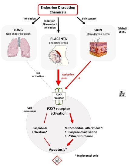
Apoptosis is characterized by numerous morphological changes, including chromatin condensation that allows DNA fragmentation. We investigated chromatin condensation after incubation with EDCs, using the Hoechst 33342 assay. A statistically significant elevated chromatin condensation was observed with diethylstilbestrol at 7.5 µM (×1.58), propylparaben at 100 µM (×1.55) and benzyl butyl phthalate at 10 µM (×1.20) compared with the control. Chromatin condensation induced by diethylstilbestrol was partially reversed by BBG (×1.58 without BBG versus ×1.37 with BBG, Figure 7). Bisphenol A, 4-tert-amylphenol, 4-heptylphenol, triclosan, DEHP and 3-benzylidene camphor had no significant effect on chromatin condensation.

4. Discussion

The objective of the present study was to explore the ability of different EDCs to induce toxicity in different cells by a common cellular mechanism, in particular P2X7 receptor activation. We compared the level of P2X7 receptor activation in human epithelial pulmonary cells, and keratinocytes and placental cells after incubation with EDCs that can be inhaled, directly applied to the skin and/or ingested. The placenta being a crucial organ during pregnancy and the most exposed organ to EDCs, further investigations were performed to study apoptosis, one of the major cell death pathways [39] that can be induced by P2X7 receptor activation [21,40]. Apoptosis is involved in pregnancy disorders [41,42,43].
The three cell lines were selected for their different endocrine properties and because they all express functional P2X7 receptors [13,26,34]. Human epithelial pulmonary A549 cells share similar ultrastructural characteristics and cytochromes expression to in situ type II pneumocytes, the most abundant cells in lungs. Human keratinocyte HaCaT cells have been extensively used to study epidermal homeostasis and its physiopathology. Furthermore, they are metabolically active since they have cytochromes from 1, 2, 3 and 4 families [44]. Human villous trophoblastic placental JEG-3 cells provide an appropriate model to detect placental toxicity [13,25,26]. They are also able to synthesize and secrete hormones.
We selected EDCs reported in the literature to be detected in pregnant women and placentas [8,9,31,32,33]. Exposure to bisphenol A, diethylstilbestrol, 4-tert-amylphenol, 4-heptylphenol, triclosan, propylparaben, benzyl butyl phthalate, DEHP and 3-benzylidene camphor can alter placental functions and therefore induce pregnancy outcomes and complications such as preeclampsia [5,6,7,8,10,45,46]. EDCs were tested at concentrations found in the placenta and we kept the same concentrations to study and compare EDCs effects in lungs and skin, the EDCs pathway’s entry into the body.
The present study is the first to compare EDCs effects on P2X7 receptor activation in three organs present in pregnant woman and possessing different endocrine properties. We highlighted that the P2X7 receptor in lung cells is not sensitive to EDCs and that the P2X7 receptors in skin cells are less sensitive to EDCs than in placental cells where they induce apoptosis.
Bisphenol A and benzyl butyl phthalate did not activate the P2X7 receptor in lung cells, while 3-benzylidene camphor activated the P2X7 receptor in keratinocytes, unlike propylparaben. All of the tested EDCs in human placental cells activated the P2X7 receptor (except 4-heptylphenol). These differences may be explained by the endocrine properties of the different cells. The placenta is considered the most important endocrine organ during pregnancy. Villous trophoblast cells have a lot of steroid or polypeptide hormone receptors and the ability to synthesize, secrete and control different maternal and fetus hormones. Skin, in addition to its main protective function, can also be classified as an endocrine organ endowed with local steroidogenic activities [47,48]. HaCaT keratinocytes possess different steroid receptors [49,50,51] and metabolize progesterone to deoxycorticosterone, cortisone, aldosterone and cortisol [47]. On the contrary, pulmonary epithelial cells do not possess any hormonal receptors and are not able to produce hormones. The lack of P2X7 receptor activation by EDCs in A549 cells did not allow us to arrive at a conclusion on the potential existence of a common mechanism of EDCs in pregnant women organs, but it suggests that P2X7 receptor activation could be a common mechanism in endocrine organs and its activation would be linked to hormonal dysregulation. To confirm this statement, it would be interesting to evaluate P2X7 receptor activation induced by the same EDCs in other endocrine cells, such as adrenal H295 cells, used for the OECD Steroidogenesis Assay (OECD Guidelines for the Testing of Chemicals, Section 4 Test No. 456).
P2X7 receptor activation is known to trigger pregnancy disorders such as preeclampsia and preterm birth [14,16]. In this study, we have shown that EDCs, known in the literature to induce the same disorders, activate the P2X7 receptor. Our results suggest that EDCs could trigger pregnancy disorders through P2X7 receptor activation. P2X7 receptor activation could be a key element in the understanding of placental disorders induced by EDCs.
Prolonged activation of the P2X7 receptor has been linked to apoptosis [21,40]. Depending on the cleaved caspase, apoptosis can be initiated through two major pathways [52]: the extrinsic receptor mediated pathway through caspase-8 activation [53,54] or the intrinsic-mediated pathway, resulting in caspase-9 activation [21,55]. Mitochondrial damages result in cytochrome c release and formation of the apoptosome, a multimeric protein complex containing Apaf-1, cytochrome c and caspase-9, resulting in caspase-9 activation [21,55]. Extrinsic and intrinsic-mediated pathways lead to caspase-3 activation [56], followed by chromatin condensation [57]. Most tested EDCs, despite different chemical structures, triggered apoptosis in placental cells, through different pathways depending on the studied EDC (Table 1). Bisphenol A and 3-benzylidene camphor induced P2X7 receptor activation and mitochondrial membrane potential disturbance. Diethylstilbestrol induced the activation of all the assessed apoptosis markers, meaning that both extrinsic and intrinsic-mediated apoptosis are triggered through P2X7 receptor activation. Propylparaben also induced both extrinsic and intrinsic-mediated apoptosis through P2X7 receptor activation, but contrary to diethylstilbestrol did not induce mitochondrial membrane potential disturbance. Benzyl butyl phthalate induced both extrinsic and intrinsic-mediated apoptosis, but only the intrinsic-mediated apoptosis was associated with P2X7 receptor activation. 4-heptylphenol induced extrinsic and intrinsic-mediated apoptosis, but it was independent from P2X7 receptor activation. 4-tert-amylphenol and DEHP induced P2X7 receptor-dependent intrinsic-mediated apoptosis with mitochondrial membrane potential disturbance. Triclosan induced P2X7 receptor-dependent intrinsic-mediated apoptosis without mitochondrial membrane potential disturbance.
Our study highlights that most of the tested EDCs, belonging to different chemical families, induced mitochondrial alterations through either intrinsic-mediated apoptosis or membrane potential disturbance or both. We were unfortunately unable to link P2X7 receptor activation with mitochondrial membrane potential alteration due to a technical artifact with BBG in the JC-1 assay. The P2X7 receptor and mitochondria have a close connection to ATP. ATP, produced by mitochondria, is the natural ligand of the P2X7 receptor. In syncytiotrophoblast, the majority of ATP is utilized for cholesterol transport and steroidogenesis [58]. Placental mitochondria play a key role in steroidogenesis with huge production of progesterone necessary for maintaining pregnancy [58]. Furthermore, progesterone increases mitochondrial membrane potential [59,60]. As mitochondria are required for steroidogenesis, mitochondrial disorders may contribute to preeclampsia [61,62] and other endocrine diseases such as diabetes mellitus and obesity. All these data suggest that the P2X7 receptor along with mitochondria are specific targets for EDCs to exert their effects on hormones alteration.

5. Conclusions

In conclusion, the results of our study suggest that the P2X7 receptor would be a common cellular mechanism of EDCs toxicity in endocrine pregnant women cells. In placental cells, EDCs induce P2X7 receptor activation and mitochondrial alterations, reported to trigger preeclampsia and preterm birth in clinics. P2X7 receptor activation and mitochondrial alterations could be key elements in understanding placental disorders induced by EDCs.

Author Contributions

Conceptualization, S.F., M.D. and E.O.; methodology, S.F.; investigation, S.F. and P.L.; writing—original draft preparation, S.F.; writing—review and editing, S.F., M.D. and E.O.; supervision, P.R. All authors have read and agreed to the published version of the manuscript.

Funding

This manuscript received no external funding.

Institutional Review Board Statement

Not applicable.

Informed Consent Statement

Not applicable.

Data Availability Statement

The data presented in this study are available on request from the corresponding author.

Acknowledgments

The authors would like to thank Laboratoires Léa Nature for the grant CIFRE with ANRT (French national agency for research) and Adebiopharm ER67 for their financial support.

Conflicts of Interest

The authors declare no conflict of interest.

References

  1. IPCS. Global Assessment of the State-of-the-Science of Endocrine Disruptor; World Health Organization: Geneva, Switzerland, 2002. [Google Scholar]
  2. Paciência, I.; Cavaleiro Rufo, J.; Silva, D.; Martins, C.; Mendes, F.; Farraia, M.; Delgado, L.; de Oliveira Fernandes, E.; Padrão, P.; Moreira, P.; et al. Exposure to indoor endocrine-disrupting chemicals and childhood asthma and obesity. Allergy 2019, 74, 1277–1291. [Google Scholar] [CrossRef] [PubMed]
  3. Ju, Q.; Zouboulis, C.C. Endocrine-disrupting chemicals and skin manifestations. Rev. Endocr. Metab. Disord. 2016, 17, 449–457. [Google Scholar] [CrossRef] [PubMed]
  4. Bamigboye, A.A.; Morris, J. Oestrogen supplementation, mainly diethylstilbestrol, for preventing miscarriages and other adverse pregnancy outcomes. Cochrane Database Syst. Rev. 2003, 3, CD004353. [Google Scholar] [CrossRef] [PubMed]
  5. Chen, X.; Chen, M.; Xu, B.; Tang, R.; Han, X.; Qin, Y.; Xu, B.; Hang, B.; Mao, Z.; Huo, W.; et al. Parental phenols exposure and spontaneous abortion in Chinese population residing in the middle and lower reaches of the Yangtze River. Chemosphere 2013, 93, 217–222. [Google Scholar] [CrossRef]
  6. Huang, Y.; Li, J.; Garcia, J.M.; Lin, H.; Wang, Y.; Yan, P.; Wang, L.; Tan, Y.; Luo, J.; Qiu, Z.; et al. Phthalate levels in cord blood are associated with preterm delivery and fetal growth parameters in Chinese women. PLoS ONE 2014, 9, e87430. [Google Scholar] [CrossRef] [Green Version]
  7. Cantonwine, D.E.; Meeker, J.D.; Ferguson, K.K.; Mukherjee, B.; Hauser, R.; McElrath, T.F. Urinary Concentrations of Bisphenol A and Phthalate Metabolites Measured during Pregnancy and Risk of Preeclampsia. Environ. Health Perspect. 2016, 124, 1651–1655. [Google Scholar] [CrossRef]
  8. Etzel, T.M.; Calafat, A.M.; Ye, X.; Chen, A.; Lanphear, B.P.; Savitz, D.A.; Yolton, K.; Braun, J.M. Urinary triclosan concentrations during pregnancy and birth outcomes. Environ. Res. 2017, 156, 505–511. [Google Scholar] [CrossRef]
  9. Aker, A.M.; Ferguson, K.K.; Rosario, Z.Y.; Mukherjee, B.; Alshawabkeh, A.N.; Calafat, A.M.; Cordero, J.F.; Meeker, J.D. A repeated measures study of phenol, paraben and Triclocarban urinary biomarkers and circulating maternal hormones during gestation in the Puerto Rico PROTECT cohort. Environ. Health 2019, 18, 28. [Google Scholar] [CrossRef] [Green Version]
  10. Mustieles, V.; Zhang, Y.; Yland, J.; Braun, J.M.; Williams, P.L.; Wylie, B.J.; Attaman, J.A.; Ford, J.B.; Azevedo, A.; Calafat, A.M.; et al. Maternal and paternal preconception exposure to phenols and preterm birth. Environ. Int. 2020, 137, 105523. [Google Scholar] [CrossRef]
  11. Müller, T.; Vieira, R.P.; Grimm, M.; Dürk, T.; Cicko, S.; Zeiser, R.; Jakob, T.; Martin, S.F.; Blumenthal, B.; Sorichter, S.; et al. A potential role for P2X7R in allergic airway inflammation in mice and humans. Am. J. Respir. Cell Mol. Biol. 2011, 44, 456–464. [Google Scholar] [CrossRef]
  12. Da Silva, G.L.; Sperotto, N.D.M.; Borges, T.J.; Bonorino, C.; Takyia, C.M.; Coutinho-Silva, R.; Campos, M.M.; Zanin, R.F.; Morrone, F.B. P2X7 receptor is required for neutrophil accumulation in a mouse model of irritant contact dermatitis. Exp. Dermatol. 2013, 22, 184–188. [Google Scholar] [CrossRef]
  13. Olivier, E.; Dutot, M.; Regazzetti, A.; Laprévote, O.; Rat, P. 25-Hydroxycholesterol induces both P2X7-dependent pyroptosis and caspase-dependent apoptosis in human skin model: New insights into degenerative pathways. Chem. Phys. Lipids 2017, 207, 171–178. [Google Scholar] [CrossRef]
  14. Tsimis, M.E.; Lei, J.; Rosenzweig, J.M.; Arif, H.; Shabi, Y.; Alshehri, W.; Talbot, C.C.; Baig-Ward, K.M.; Segars, J.; Graham, E.M.; et al. P2X7 receptor blockade prevents preterm birth and perinatal brain injury in a mouse model of intrauterine inflammation. Biol. Reprod. 2017, 97, 230–239. [Google Scholar] [CrossRef]
  15. Urabe, S.; Miyoshi, H.; Fujiwara, H.; Yamaoka, K.; Kudo, Y. Enhanced expression of P2X4 and P2X7 purinergic receptors in the myometrium of pregnant rats in preterm delivery models. Reprod. Sci. 2009, 16, 1186–1192. [Google Scholar] [CrossRef]
  16. Fodor, P.; White, B.; Khan, R. Inflammation-The role of ATP in pre-eclampsia. Microcirculation 2020, 27, e12585. [Google Scholar] [CrossRef]
  17. Adinolfi, E.; Giuliani, A.L.; De Marchi, E.; Pegoraro, A.; Orioli, E.; Di Virgilio, F. The P2X7 receptor: A main player in inflammation. Biochem. Pharmacol. 2018, 151, 234–244. [Google Scholar] [CrossRef]
  18. Zeng, D.; Yao, P.; Zhao, H. P2X7, a critical regulator and potential target for bone and joint diseases. J. Cell Physiol. 2019, 234, 2095–2103. [Google Scholar] [CrossRef]
  19. Francistiová, L.; Bianchi, C.; Di Lauro, C.; Sebastián-Serrano, Á.; de Diego-García, L.; Kobolák, J.; Dinnyés, A.; Díaz-Hernández, M. The Role of P2X7 Receptor in Alzheimer’s Disease. Front. Mol. Neurosci. 2020, 13, 94. [Google Scholar] [CrossRef]
  20. Ribeiro, D.E.; Oliveira-Giacomelli, Á.; Glaser, T.; Arnaud-Sampaio, V.F.; Andrejew, R.; Dieckmann, L.; Baranova, J.; Lameu, C.; Ratajczak, M.Z.; Ulrich, H. Hyperactivation of P2X7 receptors as a culprit of COVID-19 neuropathology. Mol. Psychiatry 2021, 26, 1044–1059. [Google Scholar] [CrossRef]
  21. Mackenzie, A.B.; Young, M.T.; Adinolfi, E.; Surprenant, A. Pseudoapoptosis induced by brief activation of ATP-gated P2X7 receptors. J. Biol. Chem. 2005, 280, 33968–33976. [Google Scholar] [CrossRef] [Green Version]
  22. Kong, Q.; Wang, M.; Liao, Z.; Camden, J.M.; Yu, S.; Simonyi, A.; Sun, G.Y.; Gonzalez, F.A.; Erb, L.; Seye, C.I.; et al. P2X(7) nucleotide receptors mediate caspase-8/9/3-dependent apoptosis in rat primary cortical neurons. Purinergic Signal. 2005, 1, 337–347. [Google Scholar] [CrossRef] [PubMed] [Green Version]
  23. Nishida, K.; Nakatani, T.; Ohishi, A.; Okuda, H.; Higashi, Y.; Matsuo, T.; Fujimoto, S.; Nagasawa, K. Mitochondrial dysfunction is involved in P2X7 receptor-mediated neuronal cell death. J. Neurochem. 2012, 122, 1118–1128. [Google Scholar] [CrossRef] [PubMed]
  24. Bartlett, R.; Yerbury, J.J.; Sluyter, R. P2X7 receptor activation induces reactive oxygen species formation and cell death in murine EOC13 microglia. Mediators Inflamm. 2013, 2013, 271813. [Google Scholar] [CrossRef] [PubMed]
  25. Fouyet, S.; Olivier, E.; Leproux, P.; Dutot, M.; Rat, P. Bisphenol A, Bisphenol F, and Bisphenol S: The Bad and the Ugly. Where Is the Good? Life 2021, 11, 314. [Google Scholar] [CrossRef]
  26. Wakx, A.; Regazzetti, A.; Dargère, D.; Auzeil, N.; Gil, S.; Evain-Brion, D.; Laprévote, O.; Rat, P. New in vitro biomarkers to detect toxicity in human placental cells: The example of benzo[A]pyrene. Toxicol. In Vitro 2016, 32, 76–85. [Google Scholar] [CrossRef]
  27. Makrantonaki, E.; Zouboulis, C.C. Androgens and ageing of the skin. Curr. Opin. Endocrinol. Diabetes Obes. 2009, 16, 240–245. [Google Scholar] [CrossRef]
  28. Roger, S.; Jelassi, B.; Couillin, I.; Pelegrin, P.; Besson, P.; Jiang, L.-H. Understanding the roles of the P2X7 receptor in solid tumour progression and therapeutic perspectives. Biochim. Biophys. Acta (BBA)-Biomembr. 2015, 1848, 2584–2602. [Google Scholar] [CrossRef] [Green Version]
  29. Gönczi, M.; Telek, A.; Czifra, G.; Balogh, A.; Blumberg, P.M.; Bíró, T.; Csernoch, L. Altered calcium handling following the recombinant overexpression of protein kinase C isoforms in HaCaT cells. Exp. Dermatol. 2008, 17, 584–591. [Google Scholar] [CrossRef]
  30. Gupta, R.; Gupta, R. Placental Toxicity. In Developmental Toxicology; Elsevier: Amsterdam, The Netherlands, 2017; Chapter 68; pp. 1301–1325. [Google Scholar]
  31. Gingrich, J.; Ticiani, E.; Veiga-Lopez, A. Placenta Disrupted: Endocrine Disrupting Chemicals and Pregnancy. Trends Endocrinol. Metab. 2020, 31, 508–524. [Google Scholar] [CrossRef]
  32. Bai, X.; Zhang, B.; He, Y.; Hong, D.; Song, S.; Huang, Y.; Zhang, T. Triclosan and triclocarbon in maternal-fetal serum, urine, and amniotic fluid samples and their implication for prenatal exposure. Environ. Pollut. 2020, 266, 115117. [Google Scholar] [CrossRef]
  33. Jiménez-Díaz, I.; Molina-Molina, J.M.; Zafra-Gómez, A.; Ballesteros, O.; Navalón, A.; Real, M.; Sáenz, J.M.; Fernández, M.F.; Olea, N. Simultaneous determination of the UV-filters benzyl salicylate, phenyl salicylate, octyl salicylate, homosalate, 3-(4-methylbenzylidene) camphor and 3-benzylidene camphor in human placental tissue by LC-MS/MS. Assessment of their in vitro endocrine activity. J. Chromatogr. B Analyt. Technol. Biomed. Life Sci. 2013, 936, 80–87. [Google Scholar] [CrossRef]
  34. Dutot, M.; Olivier, E.; Fouyet, S.; Magny, R.; Hammad, K.; Roulland, E.; Rat, P.; Fagon, R. In Vitro Chemopreventive Potential of Phlorotannins-Rich Extract from Brown Algae by Inhibition of Benzo[a]pyrene-Induced P2X7 Activation and Toxic Effects. Mar. Drugs 2021, 19, 34. [Google Scholar] [CrossRef]
  35. Olivier, E.; Wakx, A.; Fouyet, S.; Dutot, M.; Rat, P. JEG-3 placental cells in toxicology studies: A promising tool to reveal pregnancy disorders. Anat. Cell Biol. 2020, 54, 83–92. [Google Scholar] [CrossRef]
  36. Ihde, E.S.; Zamudio, S.; Loh, J.M.; Zhu, Y.; Woytanowski, J.; Rosen, L.; Liu, M.; Buckley, B. Application of a Novel Mass Spectrometric (MS) Method to Examine Exposure to Bisphenol-A and Common Substitutes in a Maternal Fetal Cohort. Hum. Ecol. Risk Assess 2018, 24, 331–346. [Google Scholar] [CrossRef]
  37. Tan, B.L.L.; Ali Mohd, M. Analysis of Selected Pesticides and Alkylphenols in Human Cord Blood by Gas Chromatograph-Mass Spectrometer. Talanta 2003, 61, 385–391. [Google Scholar] [CrossRef]
  38. Rat, P.; Olivier, E.; Tanter, C.; Wakx, A.; Dutot, M. A fast and reproducible cell- and 96-well plate-based method for the evaluation of P2X7 receptor activation using YO-PRO-1 fluorescent dye. J. Biol. Methods 2017, 4, e64. [Google Scholar] [CrossRef] [Green Version]
  39. Duprez, L.; Wirawan, E.; Berghe, T.V.; Vandenabeele, P. Major cell death pathways at a glance. Microbes Infect. 2009, 11, 1050–1062. [Google Scholar] [CrossRef]
  40. Zheng, L.M.; Zychlinsky, A.; Liu, C.C.; Ojcius, D.M.; Young, J.D. Extracellular ATP as a trigger for apoptosis or programmed cell death. J. Cell Biol. 1991, 112, 279–288. [Google Scholar] [CrossRef] [Green Version]
  41. Leung, D.N.; Smith, S.C.; To, K.F.; Sahota, D.S.; Baker, P.N. Increased placental apoptosis in pregnancies complicated by preeclampsia. Am. J. Obstet. Gynecol. 2001, 184, 1249–1250. [Google Scholar] [CrossRef]
  42. Minas, V.; Jeschke, U.; Kalantaridou, S.N.; Richter, D.U.; Reimer, T.; Mylonas, I.; Friese, K.; Makrigiannakis, A. Abortion is associated with increased expression of FasL in decidual leukocytes and apoptosis of extravillous trophoblasts: A role for CRH and urocortin. Mol. Hum. Reprod. 2007, 13, 663–673. [Google Scholar] [CrossRef]
  43. Travaglino, A.; Raffone, A.; Saccone, G.; Migliorini, S.; Maruotti, G.M.; Esposito, G.; Mollo, A.; Martinelli, P.; Zullo, F.; D’Armiento, M. Placental morphology, apoptosis, angiogenesis and epithelial mechanisms in early-onset preeclampsia. Eur. J. Obstet. Gynecol. Reprod. Biol. 2019, 234, 200–206. [Google Scholar] [CrossRef] [PubMed]
  44. Janmohamed, A.; Dolphin, C.T.; Phillips, I.R.; Shephard, E.A. Quantification and cellular localization of expression in human skin of genes encoding flavin-containing monooxygenases and cytochromes P450. Biochem. Pharmacol. 2001, 62, 777–786. [Google Scholar] [CrossRef]
  45. Troisi, R.; Hyer, M.; Hatch, E.E.; Titus-Ernstoff, L.; Palmer, J.R.; Strohsnitter, W.C.; Herbst, A.L.; Adam, E.; Hoover, R.N. Medical conditions among adult offspring prenatally exposed to diethylstilbestrol. Epidemiology 2013, 24, 430–438. [Google Scholar] [CrossRef] [PubMed]
  46. Philippat, C.; Heude, B.; Botton, J.; Alfaidy, N.; Calafat, A.M.; Slama, R. EDEN Mother–Child Cohort Study Group Prenatal Exposure to Select Phthalates and Phenols and Associations with Fetal and Placental Weight among Male Births in the EDEN Cohort (France). Environ. Health Perspect. 2019, 127, 17002. [Google Scholar] [CrossRef] [Green Version]
  47. Slominski, A.; Zbytek, B.; Nikolakis, G.; Manna, P.R.; Skobowiat, C.; Zmijewski, M.; Li, W.; Janjetovic, Z.; Postlethwaite, A.; Zouboulis, C.C.; et al. Steroidogenesis in the skin: Implications for local immune functions. J. Steroid. Biochem. Mol. Biol. 2013, 137, 107–123. [Google Scholar] [CrossRef] [Green Version]
  48. Slominski, A.T.; Manna, P.R.; Tuckey, R.C. On the role of skin in the regulation of local and systemic steroidogenic activities. Steroids 2015, 103, 72–88. [Google Scholar] [CrossRef] [Green Version]
  49. Liang, T.; Hoyer, S.; Yu, R.; Soltani, K.; Lorincz, A.L.; Hiipakka, R.A.; Liao, S. Immunocytochemical localization of androgen receptors in human skin using monoclonal antibodies against the androgen receptor. J. Investig. Dermatol. 1993, 100, 663–666. [Google Scholar] [CrossRef] [Green Version]
  50. Liu, W.; Wang, F.; Li, C.; Otkur, W.; Hayashi, T.; Mizuno, K.; Hattori, S.; Fujisaki, H.; Onodera, S.; Ikejima, T. Silibinin treatment protects human skin cells from UVB injury through upregulation of estrogen receptors. J. Photochem. PhotoBiol. B 2021, 216, 112147. [Google Scholar] [CrossRef]
  51. Zhang, Q.; Wu, S.; Liu, L.; Hou, X.; Jiang, J.; Wei, X.; Hao, W. Effects of bisphenol A on gap junctions in HaCaT cells as mediated by the estrogen receptor pathway. J. Appl. Toxicol. 2019, 39, 271–281. [Google Scholar] [CrossRef]
  52. Galluzzi, L.; Vitale, I.; Aaronson, S.A.; Abrams, J.M.; Adam, D.; Agostinis, P.; Alnemri, E.S.; Altucci, L.; Amelio, I.; Andrews, D.W.; et al. Molecular mechanisms of cell death: Recommendations of the Nomenclature Committee on Cell Death 2018. Cell Death Differ. 2018, 25, 486–541. [Google Scholar] [CrossRef]
  53. Ferrari, D.; Los, M.; Bauer, M.K.; Vandenabeele, P.; Wesselborg, S.; Schulze-Osthoff, K. P2Z purinoreceptor ligation induces activation of caspases with distinct roles in apoptotic and necrotic alterations of cell death. FEBS Lett. 1999, 447, 71–75. [Google Scholar] [CrossRef] [Green Version]
  54. Aguirre, A.; Shoji, K.F.; Sáez, J.C.; Henríquez, M.; Quest, A.F.G. FasL-triggered death of Jurkat cells requires caspase 8-induced, ATP-dependent cross-talk between Fas and the purinergic receptor P2X(7). J. Cell Physiol. 2013, 228, 485–493. [Google Scholar] [CrossRef]
  55. Hajra, K.M.; Liu, J.R. Apoptosome dysfunction in human cancer. Apoptosis 2004, 9, 691–704. [Google Scholar] [CrossRef]
  56. Wang, Q.; Wang, L.; Feng, Y.-H.; Li, X.; Zeng, R.; Gorodeski, G.I. P2X7 receptor-mediated apoptosis of human cervical epithelial cells. Am. J. Physiol Cell Physiol 2004, 287, C1349–C1358. [Google Scholar] [CrossRef] [Green Version]
  57. Porter, A.G.; Jänicke, R.U. Emerging roles of caspase-3 in apoptosis. Cell Death Differ. 1999, 6, 99–104. [Google Scholar] [CrossRef]
  58. Miller, W.L. Steroid hormone synthesis in mitochondria. Mol. Cell Endocrinol. 2013, 379, 62–73. [Google Scholar] [CrossRef]
  59. Tantibhedhyangkul, J.; Hawkins, K.C.; Dai, Q.; Mu, K.; Dunn, C.N.; Miller, S.E.; Price, T.M. Expression of a mitochondrial progesterone receptor in human spermatozoa correlates with a progestin-dependent increase in mitochondrial membrane potential. Andrology 2014, 2, 875–883. [Google Scholar] [CrossRef] [Green Version]
  60. Dai, Q.; Provost, M.P.; Raburn, D.J.; Price, T.M. Progesterone Increases Mitochondria Membrane Potential in Non-human Primate Oocytes and Embryos. Reprod. Sci. 2020, 27, 1206–1214. [Google Scholar] [CrossRef]
  61. Wang, Y.; Walsh, S.W. Placental mitochondria as a source of oxidative stress in pre-eclampsia. Placenta 1998, 19, 581–586. [Google Scholar] [CrossRef]
  62. Padmini, E.; Lavanya, S.; Uthra, V. Preeclamptic placental stress and over expression of mitochondrial HSP70. Clin. Chem Lab. Med. 2009, 47, 1073–1080. [Google Scholar] [CrossRef]
Figure 1. EDCs chemical structures: (a) bisphenol A, (b) diethylstilbestrol, (c) 4-tert-amylphenol, (d) 4-heptylphenol, (e) triclosan, (f) propylparaben, (g) benzyl butyl phthalate, (h) DEHP and (i) 3-benzylidene camphor.
Cells 11 00495 g001
Figure 2. Cell viability was evaluated using the neutral red assay after (a) bisphenol A and (b) benzyl butyl phthalate incubation for 72 h in A549 cells (blue bars) and JEG-Tox cells (black bars); (c) 3-benzylidene camphor and (d) propylparaben in HaCaT (red bars) and JEG-Tox cells (black bars); (e) diethylstilbestrol, (f) 4-ter-amylphenol, (g) 4-heptylphenol, (h) triclosan and (i) DEHP in JEG-Tox cells (black bars).
Cells 11 00495 g002aCells 11 00495 g002b
Figure 3. Effects of EDCs on P2X7 receptor activation in A549 cells, HaCaT cells and JEG-Tox cells after incubation for 72 h (YO-PRO-1® assay). The cells were preincubated with either PBS (solid bars) or with Brilliant Blue G at 25 µM (hatched bars) for 15 min. The solutions were removed and A549 cells were incubated with (a) bisphenol A and (c) benzyl butyl phthalate; HaCaT cells were incubated with (e) propylparaben and (g) 3-benzylidene camphor; JEG-Tox cells were incubated with (b) bisphenol A, (d) benzyl butyl phthalate, (f) propylparaben, (h) 3-benzylidene camphor, (i) diethylstilbestrol, (j) 4-tert-amylphenol, (k) 4-heptylphenol, (l) triclosan and (m) DEHP. The significance thresholds were **** p < 0.0001, *** p < 0.001 and ** p < 0.01 compared with the control and #### p < 0.0001, ### p < 0.001, ## p < 0.01 and # p < 0.1.
Cells 11 00495 g003aCells 11 00495 g003bCells 11 00495 g003c
Figure 4. Bisphenol A (BPA), diethylstilbestrol (DES), 4-tert-amylphenol (AP), 4-heptylphenol (HP), triclosan (T), propylparaben (PrP), benzyl butyl phthalate (BBP), DEHP and 3-benzylidene camphor (3BC) effects on caspase-8 (a) and caspase-9 (b) activity in JEG-Tox cells. The cells were preincubated with either PBS (solid bars) or BBG at 25µM (hatched bars) for 15 min. The solutions were removed and the cells were incubated with EDCs for 72 h. For clarity’s sake, EDCs abbreviations are used in Figure 4, Figure 6 and Figure 7. The significance thresholds were **** p < 0.0001, *** p < 0.001 and ** p < 0.01 compared with the control and ## p < 0.01 and # p < 0.1.
Cells 11 00495 g004
Figure 5. Fluorescence microscopy images of JEG-Tox cells stained for caspase-3/7 activity. After 72 h incubation with (a) control, (b) DMSO, (c) ethanol, (d) bisphenol A at 20 µM, (e) diethylstilbestrol at 7.5 µM, (f) 4-tert-amylphenol at 50 µM, (g) 4-heptylphenol at 10 µM, (h) triclosan at 1 µM, (i) propylparaben at 100 µM, (j) benzyl butyl phthalate at 10 µM, (k) DEHP at 10 µM and (l) 3-benzylidene camphor at 10 µM, the cells were stained using Caspase-3/7 Green ReadyProbes™. Representative images from three independent experiments are shown. Bar scale: 400 µm. Magnification: 10×.
Cells 11 00495 g005
Figure 6. Mitochondrial membrane potential was evaluated using the JC-1 assay after incubation with bisphenol A (BPA), diethylstilbestrol (DES, 4-tert-amylphenol (AP), 4-heptylphenol (HP), triclosan (T), propylparaben (PrP), benzyl butyl phthalate (BBP), DEHP and 3-benzylidene camphor (3BC) for 72 h in JEG-Tox cells. Control cells were preincubated with either PBS (solid bars) or BBG at 25µM (hatched bars) for 15 min. The solutions were removed and the cells were incubated with culture medium (control) and CCCP (positive control) for 15 min. For clarity’s sake, EDCs abbreviations are used in Figure 4, Figure 6 and Figure 7. The significance thresholds were **** p < 0.0001 and *** p < 0.001 compared with the control and #### p < 0.0001.
Cells 11 00495 g006
Figure 7. Chromatin condensation was assessed using the Hoechst 33342 assay after bisphenol A (BPA), diethylstilbestrol (DES, 4-tert-amylphenol (AP), 4-heptylphenol (HP), triclosan (T), propylparaben (PrP), benzyl butyl phthalate (BBP), DEHP and 3-benzylidene camphor (3BC) incubation for 72 h in JEG-Tox cells. The cells were preincubated with either PBS (solid bars) or with BBG at 25 µM (hatched bars) for 15 min. The solutions were removed and the cells were incubated with diethylstilbestrol, 4-heptylphenol, butyl benzyl phthalate (BBP) and DEHP. For clarity’s sake, EDCs abbreviations are used in Figure 4, Figure 6 and Figure 7. The significance thresholds were **** p < 0.0001 and * p < 0.1 compared with the control and # p < 0.1.
Cells 11 00495 g007
Table 1. Summary table of EDCs effects in HaCaT cells, A549 cells and JEG-Tox cell. (/: not tested).
Chemical
Family
Chemicals
Substances
CytotoxicityP2X7 ActivationCaspase-8
Activity
Caspase-9
Activity
Caspase-3
Activity
Mitochondrial
Membrane
Potential
Chromatin
Condensation
HaCaT
Skin Cells
A549
Pulmonary Cells
JEG-Tox
Placental Cells
HaCaT Skin CellsA549
Pulmonary Cells
JEG-Tox
Placental Cells
JEG-Tox
Placental Cells
BisphenolsBisphenol A/NoNo/NoYesNoNoNoYesNo
Diethylstilbestrol//Yes//YesYesYesYesYesYes
Alkylphenols4-tert-amylphenol//No//YesNoYesNoYesNo
4-heptylphenol//Yes//NoYesYesYesYesNo
Chlorophenol
derivatives
Triclosan//Yes//YesNoYesNoNoNo
ParabensPropylparabenNo/NoNo/YesYesYesYesNoYes
PhthalatesBenzyl butyl
phthalate
/NoYes/NoYesYesYesNoYesYes
Di(2-éthylhexyle)
phthalate
DEHP
//Yes//YesNoYesNoYesNo
Camphor
derivatives
3-benzylidene
camphor
No/YesYes/YesNoNoYesNoNo
Publisher’s Note: MDPI stays neutral with regard to jurisdictional claims in published maps and institutional affiliations.

