Next Issue
Volume 11, February-2
Previous Issue
Volume 11, January-2


Cells, Volume 11, Issue 3 (February-1 2022) – 279 articles

Cover Story (view full-size image): 
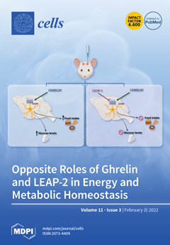
Recently gleaned data show that ghrelin, a stomach-derived peptide, and liver-expressed antimicrobial peptide 2 (LEAP-2) play opposite roles in food intake. Nevertheless, some key questions regarding the interplay among ghrelin and LEAP-2 remain to be answered. In this work we extend previous findings regarding the relevance of LEAP-2 as an anorexigenic signal. Specifically, our data, using different experimental models: lean, obese (ob/ob and DIO), aged rats and GH-deficient rats, show that LEAP-2 is a potent anorexigenic agent and maintains its capacity to blunt ghrelin-induced hyperphagia. In contrast, the inhibitory effect on glucose levels can only be observed in some specific experimental models. Taken together from these data, LEAP-2 emerged as a potential candidate to be therapeutically useful in obesity. View this paper
  • Issues are regarded as officially published after their release is announced to the table of contents alert mailing list.
  • You may sign up for e-mail alerts to receive table of contents of newly released issues.
  • PDF is the official format for papers published in both, html and pdf forms. To view the papers in pdf format, click on the "PDF Full-text" link, and use the free Adobe Reader to open them.
Order results
Result details
Section
Select all
Export citation of selected articles as:
14 pages, 1863 KiB  
Article
The Critical Role Played by Mitochondrial MITF Serine 73 Phosphorylation in Immunologically Activated Mast Cells
by Lakshmi Bhargavi Paruchuru, Sharmila Govindaraj and Ehud Razin
Cells 2022, 11(3), 589; https://doi.org/10.3390/cells11030589 - 08 Feb 2022
Cited by 5 | Viewed by 2454
Abstract
In recent years, growing evidence has indicated the pivotal role of mitochondria in mast cell immunological activation. We have previously reported a decrease in degranulation and cytokine secretion following the inhibition of pyruvate dehydrogenase (PDH) either by CPI-613 (PDH inhibitor/anti-cancer drug) or through [...] Read more.
In recent years, growing evidence has indicated the pivotal role of mitochondria in mast cell immunological activation. We have previously reported a decrease in degranulation and cytokine secretion following the inhibition of pyruvate dehydrogenase (PDH) either by CPI-613 (PDH inhibitor/anti-cancer drug) or through its interaction with mitochondrial microphthalmia-associated transcription factor (MITF). In the present study, we further explored the role played by mitochondrial MITF in mast cell exocytosis using rat basophil leukemia cells [RBL], as well as mouse bone marrow-derived mast cells (BMMCs). Here, we report that mast cell degranulation, cytokine secretion and oxidative phosphorylation (OXPHOS) activities were associated with phosphorylation of Serine 73 of mitochondrial MITF, controlled by extracellular signals regulated by protein kinase (ERK1/2) activity. Also, we report here that decreased OXPHOS activity following ERK1/2 inhibition (U0126 treatment) during IgE-Ag activation was mediated by the dephosphorylation of Serine 73 mitochondrial MITF, which inhibited its association with PDH. This led to a reduction in mast cell reactivity. In addition, a phosphorylation-mimicking mitochondrial MITF-S73D positively regulated the mitochondrial activity, thereby supporting mast cell degranulation. Thus, the present research findings highlight the prominence of mitochondrial MITF Serine 73 phosphorylation in immunologically activated mast cells. Full article
(This article belongs to the Collection Mast Cells in Health and Diseases)
Show Figures

Graphical abstract

27 pages, 1228 KiB  
Review
Biomarker Development in Cardiology: Reviewing the Past to Inform the Future
by Katharine A. Kott, Michael Bishop, Christina H. J. Yang, Toby M. Plasto, Daniel C. Cheng, Adam I. Kaplan, Louise Cullen, David S. Celermajer, Peter J. Meikle, Stephen T. Vernon and Gemma A. Figtree
Cells 2022, 11(3), 588; https://doi.org/10.3390/cells11030588 - 08 Feb 2022
Cited by 4 | Viewed by 3798
Abstract
Cardiac biomarkers have become pivotal to the clinical practice of cardiology, but there remains much to discover that could benefit cardiology patients. We review the discovery of key protein biomarkers in the fields of acute coronary syndrome, heart failure, and atherosclerosis, giving an [...] Read more.
Cardiac biomarkers have become pivotal to the clinical practice of cardiology, but there remains much to discover that could benefit cardiology patients. We review the discovery of key protein biomarkers in the fields of acute coronary syndrome, heart failure, and atherosclerosis, giving an overview of the populations they were studied in and the statistics that were used to validate them. We review statistical approaches that are currently in use to assess new biomarkers and overview a framework for biomarker discovery and evaluation that could be incorporated into clinical trials to evaluate cardiovascular outcomes in the future. Full article
(This article belongs to the Special Issue Understanding Biomarkers in Cardiology)
Show Figures

Figure 1

11 pages, 1379 KiB  
Review
Challenging the Paradigm: Anti-Inflammatory Interleukins and Angiogenesis
by Amanda M. Peluzzo and Michael V. Autieri
Cells 2022, 11(3), 587; https://doi.org/10.3390/cells11030587 - 08 Feb 2022
Cited by 22 | Viewed by 3591
Abstract
Angiogenesis is a vital biological process, and neovascularization is essential for the development, wound repair, and perfusion of ischemic tissue. Neovascularization and inflammation are independent biological processes that are linked in response to injury and ischemia. While clear that pro-inflammatory factors drive angiogenesis, [...] Read more.
Angiogenesis is a vital biological process, and neovascularization is essential for the development, wound repair, and perfusion of ischemic tissue. Neovascularization and inflammation are independent biological processes that are linked in response to injury and ischemia. While clear that pro-inflammatory factors drive angiogenesis, the role of anti-inflammatory interleukins in angiogenesis remains less defined. An interleukin with anti-inflammatory yet pro-angiogenic effects would hold great promise as a therapeutic modality to treat many disease states where inflammation needs to be limited, but revascularization and reperfusion still need to be supported. As immune modulators, interleukins can polarize macrophages to a pro-angiogenic and reparative phenotype, which indirectly influences angiogenesis. Interleukins could also potentially directly induce angiogenesis by binding and activating its receptor on endothelial cells. Although a great deal of attention is given to the negative effects of pro-inflammatory interleukins, less is described concerning the potential protective effects of anti-inflammatory interleukins on various disease processes. To focus this review, we will consider IL-4, IL-10, IL-13, IL-19, and IL-33 to be anti-inflammatory interleukins, all of which have recognized immunomodulatory effects. This review will summarize current research concerning anti-inflammatory interleukins as potential drivers of direct and indirect angiogenesis, emphasizing their role in future therapeutics. Full article
(This article belongs to the Collection The Increasingly Fascinating World of Interleukins)
Show Figures

Graphical abstract

13 pages, 45311 KiB  
Article
Arabinogalactan Proteins in the Digestive Glands of Dionaea muscipula J.Ellis Traps
by Bartosz J. Płachno, Małgorzata Kapusta, Piotr Stolarczyk and Piotr Świątek
Cells 2022, 11(3), 586; https://doi.org/10.3390/cells11030586 - 08 Feb 2022
Cited by 8 | Viewed by 2667
Abstract
The arabinogalactan proteins (AGP) play important roles in plant growth and developmental processes. However, to the best of our knowledge, there is no information on the spatial distribution of AGP in the plant organs and tissues of carnivorous plants during their carnivorous cycle. [...] Read more.
The arabinogalactan proteins (AGP) play important roles in plant growth and developmental processes. However, to the best of our knowledge, there is no information on the spatial distribution of AGP in the plant organs and tissues of carnivorous plants during their carnivorous cycle. The Dionaea muscipula trap forms an “external stomach” and is equipped with an effective digestive-absorbing system. Because its digestive glands are composed of specialized cells, the hypothesis that their cell walls are also very specialized in terms of their composition (AGP) compared to the cell wall of the trap epidermal and parenchyma cells was tested. Another aim of this study was to determine whether there is a spatio-temporal distribution of the AGP in the digestive glands during the secretory cycle of D. muscipula. Antibodies that act against AGPs, including JIM8, JIM13 and JIM14, were used. The localization of the examined compounds was determined using immunohistochemistry techniques and immunogold labeling. In both the un-fed and fed traps, there was an accumulation of AGP in the cell walls of the gland secretory cells. The epitope, which is recognized by JIM14, was a useful marker of the digestive glands. The secretory cells of the D. muscipula digestive glands are transfer cells and an accumulation of specific AGP was at the site where the cell wall labyrinth occurred. Immunogold labeling confirmed an occurrence of AGP in the cell wall ingrowths. There were differences in the AGP occurrence (labeled with JIM8 and JIM13) in the cell walls of the gland secretory cells between the unfed and fed traps. Full article
(This article belongs to the Special Issue Research on Plant Cell Wall Biology)
Show Figures

Figure 1

11 pages, 3433 KiB  
Article
Shear-Stress-Gradient and Oxygen-Gradient Ektacytometry in Sickle Cell Patients at Steady State and during Vaso-Occlusive Crises
by Camille Boisson, Elie Nader, Céline Renoux, Alexandra Gauthier, Solène Poutrel, Yves Bertrand, Emeric Stauffer, Emilie Virot, Arnaud Hot, Romain Fort, Giovanna Cannas, Philippe Joly and Philippe Connes
Cells 2022, 11(3), 585; https://doi.org/10.3390/cells11030585 - 08 Feb 2022
Cited by 4 | Viewed by 1680
Abstract
Oxygen gradient ektacytometry (oxygenscan) measures the changes in red blood cell (RBC) deformability in normoxia and during deoxygenation. We investigated the changes in RBC deformability, measured by both oxygenscan and classical shear-stress-gradient ektacytometry, in 10 patients with sickle cell disease (SCD) during vaso-occlusive [...] Read more.
Oxygen gradient ektacytometry (oxygenscan) measures the changes in red blood cell (RBC) deformability in normoxia and during deoxygenation. We investigated the changes in RBC deformability, measured by both oxygenscan and classical shear-stress-gradient ektacytometry, in 10 patients with sickle cell disease (SCD) during vaso-occlusive crisis (VOC) versus steady state. Oxygenscan and shear-stress-gradient ektacytometry parameters were also measured in 38 SCD patients at steady state on two different occasions. Shear-stress-gradient ektacytometry parameters, maximal RBC deformability at normoxia and the minimum RBC deformability during deoxygenation were lower during VOC compared to steady state. The oxygen partial pressure at which RBCs started to sickle (PoS) was not significantly affected by VOC, but the results were very heterogeneous: the PoS increased in 5 in 10 patients and decreased in 4 in 10 patients. Both oxygenscan and shear-stress-gradient ektacytometry parameters remained unchanged in patients at steady state between two sets of measurements, performed at 17 ± 8 months intervals. In conclusion, the present study showed that both oxygen gradient ektacytometry and shear-stress-gradient ektacytometry are sensitive to disease activity in SCD, and that both techniques give comparable results; however, the oxygen-dependent propensity of RBCs to sickle was highly variable during VOC. Full article
(This article belongs to the Collection Advances in Cell Culture and Tissue Engineering)
Show Figures

Figure 1

13 pages, 3205 KiB  
Article
MicroRNA-Mediated Downregulation of HMGB2 Contributes to Cellular Senescence in Microvascular Endothelial Cells
by Hye-Ram Jo and Jae-Hoon Jeong
Cells 2022, 11(3), 584; https://doi.org/10.3390/cells11030584 - 08 Feb 2022
Cited by 6 | Viewed by 2307
Abstract
High mobility group box 2 (HMGB2) is a non-histone chromosomal protein involved in various biological processes, including cellular senescence. However, its role in cellular senescence has not been evaluated extensively. To determine the regulatory role and mechanism of HMGB2 in cellular senescence, we [...] Read more.
High mobility group box 2 (HMGB2) is a non-histone chromosomal protein involved in various biological processes, including cellular senescence. However, its role in cellular senescence has not been evaluated extensively. To determine the regulatory role and mechanism of HMGB2 in cellular senescence, we performed gene expression analysis, senescence staining, and tube formation assays using young and senescent microvascular endothelial cells (MVECs) after small RNA treatment or HMGB2 overexpression. HMGB2 expression decreased with age and was regulated at the transcriptional level. siRNA-mediated downregulation inhibited cell proliferation and accelerated cellular senescence. In contrast, ectopic overexpression delayed senescence and maintained relatively higher tube-forming activity. To determine the HMGB2 downregulation mechanism, we screened miRNAs that were significantly upregulated in senescent MVECs and selected HMGB2-targeting miRNAs. Six miRNAs, miR-23a-3p, 23b-3p, -181a-5p, -181b-5p, -221-3p, and -222-3p, were overexpressed in senescent MVECs. Ectopic introduction of miR-23a-3p, -23b-3p, -181a-5p, -181b-5p, and -221-3p, with the exception of miR-222-3p, led to the downregulation of HMGB2, upregulation of senescence-associated markers, and decreased tube formation activity. Inhibition of miR-23a-3p, -181a-5p, -181b-5p, and -221-3p delayed cellular senescence. Restoration of HMGB2 expression using miRNA inhibitors represents a potential strategy to overcome the detrimental effects of cellular senescence in endothelial cells. Full article
(This article belongs to the Special Issue Epigenetic Mechanisms of Longevity and Aging)
Show Figures

Figure 1

17 pages, 2040 KiB  
Review
What Have We Learned in 30 Years of Investigations on Bari Transposons?
by Antonio Palazzo, Ruggiero Caizzi, Roberta Moschetti and René Massimiliano Marsano
Cells 2022, 11(3), 583; https://doi.org/10.3390/cells11030583 - 08 Feb 2022
Cited by 3 | Viewed by 1817
Abstract
Transposable elements (TEs) have been historically depicted as detrimental genetic entities that selfishly aim at perpetuating themselves, invading genomes, and destroying genes. Scientists often co-opt “special” TEs to develop new and powerful genetic tools, that will hopefully aid in changing the future of [...] Read more.
Transposable elements (TEs) have been historically depicted as detrimental genetic entities that selfishly aim at perpetuating themselves, invading genomes, and destroying genes. Scientists often co-opt “special” TEs to develop new and powerful genetic tools, that will hopefully aid in changing the future of the human being. However, many TEs are gentle, rarely unleash themselves to harm the genome, and bashfully contribute to generating diversity and novelty in the genomes they have colonized, yet they offer the opportunity to develop new molecular tools. In this review we summarize 30 years of research focused on the Bari transposons. Bari is a “normal” transposon family that has colonized the genomes of several Drosophila species and introduced genomic novelties in the melanogaster species. We discuss how these results have contributed to advance the field of TE research and what future studies can still add to the current knowledge. Full article
Show Figures

Figure 1

15 pages, 4256 KiB  
Article
Functional Genomic Screening in Human Pluripotent Stem Cells Reveals New Roadblocks in Early Pancreatic Endoderm Formation
by Jana Krüger, Markus Breunig, Lino Pascal Pasquini, Mareen Morawe, Alexander Groß, Frank Arnold, Ronan Russell, Thomas Seufferlein, Ninel Azoitei, Hans A. Kestler, Cécile Julier, Sandra Heller, Meike Hohwieler and Alexander Kleger
Cells 2022, 11(3), 582; https://doi.org/10.3390/cells11030582 - 08 Feb 2022
Cited by 3 | Viewed by 2712
Abstract
Human pluripotent stem cells, with their ability to proliferate indefinitely and to differentiate into virtually all cell types of the human body, provide a novel resource to study human development and to implement relevant disease models. Here, we employed a human pancreatic differentiation [...] Read more.
Human pluripotent stem cells, with their ability to proliferate indefinitely and to differentiate into virtually all cell types of the human body, provide a novel resource to study human development and to implement relevant disease models. Here, we employed a human pancreatic differentiation platform complemented with an shRNA screen in human pluripotent stem cells (PSCs) to identify potential drivers of early endoderm and pancreatic development. Deep sequencing followed by abundancy ranking pinpointed six top hit genes potentially associated with either improved or impaired endodermal differentiation, which were selected for functional validation in CRISPR-Cas9 mediated knockout (KO) lines. Upon endoderm differentiation (DE), particularly the loss of SLC22A1 and DSC2 led to impaired differentiation efficiency into CXCR4/KIT-positive DE cells. qPCR analysis also revealed changes in differentiation markers CXCR4, FOXA2, SOX17, and GATA6. Further differentiation of PSCs to the pancreatic progenitor (PP) stage resulted in a decreased proportion of PDX1/NKX6-1-positive cells in SLC22A1 KO lines, and in DSC2 KO lines when differentiated under specific culture conditions. Taken together, our study reveals novel genes with potential roles in early endodermal development. Full article
(This article belongs to the Special Issue Novel Approaches in Stem Cell Research)
Show Figures

Graphical abstract

49 pages, 2992 KiB  
Review
Glycomic and Glycoproteomic Techniques in Neurodegenerative Disorders and Neurotrauma: Towards Personalized Markers
by Firas Kobeissy, Abir Kobaisi, Wenjing Peng, Chloe Barsa, Mona Goli, Ahmad Sibahi, Samer El Hayek, Samar Abdelhady, Muhammad Ali Haidar, Mirna Sabra, Matej Orešič, Giancarlo Logroscino, Stefania Mondello, Ali H. Eid and Yehia Mechref
Cells 2022, 11(3), 581; https://doi.org/10.3390/cells11030581 - 08 Feb 2022
Cited by 13 | Viewed by 4280
Abstract
The proteome represents all the proteins expressed by a genome, a cell, a tissue, or an organism at any given time under defined physiological or pathological circumstances. Proteomic analysis has provided unparalleled opportunities for the discovery of expression patterns of proteins in a [...] Read more.
The proteome represents all the proteins expressed by a genome, a cell, a tissue, or an organism at any given time under defined physiological or pathological circumstances. Proteomic analysis has provided unparalleled opportunities for the discovery of expression patterns of proteins in a biological system, yielding precise and inclusive data about the system. Advances in the proteomics field opened the door to wider knowledge of the mechanisms underlying various post-translational modifications (PTMs) of proteins, including glycosylation. As of yet, the role of most of these PTMs remains unidentified. In this state-of-the-art review, we present a synopsis of glycosylation processes and the pathophysiological conditions that might ensue secondary to glycosylation shortcomings. The dynamics of protein glycosylation, a crucial mechanism that allows gene and pathway regulation, is described. We also explain how—at a biomolecular level—mutations in glycosylation-related genes may lead to neuropsychiatric manifestations and neurodegenerative disorders. We then analyze the shortcomings of glycoproteomic studies, putting into perspective their downfalls and the different advanced enrichment techniques that emanated to overcome some of these challenges. Furthermore, we summarize studies tackling the association between glycosylation and neuropsychiatric disorders and explore glycoproteomic changes in neurodegenerative diseases, including Alzheimer’s disease, Parkinson’s disease, Huntington disease, multiple sclerosis, and amyotrophic lateral sclerosis. We finally conclude with the role of glycomics in the area of traumatic brain injury (TBI) and provide perspectives on the clinical application of glycoproteomics as potential diagnostic tools and their application in personalized medicine. Full article
(This article belongs to the Collection Deciphering the Proteome in Cell Biology and Diseases)
Show Figures

Figure 1

16 pages, 2487 KiB  
Review
Immune Dysfunction, Cytokine Disruption, and Stromal Changes in Myelodysplastic Syndrome: A Review
by Olivia F. Lynch and Laura M. Calvi
Cells 2022, 11(3), 580; https://doi.org/10.3390/cells11030580 - 08 Feb 2022
Cited by 8 | Viewed by 3244
Abstract
Myelodysplastic syndromes (MDS) are myeloid neoplasms characterized by bone marrow dysfunction and increased risk of transformation to leukemia. MDS represent complex and diverse diseases that evolve from malignant hematopoietic stem cells and involve not only the proliferation of malignant cells but also the [...] Read more.
Myelodysplastic syndromes (MDS) are myeloid neoplasms characterized by bone marrow dysfunction and increased risk of transformation to leukemia. MDS represent complex and diverse diseases that evolve from malignant hematopoietic stem cells and involve not only the proliferation of malignant cells but also the dysfunction of normal bone marrow. Specifically, the marrow microenvironment—both hematopoietic and stromal components—is disrupted in MDS. While microenvironmental disruption has been described in human MDS and murine models of the disease, only a few current treatments target the microenvironment, including the immune system. In this review, we will examine current evidence supporting three key interdependent pillars of microenvironmental alteration in MDS—immune dysfunction, cytokine skewing, and stromal changes. Understanding the molecular changes seen in these diseases has been, and will continue to be, foundational to developing effective novel treatments that prevent disease progression and transformation to leukemia. Full article
(This article belongs to the Special Issue The Impact of Immune Activation on Hematopoiesis)
Show Figures

Figure 1

19 pages, 804 KiB  
Review
Cytokines and Chemokines in Cancer Cachexia and Its Long-Term Impact on COVID-19
by Santosh Kumar Singh and Rajesh Singh
Cells 2022, 11(3), 579; https://doi.org/10.3390/cells11030579 - 08 Feb 2022
Cited by 1 | Viewed by 3870
Abstract
Cancer cachexia remains a serious public health concern worldwide, particularly as cancer rates rise. Treatment is endangered, and survival is reduced, because this illness is commonly misdiagnosed and undertreated. Although weight loss is the most evident sign of cachexia, there are other early [...] Read more.
Cancer cachexia remains a serious public health concern worldwide, particularly as cancer rates rise. Treatment is endangered, and survival is reduced, because this illness is commonly misdiagnosed and undertreated. Although weight loss is the most evident sign of cachexia, there are other early metabolic and inflammatory changes that occur before the most obvious symptoms appear. Cachexia-related inflammation is induced by a combination of factors, one of which is the release of inflammation-promoting chemicals by the tumor. Today, more scientists are beginning to believe that the development of SARS-CoV-2 (COVID-19) related cachexia is similar to cancer-related cachexia. It is worth noting that patients infected with COVID-19 have a significant inflammatory response and can develop cachexia. These correlations provide feasible reasons for the variance in the occurrence and severity of cachexia in human malignancies, therefore, specific therapeutic options for these individuals must be addressed based on disease types. In this review, we highlighted the role of key chemokines, cytokines, and clinical management in relation to cancer cachexia and its long-term impact on COVID-19 patients. Full article
(This article belongs to the Special Issue Cancer-Induced Cachexia)
Show Figures

Figure 1

17 pages, 4397 KiB  
Article
Multifaceted Analyses of Isolated Mitochondria Establish the Anticancer Drug 2-Hydroxyoleic Acid as an Inhibitor of Substrate Oxidation and an Activator of Complex IV-Dependent State 3 Respiration
by Kumudesh Mishra, Mária Péter, Anna Maria Nardiello, Guy Keller, Victoria Llado, Paula Fernandez-Garcia, Ulf D. Kahlert, Dinorah Barasch, Ann Saada, Zsolt Török, Gábor Balogh, Pablo V. Escriba, Stefano Piotto and Or Kakhlon
Cells 2022, 11(3), 578; https://doi.org/10.3390/cells11030578 - 07 Feb 2022
Cited by 2 | Viewed by 2591
Abstract
The synthetic fatty acid 2-hydroxyoleic acid (2OHOA) has been extensively investigated as a cancer therapy mainly based on its regulation of membrane lipid composition and structure, activating various cell fate pathways. We discovered, additionally, that 2OHOA can uncouple oxidative phosphorylation, but this has [...] Read more.
The synthetic fatty acid 2-hydroxyoleic acid (2OHOA) has been extensively investigated as a cancer therapy mainly based on its regulation of membrane lipid composition and structure, activating various cell fate pathways. We discovered, additionally, that 2OHOA can uncouple oxidative phosphorylation, but this has never been demonstrated mechanistically. Here, we explored the effect of 2OHOA on mitochondria isolated by ultracentrifugation from U118MG glioblastoma cells. Mitochondria were analyzed by shotgun lipidomics, molecular dynamic simulations, spectrophotometric assays for determining respiratory complex activity, mass spectrometry for assessing beta oxidation and Seahorse technology for bioenergetic profiling. We showed that the main impact of 2OHOA on mitochondrial lipids is their hydroxylation, demonstrated by simulations to decrease co-enzyme Q diffusion in the liquid disordered membranes embedding respiratory complexes. This decreased co-enzyme Q diffusion can explain the inhibition of disjointly measured complexes I–III activity. However, it doesn’t explain how 2OHOA increases complex IV and state 3 respiration in intact mitochondria. This increased respiration probably allows mitochondrial oxidative phosphorylation to maintain ATP production against the 2OHOA-mediated inhibition of glycolytic ATP production. This work correlates 2OHOA function with its modulation of mitochondrial lipid composition, reflecting both 2OHOA anticancer activity and adaptation to it by enhancement of state 3 respiration. Full article
(This article belongs to the Collection The Pathomechanism of Mitochondrial Diseases)
Show Figures

Figure 1

21 pages, 2299 KiB  
Review
Regulation of LncRNAs in Melanoma and Their Functional Roles in the Metastatic Process
by Marine Melixetian, Pier Giuseppe Pelicci and Luisa Lanfrancone
Cells 2022, 11(3), 577; https://doi.org/10.3390/cells11030577 - 07 Feb 2022
Cited by 12 | Viewed by 2919
Abstract
Long non-coding RNAs (lncRNAs) are key regulators of numerous intracellular processes leading to tumorigenesis. They are frequently deregulated in cancer, functioning as oncogenes or tumor suppressors. As they act through multiple mechanisms, it is not surprising that they may exert dual functions in [...] Read more.
Long non-coding RNAs (lncRNAs) are key regulators of numerous intracellular processes leading to tumorigenesis. They are frequently deregulated in cancer, functioning as oncogenes or tumor suppressors. As they act through multiple mechanisms, it is not surprising that they may exert dual functions in the same tumor. In melanoma, a highly invasive and metastatic tumor with the propensity to rapidly develop drug resistance, lncRNAs play different roles in: (i) guiding the phenotype switch and leading to metastasis formation; (ii) predicting the response of melanoma patients to immunotherapy; (iii) triggering adaptive responses to therapy and acquisition of drug resistance phenotypes. In this review we summarize the most recent findings on the lncRNAs involved in melanoma growth and spreading to distant sites, focusing on their role as biomarkers for disease diagnosis and patient prognosis, or targets for novel therapeutic approaches. Full article
(This article belongs to the Special Issue Role of Non-coding RNA in Health and Disease)
Show Figures

Figure 1

23 pages, 14661 KiB  
Review
Targeting Breast Cancer and Their Stem Cell Population through AMPK Activation: Novel Insights
by Bhawna Uprety and Heidi Abrahamse
Cells 2022, 11(3), 576; https://doi.org/10.3390/cells11030576 - 07 Feb 2022
Cited by 14 | Viewed by 3704
Abstract
Despite some significant advancements, breast cancer has become the most prevalent cancer in the world. One of the main reasons for failure in treatment and metastasis has been attributed to the presence of cancer initiating cells—cancer stem cells. Consequently, research is now being [...] Read more.
Despite some significant advancements, breast cancer has become the most prevalent cancer in the world. One of the main reasons for failure in treatment and metastasis has been attributed to the presence of cancer initiating cells—cancer stem cells. Consequently, research is now being focussed on targeting cancer cells along with their stem cell population. Non-oncology drugs are gaining increasing attention for their potent anticancer activities. Metformin, a drug commonly used to treat type 2 diabetes, is the best example in this regard. It exerts its therapeutic action by activating 5′ adenosine monophosphate-activated protein kinase (AMPK). Activated AMPK subsequently phosphorylates and targets several cellular pathways involved in cell growth and proliferation and the maintenance of stem-like properties of cancer stem cells. Therefore, AMPK is emerging as a target of choice for developing effective anticancer drugs. Vanadium compounds are well-known PTP inhibitors and AMPK activators. They find extensive applications in treatment of diabetes and obesity via PTP1B inhibition and AMPK-mediated inhibition of adipogenesis. However, their role in targeting cancer stem cells has not been explored yet. This review is an attempt to establish the applications of insulin mimetic vanadium compounds for the treatment of breast cancer by AMPK activation and PTP1B inhibition pathways. Full article
(This article belongs to the Special Issue Advances in AMPK Research: Basic and Translational Aspects)
Show Figures

Graphical abstract

24 pages, 1094 KiB  
Review
Roles of Podoplanin in Malignant Progression of Tumor
by Hiroyuki Suzuki, Mika K. Kaneko and Yukinari Kato
Cells 2022, 11(3), 575; https://doi.org/10.3390/cells11030575 - 07 Feb 2022
Cited by 35 | Viewed by 8003
Abstract
Podoplanin (PDPN) is a cell-surface mucin-like glycoprotein that plays a critical role in tumor development and normal development of the lung, kidney, and lymphatic vascular systems. PDPN is overexpressed in several tumors and is involved in their malignancy. PDPN induces platelet aggregation through [...] Read more.
Podoplanin (PDPN) is a cell-surface mucin-like glycoprotein that plays a critical role in tumor development and normal development of the lung, kidney, and lymphatic vascular systems. PDPN is overexpressed in several tumors and is involved in their malignancy. PDPN induces platelet aggregation through binding to platelet receptor C-type lectin-like receptor 2. Furthermore, PDPN modulates signal transductions that regulate cell proliferation, differentiation, migration, invasion, epithelial-to-mesenchymal transition, and stemness, all of which are crucial for the malignant progression of tumor. In the tumor microenvironment (TME), PDPN expression is upregulated in the tumor stroma, including cancer-associated fibroblasts (CAFs) and immune cells. CAFs play significant roles in the extracellular matrix remodeling and the development of immunosuppressive TME. Additionally, PDPN functions as a co-inhibitory molecule on T cells, indicating its involvement with immune evasion. In this review, we describe the mechanistic basis and diverse roles of PDPN in the malignant progression of tumors and discuss the possibility of the clinical application of PDPN-targeted cancer therapy, including cancer-specific monoclonal antibodies, and chimeric antigen receptor T technologies. Full article
Show Figures

Figure 1

15 pages, 2208 KiB  
Review
Regulation of Nucleolar Activity by MYC
by Isabella N. Brown, M. Carmen Lafita-Navarro and Maralice Conacci-Sorrell
Cells 2022, 11(3), 574; https://doi.org/10.3390/cells11030574 - 07 Feb 2022
Cited by 7 | Viewed by 2916
Abstract
The nucleolus harbors the machinery necessary to produce new ribosomes which are critical for protein synthesis. Nucleolar size, shape, and density are highly dynamic and can be adjusted to accommodate ribosome biogenesis according to the needs for protein synthesis. In cancer, cells undergo [...] Read more.
The nucleolus harbors the machinery necessary to produce new ribosomes which are critical for protein synthesis. Nucleolar size, shape, and density are highly dynamic and can be adjusted to accommodate ribosome biogenesis according to the needs for protein synthesis. In cancer, cells undergo continuous proliferation; therefore, nucleolar activity is elevated due to their high demand for protein synthesis. The transcription factor and universal oncogene MYC promotes nucleolar activity by enhancing the transcription of ribosomal DNA (rDNA) and ribosomal proteins. This review summarizes the importance of nucleolar activity in mammalian cells, MYC’s role in nucleolar regulation in cancer, and discusses how a better understanding (and the potential inhibition) of aberrant nucleolar activity in cancer cells could lead to novel therapeutics. Full article
(This article belongs to the Special Issue Nucleolar Organization and Functions in Health and Disease II)
Show Figures

Graphical abstract

17 pages, 10626 KiB  
Article
Metabolomics of Acute vs. Chronic Spinach Intake in an Apc–Mutant Genetic Background: Linoleate and Butanoate Metabolites Targeting HDAC Activity and IFN–γ Signaling
by Ying-Shiuan Chen, Jia Li, Sultan Neja, Sabeeta Kapoor, Jorge Enrique Tovar Perez, Chakrapani Tripathi, Rani Menon, Arul Jayaraman, Kyongbum Lee, Wan Mohaiza Dashwood, Shan Wang, Ke Zhang, Koichi Kobayashi, Praveen Rajendran and Roderick Dashwood
Cells 2022, 11(3), 573; https://doi.org/10.3390/cells11030573 - 07 Feb 2022
Cited by 3 | Viewed by 3155
Abstract
There is growing interest in the crosstalk between the gut microbiome, host metabolomic features, and disease pathogenesis. The current investigation compared long–term (26 week) and acute (3 day) dietary spinach intake in a genetic model of colorectal cancer. Metabolomic analyses in the polyposis [...] Read more.
There is growing interest in the crosstalk between the gut microbiome, host metabolomic features, and disease pathogenesis. The current investigation compared long–term (26 week) and acute (3 day) dietary spinach intake in a genetic model of colorectal cancer. Metabolomic analyses in the polyposis in rat colon (Pirc) model and in wild–type animals corroborated key contributions to anticancer outcomes by spinach–derived linoleate bioactives and a butanoate metabolite linked to increased α–diversity of the gut microbiome. Combining linoleate and butanoate metabolites in human colon cancer cells revealed enhanced apoptosis and reduced cell viability, paralleling the apoptosis induction in colon tumors from rats given long–term spinach treatment. Mechanistic studies in cell–based assays and in vivo implicated the linoleate and butanoate metabolites in targeting histone deacetylase (HDAC) activity and the interferon–γ (IFN–γ) signaling axis. Clinical translation of these findings to at–risk patients might provide valuable quality–of–life benefits by delaying surgical interventions and drug therapies with adverse side effects. Full article
Show Figures

Figure 1

18 pages, 1595 KiB  
Review
The Biogenesis of miRNAs and Their Role in the Development of Amyotrophic Lateral Sclerosis
by Jinmeng Liu, Fenghua Zhou, Yingjun Guan, Fandi Meng, Zhenhan Zhao, Qi Su, Weiwei Bao, Xuemei Wang, Jiantao Zhao, Zijun Huo, Lingyun Zhang, Shuanhu Zhou, Yanchun Chen and Xin Wang
Cells 2022, 11(3), 572; https://doi.org/10.3390/cells11030572 - 07 Feb 2022
Cited by 21 | Viewed by 5390
Abstract
Amyotrophic lateral sclerosis (ALS) is a neurodegenerative disease that affects upper and lower motor neurons. As there is no effective treatment for ALS, it is particularly important to screen key gene therapy targets. The identifications of microRNAs (miRNAs) have completely changed the traditional [...] Read more.
Amyotrophic lateral sclerosis (ALS) is a neurodegenerative disease that affects upper and lower motor neurons. As there is no effective treatment for ALS, it is particularly important to screen key gene therapy targets. The identifications of microRNAs (miRNAs) have completely changed the traditional view of gene regulation. miRNAs are small noncoding single-stranded RNA molecules involved in the regulation of post-transcriptional gene expression. Recent advances also indicate that miRNAs are biomarkers in many diseases, including neurodegenerative diseases. In this review, we summarize recent advances regarding the mechanisms underlying the role of miRNAs in ALS pathogenesis and its application to gene therapy for ALS. The potential of miRNAs to target diverse pathways opens a new avenue for ALS therapy. Full article
Show Figures

Figure 1

12 pages, 730 KiB  
Article
Imatinib Mesylate for the Treatment of Canine Mast Cell Tumors: Assessment of the Response and Adverse Events in Comparison with the Conventional Therapy with Vinblastine and Prednisone
by Thais Rodrigues Macedo, Genilson Fernandes de Queiroz, Thaís Andrade Costa Casagrande, Pâmela Almeida Alexandre, Paulo Eduardo Brandão, Heidge Fukumasu, Samanta Rios Melo, Maria Lucia Zaidan Dagli, Ana Carolina B. C. Fonseca Pinto and Julia Maria Matera
Cells 2022, 11(3), 571; https://doi.org/10.3390/cells11030571 - 07 Feb 2022
Cited by 4 | Viewed by 3369
Abstract
Mast cell tumors (MCTs) are common neoplasms in dogs, and treatments for these diseases include surgery, polychemotherapy and targeted therapy with tyrosine kinase inhibitors. This study aimed to evaluate the response and the adverse events of treatment with imatinib mesylate (IM) compared to [...] Read more.
Mast cell tumors (MCTs) are common neoplasms in dogs, and treatments for these diseases include surgery, polychemotherapy and targeted therapy with tyrosine kinase inhibitors. This study aimed to evaluate the response and the adverse events of treatment with imatinib mesylate (IM) compared to conventional therapy using vinblastine and prednisolone (VP) in canine cutaneous MCTs. Twenty-four dogs were included in the study; 13 animals were treated with IM and 11 with VP. Tumor tissue samples were submitted for histological diagnosis, grading and KIT immunostaining. The response to treatment was assessed by tomographic measurements according to VCOG criteria. Adverse events were classified according to VCOG-CTCAE criteria. The IM and VP groups had dogs with similar breeds, gender, ages, MCT localization, WHO stages and lymph node metastasis profiles. Most MCTs were grade 2/low and had KIT- patterns 2 and 3. The objective response rate (ORR) was significantly higher (30.79%) in the IM group then in VP group (9.09%). Adverse events (AE) in IM group were all grade 1, significantly different from VP. In conclusion, IM presented better ORR and less severe adverse events when compared to VP, representing a suitable option for the treatment of low-grade canine MCTs. Full article
Show Figures

Figure 1

23 pages, 4332 KiB  
Review
Carboxypeptidase A3—A Key Component of the Protease Phenotype of Mast Cells
by Dmitri Atiakshin, Andrey Kostin, Ivan Trotsenko, Vera Samoilova, Igor Buchwalow and Markus Tiemann
Cells 2022, 11(3), 570; https://doi.org/10.3390/cells11030570 - 06 Feb 2022
Cited by 17 | Viewed by 3050
Abstract
Carboxypeptidase A3 (CPA3) is a specific mast cell (MC) protease with variable expression. This protease is one of the preformed components of the secretome. During maturation of granules, CPA3 becomes an active enzyme with a characteristic localization determining the features of the cytological [...] Read more.
Carboxypeptidase A3 (CPA3) is a specific mast cell (MC) protease with variable expression. This protease is one of the preformed components of the secretome. During maturation of granules, CPA3 becomes an active enzyme with a characteristic localization determining the features of the cytological and ultrastructural phenotype of MC. CPA3 takes part in the regulation of a specific tissue microenvironment, affecting the implementation of innate immunity, the mechanisms of angiogenesis, the processes of remodeling of the extracellular matrix, etc. Characterization of CPA3 expression in MC can be used to refine the MC classification, help in a prognosis, and increase the effectiveness of targeted therapy. Full article
(This article belongs to the Collection Mast Cells in Health and Diseases)
Show Figures

Figure 1

15 pages, 1251 KiB  
Review
Research Advances in Cadmium Uptake, Transport and Resistance in Rice (Oryza sativa L.)
by Jialiang Zhang, Yanchun Zhu, Lijuan Yu, Meng Yang, Xiao Zou, Changxi Yin and Yongjun Lin
Cells 2022, 11(3), 569; https://doi.org/10.3390/cells11030569 - 06 Feb 2022
Cited by 49 | Viewed by 5643
Abstract
Rice (Oryza sativa L.) is one of the most important food crops, feeding half of the world’s population. However, rice production is affected by cadmium (Cd) toxicity. Due to an increase in Cd-contaminated soil and rice grains, and the serious harm to [...] Read more.
Rice (Oryza sativa L.) is one of the most important food crops, feeding half of the world’s population. However, rice production is affected by cadmium (Cd) toxicity. Due to an increase in Cd-contaminated soil and rice grains, and the serious harm to human health from Cd, research on Cd uptake, transport and resistance in rice has been widely conducted, and many important advances have been made. Rice plants absorb Cd mainly from soil through roots, which is mediated by Cd absorption-related transporters, including OsNramp5, OsNramp1, OsCd1, OsZIP3, OsHIR1,OsIRT1 and OsIRT2. Cd uptake is affected by soil’s environmental factors, such as the concentrations of Cd and some other ions in soil, soil properties, and other factors can affect the bioavailability of Cd in soil. Then, Cd is transported within rice plants mediated by OsZIP6, OsZIP7, OsLCD, OsHMA2, CAL1, OsCCX2, OsLCT1 and OsMTP1, from roots to shoots and from shoots to grains. To resist Cd toxicity, rice has evolved many resistance strategies, including the deposition of Cd in cell walls, vacuolar Cd sequestration, Cd chelation, antioxidation and Cd efflux. In addition, some unresolved scientific questions surrounding Cd uptake, transport and resistance in rice are proposed for further study. Full article
(This article belongs to the Section Plant, Algae and Fungi Cell Biology)
Show Figures

Figure 1

32 pages, 1827 KiB  
Review
The Pivotal Role of the Placenta in Normal and Pathological Pregnancies: A Focus on Preeclampsia, Fetal Growth Restriction, and Maternal Chronic Venous Disease
by Miguel A. Ortega, Oscar Fraile-Martínez, Cielo García-Montero, Miguel A. Sáez, Miguel Angel Álvarez-Mon, Diego Torres-Carranza, Melchor Álvarez-Mon, Julia Bujan, Natalio García-Honduvilla, Coral Bravo, Luis G. Guijarro and Juan A. De León-Luis
Cells 2022, 11(3), 568; https://doi.org/10.3390/cells11030568 - 06 Feb 2022
Cited by 41 | Viewed by 7579
Abstract
The placenta is a central structure in pregnancy and has pleiotropic functions. This organ grows incredibly rapidly during this period, acting as a mastermind behind different fetal and maternal processes. The relevance of the placenta extends far beyond the pregnancy, being crucial for [...] Read more.
The placenta is a central structure in pregnancy and has pleiotropic functions. This organ grows incredibly rapidly during this period, acting as a mastermind behind different fetal and maternal processes. The relevance of the placenta extends far beyond the pregnancy, being crucial for fetal programming before birth. Having integrative knowledge of this maternofetal structure helps significantly in understanding the development of pregnancy either in a proper or pathophysiological context. Thus, the aim of this review is to summarize the main features of the placenta, with a special focus on its early development, cytoarchitecture, immunology, and functions in non-pathological conditions. In contraposition, the role of the placenta is examined in preeclampsia, a worrisome hypertensive disorder of pregnancy, in order to describe the pathophysiological implications of the placenta in this disease. Likewise, dysfunction of the placenta in fetal growth restriction, a major consequence of preeclampsia, is also discussed, emphasizing the potential clinical strategies derived. Finally, the emerging role of the placenta in maternal chronic venous disease either as a causative agent or as a consequence of the disease is equally treated. Full article
(This article belongs to the Special Issue The Pathophysiology of Preeclampsia and Eclampsia)
Show Figures

Figure 1

19 pages, 11682 KiB  
Article
Brite Adipocyte FGF21 Attenuates Cardiac Ischemia/Reperfusion Injury in Rat Hearts by Modulating NRF2
by Hanbyeol Moon, Jung-Won Choi, Byeong-Wook Song, Il-Kwon Kim, Soyeon Lim, Seahyoung Lee, Gyoonhee Han, Ki-Chul Hwang and Sang Woo Kim
Cells 2022, 11(3), 567; https://doi.org/10.3390/cells11030567 - 06 Feb 2022
Cited by 4 | Viewed by 2687
Abstract
Although the optimal therapy for myocardial infarction includes reperfusion to restore blood flow to the ischemic area, myocardial injury after ischemia/reperfusion usually leads to an inflammatory response, oxidative stress, and cardiomyocyte apoptosis. In this study, rat adipose-derived stem cells were differentiated into low-thermogenic [...] Read more.
Although the optimal therapy for myocardial infarction includes reperfusion to restore blood flow to the ischemic area, myocardial injury after ischemia/reperfusion usually leads to an inflammatory response, oxidative stress, and cardiomyocyte apoptosis. In this study, rat adipose-derived stem cells were differentiated into low-thermogenic beige adipocytes (LBACs) and high-thermogenic beige adipocytes (HBACs) to study the different cardioprotective effects of heterogeneous expression of brown adipocytes. We found that antioxidant and antiapoptotic factors in H9c2 cardiomyocytes were upregulated by high levels of secreted FGF21 in HBAC conditioned medium (HBAC-CM), whereas FGF21 in HBAC-CM did not affect antioxidative or antiapoptotic cell death in H9c2 cardiomyocytes with Nrf2 knockdown. These results show that NRF2 mediates antioxidative and antiapoptotic effects through the HBAC-secreted factor FGF21. Consistent with this finding, the expression of antioxidant and antiapoptotic genes was upregulated by highly secreted FGF21 after HBAC-CM treatment compared to LBAC-CM treatment in H9c2 cardiomyocytes via NRF2 activation. Furthermore, HBAC-CM significantly attenuated ischemic rat heart tissue injury via NRF2 activation. Based on these findings, we propose that HBAC-CM exerts beneficial effects in rat cardiac ischemia/reperfusion injury by modulating NRF2 and has potential as a promising therapeutic agent for myocardial infarction. Full article
Show Figures

Graphical abstract

19 pages, 6351 KiB  
Article
Voluntary Wheel Running in Old C57BL/6 Mice Reduces Age-Related Inflammation in the Colon but Not in the Brain
by Marie-Luise Ederer, Madlen Günther, Lena Best, Julia Lindner, Christoph Kaleta, Otto W. Witte, Rowena Simon and Christiane Frahm
Cells 2022, 11(3), 566; https://doi.org/10.3390/cells11030566 - 06 Feb 2022
Cited by 4 | Viewed by 2890
Abstract
Inflammation is considered a possible cause of cognitive decline during aging. This study investigates the influence of physical activity and social isolation in old mice on their cognitive functions and inflammation. The Barnes maze task was performed to assess spatial learning and memory [...] Read more.
Inflammation is considered a possible cause of cognitive decline during aging. This study investigates the influence of physical activity and social isolation in old mice on their cognitive functions and inflammation. The Barnes maze task was performed to assess spatial learning and memory in 3, 9, 15, 24, and 28 months old male C57BL/6 mice as well as following voluntary wheel running (VWR) and social isolation (SI) in 20 months old mice. Inflammatory gene expression was analyzed in hippocampal and colonic samples by qPCR. Cognitive decline occurs in mice between 15 and 24 months of age. VWR improved cognitive functions while SI had negative effects. Expression of inflammatory markers changed during aging in the hippocampus (Il1a/Il6/S100b/Iba1/Adgre1/Cd68/Itgam) and colon (Tnf/Il6/Il1ra/P2rx7). VWR attenuates inflammaging specifically in the colon (Ifng/Il10/Ccl2/S100b/Iba1), while SI regulates intestinal Il1b and Gfap. Inflammatory markers in the hippocampus were not altered following VWR and SI. The main finding of our study is that both the hippocampus and colon exhibit an increase in inflammatory markers during aging, and that voluntary wheel running in old age exclusively attenuates intestinal inflammation. Based on the existence of the gut-brain axis, our results extend therapeutic approaches preserving cognitive functions in the elderly to the colon. Full article
(This article belongs to the Special Issue How Old is Our Brain and Why Does it Age?)
Show Figures

Figure 1

17 pages, 2191 KiB  
Review
Protein Lipidation by Palmitate Controls Macrophage Function
by Jeroen Guns, Sam Vanherle, Jerome J. A. Hendriks and Jeroen F. J. Bogie
Cells 2022, 11(3), 565; https://doi.org/10.3390/cells11030565 - 06 Feb 2022
Cited by 5 | Viewed by 4119
Abstract
Macrophages are present in all tissues within our body, where they promote tissue homeostasis by responding to microenvironmental triggers, not only through clearance of pathogens and apoptotic cells but also via trophic, regulatory, and repair functions. To accomplish these divergent functions, tremendous dynamic [...] Read more.
Macrophages are present in all tissues within our body, where they promote tissue homeostasis by responding to microenvironmental triggers, not only through clearance of pathogens and apoptotic cells but also via trophic, regulatory, and repair functions. To accomplish these divergent functions, tremendous dynamic fine-tuning of their physiology is needed. Emerging evidence indicates that S-palmitoylation, a reversible post-translational modification that involves the linkage of the saturated fatty acid palmitate to protein cysteine residues, directs many aspects of macrophage physiology in health and disease. By controlling protein activity, stability, trafficking, and protein–protein interactions, studies identified a key role of S-palmitoylation in endocytosis, inflammatory signaling, chemotaxis, and lysosomal function. Here, we provide an in-depth overview of the impact of S-palmitoylation on these cellular processes in macrophages in health and disease. Findings discussed in this review highlight the therapeutic potential of modulators of S-palmitoylation in immunopathologies, ranging from infectious and chronic inflammatory disorders to metabolic conditions. Full article
(This article belongs to the Special Issue Macrophages in Tissue Repair, Regeneration, and Fibrosis)
Show Figures

Figure 1

15 pages, 4809 KiB  
Article
Dynamic Changes in miR-21 Regulate Right Ventricular Dysfunction in Congenital Heart Disease-Related Pulmonary Arterial Hypertension
by Wei-Ting Chang, Chia-Chun Wu, Yu-Wen Lin, Jhih-Yuan Shih, Zhih-Cherng Chen, Sheng-Nan Wu, Chia-Ching Wu and Chih-Hsin Hsu
Cells 2022, 11(3), 564; https://doi.org/10.3390/cells11030564 - 06 Feb 2022
Cited by 5 | Viewed by 2305
Abstract
Right ventricular (RV) failure is a major cause of mortality in pulmonary arterial hypertension (PAH), but its mechanism remains largely unknown. MicroRNA-21 (miR-21) is involved in flow-mediated stress in the vasculature, but its effects on RV remodeling require investigations. Herein, we aim to [...] Read more.
Right ventricular (RV) failure is a major cause of mortality in pulmonary arterial hypertension (PAH), but its mechanism remains largely unknown. MicroRNA-21 (miR-21) is involved in flow-mediated stress in the vasculature, but its effects on RV remodeling require investigations. Herein, we aim to study the mechanism of miR-21 in the early (compensated) and late (decompensated) phases of PAH-induced RV dysfunction. Using aorto-venous fistula (AVS) surgery, we established a rat model of PAH. To mimic the microenvironment of PAH, we treated cardiomyocytes with flow-mediated shear stress in 6 dyne for 3 and 8 h. To evaluate whether miR-21 could be a biomarker, we prospectively collected the sera of patients with congenital heart disease- (CHD) related PAH. Additionally, clinical, echocardiographic and right heart catheterization information was collected. The primary endpoint was hospitalization for decompensated heart failure (HF). It is of note that, despite an initial increase in miR-21 expression in hypertrophic RV post AVS, miR-21 expression decreased with RV dysfunction thereafter. Likewise, the activation of miR-21 in cardiomyocytes under shear stress at 3 h was downregulated at 6 h. The downregulated miR-21 at the late phase was associated with increased apoptosis in cardiomyocytes while miR-21 mimic rescued it. Among 76 CHD-induced PAH patients, 19 who were hospitalized for heart failure represented with a significantly lower expression of circulating miR-21. Collectively, our study revealed that the upregulation of miR-21 in the early phase (RV hypertrophy) and downregulation in the late phase (RV dysfunction) under PAH triggered a biphasic regulation of cardiac remodeling and cardiomyocyte apoptosis. Full article
Show Figures

Figure 1

18 pages, 3581 KiB  
Article
Interferon-γ Preferentially Promotes Necroptosis of Lung Epithelial Cells by Upregulating MLKL
by Qin Hao, Sreerama Shetty, Torry A. Tucker, Steven Idell and Hua Tang
Cells 2022, 11(3), 563; https://doi.org/10.3390/cells11030563 - 06 Feb 2022
Cited by 10 | Viewed by 2992
Abstract
Necroptosis, a form of programmed lytic cell death, has emerged as a driving factor in the pathogenesis of acute lung injury (ALI). As ALI is often associated with a cytokine storm, we determined whether pro-inflammatory cytokines modulate the susceptibility of lung cells to [...] Read more.
Necroptosis, a form of programmed lytic cell death, has emerged as a driving factor in the pathogenesis of acute lung injury (ALI). As ALI is often associated with a cytokine storm, we determined whether pro-inflammatory cytokines modulate the susceptibility of lung cells to necroptosis and which mediators dominate to control necroptosis. In this study, we pretreated/primed mouse primary lung epithelial and endothelial cells with various inflammatory mediators and assessed cell type-dependent responses to different necroptosis inducers and their underlying mechanisms. We found that interferon-γ (IFNγ) as low as 1 ng/mL preferentially promoted necroptosis and accelerated the release of damage-associated molecular patterns from primary alveolar and airway epithelial cells but not lung microvascular endothelial cells. Type-I IFNα was about fifty-fold less effective than IFNγ. Conversely, TNFα or agonists of Toll-like receptor-3 (TLR3), TLR4, TLR7 and TLR9 had a minor effect. The enhanced necroptosis in IFNγ-activated lung epithelial cells was dependent on IFNγ signaling and receptor-interacting protein kinase-3. We further showed that necroptosis effector mixed lineage kinase domain-like protein (MLKL) was predominantly induced by IFNγ, contributing to the enhanced necroptosis in lung epithelial cells. Collectively, our findings indicate that IFNγ is a potent enhancer of lung epithelial cell susceptibility to necroptosis. Full article
(This article belongs to the Section Cellular Pathology)
Show Figures

Figure 1

16 pages, 789 KiB  
Review
The Origins of NAFLD: The Potential Implication of Intrauterine Life and Early Postnatal Period
by Francesco Valentini, Giulia Rocchi, Umberto Vespasiani-Gentilucci, Michele Pier Luca Guarino, Annamaria Altomare and Simone Carotti
Cells 2022, 11(3), 562; https://doi.org/10.3390/cells11030562 - 05 Feb 2022
Cited by 6 | Viewed by 3024
Abstract
Fetal life and the first few months after birth represent a plastic age, defined as a “window of opportunity”, as the organism is particularly susceptible to environmental pressures and has to adapt to environmental conditions. Several perturbations in pregnancy, such as excessive weight [...] Read more.
Fetal life and the first few months after birth represent a plastic age, defined as a “window of opportunity”, as the organism is particularly susceptible to environmental pressures and has to adapt to environmental conditions. Several perturbations in pregnancy, such as excessive weight gain, obesity, gestational diabetes mellitus and an inadequate or high-fat diet, have been associated with long-term metabolic consequences in offspring, even without affecting birth weight. Moreover, great interest has also been focused on the relationship between the gut microbiome of early infants and health status in later life. Consistently, in various epidemiological studies, a condition of dysbiosis has been associated with an increased inflammatory response and metabolic alterations in the host, with important consequences on the intestinal and systemic health of the unborn child. This review aims to summarize the current knowledge on the origins of NAFLD, with particular attention to the potential implications of intrauterine life and the early postnatal period. Due to the well-known association between gut microbiota and the risk of NAFLD, a specific focus will be devoted to factors affecting early microbiota formation/composition. Full article
(This article belongs to the Section Intracellular and Plasma Membranes)
Show Figures

Figure 1

32 pages, 1613 KiB  
Review
Mechanisms of Cisplatin Resistance in HPV Negative Head and Neck Squamous Cell Carcinomas
by Ana Belén Griso, Lucía Acero-Riaguas, Beatriz Castelo, José Luis Cebrián-Carretero and Ana Sastre-Perona
Cells 2022, 11(3), 561; https://doi.org/10.3390/cells11030561 - 05 Feb 2022
Cited by 18 | Viewed by 4003
Abstract
Head and neck squamous cell carcinomas (HNSCCs) are the eighth most common cancers worldwide. While promising new therapies are emerging, cisplatin-based chemotherapy remains the gold standard for advanced HNSCCs, although most of the patients relapse due to the development of resistance. This review [...] Read more.
Head and neck squamous cell carcinomas (HNSCCs) are the eighth most common cancers worldwide. While promising new therapies are emerging, cisplatin-based chemotherapy remains the gold standard for advanced HNSCCs, although most of the patients relapse due to the development of resistance. This review aims to condense the different mechanisms involved in the development of cisplatin resistance in HNSCCs and highlight future perspectives intended to overcome its related complications. Classical resistance mechanisms include drug import and export, DNA repair and oxidative stress control. Emerging research identified the prevalence of these mechanisms in populations of cancer stem cells (CSC), which are the cells mainly contributing to cisplatin resistance. The use of old and new CSC markers has enabled the identification of the characteristics within HNSCC CSCs predisposing them to treatment resistance, such as cell quiescence, increased self-renewal capacity, low reactive oxygen species levels or the acquisition of epithelial to mesenchymal transcriptional programs. In the present review, we will discuss how cell intrinsic and extrinsic cues alter the phenotype of CSCs and how they influence resistance to cisplatin treatment. In addition, we will assess how the stromal composition and the tumor microenvironment affect drug resistance and the acquisition of CSCs’ characteristics through a complex interplay between extracellular matrix content as well as immune and non-immune cell characteristics. Finally, we will describe how alterations in epigenetic modifiers or other signaling pathways can alter tumor behavior and cell plasticity to induce chemotherapy resistance. The data generated in recent years open up a wide range of promising strategies to optimize cisplatin therapy, with the potential to personalize HNSCC patient treatment strategies. Full article
Show Figures

Figure 1

18 pages, 3771 KiB  
Article
Treatment of Pheochromocytoma Cells with Recurrent Cycles of Hypoxia: A New Pseudohypoxic In Vitro Model
by Jana Helm, Stephan Drukewitz, Isabel Poser, Susan Richter, Markus Friedemann, Doreen William, Hermine Mohr, Svenja Nölting, Mercedes Robledo, Stefan R. Bornstein, Graeme Eisenhofer and Nicole Bechmann
Cells 2022, 11(3), 560; https://doi.org/10.3390/cells11030560 - 05 Feb 2022
Cited by 3 | Viewed by 2505
Abstract
Continuous activation of hypoxia pathways in pheochromocytomas and paragangliomas (PPGLs) is associated with higher disease aggressiveness, for which effective treatment strategies are still missing. Most of the commonly used in vitro models lack characteristics of these pseudohypoxic tumors, including elevated expression of hypoxia-inducible [...] Read more.
Continuous activation of hypoxia pathways in pheochromocytomas and paragangliomas (PPGLs) is associated with higher disease aggressiveness, for which effective treatment strategies are still missing. Most of the commonly used in vitro models lack characteristics of these pseudohypoxic tumors, including elevated expression of hypoxia-inducible factor (HIF) 2α. To address this shortcoming, we investigated whether recurrent hypoxia cycles lead to continuous activation of hypoxia pathways under normoxic conditions and whether this pseudohypoxia is associated with increased cellular aggressiveness. Rat pheochromocytoma cells (PC12) were incubated under hypoxia for 24 h every 3–4 days, up to 20 hypoxia–reoxygenation cycles, resulting in PC12 Z20 cells. PC12 Z20 control cells were obtained by synchronous cultivation under normoxia. RNA sequencing revealed upregulation of HIF2α in PC12 Z20 cells and a pseudohypoxic gene signature that overlapped with the gene signature of pseudohypoxic PPGLs. PC12 Z20 cells showed a higher growth rate, and the migration and adhesion capacity were significantly increased compared with control cells. Changes in global methylation, together with the pseudohypoxic conditions, may be responsible for the increased aggressiveness of this new model. The established sub-cell line with characteristics of pseudohypoxic PPGLs represent a complementary model for further investigations, for example, with regard to new therapeutic approaches. Full article
(This article belongs to the Special Issue Hypoxia and Cancer: Mechanisms of Resistance and Metastasis)
Show Figures

Figure 1

Previous Issue
Back to TopTop