Article

A Spontaneous Inversion of the X Chromosome Heterochromatin Provides a Tool for Studying the Structure and Activity of the Nucleolus in Drosophila melanogaster

by
Tatyana D. Kolesnikova
1,2,*,
Mikhail S. Klenov
3,
Alina R. Nokhova
2,
Sergey A. Lavrov
3,
Galina V. Pokholkova
1,
Veit Schubert
4,
Svetlana V. Maltseva
2,
Kevin R. Cook
5,
Michael J. Dixon
5 and
Igor F. Zhimulev
1
1
Institute of Molecular and Cellular Biology SB RAS, 630090 Novosibirsk, Russia
2
Faculty of Natural Sciences, Novosibirsk State University, 630090 Novosibirsk, Russia
3
Department of Molecular Genetics of the Cell, Institute of Molecular Genetics of National Research Centre “Kurchatov Institute”, 123182 Moscow, Russia
4
Leibniz Institute of Plant Genetics and Crop Plant Research Gatersleben, 06466 Seeland, Germany
5
Bloomington Drosophila Stock Center, Department of Biology, Indiana University, Bloomington, IN 47405, USA
*
Author to whom correspondence should be addressed.
Cells 2022, 11(23), 3872; https://doi.org/10.3390/cells11233872
Submission received: 9 November 2022 / Revised: 24 November 2022 / Accepted: 28 November 2022 / Published: 1 December 2022

Abstract

:
The pericentromeric heterochromatin is largely composed of repetitive sequences, making it difficult to analyze with standard molecular biological methods. At the same time, it carries many functional elements with poorly understood mechanisms of action. The search for new experimental models for the analysis of heterochromatin is an urgent task. In this work, we used the Rif1 mutation, which suppresses the underreplication of all types of repeated sequences, to analyze heterochromatin regions in polytene chromosomes of Drosophila melanogaster. In the Rif1 background, we discovered and described in detail a new inversion, In(1)19EHet, which arose on a chromosome already carrying the In(1)sc8 inversion and transferred a large part of X chromosome heterochromatin, including the nucleolar organizer to a new euchromatic environment. Using nanopore sequencing and FISH, we have identified the eu- and heterochromatin breakpoints of In(1)19EHet. The combination of the new inversion and the Rif1 mutation provides a promising tool for studies of X chromosome heterochromatin structure, nucleolar organization, and the nucleolar dominance phenomenon. In particular, we found that, with the complete polytenization of rDNA repeats, the nucleolus consists of a cloud-like structure corresponding to the classical nucleolus of polytene chromosomes, as well as an unusual intrachromosomal structure containing alternating transcriptionally active and inactive regions.

Graphical Abstract

1. Introduction

A significant part of eukaryotic genomes consists of constitutive heterochromatin, which is mainly located adjacent to the centromeres. Although pericentromeric heterochromatin regions are largely composed of repetitive sequences, they carry vital loci, such as ribosomal DNA (rDNA) repeats, as well as many functional elements with poorly understood mechanisms of action.
Heterochromatin accounts for ~59 megabases (Mb) of the 176-Mb genome of the D. melanogaster female, and the 41-Mb male Y chromosome is entirely heterochromatic [1,2]. In the heterochromatin of the major autosomes (chromosomes 2 and 3), satellite DNA alternates with islands of complex DNA containing many vital genes. The genetic organization of the D. melanogaster X chromosome appears to differ markedly from that of autosomes. The only vital locus mapped within the X chromosome heterochromatin (Xh) is a repetitive rDNA cluster encoding 18S, 5.8S and 28S rRNAs [3,4]. While boundaries between eu- and heterochromatin can be clearly delineated in autosomes, it is quite difficult to identify the eu-/heterochromatin boundary of the X chromosome since a gradient of heterochromatin properties is observed (reviewed in [5]). In addition, an extended block of constitutive heterochromatin in section 20BC of the X chromosome is followed by a gene-rich section, which is attributed by different authors either to eu- or heterochromatin. Although most and probably all of the Xh located distal to the rDNA locus is not required for viability or fertility, this region is thought to be biologically significant. In particular, it influences the expression of the maternal-effect mutation abnormal oocyte (abo) located within the chromosome 2 euchromatin [6] and carries a group of still poorly characterized genetic loci that are thought to be composed of repetitive elements: compensatory response (cr), collochore (col), and Ribosomal exchange (Rex) with its suppressor (reviewed in [7]).
rDNA clusters are located both in Xh and on the Y chromosome, being about 2.2 and 2.8 Mb long, respectively, although their sizes vary greatly among D. melanogaster lines. The basic rDNA units encoding 18S, 5.8S, 2S and 28S rRNAs are approximately 12 kb long but can differ in intergenic spacer (IGS) length, as well as in the presence of insertions of R1 and R2 retrotransposons [8,9,10,11]. The rDNA copy number ranges from 80 to 600 [10,12,13,14], whereas individuals with ~130 rDNA copies or fewer display a bobbed mutant phenotype, which is represented by shortened and abnormally thin scutellar bristles and by delayed development [15]. rDNA clusters are dynamic genetic elements in which the rDNA copy number can change over generations or cell divisions, subject to the control of a recovery mechanism in the germline [16]. An actively transcribed rDNA cluster forms the nucleolus, a membrane-less organelle that, in addition to the biogenesis and assembly of ribosomes, performs many other functions [17,18]. In various organisms, the phenomenon of inactivation of individual rDNA clusters, known as “nucleolar dominance”, has been described. In males of D. melanogaster, the Y chromosome rDNA locus is usually thought to dominate over the X chromosome locus [19,20,21]. The choice of an active rDNA cluster is a complex phenomenon, which depends, among other things, on cis-acting factors, such as the distal pericentromeric heterochromatin of the X chromosome [21,22].
Inversions with breakpoints in heterochromatin serve as important tools for the exploration and mapping of functional elements in Xh [4,23,24]. The first classical inversions, including In(1)sc8, were obtained by irradiation [25,26,27]. Later, researchers obtained more complex rearrangements from recombination between the inverted chromosomes, including minichromosomes, deletions, multiple inversions, and balancer chromosomes. Many X chromosome balancers include an In(1)sc8 inversion [28], which inverts almost the entire X chromosome, with one breakpoint near the telomere in section 1B2 (X:382,384–382,384 (In(1)sc8:bk1)) and a second in the h32 block near the centromere (X:21,368,973–23,542,271 (In(1)sc8:bk2)). The heterochromatin breakpoint is located within repeats of 1.688 satellite DNA, which cover about half of the X chromosome pericentromeric heterochromatin [28]. X chromosome eu/heterochromatin inversions are also the main classical tools for studying position-effect variegation [29,30,31,32,33], but few have been characterized molecularly [28,34]. Third-generation sequencing platforms, in particular, Oxford Nanopore Technologies, allow us to obtain sequence reads up to hundreds of kb, which are suitable for detailed analysis of inversion breakpoints, particularly those flanked by repeated DNA [35,36]. Using this approach, Solodovnikov and Lavrov [36] recently characterized the breakpoints of the In(1)wm4 rearrangement, which has been used more than any other inversion in studies of position-effect variegation.
Since heterochromatin has a repeated nature, cytological methods remain relevant and important for its study. In Drosophila polytene chromosomes, heterochromatin is subject to underreplication that can be suppressed by several mutations. The strongest effect was found in Rif1 gene mutants, in which all types of repeated heterochromatin sequences, including satellite DNA, were polytenized [37,38]. In the Rif1 mutants, as in the wild type, most of the heterochromatin becomes replicated during the late S phase, but a significantly increased number of heterochromatin replicons was noted. Therefore, it was assumed that Rif1 regulates the activation probability of heterochromatic origins in the satellite DNA region [38]. Complete polytenization of Xh allowed us, for the first time, to visualize the 11 Mb 1.688 g/cm3 (hereinafter, 1.688) satellite block and AATAT and AAGAG satellites adjacent to the centromere. Different satellite sequences appeared to vary significantly in DAPI staining intensity. The alternation of DAPI-positive and -negative zones allowed researchers to isolate many discrete blocks in heterochromatin in Rif1 polytene chromosomes [38]. In situ hybridization indicated a correspondence between these blocks and DAPI-positive satellites (AATAT, 1.688) or DAPI-negative satellites (AAGAG and others).
The polytene chromosomes of Rif1 mutants can also be useful for visualizing Xh adjacent to the nucleolus. In Rif1 mutants, the nucleolus-forming region does not appear to be separated from the chromosome by underreplicated chromatin in squash preparations. Therefore, a typical structure characteristic of the nucleolus, a cloud of diffuse chromatin that stains for rDNA and has characteristic contours on phase-contrast images, appears attached to a specific region of the chromosome. It remains unclear whether this zone of the chromosome is part of the nucleolar organizer and whether it is transcriptionally active [38].
In this work, we have discovered and analyzed in detail a new inversion In(1)19Ehet that arose on an X chromosome carrying the classical In(1)sc8 inversion, which moves a large portion of the Xh from its normal position adjacent to the centromere to a position near the distal X tip. The new double-inversion transferred most of this heterochromatin region much deeper into euchromatin. As a result, a large block of Xh, including the nucleolar organizer, is located separately from the massive heterochromatic chromocenter on polytene chromosome preparations. When placed in a Rif1 mutant background, this double-inversion chromosome provides an exceptional opportunity to analyze Xh structure, nucleolus activity and the effects of repositioning heterochromatin within the nucleus.

2. Materials and Methods

2.1. Flies

The fly stocks mentioned in this work are described in Table 1. For all types of cytological analysis of polytene chromosomes, larvae were reared on enriched semolina (36 g/L) medium with raisins at 18 °C. For other experiments, flies were reared on a standard medium at 25 °C.

2.2. Morphological Analyses and Microscopy of Polytene Chromosomes

Polytene chromosomes were fixed and stained with aceto-orcein by the method of Zhimulev et al. [39]. Phase contrast images were acquired with an Olympus BX51 microscope using a 100×/1.30 Uplan FI objective and a DP52 camera (Olympus, Tokyo, Japan). The same fixation procedure was used to prepare polytene chromosomes for three-dimensional, structured illumination microscopy (3D-SIM), which was performed with a Zeiss Elyra PS.1 microscope system using a Plan-Apochromat 63×/1.4 oil objective and the ZENBlack 2.1 software (Carl Zeiss GmbH, Jena, Germany). Image stacks for each fluorochrome were generated with 561, 488 and 405 nm laser excitation and appropriate emission filters [40].

2.3. MinIon Sequencing

High molecular weight DNA (HMW DNA) was extracted from ~100 larval imaginal discs and brains or salivary glands using a variation on the standard proteinase K treatment—phenol-chloroform extraction—ethanol precipitation protocol [36]. Liquid handling during DNA isolation was carried out gently (without vortexing steps and using cut-off pipette tips) to avoid shearing the DNA. The concentration of prepared DNA was measured using a Qubit fluorimeter (dsDNA broad range kit, Thermo Fisher Scientific, Waltham, MA, USA) [41]. The sequencing library was prepared from 0.5 μg of DNA using a Ligation Sequencing Kit (SQK-LSK109) and Native Barcoding Expansion (EXP-NBD104) from Oxford Nanopore Technologies (ONT), Oxford, UK, according to the manufacturer’s protocol. A MinIon R 9.4.1. flowcell (ONT) was used to sequence the library without basecalling. The fast5 files generated by sequencing were basecalled using the dna_r9.4.1_450bps_hac profile and barcode demultiplexing in Guppy (version 3.5.2) running on a standalone GPU-enabled server.

2.4. Nanopore Sequencing Data Treatment and Breakpoint Detection

Reads with Q < 7 were filtered out in the course of basecalling. The resulting FASTQ files were loaded to the local Galaxy instance for quality checks and analysis. The default settings in Porechop (https://github.com/rrwick/Porechop accessed on 12 May 2022) were used to trim adapters. Reads with a middle adapter were split. Four samples of DNA from the In(1)sc8, In(1)19EHet flies were sequenced. These datasets were combined into one, and the reads of less than 15 kb in size were filtered out using NanoFilt (Galaxy Version 0.1.0). The resulting dataset contains ~2.9 Gb of nucleotides with N50 = 39722. Reads from the processed dataset were aligned to the Drosophila melanogaster reference genome (release 6.22) using the Oxford Nanopore read-to-reference profile (minimap2-x map-ont) in Minimap2 [42], and the bam output file was displayed for analysis in a local copy of the UCSC Genome Browser (https://genome.ucsc.edu/ accessed on 12 May 2022). FASTQ output files were converted to FASTA format using a Galaxy FASTQ-to-FASTA script [43].
In order to find the original breakpoints of In(1)sc8 [28], we visually inspected the chrX:382,384–382,389 region in the UCSC Genome Browser and found a gap in coverage in the predicted position. To extract the reads overlapping the breakpoint and containing heterochromatin beyond the breakpoint, we picked two short fragments of DNA leftmost and rightmost to the breakpoint (sc8_bp_left_left, chrX:381,979-382,378 and sc8_bp_left_right, chrX:382,391-382,719) and blasted the read dataset against these sequences. The BLAST results were then filtered based on the read length and the distance between the end of the read and the query match position. The two longest reads, encompassing ~30 kb of heterochromatin sequences (bda1abf7-651b-43e0-b37a-5f8076b4eb18 and 043b5081-ff6e-4b31-9806-8ef1052143dc), were selected for mapping via RepeatMasker (http://www.repeatmasker.org/cgi-bin/WEBRepeatMasker accessed on 12 May 2022). The output was converted to bed format manually and displayed in IGV (https://software.broadinstitute.org/software/igv/ accessed on 12 May 2022) with color coding for different repeat types. Additionally, several shorter reads from this region were examined in RepeatMasker in order to check the reliability of the received feature map, and the distribution of heterochromatin elements was confirmed. The position of In(1)19EHet rearrangement was inspected similarly to In(1)sc8. The longest reads overlapping In(1)19EHet breakpoints are 6ea4eb01-9107-4c78-9973-da248451fe66 and 53be4a6f-1768-418d-bdd9-33de00fca102.

2.5. PCR Primers and FISH Probes

To verify the results of inversion mapping obtained using nanopore sequencing, PCR reactions were performed with various pairwise combinations of the following primers:
  • P1 5′-ACTTTGATGCCTGCTCCAGT-3′;
  • P2 5′-ATTGACACAACCCATTTAAGAG-3′;
  • P3 5′-GGGCAGGTTCGAGGTTGGGAAGC-3′;
  • P4 5′-CCTTTGCCAGTTGAGTTTTCTATGCCG-3′;
  • P5 5′-GCCATTGTCCAGCAATCGCCAAA-3′;
  • P6 5′-GCCAAGTACTTTGCCATCTTTCG-3′;
  • P7 5′-GTCTGGAGCGAGAGCGGCCCTC-3′;
  • P8 5′-CGCGCACGCTTTCTGCAAAA-3′;
  • P9 5′-CGCTTAAGAGCGTAAAATGCATGGAG-3′.
The 19E FISH probe, corresponding to a 1151 bp genome fragment crossing the In(1)19EHet inversion breakpoint, was generated by PCR with the primers P1 and P2.
The 19EL FISH probe, corresponding to an 858 bp genome fragment to the left of the In(1)19EHet breakpoint, was generated by PCR with the primers 5′-TGGTGTCGGTTATCAGTTACG-3′ and 5′-TAGCACAATGTCAACAGTTGC-3′.
The 19ER FISH probe, corresponding to a 1091 bp genome fragment to the right of the In(1)19EHet breakpoint, was generated by PCR with the primers 5′-TGATGGTGAGCACTCATTTAG-3′ and 5′-CGGGCATTAGACTATGAATCA-3′.
The probe for heterochromatin-specific variants of the 359-bp satellite 1.688Xhet [44] was amplified with the primers 5′-TAGGGATCGTTAGCACTGGT-3′ and 5′-ACGAGCTCAGTGAGATATGA-3′.
For rDNA detection, a DNA clone with a 0.9-kb HindIII fragment of the 28S ribosomal RNA (rRNA) gene [45] was used.
DNA clones were random-prime labeled with a Klenow fragment using Tamra-5-dUTP or FLu-12-dUTP (Biosan, Novosibirsk, Russia).

2.6. Fluorescence In Situ Hybridization (FISH) and Indirect Immunostaining

Polytene chromosome preparation, FISH analysis and indirect immunostaining were performed according to the protocol described in [38].
Rabbit polyclonal anti-Fibrillarin (Abcam, ab5821, 1:5000) or guinea pig anti-Udd (1:300, [46], kindly provided by M. Buszczak) primary antibodies and Alexa Fluor 488 anti-guinea pig or/and Alexa Fluor 568-conjugated goat anti-rabbit secondary antibodies (1:500; Thermo Fisher Scientific, Waltham, MA, USA) were used for immunostaining.

2.7. EU Incorporation and Detection

Salivary glands were dissected and stored in 1 × PBS (137 mM NaCl, 3 mM KCl, 8 mM NaH2PO4, 2 mM KH2PO4). EU incorporation was carried out in a 0.2 mM EU (Sigma Aldrich, Burlington, MA, USA) solution in 1 × PBS for 20 min. Further procedures for preparing polytene chromosome squashes and EU detection were performed according to the EdU detection protocol described in [38].

2.8. Data Availability

The datasets with the results of Nanopore sequencing of In(1)sc8, In(1)19EHet fly DNA are available from the SRA (SRR22071703, SRR22071702, SRR22071691, SRR22071690).

3. Results

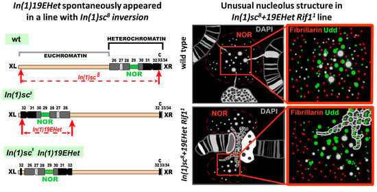
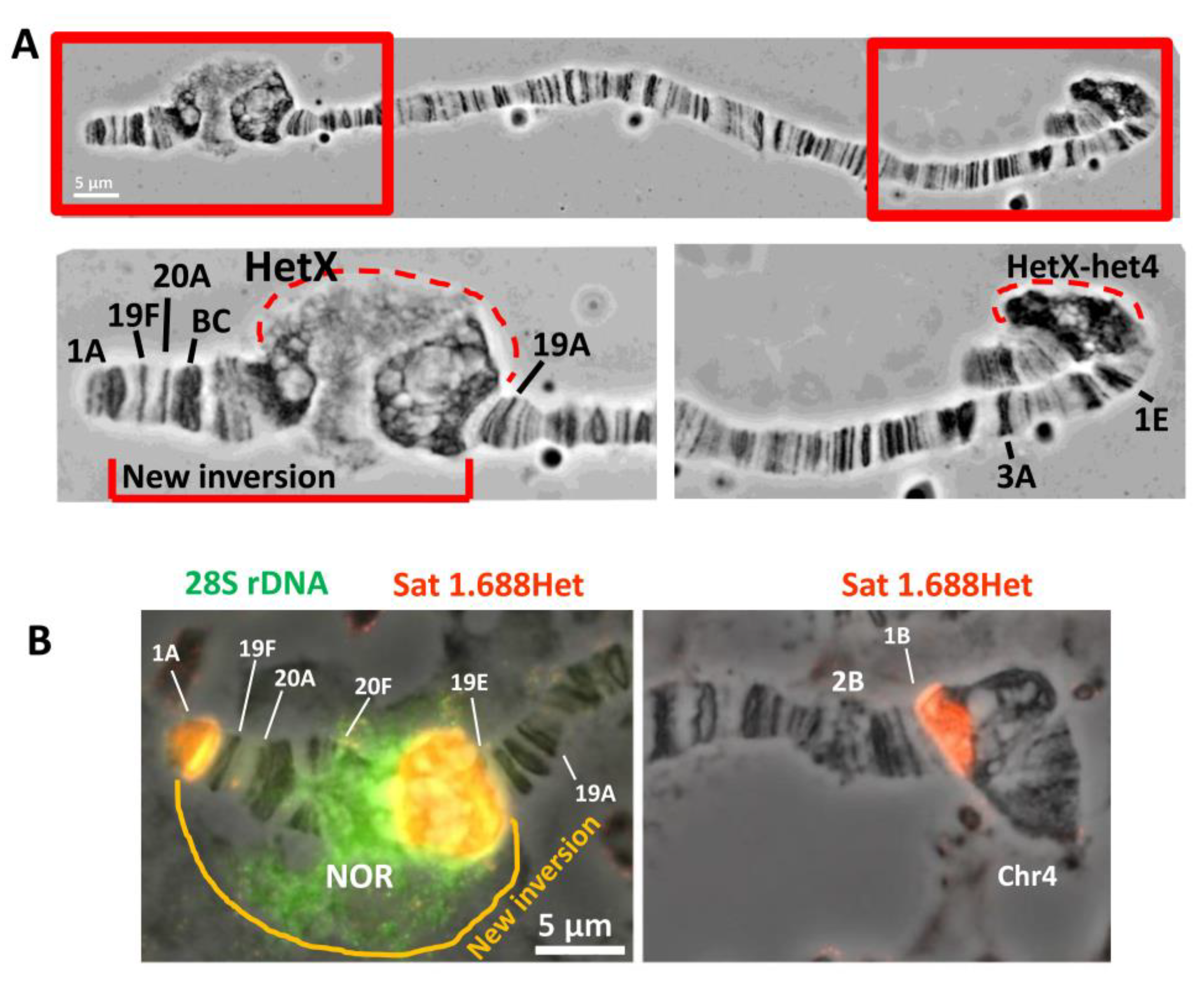
3.1. Detection of a New Inversion That Spontaneously Appeared in BDSC Line #798 with In(1)sc8 Inversion

The In(1)sc8 inversion was created in the laboratory of A. Serebrovsky by X-ray mutagenesis of wa flies [21,22,25,26,27]. The In(1)sc8, sc8 y31d wa stock was transferred to the Bloomington Drosophila Stock Center (BDSC) from the California Institute of Technology (Caltech) in 1986 and assigned the stock number 798. In 2012, the stock was split into two independent sublines (hereinafter, #798 main copy and backup copy). In 2020, we obtained flies of the #798 main copy stock from the BDSC and founded a laboratory subline (hereinafter, #798 2020 subline). Then, we performed genetic crosses to combine the X chromosome from the #798 2020 subline with the second chromosome carrying the Rif11 mutation to obtain a new stock with the genotype In(1)sc8, sc8 y31d w a; Rif11. The Rif11 mutation completely suppresses underreplication in the pericentromeric and intercalary heterochromatin [37,38]. Therefore, the Rif11 mutant background allows us to observe all D. melanogaster heterochromatic regions on polytene chromosome preparations. Morphological analysis of polytene chromosome squashes obtained from the new In(1)sc8, sc8 y31d w a; Rif11 subline demonstrated the presence of an additional rearrangement on the X chromosome. In phase-contrast images after aceto-orcein staining, part of the X chromosome—including the proximal end of region 19, region 20, and a significant part of the X heterochromatin—appeared to be inverted (Figure 1A) compared to the known In(1)sc8 rearrangement, which has been previously characterized by polytene chromosome and genomic sequence analyses. After analyzing about fifty X chromosomes on ten polytene chromosome squashes, we were able to localize one of the new inversion breakpoints to the proximal end of the intercalary heterochromatin band 19E1–4 (Figure 1A). The second breakpoint is positioned deep in heterochromatin, very close to the heterochromatic breakpoint of the progenitor In(1)sc8 inversion. We named the new inversion In(1)19Ehet and the complex chromosomal rearrangement (both inversions) In(1)sc8 + 19EHet. Schematic diagrams are shown in Figure 2. To better understand the exact breakpoint position in heterochromatin, we performed FISH with probes for rDNA and a heterochromatic variant of the 1.688 satellite and confirmed that the 11 Mb X chromosome 1.688 satellite block is broken into three blocks by the In(1)sc8 + 19EHet double inversion (Figure 1B). The largest block is transferred to the 19E region and lies between 19E and the nucleolus. Another block remains near the centromere of the X chromosome. A small block remains near the In(1)sc8 breakpoint at the telomeric end of the chromosome (Figure 1B).
To determine the origin of In(1)19EHet, we checked for the presence of the inversion in both the main and backup copies of the #798 line at the BDSC. Without the Rif1 mutant background, it is impossible to accurately confirm the presence of a rearrangement in heterochromatin due to its underreplication (see Figure S1A as an example of the morphology of the X chromosome for the #798 main copy subline without the Rif11 mutation). Therefore, we crossed both sublines with the line carrying the Rif11 mutation. On polytene chromosome squashes of the heterozygous offspring resulting from crosses, we clearly saw that only the #798 main copy subline had the double inversion, while the #798 backup copy subline had only the In(1)sc8 inversion (Figure S1B,C). Thus, we conclude that the inversion event occurred in the #798 main copy subline after 2012 when the #798 stock was split.

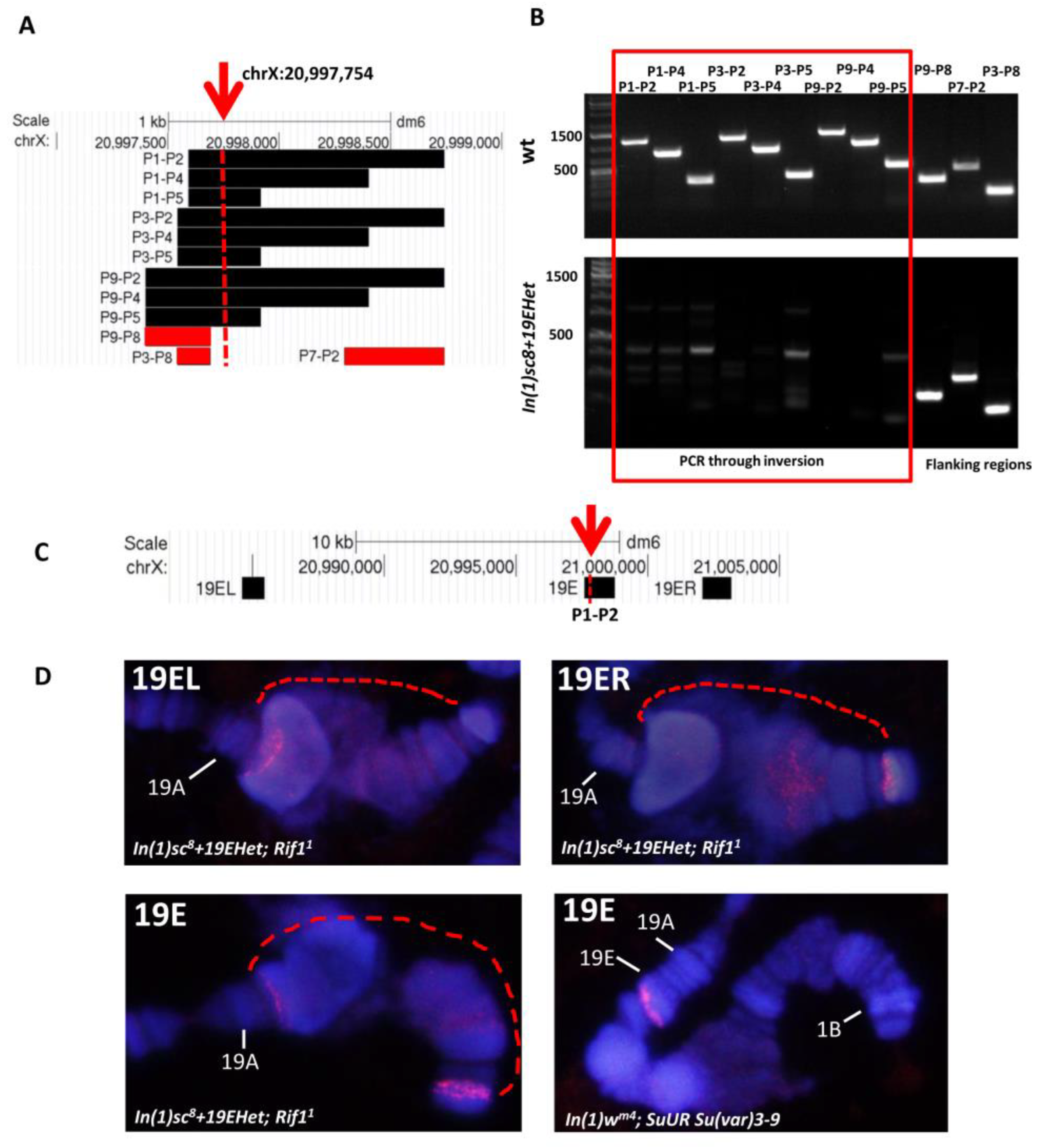
3.2. Detection of Inversion Breakpoints Using Nanopore Sequencing

To investigate the In(1)sc8 + 19EHet chromosome at the molecular level, we performed nanopore sequencing of the corresponding genome. We obtained a dataset with about 20-fold coverage of the D. melanogaster genome, with half of the reads being longer than 40 kb. In(1)sc8 + 19EHet is a derivative of In(1)sc8, and therefore, it should contain the original breakpoints of In(1)sc8, which were mapped previously [28]. According to Miller et al., the distal breakpoint is located at chrX:382,384–382,389 (dm6), while the proximal breakpoint occurred somewhere in pericentric heterochromatin in the 1.688 satellite block. Indeed, we found a gap in coverage by reads in the predicted position (chrX:382,384–382,389 region) (Figure 3A). Thus, we confirmed that In(1)19EHet arose on a chromosome carrying In(1)sc8. We also found chimeric reads overlapping the In(1)sc8 breakpoint. The two longest reads, encompassing ~30 kb of heterochromatin sequences, were selected for analysis (Figure 3A). These reads contain 1.688 satellite sequences (designated as SAR_DM or SAR2_DM in Figure 3A) juxtaposed to euchromatic regions (Figure 2A). Our analysis showed that the heterochromatin beyond the In(1)sc8 breakpoint is represented by a long (>50 kb) region of a 1.688 satellite, but we identified no sequence features allowing us to locate the heterochromatic breakpoint on the genome map.
Since our cytological analysis placed one of the In(1)19EHet breakpoints in the 19E1–4 polytene chromosome region (Figure 1A), we analyzed nanopore reads in this region to identify the new breakpoint. As for In(1)sc8, we inspected the sequence alignments and identified an interruption in coverage at position chrX:20,997,754 (Figure 3B). Two reads overlapping the expected breakpoint were selected and analyzed by RepeatMasker (Figure 3B) to place the In(1)19EHet breakpoint at position chrX:20,997,754, about 30 kb from the border of the 19E1–4 intercalary heterochromatin region (according to [47]) in a long (~30 kb) intergenic interval between the genes CG11666 and r-cup (Figure 3D). The heterochromatin breakpoint of In(1)19EHet lies in a region composed of a 1.688 satellite with occasional insertions of DOC transposable elements.
To verify the chrX:20,997,754 breakpoint predicted by nanopore sequencing, we performed PCR on DNA isolated from In(1)sc8 + 19EHet and wild-type flies using different combinations of P1–P9 primers (see materials and methods) (Figure 4A,B). PCR on genomic DNA from wild-type flies produced amplicons of the expected lengths with all primers used (Figure 4B, upper panel). PCR on DNA from In(1)19Ehet flies produced amplicons with primers from regions upstream (P9–P8, P3–P8 combinations) and downstream (P7–P2) of the breakpoint. No specific PCR products were observed during amplification with primers flanking the breakpoint (Figure 4B, bottom panel). We also confirmed our mapping by DNA FISH with three probes, one of which overlapped the supposed breakpoint (19E probe obtained with P1 and P2 primers), and with the other two lying to the right and left of the breakpoint within 10 kb (Figure 4C,D). The In(1)w4 inverted chromosome was used for a control to demonstrate the absence of a signal in the 1AB region. The SuUR Su(var)3-9 background was used to suppress under-replication in the 19E intercalary heterochromatin region.
According to our cytological analysis, the additional inversion was present in the #798 main copy stock in BDSC but absent in the #798 backup copy stock. In order to obtain pure stocks carrying only the In(1)sc8 + 19EHet double inversion chromosome, we made ~30 matings of single females to single males from the #798 main copy. PCR amplifications from a sampling of these lines confirmed that the double-inverted X chromosome had completely replaced the single-inverted X chromosome in the #798 main copy stock (Figure S2). Two stocks were thus distributed by the BDSC: the In(1)sc8, y31d sc8 wa stock (originally the backup copy) retained stock number 798, while In(1)sc8, In(1)19EHet, y31d sc8 wa (one of the 798 main copy lines validated by PCR) was assigned stock number 94727.

3.3. Morphology of the Nucleolar Organizer in In(1)sc8 + 19EHet; Rif11 Polytene Chromosomes

In preparations of wild-type polytene chromosomes, the nucleolus usually lies outside the chromosomes (Figure 5A). Mutations that prevent the underreplication of heterochromatin may allow us to visualize a part of the nucleolus organizer region (NOR) that is normally hidden as well as adjacent chromosome regions. In preparations of SuUR mutant chromosomes, which undergo additional polytenization of some pericentromeric sequences [5,48], the nucleolus is connected with distal heterochromatin by DAPI-stained strands (Figure 5B), but it still appears as a “cloud” of granules lying outside of the chromosome. In lines with Rif1 mutations, the chromosomes are completely polytenized, and the NOR does not separate from the X chromosome [38], but visualization of the nucleolus is hampered by the fact that the NOR and neighboring sequences are usually combined into a single chromocenter together with heterochromatin regions belonging to other chromosomes (not shown). Because the NOR and most pericentromeric heterochromatin are relocated to a position near the X tip by the In(1)sc8 and 19EHet inversions, these regions are often positioned away from the chromocenter in preparations made from Rif11 mutants (Figure S3). Thus, the combination of the In(1)sc8 + 19EHet chromosome and the Rif11 mutation can be used as a suitable model system for standalone imaging of fully replicated NOR and adjacent heterochromatin regions.
Figure 5C shows the result of 28S rDNA FISH on In(1)sc8 + 19EHet; Rif11 polytene chromosomes. The FISH signal was detected between the large block of chromatin stained strongly with DAPI corresponding to the 1.688 satellite and a distal heterochromatin block, which is very compact on the phase-contrast image and not stained brightly by DAPI (orange arrows in Figure 5C). This block of distal heterochromatin is usually designated as 20F and corresponds to the region on the genomic map lying proximal to the su(f) gene mapped in 20E [2,5]. The rDNA FISH signal is much wider than the DAPI-stained chromosome and also forms a cloud outside of the chromosome territory (Figure 5C), where it strongly coincides with the DAPI-stained network of filaments and granules (Figure S4). In this particular aspect, the structure of the nucleolar cloud of Rif11 mutants retains similarity with wild-type preparations (Figure 5A and Figure S4).
DAPI staining with 3D-SIM microscopy of In(1)sc8 + 19EHet; Rif11(Figure 5D) and Rif11 (Figure 5E) preparations revealed that the nucleolar part of the chromosome forms an unusual spongy structure (red arrows in Figure 5D,E), which is located in the same place as the rDNA FISH signal (Figure 5C). In some 3D-SIM images, we observed filaments connecting this spongy zone of NOR with the surrounding cloud of individual DAPI dots (Figure 5D, yellow arrow). Since this spongy structure has never been observed in wild-type chromosomes and is not found in In(1)sc8 + 19EHet line without Rif1+ mutation (Figure 5F), we suggested it arises due to the loss of the underreplication of a substantial part of the rDNA cluster in Rif1 mutants. Therefore, the spongy structure likely consists of rDNA units that are not normally polytenized. Of note, this structure is absent in SuUR mutants (Figure 5B), in which the level of centromeric heterochromatin polytenization increases less than in the Rif11 mutants.

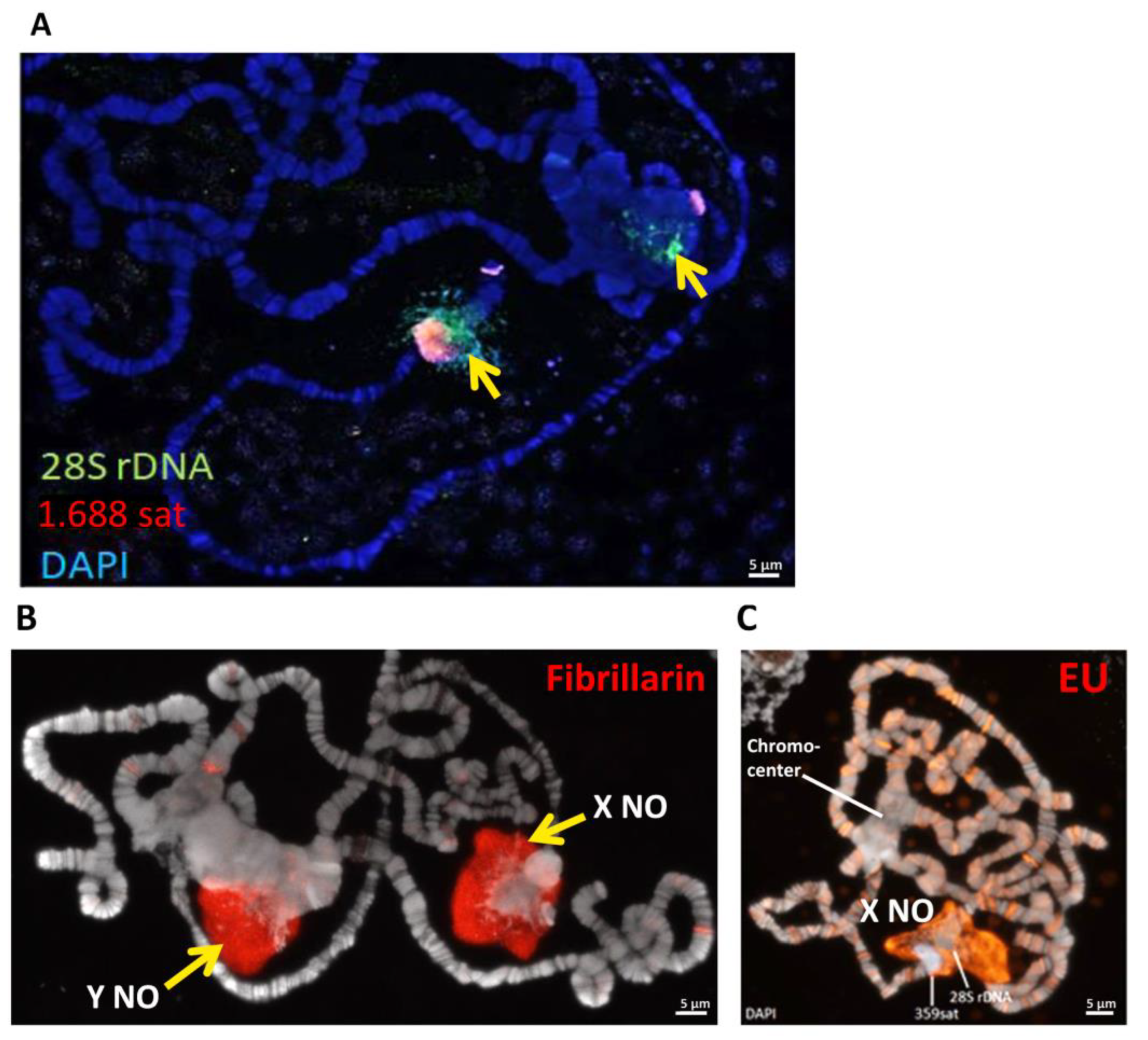
3.4. Identification of the Transcriptionally Active Part of the Nucleolus in In(1)sc8 + 19EHet; Rif11 Polytene Chromosomes

It is well known that only a part of D. melanogaster rDNA repeats is transcriptionally active, while other rDNA units, including those containing insertions of retroelements, are in a silent state [49,50,51,52]. In preparations of the wild-type salivary gland nucleolus, individual DAPI-stained specks have been shown to correspond to silent rDNA units enriched with repressive chromatin marks, whereas transcriptionally active units were mixed with them and poorly stained by DAPI [53]. To reveal functional areas of the unusual nucleolus in Rif1 mutants, we visualized several nucleolar markers. Immunostaining for fibrillarin, an rRNA 2′-O-methyltransferase involved in post-transcriptional rRNA processing, showed its predominant localization in the extrachromosomal zone, mostly in the outer part of the cloud (Figure 6A), which may correspond to the dense fibrillar component of the nucleolus. Nucleolar transcripts detected by 5-Ethynyl Uridine (EU) incorporation also produced more intense signals in the cloud than in the intrachromosomal spongy structure (Figure 6B) and did not coincide with DAPI-dense regions. However, it should be noted that within 20 min of EU incorporation, nascent transcripts may have time to transfer to the processing zone. In the chromosomes of wild-type larvae, EU signals also mostly do not coincide with dense DAPI granules in the nucleolus (Figure 6C).
To identify which part of the nucleolus in the polytene chromosome preparations corresponds to the zone of active DNA transcription, we performed immunostaining to identify Underdeveloped (Udd), a component of the rDNA transcription initiation complex SL1-like [46]. Udd was shown to be associated with the IGS regions and rDNA promoters of transcriptionally active rDNA units [52]. In the nucleoli of wild-type salivary glands, Udd aggregates mostly did not co-localize with the DAPI-stained specks (Figure 7A). Additionally, Udd signals did not coincide with fibrillarin-positive dots (Figure 7B), indicating the existence of multiple separate zones responsible for transcription or rRNA processing. In the wild type, these compartments seem to be intermingled (Figure 7B). In contrast, in In(1)sc8 + 19EHet; Rif11 nucleoli, Udd-stained specks appeared to be clustered separately from fibrillarin-stained specks (Figure 7C). Udd is localized in the form of separate dots both in the intrachromosomal spongy zone and in the cloud (Figure 7D). In the cloud, it is often amassed into larger spots surrounded by fibrillarin (Figure 7C). At higher magnification, it is noticeable that Udd mostly does not co-localize with condensed DAPI specks in the cloud (Figure 7E,F, orange arrows). Within the spongy structure, Udd resides predominantly in voids formed by the DAPI-stained network (Figure 7E,F, blue arrows). These data are in good agreement with the notion that condensed DAPI-stained specks correspond to transcriptionally silent rDNA units [53]. Note that Figure 7F shows immunostaining of the Rif11 line without inversions, confirming that the spongy structure with Udd foci is a feature of the Rif11 mutation and not the result of a chromosomal rearrangement.
Thus, we suggest that the spongy intrachromosomal part of the Rif11 mutant nucleolus consists of a mixture of active Udd-positive and inactive, condensed, Udd-negative rDNA repeats aggregated into a single structure as a result of complete rDNA replication. In contrast, the cloud is composed of predominantly active rDNA units looped out from the chromosomal NOR. These units are surrounded by an rRNA processing apparatus, an analogue of the dense fibrillar component of the nucleolus, which is detected by fibrillarin staining.

3.5. The In(1)sc8 + 19EHet; Rif11 Genotype Allows Visualization of the Y Chromosome Nucleolus

In males of D. melanogaster, the nucleolar organizer is present on both the X and Y chromosomes. In some cases, both nucleoli are active, while in others, an active nucleolus is formed only on one of the chromosomes—a phenomenon known as nucleolar dominance [19,20,21,54]. In polytene chromosomes of salivary glands, the Y chromosome is usually not polytenized and is not visible. However, genotypes with inversions that transfer Xh from the centromere made it possible to see the nucleolus derived from the Y chromosome in the chromocenter [4]. FISH with a 28S rDNA probe on the polytene chromosomes of In(1)sc8 + 19EHet/Y; Rif11 males revealed two loci: the X-chromosome nucleolus moved away from the chromocenter and a zone in the chromocenter (Figure 8A). This chromocenter zone likely corresponds to the Y chromosome rDNA cluster. An analysis of 10 preparations of In(1)sc8 + 19EHet/Y; Rif11 larvae by fibrillarin immunostaining or EU detection showed that the X chromosome nucleolus is active in all analyzed preparations, whereas the Y chromosome nucleolus was active in half of the cases (as exemplified in Figure 8B) and inactive in the others (as shown in Figure 8C). Further analysis of In(1)sc8 + 19EHet/Y; Rif11 polytene chromosomes may help identify new factors that affect the activity of the Y-chromosome nucleolus.

4. Discussion

Eu-heterochromatin inversions are classic tools for investigating position effect variegation, the interaction between eu- and heterochromatin, and chromatin organization in general. In particular, many heterochromatin proteins, such as HP1 and Su(var)3-9 histone methyltransferase, were discovered as position-effect variegation modifiers [55,56,57]. Here, we have shown that a new inversion, In(1)19EHet, arose spontaneously in a stock already carrying the classical In(1)sc8 inversion (the BDSC #798 line [28]) after the split of two sublines in 2012. Since then, the double inversion chromosome has completely replaced the In(1)sc8 single chromosome in the stock population. The new In(1)19EHet rearrangement transferred most of the X chromosome heterochromatin, including the rDNA cluster, to a new euchromatic environment in the 19E region (Figure 2). The In(1)19EHet inversion has a number of properties, which, in particular, make it possible to conveniently study the structure of the nucleolus, which appears to be remote from the bulk of heterochromatin belonging to other chromosomes. Although we did not directly compare the localization of the nucleoli between In(1)sc8 and In(1)sc8 + 19EHet lines, we suggest that the In(1)19EHet rearrangement may be more convenient for nucleolus analysis because the transfer of NOR from the telomeric end of the chromosome more than 3 Mb deep into euchromatin can further reduce the frequency of the association of the nucleolus with the heterochromatin of other chromosomes.
The detection of the In(1)19EHet inversion was made possible by the unique effect of the Rif11 mutation, which leads to the complete replication of heterochromatin sequences in polytene chromosomes. Our results show that Rif1 mutations are a useful tool for Drosophila heterochromatin studies. Even in a heterozygote, Rif11 allows visualization of many more heterochromatin regions than can be seen in wild-type larvae. This distinguishes it from SuUR mutations and SuUR mutations in combination with Su(var)3-9 mutation, which both have significant effects on heterochromatin underreplication only in homozygotes [48,58,59].
The combination of the new In(1)19EHet inversion and the Rif11 mutation in the homozygous state allows us to visualize fully replicated NORs and adjacent X heterochromatin regions apart from other chromosomes. In wild-type polytene chromosome preparations, the nucleolus is a cloud-like structure that is found separately from the chromosomal material, to which it is connected by thin fibers. In Rif1 mutants, an intrachromosomal part of the nucleolus appears in addition to the nucleolar cloud, similar in structure to a normal nucleolus. We have found that the unusual intrachromosomal part looks like a sponge by DAPI staining and spatial super-resolution SIM imaging and that it contains rDNA repeats as identified by FISH (Figure 5). A comparison of the ratio of rDNA to genomic DNA in polytene and diploid cells shows that only about 1/5 of rDNA repeats are normally replicated in the polytene chromosomes of the salivary glands [60,61]. Thus, the resulting spongy intrachromosomal structure likely represents the portion of the rDNA cluster that remains underreplicated in polytene chromosomes under normal conditions. Interestingly, we found that this chromosomal part of the NOR likely consists of a mix of transcriptionally active and inactive zones, although markers of nucleolus activity indicate less activity in the chromosomal part of the NOR than in the cloud around the chromosome (Figure 7).
Further, the use of the In(1)19EHet; Rif1 model may allow us to explore by FISH the distribution of specific rDNA repeats, such as those enriched for transposon insertions. This approach can help characterize the internal organization of rDNA clusters, which is currently difficult to study by other methods, such as sequencing, due to the repetitive nature of rDNA. The possible existence of functional and structural domains within rDNA clusters has been discussed in the literature [62,63]. The In(1)19EHet; Rif1 model can be used to examine the localization of chromatin proteins associated with specific intranucleolar zones and adjacent heterochromatin regions, which may reflect subfunctionalization.
D. melanogaster males contain NORs on two different chromosomes: X and Y. Since the X chromosome-derived nucleolus in In(1)19EHet; Rif1 flies is separated from the bulk of the chromocenter, this system, as well as some other inversion-based models [4], make it possible to visualize both nucleoli separately in different parts of the nucleus. Thus, the In(1)19EHet; Rif1 genotype provides additional advantages for the analysis of the Y-chromosomal nucleolus of males and the phenomenon of nucleolar dominance, whereby individual rDNA loci are entirely silenced. We have seen that this genotype exhibits variations in nucleolar dominance: the X chromosome rDNA cluster appears to be constitutively active, while the Y chromosome cluster seems to be inactive in only some males (Figure 8). Past studies have indicated that nucleolar dominance is complex. In D. melanogaster males, the Y-chromosome nucleolus usually dominates over the X-chromosome nucleolus [19,20,21], but significant effects have been observed for age, the composition of repetitive sequences in Xh and Y heterochromatin and mutations in genes encoding heterochromatin protein [19,21,22,64,65,66]. Nucleolar dominance may even vary from tissue to tissue. In salivary glands, in contrast to many other tissues, only modest Y dominance was shown [21,22].
Chromosomal structure is known to affect nucleolar dominance. Hilliker and Appels [4] used inverted X chromosomes to search for factors influencing nucleolar dominance and saw that small Xh deletions could shift dominance from the X to the Y chromosome. We do not know if the In(1)19EHet rearrangement itself leads to preferential use of the X nucleolus or if complete polytenization of the rDNA clusters by the Rif11 mutation affects the formation of X versus Y nucleoli. It is clear, however, that the In(1)sc8 + 19EHet; Rif11 system provides a new and convenient model for exploring the mechanistic bases of this mysterious phenomenon.

Supplementary Materials

The following supporting information can be downloaded at: https://www.mdpi.com/article/10.3390/cells11233872/s1, Figure S1: Cytological verification of the original #798 sublines from the BDSC for the presence of the additional inversion In(1)19EHet. (A) Without suppression of underreplication, it is difficult to unambiguously judge the presence or absence of the In(1)19EHet inversion in the subline #798 main copy. (B,C) The results of crossing #798 backup copy (B) or #798 main copy (C) with the Rif11 mutant. The offspring carrying both the inverted X chromosomes and Rif11/+ mutation in heterozygous states were analyzed. Partial heterochromatin polytenization in Rif11/+ heterozygotes allowed us to conclude that only line #798 main copy carries the double inversion, while line #798 backup copy carries only the original In(1)sc8 inversion. Figure S2: Examples of PCR amplifications from a sampling of lines started with single-pair matings from the 798 main copy vials. Control genotypes include flies from the 798 backup copy and Oregon-R (OR) flies. P1–P2 primers were used for PCR through the inversion breakpoint. Primers for the white gene were used to verify that the DNA samples could be used in PCR amplifications successfully. Figure S3: Localization of the nucleolus within nuclei of Rif11 mutants carrying wild-type (A) or In(1)sc8 + 19EHet (B) chromosomes in slightly squashed preparations of polytene chromosomes. Individual optical sections obtained by DAPI staining with subsequent 3D-SIM microscopy are shown. The nucleolus is marked by fibrillarin immunostaining. Figure S4: FISH with 28S rDNA probe (red) in Rif11 mutant and wild-type polytene chromosomes. The DAPI channel is overexposed to detect weakly stained structures. In both Rif11 and wild-type nucleoli, FISH signal strongly coincides with DAPI staining.

Author Contributions

Conceptualization, T.D.K. and M.S.K.; methodology, T.D.K., M.S.K., S.A.L. and V.S.; validation, A.R.N., S.V.M., G.V.P., K.R.C. and M.J.D.; investigation, T.D.K., A.R.N., S.A.L. and G.V.P.; writing—original draft preparation, T.D.K. and M.S.K.; writing—review and editing, K.R.C., V.S., S.V.M., S.A.L. and I.F.Z.; funding acquisition, T.D.K., M.S.K., V.S., K.R.C. and I.F.Z. All authors have read and agreed to the published version of the manuscript.

Funding

The part of the work associated with nanopore sequencing was funded by the Russian Science Foundation (RSF) (grant number 19-74-20178). Part of the work related to the visualization of nucleolar components was supported by a grant from the Russian Science Foundation (19-14-00382). The part of the work associated with ultrastructural polytene chromosome analysis was funded by a joint Russian-German grant from the Russian Foundation for Basic Research (No. 20-54-12016) and the Deutsche Forschungsgemeinschaft (Schu762/12-1). The part of the work associated with the new inversion cytological mapping was funded by the Russian Science Foundation grant No. 19-14-00051. Work at the Bloomington Drosophila Stock Center was supported by the U.S. National Institutes of Health grant P40 OD018537.

Institutional Review Board Statement

The study was approved by the Committee on the Ethics of Animal Experiments of the Institute of Molecular and Cellular Biology SB RAS. Protocol №01/22 from 26 October 2022.

Data Availability Statement

The datasets with the results of Nanopore sequencing of In(1)sc8, In(1)19EHet fly DNA are available from the SRA (SRR22071703, SRR22071702, SRR22071691, SRR22071690). Other files are attached in the supporting material.

Acknowledgments

We are grateful to M. Buszczak for Udd antibodies, N. Tchurikov for the 28S rDNA clone and V. Shloma for the help with confocal microscopy.

Conflicts of Interest

The authors declare no conflict of interest.

References

  1. Hoskins, R.A.; Smith, C.D.; Carlson, J.W.; Carvalho, A.B.; Halpern, A.; Kaminker, J.S.; Kennedy, C.; Mungall, C.J.; Sullivan, B.A.; Sutton, G.G.; et al. Heterochromatic sequences in a Drosophila whole-genome shotgun assembly. Genome Biol. 2002, 3, RESEARCH0085. [Google Scholar] [CrossRef] [PubMed] [Green Version]
  2. Hoskins, R.A.; Carlson, J.W.; Wan, K.H.; Park, S.; Mendez, I.; Galle, S.E.; Booth, B.W.; Pfeiffer, B.D.; George, R.A.; Svirskas, R.; et al. The release 6 reference sequence of the Drosophila melanogaster genome. Genome Res. 2015, 25, 445–458. [Google Scholar] [CrossRef] [PubMed] [Green Version]
  3. Ritossa, F.M.; Spiegelman, S. Localization of DNA complementary to ribosomal RNA in the nucleolus organiser region of Drosophila melanogaster. Proc. Natl. Acad. Sci. USA 1965, 53, 737–745. [Google Scholar] [CrossRef] [Green Version]
  4. Hilliker, A.J.; Appels, R. Pleiotropic effects associated with the deletion of heterochromatin surrounding rDNA on the X chromosome of Drosophila. Chromosoma 1982, 86, 469–490. [Google Scholar] [CrossRef]
  5. Kolesnikova, T.D.; Koriakov, D.E.; Semeshin, V.F.; Beliaeva, E.S.; Zhimulev, I.F. Interline differences in morphology of the precentromeric region of polytene X-chromosome in Drosophila melanogaster salivary glands. Genetika 2001, 37, 1632–1641. [Google Scholar] [PubMed]
  6. Parry, D.M.; Sandler, L. The Genetic Identification of a heterochromatic segment on the X chromosome of Drosophila melanogaster. Genetics 1974, 77, 535–539. [Google Scholar] [CrossRef]
  7. Gatti, M.; Pimpinelli, S. Functional elements in Drosophila melanogaster heterochromatin. Annu. Rev. Genet. 1992, 26, 239–275. [Google Scholar] [CrossRef]
  8. Wellauer, P.K.; Dawid, I.B.; Tartof, K.D. X and Y chromosomal ribosomal DNA of Drosophila: Comparison of spacers and insertions. Cell 1978, 14, 269–278. [Google Scholar] [CrossRef]
  9. Tautz, D.; Hancock, J.M.; Webb, D.A.; Tautz, C.; Dover, G.A. Complete sequences of the rRNA genes of Drosophila melanogaster. Mol. Biol. Evol. 1988, 5, 366–376. [Google Scholar] [CrossRef] [Green Version]
  10. Lyckegaard, E.M.; Clark, A.G. Evolution of ribosomal RNA gene copy number on the sex chromosomes of Drosophila melanog aster. Mol. Biol. Evol. 1991, 8, 458–474. [Google Scholar] [CrossRef]
  11. Bianciardi, A.; Boschi, M.; Swanson, E.E.; Belloni, M.; Robbins, L.G. Ribosomal DNA organization before and after magnification in Drosophila melanogaster. Genetics 2012, 191, 703–723. [Google Scholar] [CrossRef] [PubMed] [Green Version]
  12. Lyckegaard, E.M.; Clark, A.G. Ribosomal DNA and Stellate gene copy number variation on the Y chromosome of Drosophila melanogaster. Proc. Natl. Acad. Sci. USA 1989, 86, 1944–1948. [Google Scholar] [CrossRef] [PubMed] [Green Version]
  13. Tartof, K.D. Regulation of ribosomal RNA gene multiplicity in Drosophila melanogaster. Genetics 1973, 73, 57–71. [Google Scholar] [CrossRef] [PubMed]
  14. Terracol, R.; Prud’homme, N. Differential elimination of rDNA genes in bobbed mutants of Drosophila melanogaster. Mol. Cell Biol. 1986, 6, 1023–1031. [Google Scholar] [CrossRef] [PubMed]
  15. Mohan, J.; Ritossa, F.M. Regulation of ribosomal RNA synthesis and its bearing on the bobbed phenotype in Drosophila melanogaster. Dev. Biol. 1970, 22, 495–512. [Google Scholar] [CrossRef]
  16. Lu, K.L.; Nelson, J.O.; Watase, G.J.; Warsinger-Pepe, N.; Yamashita, Y.M. Transgenerational dynamics of rDNA copy number in Drosophila male germline stem cells. Elife 2018, 7, e32421. [Google Scholar] [CrossRef]
  17. Iarovaia, O.V.; Minina, E.P.; Sheval, E.V.; Onichtchouk, D.; Dokudovskaya, S.; Razin, S.V.; Vassetzky, Y.S. Nucleolus: A central hub for nuclear functions. Trends Cell Biol. 2019, 29, 647–659. [Google Scholar] [CrossRef]
  18. Pirogov, S.A.; Gvozdev, V.A.; Klenov, M.S. Long noncoding RNAs and stress response in the nucleolus. Cells 2019, 8, 668. [Google Scholar] [CrossRef] [Green Version]
  19. Greil, F.; Ahmad, K. Nucleolar dominance of the Y chromosome in Drosophila melanogaster. Genetics 2012, 191, 1119–1128. [Google Scholar] [CrossRef] [Green Version]
  20. Zhou, J.; Sackton, T.B.; Martinsen, L.; Lemos, B.; Eickbush, T.H.; Hartl, D.L. Y chromosome mediates ribosomal DNA silencing and modulates the chromatin state in Drosophila. Proc. Natl. Acad. Sci. USA 2012, 109, 9941–9946. [Google Scholar] [CrossRef]
  21. Warsinger-Pepe, N.; Li, D.; Yamashita, Y.M. Regulation of nucleolar dominance in Drosophila melanogaster. Genetics 2020, 214, 991–1004. [Google Scholar] [CrossRef] [PubMed] [Green Version]
  22. Kotov, A.A.; Bazylev, S.S.; Adashev, V.E.; Shatskikh, A.S.; Olenina, L.V. Drosophila as a model system for studying of the evolution and functional specialization of the Y chromosome. Int. J. Mol. Sci. 2022, 23, 4184. [Google Scholar] [CrossRef] [PubMed]
  23. Beadle, G.W.; Sturtevant, A.H. X Chromosome inversions and meiosis in Drosophila melanogaster. Proc. Natl. Acad. Sci. USA 1935, 21, 384–390. [Google Scholar] [CrossRef] [PubMed] [Green Version]
  24. Vogel, M.J.; Pagie, L.; Talhout, W.; Nieuwland, M.; Kerkhoven, R.M.; van Steensel, B. High-resolution mapping of heterochromatin redistribution in a Drosophila position-effect variegation model. Epigenetics Chromatin 2009, 2, 1. [Google Scholar] [CrossRef] [PubMed] [Green Version]
  25. Sidorov, B. A Study of step-allelomorphism in Drosophila melanogaster. A case of origination of an allelomorph of Scute producing simultaneously characters of ’Hairy Wing’ (Mutation Scute-8). Zhurnal Eksperimental Noi Biol. I Meditsiny 7 1931, 7, 28–40. [Google Scholar]
  26. Gershenson, S. Studies on the genetically inert region of the X-chromosome of Drosophila. I. Behaviour of an X-chromosome deficient for a part of its inert region. J. Genet. 1933, 28, 297–313. [Google Scholar] [CrossRef]
  27. Sturtevant, A.H.; Beadle, G.W. The relations of inversions in the X chromosome of Drosophila melanogaster to crossing over and disjunction. Genetics 1936, 21, 554–604. [Google Scholar] [CrossRef]
  28. Miller, D.E.; Cook, K.R.; Yeganeh Kazemi, N.; Smith, C.B.; Cockrell, A.J.; Hawley, R.S.; Bergman, C.M. Rare recombination events generate sequence diversity among balancer chromosomes in Drosophila melanogaster. Proc. Natl. Acad. Sci. USA 2016, 113, E1352–E1361. [Google Scholar] [CrossRef] [Green Version]
  29. Lewis, E.B. The Phenomenon of position effect. Adv. Genet. 1950, 3, 73–115. [Google Scholar] [CrossRef]
  30. Hannah, A. Localization and function of heterochromatin in Drosophila melanogaster. Adv. Genet. 1951, 4, 87–125. [Google Scholar] [CrossRef]
  31. Baker, W.K. Position-effect variegation. Adv. Genet. 1968, 14, 133–169. [Google Scholar] [PubMed]
  32. Spofford, J. Position-effect variegation in Drosophila. In The Genetics and Biology of Drosophila; Ashburner, M., Novitski, E., Eds.; Academic Press: New York, NY, USA, 1976; Volume 1, pp. 955–1018. [Google Scholar]
  33. Appels, R.; Hilliker, A.J. The cytogenetic boundaries of the rDNA region within heterochromatin in the X chromosome of Drosophila melanogaster and their relation to male meiotic pairing sites. Genet. Res. 1982, 39, 149–156. [Google Scholar] [CrossRef] [PubMed]
  34. Miller, D.E.; Cook, K.R.; Hawley, R.S. The joy of balancers. PLoS Genet. 2019, 15, e1008421. [Google Scholar] [CrossRef] [Green Version]
  35. Shao, H.; Ganesamoorthy, D.; Duarte, T.; Cao, M.D.; Hoggart, C.J.; Coin, L.J.M. NpInv: Accurate detection and genotyping of inversions using long read sub-alignment. BMC Bioinform. 2018, 19, 261. [Google Scholar] [CrossRef] [PubMed] [Green Version]
  36. Solodovnikov, A.; Lavrov, S. Exact breakpoints of the In(1)wm4 rearrangement. MicroPubl. Biol. 2022. [Google Scholar] [CrossRef]
  37. Munden, A.; Rong, Z.; Sun, A.; Gangula, R.; Mallal, S.; Nordman, J.T. Rif1 inhibits replication fork progression and controls DNA copy number in Drosophila. Elife 2018, 7, e39140. [Google Scholar] [CrossRef] [PubMed]
  38. Kolesnikova, T.D.; Kolodyazhnaya, A.V.; Pokholkova, G.V.; Schubert, V.; Dovgan, V.V.; Romanenko, S.A.; Prokopov, D.Y.; Zhimulev, I.F. Effects of mutations in the Drosophila melanogaster Rif1 gene on the replication and underreplication of pericentromeric heterochromatin in salivary gland polytene chromosomes. Cells 2020, 9, 1501. [Google Scholar] [CrossRef]
  39. Zhimulev, I.F.; Vlassova, I.E.; Belyaeva, E.S. Cytogenetic analysis of the 2B3-4--2B11 region of the X chromosome of Drosophila melanogaster. III. Puffing disturbance in salivary gland chromosomes of homozygotes for Mutation l(1)Pp1t10. Chromosoma 1982, 85, 659–672. [Google Scholar] [CrossRef]
  40. Weisshart, K.D.; Fuchs, J.; Schubert, V. Structured illumination microscopy (SIM) and photoactivated localization microscopy (PALM) to analyze the abundance and distribution of RNA Polymerase II molecules on flow-sorted Arabidopsis nuclei. Bio-Protoc. 2016, 6, e1725. [Google Scholar] [CrossRef] [Green Version]
  41. Koetsier, P.A.G.; Cantor, E.J. A simple approach for effective shearing and reliable concentration measurement of ultra-high-molecular-weight DNA. Biotechniques 2021, 71, 439–444. [Google Scholar] [CrossRef]
  42. Li, H. Minimap2: Pairwise alignment for nucleotide sequences. Bioinformatics 2018, 34, 3094–3100. [Google Scholar] [CrossRef] [PubMed] [Green Version]
  43. Blankenberg, D.; Gordon, A.; Von Kuster, G.; Coraor, N.; Taylor, J.; Nekrutenko, A.; the Galaxy Team. Manipulation of FASTQ data with Galaxy. Bioinformatics 2010, 26, 1783–1785. [Google Scholar] [CrossRef] [Green Version]
  44. Kim, M.; Ekhteraei-Tousi, S.; Lewerentz, J.; Larsson, J. The X-linked 1.688 satellite in Drosophila melanogaster promotes specific targeting by Painting of Fourth. Genetics 2018, 208, 623–632. [Google Scholar] [CrossRef] [PubMed] [Green Version]
  45. Tautz, D.; Dover, G.A. Transcription of the tandem array of ribosomal DNA in Drosophila melanogaster does not terminate at any fixed point. EMBO J. 1986, 5, 1267–1273. [Google Scholar] [CrossRef] [PubMed]
  46. Zhang, Q.; Shalaby, N.A.; Buszczak, M. Changes in rRNA transcription influence proliferation and cell fate within a stem cell lineage. Science 2014, 343, 298–301. [Google Scholar] [CrossRef] [Green Version]
  47. Belyaeva, E.S.; Goncharov, F.P.; Demakova, O.V.; Kolesnikova, T.D.; Boldyreva, L.V.; Semeshin, V.F.; Zhimulev, I.F. Late replication domains in polytene and non-polytene cells of Drosophila melanogaster. PLoS ONE 2012, 7, e30035. [Google Scholar] [CrossRef] [Green Version]
  48. Belyaeva, E.S.; Zhimulev, I.F.; Volkova, E.I.; Alekseyenko, A.A.; Moshkin, Y.M.; Koryakov, D.E. Su(UR)ES: A gene suppressing DNA underreplication in intercalary and pericentric heterochromatin of Drosophila melanogaster polytene chromosomes. Proc. Natl. Acad. Sci. USA 1998, 95, 7532–7537. [Google Scholar] [CrossRef] [Green Version]
  49. Long, E.O.; Dawid, I.B. Expression of ribosomal DNA insertions in Drosophila melanogaster. Cell 1979, 18, 1185–1196. [Google Scholar] [CrossRef]
  50. Ye, J.; Eickbush, T.H. Chromatin structure and transcription of the R1- and R2-inserted rRNA genes of Drosophila melanogaster. Mol. Cell Biol. 2006, 26, 8781–8790. [Google Scholar] [CrossRef] [Green Version]
  51. Luo, Y.; Fefelova, E.; Ninova, M.; Chen, Y.-C.A.; Aravin, A.A. Repression of interrupted and intact rDNA by the SUMO pathway in Drosophila melanogaster. Elife 2020, 9, e52416. [Google Scholar] [CrossRef]
  52. Fefelova, E.A.; Pleshakova, I.M.; Mikhaleva, E.A.; Pirogov, S.A.; Poltorachenko, V.A.; Abramov, Y.A.; Romashin, D.D.; Shatskikh, A.S.; Blokh, R.S.; Gvozdev, V.A.; et al. Impaired function of rDNA transcription initiation machinery leads to derepression of ribosomal genes with insertions of R2 retrotransposon. Nucleic Acids Res. 2022, 50, 867–884. [Google Scholar] [CrossRef] [PubMed]
  53. Plata, M.P.; Kang, H.J.; Zhang, S.; Kuruganti, S.; Hsu, S.-J.; Labrador, M. Changes in chromatin structure correlate with transcriptional activity of nucleolar rDNA in polytene chromosomes. Chromosoma 2009, 118, 303–322. [Google Scholar] [CrossRef]
  54. Endow, S.A. Nucleolar dominance in polytene cells of Drosophila. Proc. Natl. Acad. Sci. USA 1983, 80, 4427–4431. [Google Scholar] [CrossRef] [PubMed] [Green Version]
  55. Schotta, G.; Ebert, A.; Krauss, V.; Fischer, A.; Hoffmann, J.; Rea, S.; Jenuwein, T.; Dorn, R.; Reuter, G. Central role of Drosophila SU(VAR)3–9 in Histone H3-K9 methylation and heterochromatic gene silencing. EMBO J. 2002, 21, 1121–1131. [Google Scholar] [CrossRef]
  56. Schotta, G.; Ebert, A.; Reuter, G. SU(VAR)3-9 is a conserved key function in heterochromatic gene silencing. Genetica 2003, 117, 149–158. [Google Scholar] [CrossRef] [PubMed]
  57. Elgin, S.C.R.; Reuter, G. Position-Effect Variegation, Heterochromatin formation, and gene silencing in Drosophila. Cold Spring Harb. Perspect. Biol. 2013, 5, a017780. [Google Scholar] [CrossRef] [Green Version]
  58. Demakova, O.V.; Pokholkova, G.V.; Kolesnikova, T.D.; Demakov, S.A.; Andreyeva, E.N.; Belyaeva, E.S.; Zhimulev, I.F. The SU(VAR)3-9/HP1 complex differentially regulates the compaction state and degree of underreplication of X chromosome pericentric heterochromatin in Drosophila melanogaster. Genetics 2007, 175, 609–620. [Google Scholar] [CrossRef] [Green Version]
  59. Andreyeva, E.N.; Kolesnikova, T.D.; Demakova, O.V.; Mendez-Lago, M.; Pokholkova, G.V.; Belyaeva, E.S.; Rossi, F.; Dimitri, P.; Villasante, A.; Zhimulev, I.F. High-resolution analysis of Drosophila heterochromatin organization Using SuUR Su(Var)3-9 double mutants. Proc. Natl. Acad. Sci. USA 2007, 104, 12819–12824. [Google Scholar] [CrossRef] [Green Version]
  60. Jacobs-Lorena, M. Dosage of 5S and ribosomal genes during oogenesis of Drosophila melanogaster. Dev. Biol. 1980, 80, 134–145. [Google Scholar] [CrossRef]
  61. Spear, B.B.; Gall, J.G. Independent control of ribosomal gene replication in polytene chromosomes of Drosophila melanogaster. Proc. Natl. Acad. Sci. USA 1973, 70, 1359–1363. [Google Scholar] [CrossRef] [Green Version]
  62. Eickbush, D.G.; Ye, J.; Zhang, X.; Burke, W.D.; Eickbush, T.H. Epigenetic regulation of retrotransposons within the nucleolus of Drosophila. Mol. Cell Biol. 2008, 28, 6452–6461. [Google Scholar] [CrossRef] [PubMed]
  63. Zhou, J.; Eickbush, T.H. The pattern of R2 retrotransposon activity in natural populations of Drosophila simulans reflects the dynamic nature of the rDNA Locus. PLoS Genet. 2009, 5, e1000386. [Google Scholar] [CrossRef] [PubMed] [Green Version]
  64. Preuss, S.; Pikaard, C.S. rRNA gene silencing and nucleolar dominance: Insights into a chromosome-scale epigenetic on/off switch. Biochim. Biophys. Acta 2007, 1769, 383–392. [Google Scholar] [CrossRef] [PubMed] [Green Version]
  65. Tucker, S.; Vitins, A.; Pikaard, C.S. Nucleolar dominance and ribosomal RNA gene silencing. Curr Opin. Cell Biol. 2010, 22, 351–356. [Google Scholar] [CrossRef] [PubMed] [Green Version]
  66. Hawley, R.S.; Marcus, C.H. Recombinational controls of rDNA redundancy in Drosophila. Annu. Rev. Genet. 1989, 23, 87–120. [Google Scholar] [CrossRef] [PubMed]
Figure 1. Detection and cytological mapping of the new X-chromosomal eu/heterochromatin inversion. (A) Polytene X chromosome of the new line carrying the Rif11 mutation, the In(1)sc8 inversion, and the additional inversion named In(1)19EHet (In(1)sc8 + 19EHet; Rif11). The enlarged end segments of the chromosome highlighted in red rectangles are shown at the bottom. Of note, in Rif11 mutants, proximal X heterochromatin is always combined into a single block with chromosome 4 heterochromatin [38]. Aceto-orcein staining, phase contrast. (B) DNA FISH of the In(1)sc8 + 19EHet; Rif11 polytene X chromosome with probes to satellite 1.688Het (red) and 28S rDNA (green).
Cells 11 03872 g001
Figure 2. Schematic representation of metaphase and polytene X chromosomes without inversions (A), with In(1)sc8 (B) and with both In(1)sc8 and In(1)19EHet inversions (C). Schemes of polytene chromosomes are given for the Rif1+ and Rif1- backgrounds. Positions of In(1)sc8 and In(1)19EHet breakpoints are marked by arrows on chromosomes that are not rearranged. Blocks of heterochromatin are indicated according to the classical map of metaphase chromosomes [7]. The colours of the 28 S rDNA and 1.688 satellite reflect the colours of FISH probes in Figure 1B. The regions affected by the new rearrangement are largely under-replicated in wild-type polytene chromosomes, and therefore, the In(1)19Ehet inversion could not be detected without the Rif11 background.
Cells 11 03872 g002
Figure 3. Elicitation of inversion breakpoints using nanopore sequencing. (A) Genome Browser view of the distal In(1)sc8 breakpoint (blue line, chrX:382,384–382,389). Positions of two regions (chrX: 381,979–382,378 and chrX: 382,391–382,719) used for extraction of nanopore reads are shown at the top. Soft-clipped aligned reads, overlapping the breakpoint, are shown below. The position and feature maps of the two longest reads overlapping the In(1)sc8 breakpoint are shown below the alignment. In both cases, long (>20 kb) stretches of 1.688 satellite (designated as SAR_DM and SAR2_DM according to RepeatMasker) were detected beyond the breakpoint position. Parts of the chimeric reads corresponding to euchromatin sequence immediately near the breakpoint are marked in black. Other colors demark different types of repeats in the fused heterochromatin. (B) In(1)19EHet breakpoint in chrX:20,997,754. Overall arrangement of the image is the same as for A. (C) Schematic representation of repetitive sequences flanking In(1)sc8 and In(1)19EHet breakpoints. Transposable elements are designated according to RepeatMasker. (D) The chrX:20,997,754 breakpoint position relative to the intercalary heterochromatin polytene chromosome double band 19E1–4 and nearest gene positions.
Cells 11 03872 g003
Figure 4. Verification of the In(1)19EHet euchromatin breakpoint position at chrX:20,997,754 predicted by nanopore sequencing. (A) Position of PCR products obtained with various combinations of P1–P9 primers on the genomic map. The arrow shows the position of the breakpoint at chrX:20,997,754; black and red boxes indicate PCR products that overlap or do not overlap the inversion breakpoint, respectively. (B) Results of PCR with primers P1–P9. Genomic DNA samples from wild-type (upper panel) and In(1)sc8 + 19EHet (#798 2020 stock) (bottom panel) flies were used as templates. PCR results for primer combinations overlapping the breakpoint are indicated by the red box. (C) Position of DNA FISH probes 19EL, 19E and 19ER on the genomic map. The arrow shows the chrX:20,997,754 breakpoint. (D) FISH with In(1)sc8 + 19EHet; Rif11 and control (In(1)wm4h; SuUR Su(var)3-9) polytene chromosomes. The dashed line shows the inverted area. The 19EL and 19ER probes hybridize to chromosomal regions separated by the inversion event. 19E probe, broken by inversion, hybridizes on both sides of the break with the brightness of the signals corresponding to the ratio of fragment sizes. In the control line, a single probe signal 19E marks the proximal edge of the 19E region. SuUR Su(var)3-9 background in control line shows underreplication similar to Rif1 in 19E1–4.
Cells 11 03872 g004
Figure 5. Comparison of the nucleolus morphology in wild-type, SuUR and In(1)sc8 + 19EHet; Rif11 polytene chromosomes. (A) DAPI staining with subsequent 3D-SIM microscopy of wild-type polytene chromosomes. The nucleolus is not closely associated with the proximal end of the X chromosome but is connected to the chromocenter and other chromosome regions. The inset shows the nucleolus clearer via overexposure. (B) DAPI staining and 3D-SIM of SuUR mutant polytene chromosomes. (C) FISH with 28S rDNA probe (green) in In(1)sc8 + 19EHet; Rif11 polytene chromosomes. Chromosome morphology is shown by phase-contrast imaging and DAPI staining. The red arrow indicates the intrachromosomal rDNA FISH signal. The orange arrow indicates the distal heterochromatin (distHetX) in band 20F poorly stained by DAPI. (D1) DAPI staining and 3D-SIM analysis of In(1)sc8 + 19EHet; Rif11 chromosomes. The intrachromosomal part of the NO is represented by a spongy structure (indicated by red arrow) located between the block of 1.688 satellite, which is strongly stained by DAPI, and distHetX, which is poorly stained by DAPI. The boundaries of the nucleolus, which consists of the spongy structure and a cloud of DAPI-stained specks, are shown by the white dotted line. The yellow arrow indicates filaments connecting the spongy structure with the cloud. (D2) Another 3D-SIM slice. (E) DAPI staining and 3D-SIM analysis of uninverted chromosomes in a Rif11 mutant showing the morphology of X chromosome heterochromatin similar to (D). Contacts with heterochromatin of other chromosomes (red dotted line) complicate the analysis. (F) FISH with 28S rDNA probe (green) in polytene chromosomes of In(1)sc8 + 19EHet without Rif11 mutation. Chromosome morphology is shown by phase-contrast imaging and DAPI staining. The polytenized part of the inverted by 19EHet X chromosome fragment is indicated by a dotted line.
Cells 11 03872 g005
Figure 6. Fibrillarin and nascent transcripts are localized predominantly in the extrachromosomal part of nucleoli in In(1)sc8 + 19EHet; Rif11 polytene chromosome preparations. (A) Fibrillarin immunostaining in In(1)sc8 + 19EHet; Rif11 chromosome. (B) In(1)sc8 + 19EHet; Rif11 chromosome after a 15-min EU pulse. (C) EU incorporation assay in the nucleolus of wild-type chromosome preparations. A and C images represent single 3D-SIM slices. B image represents a confocal slice.
Cells 11 03872 g006
Figure 7. Udd is present both in the spongy structure and in the extrachromosomal cloud of the NOR. (A) Wild-type polytene chromosomes stained with DAPI and Udd antibodies. (B) Udd, fibrillarin and DAPI localization in wild-type polytene chromosomes. (C) In(1)sc8 + 19EHet; Rif11 chromosomes immunostained for Udd and fibrillarin. (D) Same as (B) without fibrillarin. (E) Close-up view from (D) showing Udd dots in the spongy area (blue arrows) and in the cloud (yellow arrows). (F) Rif11 chromosomes immunostained for Udd (all designations correspond to (E)). All images represent slices from 3D-SIM image stacks.
Cells 11 03872 g007
Figure 8. Detection of X- and Y-chromosome nucleoli in polytene chromosomes of In(1)sc8 + 19EHet; Rif11 male larvae. (A) FISH of 28S rDNA (green) and 1.688 satellite (red) probes. A large block of 1.688 satellite marks X-chromosome heterochromatin removed from the chromocenter by In(1)sc8 + 19EHet. Two rDNA clusters can be seen (arrows). (B) An example of two active nucleoli visualized by fibrillarin immunostaining. (C) An example of an active X chromosome nucleolus and an inactive Y chromosome nucleolus visualized by EU pulse incorporation.
Cells 11 03872 g008
Table 1. Description of the D. melanogaster lines used in this work.
StockOriginGenotypeCommentsSource
Rif11Deletion obtained via CRISPR-based mutagenesis; frameshift mutations at amino acid position 14. No detectable Rif1 protein. FBal0343570w1118; Rif11Used as a source of the Rif11 mutationKindly provided by Jared Nordman [37]
#798 “main copy” (BDSC)Sub-lines created in 2012 in BDSC by splitting line #798In(1)sc8, In(1)19EHet, y31 sc8 waOriginally used by us to create a line that combines In(1)sc8 and the Rif11 mutation, renamed #94727 after an additional inversion was discoveredBDSC
#798 “backup copy” (BDSC)In(1)sc8, y31 sc8 waSubline of #798, retaining the original genotypeBDSC
#798 2020Sub-lines created in 2020 based on the #798 “main copy” flies obtained from BDSC
#94727 (BDSC)Renamed sub-line #798 main copy after detection of a new spontaneous inversion In(1)19EHetIn(1)sc8, In(1)19EHet, y31 sc8 wa BDSC
#798 (BDSC)The BDSC stock with In(1)sc8, y31 sc8 wa genotype. After the discovery of In(1)19EHet in some flies, the line was cleared of carriers of additional inversionIn(1)sc8, y31 sc8 wa BDSC
In(1)sc8 + 19EHet; Rif11lA line obtained by introducing a second chromosome from line Rif11 into line #798 “main copy”In(1)sc8, In(1)19EHet, y31 sc8 wa; Rif11lA line created for the cytological analysis of the pericentromeric heterochromatin of the X chromosome. A new inversion In(1)19EHet was discovered in it for the first timeThis work
Publisher’s Note: MDPI stays neutral with regard to jurisdictional claims in published maps and institutional affiliations.

