
Biomaterials and Extracellular Vesicle Delivery: Current Status, Applications and Challenges

Abstract
:1. Introduction
2. Extracellular Vesicles (EVs)
3. Scaffolds as a Solution for EV Delivery
4. Biopolymers as Delivery Systems
5. Natural Biopolymer Scaffolds for Therapeutic EV Delivery
5.1. Sodium Alginate
5.2. Silk Fibroin
5.3. Chitosan
5.4. Collagen
5.5. Hyaluronic Acid
5.6. Gelatin
5.7. Natural Biopolymer Composite Scaffolds
6. Synthetic Biopolymer Scaffolds for Therapeutic EV Delivery
6.1. Polyethylene Glycol (PEG)
6.2. Polycaprolactone (PCL)
6.3. Poly(Lactic-Co-Glycolic Acid) (PLGA)
6.4. Poly(L-Lactide) (PLLA)
6.5. Poly(Lactic Acid) (PLA)
7. EV Delivery via Scaffolds for Tissue Repair
7.1. Nerve Repair
7.2. Bone Repair
7.3. Cardiovascular Tissue Repair
7.4. Wound Healing
8. EV Delivery via Scaffolds for Immunomodulation
9. Challenges in EV Loading, Integrity, Delivery, and Scaling Up
10. Conclusions
Author Contributions
Funding
Institutional Review Board Statement
Informed Consent Statement
Acknowledgments
Conflicts of Interest
References
- Liu, H.; Li, R.; Liu, T.; Yang, L.; Yin, G.; Xie, Q. Immunomodulatory Effects of Mesenchymal Stem Cells and Mesenchymal Stem Cell-Derived Extracellular Vesicles in Rheumatoid Arthritis. Front. Immunol. 2020, 11, 1912. [Google Scholar] [CrossRef] [PubMed]
- Zhao, M.; Liu, S.; Wang, C.; Wang, Y.; Wan, M.; Liu, F.; Gong, M.; Yuan, Y.; Chen, Y.; Cheng, J.; et al. Mesenchymal Stem Cell-Derived Extracellular Vesicles Attenuate Mitochondrial Damage and Inflammation by Stabilizing Mitochondrial DNA. ACS Nano 2021, 15, 1519–1538. [Google Scholar] [CrossRef] [PubMed]
- Branscome, H.; Paul, S.; Khatkar, P.; Kim, Y.; Barclay, R.A.; Pinto, D.O.; Yin, D.; Zhou, W.; Liotta, L.A.; El-Hage, N.; et al. Stem Cell Extracellular Vesicles and Their Potential to Contribute to the Repair of Damaged CNS Cells. J. Neuroimmune Pharmacol. 2020, 15, 520–537. [Google Scholar] [CrossRef] [PubMed]
- Webber, M.J.; Khan, O.F.; Sydlik, S.A.; Tang, B.C.; Langer, R. A Perspective on the Clinical Translation of Scaffolds for Tissue Engineering. Ann. Biomed. Eng. 2015, 43, 641–656. [Google Scholar] [CrossRef] [PubMed]
- Shahin, H.I.; Radnaa, E.; Tantengco, O.A.G.; Kechichian, T.; Kammala, A.K.; Sheller-Miller, S.; Taylor, B.D.; Menon, R. Microvesicles and Exosomes Released by Amnion Epithelial Cells under Oxidative Stress Cause Inflammatory Changes in Uterine Cells. Biol. Reprod. 2021, 105, 464–480. [Google Scholar] [CrossRef]
- Fonseka, P.; Marzan, A.L.; Mathivanan, S. Introduction to the Community of Extracellular Vesicles. In New Frontiers: Extracellular Vesicles; Mathivanan, S., Fonseka, P., Nedeva, C., Atukorala, I., Eds.; Subcellular Biochemistry; Springer International Publishing: Cham, Switzerland, 2021; pp. 3–18. [Google Scholar] [CrossRef]
- Théry, C.; Witwer, K.W.; Aikawa, E.; Alcaraz, M.J.; Anderson, J.D.; Andriantsitohaina, R.; Antoniou, A.; Arab, T.; Archer, F.; Atkin-Smith, G.K.; et al. Minimal Information for Studies of Extracellular Vesicles 2018 (MISEV2018): A Position Statement of the International Society for Extracellular Vesicles and Update of the MISEV2014 Guidelines. J. Extracell. Vesicles 2018, 7, 1535750. [Google Scholar] [CrossRef]
- Rai, A.; Fang, H.; Fatmous, M.; Claridge, B.; Poh, Q.H.; Simpson, R.J.; Greening, D.W. A Protocol for Isolation, Purification, Characterization, and Functional Dissection of Exosomes. Methods Mol. Biol. 2021, 2261, 105–149. [Google Scholar] [CrossRef]
- Ter-Ovanesyan, D.; Norman, M.; Lazarovits, R.; Trieu, W.; Lee, J.-H.; Church, G.M.; Walt, D.R. Framework for Rapid Comparison of Extracellular Vesicle Isolation Methods. eLife 2021, 10, e70725. [Google Scholar] [CrossRef]
- Brennan, K.; Martin, K.; FitzGerald, S.P.; O’Sullivan, J.; Wu, Y.; Blanco, A.; Richardson, C.; Mc Gee, M.M. A Comparison of Methods for the Isolation and Separation of Extracellular Vesicles from Protein and Lipid Particles in Human Serum. Sci. Rep. 2020, 10, 1039. [Google Scholar] [CrossRef] [Green Version]
- Torres Crigna, A.; Fricke, F.; Nitschke, K.; Worst, T.; Erb, U.; Karremann, M.; Buschmann, D.; Elvers-Hornung, S.; Tucher, C.; Schiller, M.; et al. Inter-Laboratory Comparison of Extracellular Vesicle Isolation Based on Ultracentrifugation. Transfus. Med. Hemother. 2021, 48, 48–59. [Google Scholar] [CrossRef]
- Sharma, S.; LeClaire, M.; Wohlschlegel, J.; Gimzewski, J. Impact of Isolation Methods on the Biophysical Heterogeneity of Single Extracellular Vesicles. Sci. Rep. 2020, 10, 13327. [Google Scholar] [CrossRef] [PubMed]
- Monguió-Tortajada, M.; Morón-Font, M.; Gámez-Valero, A.; Carreras-Planella, L.; Borràs, F.E.; Franquesa, M. Extracellular-Vesicle Isolation from Different Biological Fluids by Size-Exclusion Chromatography. Curr. Protoc. Stem Cell Biol. 2019, 49, e82. [Google Scholar] [CrossRef] [PubMed]
- Van Deun, J.; Mestdagh, P.; Agostinis, P.; Akay, Ö.; Anand, S.; Anckaert, J.; Martinez, Z.A.; Baetens, T.; Beghein, E.; Bertier, L.; et al. EV-TRACK: Transparent Reporting and Centralizing Knowledge in Extracellular Vesicle Research. Nat. Methods 2017, 14, 228–232. [Google Scholar] [CrossRef] [PubMed]
- Priglinger, E.; Strasser, J.; Buchroithner, B.; Weber, F.; Wolbank, S.; Auer, D.; Grasmann, E.; Arzt, C.; Sivun, D.; Grillari, J.; et al. Label-Free Characterization of an Extracellular Vesicle-Based Therapeutic. J. Extracell. Vesicles 2021, 10, e12156. [Google Scholar] [CrossRef] [PubMed]
- Crescitelli, R.; Lässer, C.; Lötvall, J. Isolation and Characterization of Extracellular Vesicle Subpopulations from Tissues. Nat. Protoc. 2021, 16, 1548–1580. [Google Scholar] [CrossRef]
- Shirazi, S.; Huang, C.-C.; Kang, M.; Lu, Y.; Ravindran, S.; Cooper, L.F. The Importance of Cellular and Exosomal MiRNAs in Mesenchymal Stem Cell Osteoblastic Differentiation. Sci. Rep. 2021, 11, 5953. [Google Scholar] [CrossRef]
- Kim, S.-D.; Kang, S.A.; Kim, Y.-W.; Yu, H.S.; Cho, K.-S.; Roh, H.-J. Screening and Functional Pathway Analysis of Pulmonary Genes Associated with Suppression of Allergic Airway Inflammation by Adipose Stem Cell-Derived Extracellular Vesicles. Stem Cells Int. 2020, 2020, 5684250. [Google Scholar] [CrossRef]
- Liu, W.; Yu, M.; Xie, D.; Wang, L.; Ye, C.; Zhu, Q.; Liu, F.; Yang, L. Melatonin-Stimulated MSC-Derived Exosomes Improve Diabetic Wound Healing through Regulating Macrophage M1 and M2 Polarization by Targeting the PTEN/AKT Pathway. Stem Cell Res. Ther. 2020, 11, 259. [Google Scholar] [CrossRef]
- Huang, C.-C.; Kang, M.; Shirazi, S.; Lu, Y.; Cooper, L.F.; Gajendrareddy, P.; Ravindran, S. 3D Encapsulation and Tethering of Functionally Engineered Extracellular Vesicles to Hydrogels. Acta Biomater. 2021, 126, 199–210. [Google Scholar] [CrossRef]
- Kang, M.; Huang, C.-C.; Lu, Y.; Shirazi, S.; Gajendrareddy, P.; Ravindran, S.; Cooper, L.F. Bone Regeneration Is Mediated by Macrophage Extracellular Vesicles. Bone 2020, 141, 115627. [Google Scholar] [CrossRef]
- Cho, K.-S.; Kang, S.A.; Kim, S.-D.; Mun, S.-J.; Yu, H.S.; Roh, H.-J. Dendritic Cells and M2 Macrophage Play an Important Role in Suppression of Th2-Mediated Inflammation by Adipose Stem Cells-Derived Extracellular Vesicles. Stem Cell Res. 2019, 39, 101500. [Google Scholar] [CrossRef] [PubMed]
- Lee, K.S.; Lee, J.; Kim, H.K.; Yeom, S.H.; Woo, C.H.; Jung, Y.J.; Yun, Y.E.; Park, S.Y.; Han, J.; Kim, E.; et al. Extracellular Vesicles from Adipose Tissue-derived Stem Cells Alleviate Osteoporosis through Osteoprotegerin and MiR-21-5p. J. Extracell. Vesicles 2021, 10, e12152. [Google Scholar] [CrossRef] [PubMed]
- Garcia-Martin, R.; Wang, G.; Brandão, B.B.; Zanotto, T.M.; Shah, S.; Kumar Patel, S.; Schilling, B.; Kahn, C.R. MicroRNA Sequence Codes for Small Extracellular Vesicle Release and Cellular Retention. Nature 2022, 601, 446–451. [Google Scholar] [CrossRef]
- Harrell, C.R.; Jovicic, N.; Djonov, V.; Arsenijevic, N.; Volarevic, V. Mesenchymal Stem Cell-Derived Exosomes and Other Extracellular Vesicles as New Remedies in the Therapy of Inflammatory Diseases. Cells 2019, 8, 1605. [Google Scholar] [CrossRef]
- Lv, K.; Li, Q.; Zhang, L.; Wang, Y.; Zhong, Z.; Zhao, J.; Lin, X.; Wang, J.; Zhu, K.; Xiao, C.; et al. Incorporation of Small Extracellular Vesicles in Sodium Alginate Hydrogel as a Novel Therapeutic Strategy for Myocardial Infarction. Theranostics 2019, 9, 7403–7416. [Google Scholar] [CrossRef] [PubMed]
- Xing, H.; Zhang, Z.; Mao, Q.; Wang, C.; Zhou, Y.; Zhou, X.; Ying, L.; Xu, H.; Hu, S.; Zhang, N. Injectable Exosome-Functionalized Extracellular Matrix Hydrogel for Metabolism Balance and Pyroptosis Regulation in Intervertebral Disc Degeneration. J. Nanobiotechnol. 2021, 19, 264. [Google Scholar] [CrossRef] [PubMed]
- Hao, D.; Swindell, H.S.; Ramasubramanian, L.; Liu, R.; Lam, K.S.; Farmer, D.L.; Wang, A. Extracellular Matrix Mimicking Nanofibrous Scaffolds Modified With Mesenchymal Stem Cell-Derived Extracellular Vesicles for Improved Vascularization. Front. Bioeng. Biotechnol. 2020, 8, 633. [Google Scholar] [CrossRef]
- Lazar, S.; Mor, S.; Chen, J.; Hao, D.; Wang, A. Bioengineered Extracellular Vesicle-Loaded Bioscaffolds for Therapeutic Applications in Regenerative Medicine. Extracell. Vesicles Circ. Nucl. Acids 2021, 2, 175–178. [Google Scholar] [CrossRef]
- Mallick, S.P.; Singh, B.N.; Rastogi, A.; Srivastava, P. Design and Evaluation of Chitosan/Poly(l-Lactide)/Pectin Based Composite Scaffolds for Cartilage Tissue Regeneration. Int. J. Biol. Macromol. 2018, 112, 909–920. [Google Scholar] [CrossRef]
- Akiyama, N.; Yamamoto-Fukuda, T.; Takahashi, H.; Koji, T. In Situ Tissue Engineering with Synthetic Self-Assembling Peptide Nanofiber Scaffolds, PuraMatrix, for Mucosal Regeneration in the Rat Middle-Ear. Int. J. Nanomed. 2013, 8, 2629–2640. [Google Scholar] [CrossRef]
- Abolgheit, S.; Abdelkader, S.; Aboushelib, M.; Omar, E.; Mehanna, R. Bone Marrow-Derived Mesenchymal Stem Cells and Extracellular Vesicles Enriched Collagen Chitosan Scaffold in Skin Wound Healing (a Rat Model). J. Biomater. Appl. 2021, 36, 128–139. [Google Scholar] [CrossRef] [PubMed]
- Cunnane, E.M.; Lorentz, K.L.; Ramaswamy, A.K.; Gupta, P.; Mandal, B.B.; O’Brien, F.J.; Weinbaum, J.S.; Vorp, D.A. Extracellular Vesicles Enhance the Remodeling of Cell-Free Silk Vascular Scaffolds in Rat Aortae. ACS Appl. Mater. Interfaces 2020, 12, 26955–26965. [Google Scholar] [CrossRef] [PubMed]
- Asadpour, S.; Kargozar, S.; Moradi, L.; Ai, A.; Nosrati, H.; Ai, J. Natural Biomacromolecule Based Composite Scaffolds from Silk Fibroin, Gelatin and Chitosan toward Tissue Engineering Applications. Int. J. Biol. Macromol. 2020, 154, 1285–1294. [Google Scholar] [CrossRef]
- Nooshabadi, V.T.; Khanmohamadi, M.; Valipour, E.; Mahdipour, S.; Salati, A.; Malekshahi, Z.V.; Shafei, S.; Amini, E.; Farzamfar, S.; Ai, J. Impact of Exosome-Loaded Chitosan Hydrogel in Wound Repair and Layered Dermal Reconstitution in Mice Animal Model. J. Biomed. Mater. Res. Part A 2020, 108, 2138–2149. [Google Scholar] [CrossRef]
- Shafei, S.; Khanmohammadi, M.; Heidari, R.; Ghanbari, H.; Taghdiri Nooshabadi, V.; Farzamfar, S.; Akbariqomi, M.; Sanikhani, N.S.; Absalan, M.; Tavoosidana, G. Exosome Loaded Alginate Hydrogel Promotes Tissue Regeneration in Full-Thickness Skin Wounds: An in Vivo Study. J. Biomed. Mater. Res. A 2020, 108, 545–556. [Google Scholar] [CrossRef]
- Zhang, Y.; Zhang, P.; Gao, X.; Chang, L.; Chen, Z.; Mei, X. Preparation of Exosomes Encapsulated Nanohydrogel for Accelerating Wound Healing of Diabetic Rats by Promoting Angiogenesis. Mater. Sci. Eng. C 2021, 120, 111671. [Google Scholar] [CrossRef]
- Yang, Z.; Yang, Y.; Xu, Y.; Jiang, W.; Shao, Y.; Xing, J.; Chen, Y.; Han, Y. Biomimetic Nerve Guidance Conduit Containing Engineered Exosomes of Adipose-Derived Stem Cells Promotes Peripheral Nerve Regeneration. Stem Cell Res. Ther. 2021, 12, 442. [Google Scholar] [CrossRef] [PubMed]
- Imanishi, Y.; Hata, M.; Matsukawa, R.; Aoyagi, A.; Omi, M.; Mizutani, M.; Naruse, K.; Ozawa, S.; Honda, M.; Matsubara, T.; et al. Efficacy of Extracellular Vesicles from Dental Pulp Stem Cells for Bone Regeneration in Rat Calvarial Bone Defects. Inflamm Regen 2021, 41, 12. [Google Scholar] [CrossRef]
- Xin, L.; Lin, X.; Zhou, F.; Li, C.; Wang, X.; Yu, H.; Pan, Y.; Fei, H.; Ma, L.; Zhang, S. A Scaffold Laden with Mesenchymal Stem Cell-Derived Exosomes for Promoting Endometrium Regeneration and Fertility Restoration through Macrophage Immunomodulation. Acta Biomater. 2020, 113, 252–266. [Google Scholar] [CrossRef]
- Wu, D.; Qin, H.; Wang, Z.; Yu, M.; Liu, Z.; Peng, H.; Liang, L.; Zhang, C.; Wei, X. Bone Mesenchymal Stem Cell-Derived SEV-Encapsulated Thermosensitive Hydrogels Accelerate Osteogenesis and Angiogenesis by Release of Exosomal MiR-21. Front. Bioeng. Biotechnol. 2022, 9, 829136. [Google Scholar] [CrossRef]
- Tang, Q.; Lu, B.; He, J.; Chen, X.; Fu, Q.; Han, H.; Luo, C.; Yin, H.; Qin, Z.; Lyu, D.; et al. Exosomes-Loaded Thermosensitive Hydrogels for Corneal Epithelium and Stroma Regeneration. Biomaterials 2022, 280, 121320. [Google Scholar] [CrossRef] [PubMed]
- Nikhil, A.; Kumar, A. Evaluating Potential of Tissue-Engineered Cryogels and Chondrocyte Derived Exosomes in Articular Cartilage Repair. Biotechnol. Bioeng. 2022, 119, 605–625. [Google Scholar] [CrossRef] [PubMed]
- Man, K.; Barroso, I.A.; Brunet, M.Y.; Peacock, B.; Federici, A.S.; Hoey, D.A.; Cox, S.C. Controlled Release of Epigenetically-Enhanced Extracellular Vesicles from a GelMA/Nanoclay Composite Hydrogel to Promote Bone Repair. Int. J. Mol. Sci. 2022, 23, 832. [Google Scholar] [CrossRef] [PubMed]
- Song, K.; Jiang, T.; Pan, P.; Yao, Y.; Jiang, Q. Exosomes from Tendon Derived Stem Cells Promote Tendon Repair through MiR-144-3p-Regulated Tenocyte Proliferation and Migration. Stem Cell Res. Ther. 2022, 13, 80. [Google Scholar] [CrossRef]
- Heirani-Tabasi, A.; Hosseinzadeh, S.; Rabbani, S.; Tafti, S.H.A.; Jamshidi, K.; Soufizomorrod, M.; Soleimani, M. Cartilage Tissue Engineering by Co-Transplantation of Chondrocyte Extracellular Vesicles and Mesenchymal Stem Cells, Entrapped in Chitosan–Hyaluronic Acid Hydrogel. Biomed. Mater. 2021, 16, 055003. [Google Scholar] [CrossRef]
- Kwak, G.; Cheng, J.; Kim, H.; Song, S.; Lee, S.J.; Yang, Y.; Jeong, J.H.; Lee, J.E.; Messersmith, P.B.; Kim, S.H. Sustained Exosome-Guided Macrophage Polarization Using Hydrolytically Degradable PEG Hydrogels for Cutaneous Wound Healing: Identification of Key Proteins and MiRNAs, and Sustained Release Formulation. Small 2022, 18, 2200060. [Google Scholar] [CrossRef]
- Gandolfi, M.G.; Gardin, C.; Zamparini, F.; Ferroni, L.; Esposti, M.D.; Parchi, G.; Ercan, B.; Manzoli, L.; Fava, F.; Fabbri, P.; et al. Mineral-Doped Poly(L-Lactide) Acid Scaffolds Enriched with Exosomes Improve Osteogenic Commitment of Human Adipose-Derived Mesenchymal Stem Cells. Nanomaterials 2020, 10, 432. [Google Scholar] [CrossRef]
- Wei, Y.; Wu, Y.; Zhao, R.; Zhang, K.; Midgley, A.C.; Kong, D.; Li, Z.; Zhao, Q. MSC-Derived SEVs Enhance Patency and Inhibit Calcification of Synthetic Vascular Grafts by Immunomodulation in a Rat Model of Hyperlipidemia. Biomaterials 2019, 204, 13–24. [Google Scholar] [CrossRef]
- Wang, X.; Ao, J.; Lu, H.; Zhao, Q.; Ma, Y.; Zhang, J.; Ren, H.; Zhang, Y. Osteoimmune Modulation and Guided Osteogenesis Promoted by Barrier Membranes Incorporated with S-Nitrosoglutathione (GSNO) and Mesenchymal Stem Cell-Derived Exosomes. Int. J. Nanomed. 2020, 15, 3483–3496. [Google Scholar] [CrossRef]
- Ko, K.-W.; Park, S.-Y.; Lee, E.H.; Yoo, Y.-I.; Kim, D.-S.; Kim, J.Y.; Kwon, T.G.; Han, D.K. Integrated Bioactive Scaffold with Polydeoxyribonucleotide and Stem-Cell-Derived Extracellular Vesicles for Kidney Regeneration. ACS Nano 2021, 15, 7575–7585. [Google Scholar] [CrossRef]
- Li, W.; Liu, Y.; Zhang, P.; Tang, Y.; Zhou, M.; Jiang, W.; Zhang, X.; Wu, G.; Zhou, Y. Tissue-Engineered Bone Immobilized with Human Adipose Stem Cells-Derived Exosomes Promotes Bone Regeneration. ACS Appl. Mater. Interfaces 2018, 10, 5240–5254. [Google Scholar] [CrossRef] [PubMed]
- Yang, Y.; Zhu, Z.; Gao, R.; Yuan, J.; Zhang, J.; Li, H.; Xie, Z.; Wang, Y. Controlled Release of MSC-Derived Small Extracellular Vesicles by an Injectable Diels-Alder Crosslinked Hyaluronic Acid/PEG Hydrogel for Osteoarthritis Improvement. Acta Biomater. 2021, 128, 163–174. [Google Scholar] [CrossRef] [PubMed]
- Swanson, W.B.; Zhang, Z.; Xiu, K.; Gong, T.; Eberle, M.; Wang, Z.; Ma, P.X. Scaffolds with Controlled Release of Pro-Mineralization Exosomes to Promote Craniofacial Bone Healing without Cell Transplantation. Acta Biomater. 2020, 118, 215–232. [Google Scholar] [CrossRef] [PubMed]
- Homem, N.C.; Tavares, T.D.; Miranda, C.S.; Antunes, J.C.; Amorim, M.T.P.; Felgueiras, H.P. Functionalization of Crosslinked Sodium Alginate/Gelatin Wet-Spun Porous Fibers with Nisin Z for the Inhibition of Staphylococcus aureus-Induced Infections. Int. J. Mol. Sci. 2021, 22, 1930. [Google Scholar] [CrossRef] [PubMed]
- Athamneh, T.; Amin, A.; Benke, E.; Ambrus, R.; Gurikov, P.; Smirnova, I.; Leopold, C.S. Pulmonary Drug Delivery with Aerogels: Engineering of Alginate and Alginate-Hyaluronic Acid Microspheres. Pharm. Dev. Technol. 2021, 26, 509–521. [Google Scholar] [CrossRef]
- Elbialy, N.S.; Mohamed, N. Alginate-Coated Caseinate Nanoparticles for Doxorubicin Delivery: Preparation, Characterisation, and in Vivo Assessment. Int. J. Biol. Macromol. 2020, 154, 114–122. [Google Scholar] [CrossRef]
- Sheng, Y.; Gao, J.; Yin, Z.-Z.; Kang, J.; Kong, Y. Dual-Drug Delivery System Based on the Hydrogels of Alginate and Sodium Carboxymethyl Cellulose for Colorectal Cancer Treatment. Carbohydr. Polym. 2021, 269, 118325. [Google Scholar] [CrossRef]
- Sarheed, O.; Dibi, M.; Ramesh, K.V.R.N.S.; Drechsler, M. Fabrication of Alginate-Based O/W Nanoemulsions for Transdermal Drug Delivery of Lidocaine: Influence of the Oil Phase and Surfactant. Molecules 2021, 26, 2556. [Google Scholar] [CrossRef]
- Rajalekshmy, G.; Rekha, M. Synthesis and Evaluation of an Alginate-Methacrylate Xerogel for Insulin Delivery towards Wound Healing Applications. Ther. Deliv. 2021, 12, 215–234. [Google Scholar] [CrossRef]
- Chen, Z.; Yu, P.; Miao, Z.; Zhang, H.; Xiao, H.; Xie, J.; Ding, C.; Li, J. Sulfated Alginate Based Complex for Sustained Calcitonin Delivery and Enhanced Osteogenesis. Biomed. Mater. 2021, 16, 035022. [Google Scholar] [CrossRef]
- Kumar, A.; Belhaj, M.; DiPette, D.J.; Potts, J.D. A Novel Alginate-Based Delivery System for the Prevention and Treatment of Pressure-Overload Induced Heart Failure. Front. Pharmacol. 2020, 11, 602952. [Google Scholar] [CrossRef] [PubMed]
- Alallam, B.; Altahhan, S.; Taher, M.; Mohd Nasir, M.H.; Doolaanea, A.A. Electrosprayed Alginate Nanoparticles as CRISPR Plasmid DNA Delivery Carrier: Preparation, Optimization, and Characterization. Pharmaceuticals 2020, 13, 158. [Google Scholar] [CrossRef] [PubMed]
- Han, C.; Xiao, Y.; Liu, E.; Su, Z.; Meng, X.; Liu, B. Preparation of Ca-Alginate-Whey Protein Isolate Microcapsules for Protection and Delivery of L. Bulgaricus and L. Paracasei. Int. J. Biol. Macromol. 2020, 163, 1361–1368. [Google Scholar] [CrossRef] [PubMed]
- Bossi, A.M.; Bucciarelli, A.; Maniglio, D. Molecularly Imprinted Silk Fibroin Nanoparticles. ACS Appl. Mater. Interfaces 2021, 13, 31431–31439. [Google Scholar] [CrossRef]
- Rockwood, D.N.; Preda, R.C.; Yücel, T.; Wang, X.; Lovett, M.L.; Kaplan, D.L. Materials Fabrication from Bombyx Mori Silk Fibroin. Nat. Protoc. 2011, 6, 1612–1631. [Google Scholar] [CrossRef]
- Guan, Y.; You, H.; Cai, J.; Zhang, Q.; Yan, S.; You, R. Physically Crosslinked Silk Fibroin/Hyaluronic Acid Scaffolds. Carbohydr. Polym. 2020, 239, 116232. [Google Scholar] [CrossRef]
- Lee, J.; Jang, E.H.; Kim, J.H.; Park, S.; Kang, Y.; Park, S.; Lee, K.; Kim, J.-H.; Youn, Y.-N.; Ryu, W. Highly Flexible and Porous Silk Fibroin Microneedle Wraps for Perivascular Drug Delivery. J. Control. Release 2021, 340, 125–135. [Google Scholar] [CrossRef]
- Opálková Šišková, A.; Kozma, E.; Opálek, A.; Kroneková, Z.; Kleinová, A.; Nagy, Š.; Kronek, J.; Rydz, J.; Eckstein Andicsová, A. Diclofenac Embedded in Silk Fibroin Fibers as a Drug Delivery System. Materials 2020, 13, 3580. [Google Scholar] [CrossRef]
- Ziadlou, R.; Rotman, S.; Teuschl, A.; Salzer, E.; Barbero, A.; Martin, I.; Alini, M.; Eglin, D.; Grad, S. Optimization of Hyaluronic Acid-Tyramine/Silk-Fibroin Composite Hydrogels for Cartilage Tissue Engineering and Delivery of Anti-Inflammatory and Anabolic Drugs. Mater. Sci. Eng. C 2021, 120, 111701. [Google Scholar] [CrossRef]
- Crakes, K.R.; Herrera, C.; Morgan, J.L.; Olstad, K.; Hessell, A.J.; Ziprin, P.; LiWang, P.J.; Dandekar, S. Efficacy of Silk Fibroin Biomaterial Vehicle for in Vivo Mucosal Delivery of Griffithsin and Protection against HIV and SHIV Infection Ex Vivo. J. Int. AIDS Soc. 2020, 23, e25628. [Google Scholar] [CrossRef]
- Crivelli, B.; Bari, E.; Perteghella, S.; Catenacci, L.; Sorrenti, M.; Mocchi, M.; Faragò, S.; Tripodo, G.; Prina-Mello, A.; Torre, M.L. Silk Fibroin Nanoparticles for Celecoxib and Curcumin Delivery: ROS-Scavenging and Anti-Inflammatory Activities in an in Vitro Model of Osteoarthritis. Eur. J. Pharm. Biopharm. 2019, 137, 37–45. [Google Scholar] [CrossRef] [PubMed]
- Laomeephol, C.; Ferreira, H.; Kanokpanont, S.; Neves, N.M.; Kobayashi, H.; Damrongsakkul, S. Dual-Functional Liposomes for Curcumin Delivery and Accelerating Silk Fibroin Hydrogel Formation. Int. J. Pharm. 2020, 589, 119844. [Google Scholar] [CrossRef] [PubMed]
- Pérez Quiñones, J.; Roschger, C.; Zierer, A.; Peniche-Covas, C.; Brüggemann, O. Self-Assembled Silk Fibroin-Based Aggregates for Delivery of Camptothecin. Polymers 2021, 13, 3804. [Google Scholar] [CrossRef]
- Rahmani, H.; Fattahi, A.; Sadrjavadi, K.; Khaledian, S.; Shokoohinia, Y. Preparation and Characterization of Silk Fibroin Nanoparticles as a Potential Drug Delivery System for 5-Fluorouracil. Adv. Pharm. Bull. 2019, 9, 601–608. [Google Scholar] [CrossRef]
- Yavuz, B.; Morgan, J.L.; Herrera, C.; Harrington, K.; Perez-Ramirez, B.; LiWang, P.J.; Kaplan, D.L. Sustained Release Silk Fibroin Discs: Antibody and Protein Delivery for HIV Prevention. J. Control. Release 2019, 301, 1–12. [Google Scholar] [CrossRef] [PubMed]
- Teuschl, A.H.; Zipperle, J.; Huber-Gries, C.; Kaplan, D.L. Silk Fibroin Based Carrier System for Delivery of Fibrinogen and Thrombin as Coagulant Supplements. J. Biomed. Mater. Res. A 2017, 105, 687–696. [Google Scholar] [CrossRef]
- Zhu, M.; Liu, Y.; Jiang, F.; Cao, J.; Kundu, S.C.; Lu, S. Combined Silk Fibroin Microneedles for Insulin Delivery. ACS Biomater. Sci. Eng. 2020, 6, 3422–3429. [Google Scholar] [CrossRef]
- Zhang, H.; Lai, L.; Wang, Y.; Ye, B.; Deng, S.; Ding, A.; Teng, L.; Qiu, L.; Chen, J. Silk Fibroin for CpG Oligodeoxynucleotide Delivery. ACS Biomater. Sci. Eng. 2019, 5, 6082–6088. [Google Scholar] [CrossRef]
- Li, T.; Song, X.; Weng, C.; Wang, X.; Wu, J.; Sun, L.; Gong, X.; Zeng, W.-N.; Yang, L.; Chen, C. Enzymatically Crosslinked and Mechanically Tunable Silk Fibroin/Pullulan Hydrogels for Mesenchymal Stem Cells Delivery. Int. J. Biol. Macromol. 2018, 115, 300–307. [Google Scholar] [CrossRef]
- Amiri, N.; Ajami, S.; Shahroodi, A.; Jannatabadi, N.; Amiri Darban, S.; Fazly Bazzaz, B.S.; Pishavar, E.; Kalalinia, F.; Movaffagh, J. Teicoplanin-Loaded Chitosan-PEO Nanofibers for Local Antibiotic Delivery and Wound Healing. Int. J. Biol. Macromol. 2020, 162, 645–656. [Google Scholar] [CrossRef]
- Bahmanpour, A.; Ghaffari, M.; Milan, P.B.; Moztarzadeh, F.; Mozafari, M. Synthesis and Characterization of Thermosensitive Hydrogel Based on Quaternized Chitosan for Intranasal Delivery of Insulin. Biotechnol. Appl. Biochem. 2020, 68, 247–256. [Google Scholar] [CrossRef] [PubMed]
- Esfandiari, F.; Motazedian, M.H.; Asgari, Q.; Morowvat, M.H.; Molaei, M.; Heli, H. Paromomycin-Loaded Mannosylated Chitosan Nanoparticles: Synthesis, Characterization and Targeted Drug Delivery against Leishmaniasis. Acta Trop. 2019, 197, 105072. [Google Scholar] [CrossRef] [PubMed]
- Deshkar, S.; Sikchi, S.; Thakre, A.; Kale, R. Poloxamer Modified Chitosan Nanoparticles for Vaginal Delivery of Acyclovir. Pharm. Nanotechnol. 2021, 9, 141–156. [Google Scholar] [CrossRef]
- Zaman, M.; Iqbal, A.; Haider Rizvi, S.F.; Hussain, M.A.; Jamshaid, T.; Jamshaid, M. Chitosan Based Controlled Release Drug Delivery of Mycophenolate Mofetil Loaded in Nanocarriers System: Synthesis and in-Vitro Evaluation. Drug Dev. Ind. Pharm. 2021, 47, 477–483. [Google Scholar] [CrossRef] [PubMed]
- Panão Costa, J.; Carvalho, S.; Jesus, S.; Soares, E.; Marques, A.P.; Borges, O. Optimization of Chitosan-α-Casein Nanoparticles for Improved Gene Delivery: Characterization, Stability, and Transfection Efficiency. AAPS PharmSciTech 2019, 20, 132. [Google Scholar] [CrossRef] [PubMed]
- Soliman, O.Y.; Alameh, M.G.; De Cresenzo, G.; Buschmann, M.D.; Lavertu, M. Efficiency of Chitosan/Hyaluronan-Based MRNA Delivery Systems In Vitro: Influence of Composition and Structure. J. Pharm. Sci. 2020, 109, 1581–1593. [Google Scholar] [CrossRef] [PubMed]
- Babii, O.; Wang, Z.; Liu, G.; Martinez, E.C.; van Drunen Littel-van den Hurk, S.; Chen, L. Low Molecular Weight Chitosan Nanoparticles for CpG Oligodeoxynucleotides Delivery: Impact of Molecular Weight, Degree of Deacetylation, and Mannosylation on Intracellular Uptake and Cytokine Induction. Int. J. Biol. Macromol. 2020, 159, 46–56. [Google Scholar] [CrossRef]
- Wang, X.; Ronsin, O.; Gravez, B.; Farman, N.; Baumberger, T.; Jaisser, F.; Coradin, T.; Hélary, C. Nanostructured Dense Collagen-Polyester Composite Hydrogels as Amphiphilic Platforms for Drug Delivery. Adv. Sci. 2021, 8, 2004213. [Google Scholar] [CrossRef]
- Kupper, S.; Kłosowska-Chomiczewska, I.; Szumała, P. Collagen and Hyaluronic Acid Hydrogel in Water-in-Oil Microemulsion Delivery Systems. Carbohydr. Polym. 2017, 175, 347–354. [Google Scholar] [CrossRef]
- Domene, C.; Jorgensen, C.; Abbasi, S.W. A Perspective on Structural and Computational Work on Collagen. Phys. Chem. Chem. Phys. 2016, 18, 24802–24811. [Google Scholar] [CrossRef]
- Cheng, O.T.; Stein, A.P.; Babajanian, E.; Hoppe, K.R.; Li, S.; Jung, H.; Abrol, A.; Akkus, A.; Younesi, M.; Altawallbeh, G.; et al. Heparin-Mediated Antibiotic Delivery from an Electrochemically-Aligned Collagen Sheet. Biomed. Mater. Eng. 2021, 32, 159–170. [Google Scholar] [CrossRef] [PubMed]
- Zeng, Y.; Zhou, M.; Mou, S.; Yang, J.; Yuan, Q.; Guo, L.; Zhong, A.; Wang, J.; Sun, J.; Wang, Z. Sustained Delivery of Alendronate by Engineered Collagen Scaffold for the Repair of Osteoporotic Bone Defects and Resistance to Bone Loss. J. Biomed. Mater. Res. A 2020, 108, 2460–2472. [Google Scholar] [CrossRef] [PubMed]
- Zhang, S.; Huang, D.; Lin, H.; Xiao, Y.; Zhang, X. Cellulose Nanocrystal Reinforced Collagen-Based Nanocomposite Hydrogel with Self-Healing and Stress-Relaxation Properties for Cell Delivery. Biomacromolecules 2020, 21, 2400–2408. [Google Scholar] [CrossRef] [PubMed]
- Zhou, X.; Wang, J.; Fang, W.; Tao, Y.; Zhao, T.; Xia, K.; Liang, C.; Hua, J.; Li, F.; Chen, Q. Genipin Cross-Linked Type II Collagen/Chondroitin Sulfate Composite Hydrogel-like Cell Delivery System Induces Differentiation of Adipose-Derived Stem Cells and Regenerates Degenerated Nucleus Pulposus. Acta Biomater. 2018, 71, 496–509. [Google Scholar] [CrossRef]
- Anghel, N.; Dinu, M.V.; Zaltariov, M.; Pamfil, D.; Spiridon, I. New Cellulose-Collagen-Alginate Materials Incorporated with Quercetin, Anthocyanins and Lipoic Acid. Int. J. Biol. Macromol. 2021, 181, 30–40. [Google Scholar] [CrossRef]
- Qu, Y.; Cao, C.; Wu, Q.; Huang, A.; Song, Y.; Li, H.; Zuo, Y.; Chu, C.; Li, J.; Man, Y. The Dual Delivery of KGF and BFGF by Collagen Membrane to Promote Skin Wound Healing. J. Tissue Eng. Regen. Med. 2018, 12, 1508–1518. [Google Scholar] [CrossRef]
- Ruehle, M.A.; Li, M.-T.A.; Cheng, A.; Krishnan, L.; Willett, N.J.; Guldberg, R.E. Decorin-Supplemented Collagen Hydrogels for the Co-Delivery of Bone Morphogenetic Protein-2 and Microvascular Fragments to a Composite Bone-Muscle Injury Model with Impaired Vascularization. Acta Biomater. 2019, 93, 210–221. [Google Scholar] [CrossRef]
- Gerton, M.L.; Mann, B.K. Mucoadhesive Hyaluronic Acid-Based Films for Vaginal Delivery of Metronidazole. J. Biomed. Mater. Res. B Appl. Biomater. 2021, 109, 1706–1712. [Google Scholar] [CrossRef]
- Du, H.; Liu, P.; Zhu, J.; Lan, J.; Li, Y.; Zhang, L.; Zhu, J.; Tao, J. Hyaluronic Acid-Based Dissolving Microneedle Patch Loaded with Methotrexate for Improved Treatment of Psoriasis. ACS Appl. Mater. Interfaces 2019, 11, 43588–43598. [Google Scholar] [CrossRef]
- Camara, C.I.; Bertocchi, L.; Ricci, C.; Bassi, R.; Bianchera, A.; Cantu’, L.; Bettini, R.; Del Favero, E. Hyaluronic Acid-Dexamethasone Nanoparticles for Local Adjunct Therapy of Lung Inflammation. Int. J. Mol. Sci. 2021, 22, 10480. [Google Scholar] [CrossRef]
- Cho, J.-A.; Kim, B.-J.; Hwang, Y.-J.; Woo, S.-W.; Noh, T.-S.; Suh, M.-W. Effect and Biocompatibility of a Cross-Linked Hyaluronic Acid and Polylactide-Co-Glycolide Microcapsule Vehicle in Intratympanic Drug Delivery for Treating Acute Acoustic Trauma. Int. J. Mol. Sci. 2021, 22, 5720. [Google Scholar] [CrossRef] [PubMed]
- Lai, H.; Ding, X.; Ye, J.; Deng, J.; Cui, S. PH-Responsive Hyaluronic Acid-Based Nanoparticles for Targeted Curcumin Delivery and Enhanced Cancer Therapy. Colloids Surf. B Biointerfaces 2021, 198, 111455. [Google Scholar] [CrossRef] [PubMed]
- Zhang, Y.; Zhang, K.; Wang, Z.; Hu, H.; Jing, Q.; Li, Y.; Guo, T.; Feng, N. Transcutol® P/Cremophor® EL/Ethyl Oleate-Formulated Microemulsion Loaded into Hyaluronic Acid-Based Hydrogel for Improved Transdermal Delivery and Biosafety of Ibuprofen. AAPS PharmSciTech 2019, 21, 22. [Google Scholar] [CrossRef]
- Liu, D.; Zhang, Q.; Wang, J.; Guan, S.; Cai, D.; Liu, J. Inhibition of Growth and Metastasis of Breast Cancer by Targeted Delivery of 17-Hydroxy-Jolkinolide B via Hyaluronic Acid-Coated Liposomes. Carbohydr. Polym. 2021, 257, 117572. [Google Scholar] [CrossRef] [PubMed]
- Rezaei, S.; Kashanian, S.; Bahrami, Y.; Cruz, L.J.; Motiei, M. Redox-Sensitive and Hyaluronic Acid-Functionalized Nanoparticles for Improving Breast Cancer Treatment by Cytoplasmic 17α-Methyltestosterone Delivery. Molecules 2020, 25, 1181. [Google Scholar] [CrossRef]
- Sagbas Suner, S.; Ari, B.; Onder, F.C.; Ozpolat, B.; Ay, M.; Sahiner, N. Hyaluronic Acid and Hyaluronic Acid: Sucrose Nanogels for Hydrophobic Cancer Drug Delivery. Int. J. Biol. Macromol. 2019, 126, 1150–1157. [Google Scholar] [CrossRef]
- Zhang, Q.; Wang, J.; Liu, D.; Zhu, W.; Guan, S.; Fan, L.; Cai, D. Targeted Delivery of Honokiol by Zein/Hyaluronic Acid Core-Shell Nanoparticles to Suppress Breast Cancer Growth and Metastasis. Carbohydr. Polym. 2020, 240, 116325. [Google Scholar] [CrossRef]
- Lei, M.; Chen, G.; Zhang, M.; Lei, J.; Li, T.; Li, D.; Zheng, H. A PH-Sensitive Drug Delivery System Based on Hyaluronic Acid Co-Deliver Doxorubicin and Aminoferrocene for the Combined Application of Chemotherapy and Chemodynamic Therapy. Colloids Surf. B Biointerfaces 2021, 203, 111750. [Google Scholar] [CrossRef]
- Li, M.; Li, J.-P.; Wang, Y.-S.; He, Q. [Construction and Anti-tumor Effect Evaluation of a Dual-Responsive Hyaluronic Acid Carbon Quantum Dot-Gelatin Nano-Drug Delivery System]. Sichuan Da Xue Xue Bao Yi Xue Ban 2021, 52, 577–584. [Google Scholar] [CrossRef]
- Lu, B.; Xiao, F.; Wang, Z.; Wang, B.; Pan, Z.; Zhao, W.; Zhu, Z.; Zhang, J. Redox-Sensitive Hyaluronic Acid Polymer Prodrug Nanoparticles for Enhancing Intracellular Drug Self-Delivery and Targeted Cancer Therapy. ACS Biomater. Sci. Eng. 2020, 6, 4106–4115. [Google Scholar] [CrossRef]
- Zhang, X.; Pan, J.; Yao, M.; Palmerston Mendes, L.; Sarisozen, C.; Mao, S.; Torchilin, V.P. Charge Reversible Hyaluronic Acid-Modified Dendrimer-Based Nanoparticles for SiMDR-1 and Doxorubicin Co-Delivery. Eur. J. Pharm. Biopharm. 2020, 154, 43–49. [Google Scholar] [CrossRef] [PubMed]
- Serini, S.; Cassano, R.; Bruni, M.; Servidio, C.; Calviello, G.; Trombino, S. Characterization of a Hyaluronic Acid and Folic Acid-Based Hydrogel for Cisplatin Delivery: Antineoplastic Effect in Human Ovarian Cancer Cells in Vitro. Int. J. Pharm. 2021, 606, 120899. [Google Scholar] [CrossRef] [PubMed]
- Sun, S.; Shang, E.; Ju, A.; Li, Y.; Wu, Q.; Li, Q.; Yang, Y.; Guo, Y.; Yang, D.; Lv, S. Tumor-Targeted Hyaluronic Acid-MPEG Modified Nanostructured Lipid Carriers for Cantharidin Delivery: An in Vivo and in Vitro Study. Fitoterapia 2021, 155, 105033. [Google Scholar] [CrossRef]
- Kim, Y.; Bhattaccharjee, S.A.; Beck-Broichsitter, M.; Banga, A.K. Fabrication and Characterization of Hyaluronic Acid Microneedlesto Enhance Delivery of Magnesium Ascorbyl Phosphate into Skin. Biomed Microdevices 2019, 21, 104. [Google Scholar] [CrossRef] [PubMed]
- Chen, S.; Han, Y.; Wang, Y.; Yang, X.; Sun, C.; Mao, L.; Gao, Y. Zein-Hyaluronic Acid Binary Complex as a Delivery Vehicle of Quercetagetin: Fabrication, Structural Characterization, Physicochemical Stability and in Vitro Release Property. Food Chem. 2019, 276, 322–332. [Google Scholar] [CrossRef] [PubMed]
- De Oliveira, M.M.; Nakamura, C.V.; Auzély-Velty, R. Boronate-Ester Crosslinked Hyaluronic Acid Hydrogels for Dihydrocaffeic Acid Delivery and Fibroblasts Protection against UVB Irradiation. Carbohydr. Polym. 2020, 247, 116845. [Google Scholar] [CrossRef]
- Liu, Y.; Wang, Y.; Yang, J.; Zhang, H.; Gan, L. Cationized Hyaluronic Acid Coated Spanlastics for Cyclosporine A Ocular Delivery: Prolonged Ocular Retention, Enhanced Corneal Permeation and Improved Tear Production. Int. J. Pharm. 2019, 565, 133–142. [Google Scholar] [CrossRef]
- Hamilton, M.; Harrington, S.; Dhar, P.; Stehno-Bittel, L. Hyaluronic Acid Hydrogel Microspheres for Slow Release Stem Cell Delivery. ACS Biomater. Sci. Eng. 2021, 7, 3754–3763. [Google Scholar] [CrossRef]
- Liu, F.; Hu, S.; Yang, H.; Li, Z.; Huang, K.; Su, T.; Wang, S.; Cheng, K. Hyaluronic Acid Hydrogel Integrated with Mesenchymal Stem Cell-Secretome to Treat Endometrial Injury in a Rat Model of Asherman’s Syndrome. Adv. Healthc. Mater. 2019, 8, e1900411. [Google Scholar] [CrossRef]
- Jenjob, R.; Nguyen, H.-P.; Kim, M.-K.; Jiang, Y.; Kim, J.J.; Yang, S.-G. Bisphosphonate-Conjugated Photo-Crosslinking Polyanionic Hyaluronic Acid Microbeads for Controlled BMP2 Delivery and Enhanced Bone Formation Efficacy. Biomacromolecules 2021, 22, 4138–4145. [Google Scholar] [CrossRef]
- Chen, Z.; Han, S.; Yang, X.; Xu, L.; Qi, H.; Hao, G.; Cao, J.; Liang, Y.; Ma, Q.; Zhang, G.; et al. Overcoming Multiple Absorption Barrier for Insulin Oral Delivery Using Multifunctional Nanoparticles Based on Chitosan Derivatives and Hyaluronic Acid. Int. J. Nanomed. 2020, 15, 4877–4898. [Google Scholar] [CrossRef] [PubMed]
- Derkach, S.R.; Kolotova, D.S.; Voron’ko, N.G.; Obluchinskaya, E.D.; Malkin, A.Y. Rheological Properties of Fish Gelatin Modified with Sodium Alginate. Polymers 2021, 13, 743. [Google Scholar] [CrossRef]
- Dolci, L.S.; Albertini, B.; Di Filippo, M.F.; Bonvicini, F.; Passerini, N.; Panzavolta, S. Development and in Vitro Evaluation of Mucoadhesive Gelatin Films for the Vaginal Delivery of Econazole. Int. J. Pharm. 2020, 591, 119979. [Google Scholar] [CrossRef]
- Oliveira, M.B.; da Silva, J.B.; Montanha, M.C.; Kimura, E.; Diniz, A.; Bruschi, M.L. Design and Characterization of Mucoadhesive Gelatin-Ethylcellulose Microparticles for the Delivery of Curcumin to the Bladder. Curr. Drug Deliv. 2018, 15, 1112–1122. [Google Scholar] [CrossRef]
- Patel, S.; Srivastava, S.; Singh, M.R.; Singh, D. Preparation and Optimization of Chitosan-Gelatin Films for Sustained Delivery of Lupeol for Wound Healing. Int. J. Biol. Macromol. 2018, 107, 1888–1897. [Google Scholar] [CrossRef] [PubMed]
- Zhang, S.; Kang, L.; Hu, S.; Hu, J.; Fu, Y.; Hu, Y.; Yang, X. Carboxymethyl Chitosan Microspheres Loaded Hyaluronic Acid/Gelatin Hydrogels for Controlled Drug Delivery and the Treatment of Inflammatory Bowel Disease. Int. J. Biol. Macromol. 2021, 167, 1598–1612. [Google Scholar] [CrossRef] [PubMed]
- Cao, S.; Li, L.; Du, Y.; Gan, J.; Wang, J.; Wang, T.; Liu, Y.; Liu, W.; Zhou, Y.; Gao, X.; et al. Porous Gelatin Microspheres for Controlled Drug Delivery with High Hemostatic Efficacy. Colloids Surf. B Biointerfaces 2021, 207, 112013. [Google Scholar] [CrossRef] [PubMed]
- Fan, Z.; Cheng, P.; Yin, G.; Wang, Z.; Han, J. In Situ Forming Oxidized Salecan/Gelatin Injectable Hydrogels for Vancomycin Delivery and 3D Cell Culture. J. Biomater. Sci. Polym. Ed. 2020, 31, 762–780. [Google Scholar] [CrossRef]
- Nouri-Felekori, M.; Khakbiz, M.; Nezafati, N.; Mohammadi, J.; Eslaminejad, M.B. Comparative Analysis and Properties Evaluation of Gelatin Microspheres Crosslinked with Glutaraldehyde and 3-Glycidoxypropyltrimethoxysilane as Drug Delivery Systems for the Antibiotic Vancomycin. Int. J. Pharm. 2019, 557, 208–220. [Google Scholar] [CrossRef]
- Chen, X.; Fan, M.; Tan, H.; Ren, B.; Yuan, G.; Jia, Y.; Li, J.; Xiong, D.; Xing, X.; Niu, X.; et al. Magnetic and Self-Healing Chitosan-Alginate Hydrogel Encapsulated Gelatin Microspheres via Covalent Cross-Linking for Drug Delivery. Mater. Sci. Eng. C Mater. Biol. Appl. 2019, 101, 619–629. [Google Scholar] [CrossRef]
- Jahanban-Esfahlan, R.; Derakhshankhah, H.; Haghshenas, B.; Massoumi, B.; Abbasian, M.; Jaymand, M. A Bio-Inspired Magnetic Natural Hydrogel Containing Gelatin and Alginate as a Drug Delivery System for Cancer Chemotherapy. Int. J. Biol. Macromol. 2020, 156, 438–445. [Google Scholar] [CrossRef] [PubMed]
- Vaghasiya, K.; Ray, E.; Singh, R.; Jadhav, K.; Sharma, A.; Khan, R.; Katare, O.P.; Verma, R.K. Efficient, Enzyme Responsive and Tumor Receptor Targeting Gelatin Nanoparticles Decorated with Concanavalin-A for Site-Specific and Controlled Drug Delivery for Cancer Therapy. Mater. Sci. Eng. C Mater. Biol. Appl. 2021, 123, 112027. [Google Scholar] [CrossRef] [PubMed]
- Zhou, K.; Zhu, Y.; Chen, X.; Li, L.; Xu, W. Redox- and MMP-2-Sensitive Drug Delivery Nanoparticles Based on Gelatin and Albumin for Tumor Targeted Delivery of Paclitaxel. Mater. Sci. Eng. C Mater. Biol. Appl. 2020, 114, 111006. [Google Scholar] [CrossRef] [PubMed]
- Albadran, H.A.; Monteagudo-Mera, A.; Khutoryanskiy, V.V.; Charalampopoulos, D. Development of Chitosan-Coated Agar-Gelatin Particles for Probiotic Delivery and Targeted Release in the Gastrointestinal Tract. Appl. Microbiol. Biotechnol. 2020, 104, 5749–5757. [Google Scholar] [CrossRef] [PubMed]
- Demir, G.C.; Erdemli, Ö.; Keskin, D.; Tezcaner, A. Xanthan-Gelatin and Xanthan-Gelatin-Keratin Wound Dressings for Local Delivery of Vitamin C. Int. J. Pharm. 2022, 614, 121436. [Google Scholar] [CrossRef]
- Amit, C.; Muralikumar, S.; Janaki, S.; Lakshmipathy, M.; Therese, K.L.; Umashankar, V.; Padmanabhan, P.; Narayanan, J. Designing and Enhancing the Antifungal Activity of Corneal Specific Cell Penetrating Peptide Using Gelatin Hydrogel Delivery System. Int. J. Nanomed. 2019, 14, 605–622. [Google Scholar] [CrossRef]
- Galdopórpora, J.M.; Morcillo, M.F.; Ibar, A.; Perez, C.J.; Tuttolomondo, M.V.; Desimone, M.F. Development of Silver Nanoparticles/Gelatin Thermoresponsive Nanocomposites: Characterization and Antimicrobial Activity. Curr. Pharm. Des. 2019, 25, 4121–4129. [Google Scholar] [CrossRef]
- Luo, R.; Huang, Y.; Yuan, X.; Yuan, Z.; Zhang, L.; Han, J.; Zhao, Y.; Cai, Q. Controlled Co-Delivery System of Magnesium and Lanthanum Ions for Vascularized Bone Regeneration. Biomed. Mater. 2021, 16, 065024. [Google Scholar] [CrossRef]
- Fan, C.; Zhan, S.-H.; Dong, Z.-X.; Yang, W.; Deng, W.-S.; Liu, X.; Wang, D.-A.; Sun, P. Cross-Linked Gelatin Microsphere-Based Scaffolds as a Delivery Vehicle of MC3T3-E1 Cells: In Vitro and in Vivo Evaluation. Mater. Sci. Eng. C Mater. Biol. Appl. 2020, 108, 110399. [Google Scholar] [CrossRef]
- Tang, Y.; Tong, X.; Conrad, B.; Yang, F. Injectable and in Situ Crosslinkable Gelatin Microribbon Hydrogels for Stem Cell Delivery and Bone Regeneration in Vivo. Theranostics 2020, 10, 6035–6047. [Google Scholar] [CrossRef]
- Yuan, Z.; Yuan, X.; Zhao, Y.; Cai, Q.; Wang, Y.; Luo, R.; Yu, S.; Wang, Y.; Han, J.; Ge, L.; et al. Injectable GelMA Cryogel Microspheres for Modularized Cell Delivery and Potential Vascularized Bone Regeneration. Small 2021, 17, e2006596. [Google Scholar] [CrossRef] [PubMed]
- Bai, Y.; Moeinzadeh, S.; Kim, S.; Park, Y.; Lui, E.; Tan, H.; Zhao, W.; Zhou, X.; Yang, Y.P. Development of PLGA-PEG-COOH and Gelatin-Based Microparticles Dual Delivery System and E-Beam Sterilization Effects for Controlled Release of BMP-2 and IGF-1. Part. Part. Syst. Charact. 2020, 37, 2000180. [Google Scholar] [CrossRef] [PubMed]
- Yu, J.R.; Janssen, M.; Liang, B.J.; Huang, H.-C.; Fisher, J.P. A Liposome/Gelatin Methacrylate Nanocomposite Hydrogel System for Delivery of Stromal Cell-Derived Factor-1α and Stimulation of Cell Migration. Acta Biomater. 2020, 108, 67–76. [Google Scholar] [CrossRef] [PubMed]
- Attarwala, H.Z.; Suri, K.; Amiji, M.M. Pharmacokinetics and Biodistribution Analysis of Small Interference RNA for Silencing Tissue Transglutaminase-2 in Celiac Disease After Oral Administration in Mice Using Gelatin-Based Multicompartmental Delivery Systems. Bioelectricity 2020, 2, 167–174. [Google Scholar] [CrossRef]
- Takanche, J.S.; Kim, J.-E.; Kim, J.-S.; Yi, H.-K. Guided Bone Regeneration with a Gelatin Layer and Adenoviral Delivery of C-Myb Enhances Bone Healing in Rat Tibia. Regen. Med. 2020, 15, 1877–1890. [Google Scholar] [CrossRef]
- Negishi, J.; Funamoto, S.; Hashimoto, Y.; Yanagisawa, K. PLA-Collagen Composite Scaffold Fabrication by Vacuum Pressure Impregnation. Tissue Eng. Part C Methods 2019, 25, 742–747. [Google Scholar] [CrossRef]
- Phelan, M.A.; Kruczek, K.; Wilson, J.H.; Brooks, M.J.; Drinnan, C.T.; Regent, F.; Gerstenhaber, J.A.; Swaroop, A.; Lelkes, P.I.; Li, T. Soy Protein Nanofiber Scaffolds for Uniform Maturation of Human Induced Pluripotent Stem Cell-Derived Retinal Pigment Epithelium. Tissue Eng. Part C Methods 2020, 26, 433–446. [Google Scholar] [CrossRef]
- Galler, K.M.; Brandl, F.P.; Kirchhof, S.; Widbiller, M.; Eidt, A.; Buchalla, W.; Göpferich, A.; Schmalz, G. Suitability of Different Natural and Synthetic Biomaterials for Dental Pulp Tissue Engineering. Tissue Eng. Part A 2018, 24, 234–244. [Google Scholar] [CrossRef]
- Sharma, P.K.; Singh, Y. Glyoxylic Hydrazone Linkage-Based PEG Hydrogels for Covalent Entrapment and Controlled Delivery of Doxorubicin. Biomacromolecules 2019, 20, 2174–2184. [Google Scholar] [CrossRef]
- Chai, D.; Hao, B.; Hu, R.; Zhang, F.; Yan, J.; Sun, Y.; Huang, X.; Zhang, Q.; Jiang, H. Delivery of Oridonin and Methotrexate via PEGylated Graphene Oxide. ACS Appl. Mater. Interfaces 2019, 11, 22915–22924. [Google Scholar] [CrossRef]
- Emami, J.; Maghzi, P.; Hasanzadeh, F.; Sadeghi, H.; Mirian, M.; Rostami, M. PLGA-PEG-RA-Based Polymeric Micelles for Tumor Targeted Delivery of Irinotecan. Pharm. Dev. Technol. 2018, 23, 41–54. [Google Scholar] [CrossRef] [PubMed]
- Gong, Y.-H.; Shu, M.; Xie, J.-H.; Zhang, C.; Cao, Z.; Jiang, Z.-Z.; Liu, J. Enzymatic Synthesis of PEG-Poly(Amine-Co-Thioether Esters) as Highly Efficient PH and ROS Dual-Responsive Nanocarriers for Anticancer Drug Delivery. J. Mater. Chem. B 2019, 7, 651–664. [Google Scholar] [CrossRef] [PubMed]
- Manjili, H.K.; Malvandi, H.; Mousavi, M.S.; Attari, E.; Danafar, H. In Vitro and in Vivo Delivery of Artemisinin Loaded PCL-PEG-PCL Micelles and Its Pharmacokinetic Study. Artif. Cells Nanomed. Biotechnol. 2018, 46, 926–936. [Google Scholar] [CrossRef] [PubMed]
- Nasab, S.H.; Amani, A.; Ebrahimi, H.A.; Hamidi, A.A. Design and Preparation of a New Multi-Targeted Drug Delivery System Using Multifunctional Nanoparticles for Co-Delivery of SiRNA and Paclitaxel. J. Pharm. Anal. 2021, 11, 163–173. [Google Scholar] [CrossRef]
- Nguyen, P.T.H.; Le, B.T.; Ninh, H.D.; La, D.D. Ultrasonic-Assisted Synthesis of Fe–BTC–PEG Metal–Organic Complex: An Effective and Safety Nanocarrier for Anticancer Drug Delivery. ACS Omega 2021, 6, 33419–33427. [Google Scholar] [CrossRef] [PubMed]
- Ni, R.; Duan, D.; Li, B.; Li, Z.; Li, L.; Ming, Y.; Wang, X.; Chen, J. Dual-Modified PCL-PEG Nanoparticles for Improved Targeting and Therapeutic Efficacy of Docetaxel against Colorectal Cancer. Pharm. Dev. Technol. 2021, 26, 910–921. [Google Scholar] [CrossRef]
- Wu, J.; Wang, X.; Zhu, B.; He, Q.; Ren, F.; Tong, F.; Jiang, W.; He, X. PH-Sensitive Magnetic Drug Delivery System via Layer-by-Layer Self-Assembly of CS/PEG and Its Controlled Release of DOX. J. Biomater. Sci. Polym. Ed. 2020, 31, 1057–1070. [Google Scholar] [CrossRef]
- Yang, J.-G.; Zhang, J.; Chen, X.-J.; Zhou, G. Stable Loading and Delivery of Icaritin Using PEG-PCL Micelles for Effective Treatment of Oral Squamous Cell Carcinoma. Curr. Drug. Deliv. 2021, 18, 975–983. [Google Scholar] [CrossRef]
- Alami-Milani, M.; Zakeri-Milani, P.; Valizadeh, H.; Salehi, R.; Jelvehgari, M. Preparation and Evaluation of PCL-PEG-PCL Micelles as Potential Nanocarriers for Ocular Delivery of Dexamethasone. Iran. J. Basic Med. Sci. 2018, 21, 153–164. [Google Scholar] [CrossRef]
- Sharma, P.K.; Halder, M.; Srivastava, U.; Singh, Y. Antibacterial PEG-Chitosan Hydrogels for Controlled Antibiotic/Protein Delivery. ACS Appl. Bio Mater. 2019, 2, 5313–5322. [Google Scholar] [CrossRef]
- Singh, S.; Alrobaian, M.M.; Molugulu, N.; Agrawal, N.; Numan, A.; Kesharwani, P. Pyramid-Shaped PEG-PCL-PEG Polymeric-Based Model Systems for Site-Specific Drug Delivery of Vancomycin with Enhance Antibacterial Efficacy. ACS Omega 2020, 5, 11935–11945. [Google Scholar] [CrossRef] [PubMed]
- Yamashita, S.; Katsumi, H.; Sakane, T.; Yamamoto, A. Bone-Targeting Dendrimer for the Delivery of Methotrexate and Treatment of Bone Metastasis. J. Drug Target. 2018, 26, 818–828. [Google Scholar] [CrossRef] [PubMed]
- Zhang, Y.; Lane, M.E.; Moore, D.J. An Investigation of the Influence of PEG 400 and PEG-6-Caprylic/Capric Glycerides on Dermal Delivery of Niacinamide. Polymers 2020, 12, 2907. [Google Scholar] [CrossRef] [PubMed]
- Zhang, M.; Xu, C.; Liu, D.; Han, M.K.; Wang, L.; Merlin, D. Oral Delivery of Nanoparticles Loaded With Ginger Active Compound, 6-Shogaol, Attenuates Ulcerative Colitis and Promotes Wound Healing in a Murine Model of Ulcerative Colitis. J. Crohn’s Colitis 2018, 12, 217–229. [Google Scholar] [CrossRef]
- Tan, X.; Yin, N.; Liu, Z.; Sun, R.; Gou, J.; Yin, T.; Zhang, Y.; He, H.; Tang, X. Hydrophilic and Electroneutral Nanoparticles to Overcome Mucus Trapping and Enhance Oral Delivery of Insulin. Mol. Pharm. 2020, 17, 3177–3191. [Google Scholar] [CrossRef]
- Ghuman, H.; Matta, R.; Tompkins, A.; Nitzsche, F.; Badylak, S.F.; Gonzalez, A.L.; Modo, M. ECM Hydrogel Improves the Delivery of PEG Microsphere-Encapsulated Neural Stem Cells and Endothelial Cells into Tissue Cavities Caused by Stroke. Brain Res. Bull. 2021, 168, 120–137. [Google Scholar] [CrossRef]
- Amani, A.; Kabiri, T.; Shafiee, S.; Hamidi, A. Preparation and Characterization of PLA-PEG-PLA/PEI/DNA Nanoparticles for Improvement of Transfection Efficiency and Controlled Release of DNA in Gene Delivery Systems. Iran. J. Pharm. Res. 2019, 18, 125–141. [Google Scholar]
- Poudel, S.; Napit, P.R.; Briski, K.P.; Mattheolabakis, G. Oral Delivery of Nucleic Acids with Passive and Active Targeting to the Intestinal Tissue Using Polymer-Based Nanocarriers. Pharmaceutics 2021, 13, 1075. [Google Scholar] [CrossRef]
- Yu, M.; Wang, K.; Zhang, H.; Liu, Q.; Wang, J.; Cao, L.; Li, W.; Wang, K.; Hong, Z. DOTAP-Incorporated PEG-PLGA Nanoparticles for Efficient In Vitro and In Vivo Gene Delivery. J. Biomed. Nanotechnol. 2018, 14, 281–293. [Google Scholar] [CrossRef]
- Jiang, T.; Liu, S.; Wu, Z.; Li, Q.; Ren, S.; Chen, J.; Xu, X.; Wang, C.; Lu, C.; Yang, X.; et al. ADSC-Exo@MMP-PEG Smart Hydrogel Promotes Diabetic Wound Healing by Optimizing Cellular Functions and Relieving Oxidative Stress. Mater. Today Bio 2022, 16, 100365. [Google Scholar] [CrossRef]
- Mohamady Hussein, M.A.; Guler, E.; Rayaman, E.; Cam, M.E.; Sahin, A.; Grinholc, M.; Sezgin Mansuroglu, D.; Sahin, Y.M.; Gunduz, O.; Muhammed, M.; et al. Dual-Drug Delivery of Ag-Chitosan Nanoparticles and Phenytoin via Core-Shell PVA/PCL Electrospun Nanofibers. Carbohydr. Polym. 2021, 270, 118373. [Google Scholar] [CrossRef] [PubMed]
- Zavan, B.; Gardin, C.; Guarino, V.; Rocca, T.; Cruz Maya, I.; Zanotti, F.; Ferroni, L.; Brunello, G.; Chachques, J.-C.; Ambrosio, L.; et al. Electrospun PCL-Based Vascular Grafts: In Vitro Tests. Nanomaterials 2021, 11, 751. [Google Scholar] [CrossRef] [PubMed]
- Brandt, J.V.; Piazza, R.D.; Dos Santos, C.C.; Vega-Chacón, J.; Amantéa, B.E.; Pinto, G.C.; Magnani, M.; Piva, H.L.; Tedesco, A.C.; Primo, F.L.; et al. Synthesis and Colloidal Characterization of Folic Acid-Modified PEG-b-PCL Micelles for Methotrexate Delivery. Colloids Surf. B Biointerfaces 2019, 177, 228–234. [Google Scholar] [CrossRef]
- De Lima, J.M.; Castellano, L.R.C.; Bonan, P.R.F.; de Medeiros, E.S.; Hier, M.; Bijian, K.; Alaoui-Jamali, M.A.; da Cruz Perez, D.E.; da Silva, S.D. Chitosan/PCL Nanoparticles Can Improve Anti-Neoplastic Activity of 5-Fluorouracil in Head and Neck Cancer through Autophagy Activation. Int. J. Biochem. Cell Biol. 2021, 134, 105964. [Google Scholar] [CrossRef] [PubMed]
- Hassankhani Rad, A.; Asiaee, F.; Jafari, S.; Shayanfar, A.; Lavasanifar, A.; Molavi, O. Poly(Ethylene Glycol)-Poly(ε-Caprolactone)-Based Micelles for Solubilization and Tumor-Targeted Delivery of Silibinin. Bioimpacts 2020, 10, 87–95. [Google Scholar] [CrossRef]
- Jalilzadeh, N.; Samadi, N.; Salehi, R.; Dehghan, G.; Iranshahi, M.; Dadpour, M.R.; Hamishehkar, H. Novel Nano-Vehicle for Delivery and Efficiency of Anticancer Auraptene against Colon Cancer Cells. Sci. Rep. 2020, 10, 1606. [Google Scholar] [CrossRef]
- Rezvani, M.; Mohammadnejad, J.; Narmani, A.; Bidaki, K. Synthesis and in Vitro Study of Modified Chitosan-Polycaprolactam Nanocomplex as Delivery System. Int. J. Biol. Macromol. 2018, 113, 1287–1293. [Google Scholar] [CrossRef]
- Yu, T.; Wu, C.; Zhu, C.; He, Y.; Yang, D.; Cheng, Y.; Gao, X. Oral Administration of Liposome-Apatinib and Locally Delivery of Docetaxel/MPEG-PCL by Fibrin Glue Synergistically Improve Therapeutic Effect in Colorectal Cancer. J. Biomed. Nanotechnol. 2018, 14, 2077–2091. [Google Scholar] [CrossRef]
- Mirzaeei, S.; Mansurian, M.; Asare-Addo, K.; Nokhodchi, A. Metronidazole- and Amoxicillin-Loaded PLGA and PCL Nanofibers as Potential Drug Delivery Systems for the Treatment of Periodontitis: In Vitro and In Vivo Evaluations. Biomedicines 2021, 9, 975. [Google Scholar] [CrossRef]
- Ramazani, A.; Keramati, M.; Malvandi, H.; Danafar, H.; Kheiri Manjili, H. Preparation and in Vivo Evaluation of Anti-Plasmodial Properties of Artemisinin-Loaded PCL-PEG-PCL Nanoparticles. Pharm. Dev. Technol. 2018, 23, 911–920. [Google Scholar] [CrossRef]
- Al-Lawati, H.; Vakili, M.R.; Lavasanifar, A.; Ahmed, S.; Jamali, F. Delivery and Biodistribution of Traceable Polymeric Micellar Diclofenac in the Rat. J. Pharm. Sci. 2019, 108, 2698–2707. [Google Scholar] [CrossRef] [PubMed]
- Samy, K.E.; Cao, Y.; Kim, J.; Konichi da Silva, N.R.; Phone, A.; Bloomer, M.M.; Bhisitkul, R.B.; Desai, T.A. Co-Delivery of Timolol and Brimonidine with a Polymer Thin-Film Intraocular Device. J. Ocul. Pharmacol. Ther. 2019, 35, 124–131. [Google Scholar] [CrossRef] [PubMed]
- Kurd, M.; Sadegh Malvajerd, S.; Rezaee, S.; Hamidi, M.; Derakhshandeh, K. Oral Delivery of Indinavir Using MPEG-PCL Nanoparticles: Preparation, Optimization, Cellular Uptake, Transport and Pharmacokinetic Evaluation. Artif. Cells Nanomed. Biotechnol. 2019, 47, 2123–2133. [Google Scholar] [CrossRef]
- Danafar, H.; Jaberizadeh, H.; Andalib, S. In Vitro and in Vivo Delivery of Gliclazide Loaded MPEG-PCL Micelles and Its Kinetic Release and Solubility Study. Artif. Cells Nanomed. Biotechnol. 2018, 46, 1625–1636. [Google Scholar] [CrossRef] [PubMed]
- Dziadek, M.; Dziadek, K.; Checinska, K.; Zagrajczuk, B.; Golda-Cepa, M.; Brzychczy-Wloch, M.; Menaszek, E.; Kopec, A.; Cholewa-Kowalska, K. PCL and PCL/Bioactive Glass Biomaterials as Carriers for Biologically Active Polyphenolic Compounds: Comprehensive Physicochemical and Biological Evaluation. Bioact. Mater. 2020, 6, 1811–1826. [Google Scholar] [CrossRef] [PubMed]
- Li, L.; Gatto, G.J.; Brand, R.M.; Krovi, S.A.; Cottrell, M.L.; Norton, C.; van der Straten, A.; Johnson, L.M. Long-Acting Biodegradable Implant for Sustained Delivery of Antiretrovirals (ARVs) and Hormones. J. Control. Release 2021, 340, 188–199. [Google Scholar] [CrossRef] [PubMed]
- Khodaei, M.; Rostamizadeh, K.; Taromchi, A.H.; Monirinasab, H.; Fathi, M. DDAB Cationic Lipid-MPEG, PCL Copolymer Hybrid Nano-Carrier Synthesis and Application for Delivery of SiRNA Targeting IGF-1R into Breast Cancer Cells. Clin. Transl. Oncol. 2021, 23, 1167–1178. [Google Scholar] [CrossRef]
- Zhang, H.-T.; Yu, M.; Niu, Y.-J.; Liu, W.-Z.; Pang, W.-H.; Ding, J.; Wang, J.-C. Polyarginine-Mediated SiRNA Delivery: A Mechanistic Study of Intracellular Trafficking of PCL-R15/SiRNA Nanoplexes. Mol. Pharm. 2020, 17, 1685–1696. [Google Scholar] [CrossRef]
- Kong, D.; Shi, Y.; Gao, Y.; Fu, M.; Kong, S.; Lin, G. Preparation of BMP-2 Loaded MPEG-PCL Microspheres and Evaluation of Their Bone Repair Properties. Biomed. Pharmacother. 2020, 130, 110516. [Google Scholar] [CrossRef]
- Chen, M.; Feng, Z.; Guo, W.; Yang, D.; Gao, S.; Li, Y.; Shen, S.; Yuan, Z.; Huang, B.; Zhang, Y.; et al. PCL-MECM-Based Hydrogel Hybrid Scaffolds and Meniscal Fibrochondrocytes Promote Whole Meniscus Regeneration in a Rabbit Meniscectomy Model. ACS Appl. Mater. Interfaces 2019, 11, 41626–41639. [Google Scholar] [CrossRef]
- Qian, Y.; Zhou, X.; Zhang, F.; Diekwisch, T.G.H.; Luan, X.; Yang, J. Triple PLGA/PCL Scaffold Modification Including Silver-Impregnation, Collagen-Coating, and Electrospinning Significantly Improve Biocompatibility, Antimicrobial, and Osteogenic Properties for Oro-Facial Tissue Regeneration. ACS Appl. Mater. Interfaces 2019, 11, 37381–37396. [Google Scholar] [CrossRef] [PubMed]
- Stromberg, Z.R.; Lisa Phipps, M.; Magurudeniya, H.D.; Pedersen, C.A.; Rajale, T.; Sheehan, C.J.; Courtney, S.J.; Bradfute, S.B.; Hraber, P.; Rush, M.N.; et al. Formulation of Stabilizer-Free, Nontoxic PLGA and Elastin-PLGA Nanoparticle Delivery Systems. Int. J. Pharm. 2021, 597, 120340. [Google Scholar] [CrossRef] [PubMed]
- Anversa Dimer, F.; de Souza Carvalho-Wodarz, C.; Goes, A.; Cirnski, K.; Herrmann, J.; Schmitt, V.; Pätzold, L.; Abed, N.; De Rossi, C.; Bischoff, M.; et al. PLGA Nanocapsules Improve the Delivery of Clarithromycin to Kill Intracellular Staphylococcus Aureus and Mycobacterium Abscessus. Nanomedicine 2020, 24, 102125. [Google Scholar] [CrossRef] [PubMed]
- Gebreel, R.M.; Edris, N.A.; Elmofty, H.M.; Tadros, M.I.; El-Nabarawi, M.A.; Hassan, D.H. Development and Characterization of PLGA Nanoparticle-Laden Hydrogels for Sustained Ocular Delivery of Norfloxacin in the Treatment of Pseudomonas Keratitis: An Experimental Study. Drug Des. Dev. Ther. 2021, 15, 399–418. [Google Scholar] [CrossRef]
- Govoni, M.; Lamparelli, E.P.; Ciardulli, M.C.; Santoro, A.; Oliviero, A.; Palazzo, I.; Reverchon, E.; Vivarelli, L.; Maso, A.; Storni, E.; et al. Demineralized Bone Matrix Paste Formulated with Biomimetic PLGA Microcarriers for the Vancomycin Hydrochloride Controlled Delivery: Release Profile, Citotoxicity and Efficacy against S. Aureus. Int. J. Pharm. 2020, 582, 119322. [Google Scholar] [CrossRef]
- Jadidi, A.; Salahinejad, E.; Sharifi, E.; Tayebi, L. Drug-Delivery Ca-Mg Silicate Scaffolds Encapsulated in PLGA. Int. J. Pharm. 2020, 589, 119855. [Google Scholar] [CrossRef]
- Sun, M.; Zhu, C.; Long, J.; Lu, C.; Pan, X.; Wu, C. PLGA Microsphere-Based Composite Hydrogel for Dual Delivery of Ciprofloxacin and Ginsenoside Rh2 to Treat Staphylococcus Aureus-Induced Skin Infections. Drug Deliv. 2020, 27, 632–641. [Google Scholar] [CrossRef]
- Allavena, P.; Palmioli, A.; Avigni, R.; Sironi, M.; La Ferla, B.; Maeda, A. PLGA Based Nanoparticles for the Monocyte-Mediated Anti-Tumor Drug Delivery System. J. Biomed. Nanotechnol. 2020, 16, 212–223. [Google Scholar] [CrossRef]
- Chung, K.; Ullah, I.; Kim, N.; Lim, J.; Shin, J.; Lee, S.C.; Jeon, S.; Kim, S.H.; Kumar, P.; Lee, S.-K. Intranasal Delivery of Cancer-Targeting Doxorubicin-Loaded PLGA Nanoparticles Arrests Glioblastoma Growth. J. Drug Target. 2020, 28, 617–626. [Google Scholar] [CrossRef]
- Nguyen, H.X.; Banga, A.K. Delivery of Methotrexate and Characterization of Skin Treated by Fabricated PLGA Microneedles and Fractional Ablative Laser. Pharm. Res. 2018, 35, 68. [Google Scholar] [CrossRef]
- Trujillo-Nolasco, R.M.; Morales-Avila, E.; Ocampo-García, B.E.; Ferro-Flores, G.; Gibbens-Bandala, B.V.; Escudero-Castellanos, A.; Isaac-Olive, K. Preparation and in Vitro Evaluation of Radiolabeled HA-PLGA Nanoparticles as Novel MTX Delivery System for Local Treatment of Rheumatoid Arthritis. Mater. Sci. Eng. C Mater. Biol. Appl. 2019, 103, 109766. [Google Scholar] [CrossRef] [PubMed]
- Yao, W.; Yao, J.; Qian, F.; Que, Z.; Yu, P.; Luo, T.; Zheng, D.; Zhang, Z.; Tian, J. Paclitaxel-Loaded and Folic Acid-Modified PLGA Nanomedicine with Glutathione Response for the Treatment of Lung Cancer. Acta Biochim. Biophys. Sin. 2021, 53, 1027–1036. [Google Scholar] [CrossRef] [PubMed]
- Yin, Y.; Wang, J.; Yang, M.; Du, R.; Pontrelli, G.; McGinty, S.; Wang, G.; Yin, T.; Wang, Y. Penetration of the Blood-Brain Barrier and the Anti-Tumour Effect of a Novel PLGA-LysoGM1/DOX Micelle Drug Delivery System. Nanoscale 2020, 12, 2946–2960. [Google Scholar] [CrossRef]
- Chatzitaki, A.-T.; Jesus, S.; Karavasili, C.; Andreadis, D.; Fatouros, D.G.; Borges, O. Chitosan-Coated PLGA Nanoparticles for the Nasal Delivery of Ropinirole Hydrochloride: In Vitro and Ex Vivo Evaluation of Efficacy and Safety. Int. J. Pharm. 2020, 589, 119776. [Google Scholar] [CrossRef] [PubMed]
- Nigam, K.; Kaur, A.; Tyagi, A.; Nematullah, M.; Khan, F.; Gabrani, R.; Dang, S. Nose-to-Brain Delivery of Lamotrigine-Loaded PLGA Nanoparticles. Drug Deliv. Transl. Res. 2019, 9, 879–890. [Google Scholar] [CrossRef]
- Sun, Y.; Long, D. Preparation, Characterization and in Vitro/in Vivo Evaluation of Lovastatin-Loaded PLGA Microspheres by Local Administration for Femoral Head Necrosis. Drug Des. Dev. Ther. 2021, 15, 601–610. [Google Scholar] [CrossRef]
- Saffari, T.M.; Chan, K.; Saffari, S.; Zuo, K.J.; McGovern, R.M.; Reid, J.M.; Borschel, G.H.; Shin, A.Y. Combined Local Delivery of Tacrolimus and Stem Cells in Hydrogel for Enhancing Peripheral Nerve Regeneration. Biotechnol. Bioeng. 2021, 118, 2804–2814. [Google Scholar] [CrossRef]
- Pan, S.; Qi, Z.; Li, Q.; Ma, Y.; Fu, C.; Zheng, S.; Kong, W.; Liu, Q.; Yang, X. Graphene Oxide-PLGA Hybrid Nanofibres for the Local Delivery of IGF-1 and BDNF in Spinal Cord Repair. Artif. Cells Nanomed. Biotechnol. 2019, 47, 651–664. [Google Scholar] [CrossRef]
- Attias Cohen, S.; Kingma, P.S.; Whitsett, J.A.; Goldbart, R.; Traitel, T.; Kost, J. SP-D Loaded PLGA Nanoparticles as Drug Delivery System for Prevention and Treatment of Premature Infant’s Lung Diseases. Int. J. Pharm. 2020, 585, 119387. [Google Scholar] [CrossRef]
- Mahmoud, M.Y.; Sapare, S.; Curry, K.C.; Demuth, D.R.; Steinbach-Rankins, J.M. Rapid Release Polymeric Fibers for Inhibition of Porphyromonas Gingivalis Adherence to Streptococcus Gordonii. Front. Chem. 2019, 7, 926. [Google Scholar] [CrossRef]
- Song, Y.; Shi, Y.; Zhang, L.; Hu, H.; Zhang, C.; Yin, M.; Chu, L.; Yan, X.; Zhao, M.; Zhang, X.; et al. Synthesis of CSK-DEX-PLGA Nanoparticles for the Oral Delivery of Exenatide to Improve Its Mucus Penetration and Intestinal Absorption. Mol. Pharm. 2019, 16, 518–532. [Google Scholar] [CrossRef] [PubMed]
- Mohammadpour, F.; Hadizadeh, F.; Tafaghodi, M.; Sadri, K.; Mohammadpour, A.H.; Kalani, M.R.; Gholami, L.; Mahmoudi, A.; Chamani, J. Preparation, in Vitro and in Vivo Evaluation of PLGA/Chitosan Based Nano-Complex as a Novel Insulin Delivery Formulation. Int. J. Pharm. 2019, 572, 118710. [Google Scholar] [CrossRef] [PubMed]
- Wang, W.; Yu, C.; Zhang, F.; Li, Y.; Zhang, B.; Huang, J.; Zhang, Z.; Jin, L. Improved Oral Delivery of Insulin by PLGA Nanoparticles Coated with 5β-Cholanic Acid Conjugated Glycol Chitosan. Biomed. Mater. 2021, 16, 064103. [Google Scholar] [CrossRef] [PubMed]
- Jo, A.; Ringel-Scaia, V.M.; McDaniel, D.K.; Thomas, C.A.; Zhang, R.; Riffle, J.S.; Allen, I.C.; Davis, R.M. Fabrication and Characterization of PLGA Nanoparticles Encapsulating Large CRISPR-Cas9 Plasmid. J. Nanobiotechnol. 2020, 18, 16. [Google Scholar] [CrossRef] [PubMed]
- Zhu, J.; Qin, F.; Ji, Z.; Fei, W.; Tan, Z.; Hu, Y.; Zheng, C. Mannose-Modified PLGA Nanoparticles for Sustained and Targeted Delivery in Hepatitis B Virus Immunoprophylaxis. AAPS PharmSciTech 2019, 21, 13. [Google Scholar] [CrossRef]
- Ramesh, B.; Cherian, K.M.; Fakoya, A.O.J. Fabrication and Electrospinning of 3D Biodegradable Poly-l-Lactic Acid (PLLA) Nanofibers for Clinical Application. In Stem Cell Nanotechnology: Methods and Protocols; Turksen, K., Ed.; Methods in Molecular Biology; Springer: New York, NY, USA, 2020; pp. 119–128. [Google Scholar] [CrossRef]
- Ghaeini-Hesaroeiye, S.; Boddohi, S.; Vasheghani-Farahani, E. Dual Responsive Chondroitin Sulfate Based Nanogel for Antimicrobial Peptide Delivery. Int. J. Biol. Macromol. 2020, 143, 297–304. [Google Scholar] [CrossRef]
- He, J.; Hu, X.; Cao, J.; Zhang, Y.; Xiao, J.; Peng, L.; Chen, D.; Xiong, C.; Zhang, L. Chitosan-Coated Hydroxyapatite and Drug-Loaded Polytrimethylene Carbonate/Polylactic Acid Scaffold for Enhancing Bone Regeneration. Carbohydr. Polym. 2021, 253, 117198. [Google Scholar] [CrossRef]
- Kiani, M.H.; Ali, S.; Qadry, A.; Arshad, R.; Aslam, A.; Shahnaz, G. Polyethylene Imine Conjugated Supramolecular Stereocomplexed Nanomicelles for Intracellular Delivery of Rifampicin against Mycobacterium Bovis. Colloids Surf. B Biointerfaces 2021, 206, 111976. [Google Scholar] [CrossRef]
- Lim, D.-J.; McCormick, J.; Skinner, D.; Zhang, S.; Elder, J.B.; McLemore, J.G.; Allen, M.; West, J.M.; Grayson, J.W.; Rowe, S.M.; et al. Controlled Delivery of Ciprofloxacin and Ivacaftor via Sinus Stent in a Preclinical Model of Pseudomonas Sinusitis. Int. Forum Allergy Rhinol. 2020, 10, 481–488. [Google Scholar] [CrossRef]
- Cardoso, M.M.; Peca, I.N.; Lopes, T.; Gardner, R.; Bicho, A. Double-Walled Poly-(D,L-Lactide-Co-Glycolide) (PLGA) and Poly(L-Lactide) (PLLA) Nanoparticles for the Sustained Release of Doxorubicin. Polymers 2021, 13, 3230. [Google Scholar] [CrossRef]
- Tan, W.; Gao, C.; Feng, P.; Liu, Q.; Liu, C.; Wang, Z.; Deng, Y.; Shuai, C. Dual-Functional Scaffolds of Poly(L-Lactic Acid)/Nanohydroxyapatite Encapsulated with Metformin: Simultaneous Enhancement of Bone Repair and Bone Tumor Inhibition. Mater. Sci. Eng. C Mater. Biol. Appl. 2021, 120, 111592. [Google Scholar] [CrossRef] [PubMed]
- Tu, H.; Dai, F.; Cheng, G.; Yuan, M.; Zhou, X.; Wang, Y.; Zhang, R.; Zheng, Y.; Cheng, Y.; Deng, H. Incorporation of Layered Rectorite into Biocompatible Core-Sheath Nanofibrous Mats for Sustained Drug Delivery. ACS Biomater. Sci. Eng. 2021, 7, 4509–4520. [Google Scholar] [CrossRef] [PubMed]
- Wang, Y.; Zhu, P.; Li, G.; Zhu, S.; Liu, K.; Liu, Y.; He, J.; Lei, J. Amphiphilic Carboxylated Cellulose-g-Poly(l-Lactide) Copolymer Nanoparticles for Oleanolic Acid Delivery. Carbohydr. Polym. 2019, 214, 100–109. [Google Scholar] [CrossRef] [PubMed]
- Wang, Y.; Cui, S.; Wu, B.; Zhang, Q.; Jiang, W. PLA-Based Core-Shell Structure Stereocomplexed Nanoparticles with Enhanced Loading and Release Profile of Paclitaxel. Front. Biosci. (Landmark Ed.) 2021, 26, 517–532. [Google Scholar] [CrossRef]
- Birhanu, G.; Tanha, S.; Akbari Javar, H.; Seyedjafari, E.; Zandi-Karimi, A.; Kiani Dehkordi, B. Dexamethasone Loaded Multi-Layer Poly-l-Lactic Acid/Pluronic P123 Composite Electrospun Nanofiber Scaffolds for Bone Tissue Engineering and Drug Delivery. Pharm. Dev. Technol. 2019, 24, 338–347. [Google Scholar] [CrossRef]
- Lugasi, L.; Grinberg, I.; Rudnick-Glick, S.; Okun, E.; Einat, H.; Margel, S. Designed Proteinoid Polymers and Nanoparticles Encapsulating Risperidone for Enhanced Antipsychotic Activity. J. Nanobiotechnol. 2020, 18, 149. [Google Scholar] [CrossRef]
- Nanaki, S.G.; Spyrou, K.; Bekiari, C.; Veneti, P.; Baroud, T.N.; Karouta, N.; Grivas, I.; Papadopoulos, G.C.; Gournis, D.; Bikiaris, D.N. Hierarchical Porous Carbon-PLLA and PLGA Hybrid Nanoparticles for Intranasal Delivery of Galantamine for Alzheimer’s Disease Therapy. Pharmaceutics 2020, 12, 227. [Google Scholar] [CrossRef]
- Liu, W.; Lee, B.-S.; Mieler, W.F.; Kang-Mieler, J.J. Biodegradable Microsphere-Hydrogel Ocular Drug Delivery System for Controlled and Extended Release of Bioactive Aflibercept In Vitro. Curr. Eye Res. 2019, 44, 264–274. [Google Scholar] [CrossRef]
- Rudeen, K.M.; Liu, W.; Mieler, W.F.; Kang-Mieler, J.J. Simultaneous Release of Aflibercept and Dexamethasone from an Ocular Drug Delivery System. Curr. Eye Res. 2022, 47, 1034–1042. [Google Scholar] [CrossRef]
- Chung, H.-J.; Kim, J.-T.; Kim, H.-J.; Kyung, H.-W.; Katila, P.; Lee, J.-H.; Yang, T.-H.; Yang, Y.-I.; Lee, S.-J. Epicardial Delivery of VEGF and Cardiac Stem Cells Guided by 3-Dimensional PLLA Mat Enhancing Cardiac Regeneration and Angiogenesis in Acute Myocardial Infarction. J. Control. Release 2015, 205, 218–230. [Google Scholar] [CrossRef]
- Kong, Y.; Zhao, Y.; Li, D.; Shen, H.; Yan, M. Dual Delivery of Encapsulated BM-MSCs and BMP-2 Improves Osteogenic Differentiation and New Bone Formation. J. Biomed. Mater. Res. A 2019, 107, 2282–2295. [Google Scholar] [CrossRef] [PubMed]
- Heidariyan, Z.; Ghanian, M.H.; Ashjari, M.; Farzaneh, Z.; Najarasl, M.; Rezaei Larijani, M.; Piryaei, A.; Vosough, M.; Baharvand, H. Efficient and Cost-Effective Generation of Hepatocyte-like Cells through Microparticle-Mediated Delivery of Growth Factors in a 3D Culture of Human Pluripotent Stem Cells. Biomaterials 2018, 159, 174–188. [Google Scholar] [CrossRef] [PubMed]
- Kang, I.-G.; Kim, J.; Park, S.; Kim, H.-E.; Han, C.-M. PLLA Membrane with Embedded Hydroxyapatite Patterns for Improved Bioactivity and Efficient Delivery of Growth Factor. Macromol. Biosci. 2020, 20, e2000136. [Google Scholar] [CrossRef] [PubMed]
- Wu, L.; Gu, Y.; Liu, L.; Tang, J.; Mao, J.; Xi, K.; Jiang, Z.; Zhou, Y.; Xu, Y.; Deng, L.; et al. Hierarchical Micro/Nanofibrous Membranes of Sustained Releasing VEGF for Periosteal Regeneration. Biomaterials 2020, 227, 119555. [Google Scholar] [CrossRef] [PubMed]
- Xia, B.; Lv, Y. Dual-Delivery of VEGF and NGF by Emulsion Electrospun Nanofibrous Scaffold for Peripheral Nerve Regeneration. Mater. Sci. Eng. C Mater. Biol. Appl. 2018, 82, 253–264. [Google Scholar] [CrossRef]
- Muniyandi, P.; Palaninathan, V.; Mizuki, T.; Mohamed, M.S.; Hanajiri, T.; Maekawa, T. Scaffold Mediated Delivery of Dual MiRNAs to Transdifferentiate Cardiac Fibroblasts. Mater. Sci. Eng. C Mater. Biol. Appl. 2021, 128, 112323. [Google Scholar] [CrossRef]
- Javazmi, L.; Young, A.; Ash, G.J.; Low, T. Kinetics of Slow Release of Nitrogen Fertiliser from Multi-Layered Nanofibrous Structures. Sci. Rep. 2021, 11, 4871. [Google Scholar] [CrossRef]
- Jelonek, K.; Zajdel, A.; Wilczok, A.; Kaczmarczyk, B.; Musiał-Kulik, M.; Hercog, A.; Foryś, A.; Pastusiak, M.; Kasperczyk, J. Comparison of PLA-Based Micelles and Microspheres as Carriers of Epothilone B and Rapamycin. The Effect of Delivery System and Polymer Composition on Drug Release and Cytotoxicity against MDA-MB-231 Breast Cancer Cells. Pharmaceutics 2021, 13, 1881. [Google Scholar] [CrossRef]
- Da Costa, D.; Exbrayat-Héritier, C.; Rambaud, B.; Megy, S.; Terreux, R.; Verrier, B.; Primard, C. Surface Charge Modulation of Rifampicin-Loaded PLA Nanoparticles to Improve Antibiotic Delivery in Staphylococcus Aureus Biofilms. J. Nanobiotechnol. 2021, 19, 12. [Google Scholar] [CrossRef]
- Salahuddin, N.; Abdelwahab, M.; Gaber, M.; Elneanaey, S. Synthesis and Design of Norfloxacin Drug Delivery System Based on PLA/TiO2 Nanocomposites: Antibacterial and Antitumor Activities. Mater. Sci. Eng. C Mater. Biol. Appl. 2020, 108, 110337. [Google Scholar] [CrossRef]
- Salahuddin, N.; Gaber, M.; Elneanaey, S.; Snowdon, M.R.; Abdelwahab, M.A. Co-Delivery of Norfloxacin and Tenoxicam in Ag-TiO2/Poly(Lactic Acid) Nanohybrid. Int. J. Biol. Macromol. 2021, 180, 771–781. [Google Scholar] [CrossRef] [PubMed]
- Afsharzadeh, M.; Hashemi, M.; Babaei, M.; Abnous, K.; Ramezani, M. PEG-PLA Nanoparticles Decorated with Small-Molecule PSMA Ligand for Targeted Delivery of Galbanic Acid and Docetaxel to Prostate Cancer Cells. J. Cell. Physiol. 2020, 235, 4618–4630. [Google Scholar] [CrossRef] [PubMed]
- Du, X.; Yin, S.; Xu, L.; Ma, J.; Yu, H.; Wang, G.; Li, J. Polylysine and Cysteine Functionalized Chitosan Nanoparticle as an Efficient Platform for Oral Delivery of Paclitaxel. Carbohydr. Polym. 2020, 229, 115484. [Google Scholar] [CrossRef]
- Kong, N.; Deng, M.; Sun, X.-N.; Chen, Y.-D.; Sui, X.-B. Polydopamine-Functionalized CA-(PCL-Ran-PLA) Nanoparticles for Target Delivery of Docetaxel and Chemo-Photothermal Therapy of Breast Cancer. Front. Pharmacol. 2018, 9, 125. [Google Scholar] [CrossRef] [PubMed]
- Taghavi, S.; Abnous, K.; Babaei, M.; Taghdisi, S.M.; Ramezani, M.; Alibolandi, M. Synthesis of Chimeric Polymersomes Based on PLA-b-PHPMA and PCL-b-PHPMA for Nucleoline Guided Delivery of SN38. Nanomedicine 2020, 28, 102227. [Google Scholar] [CrossRef] [PubMed]
- Wang, N.; Shi, J.; Wu, C.; Chu, W.; Tao, W.; Li, W.; Yuan, X. Design of DOX-GNRs-PNIPAM@PEG-PLA Micelle With Temperature and Light Dual-Function for Potent Melanoma Therapy. Front. Chem. 2020, 8, 599740. [Google Scholar] [CrossRef]
- Wang, Y.-R.; Yang, S.-Y.; Chen, G.-X.; Wei, P. Barbaloin Loaded Polydopamine-Polylactide-TPGS (PLA-TPGS) Nanoparticles against Gastric Cancer as a Targeted Drug Delivery System: Studies in Vitro and in Vivo. Biochem. Biophys. Res. Commun. 2018, 499, 8–16. [Google Scholar] [CrossRef]
- Yao, H.; Song, S.; Miao, X.; Liu, X.; Zhao, J.; Wang, Z.; Shao, X.; Zhang, Y.; Han, G. MPEG-PLA Micelle for Delivery of Effective Parts of Andrographis Paniculata. Curr. Drug Deliv. 2018, 15, 532–540. [Google Scholar] [CrossRef]
- Zheng, W.; Li, M.; Lin, Y.; Zhan, X. Encapsulation of Verapamil and Doxorubicin by MPEG-PLA to Reverse Drug Resistance in Ovarian Cancer. Biomed. Pharmacother. 2018, 108, 565–573. [Google Scholar] [CrossRef]
- Fayzullin, A.; Churbanov, S.; Ignatieva, N.; Zakharkina, O.; Tokarev, M.; Mudryak, D.; Khristidis, Y.; Balyasin, M.; Kurkov, A.; Golubeva, E.N.; et al. Local Delivery of Pirfenidone by PLA Implants Modifies Foreign Body Reaction and Prevents Fibrosis. Biomedicines 2021, 9, 853. [Google Scholar] [CrossRef]
- Abou-ElNour, M.; Ishak, R.A.H.; Tiboni, M.; Bonacucina, G.; Cespi, M.; Casettari, L.; Soliman, M.E.; Geneidi, A.S. Triamcinolone Acetonide-Loaded PLA/PEG-PDL Microparticles for Effective Intra-Articular Delivery: Synthesis, Optimization, in Vitro and in Vivo Evaluation. J. Control. Release 2019, 309, 125–144. [Google Scholar] [CrossRef] [PubMed]
- Ferrández-Montero, A.; Eguiluz, A.; Vazquez, E.; Guerrero, J.D.; Gonzalez, Z.; Sanchez-Herencia, A.J.; Ferrari, B. Controlled SrR Delivery by the Incorporation of Mg Particles on Biodegradable PLA-Based Composites. Polymers 2021, 13, 1061. [Google Scholar] [CrossRef] [PubMed]
- Han, S.H.; Cha, M.; Jin, Y.-Z.; Lee, K.-M.; Lee, J.H. BMP-2 and HMSC Dual Delivery onto 3D Printed PLA-Biogel Scaffold for Critical-Size Bone Defect Regeneration in Rabbit Tibia. Biomed. Mater. 2020, 16, 015019. [Google Scholar] [CrossRef] [PubMed]
- Palazzo, I.; Lamparelli, E.P.; Ciardulli, M.C.; Scala, P.; Reverchon, E.; Forsyth, N.; Maffulli, N.; Santoro, A.; Della Porta, G. Supercritical Emulsion Extraction Fabricated PLA/PLGA Micro/Nano Carriers for Growth Factor Delivery: Release Profiles and Cytotoxicity. Int. J. Pharm. 2021, 592, 120108. [Google Scholar] [CrossRef]
- Amani, A.; Dustparast, M.; Noruzpour, M.; Zakaria, R.A.; Ebrahimi, H.A. Design and Invitro Characterization of Green Synthesized Magnetic Nanoparticles Conjugated with Multitargeted Poly Lactic Acid Copolymers for Co-Delivery of SiRNA and Paclitaxel. Eur. J. Pharm. Sci. 2021, 167, 106007. [Google Scholar] [CrossRef]
- Senapati, S.; Upadhyaya, A.; Dhruw, S.; Giri, D.; Maiti, P. Controlled DNA Delivery Using Poly(Lactide) Nanoparticles and Understanding the Binding Interactions. J. Phys. Chem. B 2021, 125, 10009–10017. [Google Scholar] [CrossRef]
- Ghasemi, R.; Abdollahi, M.; Emamgholi Zadeh, E.; Khodabakhshi, K.; Badeli, A.; Bagheri, H.; Hosseinkhani, S. MPEG-PLA and PLA-PEG-PLA Nanoparticles as New Carriers for Delivery of Recombinant Human Growth Hormone (RhGH). Sci. Rep. 2018, 8, 9854. [Google Scholar] [CrossRef]
- Meena, J.; Kumar, R.; Singh, M.; Ahmed, A.; Panda, A.K. Modulation of Immune Response and Enhanced Clearance of Salmonella Typhi by Delivery of Vi Polysaccharide Conjugate Using PLA Nanoparticles. Eur. J. Pharm. Biopharm. 2020, 152, 270–281. [Google Scholar] [CrossRef]
- Cai, Y.; Xu, Z.; Shuai, Q.; Zhu, F.; Xu, J.; Gao, X.; Sun, X. Tumor-Targeting Peptide Functionalized PEG-PLA Micelles for Efficient Drug Delivery. Biomater. Sci. 2020, 8, 2274–2282. [Google Scholar] [CrossRef]
- Fan, T.; Daniels, R. Preparation and Characterization of Electrospun Polylactic Acid (PLA) Fiber Loaded with Birch Bark Triterpene Extract for Wound Dressing. AAPS PharmSciTech 2021, 22, 205. [Google Scholar] [CrossRef]
- Sun, S.; Du, X.; Fu, M.; Khan, A.R.; Ji, J.; Liu, W.; Zhai, G. Galactosamine-Modified PEG-PLA/TPGS Micelles for the Oral Delivery of Curcumin. Int. J. Pharm. 2021, 595, 120227. [Google Scholar] [CrossRef] [PubMed]
- Tsintou, M.; Dalamagkas, K.; Moore, T.L.; Rathi, Y.; Kubicki, M.; Rosene, D.L.; Makris, N. The Use of Hydrogel-Delivered Extracellular Vesicles in Recovery of Motor Function in Stroke: A Testable Experimental Hypothesis for Clinical Translation Including Behavioral and Neuroimaging Assessment Approaches. Neural. Regen. Res. 2020, 16, 605–613. [Google Scholar] [CrossRef]
- Pavia, F.C.; Di Bella, M.A.; Brucato, V.; Blanda, V.; Zummo, F.; Vitrano, I.; Di Liegro, C.M.; Ghersi, G.; Di Liegro, I.; Schiera, G. A 3D-Scaffold of PLLA Induces the Morphological Differentiation and Migration of Primary Astrocytes and Promotes the Production of Extracellular Vesicles. Mol. Med. Rep. 2019, 20, 1288–1296. [Google Scholar] [CrossRef]
- He, X.; Yang, L.; Dong, K.; Zhang, F.; Liu, Y.; Ma, B.; Chen, Y.; Hai, J.; Zhu, R.; Cheng, L. Biocompatible Exosome-Modified Fibrin Gel Accelerates the Recovery of Spinal Cord Injury by VGF-Mediated Oligodendrogenesis. J. Nanobiotechnol. 2022, 20, 360. [Google Scholar] [CrossRef] [PubMed]
- Bari, E.; Roato, I.; Perale, G.; Rossi, F.; Genova, T.; Mussano, F.; Ferracini, R.; Sorlini, M.; Torre, M.L.; Perteghella, S. Biohybrid Bovine Bone Matrix for Controlled Release of Mesenchymal Stem/Stromal Cell Lyosecretome: A Device for Bone Regeneration. Int. J. Mol. Sci. 2021, 22, 4064. [Google Scholar] [CrossRef]
- Azi, M.L.; Aprato, A.; Santi, I.; Junior, M.K.; Masse, A.; Joeris, A. Autologous Bone Graft in the Treatment of Post-Traumatic Bone Defects: A Systematic Review and Meta-Analysis. BMC Musculoskelet. Disord. 2016, 17, 465. [Google Scholar] [CrossRef]
- Rogers, G.F.; Greene, A.K. Autogenous Bone Graft: Basic Science and Clinical Implications. J. Craniofacial Surg. 2012, 23, 323–327. [Google Scholar] [CrossRef]
- Emami, A.; Talaei-Khozani, T.; Tavanafar, S.; Zareifard, N.; Azarpira, N.; Vojdani, Z. Synergic Effects of Decellularized Bone Matrix, Hydroxyapatite, and Extracellular Vesicles on Repairing of the Rabbit Mandibular Bone Defect Model. J. Transl. Med. 2020, 18, 361. [Google Scholar] [CrossRef]
- Huang, C.-C.; Kang, M.; Lu, Y.; Shirazi, S.; Diaz, J.I.; Cooper, L.F.; Gajendrareddy, P.; Ravindran, S. Functionally Engineered Extracellular Vesicles Improve Bone Regeneration. Acta Biomater. 2020, 109, 182–194. [Google Scholar] [CrossRef]
- Chen, H.; Liu, Y.; Wang, C.; Zhang, A.; Chen, B.; Han, Q.; Wang, J. Design and Properties of Biomimetic Irregular Scaffolds for Bone Tissue Engineering. Comput. Biol. Med. 2021, 130, 104241. [Google Scholar] [CrossRef]
- Yeung, E.; Fukunishi, T.; Bai, Y.; Bedja, D.; Pitaktong, I.; Mattson, G.; Jeyaram, A.; Lui, C.; Ong, C.S.; Inoue, T.; et al. Cardiac Regeneration Using Human IPSC-Derived Biomaterial-Free 3D Bioprinted Cardiac Patch in Vivo. J. Tissue Eng. Regen. Med. 2019, 13, 2031–2039. [Google Scholar] [CrossRef] [PubMed]
- Monguió-Tortajada, M.; Prat-Vidal, C.; Martínez-Falguera, D.; Teis, A.; Soler-Botija, C.; Courageux, Y.; Munizaga-Larroudé, M.; Moron-Font, M.; Bayes-Genis, A.; Borràs, F.E.; et al. Acellular Cardiac Scaffolds Enriched with MSC-Derived Extracellular Vesicles Limit Ventricular Remodelling and Exert Local and Systemic Immunomodulation in a Myocardial Infarction Porcine Model. Theranostics 2022, 12, 4656–4670. [Google Scholar] [CrossRef] [PubMed]
- Wang, C.; Liang, C.; Wang, R.; Yao, X.; Guo, P.; Yuan, W.; Liu, Y.; Song, Y.; Li, Z.; Xie, X. The Fabrication of a Highly Efficient Self-Healing Hydrogel from Natural Biopolymers Loaded with Exosomes for the Synergistic Promotion of Severe Wound Healing. Biomater. Sci. 2019, 8, 313–324. [Google Scholar] [CrossRef] [PubMed]
- Zhao, D.; Yu, Z.; Li, Y.; Wang, Y.; Li, Q.; Han, D. GelMA Combined with Sustained Release of HUVECs Derived Exosomes for Promoting Cutaneous Wound Healing and Facilitating Skin Regeneration. J. Mol. Histol. 2020, 51, 251–263. [Google Scholar] [CrossRef] [PubMed]
- Chung, L.; Maestas, D.R.; Housseau, F.; Elisseeff, J.H. Key Players in the Immune Response to Biomaterial Scaffolds for Regenerative Medicine. Adv. Drug Deliv. Rev. 2017, 114, 184–192. [Google Scholar] [CrossRef]
- Karin, M.; Clevers, H. Reparative Inflammation Takes Charge of Tissue Regeneration. Nature 2016, 529, 307–315. [Google Scholar] [CrossRef]
- Mescher, A.L.; Neff, A.W.; King, M.W. Inflammation and Immunity in Organ Regeneration. Dev. Comp. Immunol. 2017, 66, 98–110. [Google Scholar] [CrossRef]
- Chen, W.; Huang, Y.; Han, J.; Yu, L.; Li, Y.; Lu, Z.; Li, H.; Liu, Z.; Shi, C.; Duan, F.; et al. Immunomodulatory Effects of Mesenchymal Stromal Cells-Derived Exosome. Immunol. Res. 2016, 64, 831–840. [Google Scholar] [CrossRef]
- Kang, M.; Huang, C.-C.; Gajendrareddy, P.; Lu, Y.; Shirazi, S.; Ravindran, S.; Cooper, L.F. Extracellular Vesicles From TNFα Preconditioned MSCs: Effects on Immunomodulation and Bone Regeneration. Front. Immunol. 2022, 13, 878194. [Google Scholar] [CrossRef]
- Su, N.; Hao, Y.; Wang, F.; Hou, W.; Chen, H.; Luo, Y. Mesenchymal Stromal Exosome–Functionalized Scaffolds Induce Innate and Adaptive Immunomodulatory Responses toward Tissue Repair. Sci. Adv. 2021, 7, eabf7207. [Google Scholar] [CrossRef]
- Sun, Y.; Zhang, B.; Zhai, D.; Wu, C. Three-Dimensional Printing of Bioceramic-Induced Macrophage Exosomes: Immunomodulation and Osteogenesis/Angiogenesis. NPG Asia Mater. 2021, 13, 72. [Google Scholar] [CrossRef]
- Bari, E.; Scocozza, F.; Perteghella, S.; Sorlini, M.; Auricchio, F.; Torre, M.L.; Conti, M. 3D Bioprinted Scaffolds Containing Mesenchymal Stem/Stromal Lyosecretome: Next Generation Controlled Release Device for Bone Regenerative Medicine. Pharmaceutics 2021, 13, 515. [Google Scholar] [CrossRef] [PubMed]
- Xing, X.; Han, S.; Ni, Y.; Cheng, G.; Cheng, Y.; Ni, X.; Deng, Y.; Li, Z.; Li, Z. Mussel-Inspired Functionalization of Electrospun Scaffolds with Polydopamine-Assisted Immobilization of Mesenchymal Stem Cells-Derived Small Extracellular Vesicles for Enhanced Bone Regeneration. Int. J. Pharm. 2021, 609, 121136. [Google Scholar] [CrossRef] [PubMed]
- Witwer, K.W.; Van Balkom, B.W.M.; Bruno, S.; Choo, A.; Dominici, M.; Gimona, M.; Hill, A.F.; De Kleijn, D.; Koh, M.; Lai, R.C.; et al. Defining Mesenchymal Stromal Cell (MSC)-Derived Small Extracellular Vesicles for Therapeutic Applications. J. Extracell. Vesicles 2019, 8, 1609206. [Google Scholar] [CrossRef]
- Xiao, S.; Xiao, C.; Miao, Y.; Wang, J.; Chen, R.; Fan, Z.; Hu, Z. Human Acellular Amniotic Membrane Incorporating Exosomes from Adipose-Derived Mesenchymal Stem Cells Promotes Diabetic Wound Healing. Stem. Cell Res. Ther. 2021, 12, 255. [Google Scholar] [CrossRef] [PubMed]
- Jiang, S.; Tian, G.; Yang, Z.; Gao, X.; Wang, F.; Li, J.; Tian, Z.; Huang, B.; Wei, F.; Sang, X.; et al. Enhancement of Acellular Cartilage Matrix Scaffold by Wharton’s Jelly Mesenchymal Stem Cell-Derived Exosomes to Promote Osteochondral Regeneration. Bioact. Mater. 2021, 6, 2711–2728. [Google Scholar] [CrossRef]
- Fischer, T.; Hayn, A.; Mierke, C.T. Fast and Reliable Advanced Two-Step Pore-Size Analysis of Biomimetic 3D Extracellular Matrix Scaffolds. Sci. Rep. 2019, 9, 8352. [Google Scholar] [CrossRef]
- Hady, T.F.; Hwang, B.; Pusic, A.D.; Waworuntu, R.L.; Mulligan, M.; Ratner, B.; Bryers, J.D. Uniform 40-Μm-Pore Diameter Precision Templated Scaffolds Promote a pro-Healing Host Response by Extracellular Vesicle Immune Communication. J. Tissue Eng. Regen. Med. 2021, 15, 24–36. [Google Scholar] [CrossRef]


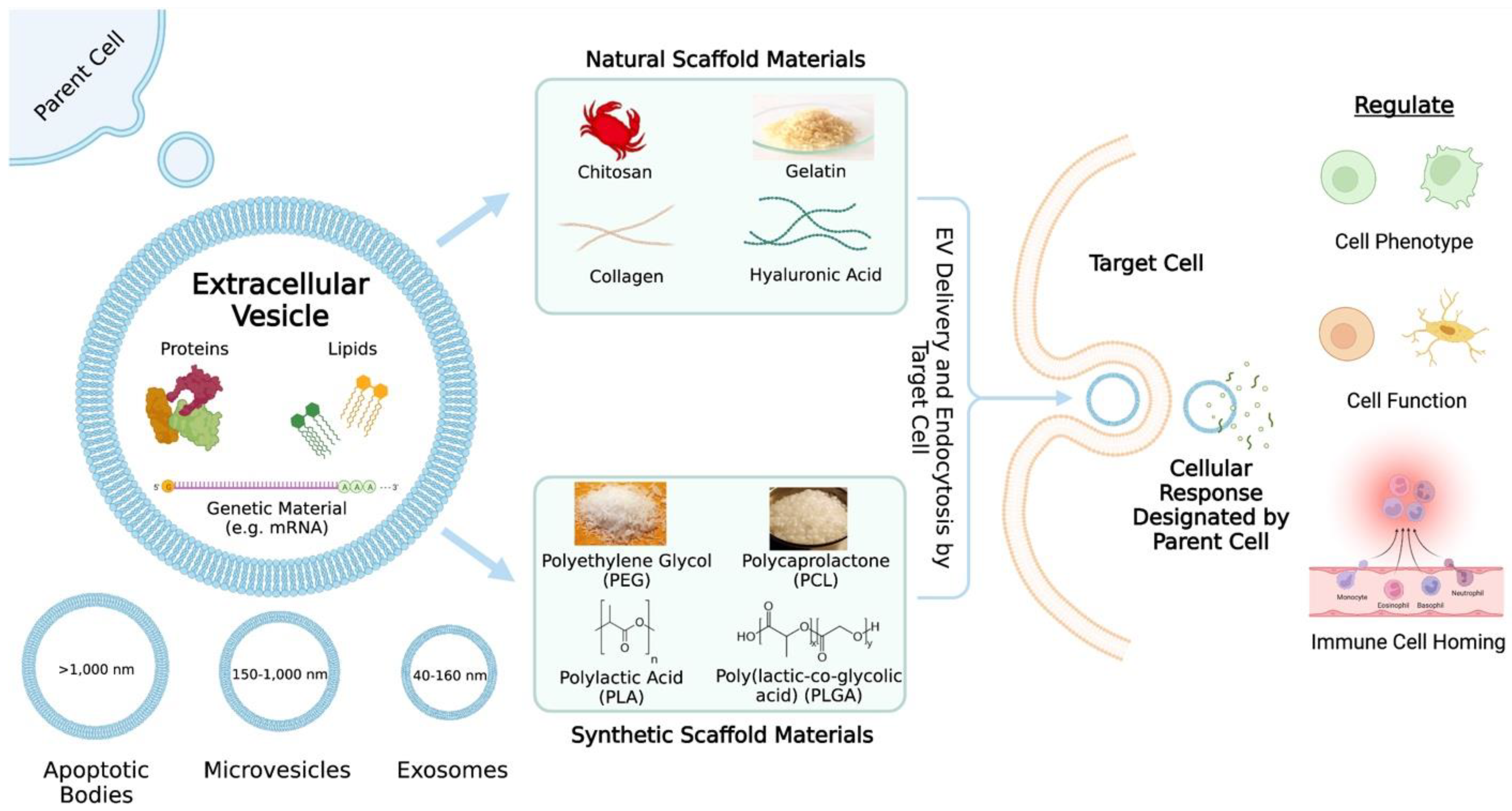
| Study | EV Source | Scaffold Biomaterial | Modifications and/or EV Incorporation Method | Disease or Tissue | Animal Models | Therapeutic Outcomes |
|---|---|---|---|---|---|---|
| [33] | Human adipose-derived mesenchymal stem cell (hADMSC) EVs | Silk-based tubular scaffold | Vacuum-seeded EVs | Cardiovascular disease | Rat abdominal aortic interposition graft model | Improved patency and matrix deposition, including increased elastin and collagen production |
| [26] | Bone marrow-derived mesenchymal stem cell (BMSC) sEVs | Sodium Alginate | sEVs mixed with sodium alginate solution | Myocardial infarction (MI) | MI induction in male rats | Decreased cardiac cell apoptosis Promotion of macrophage polarization Increased scar thickness and angiogenesis Improved cardiac function and infarct size |
| [37] | Human umbilical cord mesenchymal stem cell (HUCMSCs) Exosomes | Polyvinyl alcohol (PVA)/Alginate | Exosomes mixed with PVA/alginate solution | Diabetic wound healing | Full-thickness wounds on diabetic rat model | Proliferation, migration, and angiogenesis of HUVECs Sped up diabetic wound healing by promoting angiogenesis |
| [38] | ADSC-derived Exosomes | Sodium Alginate | Exosomes mixed with alginate solution | Peripheral nerve regeneration | Rat sciatic nerve defect | Exosomes containing neurotrophin-3 mRNA (important neurotrophic factor for peripheral nerve regeneration) in scaffold promoted nerve regeneration |
| [39] | Dental pulp stem cell (DPSC)-derived EVs | Collagen | EVs were injected into the scaffold | Bone regeneration | Rat calvarial bone defect | Bone formation in center of defects Broader angiogenesis |
| [40] | HUCMSCs | Collagen | Exosomes added dropwise onto scaffold | Endometrium regeneration and fertility restoration | Rat endometrium-damage model | Induction of endometrium regeneration, collagen remodeling Increased expression of estrogen receptor α/progesterone receptor Restored fertility Facilitated CD163+ M2 macrophage polarization, reduced inflammation, increased anti-inflammatory responses |
| [32] | BMSC EVs | Chitosan-Collagen Composite Scaffold | Scaffolds seeded with EVs | Skin wound healing | Full-thickness skin wound on adult male rats | Accelerated skin healing Enhanced macrophage count Greater collagen deposition, better collagen alignment and thus, increased mechanical strength |
| [41] | Bone mesenchymal stem cell-derived sEVs | Chitosan | sEVs added to hydrogels | Bone defect repair | Calvarial defect rat model | sEV-loaded hydrogel promoted bone healing by enhancing angiogenesis possibly via upregulation of miR-21 in sEVs |
| [42] | Induced pluripotent stem cell-derived mesenchymal stem cells (iPSC-MSCs) exosomes | Chitosan | Exosomes stirred into chitosan solution | Corneal diseases | Rat cornea anterior lamellar damage model | Promote repair of damaged corneal epithelium and stromal layer |
| [43] | Chondrocyte exosomes | Chitosan-Gelatin-Chondroitin Sulfate and Nano-Hydroxyapatite-Gelatin | Exosome suspension added dropwise over scaffold | Articular cartilage injuries | N/A | Enhance proliferation and migration of chondrocytes |
| [44] | Osteoblast-derived EVs | Gelatin Methacryloyl (GelMA) | GelMA functionalized with nanoclay laponite | Bone regeneration | N/A | Enhanced proliferation, migration, histone acetylation, mineralization of human bone marrow stromal cells |
| [45] | Tendon derived stem cells (TDSCs) exosomes | Hyaluronic acid | Loaded scaffold–not sufficiently described | Tendon repair | Rat model of tendon defects | Promoted proliferation, migration, collagen type I production, and tendon-specific markers expression in tenocytes Protected tenocytes from oxidative stress and serum deprivation Promoted early healing of injured tendons and better fiber arrangement at injury site |
| [46] | Human articular chondrocyte-derived EVs | Hyaluronic acid/Chitosan | Loaded scaffold–not sufficiently described | Osteoarthritis cartilage injuries | Rabbit osteochondral defect model | Greater cartilage regeneration Provide niche for chondrogenic differentiation of MSCs Hyalin-like cartilage in defect zone |
| [47] | M2 polarized macrophage-derived Exosomes (M2-Exos) | PEG | Dissolved freeze-dried PEG in exosome solution | Cutaneous wound healing | Mouse excisional wound splinting model | Localization and sustained release of M2-Exos Effective and prolonged conversion of M1 to M2 macrophages Enhanced efficiency and quality of wound care |
| [48] | hADMSC-derived exosomes | PLA | Mineral doped PLA scaffolds adsorbed exosomes | Bone defects including oral bone defects | N/A | Increased osteogenic commitment of MSCs |
| [49] | Mesenchymal stem cell (MSC)-derived sEVs | PCL | sEVs loaded onto heparin-modified scaffold | Cardiovascular disease | Hyperlipidemia rat model | Inhibited thrombosis and calcification and thus improved patency of graft Enhanced endothelium and vascular smooth muscle regeneration Induced polarization of M1 macrophages to M2c macrophages |
| [50] | MSC-exosomes | PCL | PCL modified with S-nitrosoglutathione (GSNO); exosomes incubated with scaffold | Bone defects | N/A | Decreased expression of pro-inflammatory genes in macrophages treated with exosome-loaded scaffold Accelerated osteogenic differentiation of mesenchymal stem cells |
| [51] | MSC-derived EVs | PLGA | EVs infused into composite scaffold; polydeoxyribonucleotide (PDRN) added | Chronic kidney disease | Partial nephrectomy mouse model | Synergistic interaction of EVs and other added compounds in scaffold alleviates fibrosis and inflammatory response Cellular proliferation Angiogenesis Effective glomerular regeneration Restoration of kidney function Develop new blood vessels and induces pro-reparative macrophages |
| [52] | Human adipose-derived stem cell exosomes | PLGA | Scaffolds submerged in exosome solution | Bone defects | Mouse calvarial defect | Enhance bone regeneration partially through osteoinductive effects and promoting mesenchymal stem cell migration and homing in newly formed bone tissue |
| [53] | MSC-sEVs | PEG/Hyaluronic Acid | sEVs mixed with scaffold solution | Osteoarthritis (OA) | Traumatic OA rat model | Improved bioavailability and therapeutic efficacy of MSC-sEVs for OA improvement |
| [54] | Human DPSC-derived exosomes | PLLA | Exosome encapsulation in triblock PLGA-PEG-PLGA microspheres and subsequently incorporated into PLLA scaffold | Bone defects | Critical size mouse calvarial bone defect | Stimulated bone tissue neogenesis Facilitated bone marrow stromal cell osteogenic differentiation Guided local progenitor cells towards osteogenic differentiation and bone healing Accelerated bone healing |
Publisher’s Note: MDPI stays neutral with regard to jurisdictional claims in published maps and institutional affiliations. |
© 2022 by the authors. Licensee MDPI, Basel, Switzerland. This article is an open access article distributed under the terms and conditions of the Creative Commons Attribution (CC BY) license (https://creativecommons.org/licenses/by/4.0/).
Share and Cite
Leung, K.S.; Shirazi, S.; Cooper, L.F.; Ravindran, S. Biomaterials and Extracellular Vesicle Delivery: Current Status, Applications and Challenges. Cells 2022, 11, 2851. https://doi.org/10.3390/cells11182851
Leung KS, Shirazi S, Cooper LF, Ravindran S. Biomaterials and Extracellular Vesicle Delivery: Current Status, Applications and Challenges. Cells. 2022; 11(18):2851. https://doi.org/10.3390/cells11182851
Chicago/Turabian StyleLeung, Kasey S., Sajjad Shirazi, Lyndon F. Cooper, and Sriram Ravindran. 2022. "Biomaterials and Extracellular Vesicle Delivery: Current Status, Applications and Challenges" Cells 11, no. 18: 2851. https://doi.org/10.3390/cells11182851