Review

Inflammation: A Target for Treatment in Spinal Cord Injury

by
Ximena Freyermuth-Trujillo
1,2,
Julia J. Segura-Uribe
3,
Hermelinda Salgado-Ceballos
1,
Carlos E. Orozco-Barrios
4 and
Angélica Coyoy-Salgado
4,*
1
Unidad de Investigación Médica en Enfermedades Neurológicas, Hospital de Especialidades Dr. Bernardo Sepúlveda, Centro Médico Nacional Siglo XXI, Instituto Mexicano del Seguro Social, Mexico City CP 06720, Mexico
2
Posgrado en Ciencias Biológicas, Universidad Nacional Autónoma de México, Mexico City CP 04510, Mexico
3
Subdirección de Gestión de la Investigación, Hospital Infantil de México Federico Gómez, Secretaría de Salud, Mexico City CP 06720, Mexico
4
CONACyT-Unidad de Investigación Médica en Enfermedades Neurológicas, Hospital de Especialidades Dr. Bernardo Sepúlveda, Centro Médico Nacional Siglo XXI, Instituto Mexicano del Seguro Social, Mexico City CP 06720, Mexico
*
Author to whom correspondence should be addressed.
Cells 2022, 11(17), 2692; https://doi.org/10.3390/cells11172692
Submission received: 17 June 2022 / Revised: 16 August 2022 / Accepted: 25 August 2022 / Published: 29 August 2022
(This article belongs to the Collection Cell Biology of Spinal Cord Injury and Repair)

Abstract

:
Spinal cord injury (SCI) is a significant cause of disability, and treatment alternatives that generate beneficial outcomes and have no side effects are urgently needed. SCI may be treatable if intervention is initiated promptly. Therefore, several treatment proposals are currently being evaluated. Inflammation is part of a complex physiological response to injury or harmful stimuli induced by mechanical, chemical, or immunological agents. Neuroinflammation is one of the principal secondary changes following SCI and plays a crucial role in modulating the pathological progression of acute and chronic SCI. This review describes the main inflammatory events occurring after SCI and discusses recently proposed potential treatments and therapeutic agents that regulate inflammation after insult in animal models.

Graphical Abstract

1. Introduction

Traumatic spinal cord injury (SCI) represents a significant cause of disability [1,2]. Depending on the severity and spinal cord anatomical level of injury, patients may experience neurological deficits ranging from loss of sensation, and bowel, bladder, sexual impairment, and autonomic dysfunction to paralysis and death [3]. Approximately 2 to 3 million people worldwide have SCI, increasing by 250,000 to 500,000 annually [4]. The leading causes of SCI are traffic accidents (50%), falls and work accidents (30%), gunshot wounds or violent crimes (11%), and sports accidents (9%) [3,5]. An SCI patient’s cumulative medical expenses range from 500,000 to 2 million USD [6]. Aside from the treatment of the injury itself, secondary costs include complications from pressure ulcers, bladder/bowel dysfunction, neuropathic pain, osteoporosis, deep vein thrombosis, cystitis, respiratory problems, pneumonia, emergency readmissions, and spinal cord injury–immune deficiency syndrome (SCI–IDS) [7,8,9].
The pathophysiology of SCI is divided into two phases: the first phase refers to the instantaneous mechanical damage generated initially [5] and the second phase includes inflammation, which involves activating resident immune cells (microglia and astrocytes) and recruiting immune cells (macrophages and neutrophils). In combination, these inflammatory reactions contribute to metabolic and cellular dysfunction of neurons, glia, and other cells at the injury site and periphery. Vascular disruption and damage to the blood–spinal cord barrier (BSCB) are significant contributors to the pathophysiology and inflammation of SCI [10,11].
The inflammatory reaction is an essential host defense mechanism. However, its effects are contradictory. Although the inflammatory process eliminates invading pathogens, removes debris, and promotes wound healing, its benefits are overshadowed by an accumulation of toxic molecules produced by inflammatory cells that damage otherwise intact tissue [12]. Inflammation in SCI involves numerous cell populations, such as astrocytes, microglia, T cells, neutrophils, monocytes, as well as non-cellular mediators [13,14,15]. Although inflammation is essential in modulating the pathological progression of acute and chronic SCI, it also regulates neuronal damage and regeneration [16].
Since the extent of secondary damage largely determines the severity of injury [17], SCI might be treatable if intervened promptly, and the high mortality rates associated with SCI could be reduced [2]. Therapeutic strategies often aim to confer neuroprotection and enhance neuroregeneration by mitigating secondary injury cascades. Based on the regulation of inflammation, some of these strategies have generated beneficial effects. In this review, we first comprehensively describe the main inflammatory events during the different stages of SCI. Second, we address potential therapies recently proposed to regulate the effects of inflammation in animal models of SCI.

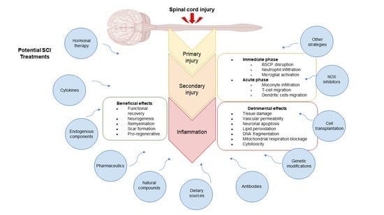
2. Main Pathological Events in SCI by Phase

The main pathological events that occur chronologically after SCI can be divided into immediate, acute, intermediate, and chronic phases (Table 1).

2.1. Immediate Phase

This phase begins immediately after SCI and lasts approximately two hours [18]. It is characterized by the immediate results of the injury, including traumatic axon rupture, rapid neural and glial cell death, and spinal shock [19], resulting in loss of function [5].
SCI directly causes necrosis and apoptosis of vascular endothelial cells, contributing to disrupting the BSCB [20]. This disruption leads to leakage of vascular material and accumulation of various inflammatory cytokines that aggravate the inflammatory reaction, edema, and secondary injury [1,20]. An SCI study demonstrated that remote ischemic preconditioning improves BSCB integrity by upregulating occludin expression [21]. Furthermore, the secretion of collagen IV and laminin is rapidly activated as a mechanism of vascular regeneration (or neovascularization) and restriction of the inflammatory reaction [20].

2.2. Acute Phase

The acute phase of SCI is further divided into the early acute phase and subacute phase. The early acute phase lasts 2 to 48 h after injury and is characterized by hemorrhage, increasing edema, and inflammation. The subacute phase lasts from two days to two weeks after injury. In this phase, the phagocytic response is maximal, eliminating cellular debris from the injury site and promoting axonal growth by removing myelin debris, which acts as a growth inhibitor [5]. Recruitment of blood monocytes to the injury site begins three days after SCI and lasts for seven days. Axonal growth and scarring by anti-inflammatory macrophages start after the first week of SCI [22].

2.3. Intermediate and Chronic Phases

The intermediate phase (2–6 weeks after SCI) is characterized by continued maturation of astroglia scarring, axonal regeneration sprouts, and the development of cysts and syrinxes. Subsequently, the chronic phase begins 6 months after SCI and lasts for life. Wallerian degeneration of injured axons is an ongoing process after SCI, and it may take years for the severed axons and their cell bodies to be removed [5]. Cytokines also play a critical role during the early stages of Wallerian degeneration by facilitating macrophage invasion to remove axon and myelin debris, an essential precondition for axonal regeneration [23]. Additionally, local and systemic inflammatory reactions during the chronic phase stimulate cavity formation and glial scarring in the medullary parenchyma and lead to neuronal and glial death [6].

3. Main Inflammatory Events in SCI

Inflammation is a fundamental mechanism that arises during the secondary phase of SCI. It occurs acutely within minutes in response to traumatic injury and can last for days or be present chronically for months or years [24]. Inflammation emerges as the host immune system response, whose main objective in SCI is to stop the degeneration of adjacent healthy tissue by clearing the injury site of cellular debris from necrosis cell death to prevent a toxic environment for axonal growth [25]. The main inflammatory events in secondary SCI are summarized in Figure 1.

3.1. Microglia Response

Concomitant with neutrophil invasion, microglial activation occurs immediately after SCI [9,22]. In a rat model of SCI, peak activation of microglia and macrophages occurs one to three days post-SCI, whereas the same phenomenon occurs three to seven days post-SCI in mice [26] (Figure 2). Microglia cells are relevant in the pathophysiology of SCI as they undergo intense activation in response to injury and are a prominent source of inflammatory mediators [27]. After SCI, however, the microglial population at the site and in the vicinity of the injury is reduced by mechanical impact and apoptosis [28].
Following SCI, toll-like receptors (TLRs) on microglial cells can detect damage-associated molecular patterns (DAMPs) released from tissue damage. Subsequently, activation and morphological hypertrophy of microglial cells occur, along with functional changes [2], including transformation to a migratory mode, retraction of processes, increased soma size, upregulation of surface antigens, and production of innate proinflammatory cytokines such as TNF-α, interleukin-1 (IL-1), interleukin-2 (IL-2), interleukin-6 (IL-6), interleukin-12 (IL-12), interleukin-18 (IL-18), and chemokines [2,12].
In addition, since microglia are phagocytes, they function as antigen-presenting cells [12]. Microglia exist in two primary polarized states dependent on external signals: the pro-inflammatory (M1) phenotype (classically activated) and the anti-inflammatory (M2) phenotype (alternatively activated), which produces anti-inflammatory cytokines, such as interleukin-10 (IL-10) and transforming growth factor-beta (TGF-β), to help maintain homeostasis [2,12]. Microglia have a neuroprotective role by inducing astrogliosis via IGF-1. Moreover, IGF1-expressing microglia are located between the astroglial and fibrotic scars [28]. Additionally, it is important to emphasize that microenvironmental factors can influence the eventual M1 or M2 phenotype and function of microglia [29].
Figure 2. Differences in intraspinal accumulation of immune cells (macrophages, microglia, dendritic cells, neutrophils, B-lymphocytes, and T-lymphocytes) after spinal cord injury in rats, mice, and humans. Presence (one arrow) and peak (two arrows) of immune cells post-SCI in the rat (Cells 11 02692 i001), mouse (Cells 11 02692 i002), and human (Cells 11 02692 i003) Refs. [14,26,30,31,32,33].
Cells 11 02692 g002
In the secondary phase of SCI, activated M1 microglia initiate a cascade of neurotoxic responses and contribute to apoptosis and necrosis of endothelial cells, neurons, axons, and oligodendrocytes [2]. Activated macrophages/microglia are a chronic source of cytotoxic substances, such as TNF-α, inducible nitric oxide synthase (iNOS), hypochlorous acid (HOCl), and reactive oxygen species (ROS) [34].
With single-cell ribonucleic acid (RNA) sequencing (scRNA-seq), several microglia phenotypes with complex functional potential have been defined in both healthy and disease states [35,36,37]. In a recent study, the unique molecular signature of the cells composing the SCI site in a mouse mid-thoracic contusion model was characterized with scRNA-seq. Four microglia subtypes were identified: homeostatic microglia identified based on their expression of P2ry12, Siglech, and Tmem119, and three non-homeostatic microglia subtypes, labeled as inflammatory, dividing, and migratory microglia based on gene ontology (GO) terms for biological processes [38].
Single-cell transcriptional analyses revealed that mouse and human spinal cord microglia exist in numerous heterogeneous subpopulations, and peripheral nerve injury-induced changes in microglia were observed to differ significantly in the acute and chronic phases of neuropathic pain. In addition, sex-specific differences in gene expression were detected: a subpopulation of selectively induced microglia was identified in males but not in females three days after peripheral nerve injury. ApoE was also found as a highly upregulated gene in microglia in chronic phases of neuropathic pain in mice [39]. Furthermore, with transcriptional analysis of individual cells in the crush-injured spinal cord in adult mice, other authors demonstrated novel microglia and astrocyte subtypes and their dynamic conversions into additional stage-specific subtypes/states. Most of the dynamic changes occurred 3 days after injury; a recovery trend was observed between 3 and 14 days after injury, and after 14 days, reactivation of microglia and a further decrease in neuronal, astrocytic, and endothelial populations along with a continued increase in leukocyte population were evident [40].
One of the most significant inflammatory factors is ROS overproduction at the site of trauma. This ROS overproduction consumes endogenous antioxidants, disrupting the redox balance and severely damaging DNA, proteins, and lipids (lipid peroxidation). In addition to uncontrolled peroxidation, the disturbed balance contributes to inflammation and concomitant swelling [41].
Activated macrophages/microglia also secrete large amounts of matrix metalloproteinases (MMPs), further contributing to vascular basement membrane degradation and increased permeability for infiltrating inflammatory cells. Reactive microgliosis after SCI involves a self-perpetuating cycle initiated by neuronal damage and myelin debris activating microglia, which upregulate proinflammatory cytokines and neurotoxic molecules that cause further tissue damage, thus continuing the activation/disruption cycle [42].

3.2. Neutrophils

Neutrophils rapidly infiltrate the spinal cord, peak 24 h after injury, decrease to a few cells in the spinal cord in rats [43,44], and remain in the injured spinal cord for up to 6 months post-SCI [30]. Conversely, neutrophils persist in the spinal cord for several months in mice [13,26] (Figure 2). Neutrophils support recovery processes through their ability to phagocytize cellular debris and summon macrophages to the injured tissue [12]; thus, neutrophil depletion after SCI has been associated with worse outcomes [45].
Neutrophil invasion requires the presence of P-selectin, a member of the endothelial leukocyte adhesion molecule family that is rapidly expressed on the surface of endothelial cells by stimuli such as thrombin, histamine, and ROS. P-selectin is responsible for mediating early interactions between neutrophils and endothelial cells that lead to the rolling state of neutrophils on the endothelium and facilitate cell adhesion, migration, and tissue injury [46]. Thus, neutrophils gather at the site of injury and release cytokines, chemokines, and various proteases, including MMPs and neutrophil elastase, aggravating tissue edema and necrosis, and promoting apoptosis of neurons and oligodendrocytes. Consequently, these actions form a local glial scar [12,47].
Upon infiltrating the spinal cord, neutrophils also release ROS and nitrosyl radicals to sterilize the injured area [12,48]. These species cause uncontrolled oxidative stress. Therefore, the antioxidant defense system can no longer cope with neutralizing the free radicals produced in the organism, resulting in irreversible cellular and body damage over time [49]. Furthermore, hydrogen peroxide (H2O2) triggers the activation of neutrophils, during which the enzyme myeloperoxidase (MPO) is also secreted. Subsequently, H2O2 and MPO react with chloride (Cl), creating a [H2O2--MPO-Cl] system that produces HOCl. This system is paradoxically beneficial and detrimental to the host since HOCl production is necessary to kill bacteria. However, it is also one of the main factors of tissue damage following inflammatory cell activation. In addition, HOCl catalyzes the deamination and decarboxylation of amino acids [50].
Moreover, neutrophils and macrophages/microglia express the nicotinamide adenine dinucleotide phosphate (NADPH) oxidase 2 (NOX2) enzyme, a primary producer of ROS in the injured spinal cord. NOX2 transfers electrons from NADPH inside the cell, across the membrane, to extracellular oxygen to generate superoxide (O2) [51].
Conversely, endogenous antioxidant enzymes, such as superoxide dismutase (SOD) and glutathione (GSH), contribute to the defense against ROS [52]. Superoxide (O2) can be reduced to H2O2 by SOD and, in turn, converted to free radicals. Although SOD has been involved in neuronal recovery and regeneration, only the mitochondrial manganese SOD (MnSOD) enzyme is induced and modulated after SCI [53]. TNF-α is an upstream signal that triggers MnSOD induction through NF-kB activation. However, MnSOD can also be induced by IL-1β, IL-6, and IFN-γ [53].

3.3. Inflammasomes

Three types of inflammasomes have been characterized in microglia: the NLRP3 (NACHT, LRR, and PYD domains-containing protein 3), the NLRC4 (NLR family, CARD domain containing 4), and the AIM2 (absent in melanoma 2) inflammasomes [16]. The NLRP3 inflammasome is activated by ROS [54]. Activation of this pathway induces neuroinflammatory responses and cell death [55]. NLRPs activation leads to the recruitment of the apoptosis-associated speck-like protein (ASC) that contains a caspase activation and recruitment domain (CARD). ASC protein interacts with the CARD of pro-caspase-1, leading to its conversion from pro-caspase-1 to caspase-1 (active form). Caspase-1 converts interleukin-1 beta (IL-1β) and IL-18 pro-forms into their respective activated configuration, thus initiating an inflammatory response [56].
Treatments targeting the NLRP3 inflammasome exert neuroprotection in a rat model of contusion SCI [56]. It is essential to consider Nrf2 (nuclear factor erythroid 2-related factor 2)—a redox-sensitive master regulator [57] that modulates the transcription of several antioxidant genes under oxidative stress and inflammation [58]. In SCI, activation of Nrf2 signaling leads to detoxification and expression of antioxidant defense genes [59]. Heme oxygenase-1 (HO-1), a target of Nrf2, is a phase II detoxifying and anti-inflammatory enzyme [60] that contributes to NLRP3 inflammasome inhibition [54].

3.4. Cytokines

Proinflammatory cytokine production also increases in the immediate phase [3,61]. Although the exact mechanism of this increase in the SCI has not been explained, it is suggested that inflammatory cytokines play a central role in triggering a cascade of events leading to apoptosis [62]. Inflammatory cytokines consist of a broad category of small soluble proteins that modulate the complex function of the immune system [1]. Early accumulation of inflammatory cytokines in and around microvessels in ischemic areas can cause spinal cord edema and neuronal apoptosis [62].
Increased levels of IL-1β and IL-6 cause upregulation of nuclear factor kappa B (NF-κB), c-Jun N-terminal kinase (JNK), and p38 mitogen-activated protein kinase (p38MAPK), which could activate apoptosis [3]. Moreover, MAPK activation is essential for producing several inflammatory cytokines [27].
Additionally, IL-1β and IL-18, whose maturation in inflammatory cells depends on inflammasome activation [16], induce upregulation of IL-6 [63], thereby enhancing vascular permeability [64] and inducing neuronal apoptosis [65]. IL-1β deficiency suppresses lesion development, promotes axonal plasticity, and improves neurological outcomes [66]. In contrast, the blockade of IL-1 receptors prevents microglial activation and promotes the survival of ventral horn neurons [67] while alleviating the severity of SCI [68] and stimulating neurological recovery [69]. IL-18 is also upregulated after SCI [70], and its inhibition reduces NF-κB phosphorylation in spinal astrocytes and suppresses the expression of astrocyte markers [71]. IL-6 induces the differentiation of neural stem cells into astrocytes. However, treatment with an antibody against the IL-6 receptor suppressed injury and glial scar formation caused by inflammatory reactions after SCI in mice, improving functional recovery [72].
IL-1β and IL-6 induce iNOS in astrocytes, microglia, macrophages, and neurons. iNOS is highly involved in inflammatory processes since it produces excessive amounts of NO molecules. NO has been implicated in microglia-dependent demyelination and apoptosis of neuronal cells [2]. NO released by endothelial NOS (eNOS) is known to play a protective role. In contrast, activation of iNOS and neuronal NOS (nNOS) is followed by an overproduction of NO, which is detrimental to neuronal cells [73].
Anti-inflammatory cytokines, such as IL-10 and TGF-β, also play an essential role in SCI. IL-10 can be synthesized by Th2, monocytes/macrophages, astrocytes, and microglia. This cytokine suppresses the inflammatory response of monocyte/macrophages and the production of multiple cytokines, cell adhesion molecules, ROS, and nitrogen intermediates. Additionally, IL-10 affects inflammation by activating microglia/macrophages and astrocytes and reducing the production of IL-1β and iNOS. This interleukin promotes neuronal survival and functional recovery in rat and mouse models of SCI [1,74]. TGF-β is a multifunctional cytokine involved in mammal damage repair and scar tissue formation. TGF-β is a potent agent of astrocyte chemotaxis in vitro, resulting in astrocyte hypertrophy and upregulation of fibronectin and collagen IV synthesis [20].
Stromal cell-derived factor-1 (SDF-1α) is a cytokine with different biological functions, such as inflammatory cell infiltration. This cytokine is upregulated in pathological conditions [75], and its administration decreases cortical neuron death after stroke by stimulating mechanisms such as angiogenesis, neurogenesis, and modulation of the inflammatory response [76]. In addition, SDF-1α is a compound of interest for regenerative medicine because it can attract different types of stem/progenitor cells to target sites.
Several inflammatory cytokines that participate in the pathophysiology of SCI can be beneficial or detrimental. Beneficial cytokines include erythropoietin (EPO), granulocyte colony-stimulating factor (G-CSF), granulocyte-macrophage colony-stimulating factor (GM-CSF), interferon-beta (IFN-β), interferon-gamma (IFN-γ), interleukin-4 (IL-4), IL-12, and interleukin-33 (IL-33). In contrast, cytokines that have shown detrimental behavior are chemokine C-X-C motif ligand 10 (CXCL10), interleukin-8 (IL-8), and interleukin-17 (IL-17) [1,77].

3.5. Interferons

TNF-α increases after SCI and may function as an apoptosis inducer in neurons and glia [78]. Different studies have shown that TNF-α can elicit trophic or toxic effects, depending on the target cell and receptors [53]. TNF-α induces and exacerbates spinal cord motoneuron death after SCI [79,80]. However, topical application of TNF-α antiserum attenuates edema, microvascular permeability, and injury in a rat model of SCI [81]. Downstream of TNF-α signaling is NF-κB, a transcriptional factor that induces inflammation-related molecules [82]. NF-κB is a redox-sensitive transcription factor that activates oxidative stress-induced-signaling cascades and plays a critical role in converting oxidative stress into inflammatory signals [83]. Within the nucleus, NF-κB induces the expression of inflammatory genes such as cyclooxygenase-2 (COX-2), iNOS, and inflammatory cytokines [84]. The NF-κB pathway also upregulates other inflammatory proteins, such as chemokines and adhesion molecules [85].

3.6. Macrophages

Macrophages, the effector cells in the inflammatory response to central nervous system (CNS) injury, are derived from microglia and hematogenous monocytes and are functionally indistinguishable in morphology and antigenic markers. In CNS injury, activated microglia are called macrophages/microglia [42]. During the first week after SCI, the populations of M1 and M2 macrophages found at the injury site are relatively evenly distributed. However, over time, macrophages eventually polarize to M1. This polarization toward M1 has been observed in different species and types of SCI, suggesting a common feature of this type of injury [86].
Peak macrophage activation is observed 7 to 14 days after injury. Macrophages function as phagocytes and serve as a reservoir for cholesterol derived from ingested myelin, which can be used to regenerate axons during remyelination [12].
Monocyte chemoattractant protein-1 (MCP-1) is a chemokine that regulates monocyte/macrophage migration and infiltration [87] and increases significantly after SCI [57]. IFN-γ-activated macrophages exert neurotoxic effects, whereas interleukin 4 (IL-4)-activated macrophages inhibit adverse immune responses and promote damage repair [88].

3.7. Dendritic Cells

Dendritic cells (DCs) are antigen-presenting cells that highly express molecular histocompatibility complex (MHC)-II proteins and proinflammatory cytokines. DCs in the spinal cord are differentiated from microglia or a circulating pool and support the ongoing inflammatory response, exacerbating the secondary injury. However, they can also produce growth factors, including neurotrophin-3, and enhance neurogenesis [12]. IL-12 is produced by DCs, macrophages, monocytes, and B cells. When secreted by DCs, IL-12 promotes functional recovery in a T8 vertebral level hemisection in female mice [89]. The recovery could be attributed to IL-12 increasing the number of activated microglia/macrophages and DCs, as well as the expression of brain-derived neurotrophic factor (BDNF), thus enhancing neurogenesis and remyelination [1].

3.8. Lymphocytes

B-lymphocytes are found near the injury site in the first few hours after SCI and persist for up to a week post-injury [12,24]. In contrast, peak T-lymphocyte infiltration occurs three to seven days post-SCI in rats but not in mice, in which it is delayed for up to two weeks. The population of MHC Class II-positive cells is more prevalent after SCI in mice than in rats [13,90] (Figure 2). T-lymphocytes can induce significant tissue damage through their proliferation after recognizing specific antigens, such as myelin basic protein. However, T-lymphocytes play complex roles in injury and repair mechanisms [12].
Conflicting results have been observed on the role of T- and B-lymphocytes in recovery after SCI. Whether lymphocytes become pathogenic or immunoregulatory probably depends on the nature of target antigens, antigen specificity of different lymphocyte populations, and the proximate microenvironment [91,92]. SCI impairs the development and mobilization of B-lymphocytes [93]. However, other studies demonstrate that SCI triggers activation of T- and B-lymphocytes, leading to impairments in the recovery of function and exacerbation of injury-induced pathology [94,95,96]. These effects are partly mediated by a subpopulation of autoreactive cells, some of which are myelin-reactive [97]. Furthermore, T-lymphocytes progressively increase in parallel with the activation of microglia and the influx of peripheral macrophages in the first week after injury [13].
T cells are attracted to the site of injury by the microenvironment and molecular signals. Chemokines are responsible for the migration of T cells and modulate their activation and effector potential at sites of inflammation. Depending on their phenotype (CD4+ or CD8+), T-lymphocytes can kill target cells and produce proinflammatory cytokines such as IFN-γ and TGF-β, as well as pro-regenerative trophic factors such as IL-10, IL-4, and interleukin-13 (IL-13) upon activation. Although chronic T-cell activation is precipitated during pathological fibrosis and scarring, some studies support the neuroprotective role of T-lymphocytes in models of CNS injury and neurodegeneration [98,99].

4. Potential Therapies to Regulate the Inflammatory Response in SCI

Although the inflammatory process generally follows a similar course in different organisms, variations have been found according to the animal model studied and to what has been observed in humans [31,32,33] (Figure 2). Knowing these differences is of utmost importance for correctly applying the proposed treatments and interpreting the outcomes and findings. With this in mind, the therapies offered to date to regulate the inflammatory response in SCI in different animal models are presented below, as well as the outcomes, advantages, and disadvantages of each treatment [100,101,102,103,104,105,106,107,108,109,110,111,112,113,114,115,116,117,118,119,120,121,122,123,124,125,126,127,128,129,130,131,132,133,134,135,136,137,138,139,140,141,142,143,144,145,146,147,148,149,150,151,152,153].
For several years, it has been known that many potential treatments and therapeutic agents for SCI regulate inflammatory cytokines and other inflammatory factors (Table 2). This review describes the results of the leading therapies tested in pre-clinical studies that appear promising for clinical use.

4.1. Hormone Therapy

Hormone therapy is a therapeutic approach to modulating inflammation in SCI based on administering estrogen, progesterone, or ghrelin to improve functional outcomes [3,100,101,102,103].
Estrogen has shown neuroprotective effects due to its anti-inflammatory effects [3]. After SCI in rats, twice-daily estrogen administration significantly reduced TNF-α and gene expression of its downstream cytokine iNOS [100], demonstrating its inhibitory effect on inflammation after SCI. Estrogen levels may be partially responsible for the improved functional outcomes in females relative to males after SCI [3]. Moreover, some estrogen receptor binding molecules—such as tamoxifen and other estrogen receptor agonists—have shown similar or identical neuroprotective functions to estrogen after SCI [3].
Progesterone is another anti-inflammatory hormone since it inhibits inflammatory cytokines. In a rat model of SCI, progesterone administration significantly reduced the expression of TNF-α and iNOS, which produce inflammatory mediators and NO [101]. Furthermore, progesterone reduces axonal dieback and neuronal death in mice after SCI by downregulating inflammatory cytokines, such as iNOS, MCP-1, and IL-1β, as well as activated caspase-3 and glial fibrillary acidic protein (GFAP) [102], a selective marker of astrocytes [104].
Moreover, ghrelin is a 28-amino acid gastric hormone with multifunctional roles in appetite, adiposity, energy balance, gastric motility, and acid secretion. In experimental autoimmune encephalomyelitis, a representative animal model of multiple sclerosis, ghrelin significantly reduced TNF-α, IL-1β, and IL-6 in the spinal cord microglia [105]. These findings suggest that ghrelin has a possible anti-inflammatory therapeutic effect on different pathologies, including SCI, where oligodendrocyte cell death and microglial activation result from the inflammatory process [103].
Despite the beneficial effects of hormone therapy, the lack of specificity and selectivity of these hormones, their short half-life, low accessibility, and rapid metabolism should be considered [106]. Another important consideration is that the therapeutic use of estradiol is limited by its peripheral hormonal actions, which increase the risk of breast, ovary, and endometrial cancer [107].

4.2. Cytokines and Interferons

Since cytokines and interferons play a significant role in regulating inflammation, their use has been proposed to control inflammation initiated after SCI. Yune et al. administered TNF-α in uninjured spinal cords to study its effect on SCI and observed an increase in MnSOD expression. Their results also revealed that NF-κB was activated and translocated to the nuclei of neurons and oligodendrocytes. In contrast, the administration of neutralizing antibodies against TNF-α in injured spinal cords attenuated the increased MnSOD expression and NF-κB activation. Their findings showed that TNF-α could induce MnSOD expression mediated by NF-κB activation and after SCI in rats [53].
Yaguchi et al. investigated the regulation of inflammation in SCI with cytokines. Using a mouse model of SCI, these authors evaluated the effect of topically IL-12 administered with a gel foam sponge [89]. Gelatin sponges are biodegradable, pH-neutral, sterile sponges with a porous structure that functions as a hemostatic agent [1]. These sponges are used in SCI models as a topically applied biomaterial system. The results showed that IL-12 increased activated amoeboid-like microglia/macrophages and BDNF expression. Increased remyelination and functional recovery were also observed [89]. This study provides encouraging evidence for using IL-12 as a potential treatment for SCI.
Stromal cell-derived factor-1 (SDF1α) is a notable cytokine in regeneration [56] that exerts neuroprotective effects after SCI. Moreover, its administration improved neural recovery after SCI by contusion in rats [75]. Zendedel et al. concluded that intrathecal administration of SDF1α reduces the inflammatory response in a rat model of weight compression SCI. They observed that SCI transiently increased the expression of NLRP3 and ASC inflammasomes and TNF-α, IL-1β, and IL-18. In contrast, SDF-1α administration significantly reduced these proinflammation cytokines and caspase-1 levels. Overall, this study suggests that the administration of SDF1α cytokine could decrease spinal cord damage and improve functional recovery after SCI [56].
Similar to hormone therapy, although it has shown promising results in several studies, cytokine therapy may have undesirable effects. For example, glial TNF-α undermines spinal learning, and its inhibition has been shown to rescue and restore adaptive spinal plasticity in SCI [108]. Additionally, most antitumor clinical trials involving IL-12 treatment did not show sustained antitumor responses and were associated with toxic side effects such as fever, fatigue, hematological changes, or hyperglycemia [109]. Further research is needed to fully understand the impact of decreasing the inflammatory response either with cytokines such as SDF-1α or any other therapy after SCI since components of the early inflammatory response after the injury may increase plasticity within injured or intact CNS axons [110].

4.3. Endogenous Components

Antioxidant enzymes such as SOD1 and catalase are highly efficient and can neutralize harmful radicals. SOD1 protects cells from constantly generated radicals and participates in antioxidant defense. Therefore, introducing antioxidant enzymes after SCI could neutralize excess ROS and promote rapid inflammation termination, thus decreasing post-traumatic neurodegeneration and neuronal death [41]. Several approaches have been developed to increase the stability and delivery of antioxidant enzymes. For example, antioxidant enzymes were encapsulated into polyionic complexes with cationic block copolymers (nanozymes). These nanozymes protect against proteases and are stable due to electrostatic interactions between the enzyme and the cation block copolymer. Encapsulated SOD1 showed stability, prolonged blood circulation, and improved recovery in animals with neurological deficits [111].
Nukolova et al. encapsulated SOD1 into polyion complexes and examined their ability to improve motor recovery in rats after contusion SCI. SOD1 was protected by either one (polycation) or two (polycation and polyanion) layers of polyions, resulting in active and stable single- or double-coat nanozymes. As a result, novel SOD1 nanoformats were developed for ROS scavenging after SCI. The multilayered polyionic SOD1 complex decreased inflammation and edema at the injury site and demonstrated efficacy in the recovery of rodents in this SCI model. Therefore, the double-coat nanozyme represents a therapeutic alternative for SCI treatment [41].
Administration of necrostatin-1 (Nec-1) is another strategy that proposes modulation of endogenous substances after SCI. Nec-1 is a receptor-interacting protein 1 (RIP1) inhibitor that prevents necroptosis (particular programmed necrosis) [112]. Wang et al. used a rat contusion model to investigate the role of Nec-1 in SCI recovery and found that Nec-1 reduced TNF-α, IL-1β, and IL-6 levels in the spinal cord 24 h after injury. Additionally, Nec1 reduced necroptosis through the recruitment of RIP1/3-MLKL (mixed lineage kinase domain-like pseudokinase) and apoptosis through the regulation of caspase 3 and Bax/Bcl-2. They also observed effective control of ROS production with Nec-1 treatment. Functional recovery and improved ethological performance after Nec-1 treatment confirmed the positive role of Nec-1 in SCI. However, whether Nec-1 can directly inhibit these cytokines or indirectly by inhibiting necrosis and apoptosis remains unknown [113].
Connexin43 (Cx43) is one of the primary gap junction proteins in CNS astrocytes. Different studies have shown that the levels of these proteins increase after SCI and that this increase may favor local glial networks involved in tissue responses to an injury. O’Carroll et al. used a rat model of moderate contusion SCI and administered a Cx43 mimetic peptide (Peptide5) directly into the lesion via an intrathecal catheter with an osmotic mini-pump. This treatment led to a significant improvement in functional recovery. Their results also showed that Peptide5 could regulate inflammation by decreasing TNF-α and IL-1β levels [114]. In addition, Peptide5 reduced Cx43 protein and increased Cx43 phosphorylation. These authors observed increased motor neuron survival, reduced astrocytosis, and activated microglia. Their results suggest that Peptide5 administration reduces secondary tissue damage after SCI and could be a potential treatment for SCI [114].
Epidermal growth factor receptor (EGFR) has recently attracted attention for its ability to regulate cell activation in the context of SCI. EGFR localizes to various cells in the CNS, including neurons, astrocytes, oligodendrocytes, and microglia. This receptor is activated once it binds to one of its several ligands, including epidermal growth factor (EGF) and TNF-α. Qu et al. hypothesized that regulating EGFR signaling could influence microglial activation and neuroinflammation. They administered inhibitors of EGFR signaling activation (C225 and AG1478) in a rat model of SCI using the weight-drop technique. Their results showed that blocking EGFR binding to its ligand reduces IL-1β and TNF-α production in microglia by inhibiting the EGFR/MAPK cascade. They also observed that SCI-induced overexpression of CD11b and GFAP was attenuated by treatment with C225 and AG1478. Overall, they concluded that by modulating the inflammatory response after SCI, inhibition of EGFR signaling reduces microglia and astrocyte activation, attenuates tissue edema, and improves morphological and functional recovery [27].
Similarly, Byrnes et al. determined the effect of regulating metabotropic glutamate receptor (mGluR) signaling in the CNS on inflammation after SCI. By administering a selective mGluR5 agonist called (RS)-2-chloro-5-hydroxyphenylglycine (CHPG) in moderate SCI at T9 in rats, they demonstrated that CHPG attenuated microglia-associated inflammatory responses in a dose-dependent manner, including the expression of ED1, Iba-1, Galectin-3, NADPH oxidase components, TNF-α, and inducible nitric oxide synthase. Furthermore, they observed that CHPG administration reduced lesion size and improved neurological recovery after SCI. Based on these results, these authors proposed that the observed effects might be mediated, in part, by modulation of microglia-associated inflammation [115].
Although using endogenous components as therapy has advantages since no adverse reaction or rejection by the organism is expected, the high cost of producing these components is a significant disadvantage. In addition, they show little stability on their own, leading to the need for sophisticated delivery techniques, such as nanozyme encapsulation of SOD1 [111]. In other cases, such as with Nec-1, the method of administration is very invasive (direct injected into the segmented vertebral cavity) to deliver the component to the lesion site [113], or with Peptide5 and EGFR, which were administered at the lesion site with an osmotic pump [27,114].

4.4. Pharmaceuticals

Given that many treatments have been proposed to regulate inflammation after SCI, it is not surprising that a large number of drugs have been evaluated. For example, meloxicam, a drug derived from enolic acid, is a potent inhibitor of prostaglandin biosynthesis under inflammatory conditions via the inhibition of COX-2 over COX-1 [116]. It has been suggested that COX-2 inhibitors may exert neuroprotective effects by reducing prostanoid and free radical synthesis or directing arachidonic acid metabolism through alternative metabolic pathways. Using a rat model of contusion SCI, Hakan et al. observed that meloxicam treatment decreased trauma-induced MPO activity (an index of neutrophil infiltration). These effects may be due directly to its antioxidant properties or its anti-inflammatory action through COX-2 inhibition. Overall, the anti-inflammatory agent meloxicam ameliorated histological and neurological deterioration after SCI. In addition to significantly inhibiting neutrophil infiltration, meloxicam inhibited free radical generation, lipid peroxidation, and DNA damage in the injured spinal cord tissue [117]. However, several side effects of meloxicam have been reported, such as abdominal pain, anemia, edema, and an increased risk of severe gastrointestinal adverse effects, including ulceration and bleeding [118].
Methylene blue exerts neuroprotective effects by targeting mitochondrial toxicity despite being a dye. Lin et al. characterized the effect of methylene blue on the inflammatory response of microglia after SCI. They first observed that IL-1β and IL-18 production by lipopolysaccharide (LPS)-stimulated microglia decreased with methylene blue. They also observed that methylene blue markedly reduced the appearance of NLRP3 and NLRC4 inflammasomes in stimulated microglia. These findings suggested that inhibition of NLRP3 and NLRC4 inflammasome activation decreased the production of mature IL-1β and IL-18, as cleaved caspase-1 was also reduced in methylene blue-treated microglia after stimulation. Finally, they confirmed neuroprotection and decreased overall neuroinflammation with methylene blue administration in the SCI rat model and concluded that methylene blue inhibits the inflammatory response of microglia after SCI by alleviating NLRP3 inflammasome activation [16].

4.5. Natural Compounds

The potential of different components of traditional medicine plants as anti-inflammatory treatments for SCI has been explored. Many elements of traditional Chinese medicine herbs, including quercetin, ligustilide, Asiatic acid, tetrandrine, Panax notoginsenoside, and plumbagin, exert anti-inflammatory effects in SCI models [119].
Quercetin is a principal flavonoid component of several Chinese herbs in foods and vegetables. This compound exhibits antioxidant, anti-inflammatory, and anticarcinogenic effects [120]. Moreover, after compression SCI, quercetin reduced neutrophil recruitment and MPO activity in rats [121].
Ligustilide is extracted from Angelica sinensis and represents one of the main active components of this plant. This compound exerts neuroprotective effects, dilates blood vessels, inhibits vascular smooth muscle cell proliferation, and has anti-cancer, anti-inflammatory, and analgesic properties. In a rat model of transection SCI, treatment with ligustilide suppressed ROS and iNOS gene expression and reduced IL-1β and TNF-α levels [122].
Asiatic acid (AA) is extracted from the Chinese herb Centella asiatica and exhibits antioxidant and anti-inflammatory properties. Jiang et al. observed that treatment with AA in the female rat model of SCI reduced cytokine levels, including IL-1β, IL-18, TNF-α, and IL-6, inhibited NLRP3 inflammasome activation, and increased Nrf2 and HO-1 levels [55].
Tetrandrine (TET) is a bis-benzylisoquinoline alkaloid extracted from the roots of Stephania tetrandrae S. Moore. TET shows anti-inflammatory, antioxidant, antihypertensive, cardioprotective, antitumorigenic, antinociceptive, and antidepressant properties [82]. In an oxygen-glucose-serum deprivation/reoxygenation (OGSD/R)-induced injury in rat spinal cord astrocytes mimicking hypoxic/ischaemic conditions in vivo, pretreatment with TET decreased the accumulation of TNF-α, IL-1β, and IL-6 in conditioned medium. Therefore, TET pretreatment attenuated OGSD/R-induced oxidative stress and inflammation through the phosphoinositide-3-kinase (PI3K)/protein kinase B (Akt)/NF-κB signaling pathway [82].
Panax notoginsenoside (PNS) is the main active compound of Panax notoginseng, an important traditional Chinese medicinal herb with anti-inflammatory, anti-edema, and anti-apoptosis effects. Primarily, PNS can protect neurons in animal models of cerebral IRI. Acute spinal cord IRI causes a significant up-regulation of IL-1β, IL-10, and TNF-α in rats, suggesting a dramatic infiltration of inflammatory cells into the grey and peripheral white matter. PNS treatment has been shown to prevent increased proinflammatory cytokines and reduced leukocyte activity. As a result, the inflammation-induced secondary injury was ameliorated. Additionally, PNS treatment exerted anti-apoptotic effects [62].
Plumbagin is extracted from the root of Plumbago zeylanica L. This compound is a vitamin K3 analog potent antioxidant with antiproliferative, chemopreventive, antimetastatic activities, and anti-inflammatory and analgesic effects [83]. In a rat model of SCI, plumbagin treatment modulated nuclear expression of Nrf-2 and NF-κB and reduced SCI-induced TNF-α and IL-1β. This study indicated that plumbagin is a potent suppressor of SCI-induced inflammation [83].
Traditional European medicine has also explored plant extracts as potential therapeutic agents for SCI. Apigenin is one of the most common flavonoids with antioxidant and anti-inflammatory properties [123]. This compound is mainly isolated from the buds and flowers of Hypericum perforatum [124], also known as St. John’s Wort [86]. However, it is also found in other plants and natural sources. Other apigenin-containing products include parsley, celery, thyme, celeriac, chamomile, onions, lemon balm, and oranges [124]. Contusion SCI in rats comes with a significant increase in IL-1β, TNF-α, and intercellular adhesion molecule-1 (ICAM-1) serum levels. Apigenin significantly reduced IL-1β, TNF-α, and ICAM-1 in this model, indicating that its protective effect is likely related to an anti-inflammatory action [86].
Rosemary and sage are rich in polyphenolic compounds, such as carnosol and rosmarinic acid. Carnosol has been identified as an excellent antioxidant with anticancer and anti-inflammatory properties [125] and exhibits cytoprotection through enhancing Nrf2-related antioxidant defense mechanisms [126]. Carnosol treatment followed by SCI modulated oxidative stress and inflammation by downregulation of NF-κB and COX-2 levels and upregulation of p-Akt and Nrf-2 levels. It also showed significant cytoprotection through the downregulation of proinflammatory cytokines (TNF-α, IL-6, and IL-1β). Therefore, carnosol’s antioxidant and anti-inflammatory function could have effectively suppressed oxidative stress and inflammation induced by SCI [125].
Rosmarinic acid—a polyphenol of the Lamiaceae family—is found in lemon balm and thyme, besides rosemary and sage [57]. Rosmarinic acid is a natural antioxidant and has potent anti-inflammatory effects. In a model of SCI, rosmarinic acid treatment exerted anti-inflammatory effects through the downregulation of NF-κB and cytokine levels (IL-6, IL-1β, TNF-α, and MCP-1). Furthermore, rosmarinic acid ameliorated Nrf2 downregulation caused by SCI and provided significant cytoprotection against SCI by preventing apoptosis [57].
Finally, tocotrienols are isomers of vitamin E with antioxidant, antitumor, and neuroprotective functions [20]. Tocotrienol treatment inhibited serum levels of NF-κB p65 unit, TNF-α, IL-1β, and IL-6, inhibiting iNOS expression, activity, and plasma NO production in rats with SCI. Moreover, tocotrienol significantly inhibited TGF-β and improved the injured spinal cord’s functional recovery and grey matter volume in rats, probably through the TGF-β, collagen type IV, and fibronectin signaling pathways [20].

4.6. Dietary Sources

Dietary modification may also be an approach to regulating inflammation after SCI. As mentioned above, apigenin is found in foods such as onions and oranges, and its protective effects on SCI are related to its anti-inflammatory actions [124]. Furthermore, allicin is a volatile oil found in a common ingredient, Allium sativum, commonly known as garlic. Garlic is a species of the onion genus that has been used for thousands of years for medicinal purposes [73]. Wang and Ren observed that allicin significantly reduced NF-κB and TNF-α levels in a mice model of SCI. This study demonstrated the protective effects of allicin following SCI through increasing heat shock protein 70 (HSP70) expression and NADH levels and decreasing iNOS and ROS levels [127].
Taurine (2-aminoethanesulfonic acid) is ubiquitously found in various tissues, including the spinal cord. Taurine modulates several neurological activities and has neuroprotective effects [128]. Increased taurine levels appear to be involved in neuroprotection and regeneration after SCI. Therefore, taurine could be beneficial as a therapeutic agent for SCI [77]. In a mice model of SCI, taurine administration decreased IL-6, COX-2, and MPO concentrations [129]. However, taurine has a strong hydrophilic and lipophobic nature and a rapid extraction rate. Therefore, some taurine analogs, such as N-chloro-taurine (NCT), have shown more effective therapeutic properties, including downregulation of cytokines (TNF-α, IL-1β, IL-6, and IL-8) and oxidative stress markers (ROS and MPO) [77].

4.7. Antibodies

Monoclonal antibodies (mAb) could be another strategy to attenuate secondary SCI damage by regulating inflammation. For example, it has been shown that mAb against P-selectin decreases the number of neutrophils at the injury site and attenuates motor impairment [46].
Bao et al. showed that treatment with mAb against the CD11d subunit of the CD11d/CD18 leukocyte integrin reduces the infiltration of neutrophils and macrophages into the spinal cord after injury and consequently reduces oxidative damage, improving neurological functions [48]. Additionally, treatment with antiCD11d mAb reduced the formation of the lipid peroxidation markers malondialdehyde (MDA) and 4-hydroxynonenal (4-HNE) [130]. 4-HNE depletion is significant in treating acute SCI, as it significantly influences secondary cell death. 4-HNE recruits phagocytic cells to the inflamed area, where treatment with anti-CD11d mAb reduces apoptosis and total cell death. As a treatment to prevent secondary damage after SCI, blocking the infiltration of activated leukocytes is superior to methods that only scavenge free radicals. Blocking leukocyte infiltration decreases free radical formation and reduces proinflammatory effects of activated leukocytes, such as cytokine production and the release of various proteases [48].
Infliximab, a chimeric human immunoglobulin G1 (IgG1) with a mouse variable fragment and a high affinity for TNF-α, is a novel immunomodulatory agent in many autoimmune diseases. Guven et al. explored its use in experimental ischemic spinal cord injury. They found that infliximab reduced the damage caused by ischemia-reperfusion injury and improved biochemical, histological, and neurological outcomes 48 h after the ischemic insult [131]. Although the potential of monoclonal antibodies as SCI therapy is exciting, unfortunately, these compounds are virtually unaffordable [132].

4.8. Genetic Modifications

Different studies have demonstrated a direct correlation between SCI and significant genetic alterations in micro ribonucleic acids (miRNAs). These non-coding RNAs can negatively regulate the expression of multiple genes by binding to the 3′-UTR of their target mRNAs, resulting in gene silencing through mRNA degradation or translational repression [133]. Therefore, the use of miRNAs as therapeutic agents for SCI has been explored in a rat model of contusion SCI. Cao et al. studied the effect of miRNA-210 by intrathecal injection of agomir-210 with osmotic pumps. Agomir-210 significantly promoted angiogenesis, attenuated lesion size, and improved functional recovery after SCI. In addition, agomir-210 reduced the levels of pro-apoptotic proteins (Bax) and cytokines (TNFα and IL-1β) and increased the levels of anti-apoptotic (Bcl-2) and anti-inflammatory (IL-10) proteins [133].
Small interfering RNAs (siRNAs) have emerged as a promising tool to control gene expression in many pathological conditions. After SCI, several target proteins have been investigated following siRNA administration [134]. Gao and Li successfully synthesized and tested siRNA-chitosan nanoparticles capable of delivering siRNA sequences with high transfection efficiency and low cytotoxicity to macrophages. They used a mouse model of SCI by compression to introduce a novel siRNA-based gene silencing strategy and reduce iNOS and NO production by targeting proinflammatory macrophages. Administration of these nanoparticles in vivo reduced the expression of iNOS and the apoptosis biomarkers Bax and Bcl-2. Their findings provide a reasonable basis for using siRNA-mediated gene silencing to reduce secondary injury propagation after SCI [134].
In 2016, Louw et al. introduced a miRNA-124 mimic into rat microglia ex vivo and in vivo using an SCI model, employing a chitosan/siRNA polyplex-like system. These authors demonstrated that miRNA-124 transfection reduced molecular histocompatibility complex-II (MHC-II), TNF-α, and ROS production in bone marrow-derived macrophages. Louw et al. altered the inflammatory response by affecting the transcriptome of local macrophages/microglia [42]. Chitosan polymer can also limit lipid peroxidation, mitigating secondary injury after SCI. Therefore, chitosan promotes functional recovery following trauma [135].
Viral vectors are another novel method that can regulate SCI inflammation through genetic modification. Injection of these vectors can express specific proteins at specific sites. In gene therapy studies targeting inflammatory cytokines after SCI, lentivirus is the most commonly used virus. Local application of lentivirus regulates several inflammatory cytokines, decreasing overexpression of TNFα and IL-1β and up-regulating anti-inflammatory cytokines IL-10 and IL-13, and promoting M2 polarization. The use of this virus also promotes functional recovery and facilitates neurorepair. Other viruses evaluated for regulating inflammation in SCI include herpes simplex virus (HSV), adenovirus, and poliovirus [1].
Each viral vector has unique advantages and disadvantages. However, the main drawback for retroviral and lentiviral vectors is insertional mutagenesis at the integration site, which originates from the disruption or inappropriate activation of transcription of a nearby host gene [136].

4.9. Cell Transplantation

Stem cells are undifferentiated and unspecialized cells with the capacity for self-renewal. Stem cell transplantation offers the potential to regulate inflammation after SCI by releasing cytokines and chemokines and facilitating functional recovery [137]. On this basis, the transplantation of cells from various sources has been evaluated in other models of SCI with promising results. The subsequent pre-clinical studies explored the potential of these stem cells.
  • Mesenchymal stem cells (MSCs) reduce the inflammatory response in rodent models of SCI. MSCs transplanted directly into the SCI lesion site reduced the expression of multiple cytokines, including TNFα, IL-1β, IL-6, IL-2, IL-4, IL-12, IFN-α, and TGF-β1, among others. Transplantation of these cells also increased the expression of IL-4, IL-13, GM-CSF, and the ciliary neurotrophic factor. However, the reduced viability of the cells in the inflamed site of the SCI is the main drawback of direct MSCs transplantation [1].
  • Bone marrow stromal cells (BMSCs) are considered an ideal cell source for treating SCI, as they can be donated for transplantation by the injured patient. Thus, immune repulsion can be prevented. BMSCs could be effectively guided to differentiate into neurons using the inverted colloidal crystal (ICC) scaffolds [138]. Yang et al. grafted two peptides to promote neurite outgrowth and cell attachment on the pore surface of ICC scaffolds and then evaluated the use of BMSBs on these peptide-modified ICC scaffolds as therapy in a rat model of contusion SCI. They observed a remarkable neuronal survival in animals treated with BMSCs in peptide-modified ICC scaffolds than in animals treated with directly administered BMSCs. In addition, they found decreased GFAP and TNF-α staining, meaning that this construct could inhibit or regulate glial scar formation and inflammation [138].
  • Hematopoietic stem cells of umbilical cord blood have already proven helpful in treating various hematological and neurological disorders and types of cancer. Previously, Dasari et al. demonstrated that human umbilical cord blood stem cells (hUCBs) downregulate Fas and caspases, leading to functional recovery of rat hind limbs after SCI [139]. The authors further validated their data by analyzing the expression profiles of apoptotic genes in a model of moderate contusion SCI in rats transplanted with hUCBs. Their results showed efficient downregulation of these genes by hUCBs in the injured spinal cord. Treatment with hUCBs also downregulated the induced increase in TNF-α to basal levels, indicating their potential use as regulators of inflammation. Overall, this study showed that hUCBs could be an important therapeutic agent for the treatment of SCI [140].
  • Neural stem cells (NSCs) can self-renew and generate neurons, oligodendrocytes, and astrocytes. NSCs immunoregulation and anti-inflammation effects have been demonstrated in vitro and in vivo. Transplantation of NSCs to the site of injury in the mouse contusion SCI model reduced neutrophils and regulated macrophage activation by inhibiting M1 macrophage activation. NSCs attenuated inflammatory cytokine mRNA levels, including TNF-α, IL-1β, IL-6 and IL-12. NSCs also inhibited the activation of bone marrow-derived macrophages, lessened the release cytokines such as TNF-α and IL-1β, and improved functional recovery after SCI [141].
  • Dental stem cells (DSCs) are mesenchymal cells originating from the cranial neural crest. Yang et al. used a complete transection SCI model and injected DSCs into both sides of the injured spinal cord. Their results showed that some transplanted cells survived, differentiated into mature neurons and oligodendrocytes, and inhibited IL-1β expression to reduce inflammatory damage [142].
  • The transplantation of stem cells from human exfoliated deciduous teeth (SHED) after SCI has also been explored. In a contusion SCI model, transplantation of SHEDs promoted functional recovery, decreased cystic cavity and glial scarring, and increased neurofilament density near the injury site. This study also demonstrated the ability of SHEDs to regulate inflammation in the context of SCI, as their transplantation reduced levels of the proinflammatory cytokine TNF-α [137].
  • Olfactory ensheathing cells (OECs) are somatic cells whose transplantation into the injured spinal cord of rats reduced GFAP, IL-1β, and iNOS 14 days after the injury [104]. Schwann cells promote axonal regeneration in the peripheral nervous system and represent another somatic cell type whose use could repair the CNS after SCI [143]. After SCI, the co-transplantation of OECs and Schwann cells increased IL-4 and decreased IFN-γ levels, simultaneously reducing the cystic cavity area and improving motor functions [88].
Regardless of their origin, embryonic or adult, the use of stem cells has several disadvantages. Apart from ethical objections, embryonic stem cells are difficult to isolate, present a risk of rejection and high risk of teratocarcinoma, require immunosuppressive therapy, have arrhythmogenic potential, and lack specific identification markers, unlike adult stem cells [144].

4.10. NOX2 Inhibitors

NOX2 components are upregulated after SCI. Therefore, several studies have found that blocking NOX2 assembly can reduce inflammation and improve recovery after SCI [145]. Gp91ds-tat is a specific NOX2 inhibitor that effectively reduces ROS release from activated microglia [146]. In a rat model of moderate contusion SCI, Cooney et al. showed that acute single central administration of the NOX2 inhibitor gp91ds-tat led to significant improvements in the quantification of recovery after injury. A substantial reduction in post-injury inflammation was observed with gp91ds-tat administration, including decreases in acute and subacute neutrophil invasion and subacute macrophage/microglial populations [146].
Other NOX inhibitors have been used in the past, such as apocynin, a non-specific NOX inhibitor [147]. Administration of apocynin after SCI in rats significantly reduced inflammatory and oxidative stress markers, neutrophil invasion, nitrotyrosine production, and proinflammatory cytokine expression [148]. Cooney et al. also demonstrated that apocynin reduced the expression of adhesion molecules, which may explain the reduction in neutrophil invasion observed with local spinal cord injection of gp91ds-tat [146].
In these preclinical studies, both NOX inhibitors, gp91ds-tat and apocynin, were administered intraperitoneally, which is considered invasive [146,148].

4.11. Other Strategies

Other promising therapies to modulate inflammation in SCI involve photobiomodulation (PBM) [149] and the application of cerium oxide nanoparticles (CONP) [74]. Veronez et al. used a rat model of mechanical SCI produced by an impactor in which functional performance and tactile sensitivity improved after PBM at 1000 J/cm2. PBM at 750 and 1000 J/cm2 reduced CD-68 protein expression (a marker of inflammation) and the lesion volume [149]. Moreover, CONPs substantially suppressed the expression of acute inflammatory and apoptotic regulatory molecules, including iNOS, COX2, Nrf2, caspase 3, IL-1β, IL-6, and TNF-α in rats, particularly at the early stage of SCI. The regulatory functions of CONPs in SCI are promising, mainly for locomotor functions [74].
Furthermore, Liu et al. worked with a rat model of spinal cord contusion. They observed that the release of cytokines (IL-1β, IL-6, and TNF-α) was attenuated by administering hydrogen-rich saline (HrS) in the acute phase of SCI. HrS also suppressed astrogliosis and decreased GFAP expression after SCI. Since IL-1α, IL-6, and TNF-α are the initial triggers of reactive astrogliosis, these results support the idea that HrS can alleviate the glial scar barrier, reduce axonal injury, and promote functional recovery [150].
Acupuncture has long been used to treat numerous diseases in oriental medicine, including neurological dysfunction in neurodegenerative disorders [151]. Previously, clinical studies demonstrated that manual acupuncture or electroacupuncture (EA) improves motor function in CNS injuries, including stroke and SCI. EA has also attenuated ischemia-induced cerebral infarction and apoptosis [151]. Choi et al. used a rat model of contusion SCI and applied manual acupuncture to the specific acupoints GV26 and GB34 immediately after injury. Acupuncture reduced apoptotic cell death of neurons and oligodendrocytes, improving functional recovery after SCI. Decreased p38MAPK activation and expression of cytokines (TNF-α, IL-1β, and IL-6) and inflammatory mediators (iNOS, COX-2, and MMP-9) involved in neuronal and oligodendrocyte apoptotic death after SCI were also observed. These results suggest that acupuncture may be a therapeutic tool to improve the outcome of SCI in humans [151].
Other authors showed that the early inflammatory response components after injury could enhance plasticity in CNS axons [152]. Interestingly, Torres-Espín et al. hypothesized that the reintroduction of inflammation after SCI could reopen a period of plasticity, improving the efficacy of motor training [153]. They induced mild neuroinflammation by frequent injections of LPS in adult female rats with chronic SCI combined with rehabilitative motor training, one of the most effective and reliable approaches to promote motor recovery after incomplete SCI [154,155]. Their results suggested that mild neuroinflammation can increase the efficacy of intensive motor training associated with a high density of corticospinal axons sprouting in the intermediate grey matter in rats with chronic SCI. Overall, these results suggest that inflammation-induced neuroplasticity may be directed by training to form functional connections that enhance motor recovery. In contrast, the results also showed that using LPS alone could be detrimental to motor recovery [153].
Because inflammation is such a complicated process that evolves, treatments could be concomitant, combined, and specific to each stage of inflammation and SCI damage (Figure 3).

5. Conclusions

Inflammation is a relevant process in the pathophysiology of SCI. Therefore, its modulation would decrease tissue and functional damage and favor nerve regeneration and plasticity for recovering functions lost due to injury. However, depending on the therapeutic strategy after SCI, the inflammatory response and the anatomical and functional effects can be affected differently.
It is vital to develop therapeutics that regulate the inflammatory response focusing on target cytokines allowing better functional recovery after SCI. In this review, we have described the main anti-inflammatory treatments tested in different models of SCI that have been proposed as potential therapies for SCI.
With a better understanding of the inflammatory process after SCI in animal models, further studies should be conducted to search for more targeted and specific yet less invasive and costly strategies so that these strategies can subsequently be applied in clinical trials. In addition to clinical studies, the cost-benefit of the treatment, its availability in healthcare institutions, the difficulty of using the treatments, and the importance of immediate or short- to medium-term application, among other factors, must be considered. Once the effects of these therapies are known in controlled clinical trials, they can be used promptly to prevent further damage due to inflammation and improve the quality of life of SCI patients.

Author Contributions

Conceptualization, A.C.-S. and X.F.-T.; investigation, X.F.-T., J.J.S.-U. and C.E.O.-B.; writing—original draft preparation, X.F.-T.; writing—review and editing, J.J.S.-U. and H.S.-C.; supervision, A.C.-S. All authors have read and agreed to the published version of the manuscript.

Funding

Consejo Nacional de Ciencia y Tecnología (Project number: 845110).

Institutional Review Board Statement

Not applicable.

Informed Consent Statement

Not applicable.

Data Availability Statement

Not applicable.

Acknowledgments

We thank Fundación IMSS for managing the funding to publish this review.

Conflicts of Interest

The authors declare no conflict of interest.

References

  1. Ren, H.; Chen, X.; Tian, M.; Zhou, J.; Ouyang, H.; Zhang, Z. Regulation of inflammatory cytokines for spinal cord injury repair through local delivery of therapeutic agent. Adv. Sci. 2018, 5, 1800529. [Google Scholar] [CrossRef]
  2. Anwar, M.A.; Al Shehabi, T.S.; Eid, A.H. Inflammogenesis of secondary spinal cord injury. Front. Cell. Neurosci. 2016, 10, 98. [Google Scholar] [CrossRef] [PubMed]
  3. Ludwig, P.E.; Patil, A.A.; Chamczuk, A.J.; Agrawal, D.K. Hormonal therapy in traumatic spinal cord injury. Am. J. Transl. Res. 2017, 9, 3881–3895. [Google Scholar] [PubMed]
  4. Quadri, S.A.; Farooqui, M.; Ikram, A.; Zafar, A.; Khan, M.A.; Suriya, S.S.; Claus, C.F.; Fiani, B.; Rahman, M.; Ramachandran, A.; et al. Recent update on basic mechanisms of spinal cord injury. Neurosurg. Rev. 2020, 43, 425–441. [Google Scholar] [CrossRef]
  5. Rowland, J.W.; Hawryluk, G.W.; Kwon, B.; Fehlings, M.G. Current status of acute spinal cord injury pathophysiology and emerging therapies: Promise on the horizon. Neurosurg. Focus 2008, 25, E2. [Google Scholar] [CrossRef]
  6. Kong, X.; Gao, J. Macrophage polarisation: A key event in the secondary phase of acute spinal cord injury. Cell. Mol. Med. 2016, 21, 941–954. [Google Scholar] [CrossRef]
  7. Riegger, T.; Conrad, S.; Schluesener, H.J.; Kaps, H.P.; Badke, A.; Baron, C.; Gerstein, J.; Dietz, K.; Abdizahdeh, M.; Schwab, J.M. Immune depression syndrome following human spinal cord injury (SCI): A pilot study. Neuroscience 2009, 158, 1194–1199. [Google Scholar] [CrossRef]
  8. Singh, A.; Tetreault, L.; Kalsi-Ryan, S.; Nouri, A.; Fehlings, M.G. Global prevalence and incidence of traumatic spinal cord injury. Clin. Epidemiol. 2014, 6, 309–331. [Google Scholar] [CrossRef]
  9. Ding, L.; Fu, W.J.; Di, H.Y.; Zhang, X.M.; Lei, Y.T.; Chen, K.Z.; Wang, T.; Wu, H.F. Expression of long non-coding RNAs in complete transection spinal cord injury: A transcriptomic analysis. Neural Regen. Res. 2020, 15, 1560–1567. [Google Scholar] [CrossRef] [PubMed]
  10. Shechter, R.; Miller, O.; Yovel, G.; Rosenzweig, N.; London, A.; Ruckh, J.; Kim, K.W.; Klein, E.; Kalchenko, V.; Bendel, P.; et al. Recruitment of beneficial M2 macrophages to injured spinal cord is orchestrated by remote brain choroid plexus. Immunity 2013, 38, 555–569. [Google Scholar] [CrossRef] [Green Version]
  11. Lukacova, N.; Kisucka, A.; Kiss Bimbova, K.; Bacova, M.; Ileninova, M.; Kuruc, T.; Galik, J. Glial-neuronal interactions in pathogenesis and treatment of spinal cord injury. Int. J. Mol. Sci. 2021, 22, 13577. [Google Scholar] [CrossRef]
  12. Trivedi, A.; Olivas, A.D.; Noble-Haeusslein, L.J. Inflammation and spinal cord injury: Infiltrating leukocytes as determinants of injury and repair processes. Clin. Neurosci. Res. 2006, 6, 283–292. [Google Scholar] [CrossRef]
  13. Donnelly, D.J.; Popovich, P.G. Inflammation and its role in neuroprotection, axonal regeneration and functional recovery after spinal cord injury. Exp. Neurol. 2008, 209, 378–388. [Google Scholar] [CrossRef]
  14. Fleming, J.C.; Norenberg, M.D.; Ramsay, D.A.; Dekaban, G.A.; Marcillo, A.E.; Saenz, A.D.; Pasquale, M.S.; Dietrich, W.D.; Weaver, L.C. The cellular inflammatory response in human spinal cords after injury. Brain 2006, 129 Pt 12, 3249–3269. [Google Scholar] [CrossRef]
  15. Popovich, P.G.; Wei, P.; Stokes, B.T. Cellular inflammatory response after spinal cord injury in Sprague-Dawley and Lewis rats. J. Comp. Neurol. 1997, 377, 443–464. [Google Scholar] [CrossRef]
  16. Lin, Z.H.; Wang, S.Y.; Chen, L.L.; Zhuang, J.Y.; Ke, Q.F.; Xiao, D.R.; Lin, W.P. Methylene blue mitigates acute neuroinflammation after spinal cord injury through inhibiting NLRP3 inflammasome activation in microglia. Front. Cell. Neurosci. 2017, 11, 391. [Google Scholar] [CrossRef]
  17. Huang, C.Y.; Chen, W.K.; Lu, C.Y.; Tsai, C.C.; Lai, H.L.; Lin, H.Y.; Guo, S.E.; Wu, L.M.; Chen, C.I. Mediating effects of social support and self-concept on depressive symptoms in adults with spinal cord injury. Spinal Cord 2015, 53, 413–416. [Google Scholar] [CrossRef]
  18. Norenberg, M.D.; Smith, J.; Marcillo, A. The pathology of human spinal cord injury: Defining the problems. J. Neurotrauma 2004, 21, 429–440. [Google Scholar] [CrossRef]
  19. Ditunno, J.F.; Little, J.W.; Tessler, A.; Burns, A.S. Spinal shock revisited: A four-phase model. Spinal Cord 2004, 42, 383–395. [Google Scholar] [CrossRef]
  20. Xun, C.; Mamat, M.; Guo, H.; Mamati, P.; Sheng, J.; Zhang, J.; Xu, T.; Liang, W.; Cao, R.; Sheng, W. Tocotrienol alleviates inflammation and oxidative stress in a rat model of spinal cord injury via suppression of transforming growth factor-β. Exp. Ther. Med. 2017, 14, 431–438. [Google Scholar] [CrossRef] [Green Version]
  21. Jing, N.; Fang, B.; Wang, Z.L.; Ma, H. Remote ischemia preconditioning attenuates blood-spinal cord barrier breakdown in rats undergoing spinal cord ischemia-reperfusion injury: Associated with activation and upregulation of CB1 and CB2 receptors. Cell. Physiol. Biochem. 2017, 43, 2516–2524. [Google Scholar] [CrossRef] [PubMed]
  22. Faden, A.I.; Wu, J.; Stoica, B.A.; Loane, D.J. Progressive inflammation-mediated neurodegeneration after traumatic brain or spinal cord injury. J. Pharmacol. 2016, 173, 681–691. [Google Scholar] [CrossRef] [PubMed]
  23. Dubový, P. Cytokines and their implication in axon degeneration and regeneration following peripheral nerve injury. In Cytokine Effector Functions in Tissues, 1st ed.; Foti, M., Locati, M., Eds.; Academic Press: Cambridge, MA, USA, 2017; Chapter 9; pp. 139–148. [Google Scholar] [CrossRef]
  24. Bloom, O.; Herman, P.E.; Spungen, A.M. Systemic inflammation in traumatic spinal cord injury. Exp. Neurol. 2020, 325, 113143. [Google Scholar] [CrossRef] [PubMed]
  25. Buzoianu-Anguiano, V.; Torres-Llacsa, M.; Doncel-Pérez, E. Role of aldynoglia cells in neuroinflammatory and neuroimmune responses after spinal cord injury. Cells 2021, 10, 2783. [Google Scholar] [CrossRef] [PubMed]
  26. Sroga, J.M.; Jones, T.B.; Kigerl, K.A.; McGaughy, V.M.; Popovich, P.G. Rats and mice exhibit distinct inflammatory reactions after spinal cord injury. J. Comp. Neurol. 2003, 462, 223–240. [Google Scholar] [CrossRef]
  27. Qu, W.S.; Tian, D.S.; Guo, Z.B.; Fang, J.; Zhang, Q.; Yu, Z.J.; Xie, M.J.; Zhang, H.Q.; Lü, J.G.; Wang, W. Inhibition of EGFR/MAPK signaling reduces microglial inflammatory response and the associated secondary damage in rats after spinal cord injury. J. Neuroinflamm. 2012, 9, 178. [Google Scholar] [CrossRef]
  28. Bellver-Landete, V.; Bretheau, F.; Mailhot, B.; Vallières, N.; Lessard, M.; Janelle, M.E.; Vernoux, N.; Tremblay, M.È.; Fuehrmann, T.; Shoichet, M.S.; et al. Microglia are an essential component of the neuroprotective scar that forms after spinal cord injury. Nat. Commun. 2019, 10, 518. [Google Scholar] [CrossRef]
  29. Kim, C.C.; Nakamura, M.C.; Hsieh, C.L. Brain trauma elicits non-canonical macrophage activation states. J. Neuroinflamm. 2016, 13, 117. [Google Scholar] [CrossRef]
  30. Beck, K.D.; Nguyen, H.X.; Galvan, M.D.; Salazar, D.L.; Woodruff, T.M.; Anderson, A.J. Quantitative analysis of cellular inflammation after traumatic spinal cord injury: Evidence for a multiphasic inflammatory response in the acute to chronic environment. Brain 2010, 133 Pt 2, 433–447. [Google Scholar] [CrossRef]
  31. Kigerl, K.A.; McGaughy, V.M.; Popovich, P.G. Comparative analysis of lesion development and intraspinal inflammation in four strains of mice following spinal contusion injury. J. Comp. Neurol. 2006, 494, 578–594. [Google Scholar] [CrossRef]
  32. Zrzavy, T.; Schwaiger, C.; Wimmer, I.; Berger, T.; Bauer, J.; Butovsky, O.; Schwab, J.M.; Lassmann, H.; Höftberger, R. Acute and non-resolving inflammation associate with oxidative injury after human spinal cord injury. Brain 2021, 144, 144–161. [Google Scholar] [CrossRef]
  33. Chio, J.; Xu, K.J.; Popovich, P.; David, S.; Fehlings, M.G. Neuroimmunological therapies for treating spinal cord injury: Evidence and future perspectives. Exp. Neurol. 2021, 341, 113704. [Google Scholar] [CrossRef] [PubMed]
  34. Gilgun, Y.S.; Melamed, E.; Offen, D. Oxidative stress induced-neurodegenerative diseases: The need for antioxidants that penetrate the blood-brain barrier. Neuropharmacology 2001, 40, 959–975. [Google Scholar] [CrossRef]
  35. Sousa, C.; Golebiewska, A.; Poovathingal, S.K.; Kaoma, T.; Pires-Afonso, Y.; Martina, S.; Coowar, D.; Azuaje, F.; Skupin, A.; Balling, R.; et al. Single-cell transcriptomics reveals distinct inflammation-induced microglia signatures. EMBO Rep. 2018, 19, e46171. [Google Scholar] [CrossRef] [PubMed]
  36. Hammond, T.R.; Dufort, C.; Dissing-Olesen, L.; Giera, S.; Young, A.; Wysoker, A.; Walker, A.J.; Gergits, F.; Segel, M.; Nemesh, J.; et al. Single-cell RNA sequencing of microglia throughout the mouse lifespan and in the injured brain reveals complex cell-state changes. Immunity 2019, 50, 253–271.e6. [Google Scholar] [CrossRef]
  37. Kubick, N.; Henckell Flournoy, P.C.; Klimovich, P.; Manda, G.; Mickael, M.E. What has single-cell RNA sequencing revealed about microglial neuroimmunology? Immun. Inflamm. Dis. 2020, 8, 825–839. [Google Scholar] [CrossRef] [PubMed]
  38. Milich, L.M.; Choi, J.S.; Ryan, C.; Cerqueira, S.R.; Benavides, S.; Yahn, S.L.; Tsoulfas, P.; Lee, J.K. Single-cell analysis of the cellular heterogeneity and interactions in the injured mouse spinal cord. J. Exp. Med. 2021, 218, e20210040. [Google Scholar] [CrossRef] [PubMed]
  39. Tansley, S.; Uttam, S.; Ureña Guzmán, A.; Yaqubi, M.; Pacis, A.; Parisien, A.; Deamond, H.; Wong, C.; Rabau, O.; Brown, N.; et al. Single-cell RNA sequencing reveals time- and sex-specific responses of mouse spinal cord microglia to peripheral nerve injury and links ApoE to chronic pain. Nat. Commun. 2022, 13, 843. [Google Scholar] [CrossRef]
  40. Li, C.; Wu, Z.; Zhou, L.; Shao, J.; Hu, X.; Xu, W.; Ren, Y.; Zhu, X.; Ge, W.; Zhang, K.; et al. Temporal and spatial cellular and molecular pathological alterations with single-cell resolution in the adult spinal cord after injury. Signal Transduct. Target. Ther. 2022, 7, 65. [Google Scholar] [CrossRef]
  41. Nukolova, N.V.; Aleksashkin, A.D.; Abakumova, T.O.; Morozova, A.Y.; Gubskiy, I.L.; Kirzhanova, E.A.; Abakumov, M.A.; Chekhonin, V.P.; Klyachko, N.L.; Kabanov, A.V. Multilayer polyion complex nanoformulations of superoxide dismutase 1 for acute spinal cord injury. J. Control. Release 2018, 270, 226–236. [Google Scholar] [CrossRef]
  42. Louw, A.M.; Kolar, M.K.; Novikova, L.N.; Kingham, P.J.; Wiberg, M.; Kjems, J.; Novikov, L.N. Chitosan polyplex mediated delivery of miRNA-124 reduces activation of microglial cells in vitro and in rat models of spinal cord injury. Nanomedicine 2016, 12, 643–653. [Google Scholar] [CrossRef] [PubMed]
  43. Bartanusz, V.; Jezova, D.; Alajajian, B.; Digicaylioglu, M. The blood-spinal cord barrier: Morphology and clinical implications. Ann. Neurol. 2011, 70, 194–206. [Google Scholar] [CrossRef] [PubMed]
  44. Tzekou, A.; Fehlings, M.G. Treatment of spinal cord injury with intravenous immunoglobulin G: Preliminary evidence and future perspectives. J. Clin. Immunol. 2014, 34 (Suppl. 1), S132–S138. [Google Scholar] [CrossRef]
  45. Stirling, D.P.; Liu, S.; Kubes, P.; Yong, V.W. Depletion of Ly6G/Gr-1 leukocytes after spinal cord injury in mice alters wound healing and worsens neurological outcome. J. Neurosci. 2009, 29, 753–764. [Google Scholar] [CrossRef] [PubMed]
  46. Taoka, Y.; Okajima, K.; Uchiba, M.; Murakami, K.; Kushimoto, S.; Johno, M.; Naruo, M.; Okabe, H.; Takatsuki, K. Role of neutrophils in spinal cord injury in the rat. Neuroscience 1997, 79, 1177–1182. [Google Scholar] [CrossRef] [PubMed]
  47. Tamai, H.; Sawamura, S.; Takeda, K.; Orii, R.; Hanaoka, K. Anti-allodynic and anti-hyperalgesic effects of nociceptin receptor antagonist, JTC-801, in rats after spinal nerve injury and inflammation. Eur. J. Pharmacol. 2005, 510, 223–228. [Google Scholar] [CrossRef]
  48. Bao, F.; Dekaban, G.A.; Weaver, L.C. Anti-CD11d antibody treatment reduces free radical formation and cell death in the injured spinal cord of rats. J. Neurochem. 2005, 94, 1361–1373. [Google Scholar] [CrossRef]
  49. Urushitani, M.; Inoue, R.; Nakamizo, T.; Sawada, H.; Shibasaki, H.; Shimohama, S. Neuroprotective effect of cyclic GMP against radical-induced toxicity in cultured spinal motor neurons. J. Neurosci. Res. 2000, 61, 443–448. [Google Scholar] [CrossRef]
  50. Zgliczynski, J.M.; Stelmasynska, T.; Domanski, J. Chloramines as intermediates of oxidation reaction of amino acids by myeloperoxidase. Biochim. Biophys. Acta 1971, 235, 419–424. [Google Scholar] [CrossRef]
  51. Khayrullina, G.; Bermudez, S.; Byrnes, K.R. Inhibition of NOX2 reduces locomotor impairment, inflammation, and oxidative stress after spinal cord injury. J. Neuroinflamm. 2015, 12, 172. [Google Scholar] [CrossRef]
  52. Jing, W.; Chunhua, M.; Shumin, W. Effects of acteoside on lipopolysaccharide-induced inflammation in acute lung injury via regulation of NF-κB pathway in vivo and in vitro. Toxicol. Appl. Pharmacol. 2015, 285, 128–135. [Google Scholar] [CrossRef] [PubMed]
  53. Yune, T.Y.; Lee, S.M.; Kim, S.J.; Park, H.K.; Oh, Y.J.; Kim, Y.C.; Markelonis, G.J.; Oh, T.H. Manganese superoxide dismutase induced by TNF-beta is regulated transcriptionally by NF-kappaB after spinal cord injury in rats. J. Neurotrauma 2004, 21, 1778–1794. [Google Scholar] [CrossRef] [PubMed]
  54. Luo, Y.P.; Jiang, L.; Kang, K.; Fei, D.S.; Meng, X.L.; Nan, C.C.; Pan, S.H.; Zhao, M.R.; Zhao, M.Y. Hemin inhibits NLRP3 inflammasome activation in sepsis-induced acute lung injury, involving heme oxygenase-1. Int. Immunopharmacol. 2014, 20, 24–32. [Google Scholar] [CrossRef] [PubMed]
  55. Jiang, W.; Li, M.; He, F.; Bian, Z.; He, Q.; Wang, X.; Yao, W.; Zhu, L. Neuroprotective effect of asiatic acid against spinal cord injury in rats. Life Sci. 2016, 157, 45–51. [Google Scholar] [CrossRef] [PubMed]
  56. Zendedel, A.; Johann, S.; Mehrabi, S.; Joghataei, M.T.; Hassanzadeh, G.; Kipp, M.; Beyer, C. Activation and regulation of NLRP3 inflammasome by intrathecal application of SDF-1a in a spinal cord injury model. Mol. Neurobiol. 2015, 53, 3063–3075. [Google Scholar] [CrossRef]
  57. Shang, A.J.; Yang, Y.; Wang, H.Y.; Tao, B.Z.; Wang, J.; Wang, Z.F.; Zhou, D.B. Spinal cord injury effectively ameliorated by neuroprotective effects of rosmarinic acid. Nutr. Neurosci. 2017, 20, 172–179. [Google Scholar] [CrossRef]
  58. Liu, X.Y.; Li, C.Y.; Bu, H.; Li, Z.; Li, B.; Sun, M.M.; Guo, Y.S.; Zhang, L.; Ren, W.B.; Fan, Z.L.; et al. The neuroprotective potential of phase II enzyme inducer on motor neuron survival in traumatic spinal cord injury in vitro. Cell. Mol. Neurobiol. 2008, 28, 769–779. [Google Scholar] [CrossRef]
  59. Wang, X.; de Rivero Vaccari, J.P.; Wang, H.; Diaz, P.; German, R.; Marcillo, A.E.; Keane, R.W. Activation of the nuclear factor E2-related factor 2/antioxidant response element pathway is neuroprotective after spinal cord injury. J. Neurotrauma 2012, 29, 936–945. [Google Scholar] [CrossRef]
  60. Lee, S.; Choi, S.Y.; Choo, Y.Y.; Kim, O.; Tran, P.T.; Dao, C.T.; Min, B.S.; Lee, J.H. Sappanone A exhibits anti-inflammatory effects via modulation of Nrf2 and NF-κB. Int. Immunopharmacol. 2015, 28, 328–336. [Google Scholar] [CrossRef]
  61. Pineau, I.; Lacroix, S. Proinflammatory cytokine synthesis in the injured mouse spinal cord: Multiphasic expression pattern and identification of the cell types involved. J. Comp. Neurol. 2007, 500, 267–285. [Google Scholar] [CrossRef]
  62. Ning, N.; Dang, X.; Bai, C.; Zhang, C.; Wang, K. Panax notoginsenoside produces neuroprotective effects in rat model of acute spinal cord ischemia-reperfusion injury. J. Ethnopharmacol. 2012, 139, 504–512. [Google Scholar] [CrossRef]
  63. Burke, S.J.; Lu, D.; Sparer, T.E.; Karlstad, M.D.; Collier, J.J. Transcription of the gene encoding TNF-α is increased by IL-1β in rat and human islets and β-cell lines. Mol. Immunol. 2014, 62, 54–62. [Google Scholar] [CrossRef] [PubMed]
  64. Sheikpranbabu, S.; Kalishwaralal, K.; Venkataraman, D.; Eom, S.H.; Park, J.; Gurunathan, S. Silver nanoparticles inhibit VEGF-and IL-1beta-induced vascular permeability via Src-dependent pathway in porcine retinal endothelial cells. J. Nanobiotechnol. 2009, 7, 8. [Google Scholar] [CrossRef]
  65. Wang, X.J.; Kong, K.M.; Qi, W.L.; Ye, W.L.; Song, P.S. Interleukin-1 beta induction of neuron apoptosis depends on p38 mitogen-activated protein kinase activity after spinal cord injury. Acta Pharmacol. Sin. 2005, 26, 934–942. [Google Scholar] [CrossRef] [PubMed]
  66. Boato, F.; Rosenberger, K.; Nelissen, S.; Geboes, L.; Peters, E.M.; Nitsch, R.; Hendrix, S. Absence of IL-1β positively affects neurological outcome, lesion development and axonal plasticity after spinal cord injury. J. Neuroinflamm. 2013, 10, 792. [Google Scholar] [CrossRef]
  67. Schizas, N.; Andersson, B.; Hilborn, J.; Hailer, N.P. Interleukin-1 receptor antagonist promotes survival of ventral horn neurons and suppresses microglial activation in mouse spinal cord slice cultures. J. Neurosci. Res. 2014, 92, 1457–1465. [Google Scholar] [CrossRef]
  68. Akuzawa, S.; Kazui, T.; Shi, E.; Yamashita, K.; Bashar, A.H.; Terada, H. Interleukin-1 receptor antagonist attenuates the severity of spinal cord ischemic injury in rabbits. J. Vasc. Surg. 2008, 48, 694–700. [Google Scholar] [CrossRef]
  69. Zong, S.; Zeng, G.; Wei, B.; Xiong, C.; Zhao, Y. Beneficial effect of interleukin-1 receptor antagonist protein on spinal cord injury recovery in the rat. Inflammation 2012, 35, 520–526. [Google Scholar] [CrossRef]
  70. Chen, M.L.; Cao, H.; Chu, Y.X.; Cheng, L.Z.; Liang, L.L.; Zhang, Y.Q.; Zhao, Z.Q. Role of P2X7 receptor-mediated IL-18/IL-18R signaling in morphine tolerance: Multiple glial-neuronal dialogues in the rat spinal cord. J. Pain 2012, 13, 945–958. [Google Scholar] [CrossRef]
  71. Miyoshi, K.; Obata, K.; Kondo, T.; Okamura, H.; Noguchi, K. Interleukin-18-mediated microglia/astrocyte interaction in the spinal cord enhances neuropathic pain processing after nerve injury. J. Neurosci. 2008, 28, 12775–12787. [Google Scholar] [CrossRef]
  72. Nakamura, M.; Okada, S.; Toyama, Y.; Okano, H. Role of IL-6 in spinal cord injury in a mouse model. Clin. Rev. Allergy Immunol. 2005, 28, 197–204. [Google Scholar] [CrossRef]
  73. Liu, S.G.; Ren, P.Y.; Wang, G.Y.; Yao, S.X.; He, X.J. Allicin protects spinal cord neurons from glutamate-induced oxidative stress through regulating the heat shock protein 70/inducible nitric oxide synthase pathway. Food Funct. 2015, 6, 321–330. [Google Scholar] [CrossRef]
  74. Kim, J.W.; Mahapatra, C.; Hong, J.Y.; Kim, M.S.; Leong, K.W.; Kim, H.W.; Hyun, J.K. Functional recovery of contused spinal cord in rat with the injection of optimal-dosed cerium oxide nanoparticles. Adv. Sci. 2017, 4, 1700034. [Google Scholar] [CrossRef] [PubMed]
  75. Zendedel, A.; Nobakht, M.; Bakhtiyari, M.; Beyer, C.; Kipp, M.; Baazm, M.; Joghataie, M.T. Stromal cell-derived factor-1 alpha (SDF-1α) improves neural recovery after spinal cord contusion in rats. Brain Res. 2012, 1473, 214–226. [Google Scholar] [CrossRef] [PubMed]
  76. Shyu, W.C.; Lin, S.Z.; Yen, P.S.; Su, C.Y.; Chen, D.C.; Wang, H.J.; Li, H. Stromal cell-derived factor-1 alpha promotes neuroprotection, angiogenesis, and mobilisation/homing of bone marrow-derived cells in stroke rats. J. Pharmacol. Exp. Ther. 2008, 324, 834–849. [Google Scholar] [CrossRef]
  77. Gupta, R.C.; Seki, Y.; Yosida, J. Role of taurine in spinal cord injury. Curr. Neurovasc. Res. 2006, 3, 225–235. [Google Scholar] [CrossRef]
  78. Yune, T.Y.; Chang, M.J.; Kim, S.J.; Lee, Y.B.; Shin, S.W.; Rhim, H.; Kim, Y.C.; Shin, M.L.; Oh, Y.J.; Han, C.T.; et al. Increased production of tumor necrosis factor-alpha induces apoptosis after traumatic spinal cord injury in rats. J. Neurotrauma 2003, 20, 207–219. [Google Scholar] [CrossRef]
  79. Ferguson, A.R.; Christensen, R.N.; Gensel, J.C.; Miller, B.A.; Sun, F.; Beattie, E.C.; Bresnahan, J.C.; Beattie, M.S. Cell death after spinal cord injury is exacerbated by rapid TNF alpha-induced trafficking of GluR2-lacking AMPARs to the plasma membrane. J. Neurosci. 2008, 28, 11391–11400. [Google Scholar] [CrossRef]
  80. Tolosa, L.; Caraballo-Miralles, V.; Olmos, G.; Lladó, J. TNF-α potentiates glutamate-induced spinal cord motoneuron death via NF-κB. Mol. Cell. Neurosci. 2011, 46, 176–186. [Google Scholar] [CrossRef]
  81. Sharma, H.S.; Winkler, T.; Stålberg, E.; Gordh, T.; Alm, P.; Westman, J. Topical application of TNF-alpha antiserum attenuates spinal cord trauma-induced edema formation, microvascular permeability disturbances and cell injury in the rat. Acta Neurochir. Suppl. 2003, 86, 407–413. [Google Scholar] [CrossRef]
  82. Bao, G.; Li, C.; Qi, L.; Wang, N.; He, B. Tetrandrine protects against oxygen-glucose-serum deprivation/reoxygenation-induced injury via PI3K/AKT/NF-κB signaling pathway in rat spinal cord astrocytes. Biomed. Pharmacother. 2016, 84, 925–930. [Google Scholar] [CrossRef]
  83. Zhang, W.; Cheng, L.; Hou, Y.; Si, M.; Zhao, Y.P.; Nie, L. Plumbagin protects against spinal cord injury-induced oxidative stress and inflammation in Wistar rats through Nrf-2 upregulation. Drug Res. 2015, 65, 495–499. [Google Scholar] [CrossRef] [PubMed]
  84. Lee, K.M.; Kang, B.S.; Lee, H.L.; Son, S.J.; Hwang, S.H.; Kim, D.S.; Park, J.S.; Cho, H.J. Spinal NF-kB activation induces COX-2 upregulation and contributes to inflammatory pain hypersensitivity. Eur. J. Neurosci. 2004, 19, 3375–3381. [Google Scholar] [CrossRef]
  85. Lawrence, T. The nuclear factor NF-kappaB pathway in inflammation. Cold Spring Harb. Perspect. Biol. 2009, 1, a001651. [Google Scholar] [CrossRef] [PubMed]
  86. Zhang, F.; Li, F.; Chen, G. Neuroprotective effect of apigenin in rats after contusive spinal cord injury. Neurol. Sci. 2014, 35, 583–588. [Google Scholar] [CrossRef] [PubMed]
  87. Deshmane, S.L.; Kremlev, S.; Amini, S.; Sawaya, B.E. Monocyte chemoattractant protein-1 (MCP-1): An overview. J. Interferon Cytokine Res. 2009, 29, 313–326. [Google Scholar] [CrossRef]
  88. Zhang, J.; Chen, H.; Duan, Z.; Chen, K.; Liu, Z.; Zhang, L.; Yao, D.; Li, B. The effects of co-transplantation of olfactory ensheathing cells and Schwann cells on local inflammation environment in the contused spinal cord of rats. Mol. Neurobiol. 2017, 54, 943–953. [Google Scholar] [CrossRef]
  89. Yaguchi, M.; Ohta, S.; Toyama, Y.; Kawakami, Y.; Toda, M. Functional recovery after spinal cord injury in mice through activation of microglia and dendritic cells after IL-12 administration. J. Neurosci. Res. 2008, 86, 1972–1980. [Google Scholar] [CrossRef]
  90. Kjell, J.; Olson, L. Rat models of spinal cord injury: From pathology to potential therapies. Dis. Model Mech. 2016, 9, 1125–1137. [Google Scholar] [CrossRef] [PubMed] [Green Version]
  91. Noble, B.T.; Brennan, F.H.; Popovich, P.G. The spleen as a neuroimmune interface after spinal cord injury. J. Neuroimmunol. 2018, 321, 1–11. [Google Scholar] [CrossRef]
  92. Goverman, J. Autoimmune T cell responses in the central nervous system. Nat. Rev. Immunol. 2009, 9, 393–407. [Google Scholar] [CrossRef] [PubMed]
  93. Carpenter, R.S.; Marbourg, J.M.; Brennan, F.H.; Mifflin, K.A.; Hall, J.C.E.; Jiang, R.R.; Mo, X.M.; Karunasiri, M.; Burke, M.H.; Dorrance, A.M.; et al. Spinal cord injury causes chronic bone marrow failure. Nat. Commun. 2020, 11, 3702. [Google Scholar] [CrossRef] [PubMed]
  94. Jones, T.B.; Basso, D.M.; Sodhi, A.; Pan, J.P.; Hart, R.P.; MacCallum, R.C.; Lee, S.; Whitacre, C.C.; Popovich, P.G. Pathological CNS autoimmune disease triggered by traumatic spinal cord injury: Implications for autoimmune vaccine therapy. J. Neurosci. 2002, 22, 2690–2700. [Google Scholar] [CrossRef] [PubMed]
  95. Schwab, J.M.; Zhang, Y.; Kopp, M.A.; Brommer, B.; Popovich, P.G. The paradox of chronic neuroinflammation, systemic immune suppression, autoimmunity after traumatic chronic spinal cord injury. Exp. Neurol. 2014, 258, 121–129. [Google Scholar] [CrossRef]
  96. Jones, T.B.; Ankeny, D.P.; Guan, Z.; McGaughy, V.; Fisher, L.C.; Basso, D.M.; Popovich, P.G. Passive or active immunization with myelin basic protein impairs neurological function and exacerbates neuropathology after spinal cord injury in rats. J. Neurosci. 2004, 24, 3752–3761. [Google Scholar] [CrossRef]
  97. Ankeny, D.P.; Popovich, P.G. Central nervous system and non-central nervous system antigen vaccines exacerbate neuropathology caused by nerve injury. Eur. J. Neurosci. 2007, 25, 2053–2064. [Google Scholar] [CrossRef]
  98. Schwartz, M.; Kipnis, J. Protective autoimmunity: Regulation and prospects for vaccination after brain and spinal cord injuries. Trends Mol. Med. 2001, 7, 252–258. [Google Scholar] [CrossRef] [PubMed]
  99. Kipnis, J.; Mizrahi, T.; Hauben, E.; Shaked, I.; Shevach, E.; Schwartz, M. Neuroprotective autoimmunity: Naturally occurring CD4+CD25+ regulatory T cells suppress the ability to withstand injury to the central nervous system. Proc. Natl. Acad. Sci. USA 2002, 99, 15620–15625. [Google Scholar] [CrossRef]
  100. Pishva, A.; Akbari, M.; Farahabadi, A.; Arabkheradmand, A.; Beyer, C.; Dashti, N.; Moradi, F.; Hassanzadeh, G. Effect of estrogen therapy on TNF-α and iNOS gene expression in spinal cord injury model. Acta Med. Iran. 2016, 54, 296–301. [Google Scholar]
  101. Farahabadi, A.; Akbari, M.; Amini Pishva, A.; Zendedel, A.; Arabkheradmand, A.; Beyer, C.; Dashti, N.; Hassanzadeh, G. Effect of progesterone therapy on TNF-α and iNOS gene expression in spinal cord injury model. Acta Med. Iran. 2016, 54, 345–351. [Google Scholar]
  102. Yang, Z.; Xie, W.; Ju, F.; Khan, A.; Zhang, S. In vivo two-photon imaging reveals a role of progesterone in reducing axonal dieback after spinal cord injury in mice. Neuropharmacology 2017, 116, 30–37. [Google Scholar] [CrossRef] [PubMed]
  103. Lee, J.Y.; Yune, T.Y. Ghrelin inhibits oligodendrocyte cell death by attenuating microglial activation. Endocrinol. Metab. 2014, 29, 371–378. [Google Scholar] [CrossRef]
  104. López, R.V.; García, G.A.; Forés, J.; Vela, J.M.; Navarro, X.; Verdú, E. Transplanted olfactory ensheathing cells modulate the inflammatory response in the injured spinal cord. Neuron Glia Biol. 2004, 1, 201–209. [Google Scholar] [CrossRef]
  105. Theil, M.M.; Miyake, S.; Mizuno, M.; Tomi, C.; Croxford, J.L.; Hosoda, H.; Theil, J.; von Horsten, S.; Yokote, H.; Chiba, A.; et al. Suppression of experimental autoimmune encephalomyelitis by ghrelin. J. Immunol. 2009, 183, 2859–2866. [Google Scholar] [CrossRef]
  106. Rey, M.; Coirini, H. Synthetic neurosteroids on brain protection. Neural Regen. Res. 2015, 10, 17–21. [Google Scholar] [CrossRef]
  107. Chuffa, L.G.; Lupi-Júnior, L.A.; Costa, A.B.; Amorim, J.P.; Seiva, F.R. The role of sex hormones and steroid receptors on female reproductive cancers. Steroids 2017, 118, 93–108. [Google Scholar] [CrossRef]
  108. Huie, J.R.; Baumbauer, K.M.; Lee, K.H.; Bresnahan, J.C.; Beattie, M.S.; Ferguson, A.R.; Grau, J.W. Glial tumor necrosis factor alpha (TNFα) generates metaplastic inhibition of spinal learning. PLoS ONE 2012, 7, e39751. [Google Scholar] [CrossRef]
  109. Tugues, S.; Burkhard, S.H.; Ohs, I.; Vrohlings, M.; Nussbaum, K.; Vom Berg, J.; Kulig, P.; Becher, B. New insights into IL-12-mediated tumor suppression. Cell Death Differ. 2015, 22, 237–246. [Google Scholar] [CrossRef] [PubMed]
  110. Chen, Q.; Smith, G.M.; Shine, H.D. Immune activation is required for NT-3-induced axonal plasticity in chronic spinal cord injury. Exp. Neurol. 2008, 209, 497–509. [Google Scholar] [CrossRef]
  111. Manickam, D.S.; Brynskikh, A.M.; Kopanic, L.J.; Sorgen, P.L.; Klyachko, N.L.; Batrakova, E.V.; Bronich, T.K.; Kabanov, A.V. Well-defined cross-linked antioxidant nanozymes for treatment of ischemic brain injury. J. Control. Release 2012, 162, 636–645. [Google Scholar] [CrossRef]
  112. Degterev, A.; Hitomi, J.; Germscheid, M.; Ch’en, I.L.; Korkina, O.; Teng, X.; Abbott, D.; Cuny, G.D.; Yuan, C.; Wagner, G.; et al. Identification of RIP1 kinase as a specific cellular target of necrostatins. Nat. Chem. Biol. 2008, 4, 313–321. [Google Scholar] [CrossRef] [PubMed] [Green Version]
  113. Wang, Y.; Wang, H.; Tao, Y.; Zhang, S.; Wang, J.; Feng, X. Necroptosis inhibitor necrostatin-1 promotes cell protection and physiological function in traumatic spinal cord injury. Neuroscience 2014, 266, 91–101. [Google Scholar] [CrossRef] [PubMed]
  114. O’Carroll, S.J.; Gorrie, C.A.; Velamoor, S.; Green, C.R.; Nicholson, L.F.B. Connexin43 mimetic peptide is neuroprotective and improves function following spinal cord injury. Neurosci. Res. 2013, 75, 256–267. [Google Scholar] [CrossRef]
  115. Byrnes, K.R.; Stoica, B.; Riccio, A.; Ganji, A.P.; Loane, D.J.; Faden, A.I. Activation of metabotropic glutamate receptor 5 improves recovery after spinal cord injury in rodents. Ann. Neurol. 2009, 66, 63–74. [Google Scholar] [CrossRef] [PubMed]
  116. Engelhardt, G.; Bogel, R.; Schnitzler, C.; Utzmann, R. Meloxicam: Influence on arachidonic acid metabolism. Biochem. Pharmacol. 1996, 51, 29–38. [Google Scholar] [CrossRef]
  117. Hakan, T.; Toklu, H.Z.; Biber, N.; Celik, H.; Erzik, C.; Ogunc, A.V.; Cetinel, S.; Sener, G. Meloxicam exerts neuroprotection on spinal cord trauma in rats. Int. J. Neurosci. 2011, 121, 142–148. [Google Scholar] [CrossRef]
  118. Khalil, N.Y.; Aldosari, K.F. Meloxicam. Profiles Drug Subst. Excip. Relat. Methodol. 2020, 45, 159–197. [Google Scholar] [CrossRef]
  119. Coyoy-Salgado, A.; Segura-Uribe, J.J.; Guerra-Araiza, C.; Orozco-Suárez, S.; Salgado-Ceballos, H.; Feria-Romero, I.A.; Gallardo, J.M.; Orozco-Barrios, C.E. The importance of natural antioxidants in the treatment of spinal cord injury in animal models: An overview. Oxidative Med. Cell. Longev. 2019, 2019, 3642491. [Google Scholar] [CrossRef]
  120. Erlund, I. Review of the flavonoids quercetin, hesperetin, and naringenin. Dietary sources, bioactivities, bioavailability, and epidemiology. Nutr. Res. 2004, 24, 851–874. [Google Scholar] [CrossRef]
  121. Schültke, E.; Griebel, R.W.; Juurlink, B.H.J. Quercetin attenuates inflammatory processes after spinal cord injury in an animal model. Spinal Cord 2010, 48, 857–861. [Google Scholar] [CrossRef]
  122. Xiao, W.; Yu, A.; Liu, D.; Shen, J.; Xu, Z. Ligustilide treatment promotes functional recovery in a rat model of spinal cord injury via preventing ROS production. Int. J. Clin. Exp. Pathol. 2015, 8, 12005–12013. [Google Scholar]
  123. Hu, X.W.; Meng, D.; Fang, J. Apigenin inhibited migration and invasion of human ovarian cancer A2780 cells through focal adhesion kinase. Carcinogenesis 2008, 29, 2369–2376. [Google Scholar] [CrossRef] [PubMed] [Green Version]
  124. Nabavi, S.F.; Khan, H.; D’onofrio, G.; Šamec, D.; Shirooie, S.; Dehpour, A.R.; Argüelles, S.; Habtemariam, S.; Sobarzo-Sanchez, E. Apigenin as neuroprotective agent: Of mice and men. Pharmacol. Res. 2017, 128, 359–365. [Google Scholar] [CrossRef] [PubMed]
  125. Wang, Z.H.; Xie, Y.X.; Zhang, J.; Qiu, X.H.; Cheng, A.B.; Tian, L.; Ma, B.Y.; Hou, Y.B. Carnosol protects against spinal cord injury through Nrf-2 upregulation. J. Recept. Signal Transduct. Res. 2015, 36, 72–78. [Google Scholar] [CrossRef] [PubMed]
  126. Chen, C.C.; Chen, H.L.; Hsieh, C.W.; Yang, Y.L.; Wung, B.S. Upregulation of NF-E2-related factor-2-dependent glutathione by carnosol provokes a cytoprotective response and enhances cell survival. Acta Pharmacol. Sin. 2011, 32, 62–69. [Google Scholar] [CrossRef]
  127. Wang, S.; Ren, D. Allicin protects traumatic spinal cord injury through regulating the HSP70/Akt/iNOS pathway in mice. Mol. Med. Rep. 2016, 14, 3086–3092. [Google Scholar] [CrossRef] [PubMed]
  128. Huxtable, R.J. Physiological actions of taurine. Physiol. Rev. 1992, 72, 101–163. [Google Scholar] [CrossRef]
  129. Nakajima, Y.; Osuka, K.; Seki, Y.; Gupta, R.C.; Hara, M.; Takayasu, M.; Wakabayashi, T. Taurine reduces inflammatory responses after spinal cord injury. J. Neurotrauma 2010, 27, 403–410. [Google Scholar] [CrossRef]
  130. Bao, F.; Chen, Y.; Dekaban, G.A.; Lynne, C.; Weaver, L.C. Early anti-inflammatory treatment reduces lipid peroxidation and protein nitration after spinal cord injury in rats. J. Neurochem. 2004, 88, 1335–1344. [Google Scholar] [CrossRef]
  131. Guven, C.; Borcek, A.O.; Cemil, B.; Kurt, G.; Yildirim, Z.; Ucankus, N.L.; Kilic, N.; Ceviker, N. Neuroprotective effects of infliximab in experimental spinal cord ischemic injury. J. Clin. Neurosci. 2010, 17, 1563–1567. [Google Scholar] [CrossRef]
  132. Shaughnessy, A.F. Monoclonal antibodies: Magic bullets with a hefty price tag. BMJ 2012, 345, e8346. [Google Scholar] [CrossRef] [PubMed]
  133. Cao, Y.; Wu, T.D.; Wu, H.; Langa, Y.; Li, D.Z.; Ni, S.F.; Lu, H.B.; Hu, J.Z. Synchrotron radiation micro-CT as a novel tool to evaluate the effect of agomir-210 in a rat spinal cord injury model. Brain Res. 2017, 1655, 55–65. [Google Scholar] [CrossRef] [PubMed]
  134. Gao, W.; Li, J. Targeted siRNA delivery reduces nitric oxide-mediated cell death after spinal cord injury. J. Nanobiotechnol. 2017, 15, 38. [Google Scholar] [CrossRef]
  135. Cho, Y.; Shi, R.; Borgens, R.B. Chitosan produces potent neuroprotection and physiological recovery following traumatic spinal cord injury. J. Exp. Biol. 2010, 213 Pt 9, 1513–1520. [Google Scholar] [CrossRef] [PubMed]
  136. Poletti, V.; Mavilio, F. Interactions between retroviruses and the host cell genome. Mol. Ther. Methods Clin. Dev. 2017, 8, 31–41. [Google Scholar] [CrossRef]
  137. Nicola, F.C.; Rodrigues, L.P.; Crestani, T.; Quintiliano, K.; Sanches, E.F.; Willborn, S.; Aristimunha, D.; Boisserand, L.; Pranke, P.; Netto, C.A. Human dental pulp stem cells transplantation combined with treadmill training in rats after traumatic spinal cord injury. Braz. J. Med. Biol. Res. 2016, 49, e5319. [Google Scholar] [CrossRef]
  138. Yang, J.T.; Kuo, Y.C.; Chiu, K.H. Peptide-modified inverted colloidal crystal scaffolds with bone marrow stromal cells in the treatment for spinal cord injury. Coll. Surf. B Biointerface 2011, 84, 198–205. [Google Scholar] [CrossRef]
  139. Dasari, V.R.; Spomar, D.G.; Li, L.; Gujrati, M.; Rao, J.S.; Dinh, D.H. Umbilical cord blood stem cell-mediated downregulation of Fas improves functional recovery of rats after spinal cord injury. Neurochem. Res. 2008, 33, 134–149. [Google Scholar] [CrossRef]
  140. Dasari, V.R.; Veeravalli, K.K.; Tsung, A.J.; Gondi, C.S.; Gujrati, M.; Dinh, D.H.; Rao, J.S. Neuronal apoptosis is inhibited by cord blood stem cells after spinal cord injury. J. Neurotrauma 2009, 26, 2057–2069. [Google Scholar] [CrossRef]
  141. Cheng, Z.; Zhu, W.; Cao, K.; Wu, F.; Li, J.; Wang, G.; Li, H.; Lu, M.; Ren, Y.; He, X. Anti-inflammatory mechanism of neural stem cell transplantation in spinal cord injury. Int. J. Mol. Sci. 2016, 17, 1380. [Google Scholar] [CrossRef]
  142. Yang, C.; Li, X.; Sun, L.; Guo, W.; Tian, W. Potential of human dental stem cells in repairing the complete transection of rat spinal cord. J. Neural Eng. 2017, 14, 026005. [Google Scholar] [CrossRef] [PubMed]
  143. Guest, J.; Santamaria, A.J.; Benavides, F.D. Clinical translation of autologous Schwann cell transplantation for the treatment of spinal cord injury. Curr. Opin. Organ Transpl. 2013, 18, 682–689. [Google Scholar] [CrossRef] [PubMed] [Green Version]
  144. Strauer, B.E.; Kornowski, R. Stem cell therapy in perspective. Circulation 2003, 107, 929–934. [Google Scholar] [CrossRef]
  145. Cooney, S.J.; Sabogal, B.S.L.; Byrnes, K.R. Cellular and temporal expression of NADPH oxidase (NOX) isotypes after brain injury. J. Neuroinflamm. 2013, 10, 155. [Google Scholar] [CrossRef]
  146. Cooney, S.J.; Zhao, Y.; Byrnes, K.R. Characterisation of the expression and inflammatory activity of NADPH oxidase after spinal cord injury. Free Radic. Res. 2014, 48, 929–939. [Google Scholar] [CrossRef]
  147. Otto, A.; Fontaine, D.; Fontaine, J.; Berkenboom, G. Rosuvastatin treatment protects against nitrate-induced oxidative stress. J. Cardiovasc. Pharmacol. 2005, 148, 544–552. [Google Scholar] [CrossRef] [PubMed]
  148. Impellizzeri, D.; Mazzon, E.; Esposito, E.; Paterniti, I.; Bramanti, P.; Cuzzocrea, S. Effect of apocynin, an inhibitor of NADPH oxidase, in the inflammatory process induced by an experimental model of spinal cord injury. Free Radic. Res. 2011, 45, 221–236. [Google Scholar] [CrossRef]
  149. Veronez, S.; Assis, L.; Campo, P.D.; Oliveira, F.D.; Castro, G.D.; Renno, A.C.M.; Medalha, C.C. Effects of different fluences of low-level laser therapy in an experimental model of spinal cord injury in rats. Lasers Med. Sci 2017, 32, 343–349. [Google Scholar] [CrossRef]
  150. Liu, F.T.; Xu, S.M.; Xiang, Z.H.; Li, X.N.; Li, J.; Yuan, H.B.; Sun, X.J. Molecular hydrogen suppresses reactive astrogliosis related to oxidative injury during spinal cord injury in rats. CNS Neurosci. Ther. 2014, 20, 778–786. [Google Scholar] [CrossRef]
  151. Choi, D.C.; Le, J.Y.; Moon, Y.J.; Kim, S.W.; Oh, T.H.; Yune, T.Y. Acupuncture-mediated inhibition of inflammation facilitates significant functional recovery after spinal cord injury. Neurobiol. Dis. 2010, 39, 272–282. [Google Scholar] [CrossRef]
  152. Schmidt, E.; Raposo, P.; Vavrek, R.; Fouad, K. Inducing inflammation following subacute spinal cord injury in female rats: A double-edged sword to promote motor recovery. Brain. Behav. Immun. 2021, 93, 55–65. [Google Scholar] [CrossRef] [PubMed]
  153. Torres-Espín, A.; Forero, J.; Fenrich, K.K.; Lucas-Osma, A.M.; Krajacic, A.; Schmidt, E.; Vavrek, R.; Raposo, P.; Bennett, D.J.; Popovich, P.G.; et al. Eliciting inflammation enables successful rehabilitative training in chronic spinal cord injury. Brain 2018, 141, 1946–1962. [Google Scholar] [CrossRef] [PubMed] [Green Version]
  154. Harkema, S.J.; Hillyer, J.; Schmidt-Read, M.; Ardolino, E.; Sisto, S.A.; Behrman, A.L. Locomotor training as a treatment of spinal cord injury and in the progression of neurologic rehabilitation. Arch. Phys. Med. Rehabil. 2012, 93, 1588–1597. [Google Scholar] [CrossRef] [PubMed]
  155. Nam, K.Y.; Kim, H.J.; Kwon, B.S.; Park, J.W.; Lee, H.J.; Yoo, A. Robot-assisted gait training (Lokomat) improves walking function and activity in people with spinal cord injury: A systematic review. J. Neuroeng. Rehabil. 2017, 14, 24. [Google Scholar] [CrossRef] [Green Version]
Figure 1. Main inflammatory events after SCI. The inflammatory mechanisms observed during secondary injury can be divided chronologically into immediate, acute, intermediate, and chronic phases. In the immediate phase, disruption of the BSCB leads to microglial activation. With microglia, neutrophils infiltrate and release proinflammatory factors that cause tissue damage, vascular permeability, neuronal apoptosis, lipid peroxidation, DNA fragmentation, and blockade of mitochondrial respiration. In the acute phase, macrophages migrate and infiltrate to promote the release of pro-inflammatory factors and help T cells to induce scar tissue formation. T cells also release pro-regenerative factors, while dendritic cells support functional recovery. Most treatments or therapeutic agents aim to regulate a particular inflammation process to improve the outcome of SCI. BSCB, blood–spinal cord barrier; Cl, chloride; COX-2, cyclooxygenase-2; DAMPs, damage-associated molecular patterns; DNA, deoxyribonucleic acid; HOCl, hypochlorous acid; IFN-γ, interferon-gamma; IL-1β, interleukin-1 beta; IL-4, interleukin-4; IL-6, interleukin-6; IL-10, interleukin-10; IL-12, interleukin-12; IL-13, interleukin-13; IL-18, interleukin-18; iNOS, inducible nitric oxide synthase; MMP, metalloproteinase; MPO, myeloperoxidase; NLRP3, NACHT, LRR, and PYD domain-containing proteins 3; NF-kB, nuclear factor-kappa beta; NO, nitric oxide; NOX2, nicotinamide adenine dinucleotide phosphate (NADPH) oxidase 2; RNS, reactive nitrogen species; ROS, reactive oxygen species; TGF-β, transforming growth factor-beta; TLRs, toll-like receptors; TNF-α, tumor necrosis factor-alpha.
Cells 11 02692 g001
Figure 3. Different potential treatments were tested in animal models according to the stage or phase of spinal cord injury (SCI). After instantaneous damage (primary injury), secondary injury initiates. The immediate phase lasts two hours, followed by the acute phase up to 48 h after injury. In the sub-acute phase (14 days after injury), progressive damage occurs for up to six months (intermediate phase). After six months, when the damage is virtually irreversible, it is considered the chronic phase of SCI. BMSCs, bone marrow stromal cells; CHPG, (RS)-2-chloro-5-hydroxyphenylglycine; CONPs, cerium oxide nanoparticles; Cx43, conexin43; DSCs, dental stem cells; EGFR, epidermal growth factor receptor; HrS, hydrogen-rich saline; hUCBs, human umbilical cord blood stem cells; HSV, herpes simplex virus; mAb, monoclonal antibodies; mGluRs, metabotropic glutamate receptors; miRNAs, micro ribonucleic acids; MSCs, mesenchymal stem cells; Nec-1, necrostatin-1; NSCs, neural stem cells; OECs, olfactory ensheathing cells; PBM, photobiomodulation; SDF-1α, stromal cell-derived factor-1; siRNAs, small interfering RNAs; SOD1, superoxide dismutase 1; TNF-α, tumor necrosis factor-alpha.
Cells 11 02692 g003
Table 1. Main pathological events in SCI by phase.
Time after SCI
Immediate≤2 h≤48 h2–13 Days2–6 Weeks≥6 Months
Primary
Injury
Secondary Injury
Immediate PhaseAcute PhaseSubacute PhaseIntermediate Phase Chronic Phase
  • Compression
  • Contusion
  • Laceration
  • Shear
  • Acute stretching
  • Acceleration-deceleration
  • Bone or disc displacement
  • Fracture-dislocation
  • Burst fracture
  • Traumatic rupture of axons
  • Traumatic axon rupture
  • Neural and glial cell death
  • Spinal shock
  • Apoptosis of vascular endothelial cells
  • Disruption of the BSCB
  • Accumulation of inflammatory cytokines
  • Edema
  • Microglia activation
  • ROS production
  • Glutamate–mediated cytotoxicity
  • Hemorrhage
  • Edema
  • Neutrophil invasion
  • Early demyelination
  • Neuronal and glial death
  • Axonal swelling
  • Inflammation
  • Phagocytic response
  • Macrophage infiltration
  • Initiation of astroglial scarring
  • Blood-brain barrier repair
  • Resolution of edema
  • Peak phagocytic response
  • Removal of cell debris
  • Axonal growth
  • Recruitment of blood monocytes to the injury site three days after SCI that lasts for seven days
  • Axonal growth and scarring by anti-inflammatory macrophages after the first week
  • Lesion stabilization
  • Continued maturation of astroglial scarring
  • Axonal regeneration sprouts
  • Prolonged Wallerian
  • Local and systemic inflammatory reactions stimulate cavity formation and glial scarring
  • Neuronal and glial death
  • Cytokines facilitating the invasion of macrophages to remove axon and myelin debris
ROS, reactive oxygen species.
Table 2. Different therapies and their effects after SCI in animal models.
TherapyTreatmentModelClinical StatusPharmacological ActivitiesRegulation of Target MoleculesFunctional and Neurological RecoveryRefs
Hormone therapyER agonists (estrogen, tamoxifen)Traumatic SCI in ratsPre-clinicalAnti-inflammatoryReduction of TNF-α and iNOS expressionImproved BBB scores
Improved MEP conductance
Increase in quantity and diameter of axons
[3]
ProgesteroneSCI in rats and micePre-clinicalPromyelinating, anti-inflammatory, and neuroprotective effects Reduction of TNF-α and iNOS expression
Downregulation of MCP-1, IL-1β, activated caspase-3, and GFAP
Improved motor function and histological outcomes
Reduced axonal dieback and neuronal death
[101,102]
GhrelinAutoimmune encephalomyelitisPre-clinicalAnti-inflammatoryReduction of TNF-α, IL-1β, and IL-6 levelsInhibited oligodendrocyte cell death
Attenuated microglial activation
[103,105]
CytokinesTNF-αTraumatic injury in ratsPre-clinicalAntioxidantIncreased MnSOD activity
Activation and nuclear translocation of NF-κB
[53]
IL-12SCI in female mice Pre-clinicalFunctional recoveryIncreased BDNF expressionImproved BBB scores
Increased remyelination
Induced neurogenesis
[89]
SDF1αSCI in male ratsPre-clinicalAnti-inflammatory
Anti-apoptosis
Neuroprotection
Reduction of NLRP3, ASC, TNF-α, IL-1β, IL-18, and caspase-1 levelsImproved functional long-term recovery
Attenuation of the inflammasome complex
[56]
Endogenous componentsSOD1Moderate SCI in male ratsPre-clinicalAnti-inflammatory
Antioxidant
Decreased GFAP expression
Reduction of ROS
Improved locomotor functions
Decreased edema
[41]
Nec-1Contusion SCI in ratsPre-clinicalAnti-inflammatory, antioxidant
Kinase inhibitor
Anti-necroptosis and anti-apoptosis
Reduction of TNF-α, IL-1β, and IL-6 levels and ROSReduced ischemia lesions
Induces functional recovery and neuroprotection
[113]
Peptide 5Moderate contusion SCI in ratsPre-clinicalAnti-inflammatoryReduction of TNF-α, IL-1β, and Cx43 levels
Increased Cx43 phosphorylation
Improved functional recovery
Increased motor neuron survival
Reduced astrocytosis, activated microglia
Prevented general secondary tissue damage
[114]
EGFR inhibition by C225 and AG1478SCI in male ratsPre-clinicalInhibition of the EGFR/MAPK cascade
Anti-inflammatory
Decreased IL-1β, TNF-α, CD11b, and GFAPReduced activation of microglia and astrocytes
Attenuated tissue edema
Improved morphological and functional recovery
[27]
CHPG (mGluR5 agonist)Moderate SCI in male ratsPre-clinicalAttenuation of microglial-associated inflammationReduction of ED1, Iba-1, Galectin-3, NADPH oxidase components, TNF-α, iNOSImproved functional motor recovery
Reduction of lesion size
[115]
PharmaceuticalsMeloxicamContusion SCI in ratsPre-clinicalInhibition of COX-2
Antioxidant and anti-inflammatory
Reduction of MPO activity (neutrophil infiltration), lipid peroxidation, and DNA damage
Inhibition of free radical generation
Ameliorated histological and neurological deterioration[117]
Methylene blueSCI in male ratsPre-clinicalAnti-inflammatory, anti-apoptosis, and antioxidant
Targeted mitochondria toxicity
Inhibition of inflammasome formation
Decreased IL-1β and IL-18, ROS, and cleaved caspase-1 levels
Reduction of NLRP3 and NLRC4 activation
Ameliorated hind limb locomotor function [16]
Natural compoundsQuercetinCompression SCI in male ratsPre-clinicalAntioxidant, anti-inflammatory, anti-carcinogenic
Reduction of neutrophil recruitment
Reduction of MPO activityDecreased white blood count in venous blood[121]
LigustilideTransection SCI in ratsPre-clinicalNeuroprotection
Anti-cancer, anti-inflammatory, and analgesic properties
Prevention of ROS production
Suppression of iNOS expression
Reduction of IL-1β, TNF-α levels
Improved BBB scale
Reduced recovery of coordination
Expands blood vessels
Inhibition of vascular smooth muscle cells proliferation
[122]
Asiatic acidSCI in female ratsPre-clinicalAntioxidant and anti-inflammatory
Inhibition of NLRP3 inflammasome activation
Suppression of MPO
Reduction of IL-1β, IL-18, TNF-α, and IL-6 levels
Upregulation of Nrf2/HO-1 levels
Increased BBB scores
Reduced inclined plane scores
[55]
TetrandrineOGSD/R-induced injury in rat spinal cord astrocytesPre-clinicalAnti-inflammatory, antioxidant, antitumor, anti-nociceptive, and antidepressantDecreased TNF-α, IL-1β, and IL-6 accumulationAttenuated oxidative stress in vitro[82]
PNSAcute spinal cord IRI in ratsPre-clinicalAnti-inflammation, anti-edema, and anti-apoptosisPrevention of IL-1β, IL-10, and TNF-α increaseIncreased BBB scores
Retained neurons
Restored neuronal morphology
Reduced leukocytes activity
[62]
PlumbaginSCI in male ratsPre-clinicalAnti-proliferative, chemo-preventive, anti-metastatic, anti-inflammatory, and analgesicDownregulation of TNF-α, IL-1β
Suppression of NF-kB expression
Enhancement of Nrf-2 nuclear levels
Reduced ROS and lipid peroxidation
Increased antioxidant pool
[83]
ApigeninTraumatic SCI in ratsPre-clinicalAntioxidant, anti-inflammatory, and anti-apoptosisDecreased IL-1β, TNF-α, and ICAM-1 levelsIncreased BBB scores
Reversed changes in MDA content, SOD, and GSH-Px activity
[86]
CarnosolSCI in ratsPre-clinicalAntioxidant, anticancer, and anti-inflammatory properties
Enhancement of Nrf2-related antioxidant defense
Downregulation of NF-κB and COX-2 levels
Decreased TNF-α, IL-6, and IL-1β levels
Upregulation of p-Akt and Nrf-2 levels
Enhancement in TAC
Declined TOS
Reduced histological damage
[125]
Rosmarinic acidSCI in male ratsPre-clinicalAntioxidant and anti-inflammatory
Anti-apoptosis
Downregulation of NF-κB, IL-6, IL-1β, TNF-α, and MCP-1 levels
Upregulation of Nrf-2 expression
Improved motor function
Decreased oxidative stress
Enhanced antioxidant status
Prevented neural apoptosis
[57]
TocotrienolSCI in female ratsPre-clinicalAnti-oxidative, anti-inflammatory, anti-apoptotic, and neuroprotection functionsReduction of NF-κB p65 unit, TNF-α, IL-1β, and IL-6 levels
Decreased iNOS expression and activity and NO production
Inhibition of TGF-β, collagen type IV, and fibronectin expression
Improved BBB scores
Reduced volume of grey matter contusions in injured spinal cords
Prevented oxidative damage
[20]
Dietary sourcesAllicinTraumatic SCI in micePre-clinicalAnti-inflammatory, antibiotic, antioxidant, and antitumorReduction of NF-κB and TNF-α levels
Inhibition of iNOS expression and ROS levels
Increased CAT and SOD activity
Increased HSP70 expression
Elevated NADH levels
Increased BBB scores
Reduced spinal cord water content
[127]
TaurineSCI in micePre-clinicalAnti-inflammatoryDecreased IL-6 and MPO levels and COX-2 expressionReduced neutrophil accumulation
Improved functional recovery in hind limbs
[129]
AntibodiesmAb against P-selectinCompression SCI in male ratsPre-clinicalAnti-inflammatoryInhibition of MPO activity
P-selectin
Improved motor functions
Attenuated intramedullary hemorrhages
Decreased accumulation of neutrophils
[46]
mAb against the CD11d subunit of CD11d/CD18SCI in female ratsPre-clinicalReduction of neutrophil and macrophage infiltration
Anti-apoptosis
Anti-inflammatory and antioxidant
Decreased ED-1 and iNOS expression
Reduction of MPO activity, protein nitrosylation, and lipid peroxidation
Reduction of ROS, RNS, MDA, 4-HNE, and caspase-3
Improved tissue preservation and neurological function
Decreased intraspinal inflammation
Reduced apoptosis and cell death
[48,130]
InfliximabIRI to the spinal cord in male rabbitsPre-clinicalReduces damage caused by ischemia-reperfusion injury
improves biochemical and
histological outcome
Decreased MDA, GSH, and AOPP levels
Increased SOD activity
Improved Tarlov scores
Reduced vascular proliferation, edema, and neuronal loss
[131]
Genetic modificationsAgomir-210Contusion SCI in male ratsPre-clinicalAnti-inflammatory
Attenuation of apoptosis
Decreased Bax, TNFα, and IL-1β
Upregulation of Bcl-2 and IL-10
Promoted angiogenesis
Attenuated the lesion size
Improved functional recovery
[133]
siRNA-chitosan nanoparticlesTraumatic SCI in female mice
Compression SCI in guinea pigs
Pre-clinicalAnti-apoptotic, antioxidant anti-inflammatory
Limitation of lipid peroxidation
Reduction of iNOS and Bax expression
Increased Bcl-2 expression and NO production
Restored nerve conduction[134,135]
miRNA-124-chitosan polyplexTraumatic SCI in female ratsPre-clinicalAnti-inflammatory
Limitation of lipid peroxidation
Reduction of MHC-II, TNF-α, and ROS production Modulated macrophage/microglia activation[42,135]
LentivirusSCI in ratsPre-clinicalAnti-inflammatory
Promotion of M2 polarisation
Neurorepair
Decreased TNFα and IL-1 β expression
Upregulation of IL-10 and IL-13
Improved motor function[1]
Cell transplantationMSCsSCI in rodentsPre-clinicalAnti-inflammatoryReduction of TNF-α, IL-1β, IL-6, IL-2, IL-12, IFN-α, TGF-β1, MMP-9, CCL2, CCL5, and CCL10 expression
Increased IL-4, IL-13, CCL5, GM-CSF, leptin, and ciliary neurotrophic factor levels
Promoted functional recovery
Limited lesion volume
Less scar tissue formation
[1]
BMSCsSCI in male ratsPre-clinicalAnti-inflammatoryReduction of GFAP and TNF-α expressionImproves neuronal survival
Reduced reactive gliosis
[138]
hUCBsSCI in male ratsPre-clinicalAnti-apoptosis, anti-inflammatoryDownregulation of Fas expression
Decreased caspases and TNF-α expression
Improved functional recovery of hind limbs
Repaired spinal cord integrity
[139]
NSCsSCI in micePre-clinicalAnti-inflammatory
Immunoregulation
Attenuation of TNF-α, IL-1β, IL-6, and IL-12 mRNA levels
Inhibition of iNOS expression
Improved BSA scores
Reduced neutrophils
Generation of neurons, oligodendrocytes, and astrocytes
Inhibits the activation of M1 macrophages
[141]
DSCsSCI in female ratsPre-clinicalAnti-inflammatory
Neuroprotection and neuro-regeneration
Inhibition of IL-1β expressionPromoted functional recovery of hind limbs
Ameliorated neural loss
[142]
SHEDsContusion SCI in male ratsPre-clinicalAnti-inflammatoryReduction of TNF-α levelsImproved locomotor recovery
Decreases cystic cavity area and glial scar formation
[137]
OECsPhotochemical SCI in female ratsPre-clinicalAnti-inflammatory, antioxidantReduction of GFAP activity, IL-1β, and iNOS levelsReduced reactive gliosis
Reduces cystic cavity area
Improved neurological and electrophysiological recovery
[104]
Co-transplantation of Schwann cells and OECsSCI in female ratsPre-clinicalAnti-inflammatoryIncreased IL-4, IL-10, and IL-13 levels
Decreased IFN-γ, IL-6 and TNF-α levels
Reduced cystic cavity area
Improved motor functions
[88]
NOX inhibitorsgp91ds-tatSCI in male ratsPre-clinicalAnti-inflammatory, antioxidantInhibition of NOX2
Suppression of IL-1β, IL-6, IL-12, and TNF-α, and other pro-inflammatory cytokines
Reduction of ROS
Reduced neutrophil and macrophage/microglia invasion
Reduced neuronal death
[146]
ApocyninCompression SCI in male micePre-clinicalAnti-inflammatory, antioxidant, anti-apoptoticBlocking NADPH oxidase activation
Attenuation of IL-1β, TNF-α, ICAM-1, P-selectin expression, and MPO activity
Reduction of nitrotyrosine, poly-ADP-ribose, FAS ligand
Prevention of Bax expression
Reduction of NF-kB and P38MAPK levels
Disinhibition of Bcl-2 expression
Prevention of IκB-α degradation
Reduced adhesion molecule expression and neutrophil infiltration
Decreased degree of tissue injury
Ameliorated the loss of limb function
[148]
Other strategiesPBMSCI in female ratsPre-clinicalAnti-inflammatoryDecreased CD68+ cellsImproved tactile sensitivity
Promoted functional recovery Reduced lesion volume
[149]
CONPsSCI in female ratsPre-clinicalAnti-inflammatory, anti-apoptotic, anti-oxidativeDownregulation of iNOS, COX2, Nrf2, caspase 3, IL-1β, IL-6, and TNF-α levels
Upregulation of IL-10
Reduced cavity size
Improved locomotor functions
[74]
HrSContusion SCI in male ratsPre-clinicalAnti-inflammatoryAttenuation of IL-1β, IL-6, and TNF-α release
Decreased GFAP, m STAT3, and p-STAT3 expression
Decreased ROS production
Suppressed reactive gliosis
Alleviated the glial scar barrier
Reduced axonal injury
Improved locomotor function
[150]
AcupunctureContusion SCI in male ratsPre-clinicalAnti-inflammatory and anti-apoptoticAttenuation of p38MAPK activation
Inhibition of caspase-3 activation
Reduction of TNF-α, IL-1β, IL-6, iNOS, COX-2, and MMP-9 expression
Improved functional recovery
Reduced size of the lesion cavity
Attenuates ischemia-induced cerebral infarction
Enhances plasticity in CNS axons
[151]
LPS + motor rehabilitation (high-intensity training)Chronic SCI in female ratsPre-clinicalInflammatory
Reopening of a plasticity period
Increases corticospinal axons sprouting into intermediate grey matter
Enhanced motor recovery in forelimbs
Regain of function
[153]
AOPP, advanced oxidation protein product; BBB, Basso, Beattie, Bresnahan locomotor rating scale; BMS, Basso Mouse Scale; BMSCs, bone marrow stromal cells; CAT, catalase; CHPG, (RS)-2-chloro-5-hydroxyphenylglycine; CONPs, cerium oxide nanoparticles; Cx43, conexin43; DSCs, dental stem cells; ED-1, EGFR, epidermal growth factor receptor; GFAP, glial fibrillary acidic protein; GSH-Px, glutathione peroxidase; 4-HNE, 4-hydroxynonenal; HrS, hydrogen-rich saline; HSV, herpes simplex virus; hUCBs, human umbilical cord blood stem cells; ICAM-1, intercellular adhesion molecule-1; IL, interleukin; iNOS, inducible nitric oxide synthase; IRI, ischemia-reperfusion injury; LPS, lipopolysaccharides; mAb, monoclonal antibodies; MCP-1, monocyte chemoattractant protein-1; MDA, malondialdehyde; MEP, motor evoked potential monitoring; mGluR, metabotropic glutamate receptor; MHC-II, molecular histocompatibility complex-II; miRNAs, micro ribonucleic acids; MPO, myeloperoxidase; MSCs, mesenchymal stem cells; Nec-1, necrostatin-1; NF-κB, nuclear factor-kappa beta; Nrf-2, nuclear factor erythroid 2-related factor 2; NO, nitric oxide; NSCs, neural stem cells; OECs, olfactory ensheathing cells; OGSD/R, oxygen-glucose-serum deprivation/reoxygenation; PBM, photo-biomodulation; PNS, Panax notoginsenoside; RNS, reactive nitrogen species; ROS, reactive oxygen species; SCI, spinal cord injury; SDF1α, stromal cell-derived factor-1; siRNAs, small interfering RNAs; SOD, superoxide dismutase; TAC, total antioxidant capacity; TGF-β, transforming growth factor-β; TNF-α, tumor necrosis factor-α; TOS, total oxidant status.
Publisher’s Note: MDPI stays neutral with regard to jurisdictional claims in published maps and institutional affiliations.

