Review

Stem Cell-Based Disease Models for Inborn Errors of Immunity

by
Aline Zbinden
,
Kirsten Canté-Barrett
,
Karin Pike-Overzet
and
Frank J. T. Staal
*
Department of Immunology, Leiden University Medical Center, 2333 ZA Leiden, The Netherlands
*
Author to whom correspondence should be addressed.
Cells 2022, 11(1), 108; https://doi.org/10.3390/cells11010108
Submission received: 1 December 2021 / Revised: 23 December 2021 / Accepted: 29 December 2021 / Published: 30 December 2021
(This article belongs to the Collection Stem Cell-Based Therapy, Disease Modeling and Drug Discovery)

Abstract

:
The intrinsic capacity of human hematopoietic stem cells (hHSCs) to reconstitute myeloid and lymphoid lineages combined with their self-renewal capacity hold enormous promises for gene therapy as a viable treatment option for a number of immune-mediated diseases, most prominently for inborn errors of immunity (IEI). The current development of such therapies relies on disease models, both in vitro and in vivo, which allow the study of human pathophysiology in great detail. Here, we discuss the current challenges with regards to developmental origin, heterogeneity and the subsequent implications for disease modeling. We review models based on induced pluripotent stem cell technology and those relaying on use of adult hHSCs. We critically review the advantages and limitations of current models for IEI both in vitro and in vivo. We conclude that existing and future stem cell-based models are necessary tools for developing next generation therapies for IEI.

Graphical Abstract

1. Introduction

Inborn errors of immunity (IEI, or primary immunodeficiencies [PIDs]) encompass a group of more than 400 inherited disorders that result in partial or complete loss of normal immune development or function [1]. They are almost all caused by monogenic mutations that affect the cells of the immune system. The clinical presentation is variable and includes severe infections, autoimmune diseases, and malignancies. Since these diseases tend to be rare, patient material is limited. Therefore, the study of these diseases requires either mouse models, which are useful but do not always phenocopy the human disease, or the use of human stem cell-based models in vitro or in vivo to recapitulate the disease. In general, two approaches are taken: (1) using patient-specific induced pluripotent stem cells (iPSCs) that carry the affected gene mutations, followed by further differentiation into the immune cell lineages concerned, or (2) use of adult hematopoietic stem cells (HSCs) that can be genetically manipulated and differentiated in vitro or in vivo after transplantation in immunodeficient mice. The term “adult stem cells” used throughout this review refers to stem cells found after birth, in opposition to embryonic stem cells (ESC), which are referring to stem cells found during development. In addition, it is important to note that ESC are pluripotent, having the capacity to develop into all three germ layers and cell types, while adult stem cells are specific to a certain organ or restricted cell types.
Stem cells have the ability to self-renew as well as differentiate along multiple cell lineages: features that can offer significant insights into developmental biology, disease modeling and regenerative medicine. Precisely, the reconstitution capacity of human HSCs (hHSCs) to efficiently repopulate both myeloid and lymphoid lineages have been of interest for decades to treat a number of immune diseases [2]. For instance, the transplantation of hHSCs is currently used for the care of hematologic malignancies such as leukemia, certain cellular immunodeficiencies and autoimmune diseases [3]. The rationale behind this approach is an initial broad lymphoablation of the defective immune cells eradiating the recipient’s immunological memory and subsequent infusion of functional hHSCs from donors, thereby allowing an extensive immunological renewal. One important drawback of allogenic hHSC transplantation, is the trigger of graft versus host disease (GvHD) with a cumulative incidence of acute GvHD of 40–60% in matched related or unrelated donors [4].
Over the last several decades, major technological advances, including in genome editing, have widened the possibilities to correct genetic defects that cause immune diseases. As a result, strategies have focused on using the patient’s own stem cells to manipulate them genetically by introducing genes or correcting genetic defects [5]. The use of autologous cellular material bypasses the current shortage issue of hHSCs collected from donors or patients and importantly, eliminates GvHD and the need for life-long immunosuppressive medication (Figure 1).

2. Paradigm of HSCs Origin and Potency: Implications for Disease Modeling

The recapitulation of certain aspects of ontogenetic processes is the common denominator of most strategies that aim to differentiate pluripotent stem cells (PSCs) towards adult hematopoietic cells and subsequent lineage-committed cells. Therefore, drawing lessons from developmental biology is of utmost importance for the development of reliable disease models, both in vitro and in vivo.
In the 50s, studies showed that mice that received a lethal dose of radiation could recover their blood cells with bone marrow (BM) cell infusion (reviewed thoroughly by Eaves et al. [6]). It was therefore established that BM had a long-term repopulation capacity. Since then, the HSC field has considerably progressed and it is now accepted that HSCs are defined by their in vivo function; that is, their self-renewal capacity and generation of all lineage-committed blood and immune subsets [7]. Interestingly, the scientific consensus slowly starts to shift from the early studies describing the HSCs as dormant at the top of the hematopoietic hierarchy regulated by a specific microenvironment in the BM, so-called HSC niche. There is now accumulating evidence that (1) HSCs are not the only producer of hematopoietic cells, (2) HSCs organigram is not a single hierarchy and may resemble more a holacratic network and (3) hematopoiesis isn’t restricted to BM and can be extramedullary (discussed below).
Embryonic hematopoiesis is driven by complex temporal and spatial patterns, which occurs sequentially as primitive hematopoiesis (so-called first wave) and definitive hematopoiesis (so-called second and third waves) [8]. Each wave is considered to have a distinct hematopoietic potential. Primitive hematopoiesis occurs in the embryonic yolk sac (YS) and primarily produces primitive erythrocytes, macrophages and megakaryocytes [9]. The second waves give rise first to multipotent progenitors, a process that is commonly termed erythro-myeloid progenitor (EMP) hematopoiesis. Subsequently, extraembryonic YS, aorta-gonad-mesonephros (AGM) region, and placenta generate multipotent progenitors with lymphoid potential, a subpopulation referred as lymphoid-primed multipotent progenitors (LMPP) [10,11]. EMP and LMPP are defined as HSC-independent. The origin of HSCs from a developmental point of view is, however, more controversial. In the late 90s, it was shown that HSCs first arise at the AGM region via endothelial-to-hematopoietic transition (EHT) [12,13], in contrast to the early postulate in the 70s, when HSCs were described to originate from the YS [14]. Later, evidence pointed out that HSCs from the AGM, major umbilical and vitelline vessel, as well as from the placenta emerged three days after the onset of the primitive hematopoiesis [15]. In addition, it was suggested that definitive HSCs emerged intra-embryonically within the para-aortic splanchnopleure, therefore disregarding the YS as source of HSCs [16,17]. After more than 50 years of debate around YS as source of HSCs, lineage tracing experiments using tamoxifen in mice could detect HSCs present in the YS at E7.5 that contributed to the HSC pool in adult mice [18,19]. Follow-up studies showed that the YS contributed up to 40% of the HSCs in fetal liver and adult BM [20]. These data are currently debated due to the long-term presence of tamoxifen in vivo [21,22]. The HSCs present at the AGM site are located in particular in the dorsal aorta (DA) [23], and due to their low cell numbers are considered transient without massive expansion [15,24,25]. At a later stage in mice, HSCs migrate and colonize the fetal liver where expansion can occur until they mobilize finally towards the BM and the thymus [26,27]. Of note, additional hematopoietic waves with HSC potential have been recently described, further adding complexity to the current model [28,29].
Although different sites have been identified as a source of HSCs during development, it remains unclear and controversial whether there is a common embryonic ancestor to all HSCs and from where exactly it would emerge. One common denominator of these hemogenic sites is the hemogenic endothelium (HE): a transient specialized endothelium with the capacity to initiate the process of EHT [30,31]. Lineage tracing experiments showed direct sprouting of HSCs from the ventral aortic endothelial, thereby reenforcing the postulate around their endothelial origin [32]. Hematopoietic cells arising from HE can be identified by the co-expression of vascular and hematopoietic markers CD34, VE-cadherin (C144), CD117, CD90 and CD105 in humans [33]. In mice, HSCs arising from HE have been defined by the expression of VE-cadherin, CD45, CD93, Kit, SCA1 and CD31 [34,35,36]. From early studies in mice, it was shown that HSCs originating from the AGM region are the earliest stem cells capable to engraft adult mice, while HSCs detected as early as E9 in the YS and para-aortic splanchno pleura had the capacity to engraft neonatal recipients but not adult mice [37,38]. These studies were the first to suggest the presence of immature HSCs (currently termed ‘pre-HSCs’) that require a specific fetal microenvironment for engraftment and further maturation. Some characteristics of these fetal microenvironmental cues could be mimicked in vitro and allow for the maturation of VE-cadherin+ E9.5-E.10 progenitor cells and subsequent engraftment in adult recipients [25,39,40,41]. Further research allowed additional maturation steps to be defined, termed, respectively, pro-HSC I and pro-HSC II. However, the question remains whether the hemogenic endothelial cells emerging at different embryonic sites are similar in terms of hemogenic potential (extensively reviewed by Lange et al. [42]).
It is generally accepted that HSCs seed the BM around birth and, by following a hierarchical process, can produce HSC-derived cells. We also know that circulating HSCs and progenitor cells (HSPCs) can be recruited to particular sites where they participate in multiple ways to the local immune response. However, until recently HSCs were rarely found in a steady state outside of the BM. Tissue-resident phenotypic HSCs in a steady state have been found in humans in the periphery [43] in the spleen [44,45], liver [46,47] and intestine [48,49], and in animal models in the periphery [50,51,52,53], spleen [54], liver [55], lungs [56], gingiva tissue [57], skeletal muscle [58] and kidney [54]. However, the self-renewal capacity still needs to be examined, because it appears that these phenotypic HSC pools do not have the same reconstitution capacity. For instance, the intestinal HSCs have a bias towards lymphoid lineages as they co-expressed CD45RA, CD7 and CD56 but not the myeloid marker CD33 [48].
Elegant lineage tracing studies in mice further demonstrated the heterogeneity of HSC clones and clonal dynamics in vivo [59,60], which forced the scientific communities to revisit the previous postulate that long-term HSCs (LT-HSCs) are the major contributors to steady-state hematopoiesis in adults [61]. Sun et al. reported only 5% contribution from LT-HSCs to mature blood cells [59]. In a follow-up study, it was shown that megakaryocytes are the immediate progeny of LT-HSCs rather than arising from other lineages in the peripheral blood (PB) [62]. Brugman et al. used DNA barcoding to trace transplanted phenotypically defined HSCs in vivo [60]. They showed clonal restriction in the thymus, as only a small fraction (10%) of the barcoded HSCs were observed in the thymus. Laurenti et al. showed heterogeneity within HSCs pools based on differential expression of the cell cycle regulator CDK6 [63]. LT-HSCs lack CDK6 protein, delaying cell cycle progression and preserving the pool long-term. Another study identified in human BM a subpopulation with self-renewal characteristics similar to LT-HSC, but with a lymphoid bias and independent of HSC contribution [64].

3. HSC-Based Immune Disease Modelling

3.1. Source of Stem Cells: Important Considerations

Disease models aim to mimic molecular mechanisms representative of human in vivo physiology. The establishment of such models, both in vitro and in vivo, relies in part on the source of cells. To model IEI, one can utilize two main sources of HSCs: adult HSCs (primary cells) or PSCs that are induced to develop towards hematopoietic lineages. PSCs can be from embryonic origin (ESCs) or from reprogrammed somatic cells into iPSCs. Here we discuss important considerations in the use of adult stem cells and iPSCs.

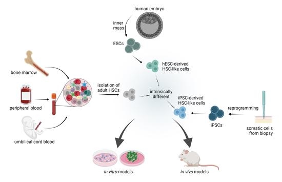
3.1.1. Adult Hematopoeitic Stem Cells

Adult HSCs are primary cells isolated from patients and are a powerful tool to study disease etiology at the molecular level. They can be enriched from PB, BM or umbilical cord blood (UCB) using CD34+ as surface marker [65]. Although all these sources of cells contain CD34+ cells, they are evolving in different physiological niches. PB CD34+ cells are collected utilizing mobilizing agents such as granulocyte colony-stimulating factor (G-CSF) which may have repercussions on cell function, compared to the BM CD34+ cells isolated directly from the BM interstitial space [66]. In general, UCB has the highest percentage of CD34+ cells in the absence of mobilizing agents, but a drawback is the limited volume of cord blood after birth. Therefore, important differences are expected to be found within the composition of CD34+ cell subsets based on their isolation source (extensively review by Tajer et al. [67]). Overall, CD34+ cells from UCB retain a higher fraction of uncommitted progenitors compared to PB and BM [68]. BM CD34+ cells have more committed progenitors such as MLP/BLP compared to PB [69]. These findings are in agreement with clinical data that shows a lower number of UCB CD34+ cells is needed for engraftment; however, the complete immune reconstitution is slower compared to other sources of CD34+ cells due to the presence of uncommitted progenitors [70,71,72,73]. Similarly, it may explain why PB CD34+ cells engraft faster than BM CD34+ cells in a clinical setting [74].
A number of studies have also shown that T cells are present in higher numbers in CD34+ enriched products from PB compared to BM or UCB (reviewed by Panch et al. [75]). In addition, the proportion of regulatory T cells (Tregs) was lower in PB CD34+ cells compared to BM CD34+ cells [76]. This may explain the result of a meta-analysis comparing BM or PB CD34+ cells for allogenic HSC transplantation in 1521 adults with acute leukemia: transplanted PB CD34+ cells were associated with a higher rate of GvHD [77]. Mature donor T cells transplanted within the PB subsets can be activated by the post-transplantation microenvironment and trigger significant damage to the host tissues [78]. In contrast, Tregs play an important role to maintain immunological self-tolerance and immune homeostasis [79]; and when infused together with conventional T cells in patients who received an allogenic CD34+ graft, Tregs successfully suppressed GvHD [80]. Of note, a recent study by the Kupper group is adding complexity to the GvHD interplay; they have identified that tissue-resident memory T cells from the host survive the pre-conditioning and contribute to the GvHD in allogenic HSC transplantation [81].
The heterogeneity of HSCs has a direct impact for disease modelling and translational medicine in terms of engraftment and reconstitution capacity. Moreover, the heterogeneity of HSCs can also affect transduction efficiency. Genovese et al. showed how different CD34+ subsets isolated from PB had differential lentiviral transduction efficiencies, with primitive progenitor subset (CD34+ CD133+ CD90+) having the lower transduction efficiency compared to early progenitors (CD34+ CD133+ CD90) and committed progenitors (CD34+ CD133) [82]. Recently, Radtke et al. performed a side-by-side comparison of most commonly enriched HSC subsets: CD34+ CD133+, CD34+ CD38low/− and CD34+ CD90+ [83]. They used phenotypic, transcriptional and functional readouts to characterize each subset and identified the CD34+CD90+ subset as having the least committed progenitors. The CD34+CD90+ subset was also the smallest sub-population of about 5% of CD34+ cells. This highly enriched population had the potential for short and long-term multilineage engraftment [83,84]. In this study, the CD34+CD90+ were successfully transduced with a high efficiency (in contrast to the previous study [82]), indicating this subset as a reasonable candidate for gene therapy and transplantation applications.
In vivo studies further showed a bias towards certain lineages from CD34+ HSC clones, including myeloid, lymphoid, megakaryocytic lineages [85,86], while others showed a balanced reconstitution [87,88]. Similar results with myeloid-biased HSCs and lymphoid-biased HSCs (mostly T cells) were found in human clinical settings [89]. The authors tracked CD34+ cells (isolation from BM and PB) over time in patients who received gene therapies for Wiskott-Aldrich syndrome (WAS), sickle cell disease (βS/βS) and thalassemia (β0/βE). The study also revealed a highly polyclonal nature with about 50–200 000 HSCs contributing to mature blood cells. Although the cohort study was small, longitudinal tracking did provide considerable information regarding the hierarchies of human hematopoiesis. The polyclonal nature of HSC clones in human was also demonstrated in a recent study by tracking mitochondrial mutations within CD34+ cells collected from patients [90]. The contribution of HSCs from BM to mature blood cells was detected in B, T, NK and myeloid cells. Interestingly, with aging it was shown that BM HSCs become biased toward myeloid lineages in humans [91].
The complexity of lineage bias is highlighted by a recent study from the Weissman group, where HSC clones were tracked using barcodes in mice [92]. They showed important differences in lineage commitment between different pre-transplantation conditioning regimens: irradiation and anti-ckit antibody treatment (i.e., ACK2) of Rag2−/− IL-2Rγ−/− mice. Three distinct groups were observed in conditioned animals representing balanced, myeloid- and lymphoid-biased subsets, while no lineage bias was observed when conditioning was absent. The underlying mechanism behind the various biases remains unclear and the characterization of each of these subsets challenging.
These studies highlight the need for a robust identification, characterization and enrichment of HSC clones with defined and optimal properties. In addition, there is a need to define a better consensus around the markers used to define the different hematopoietic populations, which currently varies greatly among studies. It will subsequently reduce the variability seen in pre-clinical and clinical studies.

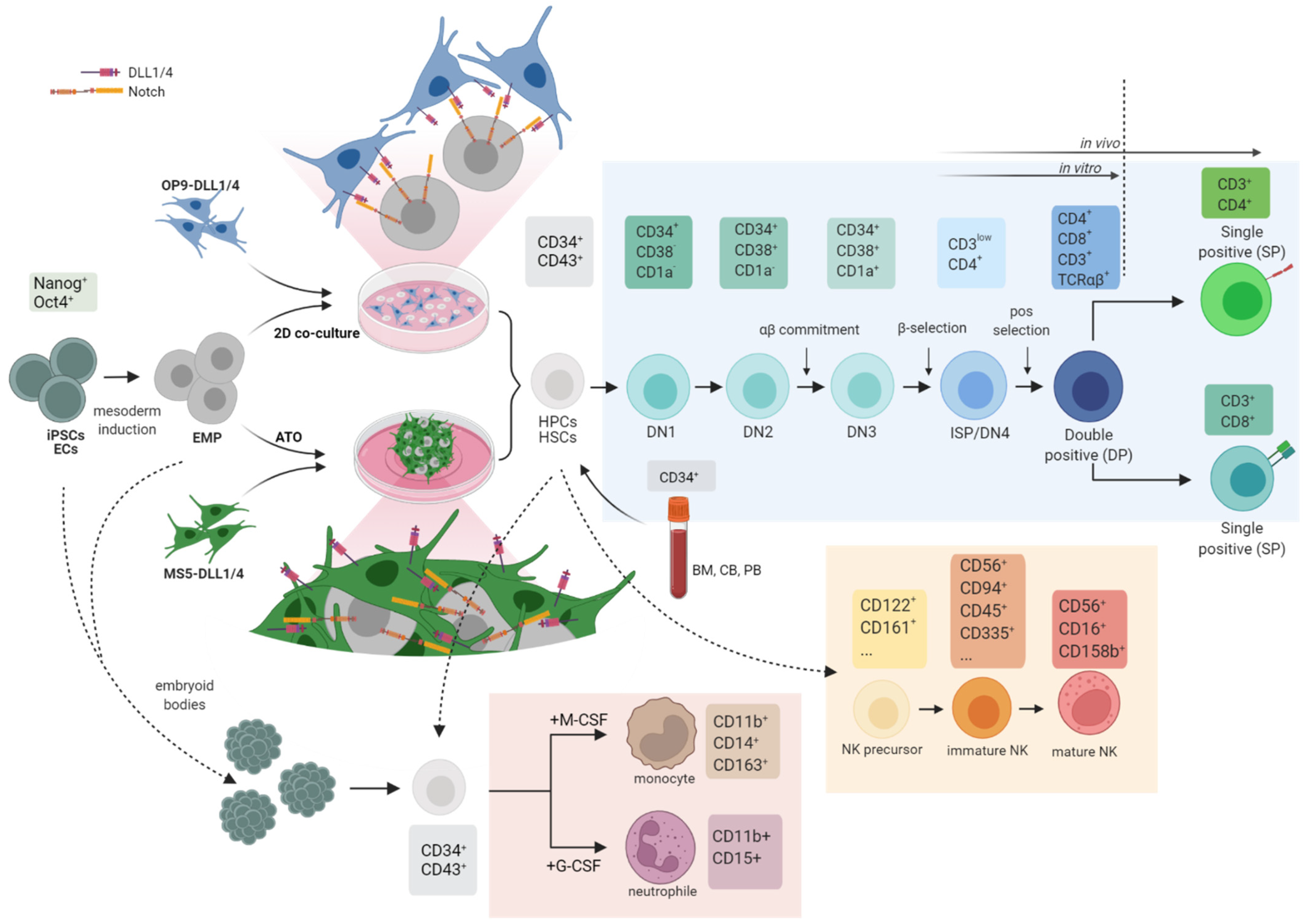
3.1.2. Differentiation Potential and De Novo Generation of HSCs from iPSCs

De novo generation of HSCs is highly interesting for regenerative medicine as it would provide an unlimited source of cells for clinical applications. Indeed, long-term expansion of ex vivo isolated human HSCs is still challenging, in contrast to murine expansion that has now been well established [93]. This is mostly the results of important mechanistic differences between mouse and human development and adult hematopoiesis. In addition, generating HSCs from autologous iPSCs has addresses the issue of GvHD. However, the current unknowns around the HSC origin, heterogeneity and plasticity makes the establishment of a defined differentiation protocol for PSCs challenging (i.e., iPSC, ESCs and adult CD34+ stem cells). Two main strategies are possible to differentiate stem cells along the hematopoietic lineage: (1) directed differentiation that sequentially uses cell-extrinsic factors in medium or from co-cultures, and (2) direct conversion and forward programming that use defined transcription factors introduced intrinsically. In some case, both strategies can be combined. Whereas the embryonic hematopoietic waves are temporally and spatially separated in vivo, the in vitro complete recapitulation of these complex patterns, so that they coexist, has proven difficult. Early differentiation protocols aiming to generate HSCs were more resembling the first transient HSC-independent wave of embryonic hematopoiesis rather than definitive hematopoiesis. One additional challenge is the lack of specific markers that could discriminate all the specific cell types from the different hematopoietic waves [94,95].
The use of more defined culture conditions has enabled significant progress towards more reliable production of hematopoietic cells. Analysis of signaling pathways have pushed our knowledge forward. For instance, recent studies have shown that the modulation between activation and repression of signaling pathways can directly impact on the production of hematopoietic progenitors resembling either primitive extra-embryonic progenitors or definitive-like hematopoietic progenitors [96,97,98]. Primitive hematopoietic cells (defined as CD235a+) are temporally dependent on the activin-nodal pathway, while the production of definitive-like progenitors (expressing RUNX1) is not [98]. Activation of the Wnt pathway while blocking the TGFβ pathway triggers the production of definitive-like progenitors marked also by upregulation of HOXA genes [97]. In addition, the extensively debated use of the HE as an intermediate step for iPSC-derived hematopoietic cultures has considerably improved the differentiation outcomes [99]. However, the scientific community agrees that these PSC-derived cells via directed differentiation, so far, are generating HPCs rather than HSCs. These PSC-derived HSC-like cells have failed to show multilineage engraftment capacity and multilineage differentiation abilities in vitro [100]. Overexpression of transcription factors is one approach to improve the HSC differentiation; forced expression of FOSB, GFI1, RUNX1 and SPI1 in endothelial cells could reconstitute primary and secondary mouse recipients [101]. Other transcription factors with differentiation potential were identified by Doulatov et al. as ERG, HOXA9, RORA, SOX4 and MYB, which were used in CD34+CD45+ myeloid precursors to generate multilineage progenitors in vivo [102]. However, a bias towards myeloid lineages was observed and only short-term engraftment was achieved. Interestingly, identification of the epigenetic regulator EZH1 by Vo et al. may improve the current differentiation protocols [103]. They showed how EZH1 is an active epigenetic repressor of hematopoietic multipotency in early mammalian embryo, therefore being an attractive target to enhance differentiation. Moreover, the knockdown of EZH1 unlocked lymphoid potential in differentiating ESCs in vitro. Sugimura et al. performed a large screen for transcription factors and identified ERG, HOXA5, HOXA5, HOA10, LCOR and RUNX1 as sufficient to generate HSC-like cells from isolated HE. These derived-HSC-like cells could engraft in primary and secondary recipients and lead to the reconstitution of myeloid, B- and T-cell lineages [104]. Using single-cell sequencing, Fidanza et al. compared iPSC-derived HSC-like cells with their in vivo counterparts and identified small populations of cells that were clearly different between the two groups [105]. They hypothesized that these differences in gene expression are responsible for the functional differences observed between in in vitro HSC-like cells and their in vivo counterparts. One additional consideration regarding the use of iPSCs is the iPSC epigenome. For instance, Cypris et al. compared the epigenetic profile of iPSCs differentiated towards hematopoietic lineages with adult CD34+ stem cells isolated from UCB, PB and BM [106]. Principal component analysis of DNA methylation profiles clearly showed separated clustering of adult CD34+ stem cells and iPSC-derived HSC-like cells.
Detailed reviews of differentiation steps and variations among protocols to generate HSCs and blood cells from PSCs (ESCs and iPSCs) have been reviewed elsewhere [8,42,100,105,107]. Overall, generated cells from iPSCs can be differentiated further towards a number of hematopoietic lineages, however, with a much lower yield compared to the HSCs obtained from isolated CD34+ cells from patients. Importantly, terminally differentiated hematopoietic cells from PSCs exhibit important differences and behave differently compared to their counterpart in vivo; and compared to their primary-derived or embryonic counterparts (discussed below). Finally, clinical translation ideally requires the development of protocols using transgene-free cells, which remains currently out of reach.

3.2. In Vitro Inborn Errors of Immunity (IEI) Disease Models

The manipulation of HSCs combined with technological advances in gene editing is an attractive strategy to study human immune cell development and generate relevant IEI disease models. In vitro IEI disease models have, so far, focused on differentiation towards T cells, NK cells, granulocytes and monocytes (Figure 2). Severe combined immunodeficiency (SCID) is characterized in patients by a lack of T cells, accompanied in some cases by a B and NK-cell defect (SCID pathophysiology reviewed by Kumrah et al. [108]). Briefly, a number of genetic aberrations cause SCID: mutations in the interleukin-2 receptor common γ-chain (IL-2Rγ) or JAK3 lead to a TB+NK phenotype in patients (i.e., lack of T and NK cells, but presence of B cells). Mutations in the adenosine deaminase (ADA) gene leads to a TBNK phenotype, while mutations in the RAG1 or RAG2 genes lead to a broader range of phenotypes.
Studies aiming to model SCID in vitro have focused mainly on using stem cells (adult CD34+ stem cells or iPSCs) that are differentiating towards lymphoid lineages [109,110,111,112,113]. Chang et al. reprogrammed primary keratinocytes obtained from SCID patients with a JAK3 mutation [109]. They differentiated iPSCs towards T cells using the OP9-DLL4 co-culture system and identified an early block in the development of the JAK3-SCID iPSCs. After 14 days, only small populations of CD7+CD16CD56 T cells and CD7+CD16+CD56+ NK cells were present in the JAK3-iPSCs compared to iPSCs from a healthy donor, and JAK3-mutant cells did not differentiate further into CD4+/CD8+ double-positive (DP) or single-positive (SP) T cells. The differentiation arrest at or before the DN2 stage correlates to in vivo studies [114] and could be lifted by restoring the mutation using CRISPR/Cas9. Similarly, Menon et al. looked at SCID-X1 iPSCs carrying a mutation in the IL-2Rγ, as well as genetically corrected iPSCs using TALEN along the T and NK-cell differentiation paths using the OP9-DLL1 system [110]. They could successfully restore the T and NK-cell differentiation of patient-derived cells to a level similar to the control iPSC line. Here, they showed the presence of CD4+CD8+ DP but no data regarding the presence of TCRαβ was provided. In contrast to the study from Chang et al., Menon et al. included ESCs as an additional positive control. Interestingly, the differentiation efficiencies towards CD16+CD56+ NK cells dropped in iPSCs compared to ESCs, highlighting the differences between the two types of stem cells.
The impact of null RAG2 mutation could be assed in vitro by using RAG2-SCID iPSCs on an OP9-DLL1 system [111]. The strongest block in T-cell development was observed from early CD7CD5 to CD4+CD8+ cells. NK-cell development was also affected and an increase in CD7CD56+CD33+ cells could be observed. Almost no CD4+CD8+ DP could be observed in the RAG2-SCID iPSCs (~2%) and no TCRδ rearrangement could be observed for CD7+CD56 sorted cells. The low numbers of CD4+CD8+ DP in RAG2-iPSCs were an aberrancy because they did not have TCR rearrangements, consistent with the absence of RAG1-RAG2 activity. Themeli et al. also generate a repaired line, which had restored capacity to induce T-cell differentiation and TCR rearrangement. NK-cell development could also be rescued [111]. RAG2 loss of function was also investigated in the 3-dimensional artificial thymic organoid (ATO) culture system [113]. ATO are co-cultures of murine stromal cells expressing DLL1 or 4 with CD34+ cells and better mimic the 3-dimensional microenvironment [115,116]. Indeed, the T-cell development on the OP9-DLL1 and -DLL4 2-dimensional stromal layer is quite poor and at best leads to varying percentages (10–70%) of DP cells and virtually no SP cells [109,110,111]. Using an ATO system, Gardner et al. obtained higher efficiencies with ~90% CD4+CD8+ DP and subsequently ~60% CD3+ TCRαβ+ cells [113]. RAG2-iPSCs generated in the ATO system had a similar block in T-cell development between CD7CD5 DN and CD4+CD8+ DP; however, with a higher percentage of remaining RAG2 CD4+CD8+ DP with no TCR rearrangement (~40%). These residual CD4+CD8+ DP cells could also be observed in iPSCs carrying various RAG1 mutations (up to ~10%) [112]. In this study, Brauer et al. used an OP9-DLL4 system with three RAG1 patient-specific iPSC lines: two were associated with a SCID clinical phenotype and one with the Omenn syndrome. In both SCID and OS cells, the block was described at an early stage in T-cell development, but with significant development of DP cells. This is in contrast to other reports in which CD34+ cells from patients were used, in such cases recombinase deficiency results in a complete block at the CD5+CD7+ DN stage without any CD4+CD8+ DP cells developing [117]. We previously have shown that hypomorphic RAG1 CD34+ patient cells transplanted into humanized mice in the context of preclinical gene therapy development for RAG1-SCID generated some CD4+CD8+ DP cells without CD3 [118], demonstrating that hypomorphic mutations and full null mutants in recombinase activity result in different developmental arrests, i.e., a full block at the CD5+CD7+ DN stage and a later arrest at CD3DP stage in an hypomorphic situation. Overall, it remains unclear whether these RAG1/2 CD4+CD8+ DP may be an artefact of in vitro iPSC based system in which T-cell development follows unusual pathways or potentially pathways that resemble fetal lymphopoiesis or if the RAG1/2 mutations would be hypomorphic and not resulting in a total loss of function of the recombinases.
In vitro models focusing on lymphoid lineage differentiation were also used to look at specific defects in T and NK-cell development due to mutation in the WAS gene [119]. WAS encode for a protein (known as WASp) that is known to activate actin polymerization required to generate actin filaments. WASp is specific to hematopoietic lineages and plays an important role in the intercellular synaptic communications. WASp deficiency is referred to as Wiskott-Aldrich syndrome and is characterized by a defect in T cells and NK cells [120]. Laskowski et al. generated iPSCs from WAS patients and differentiated them using the OP9-DLL1 co-culture system [119]. WAS-iPSCs, genetically corrected WAS-iPSCs and control ESCs progressed from CD34+CD43 endothelial cells to CD34+CD45+ HSC-like cells with the same efficiencies, indicating that the transition via the HE is not altered by the WAS mutant. CD56+CD7+ and CD56+CD94+ NK cells were significantly reduced in WAS-iPSCs compared to ESCs and corrected iPSC line. Interestingly, the few WAS-mutant NK cells showed a functional defect with upregulated interferon-γ (IFNγ) and tumor necrosis factor α (TNFα) secretion. WAS-mutant iPSC showed a defect in the generation of CD4+CD8+ DP and CD3+TCRαβ cells. Of note, the expression of CD34+ during T-cell differentiation from the control iPSCs remained high, as opposed to T-cell differentiation from CD34+ cells isolated from patients. The persistence of CD34 expression is indicating a more immature phenotype for iPSC-based T-cell differentiation. In addition, none of the studies differentiating iPSCs have included primary adult CD34+ stem cells as positive control for their differentiation.
IEI disease modelling with a focus on innate cells has been possible. For instance, chronic granulomatous disease (CGD) is caused by mutations in the gene coding for NADPH oxidases that are involved in the respiratory burst in phagocytic leukocytes [121]. As a result, CGD patients are suffering from recurrent bacterial and fungal infections due to a defective phagocytic function. Several studies have looked at modeling CGD in vitro using CGD-iPSCs [122,123,124,125,126,127,128]. Utilizing different genetic corrective strategies, such as zinc-finger targeted insertion [122,129], TALEN [124,127], BAC transgenesis [126], or CRISPR/Cas9 [125,128], it was possible to correct CYBB and NOX2 genes encoding for one of the NADPH oxidase subunits. NADPH oxidase function could be successfully restored and using bacterial killing assays, they demonstrated the rescued phagocytic function of the iPSC-derived macrophage or granulocyte. Differentiation from hPSCs towards macrophages relies on a standard protocol using IL-3 and M-CSF [130]. Modification and refinement using Stem Cell Factor (SCF), VEGF or BMP-4 has further optimized the differentiation of various subset of macrophages [131,132,133,134]. Lachmann et al. showed how either granulocytes or macrophages could be generated from iPSCs by either using M-CSF (for macrophages) or G-CSF (for granulocytes) [134]. However, the question remains whether the functionality in vitro and in vivo of iPSC-derived macrophages resemble adult macrophages or rather embryonic macrophages from the primitive hematopoietic waves. Data from Takata et al. and Buchriser et al. are pointing out that iPSC-derived macrophages may resemble macrophages arising from the YS-hematopoiesis [132,133].

3.3. In Vivo Disease Models: Towards a Fully Hummanized Immune System

In vitro differentiation capacity towards hematopoietic lineages from adult CD34+ stem cells, ESCs or iPSCs remains limited compared to their in vivo counterparts. For example, the in vitro T-cell development protocol can generate CD4+CD8+ DP (usually in low numbers) that have been positively selected for their TCR; however, no studies until now have recapitulated negative selection in vitro, a process essential for immune tolerance. In addition, rarely are B-cell development and other alternative lineages looked at in IEI in vitro models, despite the development of in vitro protocol [136]. In vivo immunodeficient mouse models and subsequent humanized mouse models offer the possibility to study the human immune system to the extent that, so far, isn’t possible in vitro.
There is a large number of immunodeficient mouse models that have been developed since the establishment of the nude mice by Flanagan in the 60s [137,138]. Nude mice phenotype relies on the disruption in the Foxn1 gene resulting in the lack of a thymus and subsequent impairment in the development of the adaptive immune system. Although nude mice have been extensively used as a model for tumor growth, they are failing to reconstitute the human immune system due to the presence of B cells and innate cells, such as highly active NK cells. As an alternative, Scid mice carry a mutation in the Prkdc gene—a protein necessary for non-homologous end joining of ds-DNA—, resulting in impaired B and T-cell development [139]. Scid mouse models are also considered to have B and T-cell leakiness (i.e., spontaneous generation of T and B cells), to be very sensitive to radiation, and have low engraftment of transplanted human PBMC and HSCs [140,141]. In contrast, the Rag1 or Rag2 deficient mice do not show this immunoglobulin leakiness or sensitivity to radiation [142,143]. Deletion of the Rag1/2 genes cause an arrest in the rearrangement of the T-and B-cell receptors and therefore leads to a lack of T and B differentiation [142,143]. Rag1−/− and Rag2−/− mice have been used as pre-clinical models for Rag1 and Rag2 gene therapies, as they exhibit similar phenotypes to patients suffering from SCID [118,144,145]. In a study from Garcia-Perez et al., BM CD34+ cells from Rag1−/− mice were isolated and genetically corrected via lentiviral transduction of codon-optimized Rag1 gene under an MND promoter [118]. Rag1−/− mice that received the transplantation of Rag1-transduced CD34+ cells had a reconstitution of the T- and B-cell development. This preclinical study and other previous work were the drivers of the NCT04797260 clinical trial for the treatment of RAG1-SCID, which is currently recruiting patients in Europe for corrective RAG1 gene therapy [146].
Although Rag1−/− mice haven been of great use to establish gene RAG1 therapy, they do not cover the high heterogeneity of the disease. Indeed, Rag1 deficiency in human is caused by mutations in the Rag1 genes and can be classified in 4 groups: (1) mutations that destabilize the tertiary structures such as for Zinc binding, (2) mutations important for DNA binding, (3) catalytic RNase H-like domain and (4) mutations involved in the RAG1/RAG2 interaction [147]. This heterogeneity in mutations results in a broad spectrum of clinical and immunological phenotypes [148]. Tackling this issue, Ott de Bruin et al. generated a number of RAG1 mouse models using CRISPR/Cas9 targeting specific regions of the RAG1 genes [149]. For instance, they targeted the region of RAG1 around residues R838-N852, which is a region with unknown functions. Sever phenotypes in mice with in frame deletion from 832–877 were observed with a block in B- cell and T-cell development at the pre-pro/pro-B cells and DN3, respectively. Interestingly, in silico modeling and algorithms led to contradictory predictions for the specific mutation H836Q. In vivo modelling of the H836Q mutation showed intact B- and T-cell development, while in silico modeling predicted deleterious effects on Rag1 function. These data highlight the need for functional in vivo assays to confirm or refute in silico models.
The Rag1−/− and Rag2−/− mouse models can also retain a high level of NK-cell activity and have been shown to have an overall limited engraftment of human HSCs compared to more recent models [142,143,150]. For instance, one important breakthrough in the field occurred with the development of the IL-2Rγ−/− mouse models [151]. The interleukin-2 receptor common γ-chain is part of a dimer forming receptors for several cytokine interleukins and is present on developing immune cells [152]. The targeted knockout of IL-2Rγ−/− impairs the development of NK cells in addition to that of T and B cells [153]. In humans, X-linked SCID (SCID-X1) is caused by a mutation in the IL-2Rγ and results in presence of very low amount of T and NK cells, as well as lack of response to the common γ-chain cytokines, while B cells are not affected. Due to these similarities, the IL-2Rγ−/− mouse background has been used to assess in vivo safety for corrective gene therapies for IL-2Rγ mutations [154,155]. For example, Poletti et al. used a IL-2Rγ−/− Rag2−/− mouse model as preclinical model for a gene therapy for X-linked SCID (SCID-X1) patients [154]. Of note, combining IL-2Rγ−/− mutation with a Rag1−/− or Rag2−/− background is advantageous, as IL-2Rγ−/− single knockout mice have been described to be leaky.
Another major breakthrough was the development of the non-obese diabetic NOD.scid.IL-2Rγ−/− (NSG) mouse models [156,157]. While NOD mice have a strong predisposition to develop spontaneous autoimmune diabetes, the NOD-Scid mice do not develop diabetes, but have a decreased NK-cell activity and a higher engraftment of human HSCs. The combination of these features with the IL-2Rγ−/− mice makes the NSG mice a powerful model to look at the human immune system in vivo with applications for personalized medicine and translation of immune-based therapies. NSG mice do not show any leakiness towards B and T cells [158], and enable long-term studies as they are surviving beyond 15 months without developing thymic lymphomas [159]. In several studies, immune reconstitution following the human developmental stages from transplanted HSCs could be observed in the NSG mice for both T and B cells, as well as myeloid and plasmacytoid dendritic cells [156,157,160,161,162]. Importantly, NSG mice humanized with CD34+ cells from patients carrying mutations are a powerful alternative to look at specific developmental defects, as mouse models with the corresponding mutations do not always recapitulate the extent of the disease phenotype. For instance, the IL-7R−/− mouse model has B and T-cell deficiencies [163], whereas mutations in the IL-7 receptors in human leads to a T-cell deficiencies without B- or NK-cell developmental arrest [164]. NSG mice were used as a model to look specifically at T-cell development arrest caused by the transplantation of CD34+ cells obtained from SCID patients [117]. The authors showed that T-cell arrests caused by IL-7 receptor α, as wells IL-2rγ, were occurring in immature thymocytes much earlier than expected compared to studies with corresponding genetic mouse models. In 2018, Tan et al. used NSG mice to look at engraftment capacity of iPSC-HSC-like cells and compared the outcome to adult CD34+ stem cells [165]. They used the transient expression of the fusion transcription factor MLL-AF4 during the differentiation of iPSCs towards hematopoietic lineages to improved differentiation efficiencies. The transplantation of from iPSC-HSC-like cells into NSG mice reestablished myelopoiesis and lymphopoiesis without lineage bias. Although both adult CD34+ stem cells and iPSC-HSC-like cells achieved long-term engraftment, only the iPSC-HSC-like cells were prone to leukemic transformation. Whether this is due to the use of MLL-AF4, which is an oncogenic fusion protein in acute myeloid leukemia and B-cell acute lymphoblastic leukemia or due to the nature of iPSCs remains unclear. Additional arguments that have been proposed are that there is a high oncogenic risk from genetic and epigenetic aberration that a due to reprogramming; and that NSG mice provide a permissive environment, which combined would allow transformation of iPSC-derived cells [166,167].
From the NSG mouse background, a number of new mouse models were developed with improved engraftments and lineage reconstitution properties. For instance, the NSG-SGM3 have a triple knock-in for the human stem cell factor (SCF), granulocyte-macrophage colony-stimulating factor (GM-CSF) and interleukin-3 (IL-3), and the MISTRG mouse model that have the knock-in for macrophage colony-stimulating factor (M-CSF), GM-CSF, IL-3, thrombopoietin and signal regulatory protein alpha (SIRPα) [168,169,170]. However, the NSG, NSG-SGM3 and MISTRG mice still require pre-conditioning before robust humanization with CD34+ cells. Different inflammatory pathways are involved during pre-conditioning that can damage the BM microenvironment, the extent of which is not yet fully understood [171]. The successful humanization with CD34+ cell without pre-conditioning was demonstrated in the NOD,B6.SCID IL-2rγ−/− kitW41/W41 (NBSGW) mice by Hess et al. [68]. NSGW41 mice are developed from the NSG by addition the kitW41/W41 allele, which results in the competitive advantages for the human transplanted cells [172]. Hess and colleagues also showed how the source of CD34+ cells (BM, PB or UCB) have a direct impact on the immune reconstitution in NBSGW mice, which is an important experimental consideration. In a follow-up study, Adigbli et al. demonstrated that the NBSGW mice could develop LT-HSC population from engrafted human UCB CD133+ cells without pre-conditioning [173]. In 2021, Coppin et al. generated mice expressing human IL-7 on a NSGW41 background mice, termed NSGW41hIL7 [174]. The expression of human IL-7 in these humanized mice leads to improved human T-cell development compared to the NSGW41 mice.
Although the NSG mice and NSG-derived models are considerably improved models for studying the humanized system, one must have the awareness that humanization can have unintended immunological consequences, as described by two recent studies from Blümich et al. [175] and Janke and colleagues [176]. From the examination of a large cohort of humanized and non-humanized NSG mice with CD34+ cells, Blümich et al. raised some important considerations, such as the poor correlation between engraftment level in the humanized mice and the presence of human circulating CD45+ cells in the PB [175]. Subsequently, one can ask weather measuring engraftment based on PB is a reliable measure for such model. The study also showed that background lesions present in the non-humanized mice were exacerbated by the humanization with CD34+ cells, which indicate some level of GvHD [177]. Janke et al. looked at large cohort of NSG-SGM3 mice humanized or non-humanized with CD34+ cells [176]. They observed 3 main features (1) mast cell hyperplasia, (2) eosinophil hyperplasia and (3) hemophagocytic lymphohistiocytosis/macrophage activation syndrome (HLH/MAS)–like disease. Importantly, naïve and humanized NSG-SGM3 mice exhibited important differences in these three features.
Therefore, a number of alternatives to immunodeficient mouse models, including rats, pigs, dogs, non-human primates or zebrafish [178,179,180]. Overall, larger animals have a longer lifespan than smaller animal models. Longer lifespans may be resourceful for disease modeling and assessing therapies and treatment options under pre-clinical settings, however it has a direct effect on research in term of costs and research speed (i.e., breeding time). Rats can be up to 10 times larger than mice, thereby can receive transplants of larger sizes. As in humans, rats express CD4 and/or CD8 on macrophages and MHC class II on endothelial cells, which is not the case in mice, mimicking therefore closer human physiology. Interestingly, no study has reported a shorter lifespan for immunodeficient rats, in contrast to immunodeficient mice such as NOD-derived models, which have a high incidence of spontaneous thymic lymphoma [181]. A number of immunodeficient rat models exists with mutations in Foxn1, Rag1, Rag2, Prkdc, Il2rg or IgM; and could be humanized via the transplantation of PBMCs or enriched CD34+ cells [182,183,184] (review extensively by Adigbli et al. [178]). Pigs have also been used to generate immunodeficient lines carrying either spontaneous or induced mutations in Rag1, Rag2, Artemis or Il2rg (review extensively by Boettcher et al. [179]). As showed by Dawson et al. [185], pigs have fewer unique immunological genes compared to human or mice, and could better replicate and predict clinical outcomes in humans, which in turn have the potential to improve the clinical translation [186]. While immunodeficient pigs can accept a number of xenografts, immune humanization in pigs remains a constant challenge and further work is required [187]. The use of canine models for HSC transplantation have been recently reviewer by Graves et al. [180].

4. Future Perspectives

The development of reliable disease models is challenging, as it requires mimicking the human pathophysiology in vitro or in vivo (Table 1). For decades, in vivo models have provided considerable insights into physiology and disease mechanisms of a large number of IEI. Important breakthroughs related to the humanization of the immune system in mouse models have addressed some of the species-specific limitations of classic mouse models, bridging the gap between animals and humans. There is no denying that data and insights provided by in vivo models have been and are essential to the development of novel therapies and cures for human diseases; however, as Justice et al. simply explained: “a model is simply that: it is not the human” [188]. Researchers must have deep knowledge and understanding about the large variety of in vivo models in order to adequately answer specific scientific questions. In parallel, enormous efforts are invested in the field of in vitro models to move from classic monolayer cultures to 3-dimensional human co-culture systems that aim to provide platforms to study human-specific developmental processes and disease mechanisms in greater details. This includes the technical advances in perfused microphysiological systems, so called organ-on-a-chip technology (OoC), stem cell technology and gene therapy. Although in vitro models for IEI are still in their infancy, OoC and 3-dimensional culture systems are gaining considerable momentum in other fields and may significantly impact the immunology field in the coming years. For instance, some progress has been made with the use of artificial thymic organoids (ATO) to study T-cell development. Bosticardo et al. used ATO to confirm that severe T-cell lymphopenia from patients with DiGeorge syndrome was caused by extrinsic stromal defects (i.e., thymic stromal cells) [189]. Such in vitro screening methods are highly valuable to determine the nature of the T-cell deficiency and to direct treatment options at an early onset. ATO could be used to address the developmental blocks in T-cell development caused by mutations in genes such as RAG1 and RAG2, ultimately giving important insight into the nature of the deficiency (i.e., hypomorphic situation or null mutations). Further optimization of the ATO system is required to switch from mouse stromal cell lines (such as MS5-DLL1/4) used to mimic the thymus stromal parts to a human source of cells that would better recapitulate the interactions between thymic epithelial cells and developing thymocytes. Indeed, in the study from Bosticardo et al., the correct in vitro development of TCRαβ+CD3+ from CD34+ cells isolated from a patient with ADA deficiency confirmed the extrinsic, metabolic origin of the deficiency in patients, highlighting that MS5-DLL4 cells in the ATO restore ADA activity and support T-cell development. Thus, the ATO only reveals intrinsic development defects. Efforts are currently invested in deriving thymic epithelial cells from hPSCs that can support human T-cell development in vitro and in vivo [190,191,192,193]. This is not only highly beneficial to improve current IEI models with genetic defects present on the hematopoietic counterpart, but also for immune deficiencies with extra-hematopoietic defects such as DiGeorge syndrome.
To conclude, the success of disease modelling relies on the optimal combination of independent models, both in vitro and in vivo, as well as a critical interpretation of results and careful comparison to data of patients included in clinical studies.

Author Contributions

A.Z., K.C.-B., K.P.-O. and F.J.T.S. discussed and wrote the manuscript. All authors have read and agreed to the published version of the manuscript.

Funding

A.Z. was funded by the Swiss National Science Foundation (SNSF). Work in the Staal laboratory is supported in part by a ZonMW E-RARE grant (40-419000-98-020) and EU H2020 grant RECOMB (755170-2) and has received funding from the European Union Horizon 2020 research and innovation program.

Institutional Review Board Statement

Not applicable.

Informed Consent Statement

Not applicable.

Data Availability Statement

Not applicable.

Acknowledgments

We thank Veronica Della Chiara for carefully reading the manuscript and members of the Staal laboratory for helpful discussions.

Conflicts of Interest

The authors declare no conflict of interest.

References

  1. Tangye, S.G.; Al-Herz, W.; Bousfiha, A.; Chatila, T.; Cunningham-Rundles, C.; Etzioni, A.; Franco, J.L.; Holland, S.M.; Klein, C.; Morio, T.; et al. Human Inborn Errors of Immunity: 2019 Update on the Classification from the International Union of Immunological Societies Expert Committee. J. Clin. Immunol. 2020, 40, 24–64. [Google Scholar] [CrossRef] [PubMed] [Green Version]
  2. Copelan, E.A. Hematopoietic Stem-Cell Transplantation. N. Engl. J. Med. 2006, 354, 1813–1826. [Google Scholar] [CrossRef] [PubMed]
  3. Duarte, R.F.; Labopin, M.; Bader, P.; Basak, G.W.; Bonini, C.; Chabannon, C.; Corbacioglu, S.; Dreger, P.; Dufour, C.; Gennery, A.R.; et al. Indications for haematopoietic stem cell transplantation for haematological diseases, solid tumours and immune disorders: Current practice in Europe, 2019. Bone Marrow Transplant. 2019, 54, 1525–1552. [Google Scholar] [CrossRef] [PubMed]
  4. Jagasia, M.; Arora, M.; Flowers, M.E.D.; Chao, N.J.; McCarthy, P.L.; Cutler, C.S.; Urbano-Ispizua, A.; Pavletic, S.Z.; Haagenson, M.D.; Zhang, M.-J.; et al. Risk factors for acute GVHD and survival after hematopoietic cell transplantation. Blood 2012, 119, 296–307. [Google Scholar] [CrossRef] [PubMed] [Green Version]
  5. Cring, M.R.; Sheffield, V.C. Gene therapy and gene correction: Targets, progress, and challenges for treating human diseases. Gene Ther. 2020, 1–10. [Google Scholar] [CrossRef]
  6. Eaves, C.J. Hematopoietic stem cells: Concepts, definitions, and the new reality. Blood 2015, 125, 2605–2613. [Google Scholar] [CrossRef] [Green Version]
  7. Seita, J.; Weissman, I.L. Hematopoietic stem cell: Self-renewal versus differentiation. WIREs Syst. Biol. Med. 2010, 2, 640–653. [Google Scholar] [CrossRef] [Green Version]
  8. Demirci, S.; Leonard, A.; Tisdale, J.F. Hematopoietic stem cells from pluripotent stem cells: Clinical potential, challenges, and future perspectives. Stem Cells Transl. Med. 2020, 9, 1549–1557. [Google Scholar] [CrossRef]
  9. Palis, J.; Robertson, S.; Kennedy, M.; Wall, C.; Keller, G. Development of erythroid and myeloid progenitors in the yolk sac and embryo proper of the mouse. Development 1999, 126, 5073–5084. [Google Scholar] [CrossRef]
  10. Lin, Y.; Yoder, M.; Yoshimoto, M. Lymphoid Progenitor Emergence in the Murine Embryo and Yolk Sac Precedes Stem Cell Detection. Stem Cells Dev. 2014, 23, 1168–1177. [Google Scholar] [CrossRef] [Green Version]
  11. McGrath, K.E.; Frame, J.M.; Fegan, K.H.; Bowen, J.R.; Conway, S.J.; Catherman, S.C.; Kingsley, P.D.; Koniski, A.D.; Palis, J. Distinct Sources of Hematopoietic Progenitors Emerge before HSCs and Provide Functional Blood Cells in the Mammalian Embryo. Cell Rep. 2015, 11, 1892–1904. [Google Scholar] [CrossRef] [Green Version]
  12. Müller, A.M.; Medvinsky, A.; Strouboulis, J.; Grosveld, F.; Dzierzakt, E. Development of hematopoietic stem cell activity in the mouse embryo. Immunity 1994, 1, 291–301. [Google Scholar] [CrossRef] [Green Version]
  13. Medvinsky, A.; Dzierzak, E. Definitive Hematopoiesis Is Autonomously Initiated by the AGM Region. Cell 1996, 86, 897–906. [Google Scholar] [CrossRef] [Green Version]
  14. Moore, M.A.S.; Metcalf, D. Ontogeny of the Haemopoietic System: Yolk Sac Origin of In Vivo and In Vitro Colony Forming Cells in the Developing Mouse Embryo. Br. J. Haematol. 1970, 18, 279–296. [Google Scholar] [CrossRef]
  15. Gekas, C.; Dieterlen-Lièvre, F.; Orkin, S.H.; Mikkola, H.K. The Placenta Is a Niche for Hematopoietic Stem Cells. Dev. Cell 2005, 8, 365–375. [Google Scholar] [CrossRef] [Green Version]
  16. Cumano, A.; Dieterlen-Lievre, F.; Godin, I. Lymphoid Potential, Probed before Circulation in Mouse, Is Restricted to Caudal Intraembryonic Splanchnopleura. Cell 1996, 86, 907–916. [Google Scholar] [CrossRef] [Green Version]
  17. Cumano, A.; Ferraz, J.C.; Klaine, M.; Di Santo, J.; Godin, I. Intraembryonic, but Not Yolk Sac Hematopoietic Precursors, Isolated before Circulation, Provide Long-Term Multilineage Reconstitution. Immunity 2001, 15, 477–485. [Google Scholar] [CrossRef] [Green Version]
  18. Samokhvalov, I.M.; Samokhvalova, N.I.; Nishikawa, S.-I. Cell tracing shows the contribution of the yolk sac to adult haematopoiesis. Nature 2007, 446, 1056–1061. [Google Scholar] [CrossRef]
  19. Tanaka, Y.; Hayashi, M.; Kubota, Y.; Nagai, H.; Sheng, G.; Nishikawa, S.-I.; Samokhvalov, I.M. Early ontogenic origin of the hematopoietic stem cell lineage. Proc. Natl. Acad. Sci. USA 2012, 109, 4515–4520. [Google Scholar] [CrossRef] [Green Version]
  20. Lee, L.K.; Ghorbanian, Y.; Wang, W.; Wang, Y.; Kim, Y.J.; Weissman, I.L.; Inlay, M.A.; Mikkola, H.K. LYVE1 Marks the Divergence of Yolk Sac Definitive Hemogenic Endothelium from the Primitive Erythroid Lineage. Cell Rep. 2016, 17, 2286–2298. [Google Scholar] [CrossRef] [Green Version]
  21. Wittamer, V.; Bertrand, J.Y. Yolk sac hematopoiesis: Does it contribute to the adult hematopoietic system? Cell. Mol. Life Sci. 2020, 77, 4081–4091. [Google Scholar] [CrossRef]
  22. Senserrich, J.; Batsivari, A.; Rybtsov, S.; Gordon-Keylock, S.; Souilhol, C.; Buchholz, F.; Hills, D.; Zhao, S.; Medvinsky, A. Analysis of Runx1 Using Induced Gene Ablation Reveals Its Essential Role in Pre-liver HSC Development and Limitations of an In Vivo Approach. Stem Cell Rep. 2018, 11, 784–794. [Google Scholar] [CrossRef]
  23. Taoudi, S.; Medvinsky, A. Functional identification of the hematopoietic stem cell niche in the ventral domain of the embryonic dorsal aorta. Proc. Natl. Acad. Sci. USA 2007, 104, 9399–9403. [Google Scholar] [CrossRef] [Green Version]
  24. Kumaravelu, P.; Hook, L.; Morrison, A.M.; Ure, J.; Zhao, S.; Zuyev, S.; Ansell, J.; Medvinsky, A. Quantitative developmental anatomy of definitive haematopoietic stem cells/long-term repopulating units (HSC/RUs): Role of the aorta-gonad-mesonephros (AGM) region and the yolk sac in colonisation of the mouse embryonic liver. Development 2002, 129, 4891–4899. [Google Scholar] [CrossRef] [PubMed]
  25. Rybtsov, S.; Sobiesiak, M.; Taoudi, S.; Souilhol, C.; Senserrich, J.; Liakhovitskaia, A.; Ivanovs, A.; Frampton, J.; Zhao, S.; Medvinsky, A. Hierarchical organization and early hematopoietic specification of the developing HSC lineage in the AGM region. J. Exp. Med. 2011, 208, 1305–1315. [Google Scholar] [CrossRef] [PubMed] [Green Version]
  26. Gao, S.; Liu, F. Fetal liver: An ideal niche for hematopoietic stem cell expansion. Sci. China Life Sci. 2018, 61, 885–892. [Google Scholar] [CrossRef] [PubMed]
  27. Ema, H.; Nakauchi, H. Expansion of hematopoietic stem cells in the developing liver of a mouse embryo. Blood 2000, 95, 2284–2288. [Google Scholar] [CrossRef] [PubMed] [Green Version]
  28. Yvernogeau, L.; Gautier, R.; Petit, L.; Khoury, H.; Relaix, F.; Ribes, V.; Sang, H.; Charbord, P.; Souyri, M.; Robin, C.; et al. In vivo generation of haematopoietic stem/progenitor cells from bone marrow-derived haemogenic endothelium. Nat. Cell Biol. 2019, 21, 1334–1345. [Google Scholar] [CrossRef] [PubMed]
  29. Beaudin, A.E.; Boyer, S.W.; Perez-Cunningham, J.; Hernandez, G.E.; Derderian, S.C.; Jujjavarapu, C.; Aaserude, E.; MacKenzie, T.; Forsberg, E.C. A Transient Developmental Hematopoietic Stem Cell Gives Rise to Innate-like B and T Cells. Cell Stem Cell 2016, 19, 768–783. [Google Scholar] [CrossRef] [Green Version]
  30. Fraser, S.T.; Ogawa, M.; Yu, R.T.; Nishikawa, S.; Yoder, M.C.; Nishikawa, S.-I. Definitive hematopoietic commitment within the embryonic vascular endothelial-cadherin+ population. Exp. Hematol. 2002, 30, 1070–1078. [Google Scholar] [CrossRef]
  31. Zovein, A.C.; Hofmann, J.J.; Lynch, M.; French, W.J.; Turlo, K.A.; Yang, Y.; Becker, M.S.; Zanetta, L.; Dejana, E.; Gasson, J.C.; et al. Fate Tracing Reveals the Endothelial Origin of Hematopoietic Stem Cells. Cell Stem Cell 2008, 3, 625–636. [Google Scholar] [CrossRef] [Green Version]
  32. Boisset, J.-C.; Van Cappellen, W.; Andrieu-Soler, C.; Galjart, N.; Dzierzak, E.; Robin, C. In vivo imaging of haematopoietic cells emerging from the mouse aortic endothelium. Nature 2010, 464, 116–120. [Google Scholar] [CrossRef]
  33. Ivanovs, A.; Rybtsov, S.; Anderson, R.A.; Turner, M.L.; Medvinsky, A. Identification of the Niche and Phenotype of the First Human Hematopoietic Stem Cells. Stem Cell Rep. 2014, 2, 449–456. [Google Scholar] [CrossRef] [Green Version]
  34. Bertrand, J.; Giroux, S.; Golub, R.; Klaine, M.; Jalil, A.; Boucontet, L.; Godin, I.; Cumano, A. Characterization of purified intraembryonic hematopoietic stem cells as a tool to define their site of origin. Proc. Natl. Acad. Sci. USA 2004, 102, 134–139. [Google Scholar] [CrossRef] [Green Version]
  35. de Bruijn, M.F.; Ma, X.; Robin, C.; Ottersbach, K.; Sánchez, M.J.; Dzierzak, E. Hematopoietic Stem Cells Localize to the Endothelial Cell Layer in the Midgestation Mouse Aorta. Immunity 2002, 16, 673–683. [Google Scholar] [CrossRef] [Green Version]
  36. Taoudi, S.; Morrison, A.M.; Inoue, H.; Gribi, R.; Ure, J.; Medvinsky, A. Progressive divergence of definitive haematopoietic stem cells from the endothelial compartment does not depend on contact with the foetal liver. Development 2005, 132, 4179–4191. [Google Scholar] [CrossRef] [Green Version]
  37. Yoder, M.C.; Hiatt, K.; Dutt, P.; Mukherjee, P.; Bodine, D.M.; Orlic, D. Characterization of Definitive Lymphohematopoietic Stem Cells in the Day 9 Murine Yolk Sac. Immunity 1997, 7, 335–344. [Google Scholar] [CrossRef] [Green Version]
  38. Yoder, M.C.; Hiatt, K.; Mukherjee, P. In vivo repopulating hematopoietic stem cells are present in the murine yolk sac at day 9.0 postcoitus. Proc. Natl. Acad. Sci. USA 1997, 94, 6776–6780. [Google Scholar] [CrossRef] [Green Version]
  39. Taoudi, S.; Gonneau, C.; Moore, K.; Sheridan, J.M.; Blackburn, C.C.; Taylor, E.; Medvinsky, A. Extensive Hematopoietic Stem Cell Generation in the AGM Region via Maturation of VE-Cadherin+CD45+ Pre-Definitive HSCs. Cell Stem Cell 2008, 3, 99–108. [Google Scholar] [CrossRef] [Green Version]
  40. Hadland, B.K.; Varnum-Finney, B.; Poulos, M.G.; Moon, R.; Butler, J.M.; Rafii, S.; Bernstein, I.D. Endothelium and NOTCH specify and amplify aorta-gonad-mesonephros–derived hematopoietic stem cells. J. Clin. Investig. 2015, 125, 2032–2045. [Google Scholar] [CrossRef] [Green Version]
  41. Rybtsov, S.; Batsivari, A.; Bilotkach, K.; Paruzina, D.; Senserrich, J.; Nerushev, O.; Medvinsky, A. Tracing the Origin of the HSC Hierarchy Reveals an SCF-Dependent, IL-3-Independent CD43− Embryonic Precursor. Stem Cell Rep. 2014, 3, 489–501. [Google Scholar] [CrossRef] [Green Version]
  42. Lange, L.; Morgan, M.; Schambach, A. The hemogenic endothelium: A critical source for the generation of PSC-derived hematopoietic stem and progenitor cells. Cell. Mol. Life Sci. 2021, 78, 4143–4160. [Google Scholar] [CrossRef]
  43. Mende, N.; Bastos, H.P.; Santoro, A.; Sham, K.; Mahbubani, K.T.; Curd, A.; Takizawa, H.; Wilson, N.K.; Göttgens, B.; Saeb-Parsy, K. Quantitative and molecular differences distinguish adult human medullary and extramedullary haematopoietic stem and progenitor cell landscapes. bioRxiv 2020. [Google Scholar] [CrossRef]
  44. Coppin, E.; Florentin, J.; Vasamsetti, S.B.; Arunkumar, A.; Sembrat, J.; Rojas, M.; Dutta, P. Splenic hematopoietic stem cells display a pre-activated phenotype. Immunol. Cell Biol. 2018, 96, 772–784. [Google Scholar] [CrossRef] [PubMed]
  45. Dor, F.J.; Ramirez, M.L.; Parmar, K.; Altman, E.L.; Huang, C.A.; Down, J.D.; Cooper, D.K. Primitive hematopoietic cell populations reside in the spleen: Studies in the pig, baboon, and human. Exp. Hematol. 2006, 34, 1573–1582. [Google Scholar] [CrossRef] [PubMed]
  46. Golden-Mason, L.; Curry, M.P.; Nolan, N.; Traynor, O.; McEntee, G.; Kelly, J.; Hegarty, J.E.; O’Farrelly, C. Differential expression of lymphoid and myeloid markers on differentiating hematopoietic stem cells in normal and tumor-bearing adult human liver. Hepatology 2000, 31, 1251–1256. [Google Scholar] [CrossRef] [PubMed]
  47. Wang, X.Q.; Lo, C.M.; Chen, L.; Cheung, C.K.; Yang, Z.F.; Chen, Y.X.; Ng, M.N.; Yu, W.C.; Ming, X.; Zhang, W.; et al. Hematopoietic chimerism in liver transplantation patients and hematopoietic stem/progenitor cells in adult human liver. Hepatology 2012, 56, 1557–1566. [Google Scholar] [CrossRef] [PubMed] [Green Version]
  48. Fu, J.; Zuber, J.; Martinez, M.; Shonts, B.; Obradovic, A.; Wang, H.; Lau, S.-P.; Xia, A.; Waffarn, E.E.; Frangaj, K.; et al. Human Intestinal Allografts Contain Functional Hematopoietic Stem and Progenitor Cells that Are Maintained by a Circulating Pool. Cell Stem Cell 2018, 24, 227–239.e8. [Google Scholar] [CrossRef] [Green Version]
  49. Lynch, L.; O’Donoghue, D.; Dean, J.; O’Sullivan, J.; O’Farrelly, C.; Golden-Mason, L. Detection and Characterization of Hemopoietic Stem Cells in the Adult Human Small Intestine. J. Immunol. 2006, 176, 5199–5204. [Google Scholar] [CrossRef]
  50. Wright, D.E.; Wagers, A.J.; Gulati, A.P.; Johnson, F.L.; Weissman, I.L. Physiological Migration of Hematopoietic Stem and Progenitor Cells. Science 2001, 294, 1933–1936. [Google Scholar] [CrossRef]
  51. Takizawa, H.; Fritsch, K.; Kovtonyuk, L.V.; Saito, Y.; Yakkala, C.; Jacobs, K.; Ahuja, A.K.; Lopes, M.; Hausmann, A.; Hardt, W.-D.; et al. Pathogen-Induced TLR4-TRIF Innate Immune Signaling in Hematopoietic Stem Cells Promotes Proliferation but Reduces Competitive Fitness. Cell Stem Cell 2017, 21, 225–240.e5. [Google Scholar] [CrossRef]
  52. Massberg, S.; Schaerli, P.; Knezevic-Maramica, I.; Köllnberger, M.; Tubo, N.; Moseman, E.A.; Huff, I.V.; Junt, T.; Wagers, A.J.; Mazo, I.B.; et al. Immunosurveillance by Hematopoietic Progenitor Cells Trafficking through Blood, Lymph, and Peripheral Tissues. Cell 2007, 131, 994–1008. [Google Scholar] [CrossRef] [Green Version]
  53. Nagai, Y.; Garrett, K.P.; Ohta, S.; Bahrun, U.; Kouro, T.; Akira, S.; Takatsu, K.; Kincade, P.W. Toll-like receptors on hematopoietic progenitor cells stimulate innate immune system replenishment. Immunity 2006, 24, 801–812. [Google Scholar] [CrossRef] [Green Version]
  54. Asakura, A. Side population cells from diverse adult tissues are capable of in vitro hematopoietic differentiation. Exp. Hematol. 2002, 30, 1339–1345. [Google Scholar] [CrossRef]
  55. Taniguchi, H.; Toyoshima, T.; Fukao, K.; Nakauchi, H. Presence of hematopoietic stem cells in the adult liver. Nat. Med. 1996, 2, 198–203. [Google Scholar] [CrossRef]
  56. Lefrancais, E.; Ortiz-Muñoz, G.; Caudrillier, A.; Mallavia, B.; Liu, F.; Sayah, D.M.; Thornton, E.E.; Headley, M.; David, T.; Coughlin, T.D.S.R.; et al. The lung is a site of platelet biogenesis and a reservoir for haematopoietic progenitors. Nature 2017, 544, 105–109. [Google Scholar] [CrossRef]
  57. Krishnan, S.; Wemyss, K.; Prise, I.E.; McClure, F.A.; O’Boyle, C.; Bridgeman, H.M.; Shaw, T.N.; Grainger, J.R.; Konkel, J.E. Hematopoietic stem and progenitor cells are present in healthy gingiva tissue. J. Exp. Med. 2021, 218, e20200737. [Google Scholar] [CrossRef]
  58. McKinney-Freeman, S.L.; Majka, S.M.; Jackson, K.A.; Norwood, K.; Hirschi, K.K.; Goodell, M. Altered phenotype and reduced function of muscle-derived hematopoietic stem cells. Exp. Hematol. 2003, 31, 806–814. [Google Scholar] [CrossRef]
  59. Sun, J.; Ramos, A.; Chapman, B.; Johnnidis, J.B.; Le, L.; Ho, Y.-J.; Klein, A.M.; Hofmann, O.; Camargo, F.D. Clonal dynamics of native haematopoiesis. Nature 2014, 514, 322–327. [Google Scholar] [CrossRef]
  60. Brugman, M.H.; Wiekmeijer, A.-S.; van Eggermond, M.; Wolvers-Tettero, I.; Langerak, A.W.; de Haas, E.F.E.; Bystrykh, L.V.; van Rood, J.J.; de Haan, G.; Fibbe, W.E.; et al. Development of a diverse human T-cell repertoire despite stringent restriction of hematopoietic clonality in the thymus. Proc. Natl. Acad. Sci. USA 2015, 112, E6020–E6027. [Google Scholar] [CrossRef] [Green Version]
  61. Bryder, D.; Rossi, D.J.; Weissman, I.L. Hematopoietic Stem Cells: The Paradigmatic Tissue-Specific Stem Cell. Am. J. Pathol. 2006, 169, 338–346. [Google Scholar] [CrossRef] [Green Version]
  62. Carrelha, J.; Meng, Y.; Kettyle, L.M.; Luis, T.C.; Norfo, R.; Alcolea, V.; Boukarabila, H.; Grasso, F.; Gambardella, A.; Grover, A.; et al. Hierarchically related lineage-restricted fates of multipotent haematopoietic stem cells. Nature 2018, 554, 106–111. [Google Scholar] [CrossRef]
  63. Laurenti, E.; Frelin, C.; Xie, S.; Ferrari, R.; Dunant, C.; Zandi, S.; Neumann, A.; Plumb, I.; Doulatov, S.; Chen, J.; et al. CDK6 Levels Regulate Quiescence Exit in Human Hematopoietic Stem Cells. Cell Stem Cell 2015, 16, 302–313. [Google Scholar] [CrossRef] [Green Version]
  64. Scala, S.; Basso-Ricci, L.; Dionisio, F.; Pellin, D.; Giannelli, S.; Salerio, F.A.; Leonardelli, L.; Cicalese, M.P.; Ferrua, F.; Aiuti, A.; et al. Dynamics of genetically engineered hematopoietic stem and progenitor cells after autologous transplantation in humans. Nat. Med. 2018, 24, 1683–1690. [Google Scholar] [CrossRef]
  65. Civin, C.I.; Strauss, L.C.; Brovall, C.; Fackler, M.J.; Schwartz, J.F.; Shaper, J.H. Antigenic analysis of hematopoiesis. III. A hematopoietic progenitor cell surface antigen defined by a monoclonal antibody raised against KG-1a cells. J. Immunol. 1984, 133, 157–165. [Google Scholar]
  66. Martins, A.; Han, J.; Kim, S.O. The multifaceted effects of granulocyte colony-stimulating factor in immunomodulation and potential roles in intestinal immune homeostasis. IUBMB Life 2010, 62, 611–617. [Google Scholar] [CrossRef] [Green Version]
  67. Tajer, P.; Pike-Overzet, K.; Arias, S.; Havenga, M.; Staal, F.J. Ex Vivo Expansion of Hematopoietic Stem Cells for Therapeutic Purposes: Lessons from Development and the Niche. Cells 2019, 8, 169. [Google Scholar] [CrossRef] [Green Version]
  68. Hess, N.J.; Lindner, P.N.; Vazquez, J.; Grindel, S.; Hudson, A.W.; Stanic, A.K.; Ikeda, A.; Hematti, P.; Gumperz, J.E. Different Human Immune Lineage Compositions Are Generated in Non-Conditioned NBSGW Mice Depending on HSPC Source. Front. Immunol. 2020, 11, 573406. [Google Scholar] [CrossRef]
  69. Dmytrus, J.; Matthes-Martin, S.; Pichler, H.; Worel, N.; Geyeregger, R.; Frank, N.; Frech, C.; Fritsch, G. Multi-color immune-phenotyping of CD34 subsets reveals unexpected differences between various stem cell sources. Bone Marrow Transplant. 2016, 51, 1093–1100. [Google Scholar] [CrossRef] [Green Version]
  70. Seggewiss, R.; Einsele, H. Immune reconstitution after allogeneic transplantation and expanding options for immunomodulation: An update. Blood 2010, 115, 3861–3868. [Google Scholar] [CrossRef]
  71. Ogonek, J.; Juric, M.K.; Ghimire, S.; Varanasi, P.; Holler, E.; Greinix, H.; Weissinger, E. Immune Reconstitution after Allogeneic Hematopoietic Stem Cell Transplantation. Front. Immunol. 2016, 7, 507. [Google Scholar] [CrossRef] [PubMed] [Green Version]
  72. Salzmann-Manrique, E.; Bremm, M.; Huenecke, S.; Stech, M.; Orth, A.; Eyrich, M.; Schulz, A.; Esser, R.; Klingebiel, T.; Bader, P.; et al. Joint Modeling of Immune Reconstitution Post Haploidentical Stem Cell Transplantation in Pediatric Patients with Acute Leukemia Comparing CD34+-Selected to CD3/CD19-Depleted Grafts in a Retrospective Multicenter Study. Front. Immunol. 2018, 9, 1841. [Google Scholar] [CrossRef] [PubMed]
  73. Kasow, K.A.; Sims-Poston, L.; Eldridge, P.; Hale, G.A. CD34+ Hematopoietic Progenitor Cell Selection of Bone Marrow Grafts for Autologous Transplantation in Pediatric Patients. Biol. Blood Marrow Transplant. 2007, 13, 608–614. [Google Scholar] [CrossRef] [PubMed] [Green Version]
  74. Bensinger, W.I. Allogeneic transplantation. Curr. Opin. Oncol. 2012, 24, 191–196. [Google Scholar] [CrossRef] [PubMed] [Green Version]
  75. Panch, S.R.; Szymanski, J.; Savani, B.N.; Stroncek, D.F. Sources of Hematopoietic Stem and Progenitor Cells and Methods to Optimize Yields for Clinical Cell Therapy. Biol. Blood Marrow Transplant. 2017, 23, 1241–1249. [Google Scholar] [CrossRef] [PubMed]
  76. Blache, C.; Chauvin, J.-M.; Marie-Cardine, A.; Contentin, N.; Pommier, P.; Dedreux, I.; François, S.; Jacquot, S.; Bastit, D.; Boyer, O. Reduced Frequency of Regulatory T Cells in Peripheral Blood Stem Cell Compared to Bone Marrow Transplantations. Biol. Blood Marrow Transplant. 2010, 16, 430–434. [Google Scholar] [CrossRef] [PubMed] [Green Version]
  77. Holtick, U.; Albrecht, M.; Chemnitz, J.M.; Theurich, S.; Shimabukuro-Vornhagen, A.; Skoetz, N.; Scheid, C.; von Bergwelt-Baildon, M. Comparison of bone marrow versus peripheral blood allogeneic hematopoietic stem cell transplantation for hematological malignancies in adults—a systematic review and meta-analysis. Crit. Rev. Oncol. 2015, 94, 179–188. [Google Scholar] [CrossRef]
  78. Magenau, J.; Runaas, L.; Reddy, P. Advances in understanding the pathogenesis of graft-versus-host disease. Br. J. Haematol. 2016, 173, 190–205. [Google Scholar] [CrossRef]
  79. Miyara, M.; Sakaguchi, S. Human FoxP3 + CD4 + regulatory T cells: Their knowns and unknowns. Immunol. Cell Biol. 2011, 89, 346–351. [Google Scholar] [CrossRef]
  80. Martelli, M.F.; Di Ianni, M.; Ruggeri, L.; Falzetti, F.; Carotti, A.; Terenzi, A.; Pierini, A.; Massei, M.S.; Amico, L.; Urbani, E.; et al. HLA-haploidentical transplantation with regulatory and conventional T-cell adoptive immunotherapy prevents acute leukemia relapse. Blood 2014, 124, 638–644. [Google Scholar] [CrossRef]
  81. DiVito, S.J.; Aasebø, A.T.; Matos, T.R.; Hsieh, P.-C.; Collin, M.; Elco, C.P.; O’Malley, J.; Bækkevold, E.S.; Reims, H.M.; Gedde-Dahl, T.; et al. Peripheral host T cells survive hematopoietic stem cell transplantation and promote graft-versus-host disease. J. Clin. Investig. 2020, 130, 4624–4636. [Google Scholar] [CrossRef]
  82. Genovese, P.; Schiroli, G.; Escobar, G.; Di Tomaso, T.; Firrito, C.; Calabria, A.; Moi, D.; Mazzieri, R.; Bonini, M.C.; Holmes, M.C.; et al. Targeted genome editing in human repopulating haematopoietic stem cells. Nature 2014, 510, 235–240. [Google Scholar] [CrossRef] [Green Version]
  83. Radtke, S.; Pande, D.; Cui, M.; Perez, A.M.; Chan, Y.-Y.; Enstrom, M.; Schmuck, S.; Berger, A.; Eunson, T.; Adair, J.E.; et al. Purification of Human CD34+CD90+ HSCs Reduces Target Cell Population and Improves Lentiviral Transduction for Gene Therapy. Mol. Ther. Methods Clin. Dev. 2020, 18, 679–691. [Google Scholar] [CrossRef]
  84. Radtke, S.; Adair, J.E.; Giese, M.A.; Chan, Y.-Y.; Norgaard, Z.K.; Enstrom, M.; Haworth, K.G.; Schefter, L.E.; Kiem, H.-P. A distinct hematopoietic stem cell population for rapid multilineage engraftment in nonhuman primates. Sci. Transl. Med. 2017, 9, eaan1145. [Google Scholar] [CrossRef] [Green Version]
  85. Yamamoto, R.; Morita, Y.; Ooehara, J.; Hamanaka, S.; Onodera, M.; Rudolph, K.L.; Ema, H.; Nakauchi, H. Clonal Analysis Unveils Self-Renewing Lineage-Restricted Progenitors Generated Directly from Hematopoietic Stem Cells. Cell 2013, 154, 1112–1126. [Google Scholar] [CrossRef] [Green Version]
  86. Kim, S.; Kim, N.; Presson, A.P.; Metzger, M.E.; Bonifacino, A.C.; Sehl, M.; Chow, S.A.; Crooks, G.M.; Dunbar, C.; An, D.S.; et al. Dynamics of HSPC Repopulation in Nonhuman Primates Revealed by a Decade-Long Clonal-Tracking Study. Cell Stem Cell 2014, 14, 473–485. [Google Scholar] [CrossRef] [Green Version]
  87. Benz, C.; Copley, M.R.; Kent, D.; Wohrer, S.; Cortes, A.; Aghaeepour, N.; Ma, E.; Mader, H.; Rowe, K.; Day, C.; et al. Hematopoietic Stem Cell Subtypes Expand Differentially during Development and Display Distinct Lymphopoietic Programs. Cell Stem Cell 2012, 10, 273–283. [Google Scholar] [CrossRef] [Green Version]
  88. Naik, S.H.; Perié, L.; Swart, E.; Gerlach, C.; Van Rooij, N.; De Boer, R.; Schumacher, T. Diverse and heritable lineage imprinting of early haematopoietic progenitors. Nature 2013, 496, 229–232. [Google Scholar] [CrossRef]
  89. Six, E.; Guilloux, A.; Denis, A.; Lecoules, A.; Magnani, A.; Vilette, R.; Male, F.; Cagnard, N.; Delville, M.; Magrin, E.; et al. Clonal tracking in gene therapy patients reveals a diversity of human hematopoietic differentiation programs. Blood 2020, 135, 1219–1231. [Google Scholar] [CrossRef]
  90. Lareau, C.A.; Ludwig, L.S.; Muus, C.; Gohil, S.H.; Zhao, T.; Chiang, Z.; Pelka, K.; Verboon, J.M.; Luo, W.; Christian, E.; et al. Massively parallel single-cell mitochondrial DNA genotyping and chromatin profiling. Nat. Biotechnol. 2020, 39, 451–461. [Google Scholar] [CrossRef]
  91. Pang, W.W.; Price, E.A.; Sahoo, D.; Beerman, I.; Maloney, W.J.; Rossi, D.J.; Schrier, S.L.; Weissman, I.L. Human bone marrow hematopoietic stem cells are increased in frequency and myeloid-biased with age. Proc. Natl. Acad. Sci. USA 2011, 108, 20012–20017. [Google Scholar] [CrossRef] [Green Version]
  92. Lu, R.; Czechowicz, A.; Seita, J.; Jiang, D.; Weissman, I.L. Clonal-level lineage commitment pathways of hematopoietic stem cells in vivo. Proc. Natl. Acad. Sci. USA 2019, 116, 1447–1456. [Google Scholar] [CrossRef] [Green Version]
  93. Wilkinson, A.C.; Ishida, R.; Kikuchi, M.; Sudo, K.; Morita, M.; Crisostomo, R.V.; Yamamoto, R.; Loh, K.M.; Nakamura, Y.; Watanabe, M.; et al. Long-term ex vivo haematopoietic-stem-cell expansion allows nonconditioned transplantation. Nature 2019, 571, 117–121, Erratum in Nature 2019, 571, E12. [Google Scholar] [CrossRef]
  94. Lacaud, G.; Kouskoff, V. Hemangioblast, hemogenic endothelium, and primitive versus definitive hematopoiesis. Exp. Hematol. 2017, 49, 19–24. [Google Scholar] [CrossRef] [Green Version]
  95. Ivanovs, A.; Rybtsov, S.; Ng, E.S.; Stanley, E.G.; Elefanty, A.G.; Medvinsky, A. Human haematopoietic stem cell development: From the embryo to the dish. Development 2017, 144, 2323–2337. [Google Scholar] [CrossRef] [Green Version]
  96. Sturgeon, C.M.; Ditadi, A.; Awong, G.; Kennedy, M.; Keller, G. Wnt signaling controls the specification of definitive and primitive hematopoiesis from human pluripotent stem cells. Nat. Biotechnol. 2014, 32, 554–561. [Google Scholar] [CrossRef] [Green Version]
  97. Ng, E.S.; Azzola, L.; Bruveris, F.F.; Calvanese, V.; Phipson, B.; Vlahos, K.; Hirst, C.; Jokubaitis, V.J.; Yu, Q.C.; Maksimovic, J.; et al. Differentiation of human embryonic stem cells to HOXA+ hemogenic vasculature that resembles the aorta-gonad-mesonephros. Nat. Biotechnol. 2016, 34, 1168–1179. [Google Scholar] [CrossRef]
  98. Bruveris, F.F.; Ng, E.S.; Leitoguinho, A.R.; Motazedian, A.; Vlahos, K.; Sourris, K.; Mayberry, R.; McDonald, P.; Azzola, L.; Davidson, N.M.; et al. Human yolk sac-like haematopoiesis generates RUNX1- and GFI1/1B-dependent blood and SOX17-positive endothelium. Development 2020, 147, dev193037. [Google Scholar] [CrossRef]
  99. Choi, K.-D.; Vodyanik, M.A.; Togarrati, P.P.; Suknuntha, K.; Kumar, A.; Samarjeet, F.; Probasco, M.D.; Tian, S.; Stewart, R.; Thomson, J.A.; et al. Identification of the Hemogenic Endothelial Progenitor and Its Direct Precursor in Human Pluripotent Stem Cell Differentiation Cultures. Cell Rep. 2012, 2, 553–567. [Google Scholar] [CrossRef] [Green Version]
  100. Vo, L.T.; Daley, G.Q. De novo generation of HSCs from somatic and pluripotent stem cell sources. Blood 2015, 125, 2641–2648. [Google Scholar] [CrossRef] [Green Version]
  101. Sandler, V.M.; Lis, R.; Liu, Y.; Kedem, A.; James, D.; Elemento, O.; Butler, J.M.; Scandura, J.; Rafii, S. Reprogramming human endothelial cells to haematopoietic cells requires vascular induction. Nat. Cell Biol. 2014, 511, 312–318. [Google Scholar] [CrossRef] [PubMed]
  102. Doulatov, S.; Vo, L.T.; Chou, S.S.; Kim, P.G.; Arora, N.; Li, H.; Hadland, B.K.; Bernstein, I.D.; Collins, J.J.; Zon, L.I.; et al. Induction of Multipotential Hematopoietic Progenitors from Human Pluripotent Stem Cells via Respecification of Lineage-Restricted Precursors. Cell Stem Cell 2013, 13, 459–470. [Google Scholar] [CrossRef] [PubMed] [Green Version]
  103. Vo, L.T.; Kinney, M.; Liu, X.; Zhang, Y.; Barragan, J.; Sousa, P.M.; Jha, D.; Han, A.; Cesana, M.; Shao, Z.; et al. Regulation of embryonic haematopoietic multipotency by EZH1. Nature 2018, 553, 506–510. [Google Scholar] [CrossRef] [PubMed]
  104. Sugimura, R.; Jha, D.K.; Han, A.; Soria-Valles, C.; da Rocha, E.L.; Lu, Y.-F.; Goettel, J.A.; Serrao, E.; Rowe, R.G.; Malleshaiah, M.; et al. Haematopoietic stem and progenitor cells from human pluripotent stem cells. Nature 2017, 545, 432–438. [Google Scholar] [CrossRef] [Green Version]
  105. Fidanza, A.; Forrester, L.M. Progress in the production of haematopoietic stem and progenitor cells from human pluripotent stem cells. J. Immunol. Regen. Med. 2021, 13, 100050. [Google Scholar] [CrossRef]
  106. Cypris, O.; Frobel, J.; Rai, S.; Franzen, J.; Sontag, S.; Goetzke, R.; De Toledo, M.A.S.; Zenke, M.; Wagner, W. Tracking of epigenetic changes during hematopoietic differentiation of induced pluripotent stem cells. Clin. Epigenetics 2019, 11, 19. [Google Scholar] [CrossRef] [Green Version]
  107. Haake, K.; Ackermann, M.; Lachmann, N. Concise Review: Towards the Clinical Translation of Induced Pluripotent Stem Cell-Derived Blood Cells— Ready for Take-Off. Stem Cells Transl. Med. 2018, 8, 332–339. [Google Scholar] [CrossRef] [Green Version]
  108. Kumrah, R.; Vignesh, P.; Patra, P.; Singh, A.; Anjani, G.; Saini, P.; Sharma, M.; Kaur, A.; Rawat, A. Genetics of severe combined immunodeficiency. Genes Dis. 2020, 7, 52–61. [Google Scholar] [CrossRef]
  109. Chang, C.-W.; Lai, Y.-S.; Westin, E.; Khodadadi-Jamayran, A.; Pawlik, K.M.; Lamb, L.S.; Goldman, F.D.; Townes, T.M. Modeling Human Severe Combined Immunodeficiency and Correction by CRISPR/Cas9-Enhanced Gene Targeting. Cell Rep. 2015, 12, 1668–1677. [Google Scholar] [CrossRef] [Green Version]
  110. Menon, T.; Firth, A.L.; Scripture-Adams, D.D.; Galic, Z.; Qualls, S.J.; Gilmore, W.; Ke, E.; Singer, O.; Anderson, L.; Bornzin, A.R.; et al. Lymphoid Regeneration from Gene-Corrected SCID-X1 Subject-Derived iPSCs. Cell Stem Cell 2015, 16, 367–372. [Google Scholar] [CrossRef] [Green Version]
  111. Themeli, M.; Chhatta, A.; Boersma, H.; Prins, H.J.; Cordes, M.; de Wilt, E.; Farahani, A.S.; Vandekerckhove, B.; van der Burg, M.; Hoeben, R.C.; et al. iPSC-Based Modeling of RAG2 Severe Combined Immunodeficiency Reveals Multiple T Cell Developmental Arrests. Stem Cell Rep. 2020, 14, 300–311. [Google Scholar] [CrossRef] [Green Version]
  112. Brauer, P.M.; Pessach, I.M.; Clarke, E.; Rowe, J.H.; De Bruin, L.O.; Lee, Y.N.; Dominguez-Brauer, C.; Comeau, A.M.; Awong, G.; Felgentreff, K.; et al. Modeling altered T-cell development with induced pluripotent stem cells from patients with RAG1-dependent immune deficiencies. Blood 2016, 128, 783–793. [Google Scholar] [CrossRef] [Green Version]
  113. Gardner, C.L.; Pavel-Dinu, M.; Dobbs, K.; Bosticardo, M.; Reardon, P.K.; Lack, J.; DeRavin, S.S.; Le, K.; Bello, E.; Pala, F.; et al. Gene Editing Rescues In vitro T Cell Development of RAG2-Deficient Induced Pluripotent Stem Cells in an Artificial Thymic Organoid System. J. Clin. Immunol. 2021, 41, 852–862. [Google Scholar] [CrossRef]
  114. Eynon, E.E.; Livak, F.; Kuida, K.; Schatz, D.G.; Flavell, R.A. Distinct effects of Jak3 signaling on alphabeta and gammadelta thymocyte development. J. Immunol. 1999, 162, 1448–1459. [Google Scholar]
  115. Seet, C.S.; He, C.; Bethune, M.T.; Li, S.; Chick, B.; Gschweng, E.H.; Zhu, Y.; Kim, K.; Kohn, D.B.; Baltimore, D.; et al. Generation of mature T cells from human hematopoietic stem and progenitor cells in artificial thymic organoids. Nat. Methods 2017, 14, 521–530. [Google Scholar] [CrossRef]
  116. Montel-Hagen, A.; Seet, C.S.; Li, S.; Chick, B.; Zhu, Y.; Chang, P.; Tsai, S.; Sun, V.; Lopez, S.; Chen, H.-C.; et al. Organoid-Induced Differentiation of Conventional T Cells from Human Pluripotent Stem Cells. Cell Stem Cell 2019, 24, 376–389.e8. [Google Scholar] [CrossRef] [Green Version]
  117. Wiekmeijer, A.-S.; Pike-Overzet, K.; Ijspeert, H.; Brugman, M.H.; Wolvers-Tettero, I.L.; Lankester, A.C.; Bredius, R.G.; Van Dongen, J.; Fibbe, W.E.; Langerak, A.W.; et al. Identification of checkpoints in human T-cell development using severe combined immunodeficiency stem cells. J. Allergy Clin. Immunol. 2015, 137, 517–526.e3. [Google Scholar] [CrossRef]
  118. Perez, L.G.; van Eggermond, M.; van Roon, L.; Vloemans, S.A.; Cordes, M.; Schambach, A.; Rothe, M.; Berghuis, D.; Lagresle-Peyrou, C.; Cavazzana, M.; et al. Successful Preclinical Development of Gene Therapy for Recombinase-Activating Gene-1-Deficient SCID. Mol. Ther. Methods Clin. Dev. 2020, 17, 666–682. [Google Scholar] [CrossRef]
  119. Laskowski, T.J.; Van Caeneghem, Y.; Pourebrahim, R.; Ma, C.; Ni, Z.; Garate, Z.; Crane, A.M.; Li, X.S.; Liao, W.; Gonzalez-Garay, M.; et al. Gene Correction of iPSCs from a Wiskott-Aldrich Syndrome Patient Normalizes the Lymphoid Developmental and Functional Defects. Stem Cell Rep. 2016, 7, 139–148. [Google Scholar] [CrossRef] [Green Version]
  120. Orange, J.S.; Ramesh, N.; Remold-O’Donnell, E.; Sasahara, Y.; Koopman, L.; Byrne, M.; Bonilla, F.A.; Rosen, F.S.; Geha, R.S.; Strominger, J.L. Wiskott-Aldrich syndrome protein is required for NK cell cytotoxicity and colocalizes with actin to NK cell-activating immunologic synapses. Proc. Natl. Acad. Sci. USA 2002, 99, 11351–11356. [Google Scholar] [CrossRef] [Green Version]
  121. Arnold, D.E.; Heimall, J.R. A Review of Chronic Granulomatous Disease. Adv. Ther. 2017, 34, 2543–2557. [Google Scholar] [CrossRef] [Green Version]
  122. Zou, J.; Sweeney, C.L.; Chou, B.-K.; Choi, U.; Pan, J.; Wang, H.; Dowey, S.N.; Cheng, L.; Malech, H. Oxidase-deficient neutrophils from X-linked chronic granulomatous disease iPS cells: Functional correction by zinc finger nuclease–mediated safe harbor targeting. Blood 2011, 117, 5561–5572. [Google Scholar] [CrossRef] [Green Version]
  123. Merling, R.K.; Sweeney, C.L.; Chu, J.; Bodansky, A.; Choi, U.; Priel, D.L.; Kuhns, D.B.; Wang, H.; Vasilevsky, S.; De Ravin, S.S.; et al. An AAVS1-Targeted Minigene Platform for Correction of iPSCs From All Five Types of Chronic Granulomatous Disease. Mol. Ther. 2015, 23, 147–157. [Google Scholar] [CrossRef] [Green Version]
  124. Dreyer, A.-K.; Hoffmann, D.; Lachmann, N.; Ackermann, M.; Steinemann, D.; Timm, B.; Siler, U.; Reichenbach, J.; Grez, M.; Moritz, T.; et al. TALEN-mediated functional correction of X-linked chronic granulomatous disease in patient-derived induced pluripotent stem cells. Biomaterials 2015, 69, 191–200. [Google Scholar] [CrossRef]
  125. Flynn, R.; Grundmann, A.; Renz, P.; Hänseler, W.; James, W.; Cowley, S.; Moore, M.D. CRISPR-mediated genotypic and phenotypic correction of a chronic granulomatous disease mutation in human iPS cells. Exp. Hematol. 2015, 43, 838–848.e3. [Google Scholar] [CrossRef] [Green Version]
  126. Laugsch, M.; Rostovskaya, M.; Velychko, S.; Richter, C.; Zimmer, A.; Klink, B.; Schröck, E.; Haase, M.; Neumann, K.; Thieme, S.; et al. Functional Restoration of gp91phox-Oxidase Activity by BAC Transgenesis and Gene Targeting in X-linked Chronic Granulomatous Disease iPSCs. Mol. Ther. 2016, 24, 812–822. [Google Scholar] [CrossRef] [Green Version]
  127. Sweeney, C.L.; Zou, J.; Choi, U.; Merling, R.K.; Liu, A.; Bodansky, A.; Burkett, S.; Kim, J.-W.; De Ravin, S.S.; Malech, H. Targeted Repair of CYBB in X-CGD iPSCs Requires Retention of Intronic Sequences for Expression and Functional Correction. Mol. Ther. 2017, 25, 321–330. [Google Scholar] [CrossRef] [Green Version]
  128. Klatt, D.; Cheng, E.; Philipp, F.; Selich, A.; Dahlke, J.; Schmidt, R.E.; Schott, J.W.; Büning, H.; Hoffmann, D.; Thrasher, A.J.; et al. Targeted Repair of p47-CGD in iPSCs by CRISPR/Cas9: Functional Correction without Cleavage in the Highly Homologous Pseudogenes. Stem Cell Rep. 2019, 13, 590–598. [Google Scholar] [CrossRef] [Green Version]
  129. Merling, R.K.; Kuhns, U.B.; Sweeney, C.L.; Wu, X.; Burkett, S.; Chu, J.; Lee, J.; Koontz, S.; Di Pasquale, G.; Afione, S.A.; et al. Gene-edited pseudogene resurrection corrects p47phox-deficient chronic granulomatous disease. Blood Adv. 2016, 1, 270–278. [Google Scholar] [CrossRef] [Green Version]
  130. Karlsson, K.R.; Cowley, S.; Martinez, F.O.; Shaw, M.; Minger, S.L.; James, W. Homogeneous monocytes and macrophages from human embryonic stem cells following coculture-free differentiation in M-CSF and IL-3. Exp. Hematol. 2008, 36, 1167–1175. [Google Scholar] [CrossRef] [Green Version]
  131. Hänseler, W.; Sansom, S.N.; Buchrieser, J.; Newey, S.E.; Moore, C.S.; Nicholls, F.; Chintawar, S.; Schnell, C.; Antel, J.; Allen, N.; et al. A Highly Efficient Human Pluripotent Stem Cell Microglia Model Displays a Neuronal-Co-culture-Specific Expression Profile and Inflammatory Response. Stem Cell Rep. 2017, 8, 1727–1742. [Google Scholar] [CrossRef] [PubMed] [Green Version]
  132. Takata, K.; Kozaki, T.; Lee, C.; Thion, M.S.; Otsuka, M.; Lim, S.; Utami, K.H.; Fidan, K.; Park, D.S.; Malleret, B.; et al. Induced-Pluripotent-Stem-Cell-Derived Primitive Macrophages Provide a Platform for Modeling Tissue-Resident Macrophage Differentiation and Function. Immunity 2017, 47, 183–198.e6. [Google Scholar] [CrossRef] [PubMed]
  133. Buchrieser, J.; James, W.; Moore, M.D. Human Induced Pluripotent Stem Cell-Derived Macrophages Share Ontogeny with MYB—Independent Tissue-Resident Macrophages. Stem Cell Rep. 2017, 8, 334–345. [Google Scholar] [CrossRef] [PubMed] [Green Version]
  134. Lachmann, N.; Ackermann, M.; Frenzel, E.; Liebhaber, S.; Brennig, S.; Happle, C.; Hoffmann, D.; Klimenkova, O.; Lüttge, D.; Buchegger, T.; et al. Large-Scale Hematopoietic Differentiation of Human Induced Pluripotent Stem Cells Provides Granulocytes or Macrophages for Cell Replacement Therapies. Stem Cell Rep. 2015, 4, 282–296. [Google Scholar] [CrossRef] [PubMed] [Green Version]
  135. Canté-Barrett, K.; Staal, F.J. An adequate human T cell repertoire from a single T cell progenitor: Lessons from an experiment of nature. EBioMedicine 2020, 60, 103015. [Google Scholar] [CrossRef]
  136. Kraus, H.; Kaiser, S.; Aumann, K.; Bönelt, P.; Salzer, U.; Vestweber, D.; Erlacher, M.; Kunze, M.; Burger, M.; Pieper, K.; et al. A Feeder-Free Differentiation System Identifies Autonomously Proliferating B Cell Precursors in Human Bone Marrow. J. Immunol. 2013, 192, 1044–1054. [Google Scholar] [CrossRef]
  137. Nehls, M.; Pfeifer, D.; Schorpp, M.; Hedrich, H.J.; Boehm, T. New member of the winged-helix protein family disrupted in mouse and rat nude mutations. Nature 1994, 372, 103–107. [Google Scholar] [CrossRef]
  138. Flanagan, S.P. ‘Nude’, a new hairless gene with pleiotropic effects in the mouse. Genet. Res. 1966, 8, 295–309. [Google Scholar] [CrossRef] [Green Version]
  139. Bosma, G.C.; Custer, R.P.; Bosma, M.J. A severe combined immunodeficiency mutation in the mouse. Nature 1983, 301, 527–530. [Google Scholar] [CrossRef]
  140. Bosma, M.J. B and T cell leakiness in the scid mouse mutant. Immunodefic. Rev. 1992, 3, 261–276. [Google Scholar]
  141. Bosma, G.C.; Fried, M.; Custer, R.P.; Carroll, A.; Gibson, D.M.; Bosma, M.J. Evidence of functional lymphocytes in some (leaky) scid mice. J. Exp. Med. 1988, 167, 1016–1033. [Google Scholar] [CrossRef] [Green Version]
  142. Mombaerts, P.; Iacomini, J.; Johnson, R.; Herrup, K.; Tonegawa, S.; Papaioannou, V. RAG-1-deficient mice have no mature B and T lymphocytes. Cell 1992, 68, 869–877. [Google Scholar] [CrossRef]
  143. Shinkai, Y. RAG-2-deficient mice lack mature lymphocytes owing to inability to initiate V(D)J rearrangement. Cell 1992, 68, 855–867. [Google Scholar] [CrossRef]
  144. Lagresle-Peyrou, C.; Yates, F.; Malassis-Séris, M.; Hue, C.; Morillon, E.; Garrigue, A.; Liu, A.; Hajdari, P.; Stockholm, D.; Danos, O.; et al. Long-term immune reconstitution in RAG-1-deficient mice treated by retroviral gene therapy: A balance between efficiency and toxicity. Blood 2005, 107, 63–72. [Google Scholar] [CrossRef]
  145. van Til, N.P.; Sarwari, R.; Visser, T.P.; Hauer, J.; Peyrou, C.L.; van der Velden, G.; Malshetty, V.; Cortes, P.; Jollet, A.; Danos, O.; et al. Recombination-activating gene 1 (Rag1)–deficient mice with severe combined immunodeficiency treated with lentiviral gene therapy demonstrate autoimmune Omenn-like syndrome. J. Allergy Clin. Immunol. 2014, 133, 1116–1123. [Google Scholar] [CrossRef]
  146. Pike-Overzet, K.; Rodijk, M.; Ng, Y.-Y.; Baert, M.R.M.; Peyrou, C.L.; Schambach, A.; Zhang, F.; Hoeben, R.C.; Hacein-Bey-Abina, S.; Lankester, A.C.; et al. Correction of murine Rag1 deficiency by self-inactivating lentiviral vector-mediated gene transfer. Leukemia 2011, 25, 1471–1483. [Google Scholar] [CrossRef]
  147. Kim, M.-S.; Lapkouski, M.; Yang, W.; Gellert, M. Crystal structure of the V(D)J recombinase RAG1–RAG2. Nature 2015, 518, 507–511. [Google Scholar] [CrossRef] [Green Version]
  148. Schwarz, K.; Gauss, G.H.; Ludwig, L.; Pannicke, U.; Li, Z.; Lindner, D.; Friedrich, W.; Seger, R.A.; Hansen-Hagge, T.E.; Desiderio, S.; et al. RAG Mutations in Human B Cell-Negative SCID. Science 1996, 274, 97–99. [Google Scholar] [CrossRef]
  149. De Bruin, L.O.; Yang, W.; Capuder, K.; Lee, Y.N.; Antolini, M.; Meyers, R.; Gellert, M.; Musunuru, K.; Manis, J.; Notarangelo, L. Rapid generation of novel models of RAG1 deficiency by CRISPR/Cas9-induced mutagenesis in murine zygotes. Oncotarget 2016, 7, 12962–12974. [Google Scholar] [CrossRef] [Green Version]
  150. Greiner, D.L.; Hesselton, R.A.; Shultz, L.D. SCID Mouse Models of Human Stem Cell Engraftment. Stem Cells 1998, 16, 166–177. [Google Scholar] [CrossRef]
  151. Di Santo, J.; Muller, W.; Guy-Grand, D.; Fischer, A.; Rajewsky, K. Lymphoid development in mice with a targeted deletion of the interleukin 2 receptor gamma chain. Proc. Natl. Acad. Sci. USA 1995, 92, 377–381. [Google Scholar] [CrossRef] [PubMed] [Green Version]
  152. Boyman, O.; Sprent, J. The role of interleukin-2 during homeostasis and activation of the immune system. Nat. Rev. Immunol. 2012, 12, 180–190. [Google Scholar] [CrossRef] [PubMed]
  153. Zamora, A.E.; Grossenbacher, S.K.; Aguilar, E.G.; Murphy, W.J. Models to Study NK Cell Biology and Possible Clinical Application. Curr. Protoc. Immunol. 2015, 110, 1–14. [Google Scholar] [CrossRef] [PubMed] [Green Version]
  154. Poletti, V.; Charrier, S.; Corre, G.; Gjata, B.; Vignaud, A.; Zhang, F.; Rothe, M.; Schambach, A.; Gaspar, H.B.; Thrasher, A.J.; et al. Preclinical Development of a Lentiviral Vector for Gene Therapy of X-Linked Severe Combined Immunodeficiency. Mol. Ther. Methods Clin. Dev. 2018, 9, 257–269. [Google Scholar] [CrossRef] [Green Version]
  155. Schiroli, G.; Ferrari, S.; Conway, A.; Jacob, A.; Capo, V.; Albano, L.; Plati, T.; Castiello, M.C.; Sanvito, F.; Gennery, A.R.; et al. Preclinical modeling highlights the therapeutic potential of hematopoietic stem cell gene editing for correction of SCID-X1. Sci. Transl. Med. 2017, 9, eaan0820. [Google Scholar] [CrossRef]
  156. Ito, M.; Hiramatsu, H.; Kobayashi, K.; Suzue, K.; Kawahata, M.; Hioki, K.; Ueyama, Y.; Koyanagi, Y.; Sugamura, K.; Tsuji, K.; et al. NOD/SCID/γcnull mouse: An excellent recipient mouse model for engraftment of human cells. Blood 2002, 100, 3175–3182. [Google Scholar] [CrossRef]
  157. Ishikawa, F.; Yasukawa, M.; Lyons, B.; Yoshida, S.; Miyamoto, T.; Yoshimoto, G.; Watanabe, T.; Akashi, K.; Shultz, L.D.; Harada, M. Development of functional human blood and immune systems in NOD/SCID/IL2 receptor γ chainnull mice. Blood 2005, 106, 1565–1573. [Google Scholar] [CrossRef] [Green Version]
  158. Katano, I.; Ito, R.; Eto, T.; Aiso, S.; Ito, M. Immunodeficient NOD-scid IL-2R.GAMMA.null Mice Do Not Display T and B Cell Leakiness. Exp. Anim. 2011, 60, 181–186. [Google Scholar] [CrossRef] [Green Version]
  159. Prochazka, M.; Gaskins, H.R.; Shultz, L.D.; Leiter, E.H. The nonobese diabetic scid mouse: Model for spontaneous thymomagenesis associated with immunodeficiency. Proc. Natl. Acad. Sci. USA 1992, 89, 3290–3294. [Google Scholar] [CrossRef] [Green Version]
  160. Yahata, T.; Ando, K.; Nakamura, Y.; Ueyama, Y.; Shimamura, K.; Tamaoki, N.; Kato, S.; Hotta, T. Functional Human T Lymphocyte Development from Cord Blood CD34+Cells in Nonobese Diabetic/Shi-scid, IL-2 Receptor γ Null Mice. J. Immunol. 2002, 169, 204–209. [Google Scholar] [CrossRef] [Green Version]
  161. Shultz, L.D.; Lyons, B.L.; Burzenski, L.M.; Gott, B.; Chen, X.; Chaleff, S.; Kotb, M.; Gillies, S.D.; King, M.; Mangada, J. Human lymphoid and myeloid cell development in NOD/LtSz-scid IL2Rγnull mice engrafted with mobilized human hemopoietic stem cells. J. Immunol. 2005, 174, 6477–6489. [Google Scholar] [CrossRef] [Green Version]
  162. Wiekmeijer, A.-S.; Pike-Overzet, K.; Brugman, M.; Salvatori, D.C.; Egeler, R.M.; Bredius, R.G.; Fibbe, W.E.; Staal, F.J. Sustained Engraftment of Cryopreserved Human Bone Marrow CD34+Cells in Young Adult NSG Mice. BioRes. Open Access 2014, 3, 110–116. [Google Scholar] [CrossRef]
  163. Peschon, J.J.; Morrissey, P.J.; Grabstein, K.H.; Ramsdell, F.J.; Maraskovsky, E.; Gliniak, B.C.; Park, L.S.; Ziegler, S.F.; Williams, D.E.; Ware, C.B.; et al. Early lymphocyte expansion is severely impaired in interleukin 7 receptor-deficient mice. J. Exp. Med. 1994, 180, 1955–1960. [Google Scholar] [CrossRef] [Green Version]
  164. Puel, A.; Leonard, W.J. Mutations in the gene for the IL-7 receptor result in T–B+NK+ severe combined immunodeficiency disease. Curr. Opin. Immunol. 2000, 12, 468–473. [Google Scholar] [CrossRef]
  165. Tan, Y.-T.; Ye, L.; Xie, F.; Beyer, A.I.; Muench, M.; Wang, J.; Chen, Z.; Liu, H.; Chen, S.-J.; Kan, Y.W. Respecifying human iPSC-derived blood cells into highly engraftable hematopoietic stem and progenitor cells with a single factor. Proc. Natl. Acad. Sci. USA 2018, 115, 2180–2185. [Google Scholar] [CrossRef] [Green Version]
  166. Ben-David, U.; Mayshar, Y.; Benvenisty, N. Large-Scale Analysis Reveals Acquisition of Lineage-Specific Chromosomal Aberrations in Human Adult Stem Cells. Cell Stem Cell 2011, 9, 97–102. [Google Scholar] [CrossRef] [Green Version]
  167. Nori, S.; Okada, Y.; Nishimura, S.; Sasaki, T.; Itakura, G.; Kobayashi, Y.; Renault-Mihara, F.; Shimizu, A.; Koya, I.; Yoshida, R.; et al. Long-Term Safety Issues of iPSC-Based Cell Therapy in a Spinal Cord Injury Model: Oncogenic Transformation with Epithelial-Mesenchymal Transition. Stem Cell Rep. 2015, 4, 360–373. [Google Scholar] [CrossRef] [Green Version]
  168. Rongvaux, A.; Willinger, T.; Martinek, J.; Strowig, T.; Gearty, S.; Teichmann, L.L.; Saito, Y.; Marches, F.; Halene, S.; Palucka, A.K.; et al. Development and function of human innate immune cells in a humanized mouse model. Nat. Biotechnol. 2014, 32, 364–372. [Google Scholar] [CrossRef]
  169. Jangalwe, S.; Shultz, L.D.; Mathew, A.; Brehm, M.A. Improved B cell development in humanized NOD-scid IL2Rγnullmice transgenically expressing human stem cell factor, granulocyte-macrophage colony-stimulating factor and interleukin-3. Immun. Inflamm. Dis. 2016, 4, 427–440. [Google Scholar] [CrossRef]
  170. Sippel, T.R.; Radtke, S.; Olsen, T.M.; Kiem, H.-P.; Rongvaux, A. Human hematopoietic stem cell maintenance and myeloid cell development in next-generation humanized mouse models. Blood Adv. 2019, 3, 268–274. [Google Scholar] [CrossRef]
  171. Tamplin, O.J.; Durand, E.M.; Carr, L.A.; Childs, S.; Hagedorn, E.J.; Li, P.; Yzaguirre, A.D.; Speck, N.A.; Zon, L.I. Hematopoietic Stem Cell Arrival Triggers Dynamic Remodeling of the Perivascular Niche. Cell 2015, 160, 241–252. [Google Scholar] [CrossRef] [Green Version]
  172. Cosgun, K.; Rahmig, S.; Mende, N.; Reinke, S.; Hauber, I.; Schäfer, C.; Petzold, A.; Weisbach, H.; Heidkamp, G.; Purbojo, A.; et al. Kit Regulates HSC Engraftment across the Human-Mouse Species Barrier. Cell Stem Cell 2014, 15, 227–238. [Google Scholar] [CrossRef] [Green Version]
  173. Adigbli, G.; Hua, P.; Uchiyama, M.; Roberts, I.; Hester, J.; Watt, S.M.; Issa, F. Development of LT-HSC-Reconstituted Non-Irradiated NBSGW Mice for the Study of Human Hematopoiesis In Vivo. Front. Immunol. 2021, 12. [Google Scholar] [CrossRef]
  174. Coppin, E.; Sundarasetty, B.S.; Rahmig, S.; Blume, J.; Verheyden, N.A.; Bahlmann, F.; Ravens, S.; Schubert, U.; Schmid, J.; Ludwig, S.; et al. Enhanced differentiation of functional human T cells in NSGW41 mice with tissue-specific expression of human interleukin-7. Leukemia 2021, 1–7. [Google Scholar] [CrossRef]
  175. Blümich, S.; Zdimerova, H.; Münz, C.; Kipar, A.; Pellegrini, G. Human CD34+ Hematopoietic Stem Cell–Engrafted NSG Mice: Morphological and Immunophenotypic Features. Veter Pathol. 2020, 58, 161–180. [Google Scholar] [CrossRef]
  176. Janke, L.J.; Imai, D.M.; Tillman, H.; Doty, R.; Hoenerhoff, M.J.; Xu, J.J.; Freeman, Z.T.; Allen, P.; Fowlkes, N.W.; Iacobucci, I.; et al. Development of Mast Cell and Eosinophil Hyperplasia and HLH/MAS-Like Disease in NSG-SGM3 Mice Receiving Human CD34+ Hematopoietic Stem Cells or Patient-Derived Leukemia Xenografts. Veter Pathol. 2020, 58, 181–204. [Google Scholar] [CrossRef]
  177. Chandra, S.; Cristofori, P.; Fonck, C.; O’Neill, C.A. Ex Vivo Gene Therapy: Graft-versus-host Disease (GVHD) in NSG™ (NOD.Cg-Prkdcscid Il2rgtm1Wjl/SzJ) Mice Transplanted with CD34+ Human Hematopoietic Stem Cells. Toxicol. Pathol. 2019, 47, 656–660. [Google Scholar] [CrossRef]
  178. Adigbli, G.; Ménoret, S.; Cross, A.R.; Hester, J.; Issa, F.; Anegon, I. Humanization of Immunodeficient Animals for the Modeling of Transplantation, Graft Versus Host Disease, and Regenerative Medicine. Transplantation 2020, 104, 2290–2306. [Google Scholar] [CrossRef]
  179. Boettcher, A.N.; Loving, C.L.; Cunnick, J.E.; Tuggle, C.K. Development of Severe Combined Immunodeficient (SCID) Pig Models for Translational Cancer Modeling: Future Insights on How Humanized SCID Pigs Can Improve Preclinical Cancer Research. Front. Oncol. 2018, 8, 559. [Google Scholar] [CrossRef] [Green Version]
  180. Graves, S.S.; Storb, R. Evolution of haematopoietic cell transplantation for canine blood disorders and a platform for solid organ transplantation. Veter Med. Sci. 2021, 7, 2156–2171. [Google Scholar] [CrossRef]
  181. Shultz, L.D.; Schweitzer, P.A.; Christianson, S.W.; Gott, B.; Schweitzer, I.B.; Tennent, B.; McKenna, S.; Mobraaten, L.; Rajan, T.V.; Greiner, D.L.; et al. Multiple defects in innate and adaptive immunologic function in NOD/LtSz-scid mice. J. Immunol. 1995, 154, 180–191. [Google Scholar] [PubMed]
  182. Ménoret, S.; Ouisse, L.-H.; Tesson, L.; Delbos, F.; Garnier, D.; Remy, S.; Usal, C.; Concordet, J.-P.; Giovannangeli, C.; Chenouard, V.; et al. Generation of Immunodeficient Rats with Rag1 and Il2rg Gene Deletions and Human Tissue Grafting Models. Transplantation 2018, 102, 1271–1278. [Google Scholar] [CrossRef] [PubMed]
  183. Ménoret, S.; Ouisse, L.-H.; Tesson, L.; Remy, S.; Usal, C.; Guiffes, A.; Chenouard, V.; Royer, P.-J.; Evanno, G.; Vanhove, B.; et al. In Vivo Analysis of Human Immune Responses in Immunodeficient Rats. Transplantation 2020, 104, 715–723. [Google Scholar] [CrossRef] [PubMed] [Green Version]
  184. Yang, X.; Zhou, J.; He, J.; Liu, J.; Wang, H.; Liu, Y.; Jiang, T.; Zhang, Q.; Fu, X.; Xu, Y. An Immune System-Modified Rat Model for Human Stem Cell Transplantation Research. Stem Cell Rep. 2018, 11, 514–521. [Google Scholar] [CrossRef] [Green Version]
  185. Dawson, H.D.; Loveland, J.E.; Pascal, G.; Gilbert, J.G.; Uenishi, H.; Mann, K.M.; Sang, Y.; Zhang, J.; Carvalho-Silva, D.; Hunt, T.; et al. Structural and functional annotation of the porcine immunome. BMC Genom. 2013, 14, 332. [Google Scholar] [CrossRef] [Green Version]
  186. Roth, J.A.; Tuggle, C.K. Livestock Models in Translational Medicine. ILAR J. 2015, 56, 1–6. [Google Scholar] [CrossRef] [Green Version]
  187. Boettcher, A.N.; Li, Y.; Ahrens, A.P.; Kiupel, M.; Byrne, K.A.; Loving, C.L.; Cino-Ozuna, A.G.; Wiarda, J.; Adur, M.; Schultz, B.; et al. Novel Engraftment and T Cell Differentiation of Human Hematopoietic Cells in ART−/−IL2RG−/Y SCID Pigs. Front. Immunol. 2020, 11, 100. [Google Scholar] [CrossRef]
  188. Justice, M.J.; Dhillon, P. Using the mouse to model human disease: Increasing validity and reproducibility. Dis. Model. Mech. 2016, 9, 101–103. [Google Scholar] [CrossRef] [Green Version]
  189. Bosticardo, M.; Pala, F.; Calzoni, E.; Delmonte, O.M.; Dobbs, K.; Gardner, C.L.; Sacchetti, N.; Kawai, T.; Garabedian, E.K.; Draper, D.; et al. Artificial thymic organoids represent a reliable tool to study T-cell differentiation in patients with severe T-cell lymphopenia. Blood Adv. 2020, 4, 2611–2616. [Google Scholar] [CrossRef]
  190. Parent, A.V.; Russ, H.A.; Khan, I.; LaFlam, T.N.; Metzger, T.C.; Anderson, M.S.; Hebrok, M. Generation of Functional Thymic Epithelium from Human Embryonic Stem Cells that Supports Host T Cell Development. Cell Stem Cell 2013, 13, 219–229. [Google Scholar] [CrossRef] [Green Version]
  191. Chhatta, A.R.; Cordes, M.; Hanegraaf, M.A.; Vloemans, S.; Cupedo, T.; Cornelissen, J.J.; Carlotti, F.; Salvatori, D.; Pike-Overzet, K.; Fibbe, W.E.; et al. De novo generation of a functional human thymus from induced pluripotent stem cells. J. Allergy Clin. Immunol. 2019, 144, 1416–1419.e7. [Google Scholar] [CrossRef] [Green Version]
  192. Otsuka, R.; Wada, H.; Tsuji, H.; Sasaki, A.; Murata, T.; Itoh, M.; Baghdadi, M.; Seino, K.-I. Efficient generation of thymic epithelium from induced pluripotent stem cells that prolongs allograft survival. Sci. Rep. 2020, 10, 224. [Google Scholar] [CrossRef]
  193. Ramos, S.A.; Morton, J.J.; Yadav, P.; Reed, B.; Alizadeh, S.I.; Shilleh, A.H.; Perrenoud, L.; Jaggers, J.; Kappler, J.; Jimeno, A.; et al. Generation of functional human thymic cells from induced pluripotent stem cells. J. Allergy Clin. Immunol. 2021, in press. [CrossRef]
Figure 1. Overview of autologous HSC transplantation and corrective strategies for inborn errors of immunity using gene therapy. Created with BioRender.com.
Cells 11 00108 g001
Figure 2. Overview of the current strategies used in IEI in vitro models to generate lineage-committed hematopoietic cells. So far, IEI in vitro models have focused on T-cell, NK-cell, monocyte and granulocyte (i.e., neutrophil) differentiations. T-cell differentiation can be performed in 2-dimension via the co-culture of mouse stromal cells (OP9) expressing delta-like ligands (DLL1 or DLL4) to stimulate Notch signaling on CD34+ cells. T cells can be differentiated in a 3-dimensional system via the use of artificial thymus organoids (ATO), where mouse stroma cells (MS5) expressing DLL1 or DLL4 are mixed with CD34+ cells. Generating NK cells in vitro is controversial; NK cells can be obtained via the OP9-DLL1/4 or ATO system but verification of their development via surface markers is currently challenging [135]. Generation of innate cells such as monocytes and neutrophils is usually performed via the formation of embryoid bodies which are induced towards mesoderm and subsequent hematopoietic progenitor cells; M-CSF and G-CSF are allowing the conversion towards one or the other lineage. CD34+ cells can be obtained from BM, UCB, PB as an adult stem cell source or via the differentiation of iPSCs or ESCs. BM: bone marrow, UCB: umbilical cord blood, PB: peripheral blood, EC: endothelial cell, EMP: erythroid-myeloid progenitor. Created with BioRender.com.
Cells 11 00108 g002
Table 1. Summary of key considerations regarding models for IEI.
Adult Stem CellshESCshiPSCs
Stem cell type
-
Potential lineage bias
-
No ethical concern
-
GvHD for allogenic transplantation
PBSC
-
Higher proportion of T cells in CD34+ enriched produced compared to BM or UCB
-
Lowest proportion of Treg
-
Transplantation outcomes with highest probabilities of GvHD
BM
-
Most committed source of cells with high percentage of CMP/CLP
-
Faster engraftment
UCB
-
Highest number of CD34+ cells compared to BM or PB
-
Highest number of uncommitted progenitors
-
Limited volume/quantity
-
Slower engraftment
-
Lineage bias
-
Ethical concern
-
Low availably
-
GvHD
-
Lineage bias
-
No ethical concern
-
Can bypass GvHD
-
Epigenomes considerations
-
Differentiation protocol are usually not xenofree (feeder cells or matrices)
-
Directed differentiation leads to a less mature phenotype compared to direct conversion and forwards programming
-
Direct conversion and forward programming involves the use of transgenes
-
Poorer engraftment properties compared to adult stem cells
In vitro IEI modelsIn vivo IEI models
Model Architecture
-
Potential for in-depth molecular and mechanistic characterization
-
Potential to generate fully human-based models
-
No ethical concerns
-
Low costs
-
Parallelization and high-throughput potentials
-
Focus mostly T-cell development
-
Reduced differentiation capacity in vitro compared to in vivo counterparts
-
Systemic effects
-
Study of the differentiation and reconstitution capacity of all lineages possible
-
Ethical concerns
-
Time consuming and expensive
-
Fundamental biological differences with humans
Publisher’s Note: MDPI stays neutral with regard to jurisdictional claims in published maps and institutional affiliations.

