Next Issue
Volume 11, January-2
Previous Issue
Volume 10, December


Cells, Volume 11, Issue 1 (January-1 2022) – 179 articles

Cover Story (view full-size image): 
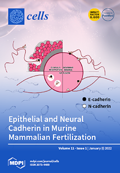
Mammalian fertilization is a Ca2+-dependent multistep process leading to gamete fusion. Herein, we report the expression of Ca2+-dependent adhesion proteins epithelial (E-cad) and neural (N-cad) cadherin in murine gametes and evidence of their involvement in fertilization. E-cad and N-cad were immunodetected in spermatozoa, cumulus cells, and oocytes. Both cadherins were found to participate in oolemma binding, fusion, and in vitro fertilization, as these processes were inhibited with specific antibodies or blocking peptides. Conversely, E-cad alone was found to have a role in cumulus penetration, as neither the N-cad antibody nor the peptide impaired this event. Our studies demonstrate the expression of members of the adherent complex in mice and confirm previous observations in the human model, reinforcing evidence on E-cad and N-cad involvement in mammalian fertilization. View this paper
  • Issues are regarded as officially published after their release is announced to the table of contents alert mailing list.
  • You may sign up for e-mail alerts to receive table of contents of newly released issues.
  • PDF is the official format for papers published in both, html and pdf forms. To view the papers in pdf format, click on the "PDF Full-text" link, and use the free Adobe Reader to open them.
Order results
Result details
Section
Select all
Export citation of selected articles as:
22 pages, 2801 KiB  
Review
Metabolic Implications of Immune Checkpoint Proteins in Cancer
by Elizabeth R. Stirling, Steven M. Bronson, Jessica D. Mackert, Katherine L. Cook, Pierre L. Triozzi and David R. Soto-Pantoja
Cells 2022, 11(1), 179; https://doi.org/10.3390/cells11010179 - 05 Jan 2022
Cited by 15 | Viewed by 4827
Abstract
Expression of immune checkpoint proteins restrict immunosurveillance in the tumor microenvironment; thus, FDA-approved checkpoint inhibitor drugs, specifically PD-1/PD-L1 and CTLA-4 inhibitors, promote a cytotoxic antitumor immune response. Aside from inflammatory signaling, immune checkpoint proteins invoke metabolic reprogramming that affects immune cell function, autonomous [...] Read more.
Expression of immune checkpoint proteins restrict immunosurveillance in the tumor microenvironment; thus, FDA-approved checkpoint inhibitor drugs, specifically PD-1/PD-L1 and CTLA-4 inhibitors, promote a cytotoxic antitumor immune response. Aside from inflammatory signaling, immune checkpoint proteins invoke metabolic reprogramming that affects immune cell function, autonomous cancer cell bioenergetics, and patient response. Therefore, this review will focus on the metabolic alterations in immune and cancer cells regulated by currently approved immune checkpoint target proteins and the effect of costimulatory receptor signaling on immunometabolism. Additionally, we explore how diet and the microbiome impact immune checkpoint blockade therapy response. The metabolic reprogramming caused by targeting these proteins is essential in understanding immune-related adverse events and therapeutic resistance. This can provide valuable information for potential biomarkers or combination therapy strategies targeting metabolic pathways with immune checkpoint blockade to enhance patient response. Full article
(This article belongs to the Special Issue Metabolic Interactions in Tumor Microenvironment (TME))
Show Figures

Figure 1

18 pages, 3983 KiB  
Protocol
Isolation and Propagation of Human Corneal Stromal Keratocytes for Tissue Engineering and Cell Therapy
by Nur Zahirah binte M. Yusoff, Andri K. Riau, Gary H. F. Yam, Nuur Shahinda Humaira binte Halim and Jodhbir S. Mehta
Cells 2022, 11(1), 178; https://doi.org/10.3390/cells11010178 - 05 Jan 2022
Cited by 20 | Viewed by 2888
Abstract
The human corneal stroma contains corneal stromal keratocytes (CSKs) that synthesize and deposit collagens and keratan sulfate proteoglycans into the stromal matrix to maintain the corneal structural integrity and transparency. In adult corneas, CSKs are quiescent and arrested in the G0 phase of [...] Read more.
The human corneal stroma contains corneal stromal keratocytes (CSKs) that synthesize and deposit collagens and keratan sulfate proteoglycans into the stromal matrix to maintain the corneal structural integrity and transparency. In adult corneas, CSKs are quiescent and arrested in the G0 phase of the cell cycle. Following injury, some CSKs undergo apoptosis, whereas the surviving cells are activated to become stromal fibroblasts (SFs) and myofibroblasts (MyoFBs), as a natural mechanism of wound healing. The SFs and MyoFBs secrete abnormal extracellular matrix proteins, leading to corneal fibrosis and scar formation (corneal opacification). The issue is compounded by the fact that CSK transformation into SFs or MyoFBs is irreversible in vivo, which leads to chronic opacification. In this scenario, corneal transplantation is the only recourse. The application of cell therapy by replenishing CSKs, propagated in vitro, in the injured corneas has been demonstrated to be efficacious in resolving early-onset corneal opacification. However, expanding CSKs is challenging and has been the limiting factor for the application in corneal tissue engineering and cell therapy. The supplementation of serum in the culture medium promotes cell division but inevitably converts the CSKs into SFs. Similar to the in vivo conditions, the transformation is irreversible, even when the SF culture is switched to a serum-free medium. In the current article, we present a detailed protocol on the isolation and propagation of bona fide human CSKs and the morphological and genotypic differences from SFs. Full article
(This article belongs to the Special Issue 10th Anniversary of Cells—Advances in Cell Techniques)
Show Figures

Figure 1

18 pages, 6420 KiB  
Article
Bioengineered Cystinotic Kidney Tubules Recapitulate a Nephropathic Phenotype
by Elena Sendino Garví, Rosalinde Masereeuw and Manoe J. Janssen
Cells 2022, 11(1), 177; https://doi.org/10.3390/cells11010177 - 05 Jan 2022
Cited by 2 | Viewed by 2438
Abstract
Nephropathic cystinosis is a rare and severe disease caused by disruptions in the CTNS gene. Cystinosis is characterized by lysosomal cystine accumulation, vesicle trafficking impairment, oxidative stress, and apoptosis. Additionally, cystinotic patients exhibit weakening and leakage of the proximal tubular segment of the [...] Read more.
Nephropathic cystinosis is a rare and severe disease caused by disruptions in the CTNS gene. Cystinosis is characterized by lysosomal cystine accumulation, vesicle trafficking impairment, oxidative stress, and apoptosis. Additionally, cystinotic patients exhibit weakening and leakage of the proximal tubular segment of the nephrons, leading to renal Fanconi syndrome and kidney failure early in life. Current in vitro cystinotic models cannot recapitulate all clinical features of the disease which limits their translational value. Therefore, the development of novel, complex in vitro models that better mimic the disease and exhibit characteristics not compatible with 2-dimensional cell culture is of crucial importance for novel therapies development. In this study, we developed a 3-dimensional bioengineered model of nephropathic cystinosis by culturing conditionally immortalized proximal tubule epithelial cells (ciPTECs) on hollow fiber membranes (HFM). Cystinotic kidney tubules showed lysosomal cystine accumulation, increased autophagy and vesicle trafficking deterioration, the impairment of several metabolic pathways, and the disruption of the epithelial monolayer tightness as compared to control kidney tubules. In particular, the loss of monolayer organization and leakage could be mimicked with the use of the cystinotic kidney tubules, which has not been possible before, using the standard 2-dimensional cell culture. Overall, bioengineered cystinotic kidney tubules recapitulate better the nephropathic phenotype at a molecular, structural, and functional proximal tubule level compared to 2-dimensional cell cultures. Full article
(This article belongs to the Special Issue Cellular and Molecular Mechanisms of Nephropathic Cystinosis)
Show Figures

Figure 1

16 pages, 28830 KiB  
Article
Targeting PGM3 as a Novel Therapeutic Strategy in KRAS/LKB1 Co-Mutant Lung Cancer
by Hyunmin Lee, Feng Cai, Neil Kelekar, Nipun K. Velupally and Jiyeon Kim
Cells 2022, 11(1), 176; https://doi.org/10.3390/cells11010176 - 05 Jan 2022
Cited by 10 | Viewed by 3904
Abstract
In non-small-cell lung cancer (NSCLC), concurrent mutations in the oncogene KRAS and tumor suppressor STK11 (also known as LKB1) confer an aggressive malignant phenotype, an unfavourability towards immunotherapy, and overall poor prognoses in patients. In a previous study, we showed that murine KRAS/LKB1 [...] Read more.
In non-small-cell lung cancer (NSCLC), concurrent mutations in the oncogene KRAS and tumor suppressor STK11 (also known as LKB1) confer an aggressive malignant phenotype, an unfavourability towards immunotherapy, and overall poor prognoses in patients. In a previous study, we showed that murine KRAS/LKB1 co-mutant tumors and human co-mutant cancer cells have an enhanced dependence on glutamine-fructose-6-phosphate transaminase 2 (GFPT2), a rate-limiting enzyme in the hexosamine biosynthesis pathway (HBP), which could be targeted to reduce survival of KRAS/LKB1 co-mutants. Here, we found that KRAS/LKB1 co-mutant cells also exhibit an increased dependence on N-acetylglucosamine-phosphate mutase 3 (PGM3), an enzyme downstream of GFPT2. Genetic or pharmacologic suppression of PGM3 reduced KRAS/LKB1 co-mutant tumor growth in both in vitro and in vivo settings. Our results define an additional metabolic vulnerability in KRAS/LKB1 co-mutant tumors to the HBP and provide a rationale for targeting PGM3 in this aggressive subtype of NSCLC. Full article
(This article belongs to the Special Issue New Aspects of Targeting Cancer Metabolism in Therapeutic Approach)
Show Figures

Figure 1

27 pages, 14903 KiB  
Article
Accelerated Growth, Differentiation, and Ploidy with Reduced Proliferation of Right Ventricular Cardiomyocytes in Children with Congenital Heart Defect Tetralogy of Fallot
by Tatyana V. Sukhacheva, Roman A. Serov, Natalia V. Nizyaeva, Artem A. Burov, Stanislav V. Pavlovich, Yulia L. Podurovskaya, Maria V. Samsonova, Andrey L. Chernyaev, Aleksandr I. Shchegolev, Alexei I. Kim, Leo A. Bockeria and Gennady T. Sukhikh
Cells 2022, 11(1), 175; https://doi.org/10.3390/cells11010175 - 05 Jan 2022
Cited by 6 | Viewed by 2484
Abstract
The myocardium of children with tetralogy of Fallot (TF) undergoes hemodynamic overload and hypoxemia immediately after birth. Comparative analysis of changes in the ploidy and morphology of the right ventricular cardiomyocytes in children with TF in the first years of life demonstrated their [...] Read more.
The myocardium of children with tetralogy of Fallot (TF) undergoes hemodynamic overload and hypoxemia immediately after birth. Comparative analysis of changes in the ploidy and morphology of the right ventricular cardiomyocytes in children with TF in the first years of life demonstrated their significant increase compared with the control group. In children with TF, there was a predominantly diffuse distribution of Connexin43-containing gap junctions over the cardiomyocytes sarcolemma, which redistributed into the intercalated discs as cardiomyocytes differentiation increased. The number of Ki67-positive cardiomyocytes varied greatly and amounted to 7.0–1025.5/106 cardiomyocytes and also were decreased with increased myocytes differentiation. Ultrastructural signs of immaturity and proliferative activity of cardiomyocytes in children with TF were demonstrated. The proportion of interstitial tissue did not differ significantly from the control group. The myocardium of children with TF under six months of age was most sensitive to hypoxemia, it was manifested by a delay in the intercalated discs and myofibril assembly and the appearance of ultrastructural signs of dystrophic changes in the cardiomyocytes. Thus, the acceleration of ontogenetic growth and differentiation of the cardiomyocytes, but not the reactivation of their proliferation, was an adaptation of the immature myocardium of children with TF to hemodynamic overload and hypoxemia. Full article
(This article belongs to the Section Cell Proliferation and Division)
Show Figures

Graphical abstract

6 pages, 37108 KiB  
Commentary
Lymphoma versus Carcinoma and Other Collaborations
by Karen Pulford
Cells 2022, 11(1), 174; https://doi.org/10.3390/cells11010174 - 05 Jan 2022
Viewed by 1447
Abstract
David Mason started his research career at a time when lymphoma diagnosis was based primarily on cellular morphology, clinical symptoms and special cytochemical stains using formalin fixed tissue sections. There were occasions, however, where the morphology was unhelpful, such as in the case [...] Read more.
David Mason started his research career at a time when lymphoma diagnosis was based primarily on cellular morphology, clinical symptoms and special cytochemical stains using formalin fixed tissue sections. There were occasions, however, where the morphology was unhelpful, such as in the case of anaplastic or poorly differentiated tumours, where a distinction between lymphoma and a non-haematopoietic tumour was often problematical. Accurate diagnosis became even more important with the developments in the clinical staging of lymphoma and the availability of more effective treatments. One way forward to improve diagnosis was to use immunohistochemistry to study the antigens expressed by the tumor cells. Full article
Show Figures

Figure 1

22 pages, 4308 KiB  
Article
Dermatan Sulfate Affects Breast Cancer Cell Function via the Induction of Necroptosis
by Grzegorz Wisowski, Adam Pudełko, Krystyna Olczyk, Monika Paul-Samojedny and Ewa M. Koźma
Cells 2022, 11(1), 173; https://doi.org/10.3390/cells11010173 - 05 Jan 2022
Cited by 3 | Viewed by 2029
Abstract
Dermatan sulfate (DS) is widespread in the extracellular matrix (ECM) of animal tissues. This glycosaminoglycan is characterized by a variable structure, which is reflected in the heterogeneity of its sulfation pattern. The sulfate groups are responsible for the binding properties of DS, which [...] Read more.
Dermatan sulfate (DS) is widespread in the extracellular matrix (ECM) of animal tissues. This glycosaminoglycan is characterized by a variable structure, which is reflected in the heterogeneity of its sulfation pattern. The sulfate groups are responsible for the binding properties of DS, which determine an interaction profile of this glycan. However, the detailed role of DS in biological processes such as the neoplasm is still poorly understood. The aim of the study was to assess the effects of the structural variants of DS on breast cancer cells. We found that DS isoforms from normal and fibrotic fascia as well as from intestinal mucosa were able to quickly induce oxidative stress in the cytoplasm and affect the mitochondrial function in luminal breast cancer cells. Moreover, the variants caused the necroptosis of the cells most likely via the first of these mechanisms. This death was responsible for a reduction in the viability and number of breast cancer cells. However, the dynamics and intensity of all of the DS variants-triggered effects were strongly dependent on the cell type and the structure of these molecules. The most pronounced activity was demonstrated by those variants that shared structural features with the DS from the tumor niche. Full article
Show Figures

Figure 1

19 pages, 7538 KiB  
Review
Role and Involvement of TENM4 and miR-708 in Breast Cancer Development and Therapy
by Giulia Peppino, Federica Riccardo, Maddalena Arigoni, Elisabetta Bolli, Giuseppina Barutello, Federica Cavallo and Elena Quaglino
Cells 2022, 11(1), 172; https://doi.org/10.3390/cells11010172 - 05 Jan 2022
Cited by 4 | Viewed by 2840
Abstract
Teneurin 4 (TENM4) is a transmembrane protein that is codified by the ODZ4 gene and is involved in nervous system development, neurite outgrowth, and neuronal differentiation. In line with its involvement in the nervous system, TENM4 has also been implicated in several mental [...] Read more.
Teneurin 4 (TENM4) is a transmembrane protein that is codified by the ODZ4 gene and is involved in nervous system development, neurite outgrowth, and neuronal differentiation. In line with its involvement in the nervous system, TENM4 has also been implicated in several mental disorders such as bipolar disorder, schizophrenia, and autism. TENM4 mutations and rearrangements have recently been identified in a number of tumors. This, combined with impaired expression in tumors, suggests that it may potentially be involved in tumorigenesis. Most of the TENM4 mutations that are observed in tumors occur in breast cancer, in which TENM4 plays a role in cells’ migration and stemness. However, the functional role that TENM4 plays in breast cancer still needs to be better evaluated, and further studies are required to better understand the involvement of TENM4 in breast cancer progression. Herein, we review the currently available data for TENM4′s role in breast cancer and propose its use as both a novel target with which to ameliorate patient prognosis and as a potential biomarker. Moreover, we also report data on the tumorigenic role of miR-708 deregulation and the possible use of this miRNA as a novel therapeutic molecule, as miR-708 is spliced out from TENM4 mRNA. Full article
(This article belongs to the Special Issue Emerging Targets and Therapeutic Strategies in Cancer)
Show Figures

Figure 1

14 pages, 1515 KiB  
Review
Lithium and Erectile Dysfunction: An Overview
by Mohammad Sheibani, Mehdi Ghasemi and Ahmad Reza Dehpour
Cells 2022, 11(1), 171; https://doi.org/10.3390/cells11010171 - 05 Jan 2022
Cited by 8 | Viewed by 7242
Abstract
Lithium has been a mainstay of therapy for patients with bipolar disorders for several decades. However, it may exert a variety of adverse effects that can affect patients’ compliance. Sexual and erectile dysfunction has been reported in several studies by patients who take [...] Read more.
Lithium has been a mainstay of therapy for patients with bipolar disorders for several decades. However, it may exert a variety of adverse effects that can affect patients’ compliance. Sexual and erectile dysfunction has been reported in several studies by patients who take lithium as monotherapy or combined with other psychotherapeutic agents. The exact mechanisms underlying such side effects of lithium are not completely understood. It seems that both central and peripheral mechanisms are involved in the lithium-related sexual dysfunction. Here, we had an overview of the epidemiology of lithium-related sexual and erectile dysfunction in previous clinical studies as well as possible pathologic pathways that could be involved in this adverse effect of lithium based on the previous preclinical studies. Understanding such mechanisms could potentially open a new avenue for therapies that can overcome lithium-related sexual dysfunction and improve patients’ adherence to the medication intake. Full article
(This article belongs to the Collection Feature Papers in 'Cells of the Nervous System' Section)
Show Figures

Figure 1

11 pages, 1520 KiB  
Review
Muscle and Bone Impairment in Infantile Nephropathic Cystinosis: New Concepts
by Dieter Haffner, Maren Leifheit-Nestler, Candide Alioli and Justine Bacchetta
Cells 2022, 11(1), 170; https://doi.org/10.3390/cells11010170 - 05 Jan 2022
Cited by 2 | Viewed by 2769
Abstract
Cystinosis Metabolic Bone Disease (CMBD) has emerged during the last decade as a well-recognized, long-term complication in patients suffering from infantile nephropathic cystinosis (INC), resulting in significant morbidity and impaired quality of life in teenagers and adults with INC. Its underlying pathophysiology is [...] Read more.
Cystinosis Metabolic Bone Disease (CMBD) has emerged during the last decade as a well-recognized, long-term complication in patients suffering from infantile nephropathic cystinosis (INC), resulting in significant morbidity and impaired quality of life in teenagers and adults with INC. Its underlying pathophysiology is complex and multifactorial, associating complementary, albeit distinct entities, in addition to ordinary mineral and bone disorders observed in other types of chronic kidney disease. Amongst these long-term consequences are renal Fanconi syndrome, hypophosphatemic rickets, malnutrition, hormonal abnormalities, muscular impairment, and intrinsic cellular bone defects in bone cells, due to CTNS mutations. Recent research data in the field have demonstrated abnormal mineral regulation, intrinsic bone defects, cysteamine toxicity, muscle wasting and, likely interleukin-1-driven inflammation in the setting of CMBD. Here we summarize these new pathophysiological deregulations and discuss the crucial interplay between bone and muscle in INC. In future, vitamin D and/or biotherapies targeting the IL1β pathway may improve muscle wasting and subsequently CMBD, but this remains to be proven. Full article
(This article belongs to the Special Issue Cellular and Molecular Mechanisms of Nephropathic Cystinosis)
Show Figures

Figure 1

21 pages, 1463 KiB  
Review
LRRK2 at Striatal Synapses: Cell-Type Specificity and Mechanistic Insights
by Patrick D. Skelton, Valerie Tokars and Loukia Parisiadou
Cells 2022, 11(1), 169; https://doi.org/10.3390/cells11010169 - 05 Jan 2022
Cited by 5 | Viewed by 3148
Abstract
Mutations in leucine-rich repeat kinase 2 (LRRK2) cause Parkinson’s disease with a similar clinical presentation and progression to idiopathic Parkinson’s disease, and common variation is linked to disease risk. Recapitulation of the genotype in rodent models causes abnormal dopamine release and increases the [...] Read more.
Mutations in leucine-rich repeat kinase 2 (LRRK2) cause Parkinson’s disease with a similar clinical presentation and progression to idiopathic Parkinson’s disease, and common variation is linked to disease risk. Recapitulation of the genotype in rodent models causes abnormal dopamine release and increases the susceptibility of dopaminergic neurons to insults, making LRRK2 a valuable model for understanding the pathobiology of Parkinson’s disease. It is also a promising druggable target with targeted therapies currently in development. LRRK2 mRNA and protein expression in the brain is highly variable across regions and cellular identities. A growing body of work has demonstrated that pathogenic LRRK2 mutations disrupt striatal synapses before the onset of overt neurodegeneration. Several substrates and interactors of LRRK2 have been identified to potentially mediate these pre-neurodegenerative changes in a cell-type-specific manner. This review discusses the effects of pathogenic LRRK2 mutations in striatal neurons, including cell-type-specific and pathway-specific alterations. It also highlights several LRRK2 effectors that could mediate the alterations to striatal function, including Rabs and protein kinase A. The lessons learned from improving our understanding of the pathogenic effects of LRRK2 mutations in striatal neurons will be applicable to both dissecting the cell-type specificity of LRRK2 function in the transcriptionally diverse subtypes of dopaminergic neurons and also increasing our understanding of basal ganglia development and biology. Finally, it will inform the development of therapeutics for Parkinson’s disease. Full article
(This article belongs to the Collection Feature Papers in 'Cells of the Nervous System' Section)
Show Figures

Figure 1

15 pages, 2491 KiB  
Article
In Vitro Anticancer Screening and Preliminary Mechanistic Study of A-Ring Substituted Anthraquinone Derivatives
by Ibrahim Morgan, Ludger A. Wessjohann and Goran N. Kaluđerović
Cells 2022, 11(1), 168; https://doi.org/10.3390/cells11010168 - 05 Jan 2022
Cited by 11 | Viewed by 2738
Abstract
Anthraquinone derivatives exhibit various biological activities, e.g., antifungal, antibacterial and in vitro antiviral activities. They are naturally produced in many fungal and plant families such as Rhamnaceae or Fabaceae. Furthermore, they were found to have anticancer activity, exemplified by mitoxantrone and pixantrone, and [...] Read more.
Anthraquinone derivatives exhibit various biological activities, e.g., antifungal, antibacterial and in vitro antiviral activities. They are naturally produced in many fungal and plant families such as Rhamnaceae or Fabaceae. Furthermore, they were found to have anticancer activity, exemplified by mitoxantrone and pixantrone, and many are well known redox-active compounds. In this study, various nature inspired synthetic anthraquinone derivatives were tested against colon, prostate, liver and cervical cancer cell lines. Most of the compounds exhibit anticancer effects against all cell lines, therefore the compounds were further studied to determine their IC50-values. Of these compounds, 1,4-bis(benzyloxy)-2,3-bis(hydroxymethyl)anthracene-9,10-dione (4) exhibited the highest cytotoxicity against PC3 cells and was chosen for a deeper look into its mechanism of action. Based on flow cytometry, the compound was proven to induce apoptosis through the activation of caspases and to demolish the ROS/RNS and NO equilibrium in the PC3 cell line. It trapped cells in the G2/M phase. Western blotting was performed for several proteins related to the effects observed. Compound 4 enhanced the production of PARP and caspase-3. Moreover, it activated the conversion of LC3A/B-I to LC3A/B-II showing that also autophagy plays a role in its mechanism of action, and it caused the phosphorylation of p70 s6 kinase. Full article
(This article belongs to the Special Issue Crosstalk of Autophagy and Apoptosis)
Show Figures

Figure 1

21 pages, 4891 KiB  
Article
Carnosic Acid Attenuates the Free Fatty Acid-Induced Insulin Resistance in Muscle Cells and Adipocytes
by Danja J. Den Hartogh, Filip Vlavcheski, Adria Giacca, Rebecca E. K. MacPherson and Evangelia Tsiani
Cells 2022, 11(1), 167; https://doi.org/10.3390/cells11010167 - 05 Jan 2022
Cited by 13 | Viewed by 2836
Abstract
Elevated blood free fatty acids (FFAs), as seen in obesity, impair insulin action leading to insulin resistance and Type 2 diabetes mellitus. Several serine/threonine kinases including JNK, mTOR, and p70 S6K cause serine phosphorylation of the insulin receptor substrate (IRS) and have been [...] Read more.
Elevated blood free fatty acids (FFAs), as seen in obesity, impair insulin action leading to insulin resistance and Type 2 diabetes mellitus. Several serine/threonine kinases including JNK, mTOR, and p70 S6K cause serine phosphorylation of the insulin receptor substrate (IRS) and have been implicated in insulin resistance. Activation of AMP-activated protein kinase (AMPK) increases glucose uptake, and in recent years, AMPK has been viewed as an important target to counteract insulin resistance. We reported previously that carnosic acid (CA) found in rosemary extract (RE) and RE increased glucose uptake and activated AMPK in muscle cells. In the present study, we examined the effects of CA on palmitate-induced insulin-resistant L6 myotubes and 3T3L1 adipocytes. Exposure of cells to palmitate reduced the insulin-stimulated glucose uptake, GLUT4 transporter levels on the plasma membrane, and Akt activation. Importantly, CA attenuated the deleterious effect of palmitate and restored the insulin-stimulated glucose uptake, the activation of Akt, and GLUT4 levels. Additionally, CA markedly attenuated the palmitate-induced phosphorylation/activation of JNK, mTOR, and p70S6K and activated AMPK. Our data indicate that CA has the potential to counteract the palmitate-induced muscle and fat cell insulin resistance. Full article
(This article belongs to the Special Issue Free Fatty Acids and Pathogenesis of Diabetes Mellitus)
Show Figures

Figure 1

12 pages, 1695 KiB  
Article
Engineered Liposomes Protect Immortalized Immune Cells from Cytolysins Secreted by Group A and Group G Streptococci
by Hervé Besançon, Yu Larpin, Viktoria S. Babiychuk, René Köffel and Eduard B. Babiychuk
Cells 2022, 11(1), 166; https://doi.org/10.3390/cells11010166 - 05 Jan 2022
Viewed by 2001
Abstract
The increasing antibiotic resistance of bacterial pathogens fosters the development of alternative, non-antibiotic treatments. Antivirulence therapy, which is neither bacteriostatic nor bactericidal, acts by depriving bacterial pathogens of their virulence factors. To establish a successful infection, many bacterial pathogens secrete exotoxins/cytolysins that perforate [...] Read more.
The increasing antibiotic resistance of bacterial pathogens fosters the development of alternative, non-antibiotic treatments. Antivirulence therapy, which is neither bacteriostatic nor bactericidal, acts by depriving bacterial pathogens of their virulence factors. To establish a successful infection, many bacterial pathogens secrete exotoxins/cytolysins that perforate the host cell plasma membrane. Recently developed liposomal nanotraps, mimicking the outer layer of the targeted cell membranes, serve as decoys for exotoxins, thus diverting them from attacking host cells. In this study, we develop a liposomal nanotrap formulation that is capable of protecting immortalized immune cells from the whole palette of cytolysins secreted by Streptococcus pyogenes and Streptococcus dysgalactiae subsp. equisimilis—important human pathogens that can cause life-threatening bacteremia. We show that the mixture of cholesterol-containing liposomes with liposomes composed exclusively of phospholipids is protective against the combined action of all streptococcal exotoxins. Our findings pave the way for further development of liposomal antivirulence therapy in order to provide more efficient treatment of bacterial infections, including those caused by antibiotic resistant pathogens. Full article
Show Figures

Graphical abstract

13 pages, 2947 KiB  
Article
Role of Nse1 Subunit of SMC5/6 Complex as a Ubiquitin Ligase
by Peter Kolesar, Karel Stejskal, David Potesil, Johanne M. Murray and Jan J. Palecek
Cells 2022, 11(1), 165; https://doi.org/10.3390/cells11010165 - 04 Jan 2022
Cited by 7 | Viewed by 2640
Abstract
Structural Maintenance of Chromosomes (SMC) complexes are important for many aspects of the chromosomal organization. Unlike cohesin and condensin, the SMC5/6 complex contains a variant RING domain carried by its Nse1 subunit. RING domains are characteristic for ubiquitin ligases, and human NSE1 has [...] Read more.
Structural Maintenance of Chromosomes (SMC) complexes are important for many aspects of the chromosomal organization. Unlike cohesin and condensin, the SMC5/6 complex contains a variant RING domain carried by its Nse1 subunit. RING domains are characteristic for ubiquitin ligases, and human NSE1 has been shown to possess ubiquitin-ligase activity in vitro. However, other studies were unable to show such activity. Here, we confirm Nse1 ubiquitin-ligase activity using purified Schizosaccharomyces pombe proteins. We demonstrate that the Nse1 ligase activity is stimulated by Nse3 and Nse4. We show that Nse1 specifically utilizes Ubc13/Mms2 E2 enzyme and interacts directly with ubiquitin. We identify the Nse1 mutation (R188E) that specifically disrupts its E3 activity and demonstrate that the Nse1-dependent ubiquitination is particularly important under replication stress. Moreover, we determine Nse4 (lysine K181) as the first known SMC5/6-associated Nse1 substrate. Interestingly, abolition of Nse4 modification at K181 leads to suppression of DNA-damage sensitivity of other SMC5/6 mutants. Altogether, this study brings new evidence for Nse1 ubiquitin ligase activity, significantly advancing our understanding of this enigmatic SMC5/6 function. Full article
Show Figures

Figure 1

29 pages, 3204 KiB  
Review
Overview of Polyamines as Nutrients for Human Healthy Long Life and Effect of Increased Polyamine Intake on DNA Methylation
by Kuniyasu Soda
Cells 2022, 11(1), 164; https://doi.org/10.3390/cells11010164 - 04 Jan 2022
Cited by 28 | Viewed by 6184
Abstract
Polyamines, spermidine and spermine, are synthesized in every living cell and are therefore contained in foods, especially in those that are thought to contribute to health and longevity. They have many physiological activities similar to those of antioxidant and anti-inflammatory substances such as [...] Read more.
Polyamines, spermidine and spermine, are synthesized in every living cell and are therefore contained in foods, especially in those that are thought to contribute to health and longevity. They have many physiological activities similar to those of antioxidant and anti-inflammatory substances such as polyphenols. These include antioxidant and anti-inflammatory properties, cell and gene protection, and autophagy activation. We have first reported that increased polyamine intake (spermidine much more so than spermine) over a long period increased blood spermine levels and inhibited aging-associated pathologies and pro-inflammatory status in humans and mice and extended life span of mice. However, it is unlikely that the life-extending effect of polyamines is exerted by the same bioactivity as polyphenols because most studies using polyphenols and antioxidants have failed to demonstrate their life-extending effects. Recent investigations revealed that aging-associated pathologies and lifespan are closely associated with DNA methylation, a regulatory mechanism of gene expression. There is a close relationship between polyamine metabolism and DNA methylation. We have shown that the changes in polyamine metabolism affect the concentrations of substances and enzyme activities involved in DNA methylation. I consider that the increased capability of regulation of DNA methylation by spermine is a key of healthy long life of humans. Full article
(This article belongs to the Special Issue Epigenetic Mechanisms of Longevity and Aging)
Show Figures

Figure 1

32 pages, 2696 KiB  
Article
Co-Expression Analysis of microRNAs and Proteins in Brain of Alzheimer’s Disease Patients
by Callum N. Watson, Ghazala Begum, Emma Ashman, Daniella Thorn, Kamal M. Yakoub, Moustafa Al Hariri, Ali Nehme, Stefania Mondello, Firas Kobeissy, Antonio Belli and Valentina Di Pietro
Cells 2022, 11(1), 163; https://doi.org/10.3390/cells11010163 - 04 Jan 2022
Cited by 7 | Viewed by 2979
Abstract
Alzheimer’s disease (AD) is the most common form of dementia globally; however, the aetiology of AD remains elusive hindering the development of effective therapeutics. MicroRNAs (miRNAs) are regulators of gene expression and have been of growing interest in recent studies in many pathologies [...] Read more.
Alzheimer’s disease (AD) is the most common form of dementia globally; however, the aetiology of AD remains elusive hindering the development of effective therapeutics. MicroRNAs (miRNAs) are regulators of gene expression and have been of growing interest in recent studies in many pathologies including AD not only for their use as biomarkers but also for their implications in the therapeutic field. In this study, miRNA and protein profiles were obtained from brain tissues of different stage (Braak III-IV and Braak V-VI) of AD patients and compared to matched controls. The aim of the study was to identify in the late stage of AD, the key dysregulated pathways that may contribute to pathogenesis and then to evaluate whether any of these pathways could be detected in the early phase of AD, opening new opportunity for early treatment that could stop or delay the pathology. Six common pathways were found regulated by miRNAs and proteins in the late stage of AD, with one of them (Rap1 signalling) activated since the early phase. MiRNAs and proteins were also compared to explore an inverse trend of expression which could lead to the identification of new therapeutic targets. These results suggest that specific miRNA changes could represent molecular fingerprint of neurodegenerative processes and potential therapeutic targets for early intervention. Full article
Show Figures

Figure 1

13 pages, 1668 KiB  
Article
N-Glycosylation Facilitates 4-1BB Membrane Localization by Avoiding Its Multimerization
by Ruoxuan Sun, Alyssa Min Jung Kim, Allison A. Murray and Seung-Oe Lim
Cells 2022, 11(1), 162; https://doi.org/10.3390/cells11010162 - 04 Jan 2022
Cited by 6 | Viewed by 2516
Abstract
Leveraging the T cell immunity against tumors represents a revolutionary type of cancer therapy. 4-1BB is a well-characterized costimulatory immune receptor existing on activated T cells and mediating their proliferation and cytotoxicity under infectious diseases and cancers. Despite the accumulating interest in implementing [...] Read more.
Leveraging the T cell immunity against tumors represents a revolutionary type of cancer therapy. 4-1BB is a well-characterized costimulatory immune receptor existing on activated T cells and mediating their proliferation and cytotoxicity under infectious diseases and cancers. Despite the accumulating interest in implementing 4-1BB as a therapeutic target for immune-related disorders, less is known about the pattern of its intracellular behaviors and regulations. It has been previously demonstrated that 4-1BB is heavily modified by N-glycosylation; however, the biological importance of this modification lacks detailed elucidation. Through biochemical, biophysical, and cell-biological approaches, we systematically evaluated the impact of N-glycosylation on the ligand interaction, stability, and localization of 4-1BB. We hereby highlighted that N-glycan functions by preventing the oligomerization of 4-1BB, thus permitting its membrane transportation and fast turn-over. Without N-glycosylation, 4-1BB could be aberrantly accumulated intracellularly and fail to be sufficiently inserted in the membrane. The N-glycosylation-guided intracellular processing of 4-1BB serves as the potential mechanism explicitly modulating the “on” and “off” of 4-1BB through the control of protein abundance. Our study will further solidify the understanding of the biological properties of 4-1BB and facilitate the clinical practice against this promising therapeutic target. Full article
(This article belongs to the Special Issue Mechanism of Anti-tumor Immunity of Cells and Immunotherapy)
Show Figures

Figure 1

22 pages, 7188 KiB  
Article
Cell Type-Selective Loss of Peroxisomal β-Oxidation Impairs Bipolar Cell but Not Photoreceptor Survival in the Retina
by Daniëlle Swinkels, Yannick Das, Sai Kocherlakota, Stefan Vinckier, Eric Wever, Antoine H.C. van Kampen, Frédéric M. Vaz and Myriam Baes
Cells 2022, 11(1), 161; https://doi.org/10.3390/cells11010161 - 04 Jan 2022
Cited by 12 | Viewed by 3326
Abstract
Retinal degeneration is a common feature in peroxisomal disorders leading to blindness. Peroxisomes are present in the different cell types of the retina; however, their precise contribution to retinal integrity is still unclear. We previously showed that mice lacking the central peroxisomal β-oxidation [...] Read more.
Retinal degeneration is a common feature in peroxisomal disorders leading to blindness. Peroxisomes are present in the different cell types of the retina; however, their precise contribution to retinal integrity is still unclear. We previously showed that mice lacking the central peroxisomal β-oxidation enzyme, multifunctional protein 2 (MFP2), develop an early onset retinal decay including photoreceptor cell death. To decipher the function of peroxisomal β-oxidation in photoreceptors, we generated cell type selective Mfp2 knockout mice, using the Crx promotor targeting photoreceptors and bipolar cells. Surprisingly, Crx-Mfp2−/− mice maintained photoreceptor length and number until the age of 1 year. A negative electroretinogram was indicative of preserved photoreceptor phototransduction, but impaired downstream bipolar cell signaling from the age of 6 months. The photoreceptor ribbon synapse was affected, containing free-floating ribbons and vesicles with altered size and density. The bipolar cell interneurons sprouted into the ONL and died. Whereas docosahexaenoic acid levels were normal in the neural retina, levels of lipids containing very long chain polyunsaturated fatty acids were highly increased. Crx-Pex5−/− mice, in which all peroxisomal functions are inactivated in photoreceptors and bipolar cells, developed the same phenotype as Crx-Mfp2−/− mice. In conclusion, the early photoreceptor death in global Mfp2−/− mice is not driven cell autonomously. However, peroxisomal β-oxidation is essential for the integrity of photoreceptor ribbon synapses and of bipolar cells. Full article
(This article belongs to the Special Issue Peroxisome Biogenesis and Protein Targeting Mechanisms)
Show Figures

Graphical abstract

41 pages, 1737 KiB  
Review
Multifactorial Mechanism of Sarcopenia and Sarcopenic Obesity. Role of Physical Exercise, Microbiota and Myokines
by Jan Bilski, Piotr Pierzchalski, Marian Szczepanik, Joanna Bonior and Jerzy A. Zoladz
Cells 2022, 11(1), 160; https://doi.org/10.3390/cells11010160 - 04 Jan 2022
Cited by 53 | Viewed by 13640
Abstract
Obesity and ageing place a tremendous strain on the global healthcare system. Age-related sarcopenia is characterized by decreased muscular strength, decreased muscle quantity, quality, and decreased functional performance. Sarcopenic obesity (SO) is a condition that combines sarcopenia and obesity and has a substantial [...] Read more.
Obesity and ageing place a tremendous strain on the global healthcare system. Age-related sarcopenia is characterized by decreased muscular strength, decreased muscle quantity, quality, and decreased functional performance. Sarcopenic obesity (SO) is a condition that combines sarcopenia and obesity and has a substantial influence on the older adults’ health. Because of the complicated pathophysiology, there are disagreements and challenges in identifying and diagnosing SO. Recently, it has become clear that dysbiosis may play a role in the onset and progression of sarcopenia and SO. Skeletal muscle secretes myokines during contraction, which play an important role in controlling muscle growth, function, and metabolic balance. Myokine dysfunction can cause and aggravate obesity, sarcopenia, and SO. The only ways to prevent and slow the progression of sarcopenia, particularly sarcopenic obesity, are physical activity and correct nutritional support. While exercise cannot completely prevent sarcopenia and age-related loss in muscular function, it can certainly delay development and slow down the rate of sarcopenia. The purpose of this review was to discuss potential pathways to muscle deterioration in obese individuals. We also want to present the current understanding of the role of various factors, including microbiota and myokines, in the process of sarcopenia and SO. Full article
(This article belongs to the Collection Molecular Mechanisms of Exercise and Healthspan)
Show Figures

Figure 1

33 pages, 50489 KiB  
Review
Aptamer-Enabled Nanomaterials for Therapeutics, Drug Targeting and Imaging
by Mengping Liu, Lin Wang, Young Lo, Simon Chi-Chin Shiu, Andrew B. Kinghorn and Julian A. Tanner
Cells 2022, 11(1), 159; https://doi.org/10.3390/cells11010159 - 04 Jan 2022
Cited by 33 | Viewed by 5705
Abstract
A wide variety of nanomaterials have emerged in recent years with advantageous properties for a plethora of therapeutic and diagnostic applications. Such applications include drug delivery, imaging, anti-cancer therapy and radiotherapy. There is a critical need for further components which can facilitate therapeutic [...] Read more.
A wide variety of nanomaterials have emerged in recent years with advantageous properties for a plethora of therapeutic and diagnostic applications. Such applications include drug delivery, imaging, anti-cancer therapy and radiotherapy. There is a critical need for further components which can facilitate therapeutic targeting, augment their physicochemical properties, or broaden their theranostic applications. Aptamers are single-stranded nucleic acids which have been selected or evolved to bind specifically to molecules, surfaces, or cells. Aptamers can also act as direct biologic therapeutics, or in imaging and diagnostics. There is a rich field of discovery at the interdisciplinary interface between nanomaterials and aptamer science that has significant potential across biomedicine. Herein, we review recent progress in aptamer-enabled materials and discuss pending challenges for their future biomedical application. Full article
(This article belongs to the Special Issue Frontiers in Aptamers)
Show Figures

Graphical abstract

26 pages, 24241 KiB  
Article
Altered White Matter and microRNA Expression in a Murine Model Related to Williams Syndrome Suggests That miR-34b/c Affects Brain Development via Ptpru and Dcx Modulation
by Meitar Grad, Ariel Nir, Gilad Levy, Sari Schokoroy Trangle, Guy Shapira, Noam Shomron, Yaniv Assaf and Boaz Barak
Cells 2022, 11(1), 158; https://doi.org/10.3390/cells11010158 - 04 Jan 2022
Cited by 8 | Viewed by 3545
Abstract
Williams syndrome (WS) is a multisystem neurodevelopmental disorder caused by a de novo hemizygous deletion of ~26 genes from chromosome 7q11.23, among them the general transcription factor II-I (GTF2I). By studying a novel murine model for the hypersociability phenotype associated with [...] Read more.
Williams syndrome (WS) is a multisystem neurodevelopmental disorder caused by a de novo hemizygous deletion of ~26 genes from chromosome 7q11.23, among them the general transcription factor II-I (GTF2I). By studying a novel murine model for the hypersociability phenotype associated with WS, we previously revealed surprising aberrations in myelination and cell differentiation properties in the cortices of mutant mice compared to controls. These mutant mice had selective deletion of Gtf2i in the excitatory neurons of the forebrain. Here, we applied diffusion magnetic resonance imaging and fiber tracking, which showed a reduction in the number of streamlines in limbic outputs such as the fimbria/fornix fibers and the stria terminalis, as well as the corpus callosum of these mutant mice compared to controls. Furthermore, we utilized next-generation sequencing (NGS) analysis of cortical small RNAs’ expression (RNA-Seq) levels to identify altered expression of microRNAs (miRNAs), including two from the miR-34 cluster, known to be involved in prominent processes in the developing nervous system. Luciferase reporter assay confirmed the direct binding of miR-34c-5p to the 3’UTR of PTPRU—a gene involved in neural development that was elevated in the cortices of mutant mice relative to controls. Moreover, we found an age-dependent variation in the expression levels of doublecortin (Dcx)—a verified miR-34 target. Thus, we demonstrate the substantial effect a single gene deletion can exert on miRNA regulation and brain structure, and advance our understanding and, hopefully, treatment of WS. Full article
(This article belongs to the Special Issue Pathophysiological Mechanism of Neurodevelopmental Disorders)
Show Figures

Graphical abstract

26 pages, 6589 KiB  
Article
Recognition and Chaperoning by Pex19, Followed by Trafficking and Membrane Insertion of the Peroxisome Proliferation Protein, Pex11
by Katarzyna M. Zientara-Rytter, Shanmuga S. Mahalingam, Jean-Claude Farré, Krypton Carolino and Suresh Subramani
Cells 2022, 11(1), 157; https://doi.org/10.3390/cells11010157 - 04 Jan 2022
Cited by 5 | Viewed by 2596
Abstract
Pex11, an abundant peroxisomal membrane protein (PMP), is required for division of peroxisomes and is robustly imported to peroxisomal membranes. We present a comprehensive analysis of how the Pichia pastoris Pex11 is recognized and chaperoned by Pex19, targeted to peroxisome membranes and inserted [...] Read more.
Pex11, an abundant peroxisomal membrane protein (PMP), is required for division of peroxisomes and is robustly imported to peroxisomal membranes. We present a comprehensive analysis of how the Pichia pastoris Pex11 is recognized and chaperoned by Pex19, targeted to peroxisome membranes and inserted therein. We demonstrate that Pex11 contains one Pex19-binding site (Pex19-BS) that is required for Pex11 insertion into peroxisomal membranes by Pex19, but is non-essential for peroxisomal trafficking. We provide extensive mutational analyses regarding the recognition of Pex19-BS in Pex11 by Pex19. Pex11 also has a second, Pex19-independent membrane peroxisome-targeting signal (mPTS) that is preserved among Pex11-family proteins and anchors the human HsPex11γ to the outer leaflet of the peroxisomal membrane. Thus, unlike most PMPs, Pex11 can use two mechanisms of transport to peroxisomes, where only one of them depends on its direct interaction with Pex19, but the other does not. However, Pex19 is necessary for membrane insertion of Pex11. We show that Pex11 can self-interact, using both homo- and/or heterotypic interactions involving its N-terminal helical domains. We demonstrate that Pex19 acts as a chaperone by interacting with the Pex19-BS in Pex11, thereby protecting Pex11 from spontaneous oligomerization that would otherwise cause its aggregation and subsequent degradation. Full article
(This article belongs to the Special Issue Peroxisome Biogenesis and Protein Targeting Mechanisms)
Show Figures

Graphical abstract

18 pages, 2603 KiB  
Article
Pathologic Proteolytic Processing of N-Cadherin as a Marker of Human Fibrotic Disease
by Paul Durham Ferrell, Kristianne Michelle Oristian, Everett Cockrell and Salvatore Vincent Pizzo
Cells 2022, 11(1), 156; https://doi.org/10.3390/cells11010156 - 04 Jan 2022
Cited by 7 | Viewed by 2836
Abstract
Prior research has implicated the involvement of cell adhesion molecule N-cadherin in tissue fibrosis and remodeling. We hypothesize that anomalies in N-cadherin protein processing are involved in pathological fibrosis. Diseased tissues associated with fibrosis of the heart, lung, and liver were probed for [...] Read more.
Prior research has implicated the involvement of cell adhesion molecule N-cadherin in tissue fibrosis and remodeling. We hypothesize that anomalies in N-cadherin protein processing are involved in pathological fibrosis. Diseased tissues associated with fibrosis of the heart, lung, and liver were probed for the precursor form of N-cadherin, pro-N-cadherin (PNC), by immunohistochemistry and compared to healthy tissues. Myofibroblast cell lines were analyzed for cell surface pro-N-cadherin by flow cytometry and immunofluorescent microscopy. Soluble PNC products were immunoprecipitated from patient plasmas and an enzyme-linked immunoassay was developed for quantification. All fibrotic tissues examined show aberrant PNC localization. Cell surface PNC is expressed in myofibroblast cell lines isolated from cardiomyopathy and idiopathic pulmonary fibrosis but not on myofibroblasts isolated from healthy tissues. PNC is elevated in the plasma of patients with cardiomyopathy (p ≤ 0.0001), idiopathic pulmonary fibrosis (p ≤ 0.05), and nonalcoholic fatty liver disease with cirrhosis (p ≤ 0.05). Finally, we have humanized a murine antibody and demonstrate that it significantly inhibits migration of PNC expressing myofibroblasts. Collectively, the aberrant localization of PNC is observed in all fibrotic tissues examined in our study and our data suggest a role for cell surface PNC in the pathogenesis of fibrosis. Full article
Show Figures

Graphical abstract

25 pages, 5004 KiB  
Article
Extracellular Vesicles Derived from Bone Marrow in an Early Stage of Ionizing Radiation Damage Are Able to Induce Bystander Responses in the Bone Marrow
by Dávid Kis, Ilona Barbara Csordás, Eszter Persa, Bálint Jezsó, Rita Hargitai, Tünde Szatmári, Nikolett Sándor, Enikő Kis, Katalin Balázs, Géza Sáfrány and Katalin Lumniczky
Cells 2022, 11(1), 155; https://doi.org/10.3390/cells11010155 - 04 Jan 2022
Cited by 5 | Viewed by 2442
Abstract
Ionizing radiation (IR)-induced bystander effects contribute to biological responses to radiation, and extracellular vesicles (EVs) play important roles in mediating these effects. In this study we investigated the role of bone marrow (BM)-derived EVs in the bystander transfer of radiation damage. Mice were [...] Read more.
Ionizing radiation (IR)-induced bystander effects contribute to biological responses to radiation, and extracellular vesicles (EVs) play important roles in mediating these effects. In this study we investigated the role of bone marrow (BM)-derived EVs in the bystander transfer of radiation damage. Mice were irradiated with 0.1Gy, 0.25Gy and 2Gy, EVs were extracted from the BM supernatant 24 h or 3 months after irradiation and injected into bystander mice. Acute effects on directly irradiated or EV-treated mice were investigated after 4 and 24 h, while late effects were investigated 3 months after treatment. The acute effects of EVs on the hematopoietic stem and progenitor cell pools were similar to direct irradiation effects and persisted for up to 3 months, with the hematopoietic stem cells showing the strongest bystander responses. EVs isolated 3 months after irradiation elicited no bystander responses. The level of seven microRNAs (miR-33a-3p, miR-140-3p, miR-152-3p, miR-199a-5p, miR-200c-5p, miR-375-3p and miR-669o-5p) was altered in the EVs isolated 24 hour but not 3 months after irradiation. They regulated pathways highly relevant for the cellular response to IR, indicating their role in EV-mediated bystander responses. In conclusion, we showed that only EVs from an early stage of radiation damage could transmit IR-induced bystander effects. Full article
Show Figures

Graphical abstract

20 pages, 3421 KiB  
Article
Comprehensive Molecular Landscape of Cetuximab Resistance in Head and Neck Cancer Cell Lines
by Izabela N. F. Gomes, Renato J. da Silva-Oliveira, Luciane Sussuchi da Silva, Olga Martinho, Adriane F. Evangelista, André van Helvoort Lengert, Letícia Ferro Leal, Viviane Aline Oliveira Silva, Stéphanie Piancenti dos Santos, Flávia Caroline Nascimento, André Lopes Carvalho and Rui Manuel Reis
Cells 2022, 11(1), 154; https://doi.org/10.3390/cells11010154 - 04 Jan 2022
Cited by 8 | Viewed by 4074
Abstract
Cetuximab is the sole anti-EGFR monoclonal antibody that is FDA approved to treat head and neck squamous cell carcinoma (HNSCC). However, no predictive biomarkers of cetuximab response are known for HNSCC. Herein, we address the molecular mechanisms underlying cetuximab resistance in an in [...] Read more.
Cetuximab is the sole anti-EGFR monoclonal antibody that is FDA approved to treat head and neck squamous cell carcinoma (HNSCC). However, no predictive biomarkers of cetuximab response are known for HNSCC. Herein, we address the molecular mechanisms underlying cetuximab resistance in an in vitro model. We established a cetuximab resistant model (FaDu), using increased cetuximab concentrations for more than eight months. The resistance and parental cells were evaluated for cell viability and functional assays. Protein expression was analyzed by Western blot and human cell surface panel by lyoplate. The mutational profile and copy number alterations (CNA) were analyzed using whole-exome sequencing (WES) and the NanoString platform. FaDu resistant clones exhibited at least two-fold higher IC50 compared to the parental cell line. WES showed relevant mutations in several cancer-related genes, and the comparative mRNA expression analysis showed 36 differentially expressed genes associated with EGFR tyrosine kinase inhibitors resistance, RAS, MAPK, and mTOR signaling. Importantly, we observed that overexpression of KRAS, RhoA, and CD44 was associated with cetuximab resistance. Protein analysis revealed EGFR phosphorylation inhibition and mTOR increase in resistant cells. Moreover, the resistant cell line demonstrated an aggressive phenotype with a significant increase in adhesion, the number of colonies, and migration rates. Overall, we identified several molecular alterations in the cetuximab resistant cell line that may constitute novel biomarkers of cetuximab response such as mTOR and RhoA overexpression. These findings indicate new strategies to overcome anti-EGFR resistance in HNSCC. Full article
Show Figures

Figure 1

18 pages, 3844 KiB  
Review
The Impact of Exercise on Telomere Length, DNA Methylation and Metabolic Footprints
by Sandra Haupt, Tobias Niedrist, Harald Sourij, Stephan Schwarzinger and Othmar Moser
Cells 2022, 11(1), 153; https://doi.org/10.3390/cells11010153 - 04 Jan 2022
Cited by 6 | Viewed by 6360
Abstract
Aging as a major risk factor influences the probability of developing cancer, cardiovascular disease and diabetes, amongst others. The underlying mechanisms of disease are still not fully understood, but research suggests that delaying the aging process could ameliorate these pathologies. A key biological [...] Read more.
Aging as a major risk factor influences the probability of developing cancer, cardiovascular disease and diabetes, amongst others. The underlying mechanisms of disease are still not fully understood, but research suggests that delaying the aging process could ameliorate these pathologies. A key biological process in aging is cellular senescence which is associated with several stressors such as telomere shortening or enhanced DNA methylation. Telomere length as well as DNA methylation levels can be used as biological age predictors which are able to detect excessive acceleration or deceleration of aging. Analytical methods examining aging are often not suitable, expensive, time-consuming or require a high level of technical expertise. Therefore, research focusses on combining analytical methods which have the potential to simultaneously analyse epigenetic, genomic as well as metabolic changes. Full article
(This article belongs to the Special Issue Metabolic Inflammation and Cellular Immunity)
Show Figures

Figure 1

17 pages, 5275 KiB  
Article
The Negative Regulative Roles of BdPGRPs in the Imd Signaling Pathway of Bactrocera dorsalis
by Ping Zhang, Zhichao Yao, Shuai Bai and Hongyu Zhang
Cells 2022, 11(1), 152; https://doi.org/10.3390/cells11010152 - 04 Jan 2022
Cited by 4 | Viewed by 1757
Abstract
Peptidoglycan recognition proteins (PGRPs) are key regulators in insects’ immune response, functioning as sensors to detect invading pathogens and as scavengers of peptidoglycan (PGN) to reduce immune overreaction. However, the exact function of PGRPs in Bactrocera dorsalis is still unclear. In this study, [...] Read more.
Peptidoglycan recognition proteins (PGRPs) are key regulators in insects’ immune response, functioning as sensors to detect invading pathogens and as scavengers of peptidoglycan (PGN) to reduce immune overreaction. However, the exact function of PGRPs in Bactrocera dorsalis is still unclear. In this study, we identified and functionally characterized the genes BdPGRP-LB, BdPGRP-SB1 and BdPGRP-SC2 in B. dorsalis. The results showed that BdPGRP-LB, BdPGRP-SB1 and BdPGRP-SC2 all have an amidase-2 domain, which has been shown to have N-Acetylmuramoyl-l-Alanine amidase activity. The transcriptional levels of BdPGRP-LB and BdPGRP-SC2 were both high in adult stages and midgut tissues; BdPGRP-SB1 was found most abundantly expressed in the 2nd instar larvae stage and adult fat body. The expression of BdPGRP-LB and BdPGRP-SB1 and AMPs were significantly up-regulated after injury infected with Escherichia coli at different time points; however, the expression of BdPGRP-SC2 was reduced at 9 h, 24 h and 48 h following inoculation with E. coli. By injection of dsRNA, BdPGRP-LB, BdPGRP-SB1 and BdPGRP-SC2 were knocked down by RNA-interference. Silencing of BdPGRP-LB, BdPGRP-SB1 and BdPGRP-SC2 separately in flies resulted in over-activation of the Imd signaling pathway after bacterial challenge. The survival rate of the ds-PGRPs group was significantly reduced compared with the ds-egfp group after bacterial infection. Taken together, our results demonstrated that three catalytic PGRPs family genes, BdPGRP-LB, BdPGRP-SB1 and BdPGRP-SC2, are important negative regulators of the Imd pathway in B. dorsalis. Full article
(This article belongs to the Collection Feature Papers in ‘Cellular Immunology’)
Show Figures

Graphical abstract

14 pages, 2707 KiB  
Article
Characterization of Oxygen Levels in an Uninfected and Infected Human Blood-Cerebrospinal-Fluid-Barrier Model
by Alexander Martens, Nicole de Buhr, Hiroshi Ishikawa, Horst Schroten and Maren von Köckritz-Blickwede
Cells 2022, 11(1), 151; https://doi.org/10.3390/cells11010151 - 04 Jan 2022
Viewed by 1848
Abstract
The host–pathogen interaction during meningitis can be investigated with blood-cerebrospinal-fluid-barrier (BCSFB) cell culture models. They are commonly handled under atmospheric oxygen conditions (19–21% O2), although the physiological oxygen conditions are significantly lower in cerebrospinal fluid (CSF) (7–8% O2). We [...] Read more.
The host–pathogen interaction during meningitis can be investigated with blood-cerebrospinal-fluid-barrier (BCSFB) cell culture models. They are commonly handled under atmospheric oxygen conditions (19–21% O2), although the physiological oxygen conditions are significantly lower in cerebrospinal fluid (CSF) (7–8% O2). We aimed to characterize oxygen levels in a Streptococcus (S.) suis-infected BCSFB model with transmigrating neutrophils. A BCSFB model with human choroid plexus epithelial cells growing on transwell-filters was used. The upper “blood”-compartment was infected and blood-derived neutrophils were added. S. suis and neutrophils transmigrated through the BCSFB into the “CSF”-compartment. Here, oxygen and pH values were determined with the non-invasive SensorDish® reader. Slight orbital shaking improved the luminescence-based measurement technique for detecting free oxygen. In the non-infected BCSFB model, an oxygen value of 7% O2 was determined. However, with S. suis and transmigrating neutrophils, the oxygen value significantly decreased to 2% O2. The pH level decreased slightly in all groups. In conclusion, we characterized oxygen levels in the BCSFB model and demonstrated the oxygen consumption by cells and bacteria. Oxygen values in the non-infected BCSFB model are comparable to in vivo values determined in pigs in the CSF. Infection and transmigrating neutrophils decrease the oxygen value to lower values. Full article
(This article belongs to the Collection Advances in Cell Culture and Tissue Engineering)
Show Figures

Figure 1

22 pages, 1842 KiB  
Article
The Molecular Subtype of Adult Acute Lymphoblastic Leukemia Samples Determines the Engraftment Site and Proliferation Kinetics in Patient-Derived Xenograft Models
by Anna Richter, Catrin Roolf, Anett Sekora, Gudrun Knuebel, Saskia Krohn, Sandra Lange, Vivien Krebs, Bjoern Schneider, Johannes Lakner, Christoph Wittke, Christoph Kiefel, Irmela Jeremias, Hugo Murua Escobar, Brigitte Vollmar and Christian Junghanss
Cells 2022, 11(1), 150; https://doi.org/10.3390/cells11010150 - 03 Jan 2022
Cited by 4 | Viewed by 2126
Abstract
In acute lymphoblastic leukemia (ALL), conventional cell lines do not recapitulate the clonal diversity and microenvironment. Orthotopic patient-derived xenograft models (PDX) overcome these limitations and mimic the clinical situation, but molecular stability and engraftment patterns have not yet been thoroughly assessed. We herein [...] Read more.
In acute lymphoblastic leukemia (ALL), conventional cell lines do not recapitulate the clonal diversity and microenvironment. Orthotopic patient-derived xenograft models (PDX) overcome these limitations and mimic the clinical situation, but molecular stability and engraftment patterns have not yet been thoroughly assessed. We herein describe and characterize the PDX generation in NSG mice. In vivo tumor cell proliferation, engraftment and location were monitored by flow cytometry and bioluminescence imaging. Leukemic cells were retransplanted for up to four passages, and comparative analyses of engraftment pattern, cellular morphology and genomic hotspot mutations were conducted. Ninety-four percent of all samples were successfully engrafted, and the xenograft velocity was dependent on the molecular subtype, outcome of the patient and transplantation passage. While BCR::ABL1 blasts were located in the spleen, KMT2A-positive cases had higher frequencies in the bone marrow. Molecular changes appeared in most model systems, with low allele frequency variants lost during primary engraftment. After the initial xenografting, however, the PDX models demonstrated high molecular stability. This protocol for reliable ALL engraftment demonstrates variability in the location and molecular signatures during serial transplantation. Thorough characterization of experimentally used PDX systems is indispensable for the correct analysis and valid data interpretation of preclinical PDX studies. Full article
Show Figures

Figure 1

Previous Issue
Back to TopTop