Review

Characterization of Apis mellifera Gastrointestinal Microbiota and Lactic Acid Bacteria for Honeybee Protection—A Review

1
Department of Environmental Biotechnology, Lodz University of Technology, Wólczańska 171/173, 90-924 Łódź, Poland
2
Faculty of Law and Administration, University of Lodz, Kopcińskiego 8/12, 90-232 Łódź, Poland
*
Author to whom correspondence should be addressed.
Cells 2021, 10(3), 701; https://doi.org/10.3390/cells10030701
Submission received: 17 February 2021 / Revised: 17 March 2021 / Accepted: 19 March 2021 / Published: 22 March 2021

Abstract

:
Numerous honeybee (Apis mellifera) products, such as honey, propolis, and bee venom, are used in traditional medicine to prevent illness and promote healing. Therefore, this insect has a huge impact on humans’ way of life and the environment. While the population of A. mellifera is large, there is concern that widespread commercialization of beekeeping, combined with environmental pollution and the action of bee pathogens, has caused significant problems for the health of honeybee populations. One of the strategies to preserve the welfare of honeybees is to better understand and protect their natural microbiota. This paper provides a unique overview of the latest research on the features and functioning of A. mellifera. Honeybee microbiome analysis focuses on both the function and numerous factors affecting it. In addition, we present the characteristics of lactic acid bacteria (LAB) as an important part of the gut community and their special beneficial activities for honeybee health. The idea of probiotics for honeybees as a promising tool to improve their health is widely discussed. Knowledge of the natural gut microbiota provides an opportunity to create a broad strategy for honeybee vitality, including the development of modern probiotic preparations to use instead of conventional antibiotics, environmentally friendly biocides, and biological control agents.

Graphical Abstract

1. Introduction

The honeybee Apis mellifera is a social insect species that has successfully colonized numerous ecosystems around the world and plays a crucial role in pollinating wild and cultivated plants, with substantial implications for the global economy and natural ecosystems [1]. Honeybees provide a key link in the production of food, and their economic value to the United States alone is estimated to be as much as USD 15 billion [2]. Besides their pollination value, honeybees are important because of their great agronomic and economic potential owing to the production of valuable commercial products such as wax, pollen, propolis, royal jelly, and most importantly, honey [1].
Bees are vital for the preservation of the ecosystem as they help maintain an ecological balance. They are known to have complex interactions with their environment and a diverse range of microorganisms. Understanding the relationship between honeybees and their external environment is important to maintain a hospitable environment for both humans and bees. The honeybee microbiome is central to maintaining the individual’s health, and a disrupted microbiome makes the insect susceptible to a variety of problems. Thus, research has focused on the intestinal microbiome of honeybees; its role and function in bee health, fitness, and metabolism; and its response to many physical, biological, chemical, and environmental factors [3,4,5,6,7]. Such a broad perspective is needed, considering the importance of honeybee health and the knock-on impact on environmental protection.

2. Apis mellifera Characterization

Apis mellifera is one of the most common floral visitors in natural environments worldwide. On average, honeybees account for 13% of floral visits across all networks. Five percent of plant species are visited by A. mellifera exclusively [8]. The lifespan of honeybees varies significantly depending on the moment of their emergence. Therefore, they can be classified as either short-lived summer bees or long-lived winter bees. Bees emerging in spring and midsummer live for an average of 25–40 days, while winter bees have a much longer lifespan of more than 100 days [9]. This bimodal longevity distribution presumably results from complex dynamics associated with biotic and abiotic factors, interactions between individuals in the colony, and regulatory mechanisms of individuals influenced by intracolonial conditions [10]. It has been shown to be predominantly associated with bees’ flight activity and the change in the nature of their tasks, from those performed inside the nest to the more hazardous task of foraging. This significant transition in the life cycle of an adult bee is related to both dietary and physiological changes, including a shift from a carbohydrate–protein diet to a pure carbohydrate diet [11].
The worldwide distribution of honeybees is due to the activities of beekeepers, but their native range is also large, spanning Europe, Africa, and the Middle East [12]. There are 10 species of honeybee belonging to the genus Apis. Phylogenetic analyses involving nuclear DNA and mitochondrial (mtDNA) markers clearly approved clustering these species into three distinct groups: Cavity-nesting bees (represented by A. mellifera, A. cerana, A. koschevnikovi, and A. nulensis), giant bees (A. dorsata, A. laboriosa, A. dorsata binghami, and A. nigrocincta), and dwarf bees (A. florae and A. andreniformis) [13]. Except for A. mellifera, all species are now limited to Asia, and the lineage that brought about the A. mellifera embodies an early split from different cavity-nesting bees, so it is thought that A. mellifera may have originated from Asia [12].
Honeybees live in large communities with a complex organization that depends on cooperative and altruistically motivated individuals and communication. The colony is formed by hundreds of males (drones), sterile female workers numbering between 12,000 and 90,000 depending on the season, and a single queen [14,15]. The workers are responsible for all activities that assist with reproduction: They clean combs and feed larvae; are involved in comb building, the evaporation of nectar, and guarding of the hive; and above all, they are responsible for foraging to provide the colony with food and water [14]. The duties of the queen, after nuptial flights, are limited exclusively to laying eggs. During the period of most intense development, which usually takes place at the end of spring and beginning of summer, the queen lays about 2000 eggs. Drones appear in May, and they are crucial for the reproduction process. They copulate with the queen in the air and then die. Drones that did not participate in the reproduction process are expelled from the hive at the end of July and starve to death [16,17]. The group remains consistent due to its ability to distinguish nestmates from non-nestmates, which is denoted by the presence of the guard bees at the entrance of the hive. Their function is to prevent non-nestmates from entering the nest and allow nestmates to freely move inside [15].
In the simplest terms, bee nutrition is based on nectar and pollen, the former supplying bees with carbohydrates and the latter a source of proteins, lipids, and other micronutrients. In order to obtain optimal nutrition, bees balance the intake of nutrients from these complementary food sources [18]. Adequate nutrition is crucial for the proper growth and development of a honeybee colony, while any deficits contribute to aggravation of the negative impacts of viral and fungal diseases [19]. Nutrition can be considered at three different scales, that is, in terms of colony nutrition, adult nutrition, and larval nutrition. In a colony, nutritional levels are connected by a variety of interactions between the adult bees and the brood called trophallaxis (transferring of food from one individual to another) [19]. Both larvae and adult bees are dependent on the food stores of the colony, and adult bees can adjust foraging and strategies of brood-care in accordance with the supply of the hive’s provisions [20].
Pollen is the predominant source of lipids, proteins, vitamins, and minerals. It is essential for the growth, development, and reproductive processes of honeybees [21]. It is especially important for the development of the hypopharyngeal glands and body fat in newly emerged workers, which is necessary for brood-reading and overwintering [22]. Bees collect pollen and place it on the corbiculae—structures located on the hind legs [23]. The color of the corbiculae reveals information about the flowers that were visited by bees. They most commonly appear yellow, orange, or brownish, although they can also be white, navy blue, or black. Pollen is also stored in nest cells, to which all the workers in a colony have access [24]. During pollen collection, bees display temporary specialization toward the pollen of one species. European honeybees are especially consistent in terms of the flowers on which they specialize, and their individual pollen loads usually originate from a single source. Nevertheless, at the colony level, pollen is concomitantly collected from different sources [25]. There are some plants that produce pollen that is harmful for bees. There have also been cases of poisoning of humans after ingestion of honey from poisonous plants [26]. However, poisoning occurs relatively rarely, and only when the poisonous plant is dominant in a certain area where other pollen plants are absent, and bees suffer from a lack of water. Poisoning leads to noninfectious disease of adult insects [26].
Nectar is an aqueous solution containing sugars, amino acids, organic acids, proteins fats, vitamins, and minerals. It is produced by a specialized group of cells called nectaries [27,28]. The composition of nectar is dominated by sucrose, fructose, and glucose. Honeybees are sensitive to differences in nectar composition and prefer pure sucrose over pure glucose or fructose solutions; however, in the field, nectars containing mixtures of these sugars are most commonly found [28].
Honeybees produce many different substances, namely honey, bee pollen, propolis, bee bread, royal jelly, beeswax, and bee venom, which play various functions in the life cycle of honeybees [29,30]. What makes honeybees different compared to other insects is that they hoard food. During the hoarding process, food undergoes refinement, so it differs from its original state. There are two major forms of hoarded food: honey from nectar and bee bread from pollen. They are both stored in a comb formed of wax, produced using the wax glands of adult worker bees [25].
The process of honey formation is initiated by the collection of nectar from plants. It is stored at the bottom of the esophagus in the honey stomach [31]. During transport to the hive, the nectar undergoes an enzymatic treatment. The chemical transformation is based on the hydrolysis of sucrose performed by the addition of invertase [32]. Afterward, the nectar loads are transferred from honeybee nectar collectors to food-storer bees. The food-storer bees regurgitate the nectar and deposit it into the honeycomb. The nectar then undergoes a ripening process, which consists of the further conversion of sucrose to glucose and fructose, and water evaporation [31]. The water concentration is decreased to about 17% [32]. This conversion process takes from one to three days and is finalized by the capping of the cells filled with nectar using bee wax [31].
Pollen-collecting foragers transport their pollen loads straight to cells distributed within the comb. These cells are often already packed with previous loads, which may be from different floral sources. Pollen is then processed by young hive bees that pack it tightly and add regurgitated honey, which preserves the stored pollen through its antimicrobial properties. Pollen that is packed into cells for storage is referred to as bee bread [25]. The flow of water and food in the colony of honeybees has been described in detail by Wright et al. [25].
Another bee product is royal jelly, a substance secreted in the hypopharyngeal glands of young worker bees that is used to feed the larvae of drones and worker bees during the first three days of their lives, and to feed the queen. Worker and drone larvae are fed royal jelly along with honey and pollen. Royal jelly is the only food that the adult and larvae queen bee consumes [33,34]. The most important role of royal jelly is to provide nutrition and protection for honeybee larvae during development, and it is the crucial driving force in the process of caste determination. A fertile egg becomes either a sexually perfect future queen bee that has mature ovaries for reproduction, or a sexually immature worker, which depends strictly on the dose and timing of royal jelly consumption during larval development [34]. Fed with royal jelly exclusively, queen bees are capable of developing superior features, not only in terms of physical appearance, but also strength, stamina, and longevity (queen bees can live for up to 5–7 years) [35,36]. Proteins are the major constituent of royal jelly, most of which are water-soluble, and it is because of these that the secretion exhibits antiaging, antitumoral, and insulin-like activities [37].

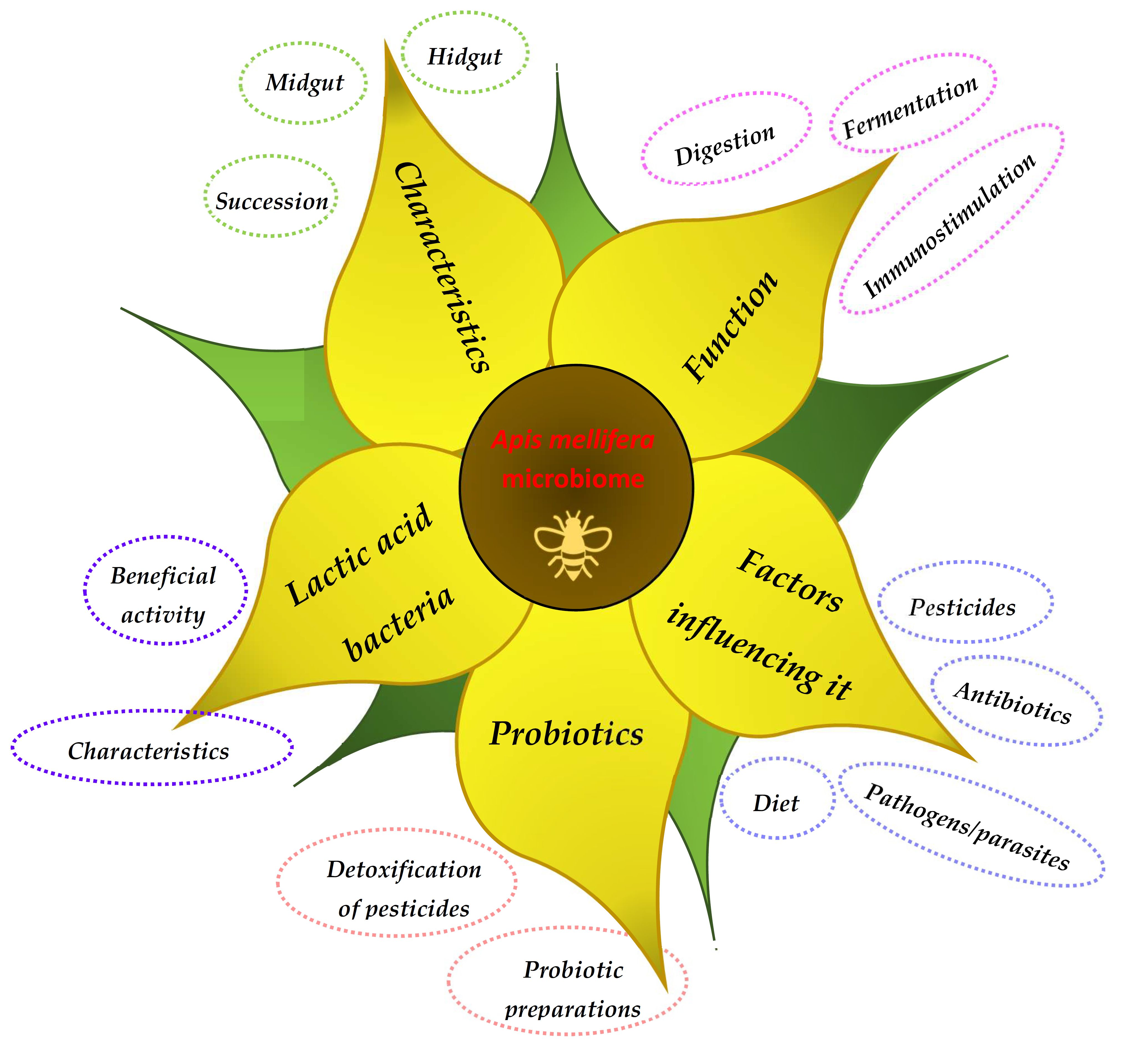
3. Honeybee Microbiota

Animals that form social communities usually employ a characteristic microbiota that is essential for various processes that occur in the body [38]. The microbiota can be defined as a complex ecosystem of microorganisms that plays a critical role in a variety of metabolic functions, including modulation of glucose and lipid homeostasis, satiety regulation, management of energy, and the production of vitamins [39,40,41]. In addition, the microbiota participates in the regulation of various biochemical and physiological mechanisms by means of the production of metabolites and other substances [42]. Furthermore, the microbiota exerts anticarcinogenetic and anti-inflammatory activities, [38] and plays a significant role in the operations of the host immune system and induction of immune responses [43]. In return, the host immune system maintains a mutualistic relationship with the microbiota. This relationship enables the induction of protective responses toward pathogens and the introduction of regulatory pathways involved in the tolerance to harmless antigens [44].
While the importance of the gut microbiota is discussed more often now, the processes responsible for the beneficial features of microbial communities remain unclear [45,46,47]. The composition of the microbial communities that inhabit the gut vary significantly between different species and within them. The diversity in composition of the gut microbiota is influenced by topographical and short-term shifts in the microbial communities, with specific microorganisms inhabiting particular niches in the host during specific growth and developmental phases of the host [48].

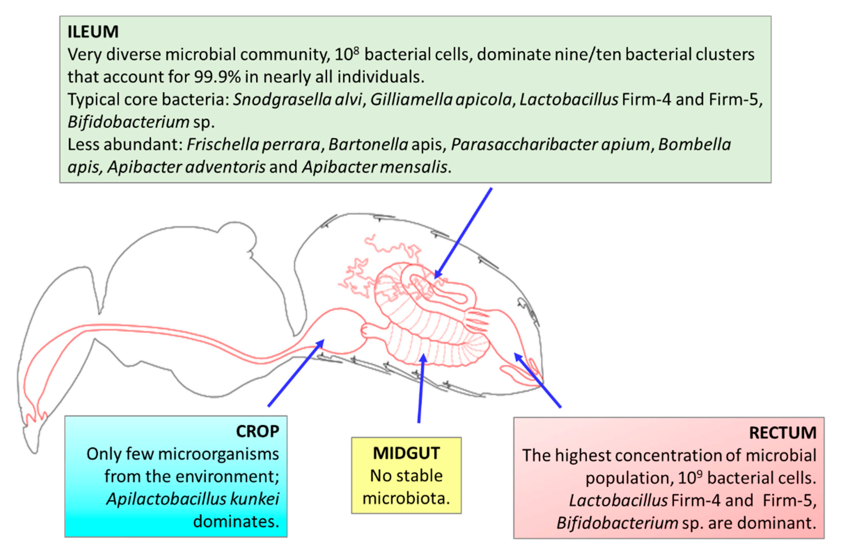
3.1. Characteristics
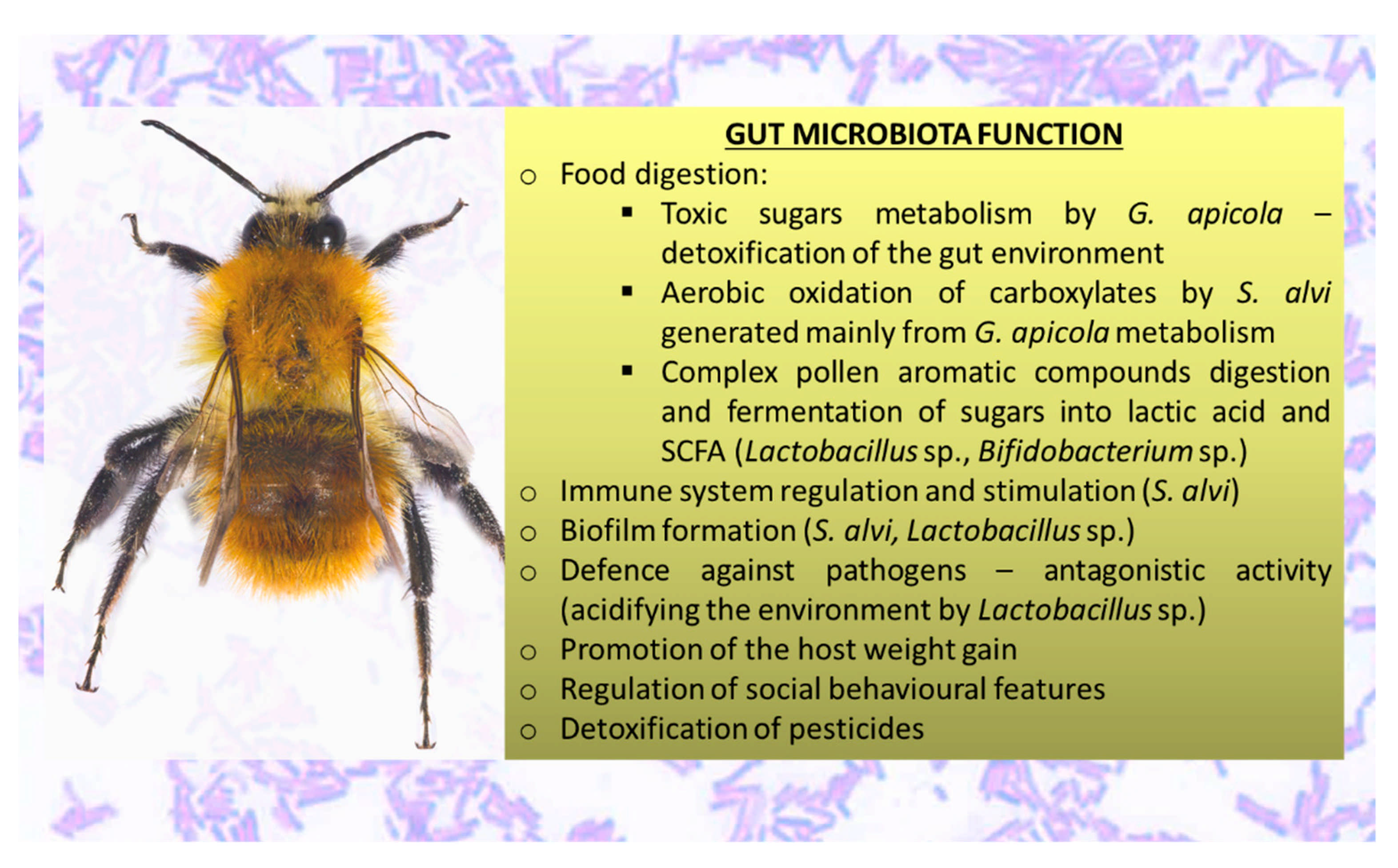
Insects represent the most diverse animal clade in terms of the number of species, the ecological habitats they inhabit, and their overall biomass [3]. A. mellifera is a useful model organism with a microbial community that displays high host adaptation. While its microbiota has some similarities with those of mammals, it has a much simpler composition. The main similarities and differences in the honeybee and human gut microbiota were reviewed previously [49].
Honeybees form huge colonies that contain thousands of nonreproductive female workers, hundreds of male drones, and only one reproductive queen [14]. Newly emerged workers have a reduced core gut microbiota or may lack it entirely [50]. Their bodies are colonized by microbial communities orally by means of social interactions with nurse bees within a few days of emergence [51,52]. During metamorphosis into pupae, the gut bacteria are excreted via defecation along with the gut epithelium, and the next colonization starts due to trophallaxis, contact with other bees, as well as from the hive [53]. The abundance of bacteria in the whole gut reaches its peak 3–5 days post-adult emergence [54]. However, taxonomic shifts take place after 3–8 days, which suggests pioneer or niche construction strains. The rectum community seems to finish the development of an emergent structure after three days. The ileum is more variable, with its final structure emerging after eight days. The most important factor influencing this process is the prevalence of core species, the host immune response related to it, and the successional alternation of the environment of ileum [4]. The workers are involved in age-associated tasks, and newly emerged bees are usually associated with hive maintenance and cleaning tasks. Therefore, the interactions with adult bees, contact with the comb, and consumption of bee bread are all potential routes of inoculation [54,55]. Dong et al. [50] analyzed the succession of A. mellifera workers gut microbiota from birth to senescence, i.e., from 0–40 days postemergence (dpe). The genera Gilliamella, Frischella, and Snodgrassella colonized the honeybee gut at 1 dpe; Lactobacillus, Bifidobacterium, and Commensalibacter colonized at 3 dpe, while a simultaneous reduction in Gilliamella was observed. At 12 dpe, significant colonization by L. kunkeei and Bartonella sp. appeared, while Bacteroides sp., Escherichia sp., Shigella sp., and Porphyromonadaceae decreased between 19 and 25 dpe. Commensalibacter sp. and Bifidobacterium sp. abundance was reduced at 25 dpe [50].
The microbiota of honeybees are located in different parts of the gut, including the crop (located between the esophagus and ventriculus, and used for storage and transport of nectar to the hive; also called stomach or sack); midgut; the hindgut, consisting of the ileum (a narrow tube containing six longitudinal folds) and lumen; and the distal rectum [56,57]. Only Parasaccharibacter sp. was found in relative abundance in worker hypopharyngeal glands [58].
It was estimated that adult workers’ guts are inhabited by characteristic, specialized microorganisms belonging to nine clusters of bacterial species [59]. Each of the clusters represents a set of bacterial strains that are related. Similar to human hosts, the microbial communities in honeybees are dominated by host-adapted species, which are highly intolerant of atmospheric oxygen; therefore, the transmission of bacterial species takes place by social interactions between hosts [60]. However, unlike mammalian gut microbiota, all of the bacterial species can be cultured in a laboratory [61].
Using 16S rDNA community surveys and metagenomics of the total DNA, it was determined that guts of worker honeybees are inhabited by nine bacterial species clusters that account for 95–99.9% of the bacteria in almost all individuals [59,62,63]. Two ubiquitous Gram-negative species—Snodgrasella alvi (nonfermenting sugar bacteria that form a film directly on the gut wall; family Neisseriaceae) and Gilliamella apicola (bacteria with the ability to ferment sugar that inhabits areas directed toward the center of the lumen; family Orbaceae)—that are members of the Proteobacteria phylum can be distinguished [2,59,63]. There are two Gram-positive species belonging to phylum Firmicutes that are ubiquitous and abundant; namely, Lactobacillus Firm-4 and Lactobacillus Firm-5, which inhabit the distal rectum [2,59]. In the majority of adult workers, Bifidobacterium asteroides is also found (albeit with much lower abundance) [53,61]. The mentioned bacterial species clusters are the most essential microorganisms in the honeybee gut, the so-called “core bacteria” [64]. There are also less-abundant/stable species from Proteobacteria: The Gammaproteobacteria Frischella perrara (Orbaceae family); the Alphaproteobacteria Parasaccharibacter apium, Bombella favorum, Bombella mellum, Bombella apis (Acetobacteraceae family, Alpha 2.2); and Commensalibacter sp. (Alpha 2.1) and Bartonella apis (Alpha 1) from the Rhizobiaceae family [50,53,59,63,65,66]. Representatives of phylum Bacteroidetes have also been identified in the honeybee gut—Apibacter adventoris and Apibacter mensalis [67,68].
A previous study [69] detected 10 taxa dominant in bee samples—four representatives of Lactobacillus sp., two Gilliamella sp., one Bifidobacterium sp., and one Snodgrassella sp.—that are considered to be part of the core gut microbiome of honeybees. Two of the taxa, from Frischella sp. and Bartonella sp., may vary depending on the environment. They are noncore members of honeybee gut [64]. Wang et al. [70,71] showed that the dominant phyla in honeybee GIT are Proteobacteria (63.2%), Firmicutes—(17.6%, with 15.9% of Lactobacillus sp.), Actinobacteria (4.1%, with 3.34% of Bifidobacterium sp.), and Bacteroidetes (1.7%, with 0.23% of Bacteroides sp.). The core member Lactobacillus Firm-4 was detectable in 98.4% of all analyzed bees in the study by Kešnerová et al. [64]. Tola et al. [63] analyzed A. mellifera gut microbiota from sub-Saharan African regions of Kenya, where indigenous and traditional management methods involving very little human intervention are practiced in beekeeping, unlike those practiced in Europe. They confirmed the core honeybee gut microbiota members were from the genera Gilliamella, Snodgrassella, Lactobacillus (Firm-4 and Firm-5), Bifidobacterium, Frischella, Commensalibacter, Bombella, Apibacter, and Bartonella, and that Frischella sp. was the third most dominant genus (16.9%), while Lactobacillus (Firm-4 and Firm-5) exhibited a lower abundance than has been demonstrated in other studies [63]. A summary of the GIT microbiota in honeybees is presented in Figure 1.

3.2. Functions

Considering an ecological perspective, gut microorganisms play a critical role in the process of codevelopment of insect-symbiotic interactions by means of secondary metabolites. Gut microbes take part in insects’ growth, development, and reproduction, and above all they contribute significantly to their metabolism [70]. Gut microorganisms synthesize essential nutritional compounds, increase the efficiency of digestion, and support insects in absorption of nutrients [72]. Most insects are inhabited by relatively few species (in comparison to mammalian gut), of which the majority is cultivable in the laboratory, but some harbor numerous communities of specialized bacteria. The factor defining limitation in gut microbiota in most insects is the lack of transmission routes between individuals. Exceptions are social insects such as termites, ants, and most importantly, bees. Social interactions give opportunities for transfer of gut microorganisms, therefore some of the most consistent and specialized gut communities, with significant functions in nutrition and protection, have been identified in social insects, such as honeybees [73].
Studies that concentrated on the beneficial health activities that microbes confer to their host have shown that the gut microbiota of honeybees plays as important a role as it does in mammals [2,3,4,45,48,49,50,72,74,75]. Two well-established functions of gut microbiota are nutrient biosynthesis and biomass deconstruction. The nutritional function was extensively studied in experiments comprising insects feeding with unbalanced and poor diets that lacked essential nutrients like amino acids and vitamins. These studies proved that insect endosymbionts help to produce nutrients that are not present in food [76]. The second function of some insect microbiota is biomass deconstruction and digestion. Both symbiotic microorganisms and host insects can release cellulolytic enzymes responsible for the deconstruction and hydrolysis of biomass, although studies have shown that microorganism activity increases the efficiency of these processes [76]. Gut microorganisms significantly contribute to the digestion of lipids and proteins, as well as the detoxification of secondary plant compounds. They also affect survival, overall size, and egg production. Moreover, they have been shown to play an important role in insect resistance against insecticides [77].
Gut microorganisms inhabiting insects can indirectly exert beneficial health effects on humans, in the case of parasitic diseases transmitted by insect vectors [78]. It was observed that in the gut of insect vectors, parasites ingested with bloodmeal reduced in number before coming into contact with host tissues. Microbial communities are thought to be an important factor influencing this effect. It was concluded that gut microorganisms contribute to the modulation of the competence of insect vectors. One of the possible mechanisms through which microbes support insects against parasites is through modification of the gut environment to constrain parasite development or induce an immune response of the host. They are also capable of producing antimicrobial peptides, which play a key role in the control of parasites and bacterial pathogens. In the study referred to above, after bloodmeal was ingested, the population of bacteria in the vector gut expanded rapidly. However, the microbiota were able to kill all parasites present [78,79]. The application of microbial symbionts to reduce vector competence is a promising approach to control the spread of insect vector transmitted pathogens [79].
Compared with the gut microorganisms of other animals, the honeybee microbiota is heavily involved in functions associated with carbohydrates, which reflects specific adaptations to a host’s diet that is rich in sugars. It provides the honeybee with sugar uptake systems belonging to various phosphotransferase systems. Many of these transporters are classified in the mannose family [73]. This feature of bacteria is important because only trace amounts of mannose are present in nectar, but it is highly poisonous when ingested at higher concentrations [73].
Another function associated with carbohydrates is enrichment of the host with arabinose efflux permeases. This family of transporters is involved in the transfer of different compounds such as antimicrobial proteins, amino acids, and sugars. A diverse set of transporters confers protection for the bacteria against a variety of pesticides applied in agriculture and naturally occurring antimicrobial proteins ingested by bees as part of their plant-based diet [3].
Furthermore, gut microorganisms influence the transformation of both nectar into honey and plant buds and exudates into propolis, owing to their fermentation properties [80]. They are also responsible for the freshness of honey [81].
One of the ways by which the gut microbiota can affect the health of honeybees is through modulation of the immune responses of the host [82]. Microorganisms impact the development and morphogenesis of the immune system and other organs and body structures [83,84]. One of the examples of how microbes affect a host is the symbiotic interaction between the fruit fly Drosophila melanogaster and the bacteria inhabiting its gut, Acetobacter pomorum [85]. This relationship influences the host’s body size, developmental rate, metabolism, activity of stem cells, and surface area of wings [85].
The primary role of gut microbiota in the functioning of mucosal immunity is not surprising, considering that the intestinal mucosa comprises the largest surface area in contact with antigens coming from the external environment, and that the dense layer of microbiota covering the mucosa constitutes the greatest proportion of antigens presented to the resident immune cells [75]. The mucosal immune system is responsible for the realization of two seemingly contradictory functions. It must tolerate microbiota inhabiting the gut to prevent the induction of harmful systemic immune responses, while controlling the number of microorganisms to avoid overgrowth and translocation [86]. Gut microorganisms are involved in the fulfillment of these objectives [75]. They control intestinal homeostasis through a variety of mechanisms involving substances like lipopolysaccharides, flagellins, and peptidoglycans. They interact with cell receptors such as Toll-like receptors, and they activate intracellular signaling pathways associated with cell survival, replication, apoptosis, and inflammatory responses [87,88,89]. In return, the host immune system controls the composition of microbes by releasing molecules like defensins, lectins, reactive oxygen species, and bacteriocins, which effectively constrain the expansion of pathogenic microorganisms [87,88,89].
Antimicrobial peptides are crucial components of innate immunity aimed at defense against the invasion of pathogens. They are determinants of the microbiota composition, as their role is to damage pathogenic microorganisms’ cells by means of membrane perforation [90]. Four families of antimicrobial peptides (abaecin, apidaecin, defensin, and hymenoptaecin) are evoked within the honeybee hemolymph during immune challenge. In one study, bees lacking gut microbiota were compared with bees inoculated with the normal gut microbiota by feeding with hive bee guts or with the bacteria S. alvi. It was observed that apidaecin and hymenoptaecin expression was upregulated in bees inoculated with gut microbiota, which indicates that the gut microbiota induces immune responses in bees [82].
The honeybee microbiota was observed to promote body-weight gains. To examine the effect of the microbiota on the growth of hosts, body-weight measurements were made in the presence and absence of gut microorganisms. Germ-free and conventional bees were received from pupae that were collected from hives and allowed to emerge in sterile laboratory conditions [2]. Bees deprived of microbiota achieved significantly lower weight gain (by 82%) than conventional bees. The weight gain was associated with the insulin/insulin-like signaling pathway, which plays a critical role in growth, reproduction, and aging, and regulates homeostasis and behavior in bees [2].
Gut microorganisms inhabiting insects do not just affect the digestive system. Various studies proved the existence of a gut microbiota–brain axis, meaning that gut microorganisms induce alteration of neurophysiology and changes in behavior of insect hosts [91,92]. For example, microorganisms can alter volatile profiles and the olfactory behavior of their insect hosts. Consequently, they regulate the ways in which individuals interact through chemical communication, aggregate in groups, and make decisions concerning foraging and mating. For instance, lower termite Reticulitermes speratus conspecific intruders are more quickly recognized and attacked when they are colonized by foreign gut bacteria releasing unfamiliar scents. Another example is found with the leaf-cutting ant Acromyrmex echinatior, in which suppression of the gut microbiota induces aggression among non-nestmates through alterations in cuticular hydrocarbon profiles [93]. Gut microorganisms can also increase the longevity of insects. An example of such activity of microbes is in D. melanogaster, the lifespan of which was significantly elongated after application of probiotic and symbiotic formulations. These formulations rescued metabolic stress markers through management of insulin resistance and energy-regulatory pathways [91]. Gut microorganisms also affect the neurophysiological development of the host, as they support cognition by enhancing its capacity to memorize and learn. A recent study linked gut microorganisms with markers of Alzheimer’s disease [93].
The gut microbiota of honeybees was observed to impact the neurophysiology and behavior of hosts. Microbes can also affect host behavior by alteration of the levels of biogenic amines such as serotonin, octopamine, and dopamine. Levels of these compounds vary seasonally in the worker’s brains, increasing in summer when foraging activity is the highest, and at different life stages, being lower in brains of newly emerged, germ-free bees [94]. Furthermore, the gut microbiome plays a key role in the regulation of social behavioral features in honeybees [95].
Gut-microbiota involvement in xenobiotic metabolism has been known for years, and this ability sheds light on the potential ability to maintain microbiota as a target for drugs to effectively contribute to treatment for various diseases [96,97]. As honeybees are exposed to a wide range of pesticides, an important role of their gut microbiota is the detoxification of xenobiotics, especially neonicotinoid insecticides [98]. Wu et al. [98] demonstrated that honeybee gut microbiota contribute to the host’s endogenous detoxification and resistance to thiacloprid and fluvalinate, as it promotes the expression of detoxification enzymes in the midgut. The importance of honeybee gut microbiota was also illustrated by a metagenome project in which symbionts of honeybees were affected by viruses. This led to detrimental effects on the growth and development of bees, and could be a major cause of colony collapse disorder (CCD) [76]. Undigested pollen was observed in the fecal content of honeybees that died due to CCD, and it indicated a deficit in the abundance of beneficial probiotic bacteria in the GIT. This may have been caused by pesticides and antibiotic residues [99].
The microbiota synthesizes enzymes such as proteases and glycosidases, metabolizes indigestible polysaccharides, produces essential vitamins, and conducts xenobiotic metabolism. This significantly expands the host’s biochemical capacity [100]. The fermentation of indigestible carbohydrates and oligosaccharides by bacteria belonging to the genera Bacteroides, Roseburia, Bifidobacterium, and Faecalibacterium results in the formation of short-chain fatty acids (SCFAs) including butyrate, propionate, and acetate [71,101]. These substances provide rich sources of energy for the host. Butyrate helps prevent the accumulation of toxic byproducts of metabolism [101]. Honeybee gut microbiota functions are presented in Figure 2.

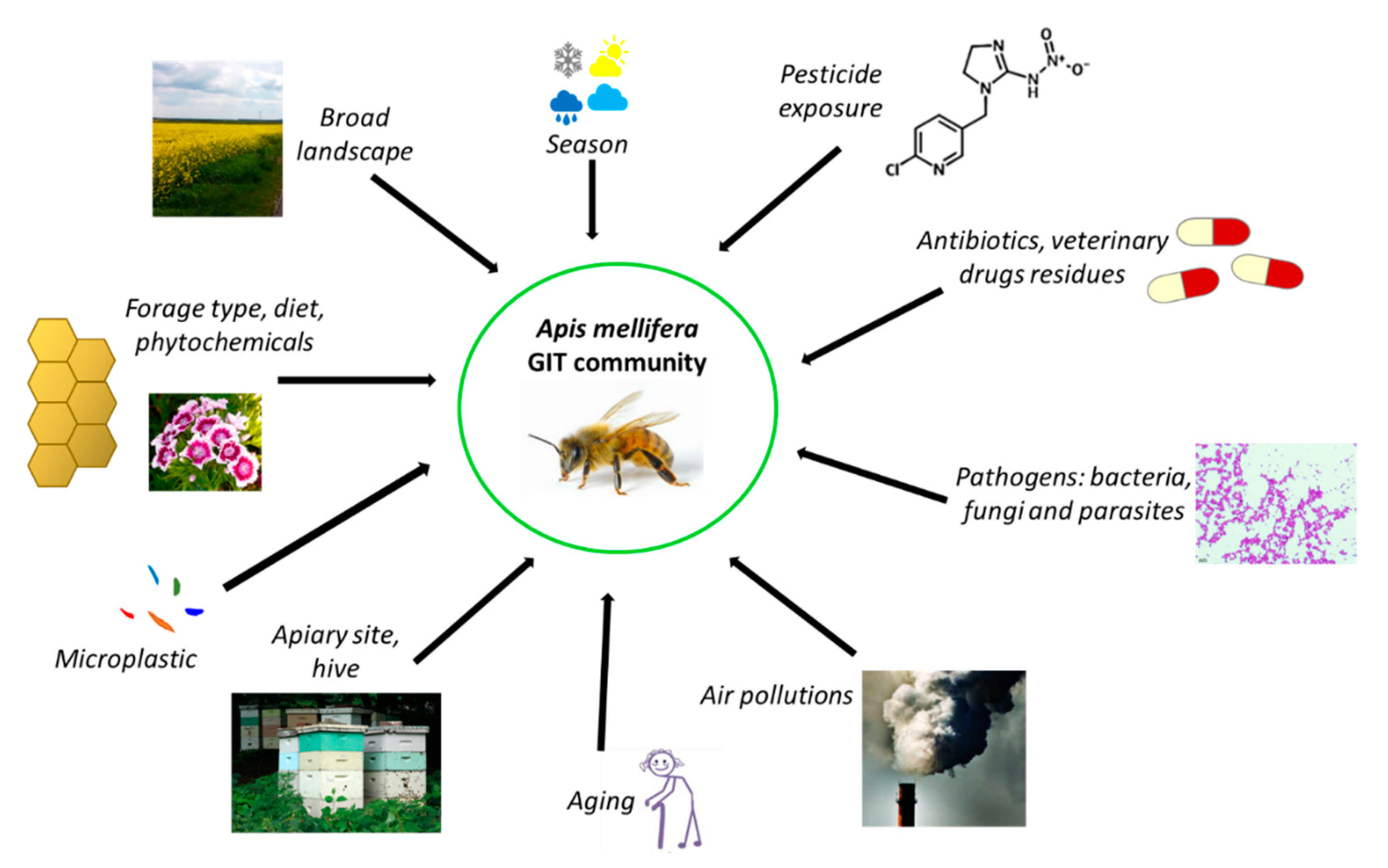
3.3. Factors Affecting Honeybee Microbiota

Interactions between the honeybee gut community and the environment are complex and not well understood. There exists a huge diversity of gut microorganisms among insects, influenced by many factors such as habitat, feeding preference, life stage, and host species. Jones et al. [59] showed that the broad landscape influenced the diversity of some members of honeybee gastrointestinal microbiota, especially those belonging to Proteobacteria and Firmicutes. Muñoz-Colmenero et al. [102] demonstrated that the environment plays the main role in determining honeybee microbiota, and that agricultural treatments cause disruption to the bacterial community.
Many pesticides (e.g., chlorothalonil, imidacloprid, and coumaphos) contribute to important adverse health effects [7,103,104,105,106] and unfavorable changes in the structure and function of the honeybee microbiome [107]. Honeybees are exposed to them through contaminated nectar, pollen, and water. The abundance of Lactobacillales in honeybees exposed to chlorothalonil was significantly lower compared to a control group [108]. Sublethal doses of insecticides, such as fipronil, imidacloprid, thiamethoxam, and coumaphos, induced significant decreases in the quantity of Lactobacillus sp. and Bifidobacterium sp. regardless of season [108]. Exposing honeybees to glyphosate negatively affected the gut microbiome, leading to a decreased total number of gut bacteria and reduced amounts of S. alvi, Bifidobacterium, and Lactobacillus (Firm-4 and Firm-5) [52]. Motta et al. [109] investigated the effects of glyphosate on bees under laboratory and field conditions, and demonstrated that honeybees transport glyphosate to the hive, which can increase the exposure of insects to xenobiotics. Furthermore, glyphosate reduced the abundance of beneficial bacteria in the honeybee gut in a dose-dependent way [109]. According to Liu et al. [110], high and very high concentrations of thiacloprid (a neonicotinoid insecticide) led to dysbiosis in the gut microbial community of honeybees. It caused a decrease in total microbial abundance in a dose-dependent manner in three treatment groups of insects. Another neonicotinoid insecticide, nitenpyram, contributed to key alterations in the microbiota community, leading to metabolic changes and a decrease in effectiveness of the immune system [111]. Alberoni et al. [112] investigated the long-term impact of two neonicotinoids (imidacloprid and thiacloprid), on worker honeybees’ gut microbiota under open-field conditions after acute and chronic exposure. Numerous negative effects were observed in several microbial species such as Frischella sp., Lactobacillus (Firm-4 and Firm-5), and Bifidobacterium sp., the changes of which contributed to gut dysbiosis. The general problem with pesticides and honeybees is that pest-control methods alter the composition, diversity, and physiology of gut microbiota, and consequently affect honeybee health, especially after long-term exposure [113,114]. Furthermore, exposing honeybees to pesticides negatively impacts their gut microbiome and increases their susceptibility to infection by opportunistic pathogens [112]. To date, there has been no research on the mechanisms of detoxification of neonicotinoid insecticides by LAB (likewise probiotic) with the application of cell lines. A prerequisite for the toxic effects of a pesticide is its uptake into the body (bioavailability). Future studies should test the reduction in uptake of pesticides or their metabolites in a Caco-2 gut model (passage through the gastrointestinal epithelium) under the influence of probiotics. The toxicity of metabolites of pesticides conducted by some LAB strains is not known (summarized in Table 1), and it is not known whether these metabolites are more or less toxic than the substrate.
Honeybees exhibit a complex social network of microorganisms that can be characterized by variations according to geographic location [5,114]. For example, in A. mellifera jemenitica, the rural honeybee characteristic of the Kingdom of Saudi Arabia, some bacteria identified in the alimentary tract—Citrobacter sp., Providencia vermicola, Exiguobacterium acetylicum, and Planomicrobium okeanokoites—are unique to this species [115]. The core honeybee intestinal microbiota is also subjected to global seasonal variations [108]. Few studies have shown how extreme modifications impact gut microbiota dynamics during overwintering. However, seasonal changes in the honeybee microbiome in Canada were investigated by Bleau et al. [53], and they observed a decrease in the abundance of Enterobacteriaceae from September to November, while the relative abundance of Neisseriaceae increased. Subotic et al. [69] found that the honeybee microbiome changes seasonally. Another study found differences in bacterial abundance of honeybee gut community members between summer and winter months that were linked to diet [64]. The lowest diversity and highest bacterial loads were observed in winter bees (with high levels of Bartonella sp. and Commensalibacter sp.) [86]. Furthermore, diet (type of sugar used in winter forage, nutritional stressors, poor-quality diet, and propolis-rich and propolis-poor diets) has been shown to determine the profile of the dominant honeybee gut community [71,116,117]. A high-fat diet (palm oil) significantly increased the abundance of Gilliamella sp., while a decreased abundance of Bartonella sp. was observed [118]. In another study, honeybees that were subjected to feeding with “aged” pollen displayed increased mortality, a higher load of Nosema sp., a pathogen of fungal origin, and a significant shift in the gut microbiota composition [6].
Due to the increasing risk of CCD, attempts have been made to treat colonies using chemical methods. Antibiotics can influence the host by altering the species of gut microbiota. Daisley et al. [119] documented the deleterious effects of antibiotics on the gut microbiome, immunity, and productivity of honeybees. Several residues of antibiotics and veterinary chemotherapeutics are detected in honey, showing that honeybees are still exposed to them, despite many countries banning their usage in beekeeping [120,121]. These stressors prompt a reduction of bacterial species in the honeybee gut, weakening their immunity and increasing their susceptibility to infections [122]. In one study, honeybees underwent treatment with antibiotics, which resulted in the elimination of their microbiota. It was found that bees were more susceptible to infections by Nosema ceranae (a frequent honeybee pathogen) due to its negative influence on the immune system, which was illustrated by the depletion of the expression of genes that encode antimicrobial peptides [54]. Another study suggests that disturbance of gut microbiota with tetracycline decreased honeybee survival, which was associated with an elevated susceptibility to the opportunistic pathogen Serratia sp. [6]. Furthermore, antibiotic residues may be found later in honeybee products. Ortiz-Alvarado et al. [123] studied the effect of two commercial beekeeping antibiotics—Terramycin (oxytetracycline) and Tylan (tylosin tartrate)—on bee physiology and behavior throughout development. The results of the study showed that antibiotic treatments increased the amount of lipids and the rate of behavioral development. The timing of the antibiotic treatment affected the age of onset of behaviors, starting with cleaning, then nursing and foraging. Bees treated during the larva–pupa stages demonstrated an accelerated behavioral development and loss of lipids, while bees treated from larva to adulthood had a delay in behavioral development and loss of lipids. These effects of antibiotic treatments suggest a role of microbiota in the interaction between the fat body and brain, which is important for honeybee behavioral development. Zheng et al. [49] presented an overview of the recent research in the field of antibiotic use. Long-term antibiotic use may have impacted the diversity within human gut communities and has resulted in high frequencies of resistance determinants [124]. In the United States and other countries where beekeepers used antibiotics since the late 1940s to control or prevent larval bacterial diseases (foulbrood), antibiotic exposure has affected gut communities of honeybees [125,126,127]. This practice has resulted in high frequencies of antibiotic resistance determinants in core gut bacteria isolated from bees in the United States, in contrast to gut bacteria of honeybees from countries that do not permit the use of antibiotics in beekeeping [120,128]. In both human and honeybee gut communities, resistance determinants have been exchanged among community members through horizontal transfer [129]. In the European Union (EU), legal permission for the application of antibiotics is connected with the food safety and protection of consumers. The new European environmental strategy “The European Green Deal” [130] stresses the role of the “from farm to fork” approach, which entails designing a fair, healthy, and environmentally friendly food system. The strategic plans will need to reflect an increased level of ambition to reduce the use and risk of chemical pesticides, as well as the use of fertilizers and antibiotics. The EU needs to develop innovative ways to protect harvests from pests and disease, and to consider the potential role of new innovative techniques to improve the sustainability of the food system, while ensuring that they are safe. The most significant act that regulates food safety is Regulation No. 178/2002 [131], which includes the basic rules on food safety and established the European Food Safety Agency. European food safety is regulated by over a hundred legal acts, and Regulation No. 415/2014 [132] established the EU reference laboratory for bee health, which coordinates the methods employed in the member states for diagnosing relevant bee diseases. In reference to the veterinary medicinal products as antibiotics in the bee sectors, member states have to comply with the European rules on veterinary medical products. The definition of honey is regulated in the Directive 2001/110/EC [133]. The Commission stresses the limited availability of veterinary medicines for bees. According to Regulation (EC) No 470/2009 [134], the veterinary medicinal products intended for use in food-producing animals like bees have to be scientifically evaluated according to human food-safety requirements. Regulation (EU) No 37/2010 [135] outlined the EU Maximum Residue Limits (MRLs) for residues of pharmacologically active substances in honey. For some substances (e.g., amitraz and coumaphos), an MRL has been established, while for others the evaluation demonstrated that no MRL was required to protect food safety (e.g., flumethrin, oxalic acid, and tau fluvalinate). Products that have not been assessed as safe according to these requirements can neither be authorized nor used otherwise for food-producing animals. A new Regulation (EU) No 6/2019 [136] on veterinary medical products will come into effect on 22 January 2022. The regulation sets out rules for the sale, manufacture, import, export, supply, distribution, control, and use of veterinary medicinal products (VMPs), aiming to modernize legislation, stimulate innovation in and increase the availability of VMPs, and strengthen the EU’s campaign against antimicrobial resistance. The regulation specifies clear and fully harmonized labeling requirements, adopts a simpler system for making decisions on exceptions, and uses a risk-based approach to pharmacovigilance and controls among the key measures. It defines clear rules for organically sourced VMPs and novel therapies that also aim to encourage the development of new VMPs. It is important that the regulation strengthens the EU’s fight against antimicrobial resistance by banning the preventive use of antibiotics in groups of animals, banning the preventive use of antimicrobials via medicated feed, restricting the use of antimicrobials as a control treatment to prevent a further spread of infection, introducing a reinforced ban on the use of antimicrobials for promoting growth and increasing yield (in addition to the 2006 prohibition of using antibiotics as growth promoters in feed), including the possibility to reserve certain antimicrobials for humans only, obligating EU countries to collect data on the sale and use of antimicrobials, introducing science-based maximum limits for cross-contamination of feed with antimicrobials, and introducing various other measures to promote the responsible use of antimicrobials.
Another factor influencing honeybee gut microbiome composition is exposure to particulate-matter air pollution [137], which has been investigated for the buff-tailed bumblebee (Bombus terrestris) [138]. Likewise, there is scant evidence on the effects of heavy metals on honeybees [139,140].
In a recent study by Wang et al. [141], the authors investigated how microplastics impact honeybee fitness. They fed newly emerged bees for 14 days with microplastics under laboratory conditions. The accumulation and degradation of microplastics in the gut and interaction with gut bacteria was observed. A significant decrease in diversity and changes in the core microbial population took place. The real challenge with environmental factors affecting the honeybee microbiome, such as air pollutants, heavy metals, and microplastics, is determining the mechanism of their action and how they should be measured. Several factors influencing the honeybee gut community are presented in Figure 3.

4. LAB as a Significant Component of the Honeybee Microbiota and Their Beneficial Activities

Similar to other animals, in honeybees LAB are an integral part of the microbiota [142]. Microaerophilic conditions dominate the honeybee digestive system, and the temperature of 35 °C and presence of sugars from nectar are ideal conditions for lactic acid bacteria [19]. They can be characterized as Gram-positive, nonsporulating, catalase-negative bacteria that are highly tolerant to low pH [143]. These bacteria attain the shape of rods and cocci [144]. They utilize carbohydrates to obtain energy, using endogenous carbon source as final electron acceptor [145]. As the name suggests, LAB produce lactic acid [146]. Based on the products of fermentation, they can be classified either as homofermentative, producing mainly lactic acid, or heterofermentative, producing other substances such as acetic acid or ethanol [147,148]. Considering taxonomy, LAB belong to two different phyla, Firmicutes and Actinobacteria [148]. In phylum Firmicutes, LAB belong to the order Lactobacillales, which includes six families: Aerococcaceae, Carnobacteriaceae, Enterococcaceae, Lactobacillaceae, Leuconostocaceae, and Streptococcaceae [149]. LAB in the Actinobacteria phylum belong to the Bifidobacterium genus [150].
The most significant representative of LAB is Lactobacillus sp. This genus comprises 261 species that display extreme diversity in terms of phenotype, ecology, and genotype. Zheng et al. [151] examined the taxonomy of Lactobacillaceae and Leuconostocaceae using whole genome sequences. Their evaluation concerned parameters including core genome phylogeny, pairwise average amino acid identity, signature genes specific for clade, physiology, and ecological characteristics. They proposed to reclassify the genus Lactobacillus into 25 genera including an amended Lactobacillus genus, Paralactobacillus, and 23 newly introduced genera: Acetilactobacillus, Agrilactobacillus, Amylolactobacillus, Apilactobacillus, Bombilactobacillus, Companilactobacillus, Dellaglioa, Fructilactobacillus, Furfurilactobacillus, Holzapfelia, Lacticaseibacillus, Lactiplantibacillus, Lapidilactobacillus, Latilactobacillus, Lentilactobacillus, Levilactobacillus, Ligilactobacillus, Limosilactobacillus, Liquorilactobacillus, Loigolactobacilus, Paucilactobacillus, Schleiferilactobacillus and Secundilactobacillus. The description of the family Lactobacillaceae was extended to include not only genera previously belonging to the family Lactobacillaceae, but also those belonging to Leuconostocacae. This reclassification improves the understanding of the beneficial health activities of these bacteria due to the fact that species that are more closely related, and thus share more physiological features, are located in the same genus [151]. In the current text, the LAB nomenclature used follows the source references.
LAB can be found in decomposing plant materials, fermented food, sourdough, and cavities of animals, including humans [145]. These bacteria are important from a food-industry perspective because they are utilized as bioconversion agents and starter cultures in food production [152]. They are involved in the preparation of dairy products (e.g., hard cheeses, butter, yogurt, and kefir), fermented meat and fish products, and fermented vegetables (e.g., sauerkraut, pickles, and olives) [153,154,155,156,157,158,159]. They are attractive starter cultures because they produce bacteriocins, which display inhibitory activity toward food-spoilage microorganisms [152].
Various species of LAB occur in the respiratory, intestinal, and genital tracts of animals [160]. In humans, they predominantly inhabit the oral cavity, ileum, colon, and vagina [161,162]. LAB in the microbiota are involved in a variety of different functions that affect the host. For instance, LAB inhibit the expansion of pathogens in the gut as they compete for nutrients [163]. Since they are primarily fermenting saccharides, but also utilize amino acids, they can significantly deplete the nutrient resources to both saccharolytic and proteolytic species [164]. Furthermore, the products of their metabolism, such as organic acids, carbon dioxide, ethanol, or hydrogen peroxide, also contribute to the fight against pathogens [165]. LAB also produce bacteriocins—proteinaceous molecules that disturb the growth of most bacteria. They are capable of the biosynthesis of many different types of antagonistic molecules [166]. As previously described, gut microbiota are significantly involved in the immunomodulation of the host, and LAB, as a constituent of the microbiota, participate in these interactions [167]. The most prominent effect of LAB is related to the enhancement of the ratio between anti-inflammatory and proinflammatory cytokines. LAB components (e.g., lipoproteins and exopolysaccharides) may also directly induce immune responses. Furthermore, it was observed that Lactobacillus johnsonii induces the aggregation of Helicobacter pylori (a pathogen that invades the gut), which contributes to the depletion of the bacterial load and facilitates the clearance of the aggregated pathogen [168]. LAB were also observed to affect the metabolism of lipids. A study performed by Kishino et al. [168] demonstrated that Lactobacillus plantarum displays the ability to metabolize fatty acids and is involved in the saturation metabolism of polyunsaturated fatty acids, which leads to the generation of hydroxyl fatty acids, oxo fatty acids, conjugated fatty acids, and partially saturated trans-fatty acids as intermediates [168]. Fatty-acid analysis in mice revealed that intestinal microbes modify the composition of fatty acids in the host [168]. LAB were also observed to efficiently protect human and animal intestinal epithelial cells from the enteric viral infections [169]. In that study, selected LAB strains were chosen based on previous in vitro trials and were incubated with animal and human intestinal cell lines (of nontumor origin), which were further exposed to rotavirus and transmissible gastroenteric virus. It was observed that various strains displayed moderate to total cell monolayer protection against viruses. The most prominent effect was recorded for Lactobacillus rhamnosus and Lactobacillus casei Shirota. A significant antiviral effect was observed for Enterococcus faecium, Lactobacillus fermentum, Lactobacillus pentosus, and L. plantarum [169].
The presence of LAB within honeybees has been extensively investigated over the years. A study conducted by Vasquez et al. [57] demonstrated the presence of 13 bacterial species representing genera Lactobacillus and Bifidobacterium in the honeybee crop. Among these species, Lactobacillus kunkei was found to be dominant [57]. Another study performed by Olofsson and Vasquez [170] examined the microorganisms inhabiting honeybee stomach. Phylogenetic research pointed out the presence of 10 different phylotypes of LAB. Among them, five were closely related to L. kunkeei, B. asteroides, and Bifidobacterium coryneforme. The other five phylotypes were more distantly related, but were mostly related to the Lactobacillus genus [170]. Another study by Vasquez et al. [171] documented the presence of Lactobacillus helveticus in the honeybee stomach. Forsgren et al. [57] isolated L. kunkeei, B. asteroides, and B. coryneforme from the crop. Olofsson et al. [172] isolated even more strains of LAB from the honeybee crop: Lactobacillus helsingborgensis, Lactobacillus kimbladii, Lactobacillus mellis, Lactobacillus mellifer, Lactobacillus melliventris, Lactobacillus apis, Lactobacillus kullabergensis, Lactobacillus apinorum, L. kunkeei, and B. coryneforme.
The rectum, which is where fecal waste is stored prior to defecation, was also shown to be abundant in Lactobacillus species and B. asteroides clusters. Lactobacillus sp. can also be found in the lumen of ileum [61]. Audisio et al. [173] performed a study and examined the whole intestinal tracts of honeybees from the esophagus to the rectum. In the research, eight strains belonging to Lactobacillus genus and five belonging to genus Enterococcus were isolated. They performed 16S rRNA analysis and identified Lactobacillus strains that belonged to species L. johnsonii and Enteroccocus strains of E. faecium. Furthermore, McFrederick et al. [174] reported the presence of three other species of Lactobacillus in the bee gut. Based on 16S rRNA analysis and fatty-acid profiling, it was established that these strains belonged to species Lactobacillus micheneri, Lactobacillus timberlakei, and Lactobacillus quenuiae. In another study performed by Janashia and Alaux [175], three different LAB species were isolated from the worker honeybee gut, namely Fructobacillus fructosus, Fructobacillus tropaeoli, and Fructobacillus pseudoficulneus. Iorizzo et al. [19] identified 24 strains from honeybee stomach and midgut of A. mellifera ligustica, a native endemic Italian subspecies. Ten strains of L. plantarum were found in the stomach, along with three strains of Apilactobacillus kunkeei, one strain of Lactococcus lactis, and one strain of F. fructosus; and eight strains of Al. kunkeei and one strain of L. plantarum were found in the midgut.
Rokop et al. [176] found bacteria belonging to genera Lactobacillus and Fructobacillus in bee pollen. Janashia and Alaux [175] isolated L. kunkeei and B. asteroides from bee pollen. Anderson et al. [177] isolated bacteria belonging to genus Lactobacillus, which were predominantly L. kunkeei. Bulgasem et al. [178] examined 15 types of this bee product from different sources. The identification procedure they performed used API 50 CH tests to prove the presence of L. plantarum, Lactobacillus curvatus, Pediococcus acidilactici, and Pediococcus pentosaceus. Aween et al. [179] conducted research using commercially available honey from Malaysia and isolated 36 strains by means of API CH 50 tests, six of which were identified as Lactobacillus acidophilus. Asama et al. [180] noted that bacteria belonging to the Lactobacillus genus were dominant among samples of honey, bee pollen, royal jelly, and the whole gut and honey stomach of bees. In whole guts of bees, Lactobacillus insectis was most abundant, while in bee pollen, royal jelly and honey the most abundant species was L. kunkeei. Libonatti et al. [181] isolated Weissella paramesenteroides from bee bread. Anderson et al. [177] also observed the presence of L. kunkeei in a sample of bee bread. Iorizzo et al. [19] identified 21 strains in bee bread: 10 strains of L. plantarum, four strains of F. fructosus, three strains of Al. kunkeei, three strains of Lactobacillus brevis, and one strain of L. lactis. Neveling et al. [182] documented the presence of fructophilic LAB (those preferring D-fructose over D-glucose) in biological materials isolated from fresh flowers, beehive elements, and honeybees collected in Stellenbosch and the Durban Botanical Garden in Durban, South Africa. These isolates were identified as L. kunkeei and L. brevis.
Magnusson et al. [183] isolated LAB from different flowers: P. pentosaceus was isolated from clover (Trifolium L.); P. pentosaceus and L. plantarum from chestnut (Castanea Mill.); Lactobacillus coryniformis, L. plantarum, Lactobacillus sakei, Pediococcus parvulus, and P. pentosaceus from dandelion (Taraxacum officinale); and L. plantarum from lilac (Syringa vulgaris). In a study performed by Rodríguez et al. [184], LAB strains were isolated from passion fruit (Passiflora edulis) flowers, custard apple flowers (Annona reticulate) and meddler (Mespilus germanica) flowers gathered in Tucumar in northern Argentina. Six different strains were isolated from passion fruit flowers, namely Enterococcus casseliflavus, Enterococcus gallinarum, Enterococcus faecalis, L. lactis ssp. lactis, Leuconostoc mesenteroides ssp. Mesenteroides, and Weisella cibara. Two strains were isolated from custard apple flowers: Enterococcus casseliflavus and L. brevis. Four strains were isolated from medlar flowers: E. casseliflavus, L. lactis, L. lactis ssp. lactis, and Leuconostoc pseudomesenteroides [184]. In research conducted by Endo et al. [185], three strains of fructophilic LAB were isolated from flowers gathered in South Africa. The biological material for isolation comprised flowers of peony (Paeonia suffruticosa) and bietou (Chrysanthemoides monilifera). The isolates were closely related to Lactobacillus fructivorans, Lactobacillus homohiochii, Lactobacillus lindneri, and Lactobacillus sanfranciscensis. Based on 16S rRNA gene analysis, these three strains were classified as a novel strain with the proposed name Lactobacillus florum sp. nov. The presence of LAB in flower pollen proves that it can be found in the honeybee GIT and its environment, and indicates the transmission of microorganisms between honeybees and flower pollen grains and nectar [163,186].
LAB are involved in a variety of functions that affect honeybees. One of their profitable activities is the contribution to bee nutrition. It was suggested that bacteria belonging to genus Bifidobacterium, Simonsiella, or Lactobacillus are capable of the production of SCFAs such as acetic acid, which are waste products of carbohydrate metabolism [187]. Assimilation of these compounds can supplement the nutrition of bees. It is possible that SCFAs can be absorbed in the rectal wall of insects and it has been determined that the greatest amount of pollen and biomass of bacterial origin among adult honeybees is located inside the rectum [187]. Among the bee gut microbiota known to produce SCFAs, Lactobacillus Firm-5 is considered the main producer of succinate and pimelate, while B. asteroides is considered the main producer of valerate [49]. A. mellifera could obtain extra nutrition from these rectal bacteria during overwintering, as consumed food storage takes place within the rectum for longer periods of time [187].
LAB also exhibit colonization resistance against microbes that are potentially harmful, preventing the dysbiosis in the gut. They can influence the host by changing the composition of gut microbiota. In honeybees, LAB can protect against pathogens contributing to CCD such as Paenibacillus larvae, Melissococcus plutonius, Serratia marcescens, Ascosphaera apis, and Nosema sp. [188,189,190,191,192,193]. Iorizzo et al. [163] tested the antagonistic activity of 85 strains of L. kunkeei against A. apis DSM 31116, of which 23 displayed high inhibitory activity toward the fungus, and nine strains caused 100% inhibition. Tejerina et al. [194] observed 80% inhibition of A. apis in vivo after feeding honeybees with three strains of Lactobacillus sp. bacteria added to sugar syrup at 105 CFU/mL concentration. L. kunkeei, Lactobacillus crispatus, and L. acidophilus showed the strongest antagonistic activity against a highly virulent bacterium, P. larvae [195]. In one study, honeybee larvae and adult bees were administered a mixture of four different strains of L. kunkeei. This resulted in reduced mortality related to infection of larvae by P. larvae, as well as a decrease in counts of N. cerenae spores in adult individuals [190]. Evans and Armstrong [125] considered the influence of gut microorganisms on infection with P. larvae, and reported that bacteria isolated from A. mellifera inhibited the growth of P. larvae. However, these host bacteria did not belong to stable, core gut microbial community. Despite successful laboratory studies against P. larvae, the application of LAB in field experiments is not always effective [196,197], but some results are promising [190,198]. The antimicrobial effect of LAB from the honeybee environment against bee pathogens were discussed in a review by Ramos et al. [199].

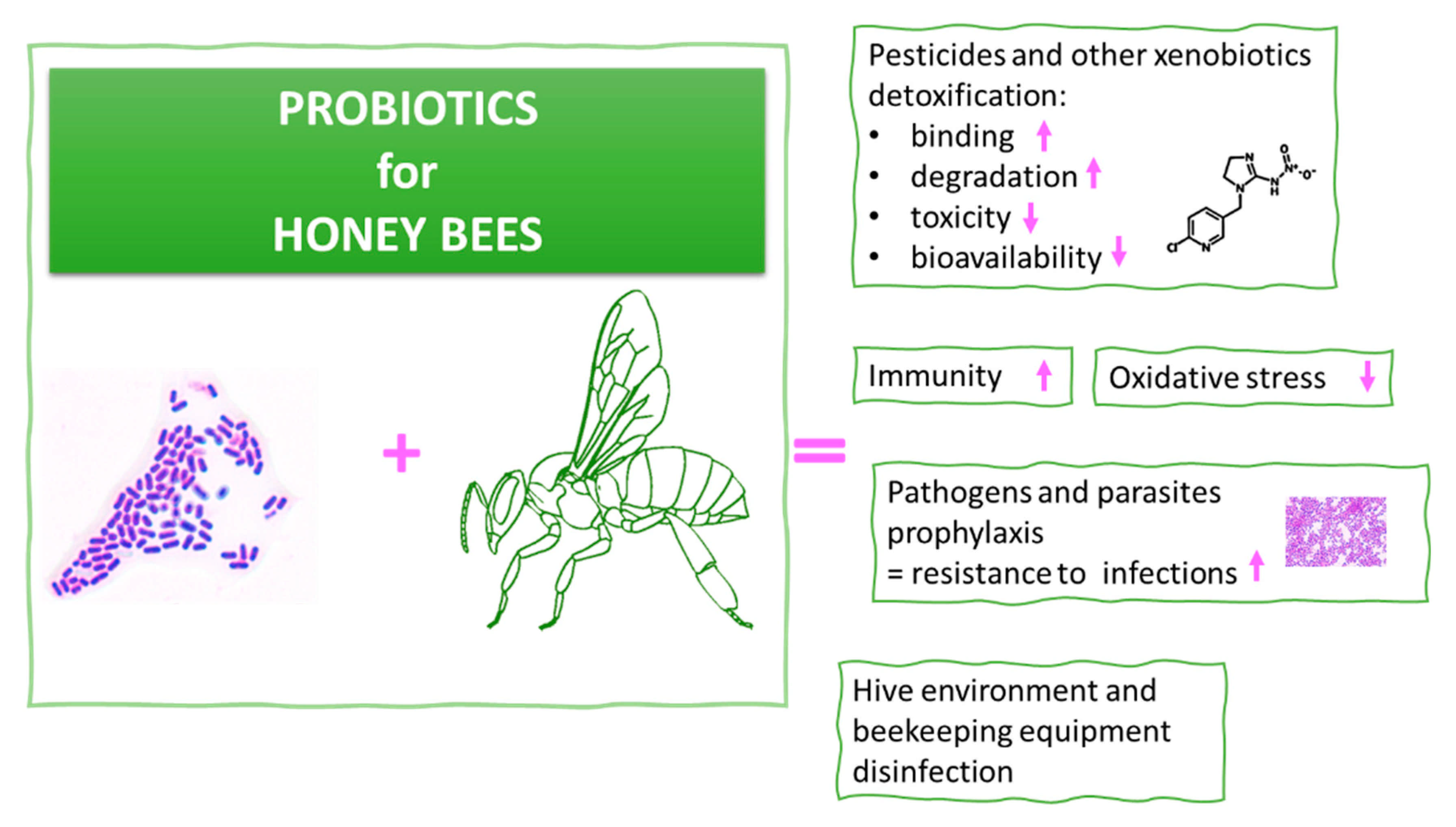
5. Probiotics for Honeybees

Due to their beneficial health effects, some LAB are considered probiotics. Probiotics are defined as live microorganisms that, if administered in adequate amounts, confer a health benefit on the host [200]. In order to identify the microorganism as a probiotic, it should fulfill a set of conditions [201]. Therefore, considering the fact that some LAB are probiotic, they are nonpathogenic, nontoxic, and achieve GRAS (Generally Recognized as Safe) status. They remain alive and active in GIT, are highly resistant to digestive enzymes and stomach acid, and have the ability to adhere to the intestinal epithelium [201,202]. Additionally, for a probiotic strain to possess the status “probiotic”, it should distinguish itself with a special feature characteristic of all LAB. For example, in the case of probiotics for honeybees, this could include immune-system stimulation; pathogen inhibition; or pesticide/xenobiotic degradation, binding, or neutralization. The supplementation of honeybees with probiotic LAB is a promising concept that could mitigate the harmful effects of pathogens and pesticides. However, there is no information regarding the molecular mechanisms of probiotics in protecting honeybees against pathogens. From the literature, data show that LAB are most effective in pesticide degradation during fermentation, which takes place in the GIT of honeybees. The protective effects of probiotics toward toxicity (cyto- and genotoxicity) of pesticides, especially neonicotinoid, have not been investigated comprehensively. A short review of LAB and pesticide interactions is presented in Table 1. The organophosphorus insecticide chlorpyrifos seems to be one of the most widely studied pesticides in relation to LAB. These studies indicate that the application of LAB in pesticide detoxification/removal is a safe and highly efficient method, both from the culture medium as well as during the fermentation of the contaminated food. Binding or biosorption is preferred to degradation, as the latter can generate toxic metabolites [203].
Table 1. Summarized effects of probiotics on pesticide mitigation, binding, degradation, metabolism, and toxicity in diverse systems.
StrainPesticide/sEffectReference
Human gut microbiota plus L. plantarum
ATCC 11095
Phoxim, chlorpyrifos,
imidacloprid, thiamethoxam, emamectin
benzoate, chlorpyrifos-d10, thiamethoxam-d4
Metabolism of pesticides in the colon digests. The rate of the metabolism was significantly increased in the presence of L. plantarum. The strain reduced the relative amounts of six pesticides by 11.40–86.51%.[204]
282 LAB strains,
L. plantarum RS60
and P. acidilactici
D15 selected as
the most efficient
Cypermethrin229 LAB strains removed the pesticide by at least 81% (binding), and 56% of cypermethrin was removed within 15 min by L. plantarum RS60 and P. acidilactici D15. No metabolites were detected.[203]
L. plantarum
LB-1 and LB-2
Chlorpyrifos,
deltamethrin
Degradation reached values of up to 96%. Metabolism of these insecticides was conducted by the esterase enzyme. Tested LAB used these compounds as carbon and energy sources.[205]
P. acidilactici
PA CNCM
MA18/5 M
Thiamethoxam,
boscalid
Tested pesticides deregulated genes involved in detoxification system (glutathione peroxidase-like 2, catalase) in honeybees. The strain abolished the harmful effects.[193]
Ent. faecium E86,
L. lactis subsp. lactis ATCC 11454;
L. rhamnosus GG;
Leuconostoc lactis ATCC 19256; L. mesenteroides subsp. mesenteroides ATCC 8293,
P. pentosaceus ATCC 43200
ChlorpyrifosAll LAB degraded chlorpyrifos by a minimum of 80.3%. In the case of P. pentosaceus, complete degradation was observed (below detection limit).[206]
L. acidophilus,
L. delbrueckii subsp.
bulgaricus, L. plantarum, L. rhamnosus, L. casei,
S. thermophilus,
Bifidobacterium bifidum used as starter cultures
Organochlorine pesticide mixture
(α-HCH, HCB, γ-HCH,
g-chlordane, α-chlordane)
The starters contributed to a significant reduction in pesticide level during the production of yogurt and cheese.[207]
121 strains of L. plantarum, of which six with
the highest activity
were selected
Dimethoate,
phorate,
omethoate
All pesticides were degraded with different effectiveness depending on the strain—with omethoate, by up to 13%; phorate, by up to 36%; and dimethoate, by up to 27%.[208]
L. plantarum
ATCC 14917
ImidaclopridLAB reduced susceptibility to infection with honeybee pathogen S. marcescens Db11 in an insect model of D. melanogaster by immune-deficiency pathway. LAB did not bind or metabolize imidacloprid.[113]
L. casei WYS3ChlorpyrifosViable pour culture bound 33.3–42% of exogenously added chlorpyrifos; acid-treated cells and heat-treated cells bound 32.0% and 77.2% chlorpyrifos, respectively. During rice straw silage fermentation, the reduction of chlorpyrifos was up to 72.0%.[209]
L. rhamnosus GG
(LGG),
L. rhamnosus GR-1
(LGR-1)
Parathion,
chlorpyrifos
Metabolism and passive binding of both pesticides by alive and heat-killed strains. Bacteria also reduced intestinal absorption of these compounds via Caco-2 Transwell model of the small intestine.[210]
L. caseiDiazinonDecrease of cytotoxicity of diazinon after treatment of HUVEC cells (human umbilical vein endothelial) with cell-free supernatant in a dose-dependent manner by nearly 51%.[211]
L. plantarum BJ0021EndosulfanProtective effect of LAB, which reduced toxicity of endosulfan in pregnant Wistar rats by amelioration of blood and urine biochemical values, and decrease in apoptosis of liver and kidney cells.[212]
10 LAB strains in skimmed milk
(L. plantarum,
L. helveticus,
L. brevis,
L. bulgaricus,
L. lactis, Streptococcus thermophilus)
Chlorpyrifos,
diazinon,
fenitrothion,
malathion,
methyl parathion
Degradation of pesticides during fermentation of milk. The metabolism was conducted by LAB phosphatase enzymes. Different combinations of strains reduced the pesticide content to a greater extent than single strains.[213]
L. plantarum
DSMZ 20174
Pirimiphos-methylDegradation of pesticide with 81% effectiveness during wheat fermentation without toxic effect on growth and activity of the strain.[214]
L. fermentum
MTCC 903,
L. lactis
MTCC 4185
ChlorpyrifosL. lactis and L. fermentum degraded chlorpyrifos to different metabolic end products—chlorpyrifos-oxon (in 61%) and 3,5,6-trichloro-2-pyridinol (in 70%), respectively.[215]
L. brevis WCP902ChlorpyrifosComplete degradation of the pesticide. Authors isolated a gene (opdB) encoding an organophosphorus hydrolase enzyme (OpdB) responsible for the degradation.[216]
L. mesenteroides WCP907,
L. brevis WCP902,
L. plantarum WCP931,
L. sakei WCP904
Chlorpyrifos,
coumaphos,
diazinon,
parathion,
methylparathion
All compounds were utilized as the sole source of carbon and phosphorus during the fermentation of kimchi. Chlorpyrifos was degraded up to 100% within 9 days. Remaining pesticides were degraded by up to 82% within 12 days.[217]
Currently, there are probiotic preparations for honeybees available on the market. Their application resulted in various profitable outcomes, including an increase in the number of bees in a colony, increased survival rates, and significant improvements of their overall health. The administration of these preparations contributed to the inhibition of development of various diseases, predominantly of fungal and bacterial origin, and the acidification of the environment, which prevents the growth of pathogens. Honeybees not only became more resistant toward pathogens, but also against stress factors [218,219]. At first it seems there are many commercial probiotic preparations for honeybees, but after screening the internet, there are several doubts related to their quality and scientific value. Some producers declare “Lactobacillus lactis” in the ingredients, but such bacteria do not exist, which can be confirmed at NCBI Taxonomy Browser (https://www.ncbi.nlm.nih.gov/Taxonomy/Browser/wwwtax.cgi, accessed on 22 March 2021). Other producers specify in the liquid product the presence of LAB and a dozen herb extracts, which are known for their antibacterial properties, so the survival and hence activity of LAB in a such product is doubtful. Some of the commercial products are described in too general a manner and do not provide information about the strain’s composition. It seems that there are few reliable probiotic supplements for honeybees, which we have detailed in Table 2.

6. Conclusions and Future Perspectives

A. mellifera is an important pollinator that strongly influences the genomic diversity of the plant community, helping to shape ecosystems. Moreover, honeybee products are used by humans in traditional, complementary, and integrative medicine. Maintaining bee colonies in a healthy state throughout the year is one of the main concerns of apiculture. The worrying phenomenon of disappearance of honeybee colonies is determined by several factors, namely environmental pollution, biocides, and bee diseases, and it should be stopped by applying synergistic strategies based on probiotic bacteria. The supplementation of the honeybee diet with proper probiotics could fortify the natural microbiota composition, which is important in maintaining metabolic homeostasis in bee intestines. Honeybee gut bacteria originate from their surrounding habitat, and their food, nectar, pollen, and water intake must be suitable to maintain honeybees in good condition. Beekeepers should readily adopt strategies into their beekeeping habits to help prevent colony collapse. Therefore, knowledge of molecular mechanisms of probiotics in protecting honeybee colonies against pathogens is important. It enables researchers to create new formulations suitable for the age of the bees and their function. The main challenge is searching for microbial strains that possess important probiotic features specific to honeybees and the construction of proper probiotic preparations with scientifically verified properties. In particular, lactic acid bacteria isolated from honeybees has beneficial effects on bee health and reduces the prevalence of pathogens.
One of the tools that could facilitate a better understanding of the interactions between honeybees, pathogens, and probiotics, and between honeybees, pesticides, and probiotics, are cell cultures. There is no research on mechanisms of detoxification of neonicotinoid insecticides by LAB (likewise probiotic) with the application of cell lines. A prerequisite for the toxic effects of a pesticide is its uptake into the body (bioavailability). Future studies should test the reduction in uptake of pesticides or their metabolites in a Caco-2 gut model (passage through the gastrointestinal epithelium) under the influence of probiotics. To date, the toxicity of metabolites of pesticides conducted by some LAB strains is unknown (summarized in Table 1), as is whether these metabolites are more/less toxic than the substrate. There is a need to develop a continuous honeybee cell line. Until recently, only one honeybee cell line had been defined; that is, the adherent AmE-711 fibroblast-type, which was isolated from undifferentiated embryonic tissues of A. mellifera [221]. Instead, many insect cell lines are applied in honeybee research [222].
Long-term probiotic supplementation is a viable, practical, and available alternative to using chemicals and antibiotics. This option could involve natural formulations based on probiotic microorganisms, which could be applied instead of conventional antibiotics in the prophylaxis of pathogens infections, as modern biocides for hive area disinfection, and as biological control agents in plant protection. Possible future directions vary, but all strategies are interesting and beneficial to maintain healthy honeybee populations and protect the environment (Figure 4).

Author Contributions

Conceptualization, supervision, figure preparation, writing—original draft preparation, review, and editing, A.N.; writing—original draft preparation, D.S.; writing—original draft preparation, review, D.K.; writing—original draft preparation, A.G.; writing—editing, I.M. All authors have read and agreed to the published version of the manuscript.

Funding

This research received no external funding.

Conflicts of Interest

The authors declare no conflict of interest.

References

  1. Villalba, A.; Maggi, M.; Ondarza, P.M.; Szawarski, N.; Miglioranza, K.S.B. Influence of land use on chlorpyrifos and persistent organic pollutant levels in honey bees. Bee bread and honey: Beehive exposure assessment. Sci. Total Environ. 2020, 713, 136554. [Google Scholar] [CrossRef] [PubMed]
  2. Zheng, H.; Powell, J.E.; Steele, M.I.; Dietrich, C.; Moran, N.A. Honeybee gut microbiota promotes host weight gain via bacterial metabolism and hormonal signaling. Proc. Natl. Acad. Sci. USA 2017, 114, 4775–4780. [Google Scholar] [CrossRef] [PubMed] [Green Version]
  3. Engel, P.; Moran, N.A. Functional and evolutionary insights into the simple yet specific gut microbiota of the honey bee from metagenomic analysis. Gut Microbes 2013, 4, 60–65. [Google Scholar] [CrossRef] [PubMed]
  4. Anderson, K.E.; Ricigliano, V.A. Honey bee gut dysbiosis: A novel context of disease ecology. Curr. Opin. Insect Sci. 2017, 22, 125–132. [Google Scholar] [CrossRef] [PubMed]
  5. Anjum, S.I.; Shah, A.H.; Aurongzeb, M.; Kori, J.; Azim, M.K.; Ansari, M.J.; Bin, L. Characterization of gut bacterial flora of Apis mellifera from north-west Pakistan. Saudi J. Biol. Sci. 2018, 25, 388–392. [Google Scholar] [CrossRef]
  6. Bonilla-Rosso, G.; Engel, P. Functional roles and metabolic niches in the honey bee gut microbiota. Curr. Opin. Microbiol. 2018, 43, 69–76. [Google Scholar] [CrossRef] [PubMed] [Green Version]
  7. Bommuraj, V.; Chen, Y.; Birenboim, M.; Barel, S.; Shimshoni, J.A. Concentration- and time-dependent toxicity of commonly encountered pesticides and pesticide mixtures to honeybees (Apis mellifera L.). Chemosphere 2021, 266, 128974. [Google Scholar] [CrossRef] [PubMed]
  8. Hung, K.J.; Kingston, J.M.; Albrecht, M.; Holway, D.A.; Kohn, J.R. The worldwide importance of honey bees as pollinators in natural habitats. Proc. Bio. Sci. 2018, 285, 20172140. [Google Scholar] [CrossRef] [PubMed] [Green Version]
  9. Matilla, H.R.; Harris, J.L.; Otis, G.W. Timing of production of winter bees in honey bee (Apis mellifera) colonies. Insectes Sociaux 2001, 48, 88–93. [Google Scholar] [CrossRef]
  10. Amdam, G.V.; Omholt, S.W. The Regulatory Anatomy of Honeybee Lifespan. J. Theor. Biol. 2002, 216, 209–228. [Google Scholar] [CrossRef] [PubMed] [Green Version]
  11. Cardoso-Júnior, C.A.M.; Guidugli-Lazzarini, K.R.; Hartfelder, K. DNA methylation affects the lifespan of honey bee (Apis mellifera L.) workers – evidence for a regulatory module that involves vitellogenin expression but is independent of juvenile hormone function. Insect Biochem. Mol. Biol. 2018, 92, 21–29. [Google Scholar] [CrossRef]
  12. Han, F.; Wallberg, A.; Webster, M.T. From where did the Western honeybee (Apis mellifera) originate? Ecol. Evol. 2012, 2, 1949–1957. [Google Scholar] [CrossRef]
  13. Gupta, R.K. Chapter 2. Taxonomy and Distribution of Different Honeybee Species. In Beekeeping for Poverty Alleviation and Livelihood Security; Gupta, R.K., Reybroeck, W., van Veen, J.W., Gupta, A., Eds.; Springer Nature: Berlin, Germany, 2014; Volume 1. [Google Scholar]
  14. Glenny, W.; Cavigli, I.; Daughenbaugh, K.F.; Radford, R.; Kegley, S.E.; Flenniken, M.L. Honey bee (Apis mellifera) colony health and pathogen composition in migratory beekeeping operations involved in California almond pollination. PLoS ONE 2017, 12, e0182814. [Google Scholar] [CrossRef]
  15. Hewlett, S.E.; Wareham, D.M.; Barron, A.B. Honey bee (Apis mellifera) sociability and nestmate affiliation are dependent on the social environment experienced post-eclosion. J. Exp. Biol. 2017, 221, eb173054. [Google Scholar] [CrossRef] [Green Version]
  16. Amiri, E.; Strand, M.K.; Rueppell, O.; Tarpy, D.R. Queen quality and the impact of honey bee diseases on queen health: Potential for interactions between two major threats to colony health. Insects 2017, 8, 48. [Google Scholar] [CrossRef]
  17. Lee, K.V.; Goblirsch, M.; McDermott, E.; Tarpy, D.R.; Spivak, M. Is the brood pattern within a honey bee colony a reliable indicator of queen quality? Insects 2019, 10, 12. [Google Scholar] [CrossRef] [Green Version]
  18. Negri, P.; Villalobos, E.; Szawarski, N.; Damiani, N.; Gende, L.; Garrido, M.; Maggi, M.; Quintana, S.; Lamattina, L.; Eguaras, M. Towards Precision Nutrition: A Novel Concept Linking Phytochemicals, Immune Response and Honey Bee Health. Insects 2019, 10, 401. [Google Scholar] [CrossRef] [Green Version]
  19. Iorizzo, M.; Pannella, G.; Lombardi, S.J.; Ganassi, S.; Testa, B.; Succi, M.; Sorrentino, E.; Petrarca, S.; De Cristofaro, A.; Coppola, R.; et al. Inter- and intra-species diversity of lactic acid bacteria in Apis mellifera ligustica colonies. Microorganisms 2020, 8, 1578. [Google Scholar] [CrossRef]
  20. Brodschneider, R.; Crailsheim, K. Nutrition and health in honey bees. Apidologie 2010, 41, 278–294. [Google Scholar] [CrossRef]
  21. Danner, N.; Keller, A.; Härtel, S.; Steffan-Dewenter, I. Honey bee foraging ecology: Season but not landscape diversity shapes the amount and diversity of collected pollen. PLoS ONE 2017, 12, e0183716. [Google Scholar] [CrossRef] [Green Version]
  22. Hoover, S.E.; Ovinge, L.P. Pollen Collection, Honey Production, and Pollination Services: Managing Honey Bees in an Agricultural Setting. J. Econ. Entomol. 2018, 111, 1509–1516. [Google Scholar] [CrossRef] [PubMed]
  23. Vieira, K.I.C.; Azevedo Werneck, H.; Santos Júnior, J.E.; Silva Flores, D.S.; Serrão, J.E.; Campos, L.A.D.O.; Resende, H.C. Bees and the environmental impact of the rupture of the fundão dam. Integr. Environ. Assess. Manag. 2020, 16, 631–635. [Google Scholar] [CrossRef] [PubMed]
  24. Carroll, M.J.; Brown, N.; Goodall, C.; Downs, A.M.; Sheenan, T.H.; Anderson, K.E. Honey bees preferentially consume freshly stored pollen. PLoS ONE 2017, 12, e0175933. [Google Scholar] [CrossRef] [PubMed] [Green Version]
  25. Wright, G.A.; Nicolson, S.W.; Shafir, S. Nutritional physiology and ecology of honey bees. Annu. Rev. Entomol. 2018, 63, 327–344. [Google Scholar] [CrossRef]
  26. Nuru, A.; Hepburn, H.R. Pollen grains of some bee plants of Ethiopia. In Proceedings of the 37th International Apicultural Congress, Durban, South Africa, 28 October–1 November 2001. [Google Scholar]
  27. Bertazzini, M.; Medrzycki, P.; Bortolotti, L.; Maistrello, L.; Forlani, G. Amino acid content and nectar choice by forager honeybees (Apis mellifera L.). Amino Acids 2010, 39, 315–318. [Google Scholar] [CrossRef] [Green Version]
  28. Nicolson, S.W. Bee food: The chemistry and nutritional value of nectar, pollen and mixtures of the two. Afr. Zool. 2011, 46, 197–204. [Google Scholar] [CrossRef]
  29. Cornara, L.; Biagi, M.; Xiao, J.; Burlando, B. Therapeutic properties of bioactive compounds from different honeybee products. Front. Pharmacol. 2017, 8. [Google Scholar] [CrossRef]
  30. Kurek-Górecka, A.; Górecki, M.; Rzepecka-Stojko, A.; Balwierz, R.; Stojko, J. Bee products in dermatology and skin care. Molecules 2020, 25, 556. [Google Scholar] [CrossRef] [Green Version]
  31. Ball, D.W. The chemical composition of honey. J. Chem. Edu. 2007, 84, 1643. [Google Scholar] [CrossRef]
  32. Svečnjak, L.; Prđun, S.; Rogina, J.; Bubalo, D.; Jerković, I. Characterization of Satsuma mandarin (Citrus unshiu Marc.) nectar-to-honey transformation pathway using FTIR-ATR spectroscopy. Food Chem. 2017, 232, 286–294. [Google Scholar] [CrossRef]
  33. Vezeteu, T.V.; Bobiş, O.; Moritz, R.F.A.; Buttstedt, A. Food to some. poison to others—honeybee royal jelly and its growth inhibiting effect on European Foulbrood bacteria. MicrobiologyOpen 2016, 6, e00397. [Google Scholar] [CrossRef] [Green Version]
  34. Melliou, E.; Chinou, I. Chapter 8—Chemistry and bioactivities of royal jelly. In Studies in Natural Products Chemistry; Elsevier: Amsterdam, The Netherlands, 2014; Volume 43, pp. 261–290. [Google Scholar] [CrossRef]
  35. Mannoor, M.; Shimabukuro, I.; Tsukamotoa, M.; Watanabe, H.; Yamaguchi, K.; Sato, Y. Honeybee royal jelly inhibits autoimmunity in SLE-prone NZB × NZW F1 mice. Lupus 2009, 18, 44–52. [Google Scholar] [CrossRef]
  36. Anderson, K.E.; Ricigliano, V.A.; Mott, B.M.; Copeland, D.C.; Floyd, A.S.; Maes, P. The queen’s gut refines with age: Longevity phenotypes in a social insect model. Microbiome 2018, 6, 108. [Google Scholar] [CrossRef] [Green Version]
  37. Salazar-Olivo, L.A.; Paz-González, V. Screening of biological activities present in honeybee (Apis mellifera) royal jelly. Toxicol. In Vitro 2005, 19, 645–651. [Google Scholar] [CrossRef]
  38. Pascale, A.; Marchesi, N.; Marelli, C.; Coppola, A.; Luzi, L.; Govoni, S.; Gazzaruso, C. Microbiota and metabolic diseases. Endocrine 2018, 61, 357–371. [Google Scholar] [CrossRef]
  39. Greiner, T.; Bäckhed, F. Effects of the gut microbiota on obesity and glucose homeostasis. Trends Endocrinol. Metab. 2011, 22, 117–123. [Google Scholar] [CrossRef]
  40. LeBlanc, J.G.; Milani, C.; de Giori, G.S.; Sesma, F.; van Sinderen, D.; Ventura, M. Bacteria as vitamin suppliers to their host: A gut microbiota perspective. Curr. Opin. Biotechnol. 2013, 24, 160–168. [Google Scholar] [CrossRef]
  41. Gagliardi, A.; Totino, V.; Cacciotti, F.; Iebba, V.; Neroni, B.; Bonfiglio, G.; Trancassini, M.; Passariello, C.; Pantanella, F.; Schippa, S. Rebuilding the gut microbiota ecosystem. Int. J. Environ. Res. Public Health 2018, 15, 1679. [Google Scholar] [CrossRef] [Green Version]
  42. Agus, A.; Planchais, J.; Sokol, H. Gut microbiota regulation of tryptophan metabolism in health and disease. Cell Host Microbe 2018, 23, 716–724. [Google Scholar] [CrossRef] [Green Version]
  43. Molloy, M.J.; Bouladoux, N.; Belkaid, Y. Intestinal microbiota: Shaping local and systemic immune responses. Semin. Immunol. 2012, 24, 58–66. [Google Scholar] [CrossRef] [Green Version]
  44. Belkaid, Y.; Harrison, O.J. Homeostatic Immunity and the Microbiota. Immunity 2017, 46, 562–576. [Google Scholar] [CrossRef] [Green Version]
  45. Biedermann, L.; Rogler, G. The intestinal microbiota: Its role in health and disease. Eur. J. Pediatr. 2015, 174, 151–167. [Google Scholar] [CrossRef]
  46. El Aidy, S.; Dinan, T.G.; Cryan, J.F. Gut microbiota: The conductor in the orchestra of immune–neuroendocrine communication. Clin. Ther. 2015, 37, 954–967. [Google Scholar] [CrossRef]
  47. Mu, C.; Yang, Y.; Zhu, W. Gut Microbiota: The Brain Peacekeeper. Front. Microbiol. 2016, 7, 345. [Google Scholar] [CrossRef] [Green Version]
  48. Nicholson, J.K.; Holmes, E.; Kinross, J.; Burcelin, R.; Gibson, G.; Jia, W.; Pettersson, S. Host-gut microbiota metabolic interactions. Science 2012, 336, 1262–1267. [Google Scholar] [CrossRef] [Green Version]
  49. Zheng, H.; Steele, M.I.; Leonard, S.P.; Motta, E.V.S.; Moran, N.A. Honey bees as models for gut microbiota research. Lab. Animal. 2018, 47, 317–325. [Google Scholar] [CrossRef]
  50. Dong, Z.X.; Li, H.Y.; Chen, Y.F.; Wang, F.; Deng, X.Y.; Lin, L.B.; Zhang, Q.L.; Li, J.L.; Guo, J. Colonization of the gut microbiota of honey bee (Apis mellifera) workers at different developmental stages. Microbiol. Res. 2020, 231, 126370. [Google Scholar] [CrossRef]
  51. Martinson, V.G.; Moy, J.; Moran, N.A. Establishment of characteristic gut bacteria during development of the honeybee worker. Appl. Environ. Microbiol. 2012, 78, 2830–2840. [Google Scholar] [CrossRef] [Green Version]
  52. Motta, E.V.S.; Raymann, K.; Moran, N.A. Glyphosate perturbs the gut microbiota of honey bees. Proc. Natl. Acad. Sci. 2018, 115, 10305–10310. [Google Scholar] [CrossRef] [Green Version]
  53. Bleau, N.; Bouslama, S.; Giovenazzo, P.; Derome, N. Dynamics of the honeybee (Apis mellifera) gut microbiota throughout the overwintering period in Canada. Microorganisms 2020, 29, 1146. [Google Scholar] [CrossRef] [PubMed]
  54. Li, J.H.; Evans, J.D.; Li, W.F.; Zhao, Y.Z.; DeGrandi-Hoffman, G.; Huang, S.K.; Li, Z.G.; Hamilton, M.; Chen, Y.P. New evidence showing that the destruction of gut bacteria by antibiotic treatment could increase the honey bee’s vulnerability to Nosema infection. PLoS ONE 2017, 12, e0187505. [Google Scholar] [CrossRef] [PubMed]
  55. Powell, J.E.; Martinson, V.G.; Urban-Mead, K.; Moran, N.A. Routes of Acquisition of the gut microbiota of the honey bee Apis mellifera. Appl. Environ. Microbiol. 2014, 80, 7378–7387. [Google Scholar] [CrossRef] [PubMed] [Green Version]
  56. Engel, P.; Martinson, V.G.; Moran, N.A. Functional diversity within the simple gut microbiota of the honey bee. Proc. Natl. Acad. Sci. 2012, 109, 11002–11007. [Google Scholar] [CrossRef] [Green Version]
  57. Vásquez, A.; Forsgren, E.; Fries, I.; Paxton, R.J.; Flaberg, E.; Szekely, L.; Olofsson, T.C. Symbionts as major modulators of insect health: Lactic acid bacteria and honeybees. PLoS ONE 2012, 7, e33188. [Google Scholar] [CrossRef]
  58. Corby-Harris, V.; Snyder, L.A.; Schwan, M.R.; Maes, P.; McFrederick, Q.S.; Anderson, K.E. Origin and effect of Alpha 2.2 Acetobacteraceae in honey bee larvae and description of Parasaccharibacter apium gen. nov., sp. nov. Appl. Environ. Microbiol. 2014, 80, 7460–7472. [Google Scholar] [CrossRef] [Green Version]
  59. Jones, J.C.; Fruciano, C.; Hildebrand, F.; Al Toufalilia, H.; Balfour, N.J.; Bork, P.; Engel, P.; Ratnieks, F.L.W.; Hughes, W.O. Gut microbiota composition is associated with environmental landscape in honey bees. Ecol. Evol. 2017, 8, 441–451. [Google Scholar] [CrossRef] [Green Version]
  60. Alberoni, D.; Gaggìa, F.; Baffoni, L.; Di Gioia, D. Beneficial microorganisms for honey bees: Problems and progresses. App. Microbiol. Biotechnol. 2016, 100, 9469–9482. [Google Scholar] [CrossRef]
  61. Kwong, W.K.; Moran, N.A. Gut microbial communities of social bees. Nat. Rev. Microbiol. 2016, 14, 374–384. [Google Scholar] [CrossRef]
  62. Moran, N.A.; Hansen, A.K.; Powell, J.E.; Sabree, Z.L. Distinctive gut microbiota of honey bees assessed using deep sampling from individual worker bees. PLoS ONE 2020, 7, e36393. [Google Scholar] [CrossRef] [Green Version]
  63. Tola, Y.H.; Waweru, J.W.; Hurst, G.D.D.; Slippers, B.; Paredes, J.C. Characterization of the Kenyan honey bee (Apis mellifera) gut microbiota: A first look at tropical and Sub-Saharan African bee associated microbiomes. Microorganisms 2020, 8, 1721. [Google Scholar] [CrossRef]
  64. Kešnerová, L.; Emery, O.; Troilo, M.; Liberti, J.; Erkosar, B.; Engel, P. Gut microbiota structure differs between honeybees in winter and summer. ISME J. 2020, 14, 801–814. [Google Scholar] [CrossRef] [Green Version]
  65. Kešnerová, L.; Moritz, R.; Engel, P. Bartonella apis sp. nov., a honey bee gut symbiont of the class Alphaproteobacteria. Int. J. Syst. Evol. Microbiol. 2016, 66, 414–421. [Google Scholar] [CrossRef]
  66. Hilgarth, M.; Redwitz, J.; Ehrmann, M.A.; Vogel, R.F.; Jakob, F. Bombella favorum sp. nov. and Bombella mellum sp. nov., two novel species isolated from the honeycombs of Apis mellifera. Int. J. Syst. Evol. Microbiol. 2021. [Google Scholar] [CrossRef]
  67. Kwong, W.K.; Moran, N.A. Apibacter adventoris gen. nov., sp. nov., a member of the phylum Bacteroidetes isolated from honey bees. Int. J. Syst. Evol. Microbiol. 2016, 66, 1323–1329. [Google Scholar] [CrossRef]
  68. Kwong, W.K.; Steele, M.I.; Moran, N.A. Genome sequences of Apibacter spp., gut symbionts of Asian honey bees. Genome Biol. Evol. 2018, 10, 1174–1179. [Google Scholar] [CrossRef]
  69. Subotic, S.; Boddicker, A.M.; Nguyen, V.M.; Rivers, J.; Briles, C.E.; Mosier, A.C. Honey bee microbiome associated with different hive and sample types over a honey production season. PLoS ONE 2019, 14, e0223834. [Google Scholar] [CrossRef]
  70. Wang, S.; Wang, L.; Fan, X.; Yu, C.; Feng, L.; Yi, L. An insight into diversity and functionalities of gut microbiota in insects. Curr. Microbiol. 2020, 77, 1976–1986. [Google Scholar] [CrossRef]
  71. Wang, H.; Liu, C.; Liu, Z.; Wan, Y.; Ma, L.; Xu, B. The different dietary sugars modulate the composition of the gut microbiota in honeybee during overwintering. BMC Microbiol. 2020, 20, 61. [Google Scholar] [CrossRef]
  72. Pernice, M.; Simpson, S.J.; Ponton, F. Towards an integrated understanding of gut microbiota using insects as model systems. J. Insect Physiol. 2014, 69, 12–18. [Google Scholar] [CrossRef]
  73. Engel, P.; Moran, N.A. The gut microbiota of insects – diversity in structure and function. FEMS Microbiol. Rev. 2013, 37, 699–735. [Google Scholar] [CrossRef] [PubMed]
  74. Cianci, R.; Pagliari, D.; Piccirillo, C.A.; Fritz, J.H.; Gambassi, G. The microbiota and immune system crosstalk in health and disease. MediatorsInflamm 2018, 2018, 2912539. [Google Scholar] [CrossRef] [PubMed] [Green Version]
  75. Sekirov, I.; Russell, S.L.; Antunes, L.C.M.; Finlay, B.B. Gut microbiota in health and disease. Physiol. Rev. 2010, 90, 859–904. [Google Scholar] [CrossRef] [PubMed] [Green Version]
  76. Shi, W.; Syrenne, R.; Sun, J.-Z.; Yuan, J.S. Molecular approaches to study the insect gut symbiotic microbiota at the “omics” age. Insect Sci. 2010, 17, 199–219. [Google Scholar] [CrossRef]
  77. Jing, T.-Z.; Qi, F.-H.; Wang, Z.-Y. Most dominant roles of insect gut bacteria: Digestion. detoxification or essential nutrient provision? Microbiome 2020, 8. [Google Scholar] [CrossRef] [Green Version]
  78. Azambuja, P.; Garcia, E.S.; Ratcliffe, N.A. Gut microbiota and parasite transmission by insect vectors. Trends in Parasitology 2005, 21, 568–572. [Google Scholar] [CrossRef]
  79. Cirimotich, C.M.; Ramirez, J.L.; Dimopoulos, G. Native microbiota shape insect vector competence for human pathogens. Cell Host Microbe 2011, 10, 307–310. [Google Scholar] [CrossRef] [Green Version]
  80. Silva, M.S.; Rabadzhiev, Y.; Renon Eller, M.; Iliev, I.; Ivanova, I.; Santana, W.C. Microorganisms in honey. Honey analysis. IntechOpen 2017, S233–S258. [Google Scholar] [CrossRef] [Green Version]
  81. Pachila, A.; Ptaszyńska, A.A.; Wicha, M.; Oleńska, E.; Małek, W. Fascinating fructophilic lactic acid bacteria associated with various fructose-rich niches. Ann. Univ. Mariae Curie Sklodowska Med. 2017, 72, S41–S50. [Google Scholar] [CrossRef]
  82. Kwong, W.K.; Mancenido, A.L.; Moran, N.A. Immune system stimulation by the native gut microbiota of honey bees. R. Soc. Open Sci. 2017, 4, 170003. [Google Scholar] [CrossRef] [Green Version]
  83. Egert, M.; Simmering, R. The microbiota of the human skin. advances in experimental medicine and biology. Adv. Exp. Med. Biol. 2016, 90, 61–81. [Google Scholar] [CrossRef]
  84. Schroeder, B.O.; Bäckhed, F. Signals from the gut microbiota to distant organs in physiology and disease. Nature Med. 2016, 22, 1079–1089. [Google Scholar] [CrossRef] [PubMed]
  85. Shin, S.C.; Kim, S.H.; You, H.; Kim, B.; Kim, A.C.; Lee, K.A.; Yoon, J.H.; Ryu, J.H.; Lee, W.J. Drosophila microbiome modulates host developmental and metabolic homeostasis via insulin signaling. Science 2011, 4, 334–670. [Google Scholar] [CrossRef] [PubMed] [Green Version]
  86. Purchiaroni, F.; Tortora, A.; Gabrielli, M.; Bertucci, F.; Gigange, G.; Laniro, G.; Ojetti, V.; Scarpellini, E.; Gasbarrini, A. The role of intestinal microbiota and the immune system. Eur. Rev. Med. Pharmacol. Sci. 2013, 17, 323–333. [Google Scholar] [CrossRef] [Green Version]
  87. Evans, J.D.; Aronstein, K.; Chen, Y.P.; Hetru, C.; Imler, J.L.; Jiang, H.; Kanost, M.; Thompson, G.J.; Zou, Z.; Hultmark, D. Immune pathways and defence mechanisms in honey bees Apis mellifera. Insect Mol. Biol. 2006, 15, 645–656. [Google Scholar] [CrossRef] [Green Version]
  88. Valentini, M.; Piermattei, A.; Di Sante, G.; Migliara, G.; Delogu, G.; Ria, F. Immunomodulation by gut microbiota: Role of toll-like receptor expressed by T cells. J. Immunol. Res. 2014, 586939, 1–8. [Google Scholar] [CrossRef] [Green Version]
  89. Yiu, J.H.C.; Dorweiler, B.; Woo, C.W. Interaction between gut microbiota and toll-like receptor: From immunity to metabolism. J. Mol. Med. 2016, 95, 13–20. [Google Scholar] [CrossRef] [Green Version]
  90. Cederlund, A.; Gudmundsson, G.H.; Agerberth, B. Antimicrobial peptides important in innate immunity. FEBS J. 2011, 78, 3942–3951. [Google Scholar] [CrossRef]
  91. Westfall, S.; Lomis, N.; Prakash, S. Longevity extension in Drosophila through gut-brain communication. Sci. Rep. 2018, 8, 8362. [Google Scholar] [CrossRef]
  92. Leger, L.; McFrederick, Q.S. The gut–brain–microbiome axis in bumble bees. Insects 2020, 11, 517. [Google Scholar] [CrossRef]
  93. Liberti, J.; Engel, P. The gut microbiota—brain axis of insects. Curr. Opin. Insect Sci. 2020, 39, 6–13. [Google Scholar] [CrossRef]
  94. Harris, J.W.; Woodring, J. Effects of stress, age, season, and source colony on levels of octopamine, dopamine and serotonin in the honey bee (Apis mellifera L.) brain. J. Ins. Physiol. 1992, 38, 29–35. [Google Scholar] [CrossRef]
  95. Vernier, C.L.; Chin, I.M.; Adu-Oppong, B.; Krupp, J.J.; Levine, J.; Dantas, G.; Ben-Shahar, Y. The gut microbiome defines social group membership in honey bee colonies. Sci. Adv. 2020, 6, eabd3431. [Google Scholar] [CrossRef] [PubMed]
  96. Backhed, F.; Ding, H.; Wang, T.; Hooper, L.V.; Koh, G.Y.; Nagy, A.; Semenkovich, C.F.; Gordon, J.I. The gut microbiota as an environmental factor that regulates fat storage. Proc. Natl. Acad. Sci. USA 2004, 101, 15718–15723. [Google Scholar] [CrossRef] [PubMed] [Green Version]
  97. Vergnolle, N. Protease inhibition as new therapeutic strategy for GI diseases. Gut 2016, 65, 1215–1224. [Google Scholar] [CrossRef] [Green Version]
  98. Wu, Y.; Zheng, Y.; Chen, Y.; Wang, S.; Chen, Y.; Hu, F.; Zheng, H. Honey bee (Apis mellifera) gut microbiota promotes host endogenous detoxification capability via regulation of P450 gene expression in the digestive tract. Microb. Biotechnol. 2020, 13, 1201–1212. [Google Scholar] [CrossRef]
  99. Van Engelsdorp, D.; Traynor, K.S.; Andree, M.; Lichtenberg, E.M.; Chen, Y.; Saegerman, C.; Cox-Foster, D.L. Colony Collapse Disorder (CCD) and bee age impact honey bee pathophysiology. PLoS ONE 2017, 12, e0179535. [Google Scholar] [CrossRef] [Green Version]
  100. Sommer, F.; Bäckhed, F. The gut microbiota—Masters of host development and physiology. Nature Rev. Microbiol. 2013, 11, 227–238. [Google Scholar] [CrossRef]
  101. Jandhyala, S.M.; Talukdar, R.; Subramanyam, C.; Vuyyuru, H.; Sasikala, M.; Nageshwar Reddy, D. Role of the normal gut microbiota. World J. Gastroenterol. 2015, 21, 8787–8803. [Google Scholar] [CrossRef]
  102. Muñoz-Colmenero, M.; Baroja-Careaga, I.; Kovačić, M.; Filipi, J.; Puškadija, Z.; Kezić, N.; Estonba, A.; Büchler, R.; Zarraonaindia, I. Differences in honey bee bacterial diversity and composition in agricultural and pristine environments—A field study. Apidologie 2020, 51, 1018–1037. [Google Scholar] [CrossRef]
  103. Fisher, A.; DeGrandi-Hoffman, G.; Smith, B.H.; Johnson, M.; Kaftanoglu, O.; Cogley, T.; Fewell, J.H.; Harrison, J.F. Colony field test reveals dramatically higher toxicity of a widely used mito-toxic fungicide on honey bees (Apis mellifera). Environ. Pollut. 2021, 269, 115964. [Google Scholar] [CrossRef]
  104. Flores, J.M.; Gámiz, V.; Gil-Lebrero, S.; Rodríguez, I.; Navas, F.J.; García-Valcárcel, A.I.; Cutillas, V.; Fernández-Alba, A.R.; Hernando, M.D. A three-year large scale study on the risk of honey bee colony exposure to blooming sunflowers grown from seeds treated with thiamethoxam and clothianidin neonicotinoids. Chemosphere 2021, 262, 127735. [Google Scholar] [CrossRef]
  105. Milone, J.P.; Tarpy, D.R. Effects of developmental exposure to pesticides in wax and pollen on honey bee (Apis mellifera) queen reproductive phenotypes. Sci. Rep. 2021, 11, 1020. [Google Scholar] [CrossRef]
  106. Miotelo, L.; Mendes Dos Reis, A.L.; Malaquias, J.B.; Malaspina, O.; Roat, T.C. Apis mellifera and Melipona scutellaris exhibit differential sensitivity to thiamethoxam. Environ. Pollut. 2021, 268, 115770. [Google Scholar] [CrossRef]
  107. Kakumanu, M.L.; Reeves, A.M.; Anderson, T.D.; Rodrigues, R.R.; Williams, M.A. Honey bee gut microbiome is altered by in-hive pesticide exposures. Front. Microbiol. 2016, 7, 1255. [Google Scholar] [CrossRef] [Green Version]
  108. Rouzé, R.; Moné, A.; Delbac, F.; Belzunces, L.; Blot, N. The honeybee gut microbiota is altered after chronic exposure to different families of insecticides and infection by Nosema ceranae. Microbes Environ. 2019, 34, 226–233. [Google Scholar] [CrossRef] [PubMed] [Green Version]
  109. Motta, E.; Mak, M.; De Jong, T.K.; Powell, J.E.; O’Donnell, A.; Suhr, K.J.; Riddington, I.M.; Moran, N.A. Oral or Topical Exposure to Glyphosate in Herbicide Formulation Impacts the Gut Microbiota and Survival Rates of Honey Bees. Appl. Environ. Microbiol. 2020, 86, e01150-20. [Google Scholar] [CrossRef]
  110. Liu, Y.J.; Qiao, N.H.; Diao, Q.Y.; Jing, Z.; Vukanti, R.; Dai, P.L.; Ge, Y. Thiacloprid exposure perturbs the gut microbiota and reduces the survival status in honeybees. J. Hazard. Mater. 2020, 5, 389. [Google Scholar] [CrossRef]
  111. Zhu, L.; Qi, S.; Xue, X.; Niu, X.; Wu, L. Nitenpyram disturbs gut microbiota and influences metabolic homeostasis and immunity in honey bee (Apis mellifera L.). Environ Pollut. 2020, 258, 113671. [Google Scholar] [CrossRef]
  112. Alberoni, D.; Favaro, R.; Baffoni, L.; Angeli, S.; Di Gioia, D. Neonicotinoids in the agroecosystem: In-field long-term assessment on honeybee colony strength and microbiome. Sci. Total. Environ. 2021, 15, 762. [Google Scholar] [CrossRef]
  113. Daisley, B.A.; Trinder, M.; McDowell, T.W.; Welle, H.; Dube, J.S.; Ali, S.N.; Leong, H.S.; Sumarah, M.W.; Reid, G. Neonicotinoid-induced pathogen susceptibility is mitigated by Lactobacillus plantarum immune stimulation in a Drosophila melanogaster model. Sci. Rep. 2017, 7, 2703. [Google Scholar] [CrossRef]
  114. Diaz, T.; Del-Val, E.; Ayala, R.; Larsen, J. Alterations in honey bee gut microorganisms caused by Nosema spp. and pest control methods. Pest. Manag. Sci. 2019, 75, 835–843. [Google Scholar] [CrossRef] [PubMed]
  115. Khan, K.A.; Ansari, M.J.; Al-Ghamdi, A.; Nuru, A.; Harakeh, S.; Iqbal, J. Investigation of gut microbial communities associated with indigenous honey bee (Apis mellifera jemenitica) from two different eco-regions of Saudi Arabia. Saudi J. Biol. Sci. 2017, 24, 1061–1068. [Google Scholar] [CrossRef] [PubMed]
  116. Castelli, L.; Branchiccela, B.; Garrido, M.; Invernizzi, C.; Porrini, M.; Romero, H.; Santos, E.; Zunino, P.; Antúnez, K. Impact of nutritional stress on honeybee gut microbiota, immunity, and Nosema ceranae infection. Microb. Ecol. 2020, 80, 908–919. [Google Scholar] [CrossRef]
  117. Saelao, P.; Borba, R.S.; Ricigliano, V.; Spivak, M.; Simone-Finstrom, M. Honeybee microbiome is stabilized in the presence of propolis. Biol. Lett. 2020, 16, 20200003. [Google Scholar] [CrossRef]
  118. Wang, X.; Zhong, Z.; Chen, X.; Hong, Z.; Lin, W.; Mu, X.; Hu, X.; Zheng, H. High-Fat Diets with Differential Fatty Acids Induce Obesity and Perturb Gut Microbiota in Honey Bee. Int. J. Mol. Sci. 2021, 22, 834. [Google Scholar] [CrossRef]
  119. Daisley, B.A.; Pitek, A.P.; Chmiel, J.A.; Gibbons, S.; Chernyshova, A.M.; Al, K.F.; Faragalla, K.M.; Burton, J.P.; Thompson, G.J.; Reid, G. Lactobacillus spp. attenuate antibiotic-induced immune and microbiota dysregulation in honey bees. Commun. Biol. 2020, 3, 534. [Google Scholar] [CrossRef]
  120. Ludvigsen, J.; Porcellato, D.; L’Abée-Lund, T.M.; Amdam, G.V.; Rudi, K. Geographically widespread honeybee-gut symbiont subgroups show locally distinct antibiotic-resistant patterns. Mol. Ecol. 2017, 26, 6590–6607. [Google Scholar] [CrossRef]
  121. Reybroeck, W. Residues of antibiotics and chemotherapeutics in honey. J. Api. Res. 2017, 57, 97–112. [Google Scholar] [CrossRef]
  122. Raymann, K.; Bobay, L.M.; Moran, N.A. Antibiotics reduce genetic diversity of core species in the honeybee gut microbiome. Mol. Ecol. 2018, 27, 2057–2066. [Google Scholar] [CrossRef]
  123. Ortiz-Alvarado, Y.; Clark, D.R.; Vega-Melendez, C.J.; Flores-Cruz, Z.; Domingez-Bello, M.G.; Giray, T. Antibiotics in hives an their effect on honey bee physiology and behavioral development. Biol. Open 2020, 9, bio053884. [Google Scholar] [CrossRef]
  124. Blaser, M.J. Antibiotic use and its consequences for the normal microbiome. Science 2016, 352, 544–545. [Google Scholar] [CrossRef] [Green Version]
  125. Evans, J.; Armstrong, T.N. Inhibition of the American foulbrood bacterium, Paenibacillus larvae, by bacteria isolated from honey bees. J. Apic. Res. 2005, 44, 168–171. [Google Scholar] [CrossRef]
  126. Evans, J.D.; Spivak, M. Socialized medicine: Individual and communal disease barriers in honey bees. J. Invertebr. Pathol. 2010, 103, S62–S72. [Google Scholar] [CrossRef]
  127. Reybroeck, W.; Daeseleire, E.; De Brabander, H.F.; Herman, L. Antimicrobials in beekeeping. Vet. Microbiol. 2012, 158, 1–11. [Google Scholar] [CrossRef]
  128. Tian, B.; Fadhil, N.H.; Powell, J.E.; Kwong, W.K.; Moran, N.A. Long-term exposure to antibiotics has caused accumulation of resistance determinants in the gut microbiota of honeybees. mBio 2012, 3, e00377-12. [Google Scholar] [CrossRef] [Green Version]
  129. Ludvigsen, J.; Amdam, G.V.; Rudi, K.; L’Abée-Lund, T.M. Detection and characterization of streptomycin resistance (strA-strB) in a honeybee gut symbiont (Snodgrassella alvi) and the associated risk of antibiotic resistance transfer. Microb. Ecol. 2018, 76, 588–591. [Google Scholar] [CrossRef]
  130. The European Green Deal. Communication from the Commission to the European Parliament, the European Council. In Proceedings of the European Economic and Social Committee and the Committee of the Regions, Brussels, Belgium, 11 December 2019.
  131. Regulation (EC) No 178/2002 of the European Parliament and of the Council of 28 January 2002 Laying down the General Principles and Requirements of Food Law, Establishing the European Food Safety Authority and Laying down Procedures in Matters of Food Safety, O.J.L. 31. 1 February 2002; 1–24.
  132. Commission Regulation (EU) No 415/2013 of 6 May 2013 Laying down Additional Responsibilities and Tasks for the EU Reference Laboratories for Rabies, Bovine Tuberculosis and Bee Health, Amending Regulation (EC) No 737/2008 and Repealing Regulation (EU) No 87/2011, O.J.L. 125. 7 May 2013; 7–12.
  133. Council Directive 2001/110/EC of 20 December 2001 Relating to Honey, O.J.L. 10. 12 January 2002; 47–52.
  134. Regulation (EC) No 470/2009 of the European Parliament and of the Council of 6 May 2009 Laying down Community Procedures for the Establishment of Residue Limits of Pharmacologically Active Substances in Foodstuffs of Animal Origin, Repealing Council Regulation (EEC) No 2377/90 and Amending Directive 2001/82/EC of the European Parliament and of the Council and Regulation (EC) No 726/2004 of the European Parliament and of the Council, O.J.L. 152. 16 June 2009; 11–22.
  135. Commission Regulation (EU) No 37/2010 of 22 December 2009 on Pharmacologically Active Substances and Their Classification Regarding Maximum Residue Limits in Foodstuffs of Animal Origin, O.J.L. 15. 20 January 2010; 1–72.
  136. Regulation (EU) 2019/6 of the European Parliament and of the Council of 11 December 2018 on Veterinary Medicinal Products and Repealing Directive 2001/82/EC, O.J.L. 4. 7 January 2019; 43–167.
  137. Mutlu, E.A.; Comba, I.Y.; Cho, T.; Engen, P.A.; Yazıcı, C.; Soberanes, S.; Hamanaka, R.B.; Niğdelioğlu, R.; Meliton, A.Y.; Ghio, A.J.; et al. Inhalational exposure to particulate matter air pollution alters the composition of the gut microbiome. Environ. Pollut. 2018, 240, 817–830. [Google Scholar] [CrossRef]
  138. Sampson, H.; Ketley, J.; Mallon, E.; Morrissey, J. Impact of air pollution on buff-tailed bumblebees (Bombus terrestris) and their gut microbiome. Access Microbiol. 2020, 2, 7A. [Google Scholar] [CrossRef]
  139. Costa, A.; Veca, M.; Barberis, M.; Tosti, A.; Notaro, G.; Nava, S.; Lazzari, M.; Agazzi, M.; Tangorra, F.M. Heavy metals on honeybees indicate their concentration in the atmosphere. a proof of concept. Ital. J. Anim. Sci. 2019, 18, 309–315. [Google Scholar] [CrossRef] [Green Version]
  140. Rothman, J.A.; Leger, L.; Kirkwood, J.S.; McFrederick, Q.S. Cadmium and Selenate Exposure Affects the Honey Bee Microbiome and Metabolome, and Bee-Associated Bacteria Show Potential for Bioaccumulation. Appl. Environ. Microbiol. 2019, 85, e01411-19. [Google Scholar] [CrossRef] [Green Version]
  141. Wang, K.; Li, J.; Zhao, L.; Mu, X.; Wang, C.; Wang, M.; Xue, X.; Qi, S.; Wu, L. Gut microbiota protects honey bees (Apis mellifera L.) against polystyrene microplastics exposure risks. J. Hazard. Mater. 2021, 402, 123828. [Google Scholar] [CrossRef]
  142. Piccart, K.; Vásquez, A.; Piepers, S.; De Vliegher, S.; Olofsson, T.C. Short communication: Lactic acid bacteria from the honeybee inhibit the in vitro growth of mastitis pathogens. J. Dairy Sci. 2016, 99, 2940–2944. [Google Scholar] [CrossRef] [PubMed]
  143. Batista, V.L.; da Silva, T.F.; de Jesus, L.C.L.; Tapia-Costa, A.P.; Drumond, M.M.; Azevedo, V.; Mancha-Agresti, P. Lactic Acid Bacteria as Delivery Vehicle for Therapeutics Applications. Methods Mol Biol. 2021, 2183, 447–459. [Google Scholar] [CrossRef] [PubMed]
  144. Perczak, A.; Goliński, P.; Bryła, M.; Waśkiewicz, A. The efficiency of lactic acid bacteria against pathogenic fungi and mycotoxins. Arh. Hig. Rada. Toksikol. 2018, 69, 32–45. [Google Scholar] [CrossRef] [Green Version]
  145. Mokoena, M.P. Lactic acid bacteria and their bacteriocins: Classification, biosynthesis and applications against uropathogens: A Mini-Review. Molecules 2017, 22, 1255. [Google Scholar] [CrossRef]
  146. Hatti-Kaul, R.; Chen, L.; Dishisha, T.; Enshasy, H.E. Lactic acid bacteria: From starter cultures to producers of chemicals. FEMS Microbiol. Lett. 2018, 365. [Google Scholar] [CrossRef] [Green Version]
  147. Ouattara, D.H.; Ouattara, H.G.; Goualie, B.G.; Kouame, L.M.; Niamke, S.L. Biochemical and functional properties of lactic acid bacteria isolated from Ivorian cocoa fermenting beans. J. Appl. Biosci. 2014, 77, 6489–6499. [Google Scholar] [CrossRef]
  148. Blajman, J.E.; Páez, R.B.; Vinderola, C.G.; Lingua, M.S.; Signorini, M.L. A meta-analysis on the effectiveness of homofermentative and heterofermentative lactic acid bacteria for corn silage. J. Appl. Microbiol. 2018. [Google Scholar] [CrossRef]
  149. Mora-Villalobos, J.A.; Montero-Zamora, J.; Barboza, N.; Rojas-Garbanzo, C.; Usaga, J.; Redondo-Solano, M.; Schroedter, L.; Olszewska-Widdrat, A.; López-Gómez, J.P. Multi-Product Lactic Acid Bacteria Fermentations: A Review. Fermentation 2020, 6, 23. [Google Scholar] [CrossRef] [Green Version]
  150. Liu, W.; Pang, H.; Zhang, H.; Cai, Y. Biodiversity of lactic acid bacteria. Lactic Acid Bacteria 2014, 103–203. [Google Scholar] [CrossRef]
  151. Zheng, J.; Wittouck, S.; Salvetti, E.; Franz, C.M.A.P.; Harris, H.M.B.; Mattarelli, P.; O’Toole, P.W.; Pot, B.; Vandamme, P.; Walter, J.; et al. A taxonomic note on the genus Lactobacillus: Description of 23 novel genera, emended description of the genus Lactobacillus Beijerinck 1901, and union of Lactobacillaceae and Leuconostocaceae. Int. J. Syst. Evol. Microbiol. 2020, 70, 2782–2858. [Google Scholar] [CrossRef]
  152. Giraffa, G. Selection and design of lactic acid bacteria probiotic cultures. Engineering in Life Sciences 2012, 12, 391–398. [Google Scholar] [CrossRef]
  153. Tanasupawat, S.; Okada, S.; Komagata, K. Lactic acid bacteria found in fermented fish in Thailand. J. Gen. Appl. Microbiol. 1998, 44, 193–200. [Google Scholar] [CrossRef] [Green Version]
  154. Simova, E.; Beshkova, D.; Angelov, A.; Hristozova, T.; Frengova, G.; Spasov, Z. Lactic acid bacteria and yeasts in kefir grains and kefir made from them. J. Ind. Microbiol. Biotechnol. 2002, 28, 1–6. [Google Scholar] [CrossRef]
  155. Reina, L.D.; Breidt, F.; Fleming, H.P.; Kathariou, S. Isolation and selection of lactic acid bacteria as biocontrol agents for nonacidified, refrigerated pickles. J. Food Sci. 2005, 70, M7–M11. [Google Scholar] [CrossRef]
  156. Smit, G.; Smit, B.A.; Engels, W.J.M. Flavour formation by lactic acid bacteria and biochemical flavour profiling of cheese products. FEMS Microbiol. Rev. 2005, 29, 591–610. [Google Scholar] [CrossRef]
  157. Plengvidhya, V.; Breidt, F.; Lu, Z.; Fleming, H.P. DNA fingerprinting of lactic acid bacteria in sauerkraut fermentations. Appl. Environ. Microbiol. 2007, 73, 7697–7702. [Google Scholar] [CrossRef] [Green Version]
  158. Hurtado, A.; Reguant, C.; Bordons, A.; Rozès, N. Lactic acid bacteria from fermented table olives. Food Microbiol. 2012, 31, 1–8. [Google Scholar] [CrossRef]
  159. Bettache, G.; Fatma, A.; Miloud, H.M.; Mebrouk, K.M. Isolation and identification of lactic acid bacteria from Dhan, a traditional butter and their major technological traits guessas bettache. World J. Dairy Food Sci. 2013, 7, 101–108. [Google Scholar] [CrossRef]
  160. George, F.; Daniel, C.; Thomas, M.; Singer, E.; Guilbaud, A.; Tessier, F.J.; Revol-Junelles, A.M.; Borges, F.; Foligné, B. Occurrence and dynamism of lactic acid bacteria in distinct ecological niches: A multifaceted functional health perspective. Front. Microbiol. 2018, 9, 2899. [Google Scholar] [CrossRef] [Green Version]
  161. Nielsen, D.S.; Moller, P.L.; Rosenfeldt, V.; Paerregaard, A.; Michaelsen, K.F.; Jakobsen, M. Case study of the distribution of mucosa-associated Bifidobacterium species, Lactobacillus species, and other lactic acid bacteria in the human colon. Appl. Environ. Microbiol. 2003, 69, 7545–7548. [Google Scholar] [CrossRef] [PubMed] [Green Version]
  162. Jin, L.; Tao, L.; Pavlova, S.I.; So, J.-S.; Kiwanuka, N.; Namukwaya, Z.; Saberbein, B.A.; Wawer, M. Species diversity and relative abundance of vaginal lactic acid bacteria from women in Uganda and Korea. J. Appl. Microbiol. 2006, 102, 1107–1115. [Google Scholar] [CrossRef] [PubMed]
  163. Iorizzo, M.; Lombardi, S.J.; Ganassi, S.; Testa, B.; Ianiro, M.; Letizia, F.; Succi, M.; Tremonte, P.; Vergalito, F.; Cozzolino, A.; et al. Antagonistic activity against Ascosphaera apis and functional properties of Lactobacillus kunkeei strains. Antibiotics 2020, 9, 262. [Google Scholar] [CrossRef] [PubMed]
  164. Pessione, E. Lactic acid bacteria contribution to gut microbiota complexity: Lights and shadows. Front Cell Infect. Microbiol. 2012, 2, 86. [Google Scholar] [CrossRef] [Green Version]
  165. Serna-Cock, L.; Rojas-Dorado, M.; Ordoñez-Artunduaga, D.; García-Salazar, A.; García-González, E.; Aguilar, C.N. Crude extracts of metabolites from co-cultures of lactic acid bacteria are highly antagonists of Listeria monocytogenes. Heliyon 2019, 5, e02448. [Google Scholar] [CrossRef] [Green Version]
  166. Alvarez-Sieiro, P.; Montalbán-López, M.; Mu, D.; Kuipers, O.P. Bacteriocins of lactic acid bacteria: Extending the family. Applied Microbiol. Biotechnol. 2016, 100, 2939–2951. [Google Scholar] [CrossRef] [Green Version]
  167. Foligne, B.; Nutten, S.; Grangette, C.; Dennin, V.; Goudercourt, D.; Poiret, S.; Dewulf, J.; Brassart, D.; Mercenier, A.; Pot, B. Correlation between in vitro and in vivo immunomodulatory properties of lactic acid bacteria. World J. Gastroenterol. 2007, 13, 236–243. [Google Scholar] [CrossRef] [Green Version]
  168. Kishino, S.; Takeuchi, M.; Park, S.-B.; Hirata, A.; Kitamura, N.; Kunisawa, J.; Hiroshi, K.; Iwamoto, R.; Isobe, Y.; Arita, M.; et al. Polyunsaturated fatty acid saturation by gut lactic acid bacteria affecting host lipid composition. Proc. Natl. Acad. Sci. USA 2013, 110, 17808–17813. [Google Scholar] [CrossRef] [Green Version]
  169. Maragkoudakis, P.A.; Chingwaru, W.; Gradisnik, L.; Tsakalidou, E.; Cencic, A. Lactic acid bacteria efficiently protect human and animal intestinal epithelial and immune cells from enteric virus infection. Int. J. Food Microbiol. 2010, 141, S91–S97. [Google Scholar] [CrossRef]
  170. Olofsson, T.C.; Vásquez, A. Detection and identification of a novel lactic acid bacterial flora within the honey stomach of the honeybee Apis mellifera. Curr. Microbiol. 2008, 57, 356–363. [Google Scholar] [CrossRef]
  171. Vásquez, A.; Olofsson, T.C.; Sammataro, D. A scientific note on the lactic acid bacterial flora in honeybees in the USA—A comparison with bees from Sweden. Apidologie 2008, 40, 26–28. [Google Scholar] [CrossRef] [Green Version]
  172. Olofsson, T.C.; Butler, È.; Markowicz, P.; Lindholm, C.; Larsson, L.; Vásquez, A. Lactic acid bacterial symbionts in honeybees—an unknown key to honey’s antimicrobial and therapeutic activities. Int. Wound J. 2014, 13, 668–679. [Google Scholar] [CrossRef]
  173. Carina Audisio, M.; Torres, M.J.; Sabaté, D.C.; Ibarguren, C.; Apella, M.C. Properties of different lactic acid bacteria isolated from Apis mellifera L. bee-gut. Microbiol. Res. 2011, 166, 1–13. [Google Scholar] [CrossRef]
  174. McFrederick, Q.S.; Vuong, H.Q.; Rothman, J.A. Lactobacillus micheneri sp. nov., Lactobacillus timberlakei sp. nov. and Lactobacillus quenuiae sp. nov., lactic acid bacteria isolated from wild bees and flowers. Int. J. Syst. Evol. Microbiol. 2018, 68, 1879–1884. [Google Scholar] [CrossRef]
  175. Janashia, I.; Alaux, C. Specific immune stimulation by endogenous bacteria in honey bees (Hymenoptera: Apidae). J. Econ. Entomol. 2016, 109, 1474–1477. [Google Scholar] [CrossRef]
  176. Rokop, Z.P.; Horton, M.A.; Newton, I.L.G. Interactions between cooccurring lactic acid bacteria in honey bee hives. Appl. Environ. Microbiol. 2015, 81, 7261–7270. [Google Scholar] [CrossRef] [PubMed] [Green Version]
  177. Anderson, K.E.; Sheehan, T.H.; Mott, B.M.; Maes, P.; Snyder, L.; Schwan, M.R.; Walton, A.; Jones, B.M.; Corby-Harris, V. Microbial Ecology of the hive and pollination landscape: Bacterial associates from floral nectar, the alimentary tract and stored food of honey bees (Apis mellifera). PLoS ONE 2013, 8, e83125. [Google Scholar] [CrossRef] [Green Version]
  178. Bulgasem, B.Y.; Lani, M.N.; Hassan, Z.; Yusoff, W.M.W.; Fnaish, S.G. Antifungal activity of lactic acid bacteria strains isolated from natural honey against pathogenic candida species. Mycobiology 2016, 44, 302–309. [Google Scholar] [CrossRef] [Green Version]
  179. Aween, M.M.; Hassan, Z.; Muhialdin, B.J.; Eljamel, Y.A.; Al-Mabrok, A.S.W.; Lani, M.N. Antibacterial activity of Lactobacillus acidophilus strains isolated from honey marketed in Malaysia against selected multiple antibiotic resistant (MAR) gram-positive bacteria. J. Food Sci. 2012, 77, M364–M371. [Google Scholar] [CrossRef]
  180. Asama, T.; Arima, T.H.; Gomi, T.; Keishi, T.; Tani, H.; Kimura, Y.; Tatefuji, T.; Hashimoto, K. Lactobacillus kunkeei YB38 from honeybee products enhances IgA production in healthy adults. J. Appl. Microbiol. 2015, 119, 818–826. [Google Scholar] [CrossRef]
  181. Libonatti, C.; Agüeria, D.; García, C.; Basualdo, M. Weissella paramesenteroides encapsulation and its application in the use of fish waste. Revista Argentina de Microbiología 2018, 52, 81–83. [Google Scholar] [CrossRef] [PubMed]
  182. Neveling, D.P.; Endo, A.; Dicks, L.M.T. Fructophilic Lactobacillus kunkeei and Lactobacillus brevis isolated from fresh flowers, bees and bee-hives. Curr. Microbiol. 2012, 65, 507–515. [Google Scholar] [CrossRef] [PubMed]
  183. Magnusson, J.; Ström, K.; Roos, S.; Sjögren, J.; Schnürer, J. Broad and complex antifungal activity among environmental isolates of lactic acid bacteria. FEMS Microbiol. Lett. 2003, 219, 129–135. [Google Scholar] [CrossRef] [Green Version]
  184. Ruiz Rodríguez, L.G.; Mohamed, F.; Bleckwedel, J.; Medina, R.; De Vuyst, L.; Hebert, E.M.; Mozzi, F. Diversity and functional properties of lactic acid bacteria isolated from wild fruits and flowers present in northern Argentina. Front. Microbiol. 2019, 10, 1091. [Google Scholar] [CrossRef]
  185. Endo, A.; Futagawa-Endo, Y.; Sakamoto, M.; Kitahara, M.; Dicks, L.M.T. Lactobacillus florum sp. nov., a fructophilic species isolated from flowers. Int. J. Syst. Evol. Microbiol. 2010, 60, 2478–2482. [Google Scholar] [CrossRef]
  186. Ushio, M.; Yamasaki, E.; Takasu, H.; Nagano, A.J.; Fujinaga, S.; Honjo, M.N.; Ikemoto, M.; Sakai, S.; Kudoh, H. Microbial communities on flower surfaces act as signatures of pollinator visitation. Sci. Rep. 2015, 5, 8695. [Google Scholar] [CrossRef]
  187. Martinson, V.G.; Danforth, B.N.; Minckley, R.L.; Rueppell, O.; Tingek, S.; Moran, N.A. A simple and distinctive microbiota associated with honey bees and bumble bees. Mol. Ecol. 2010, 20, 619–628. [Google Scholar] [CrossRef]
  188. Wu, M.; Sugimura, Y.; Iwata, K.; Takaya, N.; Takamatsu, D.; Kobayashi, M.; Taylor, D.; Kimura, K.; Yoshiyama, M. Inhibitory effect of gut bacteria from the Japanese honey bee, Apis cerana japonica, against Melissococcus plutonius, the causal agent of European foulbrood disease. J. Insect. Sci. 2014, 14, 129. [Google Scholar] [CrossRef]
  189. Vahedi-Shahandashti, R.; Kasra-Kermanshahi, R.; Shokouhfard, M.; Ghadam, P.; Feizabadi, M.M.; Teimourian, S. Antagonistic activities of some probiotic lactobacilli culture supernatant on Serratia marcescens swarming motility and antibiotic resistance. Iran J. Microbiol. 2017, 9, 348–355. [Google Scholar]
  190. Arredondo, D.; Castelli, L.; Porrini, M.P.; Garrido, P.M.; Eguaras, M.J.; Zunino, P.; Antúnez, K. Lactobacillus kunkeei strains decreased the infection by honey bee pathogens Paenibacillus larvae and Nosema ceranae. Beneficial Microbes 2018, 9, 279–290. [Google Scholar] [CrossRef]
  191. Fünfhaus, A.; Ebeling, J.; Genersch., E. Bacterial pathogens of bees. Curr. Opin. Insect. Sci. 2018, 26, 89–96. [Google Scholar] [CrossRef]
  192. Nowak, I.; Nowak, A.; Leska, A. American foulbrood, as an infectious disease of honey bees—Selected legal and environmental aspects. Studies in Law and Economics 2020, 115, 87–108. [Google Scholar] [CrossRef]
  193. Peghaire, E.; Moné, A.; Delbac, F.; Debroas, D.; Chaucheyras-Durand, F.; El Alaoui, H. A Pediococcus strain to rescue honeybees by decreasing Nosema ceranae-and pesticide-induced adverse effects. Pestic. Biochem. Physiol. 2020, 163, 138–146. [Google Scholar] [CrossRef]
  194. Tejerina, M.R.; Cabana, M.J.; Benitez-Ahrendts, M.R. Strains of Lactobacillus spp. reduce chalkbrood in Apis mellifera. J. Invertebr. Pathol. 2020, 178, 107521. [Google Scholar] [CrossRef]
  195. Kačániová, M.; Terentjeva, M.; Žiarovská, J.; Kowalczewski, P.Ł. In vitro antagonistic effect of gut bacteriota isolated from indigenous honey bees and essential oils against Paenibacillus larvae. Int. J. Mol. Sci. 2020, 21, 6736. [Google Scholar] [CrossRef]
  196. Stephan, J.G.; Lamei, S.; Pettis, J.S.; Riesbeck, K.; de Miranda, J.R.; Forsgren, E. Honeybee-specific lactic acid bacterium supplements have no effect on American foulbrood-infected honeybee colonies. Appl. Environ. Microbiol. 2019, 17, 85–e00606. [Google Scholar] [CrossRef] [Green Version]
  197. Lamei, S.; Stephan, J.G.; Nilson, B.; Sieuwerts, S.; Riesbeck, K.; de Miranda, J.R.; Forsgren, E. Feeding honeybee colonies with honeybee-specific lactic acid bacteria (Hbs-LAB) does not affect colony-level Hbs-LAB composition or Paenibacillus larvae spore levels, although American foulbrood affected colonies harbor a more diverse Hbs-LAB community. Microb. Ecol. 2020, 79, 743–755. [Google Scholar] [CrossRef] [Green Version]
  198. Daisley, B.A.; Pitek, A.P.; Chmiel, J.A.; Al, K.F.; Chernyshova, A.M.; Faragalla, K.M.; Burton, J.P.; Thompson, G.J.; Reid, G. Novel probiotic approach to counter Paenibacillus larvae infection in honey bees. ISME J. 2020, 14, 476–491. [Google Scholar] [CrossRef] [Green Version]
  199. Ramos, O.Y.; Basualdo, M.; Libonatti, C.; Vega, M.F. Current status and application of lactic acid bacteria in animal production systems with a focus on bacteria from honey bee colonies. J. Appl. Microbiol. 2020, 128, 1248–1260. [Google Scholar] [CrossRef]
  200. Hill, C.; Guarner, F.; Reid, G.; Gibson, G.R.; Marenstein, D.J.; Pot, B.; Morelli, L.; Canani, R.B.; Flint, H.J.; Salminen, S.; et al. The International Scientific Association for Probiotics and Prebiotics consensus statement on the scope and appropriate use of the term probiotic. Nature Rev. Gastroenterol. Hepatol. 2014, 11, 506–514. [Google Scholar] [CrossRef] [Green Version]
  201. Zommiti, M.; Feuilloley, M.G.J.; Connil, N. Update of Probiotics in Human World: A Nonstop Source of Benefactions till the End of Time. Microorganisms 2020, 8, 1907. [Google Scholar] [CrossRef]
  202. Nowak, A.; Paliwoda, A.; Błasiak, J. Anti-proliferative, pro-apoptotic and anti-oxidative activity of Lactobacillus and Bifidobacterium strains: A Review of mechanisms and therapeutic perspectives. Crit. Rev. Food Sci. Nutr. 2019, 59, 3456–3467. [Google Scholar] [CrossRef]
  203. Zhang, M.; Ming, Y.; Guo, H.; Zhu, Y.; Yang, T.; Chen, S.; He, L.; Ao, X.; Liu, A.; Zhou, K.; et al. Screening of lactic acid bacteria for their capacity to bind cypermethrin in vitro and the binding characteristics and its application. Food Chem. 2021, 347, 715–721. [Google Scholar] [CrossRef]
  204. Shi, Y.H.; Xiao, J.J.; Liu, Y.Y.; Deng, Y.J.; Feng, W.Z.; Wei, D.; Liao, M.; Cao, H.Q. Gut microbiota influence on oral bioaccessibility and intestinal transport of pesticides in Chaenomeles speciosa. Food Chem. 2021, 339, 127985. [Google Scholar] [CrossRef]
  205. Kumral, A.; Kumral, N.A.; Gurbuz, O. Chlorpyrifos and deltamethrin degradation potentials of two Lactobacillus plantarum (Orla-Jensen, 1919) (Lactobacillales: Lactobacillaceae) strains. Turkiye Entomoloji Dergisi 2020, 44, 165–176. [Google Scholar] [CrossRef] [Green Version]
  206. Pinto, G.D.A.; Castro, I.M.; Miguel, M.A.L.; Koblitz, M.G.B. Lactic acid bacteria—promising technology for organophosphate degradation in food: A pilot study. LWT 2019, 110, 353–359. [Google Scholar] [CrossRef]
  207. Duan, J.; Cheng, Z.; Bi, J.; Xu, Y. Residue behavior of organochlorine pesticides during the production process of yogurt and cheese. Food Chem. 2018, 15, 119–124. [Google Scholar] [CrossRef]
  208. Li, C.; Ma, Y.; Mi, Z.; Huo, R.; Zhou, T.; Hai, H.; Kwok, L.Y.; Sun, Z.; Chen, Y.; Zhang, H. Screening for Lactobacillus plantarum Strains That Possess Organophosphorus Pesticide-Degrading Activity and Metabolomic Analysis of Phorate Degradation. Frot. Microbiol. 2018, 9, 2048. [Google Scholar] [CrossRef] [Green Version]
  209. Wang, Y.S.; Wu, T.H.; Yang, Y.; Zhu, C.L.; Ding, C.L.; Dai, C.C. Binding and detoxification of chlorpyrifos by lactic acid bacteria on rice straw silage fermentation. J. Environ. Sci. Health B 2016, 51, 316–325. [Google Scholar] [CrossRef]
  210. Trinder, M.; McDowell, T.W.; Daisley, B.A.; Ali, S.N.; Leong, H.S.; Sumarah, M.W.; Reid, G. Probiotic Lactobacillus rhamnosus Reduces Organophosphate Pesticide Absorption and Toxicity to Drosophila melanogaster. Appl. Environ. Microbiol. 2016, 82, 6204–6213. [Google Scholar] [CrossRef] [PubMed] [Green Version]
  211. Bagherpour Shamloo, H.; Golkari, S.; Faghfoori, Z.; Movassaghpour, A.; Lotfi, H.; Barzegari, A.; Yari Khosroushahi, A. Lactobacillus casei decreases organophosphorus pesticide diazinon cytotoxicity in human HUVEC cell line. Adv. Pharm. Bull. 2016, 6, 201–210. [Google Scholar] [CrossRef] [PubMed]
  212. Bouhafs, L.; Moudilou, E.N.; Exbrayat, J.M.; Lahouel, M.; Idoui, T. Protective effects of probiotic Lactobacillus plantarum BJ0021 on liver and kidney oxidative stress and apoptosis induced by endosulfan in pregnant rats. Ren. Fail. 2015, 37, 1370–1378. [Google Scholar] [CrossRef] [PubMed]
  213. Zhang, Y.H.; Xu, D.; Liu, J.Q.; Zhao, X.H. Enhanced degradation of five organophosphorus pesticides in skimmed milk by lactic acid bacteria and its potential relationship with phosphatase production. Food Chem. 2014, 164, 173–178. [Google Scholar] [CrossRef] [PubMed]
  214. Dorđević, T.M.; Siler-Marinković, S.S.; Durović-Pejčev, R.D.; Dimitrijević-Branković, S.I.; Gajić Umiljendić, J.S. Dissipation of pirimiphos-methyl during wheat fermentation by Lactobacillus plantarum. Lett. Appl. Microbiol. 2013, 57, 412–419. [Google Scholar] [CrossRef]
  215. Harishankar, M.K.; Sasikala, C.; Ramya, M. Efficiency of the intestinal bacteria in the degradation of the toxic pesticide, chlorpyrifos. 3 Biotech 2013, 3, 137–142. [Google Scholar] [CrossRef] [Green Version]
  216. Islam, S.M.; Math, R.K.; Cho, K.M.; Lim, W.J.; Hong, S.Y.; Kim, J.M.; Yun, M.G.; Cho, J.J.; Yun, H.D. Organophosphorus hydrolase (OpdB) of Lactobacillus brevis WCP902 from kimchi is able to degrade organophosphorus pesticides. J. Agric. Food Chem. 2010, 12, 5380–5386. [Google Scholar] [CrossRef]
  217. Cho, K.M.; Math, R.K.; Islam, S.M.; Lim, W.J.; Hong, S.Y.; Kim, J.M.; Yun, M.G.; Cho, J.J.; Yun, H.D. Biodegradation of chlorpyrifos by lactic acid bacteria during kimchi fermentation. J. Agric. Food Chem. 2009, 57, 1882–1889. [Google Scholar] [CrossRef]
  218. Ptaszyńska, A.A.; Małek, W.; Borsuk, G.; Grzęda, M.; Wicha, M.; Pachla, A. Bacterial Strains of the Lactobacillus and Fructobacillus Genera, Isolated from Alimentary Tract of Honeybees to Be Applied for Fighting and Prevention of Bees’ Diseases and the Probiotic Preparations Based on Such Bacteria Strains. Application for Patent No. P.423363, 2017. The Patent Office of the Republic of Poland. Available online: https://ewyszukiwarka.pue.uprp.gov.pl (accessed on 21 March 2021).
  219. Maruščáková, I.C.; Schusterová, P.; Bielik, B.; Toporčák, J.; Bíliková, K.; Mudroňová, D. Effect of application of probiotic pollen suspension on immune response and gut microbiota of honey bees (Apis mellifera). Probiotics Antimicrob. Proteins 2020, 12, 929–936. [Google Scholar] [CrossRef]
  220. Tlak Gajger, I.; Vlainić, J.; Šoštarić, P.; Prešern, J.; Bubnič, J.; Smodiš Škerl, M.I. Effects on Some Therapeutical, Biochemical, and Immunological Parameters of Honey Bee (Apis mellifera) Exposed to Probiotic Treatments, in Field and Laboratory Conditions. Insects 2020, 11, 638. [Google Scholar] [CrossRef]
  221. Goblirsch, M.J.; Spivak, M.S.; Kurtti, T.J. A cell line resource derived from honey bee (Apis mellifera) embryonic tissues. PLoS ONE 2013, 8, e69831. [Google Scholar] [CrossRef] [Green Version]
  222. Genersch, E.; Gisder, S.; Hedtke, K.; Hunter, W.B.; Möckel, N.; Müller, U. Standard methods for cell cultures in Apis mellifera research. J. Apic. Res. 2013, 52, 1–8. [Google Scholar] [CrossRef] [Green Version]
Figure 1. The gastrointestinal microbiota of an adult worker honeybee (Apis mellifera) (references in the text). Figure taken from http://honeybee.drawwing.org/book/crop (accessed on 22 March 2021) with the permission of the author.
Cells 10 00701 g001
Figure 2. Summary of the main functions of Apis mellifera gut microbiota (references in the text).
Cells 10 00701 g002
Figure 3. Possible factors affecting the microbiome of A. mellifera GIT (gastrointestinal tract) (references in the text).
Cells 10 00701 g003
Figure 4. Selected challenges linked to probiotics for honeybees. The figure of the honeybee was taken from http://honeybee.drawwing.org/book/worker (accessed on 22 March 2021) with the permission of the author.
Cells 10 00701 g004
Table 2. Short screening of probiotic honeybee supplements worldwide.
Preparation NameProducerShort CharacteristicsEffects
ApifloraBiowet, PolandLyophilized selected Lactobacillus strains; 1×10⁸ CFU/vial; application in water or sugar syrup. Elaborated with Maria Curie-Skłodowska University in Lublin and University of Life Sciences in Lublin, Poland.Colonization of honeybee gut. Antagonistic effect toward P. larvae and N. ceranae. Increase of honeybee survival rate.
Available at: https://biowet.pl/en/produkty/apiflora-2/, accessed on 22 March 2021
EM®
PROBIOTIC FOR BEES
EMRO, JapanMultiple species of lactic acid bacteria, yeast, and photosynthetic bacteria. No detailed information given.Inhibition of nosemosis: reduction of spore counts in colonies; colonies’ strength increased. Positive physiological changes in probiotic-treated groups of adult bees [220].
SuperDFM®-HoneybeeStrong
Microbials, USA
Dried: L. acidophilus, Ent. faecium, B. bifidum, L. plantarum, Saccharomyces cerevisiae, Bacillus subtilis, Bacillus licheniformis, Bacillus pumilus fermentation products; dried B. subtilis fermentation extract. Total min. LAB count: 1.5 billion CFU/g. Total min. yeast count: 1 billion CFU/g.Digestion and nutrient absorption improvement, gut health promotion, renewal of the microbes. Available at: https://www.strongmicrobials.com/honeybee, accessed on 22 March 2021
SuperDFM® +P801™Strong
Microbials, USA
Composition as in the case of SuperDFM®-Honeybee plus P. acidilactici P801 fermentation product. Total min. LAB count: 2 billion CFU/g.Strengthen and stimulate the immune system, aiding optimal nutrient absorption, better survivorship to honeybees exposed to pesticides. Available at: https://www.strongmicrobials.com/superdfm-p801, accessed on 22 March 2021
Publisher’s Note: MDPI stays neutral with regard to jurisdictional claims in published maps and institutional affiliations.

