Article

Biological Exploration and Physicochemical Characteristics of Tomato Brown Rugose Fruit Virus in Several Host Crops

by
Ubilfrido Vasquez Gutierrez
1,†,
Henry López López
1,
Gustavo Alberto Frías Treviño
1,†,
Juan Carlos Delgado Ortiz
1,2,
Alberto Flores Olivas
1,*,
Luis Alberto Aguirre Uribe
1 and
Agustín Hernández Juarez
1
1
Parasitología Agrícola, Universidad Autónoma Agraria Antonio Narro, Calzada Antonio Narro, Saltillo 25315, Mexico
2
Consejo Nacional de Humanidades, Ciencia y Tecnología, Ciudad de México 03940, Mexico
*
Author to whom correspondence should be addressed.
These authors contributed equally to this work.
Agronomy 2024, 14(2), 388; https://doi.org/10.3390/agronomy14020388
Submission received: 2 January 2024 / Revised: 4 February 2024 / Accepted: 15 February 2024 / Published: 17 February 2024

Abstract

:
Tomato brown rugose fruit virus (ToBRFV) is considered an emerging disease and a viral pandemic for tomato consumers. The objectives of this research were to analyze the biological and physicochemical characteristics of ToBRFV in tomato and tobacco plants, as well as to evaluate its natural host range. Inoculant seeds were recovered from ToBRFV-infected tomato samples in Coahuila, Mexico, and confirmed by RT-PCR. In the first greenhouse experiment, tomato plants of the F1 hybrid variety 172–300 (Yüksel), infected with ToBRFV, were used to evaluate viral inclusions (VI), dilution endpoint (DEP), the incubation period (IP), and latency period (LP). In a greenhouse experiment, Nicotiana longiflora plants were inoculated with ToBRFV to determine the in vitro longevity (IVL) and thermal inactivation (TI) of the virus in sap. Finally, the inoculation of tomato plants grown in open fields was carried out to evaluate transmission to natural hosts. The plants tested for possible ToBRFV reservoirs near the inoculum source were inspected and confirmed by a double-antibody sandwich enzyme-linked immunosorbent assay (DAS-ELISA). The results indicate that the VIs on tomato leaves manifested as X-bodies and rounded, stacked plaques within epidermal cells. The DEP required to induce the infection in plants was from a ToBRFV concentration of 1 × 10−5.5, the IP of ToBRFV occurred 9 to 12 days post-inoculation, and LP could be detected one day after inoculation. The TI of ToBRFV in N. longiflora plants occurred at 85 °C for 30 min. Ipomoea purpurea, Mirabilis jalapa, Clematis drummondii, and Solanum tuberosum were newly identified hosts of ToBRFV. The results found contribute to a better understanding of the impact of ToBRFV, managing and preventing the spread of ToBRFV in diverse environments.

Graphical Abstract

1. Introduction

Tomatoes stand out as one of the most widely used vegetables worldwide, and their popularity is due not only to their culinary versatility but also to their remarkable nutritional contribution, which includes vitamins, minerals, and antioxidants essential for a balanced and healthy diet [1]. Tomato brown rugose fruit virus (ToBRFV) is a disease that mainly impacts tomato (Solanum lycopersicum L.) and bell pepper (Capsicum annum L.) crops [2], and it has become recognized as a global phytosanitary challenge. Since its initial detection in Jordan and Israel [3,4] and its introduction in Mexico in 2018 [5,6], phytosanitary measures for ToBRFV management have focused on reducing its impact on vulnerable crops. Due to its epidemiology, ToBRFV spreads rapidly, mainly via contaminated seeds, which constitute a source of primary inoculum [7]. ToBRFV is currently present in 47 countries and 5 continents [8].
Research has centered on different aspects of the ecological management of the virus, the epidemiology of the pathosystem, gene expression, variants, strategies such as cultural control using sanitizers, and antiviral compounds, as well as diagnosis using rapid, sensitive, and sophisticated methods, and the use of resistance elicitors for the management of ToBRFV in greenhouse conditions [9]. Since the discovery of the first virus infecting tobacco plants, Tobacco mosaic virus (TMV) [10], identification has been based on the symptoms they produced, and thus the diseases caused by the virus were named [11]. This approach led to the first virus diagnosis by differential plants, an ancestral method widely used for the identification and classification of viruses of economic relevance; however, the application of these pictorial keys became complex to distinguish viruses between species, which caused this method to be considered secondary because they did not comply with the principle of a rapid and sensitive diagnosis [12].
Subsequently, the study with differential plants proved relevant by serving as indicator plants for local lesions, thus opening new perspectives in research on emerging viruses in agriculture [13]. Relevance attaches great importance to the genus Tobamovirus, which contemplates a wide variety of plant parasites, including ToBRFV; this contributes to demonstrating the number of hosts susceptible to the virus, which can be a source of primary inoculum [4,14]. Studies indicate that ToBRFV has a host range equivalent to the TMV and ToMV due to its phylogenetic origin [15]. Recently, researchers have focused on virus inactivation from exposure of infective sap to different temperatures [16]. Other studies have evaluated the persistence of ToBRFV after a culture cycle by subjecting various materials to temperatures, tools, substrates, and clothing contaminated with ToBRFV viral particles to determine virus persistence after a culture cycle [17]. In this research, the physicochemical characteristics of ToBRFV in S. lycopersicum and N. longiflora as experimental hosts were used. We proposed the investigation of poorly explored parameters of ToBRFV like dilution endpoint, latency, the incubation period of ToBRFV, thermal inactivation point, and in vitro longevity. Epifluorescence microscopy showed the presence of viral inclusions in the epidermal cells caused by ToBRFV infection. Finally, the surrounding plants of the tomato crop were examined to identify possible reservoirs of ToBRFV not previously reported.

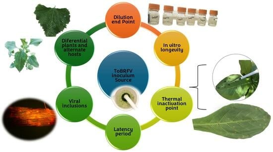
2. Materials and Methods

2.1. Plant Growth Condition

The research was conducted at the Agricultural Parasitology greenhouse of Universidad Autónoma Agraria Antonio Narro, located in Saltillo, Coahuila, Mexico, from August 2022 to October 2023. Initially, ToBRFV was isolated from commercial greenhouses at General Cepeda, Coahuila, Mexico (N 25°19′04.0″ W 101°24′10.6″). This step required the identification of symptomatic leaflets and fruits using pictorial keys described by [4,5]. Subsequently, rapid detection by immunoStrip (Agdia, Elkhart, IN, USA) was performed to confirm the virus in the collected isolates.
The isolates were moved to the greenhouse under controlled conditions for inoculation on Rio Grande tomato plants, which served as the main source of inoculum for the assays. Virus identification was initially confirmed by DAS-ELISA using the SRA 668/0500 Reagent Set (Agdia, Elkhart, IN, USA) specific for ToBRFV [18] and later by the RT-PCR technique, following the methodology proposed by [19]. Of the three isolates examined in a previous phase [20], only the last one (Isolate FQ3) was used in the assays, considering its pathogenic characteristics previously identified.

2.2. Dilution Endpoint from Inoculations on Tomato Plants var. 172–300

For the implementation of the experiment, ball tomato var. 172–300 (Yüksel Tohum, Antalya, Turkey) were placed in seedbeds containing 200 cavities, using a substrate composed of solid organic fertilizer, coarse sphagnum peat, perlite, and vermiculite in a 1:1:1:1 ratio. Thirty days post-germination (dpg), seedlings were transplanted into 1.5 L plastic containers using the same substrate mix mentioned earlier.
Plants were fertilized by supplying nutrients for stages V1 and V2, with Steiner’s nutrient solution at 25% [21] applied twice a week. A completely randomized design was used with seven treatments, ten replications, and ten plants as the absolute control. Treatments consisted of serial dilutions of D1 (1 × 10⁻³), D2 (1 × 10−3.5), D3 (1 × 10⁻⁴), D4 (1 × 10−4.5), D5 (1 × 10⁻⁵), D6 (1 × 10−5.5), and D7 (1 × 10⁻⁶), using symptomatic leaf tissue with ToBRFV. The dilutions started from a first viral concentration of 1 × 101, that is, of the infected tissue preparation at 1 × 10 (w/v) in a 0.01 M phosphate-buffered solution (PBS), pH 8, and the control was PBS and celite (Merk, Mexico City, Mexico). Inoculation was carried out ten days after transplanting (dat), following the methodology established by [22].

2.2.1. Evaluation of Agronomic Variables

Parameters measured for agronomic variables covered a period of 40 days post-inoculation (dpi) until the end of the experiment. Evaluated parameters included plant height (PH), which underwent assessment using a flexometer, stem diameter (SD) with an electronic vernier, chlorophyll index (SPAD), and nitrogen (N) units, measured in real time using a Minolta SPAD 502 plus chlorophyllometer (Copersa, Spain, Barcelona). To evaluate foliage fresh weight (FFW), root fresh weight (RFW), foliage dry weight (FDW), and root dry weight (RDW), a drying oven FE-291 (Felisa, Guadalajara, Mexico) and an electronic balance XY-C (XingYun, Changzhou, China) were used [1]. Data underwent an analysis of variance (ANOVA) and means with Tukey’s test (p = 0.05) comparisons when a statistically significant difference emerged. The results were analyzed using the SAS version 9.1 statistical program.
Incidence was determined using the following equation [23]:
Incidence   ( % ) Number   of   plants   observed   showing   ToBRFV   disease   symptom   scored Total   number of   plants   on   the   field × 100
Severity was assessed, and the disease index was calculated using the equation proposed by [24,25]:
Severity   ( % ) Area   of   diseased   tissue Total   Area × 100
DSI   % = e = 0 e R e × 100 5 N × 100
where DSI is the disease severity index; e is the class; Re is the number of plants in class (e), and N is the total number of plants. The severity diagrammatic scale for ToBRFV, as detailed by [22], was utilized in this study. All variables were assessed in a completely randomized design using ANOVA, and mean comparisons utilized the Tukey test (p = 0.05).

2.2.2. Determination of Viral Concentration by DAS-ELISA

Processed samples came from one plant per replicate, in which dilutions were the treatments. In total, there were two replicates per treatment; a total of nine treatments were processed coming from the first vegetative sprout. The assay started with plate sensitization, in which the capture antibody ACC 00960 (Agdia, Elkhart, IN, USA) was added at a concentration of 1:200 µL in 1× Carbonate Coating Buffer (CCB) solution and incubated under refrigeration (12 h at 6 ± 2 °C). Subsequently, the symptomatic tissue was macerated at a concentration of 1 × 10 (w/v) in extraction bags (12 × 15 mm) (BioCiencia, Nuevo León, Mexico) with 1× General Extract Buffer (GEB) (pH 7.5); the plate was triple rinsed with PBST Buffer (1×) and 100 µL of the extracted samples, Control+, and Control were added. Finally, the enzyme conjugate ECA ACC 00960 at a concentration of 1:200 µL in 1× ECI Buffer (pH 7.5) on the plate was washed eight times and incubated for 2 h at room temperature. Later, a PNP substrate was previously prepared in 1× Buffer (pH 9.8) at a concentration of 1 mg mL−1, poured 100 µL into each well of the plate, incubated for 30 min, and proceeded to read the optical density (O.D.) at 405 nm using a Multiskan 60 plate reader (Thermo FisherTM, Madrid, Spain). We take a total of three readings every 15 min. The viral concentration was determined based on the absorbance averages of the evaluated treatments, and the plotted data were serial dilutions and the negative control (healthy).

2.2.3. Pearson’s Correlation Analysis

Pearson’s Correlation Analysis evaluates the dilution endpoint and the effect of the variables studied on the treatments performed. Evaluated variables were VC (virus concentration), severity, DSI (disease severity index), PH (plant height), FFW (foliage fresh weight), FDW (foliage dry weight), RFW (root fresh weight), RDW (root dry weight), SPAD (leaf chlorophyll content), and SD (stem diameter).

2.3. Determination of Latency and Incubation Period from Inoculations in Tomato Plants var. 172–300

In this experiment, ball-type tomato plants var. 172–300 (Yüksel Tohum) of 40 dpg were used and transplanted into 1.5 L plastic pots using a mixture of peat moss substrate and vermiculite in a ratio of 1:1, and Steiner’s 25% nutrient solution was used [21]. Apical pruning of the plants was carried out, inducing vegetative shoots, and five days after the appearance of the first shoots, initiation of the trial began.
There were two experimental blocks, with the first block designated as an experimental inoculum source with ten treatments and four replicates, considered an infected control. The ToBRFV inoculum was prepared in a phosphate-buffered solution (PBS) with a viral concentration of 1:10 (w/v) (Figure 1A) and inoculated at 17 h with 200 µL in each first vegetative shoot of plants [22,26]. After a period of 24 h, the tissue from the second apical shoot of the four replicates of the previously inoculated plants was extracted. Such tissue served to prepare inoculum at the same concentration mentioned above and was inoculated (200 µL) on plants from the second block on the first shoot (Figure 1B), with four replicates per treatment. Inoculation was repeated on day 1(IPD1), 2 (IPD2), 3 (IPD3), 4 (IPD4), 5 (IPD5), 6 (IPD6), 7 (IPD7), 8 (IPD8), 9 (IPD9), and 10 (IPD10). A control without virus (Control−) and a control infected with ToBRFV, previously described (Control+), were also included.

2.3.1. Determination of Viral Concentration by DAS-ELISA

The evaluation of viral concentration was measured by absorbance using DAS-ELISA (Agdia, Elkhart, IN, USA) at 405 nm, 45 dpi of the experimental source (Control+). The processed samples corresponded to one plant per replicate, with treatments being the application of the 1:10 (w/v) concentration on plants with different dpi and those mentioned earlier; 2 replicates per treatment processed, which came from the second vegetative shoot (Figure 1C). The assay started with plate sensitization, in which ACC 00960 capture antibody (Agdia, Elkhart, IN, USA) at a concentration of 1:200 µL in 1× Carbonate Coating Buffer (CCB) solution was added and incubated (12 h at 6 ± 2 °C) under refrigeration. Subsequently, symptomatic tissue was macerated at a concentration of 1:10 (w/v) in extraction bags (12 × 15 mm) (BioCiencia, Nuevo León, Mexico) with General Extract Buffer (GEB) 1× (pH 7.5), the plate was rinsed three times with PBST Buffer (1×), and 100 µL of the extracted samples, Control+ and Control−, were added. Finally, we added the enzymatic conjugate ECA ACC 00960 at a concentration of 1:200 µL in ECI Buffer 1× (pH 7.5), washed the plate eight times, and incubated it for 2 h at room temperature. A PNP substrate was prepared in Buffer 1× (pH 9.8) at a concentration of 1 mg mL−1, poured 100 µL into each well of the plate, incubated for 30 min, and proceeded to read the optical density (O.D.) at 405 nm using a Multiskan 60 plate reader (Thermo FisherTM, Madrid, Spain); three readings were taken every 15 min. The viral concentration was determined based on the average absorbance of the evaluated treatments, considering the positive control as the source of the experimental inoculum.

2.3.2. Determination of Severity and Area under the Disease Progress Curve (AUDPC)

Severity was evaluated at intervals of 15, 30, and 45 dat, using the diagrammatic severity scale for ToBRFV (DSST) developed by [22]. Once data were obtained, the following equation for analysis was applied [24]:
S e v e r i t y   ( % ) Area   of   diseased   tissue Total   Area × 100
To quantify the accumulation of damage caused by ToBRFV in each day corresponding to the latency period (LP), the area under the disease progress curve produced by ToBRFV (AUDPC) using the equation proposed by [27] was calculated:
A U D P C = i = 1 n Y i + Y i + 1 2 ( + X i + 1 X i )
Variables were evaluated in a completely randomized design, using an ANOVA, and mean comparisons with Tukey’s test were performed, with a significance level of p = 0.05.

2.4. The Presence of ToBRFV Infective Sap in Plants of Nicotiana longiflora

2.4.1. Thermal Inactivation Points of ToBRFV in Plants of Nicotiana longiflora

N. longiflora seeds were put in trays containing 50 cavities, using a mixture of peat moss and vermiculite (1:1). Subsequently, at 30 dpg, seeds were transplanted into a 1.5 L pots and fertilized with a 25% diluted Steiner solution to promote leaf elongation.
Inoculum is used to determine the thermal inactivation point of ToBRFV with a viral concentration of 1 × 101 (w/v) following [28] with modifications. The extracts came from the extraction of one gram of infected tissue, ground in phosphate solution (PBS) (0.01 M, pH 8), and centrifuged at 6000 rpm in Eppendorf tubes. Samples were then exposed to six different temperatures, including −15 °C for 24 h of incubation at 100 rpm in a Mini Dry Bath MDB100C (Joanlab, Huzhou, China). Similarly, they underwent temperatures of 85 °C (for 15 and 30 min), 95 °C (for 15 and 30 min), and 99.5 °C (for 30 min) at 100 rpm. An IncumixTM Incubator Shaker (Daeil Tech, Gangnam, Republic of Korea) incubated all samples. An amount of 100 µL of the prepared sap was taken for each treatment and inoculated into half leaves of N. longiflora corresponding to each treatment. There were 20 replicates per treatment and readings at eight dpi were carried out by counting the number of necrotic local lesions (NLL) produced [29]. An ANOVA test and a means comparison using Tukey’s multiple range test (p = 0.05) were performed.

2.4.2. In Vitro Longevity from ToBRFV Inoculations in Nicotiana longiflora Plants

In this experiment, N. longiflora plants were grown in 50-cavity trays containing a substrate mixture composed of solid organic fertilizer, peat moss, perlite, and vermiculite in a 1:1:1:1:1 ratio, and Steiner’s nutrient solution was used for nutrient supply [21].

2.4.3. In Vitro Longevity of ToBRFV in N. Longiflora Plants

During this first stage of the longevity assay, the persistence of infective sap tissue infected with the FQ3 isolate was examined using tissue from infected plants with the FQ3 isolate. The infective inoculum was prepared in sterile distilled water at 1 × 101 (w/v) concentration in test vials and serial dilutions at concentrations of 1 × 10⁻², 1 × 10⁻³, 1 × 10⁻⁴, and 1 × 10⁻⁵. The inoculates remain at room temperature of 22 ± 2 °C and stored for up to 330 days. In addition, dehydrated infected tissue was preserved under the above-mentioned environmental conditions, and at the end of the storage period (330 days), an inoculum was prepared at 1 × 101. After the storage period, 15-day-old N. longiflora plants were inoculated with 100 µL of preserved sap applied to half of the leaf. Subsequently, it was concentrated uniformly using a bacteriological loop until the selected area was covered according to that performed by [17], with modifications. The treatment was repeated 20 times, with each experimental unit consisting of half a sheet, and LLN evaluation was carried out at 10 dpi. The obtained results underwent an ANOVA, and the means were Tukey’s multiple range tests (p = 0.05).

2.4.4. Persistence of ToBRFV in Geometric Progression in Nicotiana longiflora Plants

The second phase of the experiment started 15 dat; a crude sap solution from plants infected with the FQ3-ToBRFV isolate, diluted to a concentration of 1 × 101 (w/v) in sterile distilled water, was prepared [30]. Since no information was available on the stability of ToBRFV in sap, we established intervals in a geometric progression of 1, 4, 8, 16, and 32 days. For the execution of the trial, five treatments for the first day, D1, D4 for the fourth day, D8 for the eighth day, D16 for the sixteenth day, and D32 for the thirty-second day each were designated. For each treatment, we performed 20 replicates, using half-detached leaves of N. longiflora as the replicate unit. The inoculation process was to apply 100 µL of the solution to the detached half-leaf, followed by complete dispersion using a bacterial dipper [17] and counting of LLN 10 dpi. The counting and analysis of the NLL were conducted using an ANOVA, and means were compared via Tukey’s mean test (p = 0.05) using the statistical software SAS version 9.1.

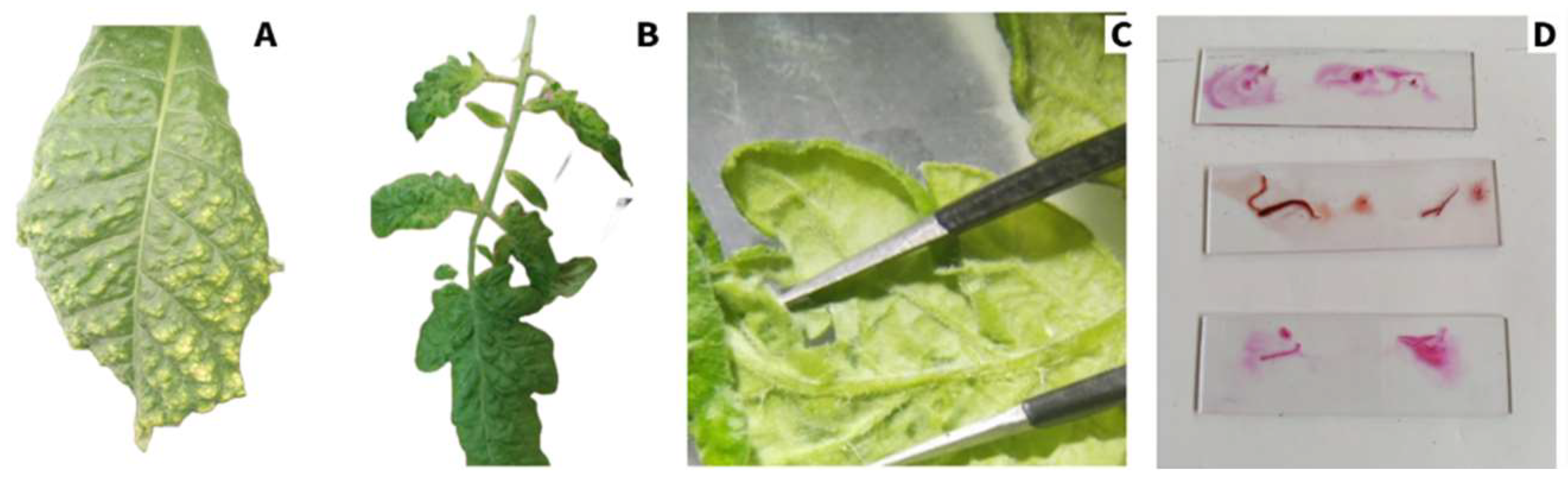
2.5. Viral Inclusions in Tomato var. 172–300 and Nicotiana Tabacum Plants

Tomato var. 172–300 and N. tabacum plants obtained at 40 dpi underwent inoculation with 200 µL of ToBRFV inoculum derived from isolate FQ3. After 30 dpi, tomato leaves and fruits showing apparent symptoms such as severe mosaic, yellowing, blistering, and blossoms were inspected and collected (Figure 2B). In the case of N. tabacum plants, symptoms of severe yellowing and blistering of the leaf blades appeared (Figure 2A). Sample preparation following the methodology proposed by [31] (with modifications) for identification of viral inclusions in ToBRFV, for tomato, a cut was made in the transverse section of the fruit pericarp. There were 20 replicates per species, using Methyl Green (Sigma-Aldrich, St. Louis, MO, USA, CAS: 7114-03-6) and Bengal rose (Sigma-Aldrich, CAS: 632-69-9) as dyes to stain the tissue (Figure 2C,D). Viral inclusions were reported in an OMAX advanced EPI-Fluorescence OMAX microscope (OMAX, Kent, WA, USA), equipped with a 100 W HBO power supply and a mercury lamp, adapted to a 9 MP digital camera (CMOS), set to a 1000× magnification (100×) objective at 2500× magnification.

2.6. Study of Alternate Hosts for ToBRFV Identified in the Natural Environment

2.6.1. Differential Plant Diagnosis

The trial was carried out in the greenhouse, using the same inoculum source as previous trials (isolate FQ3). Potential plant species that could serve as indicators of NLL, which could facilitate the diagnosis of ToBRFV, were identified. These species included Nicotiana longiflora, N. tabacum, N. glauca, Chenopodium giganteum, Ch. murale, and Ch. album Seeds of these species were grown in plastic pots with peat moss as a substrate. At 40 dpg, a ToBRFV inoculum was made at a concentration of 1 × 101 (w/v) using symptomatic leaf tissue in a phosphate-buffered solution (0.01 M, pH 8). Overall, 200 µL and the first leaves of each species under study were inoculated, with a total of 15 replicates for each species [32]. The screening of NLL and chlorotic (CLL) took place at 20 dpi.

2.6.2. Potential Hosts of ToBRFV

The second part of this research focused on new alternate host identification for ToBRFV by implementing an open-field experiment [33]. The absence of ToBRFV-infected bell pepper and tomato crops next to the experimental area was guaranteed. The experiment proceeded by planting eight 15 m long furrows with ball-type tomato plants var. 172–300 (Yüksel Tohum), with 30 m between plants. Inoculum with a concentration of 1 × 101 (w/v) of symptomatic leaf tissue sample was prepared in a phosphate-buffered solution (0.01 M, pH 8), and 200 µL was isolated from each plant. The evaluation occurred from 40 dpi onwards, inspecting plant species at approximately five meters. Hosts showing local (NLL and CLL) and systemic symptoms are reported.

2.6.3. Confirmation by DAS-ELISA

Confirmation of the presence of the virus in each host was examined by the DAS-ELISA technique, using the Reagent Set SRA 668/0500 (Agdia, Elkhart, IN, USA), following the methodology established by [22,32], using only hosts that exhibited both systemic and local symptoms. Samples containing a negative control (NC) were obtained from the healthy tissue of the host species under study, a positive control (PC) derived from FQ3, and samples with ToBRFV symptoms from each species tested. Three optical density measurements (405 nm) were performed at 15 min intervals using a Multiskang 60 plate reader (Thermo FisherTM, Madrid, Spain). For results to be acceptable, any sample with a value greater than twice the NC average had a positive ToBRFV infection, whereas samples with a value less than twice the NC average were negative for ToBRFV infection.

2.7. Statistical Analysis

Dilution endpoint from inoculations on tomato plants and the determination of latency and incubation period from inoculations in tomato plants tests were performed by one-way ANOVA and Tukey’s tests (p = 0.05) in the Statistical Analysis System (SAS) version 9.1 statistical software.

3. Results

3.1. Confirmation of the ToBRFV

Before the assays started, the identification of the inoculum source was confirmed, first by immunoStrip (Agdia, Elkhart, IN, USA), by DAS-ELISA, and finally by RT-PCR, which was confirmed positive for the first two tests, and for ToBRFV identity, it amplified a 475 bp fragment, which confirms its presence.

3.2. Dilution Endpoint from ToBRFV Inoculations in Tomato Plants

It was observed that tomato plants var. 172–300, which were inoculated with ToBRFV (Table 1), had a lower impact by treatment D7 on the variables PFF, PSF, PFR, SPAD, and N, as compared with the control. Likewise, this treatment did not cause the appearance of infection symptoms (severity). Similarly, the D6 treatment did not cause more severe symptoms in PH, SPAD, N, or disease severity.
Analysis of intensity, severity, and viral load by ELISA in response to ToBRFV infection (Figure 3) showed that the 1 × 10⁻⁶ dilution (D7) had significantly lower severity values (0), intensity (0.4), and ELISA (0.21). These results were remarkably like those reported for the control (0, 0, and 0.06, respectively).
Figure 4 shows a negative correlation between viral concentration and nitrogen levels in tomato leaves (r = −0.97, p ˂ 7.93 × 10⁻⁵), PFR (r = −0.962, p ˂ 1.33 × 10⁻⁴), and PSF (r = −0.96, p ˂ 1.53 × 10⁻⁴); in addition, negative reports of disease intensity and chlorophyll (r = 0.95, p ˂ 2.81 × 10⁻⁴) and nitrogen (r = −0.95, p ˂ 2.83 × 10⁻⁴) contents occurred. On the other hand, there is a positive correlation between the effect of PSF with PFR (r = 0.99, p ˂ 1.77 × 10⁻⁶) and chlorophyll content (r = 0.99, p ˂ 2.27 × 10⁻⁶), as well as between the intensity and severity of disease caused by ToBRFV (r = 0.98, p ˂ 1.35 × 10⁻⁵).

3.3. Determination of Latency and Incubation Period in Tomato Plants var. 172–300 Inoculated with ToBRFV

In tomato plants infected with ToBRFV at a concentration of 1 × 101 (w/v) it was shown that plants inoculated on days 9 (PID9) and 10 (PID10) had a decrease in viral load of 36.7% and 42.02%, respectively, when compared to the control infected with ToBRFV (Control+) (Figure 5).
The severity caused by different days of ToBRFV inoculation on tomato plants (Table 2) showed that the PID10 treatment presented a significant reduction of 0.25 at 15 dpi, 4.25 at 30 dpi, and 12.55 at 45 dpi, as compared to the infected control (Control+). Treatments PID7, IPD8, and PID9 also showed a decrease in severity at the beginning of the experiment (15 dpi), from 0.43 to 0.76 compared to the Control+. The statistically significant reduction in disease expression caused by ToBRFV (AUDPC) was reported in the PID10 treatment, which generated a disease progression of 174 compared to the infected control with 302.06 (Table 2).

3.4. Presence of ToBRFV Infective Sap in Nicotiana longiflora Plants

3.4.1. Thermal Inactivation Points of ToBRFV in Nicotiana longiflora Plants

After subjecting ToBRFV-infected saps to different temperatures, they showed that at 85 °C for 15 min followed by 85 °C for 30 min, they presented NLL on N. longiflora leaves, indicating that they did not inactivate ToBRFV but managed to reduce the number of lesions by 51.8% (52.6 NLL) and 96.1% (4.22 NLL), respectively, compared to the ToBRFV-infected control (109.2 NLL). The other temperatures evaluated were effective in inactivating ToBRFV, which prevented the appearance of NLL on tobacco leaves (Figure 6).

3.4.2. In Vitro Longevity of Nicotiana longiflora following Inoculation with ToBRFV

The persistence of infective sap with ToBRFV preserved for 330 days, evaluated on N. longiflora plants, resulted in dilutions that reduced the number of local necrotic lesions (NLL) to 1 × 10⁻⁴ (13.33) and 1 × 10⁻⁵ (10), compared to dilution of 1 × 101 (158.97), demonstrating the survival of infectious viral particles, even while remaining dormant (Figure 7). Furthermore, ToBRFV-infected tissue stored for 330 days is capable of harboring viral particles even without being in liquid media, which acquired an amount of NLL formed in N. longiflora leaves (135.33), almost equivalent to that reported with the 1 × 101 dilution.

3.4.3. Persistence of ToBRFV in Geometric Progression in Nicotiana longiflora Plants

The geometric progression of ToBRFV infection in N. longiflora plants (Figure 8) made it possible to evaluate the infection with viral particles. Inoculation of sap preserved for 32 days registered a decrease of 138.05 NLL and 188.82 NLL at 16 days, a reduction of 50.2% compared to inoculation on day 1 (378.96 NLL). In addition, inoculation of N. longiflora plants with fresh infected tomato sap at a concentration of 1 × 101 (Figure 8) generated more than twice as much NLL (378.96) compared to the same concentration kept for 330 days (158.97) (Figure 7), showing that virions can remain infectious for short periods.

3.5. Viral Inclusions in Tomato var. 172–300 and Nicotiana Tabacum Plants in Response to ToBRFV Infection

Tobamoviruses have the peculiarity of inducing physicochemical alterations in cells of infected tissue after infection with their host. The most prominent inclusions were those stained with Methyl Green in S. lycopersicum and N. tabacum tissue (Figure 9A–D). In S. lycopersicum, inclusions reported within the parenchymal palisade were rounded and stacked plaques (Figure 9A) with a size of 44.89 μm long and 30.80 μm wide, an association of X-bodies in a mass of aggregates with uniform sizes (Figure 9B) of 18.32–73.91 μm long and 18.32–28.32 μm wide, and the inclusion of X-bodies with the following dimensions: width 37.88 μm and length 54.05 μm (Figure 9C,D). In N. tabacum, in the upper epidermis, X-bodies in a mass of aggregates (Figure 9E–G) with sizes ranging from 46.45–61.98 μm wide to 48.55–102.02 μm were found, as were inclusions inside a plant cell with a size of 31.14–37.55 μm (width) and 55.31–82.01 μm long (Figure 9H). When the pericarp of severely symptomatic fruits could be visualized, chromoplasts (Figure 9I) with a diameter of 8.95 to 10.76 μm were found, with spots in the center of the epidermal.

3.6. Diagnosis of ToBRFV by Differential Plants

Table 3 shows indicator plants and possible alternative reservoirs for ToBRFV to facilitate the diagnosis of ToBRFV. As an alternative reservoir, we selected N. longiflora, Chenopodium murale, and N. glauca as indicator plants for the local lesions we selected. These species showed local symptoms, such as chlorosis and necrosis, in response to hypersensitivity (RH). Notably, these species have not previously served for virus diagnosis, making them viable options to play the role of differential plants in detecting ToBRFV. They also provide a valuable tool for evaluating the efficacy of disinfectant compounds against ToBRFV by assessing local lesions.

Host Plants Susceptible to Inoculation with ToBRFV

Weeds are a potential source of inoculum for ToBRFV, with the risk of triggering outbreaks in tomato crops either in the field or in greenhouses (Figure 10). About 28 potential hosts displaying both local and systemic symptoms were tested, which could contribute to the ToBRFV infection. ELISA tests revealed that 85.7% of these species were found to be positive for ToBRFV infection, whereas only five species are registered as alternate hosts (Table 4). Therefore, it suggests that maintaining a weed-free area could significantly reduce the spread of ToBRFV. Some weeds identified in this study belong to perennial and easily dispersed species, being reservoirs of the virus and facilitating its dissemination to tomato plants over time.

4. Discussion

4.1. Impact of ToBRFV Infection at Diverse Sap Concentrations in Tomato var. 172–300 Plants

The viral species that conform to the genus Tobamovirus are subject to relevant attention in plant pathology [36], mainly due to the remarkable stability of virions, which can reproduce by contact and often cause significant diseases in various crops [37]. Knowledge about the transmission of viral particles between plants and their roots attaches mainly to contamination of the nutrient solution, contamination of plant residues, and raw materials such as plastics, equipment, and tools [17,38]. By performing serial dilutions, we were able to identify both the dilution endpoint (DEP) and examine the effects of ToBRFV infection on tomato plants. Results indicate that ToBRFV requires at least a dilution of 1 × 10−5.5 (D6) to trigger infection in tomato var. 172–300 plants and affect agronomic variables (Table 1).
Previous research studies, such as that of [39], reported that TMV infection at a concentration of 1 × 101 reduces tomato hybrid growth, coinciding with these results that revealed a decrease in PH (21.6 cm) compared to the control (35.4 cm). In another study by [26], ToBRFV infection was evaluated in eight chili bell pepper varieties, reporting growth retardation ranging from 12 to 30%. On the other hand, results showed a loss in PH ranging from 13.56% (D7) to 38.99% (D1) (Table 1). These findings confirm those reported by [40], which mention that size reduction stands out as a principal symptom caused by virus infection in susceptible tomato hybrids.
Regarding FFW and FDW, [26] found a difference of 15 to 33% in the biomass of roots of native and jalapeño peppers infected with ToBRFV, but no statistical difference was found in foliage fresh weight between treatments. On the other hand, [41] showed that infection by the White Clover Mosaic Virus (WClMV) caused a decrease in the root biomass of Trifolium repens. The variable SD showed a minor difference between treatments and control, ranging from 70.1% (D1) to 84.25% (D7) compared with the control (Table 1). They obtained similar findings to [26] because no statistical differences emerged in the SD of the chili bell pepper treatments they evaluated. Regarding the relationship of chlorophyll (SPAD) and nitrogen (N) levels detailed in Table 1, it coincides with the research [9] showing that ToBRFV hurts chlorophyll and N production in tomato plants; however, they found that applying external elicitor therapies, such as Supermagro and enhanced Supermagro, significantly increased these variables by 27% and 26%, respectively. The results observed are consistent with the conclusions of [42], who suggest that virus infections, such as the Obuda pepper virus (OPV), affect the photosynthetic system of plants, even before visible symptoms appear, which manifests itself in the chlorophyll content.
Severity expression was symptomless only on D7 (Table 1). Several studies indicate that severity may vary in ToBRFV-infected plants due to differences in plant susceptibility, environmental conditions, the diversity of virus isolates, and the presence or absence of resistance genes in specific genotypes [43,44]. These studies were implemented on N. tabacum cv. Xanthi, targeted to assess the infective viral load of ToBRFV, confirmed that the local infection limit of the virus is 1.28 × 10⁻⁴ mg/mL [29]. Other studies carried out inoculations of different dilutions of ToBRFV in tomato plants and identified a viral load up to a dilution of (1 × 10⁻⁸, Cq = 25) from RT-qPCR [45]. The results in Figure 1 indicate that the viral concentration measured by DAS-ELISA was 0.21 absorbance (405 nm) at 45 dpi, using a dilution of 1 × 10⁻⁶, indicating that ToBRFV probably remained latent without expressing symptoms in the plants, even though the virus continued to replicate within the host, which happened in potato and eggplant [46]. Another study [47] detected ToBRFV at dilutions of 1 × 10⁻⁴ (using the 4B10 antibody) and 1 × 10−3.5 (with the 5A6 antibody), both monoclonal. This study highlighted that the sensitivity of antibodies can vary depending on whether they are monoclonal or polyclonal, which affects detection ability. In this research, monoclonal antibodies with a detection limit of 3.86 × 10⁻⁶ were used, according to the validation provided by the manufacturer.
Figure 2 shows that negative correlations exist between several agronomic variables and the presence of ToBRFV, which correspond to severity, intensity, and viral load. Various studies on the impact of tobamoviruses on plants have revealed that these viruses mainly use the phloem as the medium for their distribution in the plant, followed by cell-to-cell movement [11,14]. According to reports, their principal involvement is in the photosynthetic electron transport chain, connected to photosystem II and the Calvin cycle. This effect has an impact on chlorophyll content and shows a relationship between viral distribution in the plant and infection of the vascular system, affecting leaf fluorescence [48,49]. In addition, the concentration of viral load can influence this phenomenon, where decreased inoculum concentrations can lead to the absence of symptoms [50]. In tomato plants, it became evident that ToBRFV exhibited potential developmental damage, resulting in the depletion of essential phytohormones, such as auxin [16]. Considering the above-mentioned, the effects on tomato plants caused by ToBRFV depend directly on the amount of viral load in the plant.

4.2. Determination of the Latency and Incubation Period of ToBRFV in Tomato Plants var. 172–300

The ability of tobamoviruses to manifest an LP in infected tissues is because viruses can release their nucleic acid from the capsid after infection and before the replication process [51]. An analysis performed with TMV-infected tissue (0.1 g) in tobacco plants revealed an exponential increase in viral load with time, starting from the early stages [52]. Results obtained indicate a constant and accelerated increased viral load in tomato plants during the study period (Figure 5), particularly when the infection is established at 24 h of the experimental process, together with a virus concentration of 1.28 × 10⁻⁴ mg mL−1 [29], which showed that tomato plants exposed to ToBRFV can transmit to other tomato plants, one day after inoculation. The spread of ToBRFV in protected agriculture is attributed to mechanical damage and the use of contaminated tools, discarding seed as a source of inoculum; therefore, for a plant to show symptoms, it takes 9 to 12 days after exposure to the virus, according to the incubation period [2]. It has been demonstrated that only a 1.45% incidence of ToBRFV in a greenhouse containing tomato plants at the commencement of the crop is sufficient to achieve an infection rate of 80% after four months [51].
This study also examined how the severity of ToBRFV infection evolved in tomato plants at 15, 30, and 45 dpi and the accumulation of ToBRFV-generated AUDPC in tomato plants (Table 2). It has become documented that virus spread among plants, absent seed contamination in greenhouse tomato crops, could have a severity rate directly related to the incidence at 21 weeks post-transplanting [2]. Results in this research showed that four weeks after infection (30 dpi), there were significant levels of ToBRFV damage, which could imply an increasing correlation between the incidence and severity of infection in tomato F1 hybrids. To our knowledge, this is the only report combining aspects of latency period and disease progress with viral load accumulation.
The spread severity of ToBRFV is abiotic since mechanical manipulation of plants during routine crop work increases the spread of the disease [36,53]. Early in the emergence of ToBRFV, effective transmission involved damaging the roots and leaves of diseased plants; however, it is known that the most common way to spread the disease is by physical attachment [36]. The transmission rate between roots and seeds sown with contaminated soil is usually low [38] compared to seedlings spread in contaminated soil [54]; therefore, hands, equipment, and tools are the most effective mechanisms for ToBRFV transmission [36]. In a trial by [54], it is mentioned that three weeks after inoculation of CGMMV in tomato plants, it produced an 86% infection (without any management); however, in tomato greenhouses, 11 plants were found infected by CGMMV, which constituted 0.5% of the incidence; after the continuous management of plants and the spread of the virus in the crop, an increase of 32% of the incidence occurred after 40 days. Other studies evaluated 44 genotypes, including wild and commercial ones, and found that S. pimpinellifolium (LA1651), S. penellii (LA0716), and S. chilense (LA4117A, LA2747) had resistance to ToBRFV and did not express significant symptoms 30 dpi, whereas the hybrids evaluated showed high susceptibility, as in the case of Torry F1 [44]. According to what was described above, there is limited information on the study focused on the tolerance of tomato hybrids, whereas most of the works support the idea that wild species represent a source of resistance to ToBRFV, which can provide opportunities for the improvement in resistance genotypes via wild species.

4.3. Presence of ToBRFV Infective Sap in Nicotiana longiflora Plants

Most tobamoviruses can remain in the soil; data from [55] show that CGMMV can survive in irrigated soils, facilitating its transmission to watermelon plants. Also, it has become apparent that the presence of YTMMV in hydroponic systems could trigger epidemics via infection of the roots of healthy plants [56]. The persistence of these viruses in water contributes to their dissemination in agricultural systems, demonstrating the presence of infectious ToBRFV viral particles in wastewater located at some distance from production areas [56,57]. Lately, the persistence of ToBRFV viral particles has become apparent in conventionally treated wastewater, and these particles maintain their infectious capacity even after treatment [58]. Likewise, virulent ToBRFV particles obtained from infected persons have been ascertained in the gastrointestinal tract and oropharyngeal cavity [59]. This situation could well reflect the ability of ToBRFV to survive in plant debris after a culture cycle, although no detailed information regarding the period during which it retains its infectious capacity is provided [2]. The study conducted by [60] on the spread of ToBRFV on materials and tools indicates that the main route of pathogen dissemination in production areas is via hands, clothing, personal protective items, and footwear. Due to this circumstance, in vitro longevity, also known as persistence, has emerged as a critical descriptor for tobamoviruses, considering the point of thermal inactivation and the duration of their viability outside the organism.
Current research on the thermal inactivation point of ToBRFV in vitro experiments focuses on applying thermotherapy to seeds, tools, and substrates. In this setting, Ref. [61] extracted contaminated tomato seeds, applying thermal treatments at 72 °C for 72 h without achieving disinfection. Meanwhile, Ref. [28] used an efficient heat treatment of 92 °C for 30 min to disinfect substrates, plastic articles, and other greenhouse utensils. Similarly, [62] has indicated that the thermal inactivation point for ToBRFV is 90 °C, according to an in vitro assay with sap applied to plastic carriers and the use of Sodium Hypochlorite. The results presented in Figure 5 indicate that inhibition of ToBRFV occurred at both low (−15 °C) and high temperatures (85 °C to 99.5 °C). These varied temperatures are applicable in treatments to decontaminate equipment, tools, substrates, clothing, and seeds after a crop cycle in which the virus was present. The purpose is to reduce the source of inoculum that could trigger an epidemic in tomato plants during a subsequent growing season.
The in vitro longevity experiment reported that sap preserved for 330 days at a temperature of 22 °C, in 1 × 10⁻⁴ and 1 × 10⁻⁵ dilutions, generated a lower number of NLL; however, 1 × 101 dilutions of sap (158.9) and tissue (135.3) showed a higher number of NLL (Figure 7). The results obtained coincide with the findings of [45], in which inoculations on tomato plants cv. Moneymaker reported that ToBRFV persistence in aqueous environments was maintained for four weeks at dilutions of 1 × 10⁻² and 1 × 10⁻⁴, decreasing three weeks at a dilution of 1 × 10⁻⁶, while the viral load quantified by RT-qPCR continued to increase after nine weeks (1 × 10⁻², Cq 11), (1 × 10⁻⁴, Cq 21), and (1 × 10⁻⁶, Cq 21). This trend could be related to the analysis of [63], who reported that purified TMV can maintain its infectious capacity after 50 years.

4.4. Viral Inclusions

The International Committee on Taxonomy of Viruses has established that inclusion bodies are considered classification criteria for infected viruses in plants [64]. Fenner made the first descriptions of VI in 1976, and it was in 1978 that cytoplasmic inclusions became detectable [65]. They also reported elongated paracrystals, rounded and stacked plaques, and X-bodies, which were later associated with tobamovirus infection and named viroplasms [66]. This research shows inclusions like those described for tobacco mosaic virus (Figure 9A–I), where the presence of X-bodies in S. lycopersicum and N. tabacum induced by ToBRFV was found, indicating that inclusions produced do not vary in function of the host. Also, the identified inclusions in palisade parenchymal cells (Figure 9D) indicate that these inclusions may be related to membranes harboring viral replication complexes. These complexes may be highly organized structures involving elements of the cytoskeleton, allowing interaction with protein movement (PM) and facilitating its movement between cells, which favors the spread of viral infection [67]. In addition, PM increases the size of plasmodesmata so that the virus can transmit through the cytoplasm of cells [68]. The capsid protein (CP) of tobamoviruses maintains a critical role in long-distance virus dissemination [69], as it is necessary for viral movement between the vascular parenchymal and the accompanying cells [70]. The significance of the CP in interacting with host plant factors has not been thoroughly investigated [71]. Nevertheless, epidermis and mesophyll cells are pivotal scenarios for processing RNA silencing and inducing plant resistance programs [72].
There is controversy regarding the purpose of studying viral inclusions and using them as viral classification criteria. In certain circumstances, mixed infections with viruses of different genera make identification according to the species under study problematic [73]. Nevertheless, research on viral inclusions in the host has primarily aimed not at sorting but at comprehending the physicochemical alterations impacting the virus following infection [53]. Some studies suggest that X-bodies act as virus factories in the cytoplasm of a cell during the initial stages of infection, promoting virus replication and causing damage to the host [74]. From 40 dpi onwards, the presence of viroplasms that had colonized the parenchymal palisade could be observed (Figure 9). Not only do virus factories harbor PM particles, but they also store virus movement proteins [75]. These PMs play an essential role in cell-to-cell virus motility via plasmodesmata, resulting in the expression of symptoms in plants [76]. The accumulation of these particles in cells contributes to increased infection, although the host regulates and inhibits particle replication via its immune system as a defense mechanism [75]. Studies also mention that tobamovirus-infected cells, when stained with antibodies, show that replication proteins colocalize in the cytoplasmic granular bodies, known as X-bodies or viroplasms [76]. The results presented allowed us to establish a relationship between these alterations in S. lycopersicum and N. tabacum, increasing the intensity of the disease, which contributes to the cytopathology and damage caused by ToBRFV.

4.5. Diagnostic Hosts and Alternate Reservoirs of ToBRFV

Most tobamoviruses exhibit characteristic symptoms that distinguish them from other virus species [77]. According to research, ToBRFV shares a similar host range to ToMV and TMV due to its phylogenetic origin [14]. ToBRFV can infect more than 40 species spanning diverse families, such as Amaranthaceae, Apocynaceae, Asteraceae, and Solanaceae [4]; however, it cannot affect species of the families Brassicaceae, Cucurbitaceae, and Verbenaceae [32]. Tomato and bell pepper species are susceptible to natural infection by ToBRFV [14]; therefore, indicator plant screening emerges as a valuable tool to improve the ability to distinguish and diagnose ToBRFV in other plant species and to analyze susceptible hosts that served as principal sources of inoculum [2,78].
Results of this research (Table 3) show that of the six species considered as differential plants, N. longiflora, Ch. murale, and N. glauca have not yet registered as susceptible to ToBRFV infection. This finding is of significant phytosanitary concern. N. longiflora has become a model to address pest and disease resistance [79,80]. On the other hand, Ch. murale underwent research due to its insecticidal, fungicidal, and bactericidal properties, supported by its pharmacological and biological activities; however, in natural environments, it is usually an annual plant that commonly grows in association with other weeds [81]. As for N. glauca, it is a perennial invasive species that generates disturbances in response to climatic conditions, such as heavy rains and floods, demonstrating its resistance to extreme temperatures [82] and identifying it as a host highly susceptible to TMV [83]. The data collected suggest a potential risk for these species as alternative hosts of ToBRFV, which could affect tomato crops in this region of Coahuila, Mexico.
In the analysis of plants susceptible to ToBRFV inoculation under field conditions (Table 4), it was observed that eight species belong to the Solanaceae family, six to Asteraceae, three to Chenopodiaceae, two to Malvaceae, and two to Amaranthaceae. Likewise, one plant species each in the families Convolvulaceae, Labiatae, Oxalidaceae, Resedaceae, Araliaceae, Plantaginaceae, Polygonaceae, Rubiaceae, Vitaceae, Rosaceae, Ranunculaceae, and Euphorbiaceae was identified (Figure 10). Among these plants, 23 species have not previously received identification as potential hosts of ToBRFV. An analysis of the species A. hybridus, known as A. retroflexus, via ELISA and RT-PCR did not show ToBRFV [33]. These results are consistent with the findings of these researchers (Table 4). Most species identified are species considered weeds and play an essential role in the spread of viral pathogens, being a source of inoculum for dispersal via work personnel; therefore, it is important to implement appropriate management strategies, including crop rotation with non-host species and specific control of these weeds [14].
In previous reports, S. melongena and S. tuberosum have demonstrated that they play a host role for ToBRFV, being able to develop latent infections [46]. The analysis of species belonging to the Apocynaceae and Asteraceae families highlights the importance of ornamental plants susceptible to ToBRFV, such as Catharanthus roseus (an annual weed) and Glebionis coronaria (cultivated as a leafy vegetable in East Asia, but also considered as an invasive weed in California) [14]. Table 4 displays the assessment of cultivated plants near rows of tomato plants infected with ToBRFV. The plants, so far not reported as susceptible, include Vitis vinifera, Hedera helix, Oxalis latifolia, and Helianthus annus, which showed local symptoms, whereas Rubus idaeus showed no apparent symptoms; however, all species tested positive for virus infection by ELISA. Although [32] identified 12 wild species, highlighting that Ch. murale and S. nigrum are natural hosts of ToBRFV, this study has revealed that Ch. murale can also be used as a plant differential for ToBRFV. In addition, it was observed that ToBRFV can infect M. jalapa, an ornamental plant in Mexico that is important in soil phytoremediation in various desert areas of the world [84]. Several researchers indicate the importance of performing a broader host range analysis to compare ToBRFV isolates and evaluate their connection with plant species examined in other studies [4,15,32]. Based on the findings of this study, it can be inferred that ToBRFV exhibits a diverse range of hosts. However, further research is required to comprehend the potential of these reservoirs as inoculum sources for transmission to healthy tomato plants, either in greenhouses or open fields.

5. Conclusions

The results obtained revealed that PI of ToBRFV occurred between 9 and 12 days post-inoculation, while LP could be detected only one day after inoculation. It was found that ToBRFV at 1 × 10−5.5 (DEP) could still induce symptoms in tomato plants. In addition, the evaluation of serial dilutions provides a novel finding on the impact of the virus on the agronomic parameters of the tomato crop. ToBRFV IT in N. longiflora plants occurred at 85 °C for 30 min. Twenty-three plant species were found to act as ToBRFV new hosts. The VIs caused by the virus on tomato leaves were characterized by the presence of X-bodies, whose plaques were found to be rounded and stacked within the epidermal cells. The study significantly enhances our understanding of addressing and mitigating the risks associated with ToBRFV. Future research could focus on developing innovative protocols that improve the disinfection of personnel and equipment, thus facilitating the identification of viral strains originating from the region in which isolation occurs.

Author Contributions

Methodology, U.V.G. and H.L.L.; Conceptualization, U.V.G., H.L.L. and G.A.F.T.; Investigation, U.V.G.; Resources, L.A.A.U.; Data curation, H.L.L., A.H.J. and L.A.A.U.; validación, G.A.F.T., J.C.D.O., L.A.A.U. and A.H.J.; Supervision, G.A.F.T., H.L.L., A.F.O. and A.H.J.; writing—review and editing, H.L.L. and G.A.F.T. All authors have read and agreed to the published version of the manuscript.

Funding

Phytosanitary inspection unit SERVESA A. C. (1223150), Universidad Autónoma Agraria Antonio Narro (1223150) and the Consejo Nacional de Humanidades, Ciencia y Tecnología (1223150).

Data Availability Statement

The underlying data of this manuscript are available upon reasonable request from the authors.

Acknowledgments

To José Ángel Villarreal Quintanilla for his support in the taxonomic identification of the plants, as well as to the ANSM herbarium of the Universidad Autónoma Agraria Antonio Narro.

Conflicts of Interest

The authors declare no conflicts of interest.

References

  1. Ruíz, A.M.Y.; Montes, M.J.A.; Castañón, G.J.H.; Gutiérrez, M.F.A.; Hernández, G.M.; López, L.H.; Ruíz, V.V.M.; Villalobos, M.J.J. Uso de la harina del pez diablo (Pterygoplichthys spp.) en la fertilización orgánica del tomate (Solanum lycopersicum L.). Rev. Int. Contam. Ambient. 2023, 39, 159–169. [Google Scholar] [CrossRef]
  2. González-Concha, L.F.; Ramírez-Gil, J.G.; García-Estrada, R.S.; Rebollar-Alviter, Á.; Tovar-Pedraza, J.M. Spatiotemporal Analyses of Tomato Brown Rugose Fruit Virus in Commercial Tomato Greenhouses. Agronomy 2021, 11, 1268. [Google Scholar] [CrossRef]
  3. Salem, N.; Mansour, A.; Ciuffo, M.; Falk, B.W.; Turina, M. A new Tobamovirus infecting tomato crops in Jordan. Arch. Virol. 2016, 161, 503–506. [Google Scholar] [CrossRef]
  4. Luria, N.; Smith, E.; Reingold, V.; Bekelman, I.; Lapidot, M.; Levin, I. A new Israeli Tobamovirus isolate infects tomato plants harboring Tm-22 resistance genes. PLoS ONE 2017, 12, e0170429. [Google Scholar] [CrossRef] [PubMed]
  5. Cambrón-Crisantos, J.M.; Rodríguez-Mendoza, J.; Valencia-Luna, J.B.; Alcasio Rangel, S.; de Jesús García-Ávila, C.; López-Buenfil, J.A.; Ochoa-Martínez, D.L.; Cambrón-Crisantos, J.M.; Rodríguez-Mendoza, J.; Valencia-Luna, J.B.; et al. First Report of Tomato Brown Rugose Fruit Virus (ToBRFV) in Michoacan, Mexico. Rev. Mex. Fitopatol. 2019, 37, 185–192. [Google Scholar] [CrossRef]
  6. Camacho-Beltrán, E.; Pérez-Villarreal, A.; Leyva-López, N.E.; Rodríguez-Negrete, E.A.; Ceniceros-Ojeda, E.A.; Méndez-Lozano, J. Occurrence of Tomato Brown Rugose Fruit Virus Infecting Tomato Crops in Mexico. Plant Dis. 2019, 103, 1440. [Google Scholar] [CrossRef]
  7. Van de Vossenberg, B.T.; Visser, M.; Bruinsma, M.; Koenraadt, H.M.S.; Westenberg, M. Real-time tracking of Tomato Brown Rugose Fruit Virus (ToBRFV) outbreaks in the Netherlands using Nextstrain. PLoS ONE 2020, 15, e0234671. [Google Scholar] [CrossRef]
  8. EPPO. European and Mediterranean Plant Protection Organization: Tomato Brown Rugose Fruit Virus. Global Database Distribution World. Available online: https://gd.eppo.int/taxon/TOBRFV/distribution (accessed on 14 April 2023).
  9. Ortiz-Martínez, L.E.; Ochoa-Martínez, D.L. Elicitors and biostimulants in the production of tomato infected with Tomato Brown Rugose Fruit Virus. J. Plant Dis. Prot. 2023, 130, 351–360. [Google Scholar] [CrossRef]
  10. Iwanowski, D. Concerning the mosaic disease of the tobacco plant. In St. Petersb. Acad. Imp. Sci. Bull., Phytopathological Classics Number 7 (1942); Translator Johnson, J.; American Phytopathological Society Press: St. Paul, MN, USA, 1892; Volume 35, pp. 67–70. [Google Scholar]
  11. Dombrovsky, A.; Tran-Nguyen, L.T.T.; Jones, R.A.C. Cucumber Green Mottle Mosaic Virus: Rapidly increasing global distribution, etiology, epidemiology, and management. Annu. Rev. Phytopathol. 2017, 55, 231–256. [Google Scholar] [CrossRef]
  12. Khamphirapaeng, P.; Cheewangkoon, R.; McGovern, R.J.; Wong, S.M.; To-Anun, C. Detection of Tobacco Mosaic Virus in petunia and tobacco by light microscopy using a simplified inclusion body staining technique. Int. J. Agric. Technol. 2017, 13, 163–168. Available online: http://www.ijat-aatsea.com/ (accessed on 28 December 2023).
  13. Jewehan, A.; Kiemo, F.W.; Salem, N. Isolation and molecular characterization of a Tomato Brown Rugose Fruit Virus mutant breaking the tobamovirus resistance found in wild Solanum species. Arch Virol. 2022, 167, 1559–1563. [Google Scholar] [CrossRef] [PubMed]
  14. Chanda, B.; Gilliard, A.; Jaiswal, N.; Ling, K.S. Comparative analysis of host range, ability to infect tomato cultivars with Tm-22 gene, and real-time reverse transcription PCR detection of Tomato Brown Rugose Fruit Virus. Plant Dis. 2021, 105, 3643–3652. [Google Scholar] [CrossRef] [PubMed]
  15. Chanda, B.; Rivera, Y.; Nunziata, S.O.; Galvez, M.E.; Gilliard, A.; Ling, K.S. Complete genome sequence of a Tomato Brown Rugose Fruit Virus isolated in the United States. Microbiol. Resour. Announc. 2020, 9, e00630-20. [Google Scholar] [CrossRef]
  16. Vaisman, M.; Hak, H.; Arazi, T.; Spiegelman, Z. The impact of tobamovirus infection on root development involves induction of auxin response factor 10a in tomato. Plant Cell Physiol. 2022, 63, 1980–1993. [Google Scholar] [CrossRef] [PubMed]
  17. Ehlers, J.; Nourinejhad Zarghani, S.; Kroschewski, B.; Büttner, C.; Bandte, M. Cleaning of Tomato Brown Rugose Fruit Virus (ToBRFV) from Contaminated Clothing of Greenhouse Employees. Horticulturae 2022, 8, 751. [Google Scholar] [CrossRef]
  18. Eads, A.; Groth-Helms, D.; Davenport, B.; Cha, X.; Li, R.; Walsh, C.; Schuetz, K. The commercial validation of three Tomato brown rugose fruit virus assays. PhytoFrontiers 2023, 3, 206–213. [Google Scholar] [CrossRef]
  19. Rodríguez-Mendoza, J.; García-Ávila, C.J.; López-Buenfil, J.A.; Araujo-Ruiz, K.; Quezada-Salinas, A.; Cambrón-Crisantos, J.M.; Ochoa-Martínez, D.L. Identificación de Tomato Brown Rugose Fruit Virus por RT-PCR de una región codificante de la replicasa (RdRP). Rev. Mex. Fitopatol. 2019, 37, 345–356. [Google Scholar] [CrossRef]
  20. Vasquez-Gutierrez, U.; Delgado-Ortiz, J.C.; Frías-Treviño, G.A.; Aguirre-Uribe, L.A.; Olivas, A.F. Pathogenicity of three Tomato brown rugose fruit virus isolates and response in tomato (Solanum lycopersicum L.). In Proceedings of the 3rd International Electronic Conference on Agronomy, Basel, Switzerland, 15–30 October 2023. [Google Scholar]
  21. Steiner, A.A. A universal method for preparing nutrient solutions of a certain desired composition. Plant Soil 1961, 15, 134–154. [Google Scholar] [CrossRef]
  22. Vasquez-Gutierrez, U.; Frias-Treviño, G.A.; Delgado-Ortiz, J.C.; Aguirre-Uribe, L.A.; Flores-Olivas, A. Severity of Tomato Brown Rugose Fruit Virus in tomato (Solanum lycopersicum L.) from a region of Coahuila, México. Int. J. Hortic. Agric. Food Sci. 2023, 7, 1–6. [Google Scholar] [CrossRef]
  23. Fajinmi, A.A.; Dokunmu, A.O.; Akheituamen, D.O.; Onanuga, K.A. Incidence and infection rate of Maize streak virus disease by Cicadulina triangular on maize plants and its distribution from the lowest diseased leaf under tropical conditions. Arch. Phytopathol. Plant Prot. 2012, 45, 1591–1598. [Google Scholar] [CrossRef]
  24. Kone, N.; Asare-Bediako, E.; Silue, S.; Kone, D.; Koita, O.; Menzel, W.; Winter, S. Influence of planting date on incidence and severity of viral disease on cucurbits under field condition. Ann. Agric. Sci. 2017, 62, 99–104. [Google Scholar] [CrossRef]
  25. Jewehan, A.; Salem, N.; Tóth, Z. Evaluation of responses to Tomato Brown Rugose Fruit Virus (ToBRFV) and selection of resistant lines in Solanum habrochaites and Solanum peruvianum germplasm. J. Gen. Plant Pathol. 2022, 88, 187–196. [Google Scholar] [CrossRef]
  26. Ortiz-Martínez, L.E.; Ochoa-Martínez, D.L.; Rojas-Martínez, R.I.; Aranda-Ocampo, S.; Cruz, M.A.G. Respuesta de variedades de chile a la infección con Tomato Brown Rugose Fruit Virus. Summa Phytopathol. 2022, 47, 209–215. [Google Scholar] [CrossRef]
  27. Shaner, G. The Effect of Nitrogen Fertilization on the Expression of Slow-Mildewing Resistance in Knox Wheat. Phytopathology 1977, 67, 1051–1056. [Google Scholar] [CrossRef]
  28. Vargas-Mejía, P.; Rodríguez-Gómez, G.; Salas-Aranda, D. A Identificación y manejo del virus del fruto rugoso marrón del tomate en invernaderos en México. Arch. Virol. 2023, 168, 135. [Google Scholar] [CrossRef]
  29. Nourinejhad Zarghani, S.; Monavari, M.; Ehlers, J.; Hamacher, J.; Büttner, C.; Bandte, M. Comparison of Models for Quantification of Tomato Brown Rugose Fruit Virus Based on a Bioassay Using a Local Lesion Host. Plants 2022, 11, 3443. [Google Scholar] [CrossRef]
  30. Francki, R.I. Limited value of the thermal inactivation point, longevity in vitro and dilution endpoint as criteria for the characterization, identification, and classification of plant viruses. Intervirology 1980, 13, 91–98. [Google Scholar] [CrossRef] [PubMed]
  31. Cárdena, S.E. Diagnóstico De Virus Mediante Inclusiones Virales, Microscopía Electrónica y Rango De Hospedantes, 3rd ed.; Colegio de Postgraduados, Instituto de Fitosanidad: Montecillo, Mexico, 1999; pp. 154–196. [Google Scholar]
  32. Salem, N.M.; Abumuslem, M.; Turina, M.; Samarah, N.; Sulaiman, A.; Abu-Irmaileh, B.; Ata, Y. New Weed Hosts for Tomato Brown Rugose Fruit Virus in Wild Mediterranean Vegetation. Plants 2022, 11, 2287. [Google Scholar] [CrossRef] [PubMed]
  33. Matzrafi, M.; Abu-Nassar, J.; Klap, C.; Smith, E.; Dombrovsky, A. Solanum elaeagnifolium and S. rostratum as potential hosts of the tomato brown rugose fruit virus. PLoS ONE 2022, 18, e0282441. [Google Scholar] [CrossRef]
  34. Sabra, A.; Amer, M.A.; Hussain, K.; Zakri, A.; AlShahwan, I.M.; Al-Saleh, M.A. Occurrence and Distribution of Tomato Brown Rugose Fruit Virus Infecting Tomato Crop in Saudi Arabia. Plants 2023, 11, 3157. [Google Scholar] [CrossRef]
  35. Zhang, S.; Griffiths, J.S.; Marchand, G.; Bernards, M.A.; Wang, A. Tomato Brown Rugose Fruit Virus: An emerging and rapidly spreading plant RNA virus that threatens tomato production worldwide. Mol. Plant Pathol. 2022, 23, 1262–1277. [Google Scholar] [CrossRef]
  36. Smith, E.; Dombrovsky, A. Aspects in Tobamovirus management in intensive agriculture. In Plant Diseases-Current Threats and Management Trends; IntechOpen: London, UK, 2019. [Google Scholar] [CrossRef]
  37. Jablonski, M.; Poghossian, A.; Keusgen, M. Detection of plant virus particles with a capacitive field-effect sensor. Anal. Bioanal. Chem. 2021, 413, 5669–5678. [Google Scholar] [CrossRef]
  38. Koh, S.H.; Li, H.; Sivasithamparam, K.; Admiraal, R.; Jones, M.G.K.; Wylie, S.J. Low root-to-root transmission of a tobamovirus, yellow tailflower mild mottle virus, and resilience of its virions. Plant Pathol. 2018, 67, 651–659. [Google Scholar] [CrossRef]
  39. Takács, A.; Kazinczi, G.; Pribék, D. Biological Decline of Solanum nigrum L. Due to Tobacco Mosaic Tobamovirus (TMV) Infection I. Growth and Nutrient Uptake. Acta Phytopathol. Entomol. Hung. 2001, 36, 9–14. [Google Scholar] [CrossRef]
  40. Bhat, A.I.; Rao, G.P. Symptoms of Virus-Infected Plants. In Characterization of Plant Viruses; Bhat, A.I., Rao, G.P., Eds.; Springer: New York, NY, USA, 2020; Volume 2, pp. 7–22. [Google Scholar]
  41. Van Mölken, T.; Stuefer, J.F. The potential of plant viruses to promote genotypic diversity via genotype× environment interactions. Ann. Bot. 2011, 107, 1391–1397. [Google Scholar] [CrossRef] [PubMed]
  42. Rys, M.; Juhász, C.; Surówka, E.; Janeczko, A.; Saja, D.; Tóbiás, I.; Gullner, G. Comparison of a compatible and an incompatible pepper-tobamovirus interaction by biochemical and non-invasive techniques: Chlorophyll a fluorescence, isothermal calorimetry, and FT-Raman spectroscopy. Plant Physiol. Biochem. 2014, 83, 267–278. [Google Scholar] [CrossRef] [PubMed]
  43. Hanssen, I.M.; Thomma, B.P. Cucumber Mosaic Virus: A Successful Pathogen That Rapidly Evolved from Emerging to Endemic in Tomato Crops. Mol. Plant Pathol. 2010, 11, 179–189. [Google Scholar] [CrossRef] [PubMed]
  44. Kabas, A.; Fidan, H.; Kucukaydin, H.; Atan, H.N. Screening of wild tomato species and interspecific hybrids for resistance/tolerance to Tomato Brown Rugose Fruit Virus (ToBRFV). Chil. J. Agric. Res. 2022, 82, 189–196. [Google Scholar] [CrossRef]
  45. Mehle, N.; Bačnik, K.; Bajde, I.; Brodarič, J.; Fox, A.; Gutiérrez-Aguirre, I.; Vučurović, A. Tomato Brown Rugose Fruit Virus in aqueous environments–survival and significance of water-mediated transmission. Front. Plant Sci. 2023, 14, 1187920. [Google Scholar] [CrossRef] [PubMed]
  46. Yan, Z.Y.; Zhao, M.S.; Liu, L.Z.; Yang, G.L.; Geng, C.; Tian, Y.; Li, X.D. Biological and molecular characterization of Tomato Brown Rugose Fruit Virus and development of quadruplex RT-PCR detection. J. Integr. Agric. 2021, 20, 1871–1879. [Google Scholar] [CrossRef]
  47. Bernabé-Orts, J.M.; Torre, C.; Méndez-López, E.; Hernando, Y.; Aranda, M.A. Nuevos recursos para la detección específica y sensible del virus emergente del fruto rugoso marrón del tomate. Virus 2021, 13, 1680. [Google Scholar] [CrossRef]
  48. Pineda, M.; Olejníčková, J.; Cséfalvay, L.; Barón, M. Tracking viral movement in plants by means of chlorophyll fluorescence imaging. J. Plant Physiol. 2011, 168, 2035–2040. [Google Scholar] [CrossRef] [PubMed]
  49. Pineda, M.; Sajnani, C.; Barón, M. Changes induced by the Pepper mild mottle tobamovirus on the chloroplast proteome of Nicotiana benthamiana. Photosynth. Res 2010, 103, 31–45. [Google Scholar] [CrossRef] [PubMed]
  50. Dombrovsky, A.; Mor, N.; Gantz, S.; Lachman, O.; Smith, E. Disinfection efficacy of Tobamovirus-contaminated soil in greenhouse-grown crops. Horticulturae 2022, 8, 563. [Google Scholar] [CrossRef]
  51. Schramm, G.; Engler, R. The latent period after infection with tobacco mosaic virus and virus nucleic acid. Nature 1958, 181, 916–917. [Google Scholar] [CrossRef]
  52. Yarwood, C.E. Latent Period and Generation Time for Two Plant Viruses. Am. J. Bot. 1952, 39, 613–618. [Google Scholar] [CrossRef]
  53. Broadbent, L. The epidemiology of tomato mosaic. XI. Seed transmission of TMV. Ann. Appl. Biol. 1965, 56, 177–205. [Google Scholar] [CrossRef]
  54. Reingold, V.; Lachman, O.; Belausov, E.; Koren, A.; Mor, N.; Dombrovsky, A. Epidemiological study of Cucumber Green Mottle Mosaic Virus in greenhouses enables reduction of disease damage in cucurbit production. Ann. Appl. Biol. 2016, 168, 29–40. [Google Scholar] [CrossRef]
  55. Li, R.; Zheng, Y.; Fei, Z.; Ling, K.S. First complete genome sequence of an emerging Cucumber Green Mottle Mosaic Virus isolate in North America. Genome Announc. 2015, 3, 10–1128. [Google Scholar] [CrossRef]
  56. Nash, D.; Ellmen, I.; Knapp, J.J.; Menon, R.; Overton, A.K.; Cheng, J.; Charles, T.C. A Novel Tiled-Amplicon Sequencing Assay Targeting the Tomato Brown Rugose Fruit Virus (ToBRFV) Genome Reveals Widespread Distribution in Municipal Wastewater Treatment Systems in the Province of Ontario, Canada. bioRxiv 2023, 11. [Google Scholar] [CrossRef]
  57. Lopez-Roblero, A.; Martínez Cano, D.J.; Diego-García, E.; Guillén-Navarro, G.K.; Iša, P.; Zarza, E. Metagenomic analysis of plant viruses in tropical fresh and wastewater. Environ. DNA 2023, 6, e416. [Google Scholar] [CrossRef]
  58. Bačnik, K.; Kutnjak, D.; Pecman, A.; Mehle, N.; Žnidarič, M.T.; Aguirre, I.G.; Ravnikar, M. Viromics and infectivity analysis reveal the release of infective plant viruses from wastewater into the environment. Water Res. 2020, 177, 115628. [Google Scholar] [CrossRef]
  59. Aguado-García, Y.; Taboada, B.; Morán, P.; Rivera-Gutiérrez, X.; Serrano-Vázquez, A.; Iša, P. Tobamoviruses can be frequently present in the oropharynx and gut of infants during their first year of life. Sci. Rep. 2020, 10, 13595. [Google Scholar] [CrossRef]
  60. Ehlers, J.; Nourinejhad Zarghani, S.; Liedtke, S.; Kroschewski, B.; Büttner, C.; Bandte, M. Análisis de la dispersión espacial del virus del fruto rugoso marrón del tomate en superficies en un sitio de producción comercial de tomate. Horticulturae 2023, 9, 611. [Google Scholar] [CrossRef]
  61. Samarah, N.; Sulaiman, A.; Salem, N.M. Disinfection treatments eliminated Tomato Brown Rugose Fruit Virus in tomato seeds. Eur. J. Plant Pathol. 2021, 159, 153–162. [Google Scholar] [CrossRef]
  62. Skelton, A.; Frew, L.; Ward, R.; Hodgson, R.; Forde, S.; McDonough, S.; Webster, G.; Chisnall, K.; Mynett, M.; Buxton-Kirk, A. Virus del fruto rugoso marrón del tomate: Eficacia de supervivencia y desinfección en superficies comunes de invernaderos. Virus 2023, 15, 2076. [Google Scholar] [CrossRef] [PubMed]
  63. Ilyas, R.; Rohde, M.J.; Richert-Pöggeler, K.R.; Ziebell, H. To Be Seen or Not to Be Seen: Latent Infection by Tobamoviruses. Plants 2022, 11, 2166. [Google Scholar] [CrossRef] [PubMed]
  64. Harrison, B.D.; Finch, J.T.; Gibbs, A.J.; Hollings, M.; Shepherd, R.J.; Valenta, V.; Wetter, C. Sixteen groups of plant viruses. Virology 1971, 45, 356. [Google Scholar] [CrossRef]
  65. Edwardson, J.R.; Christie, R.G. Use of virus-induced inclusions in classification and diagnosis. Annu. Rev. Phytopathol. 1978, 16, 31. [Google Scholar] [CrossRef]
  66. Martelli, G.P.; Russo, M. Plant virus inclusion bodies. Adv. Virus Res. 1977, 21, 175. [Google Scholar] [CrossRef]
  67. Grangeon, R.; Jiang, J.; Wan, M.; Agbeci, H.; Zheng, J.F. Laliberte 6K2-induced vesicles can move cell to cell during Turnip mosaic virus infection. Front. Microbiol. 2013, 4, 351. [Google Scholar] [CrossRef] [PubMed]
  68. Maule, A.J.; Benitez-Alfonso, Y.; Faulkner, C. Plasmodesmata–membrane tunnels with attitude. Curr. Opin. Plant Biol. 2011, 14, 683–690. [Google Scholar] [CrossRef]
  69. Saito, T.; Yamanaka, K.; Okada, Y. Long-distance movement, and viral assembly of tobacco mosaic virus mutants. Virology 1990, 176, 329–336. [Google Scholar] [CrossRef] [PubMed]
  70. Maule, A.J.; Palukaitis, P. Virus movement in infected plants. Crit. Rev. Plant Sci. 1991, 9, 457–473. [Google Scholar] [CrossRef]
  71. Hipper, C.; Brault, V.; Ziegler-Graff, V.; Revers, F. Viral and cellular factors involved in phloem transport of plant viruses. Front. Plant Sci. 2013, 4, 154. [Google Scholar] [CrossRef] [PubMed]
  72. Ding, S.W.; Voinnet, O. Antiviral immunity directed by small RNAs. Cell 2007, 130, 413–426. [Google Scholar] [CrossRef] [PubMed]
  73. Edwardson, J.R.; Christie, R.G.; Purcifull, D.E.; Petersen, M.A. Inclusions in diagnosing plant virus diseases. In Diagnosis of Plant Virus Diseases, 1st ed.; CRC Press: Boca Raton, FL, USA, 2019; pp. 101–128. [Google Scholar]
  74. Harries, P.A.; Schoelz, J.E.; Nelson, R.S. Intracellular transport of viruses and their components: Utilizing the cytoskeleton and membrane highways. Mol. Plant Microbe Interact. 2010, 23, 1381–1393. [Google Scholar] [CrossRef] [PubMed]
  75. Niehl, A.; Peña, E.J.; Amari, K.; Heinlein, M. Microtubules in viral replication and transport. Plant J. 2013, 75, 290–308. [Google Scholar] [CrossRef]
  76. Watanabe, Y.; Ohnishi, J.; Saitoh, H.; Hosokawa, D.; Okada, Y. Addition of nucleotides similar to deleted CAA repeats in the 5′ non-coding region of tomato mosaic virus RNA following propagation. J. Gen. Virol. 1996, 77, 2353–2357. [Google Scholar] [CrossRef]
  77. Roistacher, C.N. Graft-transmissible Diseases of Citrus. In Handbook for Detection and Diagnosis; IOCV, Riverside and FAO: Rome, Italy, 1991; p. 286. Available online: https://archive.org/details/bub_gb_9zY1uFGchAgC (accessed on 22 December 2023).
  78. Panno, S.; Caruso, A.G.; Barone, S.; Lo Bosco, G.; Rangel, E.A.; Davino, S. Spread of Tomato Brown Rugose Fruit Virus in Sicily and Evaluation of the Spatiotemporal Dispersion in Experimental Conditions. Agronomy 2020, 10, 834. [Google Scholar] [CrossRef]
  79. Izaguirre, M.M.; Mazza, C.A.; SvatoŠ, A.; Baldwin, I.T.; BallarÉ, C.L. La radiación solar ultravioleta B y la herbivoría de insectos desencadenan respuestas fenólicas parcialmente superpuestas en Nicotiana attenuata y Nicotiana longiflora. An. Botánica 2007, 99, 103–109. [Google Scholar] [CrossRef]
  80. Valleau, W.D.; Stokes, G.W.; Johnson, E.M. Nueve años de experiencia con el factor Nicotiana longiflora para resistencia a Phytophthora parasitica var. nicotianae en el control del jarrete negro. Tabaco 1960, 150, 12. [Google Scholar]
  81. Ahmad, B.; Jan, Q.; Bashir, S.; Nisar, M.; Shaheen, F.; Ahmad, M. Pharmacological and biological investigations of Chenopodium murale Linn. Asian J. Plant Sci. 2003, 2, 1107–1111. [Google Scholar] [CrossRef]
  82. Florentine, S.K.; Weller, S.; Graz, P.F.; Westbrooke, M.; Florentine, A.; Javaid, M.; Dowling, K. Influence of selected environmental factors on seed germination and seedling survival of the arid zone invasive species tobacco bush (Nicotiana glauca R. Graham). Rangel. J. 2016, 38, 417–425. [Google Scholar] [CrossRef]
  83. Solberg, R.A.; Bald, J.G. Distribution of a natural and an alien form of Tobacco Mosaic Virus in the shoot apex of Nicotiana glauca Grah. Virology 1963, 21, 300–308. [Google Scholar] [CrossRef]
  84. Wei, S.; Xu, L.; Dai, H. Ornamental hyperaccumulator Mirabilis jalapa L. phytoremediation combine contaminated soil enhanced by some chelators and surfactants. Environ. Sci. Pollut. Res. 2018, 25, 29699–29704. [Google Scholar] [CrossRef]
Figure 1. Determination of the latency period (LP) from ToBRFV inoculations. (A) Inoculum preparation. (B) Folioles 24 h post-inoculation. (C) Apical folioles selected for analysis by DAS-ELISA.
Agronomy 14 00388 g001
Figure 2. Preparation of samples for visualization of viral inclusions. (A,B) Tomato and tobacco synthosomes. (C,D) Sample extraction and mounting.
Agronomy 14 00388 g002
Figure 3. Inoculation of tomato plants var. 172–300 in the greenhouse with different dilutions of ToBRFV. The treatments consist of different dilutions, designated as D1 = 1 × 10⁻³, D2 = 1 × 10−3.5, D3 = 1 × 10⁻⁴, D4 = 1 × 10−4.5, D5 = 1 × 10⁻⁵, D6 = 1 × 10−5.5, and D7 = 1 × 10⁻⁶.
Agronomy 14 00388 g003
Figure 4. Pearson’s correlation analysis between dilution end point and ToBRFV inoculations in tomato plants var. 172–300. VC (viral concentration), DSI (disease severity index), PH (plant height), FFW (foliage fresh weight), FDW (foliage dry weight), RFW (root fresh weight), RDW (root dry weight), SPAD (leaf chlorophyll content), and SD (stem diameter).
Agronomy 14 00388 g004
Figure 5. Effect of 1 × 101 (w/v) ToBRFV viral concentration on tomato plants var. 172–300 in greenhouse. Means with different letters indicate statistically significant differences, according to Tukey’s test (0.05). Bars represent the standard deviation from the mean (n = 6). Control− = control without ToBRFV, Control+ = control infected with ToBRFV, PID1 = plant inoculated day 1 after infected control, PID2 = plant inoculated day 2 after infected control, PID3 = plant inoculated day 3 after infected control, PID4 = plant inoculated day 4 after infected control, PID5 = plant inoculated day 5 after infected control, PID6 = plant inoculated day 6 after infected control, PID7 = plant inoculated day 7 after infected control, PID8 = plant inoculated day 8 after infected control, PID9 = plant inoculated day 9 after infected control, and PID10 = plant inoculated day 10 after infected control.
Agronomy 14 00388 g005
Figure 6. Determination of the thermal inactivation point of ToBRFV sap inactivation in Nicotiana longiflora plants. Means correspond to 20 replicates for each treatment.
Agronomy 14 00388 g006
Figure 7. Various dilutions of ToBRFV preserved sap inoculated to Nicotiana longiflora plants. Means correspond to 20 replicates for each treatment.
Agronomy 14 00388 g007
Figure 8. Geometric progression of ToBRFV fresh sap infection in Nicotiana longiflora plants.
Agronomy 14 00388 g008
Figure 9. Viral inclusions formed by ToBRFV infection. Different types of inclusions in S. lycopersicum (AD), N. tabacum (EH), infected epidermal cells of tomato folioles. Scale bars: (A) 20 μm, (B) 50 μm, (CG) 20 μm, and (H,I) 40 μm.
Agronomy 14 00388 g009
Figure 10. Expression of systemic and local symptoms in studied hosts. Malvastrum coromandelianum (A); Ipomoea purpurea (B,Y,C); Amaranthus hybridus (D); Hedera hélix (E); Mirabilis jalapa (F); Malva neglecta (G); Bidens pilosa (H); Vitis vinífera (I); Helianthus annus (J); Chenopodium giganteum (K); Polygonum colvolvulus (L); Sonchus oleraceus (M); Bouvardia termifolia (N); Marrubium vulgare (O); Rubus idaeus (P); Ch. album (Q); A. viridis (R); Lactuca serriola (S); Nicotiana glauca (T); Clematis drummondii (U); Plantago lancelolata (V); Datura quercifolia (W); Verbesina encelioides (X); Ch. murale (Y); N. tabacum (Z,AB); Reseda luteola (AC); N. longiflora (AD); Oxalis latifolia (AE); Ricinus comunis (AF); Solanum nigrum (AG); Titonia tubaeformis (AH); and S. tuberosum (AI).
Agronomy 14 00388 g010
Table 1. Impact of varying dilutions of ToBRFV on agronomic variables in tomato var. 172–300 plants 45 days post-inoculation.
TreatmentsAgronomic Variables Evaluated
PH (cm)FFW (g)FDW (g)RFW (g)RDW (g)SD (mm)Chlorophyll (SPAD)NSeverity A
Control35.4 ± 1.14 a13.34 ± 1.54 a6.94 ± 0.36 a7.33 ± 0.30 a4.46 ± 1.96 a5.08 ± 0.04 a17.57 ± 1.30 a5.74 ± 0.58 a0 ± 0 c
D121.6 ± 3.36 e3.77 ± 1.44 f1.82 ± 0.73 g1.24 ± 0.10 h0.52 ± 0.06 d3.56 ± 0.45 b2.80 ± 2.55 e1.16 ± 1.07 e44.98 ± 21.48 a
D224.2 ± 2.17 de4.68 ± 1.18 ef2.35 ± 0.76 fg1.60 ± 0.06 g0.73 ± 0.08 d3.78 ± 0.51 b4.20 ± 3.84 de1.34 ± 1.22 de37.90 ± 21.72 ab
D326.1 ± 1.59 cde6.16 ± 0.04 de3.10 ± 0.06 ef2.08 ± 0.08 f1.05 ± 0.08 cd4.94 ± 0.27 a7.62 ± 0.04 cd2.64 ± 0.15 c16.00 ± 0.22 bc
D427 ± 2.00 cd6.88 ± 0.12 d3.46 ± 0.07 de2.69 ± 0.16 e1.30 ± 0.07 cd4.84 ± 0.63 a7.90 ± 0.07 c2.52 ± 0.04 cd11.79 ± 0.27 c
D529.8 ± 1.30 bc7.70 ± 0.15 cd4.072 ± 0.15 cd3.66 ± 0.06 d1.80 ± 0.02 bcd4.98 ± 0.13 a10.98 ± 0.16 bc3.68 ± 0.13 bc9.35 ± 0.24 c
D633.8 ± 3.56 ab8.96 ± 0.15 bc4.83 ± 0.10 bc4.43 ± 0.06 c2.38 ± 0.04 bc4.38 ± 0.27 a12.14 ± 0.27 b4.02 ± 0.08 b1.36 ± 3.04 c
D730.6 ± 1.67 bc10.70 ± 0.11 b5.48 ± 0.12 b5.88 ± 0.11 b2.90 ± 0.02 b4.28 ± 0.13 a13.12 ± 0.15 b4.46 ± 0.26 b0.00 ± 0.00 c
p value0.05
Significant differences between treatments in each column have different letters in the values (Tukey, 0.05). Each value corresponds to the mean obtained from five replicates. Treatments consist of different dilutions, designated as D1 = 1 × 10⁻³, D2 = 1 × 10−3.5, D3 = 1 × 10⁻⁴, D4 = 1 × 10−4.5, D5 = 1 × 10⁻⁵, D6 = 1 × 10−5.5, and D7 = 1 × 10⁻⁶. PH (plant height), FFW (foliage fresh weight), FDW (foliage dry weight), RFW (root fresh weight), RDW (root dry weight), SD (stem diameter), and N (nitrogen units). An Arcosine transformed data.
Table 2. Severity and area under the disease progress curve (AUDPC) in tomato var. 172–300 plants in response to ToBRFV infection.
TreatmentsSeverity (Days Post-Inoculation)AUDPC
153045
Control−0 h0 h0 g0 i
Control+1.89 ± 0.08 a9.85 ± 0.21 a16.8 ± 0.08 a302.06 ± 4.18 a
PID11.83 ± 0.09 ab9.45 ± 0.13 ab16.62 ± 0.17 a293.89 ± 3.25 a
PID21.46 ± 0.1 b9.17 ± 0.17 b16.3 ± 0.08 ab279 ± 4.02 b
PID31.45 ± 0.13 b9 ± 0.18 bc16.01 ± 0.19 abc268.93 ± 2.58 c
PID41.23 ± 0.05 c8.7 ± 0.23 c15.65 ± 0.21 bcd263.63 ± 4.17 c
PID51.13 ± 0.05 cd8.63 ± 0.17 c15.68 ± 0.09 bcd253.65 ± 4.69 d
PID60.99 ± 0.02 d8.07 ± 0.13 d15.4 ± 0.08 cd253.57 ± 2.36 d
PID70.76 ± 0.03 e7.65 ± 0.1 d15.12 ± 0.17 d241.69 ± 2.4 e
PID80.55 ± 0.06 ef6.05 ± 0.19 e14.83 ± 0.1 de212.44 ± 3.02 f
PID90.43 ± 0.05 fg5.18 ± 0.3 f14.2 ± 0.29 e195.15 ± 3.46 g
PID100.25 ± 0.19 g4.25 ± 0.25 g12.55 ± 1.11 f174 ± 5.37 h
p value0.0001
Different letters in the values (Tukey, 0.05) indicate significant differences between treatments in each column. Each value corresponds to the obtained mean from four replicates. Control+ = control infected with ToBRFV, PID1 = plant inoculated day 1 after infected control, PID2 = plant inoculated day 2 after infected control, PID3 = plant inoculated day 3 after infected control, PID4 = plant inoculated day 4 after infected control, PID5 = plant inoculated day 5 after infected control, PID6 = plant inoculated day 6 after infected control, PID7 = plant inoculated day 7 after infected control, PID8 = plant inoculated day 8 after infected control, PID9 = plant inoculated day 9 after infected control, and PID10 = plant inoculated day 10 after infected control.
Table 3. Hosts utilized as indicator plants for ToBRFV diagnosis.
FamilyHostLocal
Expressions
Systemic
Expressions
ELISA
Detection
Report
by Authors
SolanaceaeNicotiana tabacum L.NLLM, D.ANP[4]
SolanaceaeNicotiana longiflora Cav.NLLNSAP
ChenopodiaceaeChenopodium giganteum D. Don.CLLNSANP[4,15]
ChemopodiaceaeChenopodium album L.NLL, LLPNSANP[34]
ChenopodiaceaeChenopodium murale L.CLLNSAP[32]
SolanaceaeNicotiana glauca Graham.CLL, CLSNSAP
AP: analysis performed; ANP: analysis not performed; CLL: chlorotic local lesions; NLL: local lesions necrotic; M: mosaic; D: leaf deformation; NS: no symptoms; CLS: chlorotic local spots; LLP: local lesions purple.
Table 4. Alternate hosts for ToBRFV.
FamilyHostLocal
Expressions
Systemic
Expressions
ELISA
Detection
Report
by Authors
AmaranthaceaeAmaranthus hybridus L.NSNSANP[33]
ConvolvulaceaeIpomoea purpurea LNSM, DAP
AsteraceaeLactuca serriola L.NSMAP
NyctaginaceaeMirabilis jalapa L.NSM, Jl, D, ASAP
AsteraceaeVerbesina encelioides Cav.LCSNSAP
LabiataeMarrubium vulgare L.Ln, JlNSAP
MalvaceaeMalva neglecta Wallr.CLSCn, MAP
OxalidaceaeOxalis latifolia KunthCLLDAP
ResedaceaeReseda luteola L.CLLNSAP
AraliaceaeHedera hélix L.CLLNSAP
SolanaceaeSolanum eleagnifolium Cav.NSSCANP[14]
PlantaginaceaePlantago lanceolata L.CLLNSAP
PolygonaceaePolygonum convolvulus L.CLLNSAP
AmaranthaceaeAmaranthus viridis L.CLSNSAP
SolanaceaeDatura quercifolia Kunth.CLL, NLLNSANP[35]
SolanaceaeDatura innoxia Mill.LLC, ASNSAP
AsteraceaeBidens pilosa L.CLSNSAP
AsteraceaeHelianthus annus L.CLSNSAP
MalvaceaeMalvastrum coromandelianum (L.) GarckeCLLNSAP
RubiaceaeBouvardia termifolia (Cav.) Schleter.NSNSAP
SolanaceaeSolanum nigrum L.CLLNSANP[33]
VitaceaeVitis vinífera L.CLSNSAP
RosaceaeRubus idaeus LNSNSAP
RanunculaceaeClematis drummondii K.NSM, IY, DAP
AsteraceaeSonchus oleraceus L.CLLIY,AP
AsteraceaeTitonia tubaeformis (Jacq)CLSNSAP
EuphorbiaceaeRicinus comunis L.CLLM, DAP
SolanaceaeSolanum tuberosum L.NSJl, D.AP[34]
AP: analysis performed; ANP: analysis not performed; CLL: chlorotic local lesions; NLL: necrotic local lesions; M: mosaic; D: leaf deformation; IY: interval yellowing; Cn: calyx necrosis; Ln: leaflet necrosis; NS: no symptoms; CLS: chlorotic local spots; Jl: yellowing on leaves; AS: annular spot; SC: slight curliness.
Disclaimer/Publisher’s Note: The statements, opinions and data contained in all publications are solely those of the individual author(s) and contributor(s) and not of MDPI and/or the editor(s). MDPI and/or the editor(s) disclaim responsibility for any injury to people or property resulting from any ideas, methods, instructions or products referred to in the content.

