Article

Smilax aspera L. Leaf and Fruit Extracts as Antibacterial Agents for Crop Protection

by
Riccardo Fontana
1,
Eva Sánchez-Hernández
2,
Pablo Martín-Ramos
2,*,
Jesús Martín-Gil
2 and
Peggy Marconi
1,3
1
Department of Chemical, Pharmaceutical and Agricultural Sciences, University of Ferrara, 44121 Ferrara, Italy
2
Department of Agricultural and Forestry Engineering, ETSIIAA, Universidad de Valladolid, 34004 Palencia, Spain
3
Laboratory for the Technology of Advanced Therapy (LTTA), Technopole of Ferrara, 44121 Ferrara, Italy
*
Author to whom correspondence should be addressed.
Agronomy 2024, 14(2), 383; https://doi.org/10.3390/agronomy14020383
Submission received: 31 December 2023 / Revised: 9 February 2024 / Accepted: 14 February 2024 / Published: 16 February 2024

Abstract

:
Smilax aspera L. (commonly known as sarsaparilla) is recognized for its composition rich in flavonoids, phenylpropanoids, steroidal saponins, stilbenoids, and tannins, exhibiting anti-inflammatory, cytotoxic, and antimicrobial properties. This study investigates the hydromethanolic extracts of its leaves and fruits through vibrational spectroscopy and gas chromatography–mass spectrometry, evaluating their potential as biorationals for safeguarding crops. Analysis of S. aspera leaf and fruit extracts revealed the presence of phytochemicals such as lactones and other furan derivatives. In vitro assessments against three phytopathogens—Erwinia amylovora, Pseudomonas syringae pv. actinidiae, and Xanthomonas campestris pv. campestris—demonstrated strong antibacterial activity, with minimum inhibitory concentration (MIC) values of 1500 μg·mL−1 for both extracts. Biofilm tests indicated that the leaf extract reduced biofilm formation by 78–85%, while the fruit extract led to a reduction of 73–92.5%. At a concentration of 750 µg·mL−1, the extracts caused a decrease in amylovoran synthesis by 41–58%. Additionally, noticeable alterations in membrane permeability were observed at MIC and MIC/2 doses. Subsequent in vivo trials conducted on Pyrus communis L. trees utilizing the combined aerial part extract yielded substantial protection against E. amylovora at a dose of 1500 μg·mL−1, reaching 80% wilting reduction for the leaf extract. The findings presented herein cast S. aspera extracts as a promising natural-based treatment against these bacterial phytopathogens.

1. Introduction

Smilax aspera L., commonly referred to as sarsaparilla, is a perennial evergreen climbing plant within the Smilacaceae family, a characteristic species of the Mediterranean basin. Featuring stems reaching approximately 3 m in length, this plant utilizes tendrils to affix itself to other plants for support. Growing and climbing from a rhizome, it extends numerous branches, some reaching lengths of up to 15 m, and envelops shrubs and trees with an abundance of leaves. Its semi-woody stems are adorned with multiple prickles, and its glossy heart-shaped leaves bear small prickles along their margins. The flowers are arranged in branched clusters, and the plant produces succulent berries, which are initially green, maturing into red and occasionally black. These berries, with a soft, spherical shape and measuring 7–9 mm across, each harbor up to three seeds [1].
The appeal of this plant is rooted in its historical use for medicinal purposes, particularly with regard to its rhizomes, renowned for their diaphoretic, depurative, stimulant, diuretic, and tonic properties. Traditionally, they have been incorporated into soft drinks. Their therapeutic efficacy is ascribed to the existence of phenolic compounds and steroidal saponins within them [2,3].
Phenolic compounds, including caffeoyl shikimic acid, catechin, chlorogenic acid, isorhamnetin, kaempferol and its glycosides, quercetin, and rutin, have been identified in S. aspera [4]. Recent research by Kakouri et al. [3] has expanded the list to include isorhamnetin pentoside-hexoside, isoshaftoside, and luteolin glucoside. In the case of the berries, their coloration has been attributed to both their anthocyanin content [5] and carotenoids [6].
As noted above, previous studies have focused on the rhizomes of the Smilax genus and—to a lesser extent—on the chemical characterization of leaves and fruits [3]. However, based on the available literature, there is a research gap regarding the assessment of the antibacterial activity of S. aspera aerial part extracts against phytopathogens for crop protection purposes.
Specifically, three Gram-negative plant-pathogenic bacteria, namely, Pseudomonas syringae pv. actinidiae Takikawa et al. 1989 (Psa); Xanthomonas campestris pv. campestris (Pammel) Dowson (Xcc); and Erwinia amylovora (Burrill 1982) Winslow et al. 1920 (EA), were selected for evaluation in this study. Since 2008, the kiwifruit industry has faced a severe threat from a pandemic outbreak of Psa [7], the causal agent of bacterial canker, while global apple and pear production is confronting a serious challenge in the form of the rapid dissemination of EA, the causal agent of fire blight [8]. In the domain of vegetable brassica crops, Xcc has emerged as a significant menace, causing black rot. These plant diseases have posed critical challenges to their respective industries, with Psa, EA, and Xcc displaying distinctive host interactions, epidemiological patterns, and control strategies. As the kiwifruit industry grapples with the intricate ecology of Psa, the apple and pear production industry seeks eco-friendly alternatives for controlling fire blight, and managers of vegetable brassica crops strive to combat black rot with limited resistance options [9,10,11]. This context establishes the foundation for a more profound exploration of these pressing issues, aimed at advancing our understanding of potential solutions to mitigate their impact.
The aim of this research is to investigate the phytochemical components present in the hydromethanolic extracts of S. aspera leaf and fruit extracts and explore their potential as natural-based treatments against the aforementioned bacterial phytopathogens—Psa, EA, and Xcc—contributing valuable insights toward sustainable crop protection strategies.

2. Material and Methods

2.1. Plant Material and Chemicals

Samples of S. aspera were collected in Niembro (Llanes, Asturias, Spain; 43°26′08.0″ N 4°51′30.5″ W) in November 2021. Prof. Dr. B. Herrero-Villacorta from the Agricultural and Forestry Engineering Department, ETSIIAA, Universidad de Valladolid, authenticated and identified the specimens, and the corresponding voucher specimens are stored at the herbarium of the ETSIIAA. To prepare separate composite samples of leaves and fruits, plant parts from various specimens (n = 25) were blended.
Luria–Bertani (LB) agar and LB broth were acquired from Liofilchem (Roseto degli Abruzzi, TE, Italy). Propidium iodide (CAS 25535-16-4), cetylpyridinum chloride (CAS 6004-24-6), phosphate-buffered saline (PBS), monobasic potassium phosphate (CAS 7778-77-0), potassium phosphate dibasic (CAS 7758-11-4), ammonium sulfate (CAS 7783-20-2), glycerol (CAS 56-81-5), citric acid (CAS 77-92-9), magnesium sulfate (CAS 7487-88-9), sorbitol (CAS 50-70-4), and crystal violet solution (CAS 548-62-9) were supplied by Merck KGaA (Darmstadt, Germany).

2.2. Bacterial Strains

The EA, Xcc, and Psa isolates employed in this investigation were provided by the Emilia-Romagna Phytosanitary Agency (Bologna, Italy), while control strains 30,165, 3586, and 10,604, from the Leibniz Institute DSMZ—German Collection of Microorganisms (Brunswick, Germany), served as controls. Throughout the study, the bacteria were cultured on LB agar (30 g·L−1) or in LB broth, maintaining incubation temperatures at 25/28 °C. Various inoculum concentrations were chosen based on established data and protocols, which are cited in their respective sections. For biofilm formation, a higher inoculum concentration was employed to expedite the acquisition of results and ensure the robustness of the biofilm formation process. Similarly, for the in planta experiments, a highly concentrated inoculum was also chosen. Regarding the assessment of membrane permeability and amylovoran production, a range of concentrations for propidium iodide (PI) and cetylpyridinum chloride (CPC) was explored against bacterial concentrations spanning from 104 to 107 colony-forming units (CFU). Subsequently, for standardization purposes, an inoculum concentration of 105 was settled upon, aligning with the existing literature and guided by the outcomes of the experiments.

2.3. Preparation of Leaf and Fruit Extracts

Dried samples of plant parts (either leaves or fruits) were mixed with a methanol/water solution (1:17 v/v). The mixture underwent heating at 50 °C for 30 min, followed by sonication using a UIP1000hdT probe-type ultrasonicator (Hielscher Ultrasonics; Teltow, Germany), and subsequent centrifugation for 15 min at 9000 rpm. The resulting supernatant was filtered through Whatman No. 1 paper and then freeze-dried to produce a solid residue. For gas chromatography–mass spectrometry (GC–MS) analysis, the freeze-dried extract was dissolved in methanol (HPLC-grade) to acquire a 5000 μg·mL−1 solution, with additional filtration performed thereafter.

2.4. Characterization Procedures

The vibrational spectra in the infrared region of the dried plant parts of S. aspera were collected by employing a Nicolet iS50 (Thermo Scientific; Waltham, MA, USA) Fourier-transform infrared (FTIR) spectrometer equipped with a diamond attenuated total reflection (ATR) system. Spectra were acquired with a 1 cm−1 spectral resolution covering the range of 400–4000 cm−1 through the co-addition of 64 scans.
The hydromethanolic extracts were subjected to analysis using GC–MS at the Research Support Services (SSTTI) at the Universidad de Alicante (Alicante, Spain). An Agilent Technologies (Santa Clara, CA, USA) model 7890A gas chromatograph coupled with a model 5975C quadrupole mass spectrometer was employed. Chromatographic conditions comprised a 1 µL injection volume; a 280 °C injector temperature, in splitless mode; and a 60 °C initial temperature, held for 2 min, followed by a ramp of 10 °C·min−1 up to a 300 °C, which was held for 15 min. An HP-5MS UI chromatographic column with a length of 30 m, a 0.250 mm diameter, and a 0.25 µm film was utilized for compound separation. Mass spectrometer conditions comprised a 230 °C electron impact source temperature, with the quadrupole set to 150 °C, and a 70 eV ionization energy. Identification of phytoconstituents relied on comparing their mass spectra and retention times with the National Institute of Standards and Technology and Wiley databases.

2.5. Evaluation of Antibacterial Activity

2.5.1. In Vitro Tests

Determination of the Minimum Inhibitory Concentration of Smilax aspera Extracts. To ascertain the minimum inhibitory concentration (MIC) of S. aspera extracts, the microdilution method, as outlined by Akhlaghi et al. [12], was utilized. Initially, EA was cultured overnight at 28 °C in LB broth with continuous shaking at 160 rpm in a MaxQ 4000 incubator (Thermo Scientific Italia, Milano, Italy). Subsequently, aliquots of S. aspera extract (either leaf or fruit) stock solutions were added to 120 μL of LB broth, resulting in a final concentration of 1500 µg·mL−1 of extract in the first well of each row within 96-well plates (Costar Corning, Corning, NY, USA). The extract was then systematically diluted with LB medium within the 96-well microplate to generate a concentration range spanning from 1500 to 62.5 µg·mL−1, maintaining a total volume of 200 μL per well. Next, 10 μL of the overnight bacterial culture was inoculated in each well, with the inoculum being standardized to a concentration of 104 CFU·mL−1. The microplate underwent static incubation at 25 °C for 48 h. Subsequently, turbidity measurements were taken at OD600 using a GloMax (Promega; Madison, WI, USA) spectrophotometer. The same protocol was applied for Xcc and Psa. Data were derived from three separate experiments, each performed in triplicate for each bacterium. Streptomycin and tetracycline, added at their MIC concentrations (which were obtained from the literature), served as controls for comparison purposes.
Anti-Biofilm Activity. Erwinia amylovora is recognized for its ability to form biofilms, a pivotal process within its pathogenic cycle. The evaluation of its impact on biofilm formation was conducted utilizing a microplate assay incorporating crystal violet, according to the procedure outlined by Wilson et al. [13]. Initially, 106 CFU·mL−1 EA suspensions were introduced into LB broth supplemented with S. aspera extracts (either leaf or fruit extract) at non-lethal concentrations. This mixture was placed in a 96-well U-bottom microplate and incubated at 25 °C for 72 h. After incubation, the growth media, S. aspera extracts, and planktonic cells were carefully removed from the microplate and rinsed with deionized water. A 1% concentration solution of crystal violet was introduced into each well, and the microplate was incubated at room temperature for 30 min. Subsequently, the dye solution was removed through multiple washes with deionized water. To enhance crystal violet solubility, decoloring solutions consisting of 90–95% ethanol (200 μL) were introduced into each well and incubated at room temperature for 15 min. The contents of the 96-well plate were transferred to a fresh and sterile microplate, and quantification of biofilm formation was performed by measuring absorbance at 570 nm using a microplate reader (Tecan-Sunrise; Tecan Italia, Cernusco sul Naviglio, MI, Italy). The same protocol was applied for Xcc and Psa. Analyses were conducted based on data obtained from three independent experiments, each conducted in triplicate for each bacterium.
Amylovoran Production Assay. Amylovoran stands as a crucial virulence determinant in the context of EA, whose pathogenicity relies on the expression of the Type III secretion system (T3SS) and the synthesis of the exopolysaccharide known as amylovoran. The CPC assay, as outlined by Bellemann et al. [14], was employed to evaluate amylovoran production. Initially, overnight cultures of EA underwent cold centrifugation, and the resulting pellets were washed with PBS. Subsequently, these pellets were diluted 1:100 in a modified Burkholderia minimal agar (MBMA) medium, consisting of 3 g of KH2PO4, 7 g of K2HPO4, 1 g of [NH4]2SO4, 2 mL of glycerol, 0.5 g of citric acid, and 0.03 g of MgSO4, supplemented with 1% sorbitol and the extracts at their MIC concentrations. The supernatants from the MBMA cultures were analyzed for their amylovoran content by incubating them with 50 μL of a 50,000 μg·mL−1 CPC solution per milliliter of supernatant for a duration of 10 min. The control group comprised untreated EA cells. Subsequently, turbidity measurements were taken at OD600 using a spectrophotometer, using a calibration curve to relate the amylovoran concentration to the absorbance values. All analyses were conducted based on data obtained from three separate experiments, each performed in triplicate.
Membrane Permeability Assay. Bacterial suspensions of EA were cultured in LB broth at 25 °C for 24 h. Subsequently, 105 CFU·mL−1 of bacteria was divided into individual Eppendorf tubes, each containing S. aspera extracts (either leaf or fruit extract) at the corresponding MIC and MIC/2 concentrations. The suspensions underwent incubation for durations of 180, 120, 60, and 5 min. Subsequently, they were subjected to centrifugation for 5 min at 10,000 rpm and then washed with PBS. The resulting pellet was resuspended in PI (0.5%) and incubated for 15 min, avoiding exposure to light. Each suspension sample was plated on a separate 96-well plate, and measurements were acquired using a fluorescence microplate reader (Tecan-Fluoroscan; Tecan Italia, Cernusco sul Naviglio, MI, Italy). The negative control group consisted of untreated EA cells, while the positive control group comprised EA cells treated with a 10% bleach solution. The same protocol was applied for Xcc and Psa. All analyses were based on data obtained from three separate experiments, each conducted in triplicate for each bacterium.

2.5.2. In Vivo Tests

Erwinia amylovora strains were cultured overnight in 5 mL of LB broth at a temperature of 28 °C for 24 h. Subsequently, the cultures underwent centrifugation, and the resulting bacterial pellets were resuspended in a PBS solution to achieve a bacterial concentration of 107 CFU·mL−1. To evaluate the strains’ pathogenicity toward pear seedlings, two-year-old P. communis variety ‘San Pietro’ plants were employed for greenhouse experiments. Three distinct pear trees were utilized, with each tree being subjected to infection or inoculation and subsequent treatment applied to three discrete branches. Throughout the experiments, the plants were maintained under controlled conditions, characterized by a constant temperature of 25 °C and a relative humidity of 70%, with approximately 12 h of daily sunlight exposure. For the control group, a 50 μL aliquot of EA suspension (107 CFU·mL−1) was introduced into the shoots (15–20 cm) through scissor inoculation. In the treatment protocol, a 50 μL EA suspension was also initially inoculated onto the shoots, but seven days later, at the appearance of symptoms, S. aspera extracts at MIC (1500 μg·mL−1) were sprayed onto each infected shoot. Disease symptoms were observed at 14 and 28 days after the introduction of EA, with manifestations on the shoots. For each shoot, both the total shoot length and the length of lesions were measured. Shoot susceptibility to fire blight was quantified using the disease index (DI), calculated as follows: DI = (length of blighted shoot/total shoot length) × 100. The experiment was conducted in triplicate shoots to ensure robustness and reproducibility of the results. Wilting areas were quantified using ImageJ software 2.9 (Fiji ImageJ for MacOS, NIH, Bethesda, MD, USA), as described by Schneider et al. [15].

2.6. Statistical Analysis

Statistical analyses were conducted using Graphpad Prism Software v.9.0.0 for MacOS (San Diego, CA, USA). The normality and homoscedasticity assumptions were evaluated using the Shapiro–Wilk test and the Bartlett test, respectively. Based on these assessments, comparisons were made by using a two-way ANOVA followed by Dunnett’s post hoc test.

3. Results

3.1. Vibrational Spectroscopy

The infrared spectra of the dried leaf and fruit samples before extraction (refer to Table 1 and Figure S1) exhibited common absorption bands compatible with the presence of aromatic compounds, ketones, and alcohols containing multiple hydroxyl groups. The identified functional groups align with the chemical species detected in the extracts via GC–MS, as presented below.

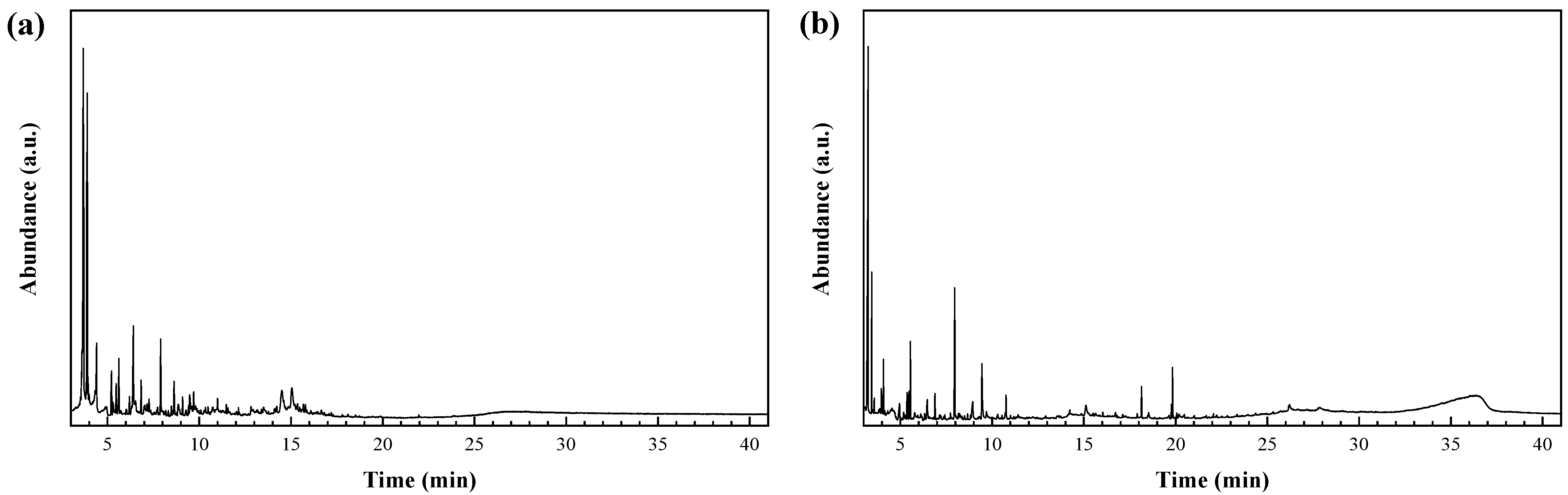
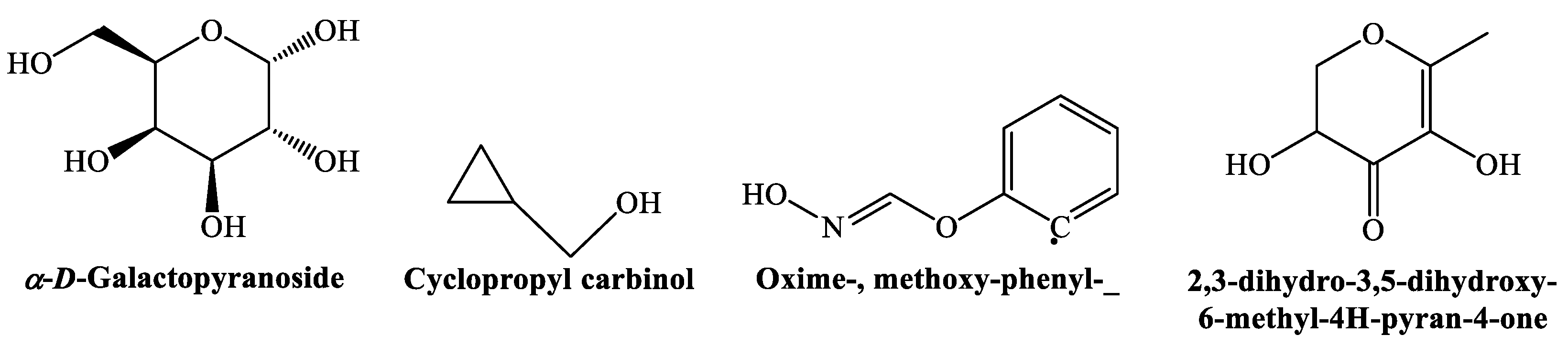
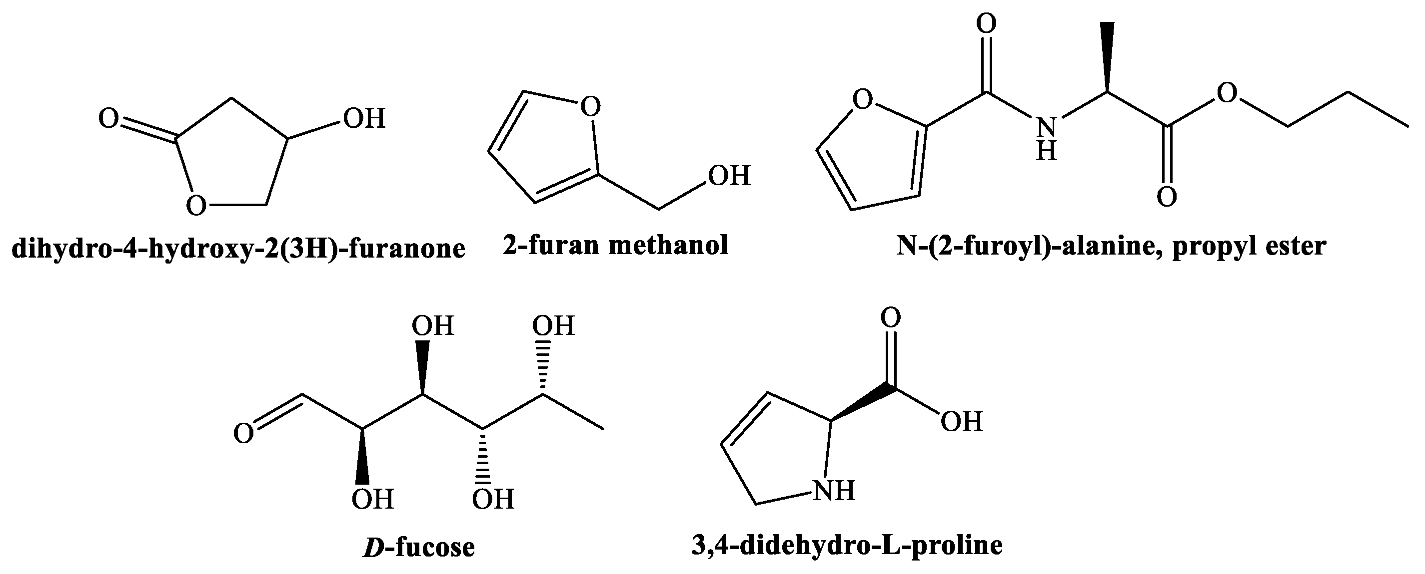
3.2. Phytochemicals Identification

The Smilax aspera leaf extract chromatogram (Figure 1a, Table S1) includes α- and β-D-galactopyranoside, methyl (5.2%); cyclopropyl carbinol (3.2%); methoxy-phenyl-oxime (2.1%); and 2,3-dihydro-3,5-dihydroxy-6-methyl-4H-pyran-4-one (1.7%), depicted in Figure 2.
Among the phytochemicals contained in the S. aspera fruit extract (Figure 1b, Table S2), three chemicals with a furan ring in their structure were found, namely, dihydro-4-hydroxy-2(3H)-furanone (or 3-hydroxy-γ-butyrolactone) (9.7%), 2-furan methanol (3.4%), and N-(2-furoyl)-alanine, and propyl ester (1.5%); the methyl esters of 11-octadecenoic and hexadecanoic acids (2.6% and 1.8%, respectively); D-fucose (1.8%); and 3,4-didehydro-proline (1.5%), as shown in Figure 3.
The phytochemicals shared by the leaf and fruit extracts (Figure 4) included 1-hydroxy-2-propanone (4.4–17.4%); dialcohols such as 2,3-butanediol (2.9–3.4%) and catechol (2.3–5.5%); and lactones such as 2-hydroxy-γ-butyrolactone (2.1–5.3%) and 2-hydroxy-2-cyclopenten-1-one (2.1–3.8%).

3.3. Antibacterial Activity

3.3.1. In Vitro Antibacterial Activity

The MIC assessment of the S. aspera extracts against phytopathogens was carried out using a microplate assay (Figure 5). The analysis indicated that the S. aspera extracts at lower concentrations, ranging from 500 to 125 µg·mL−1, did not display any efficacy. In contrast, the tested extracts exhibited a complete inhibition of bacterial growth at 1500 μg·mL−1. Further testing of these concentrations in an agar matrix inoculated with bacteria confirmed the absence of detectable growth, providing evidence that the MIC and Minimum Bactericidal Concentration (MBC) values aligned.
A comparison of the antibacterial activity of the S. aspera fruit and leaf extracts with that of antibiotics (Table 2) revealed that the extracts were less effective against the bacteria under study.

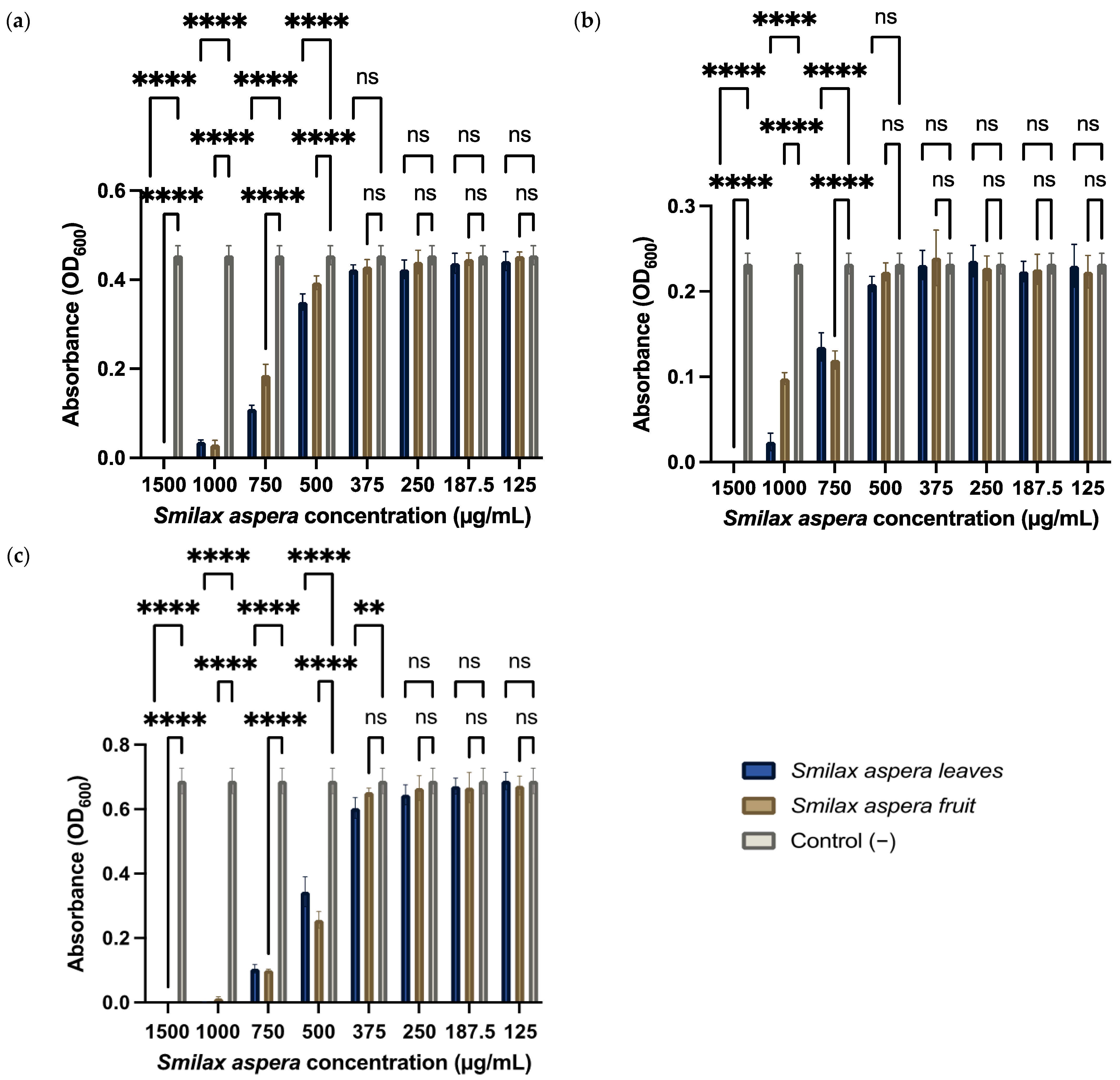
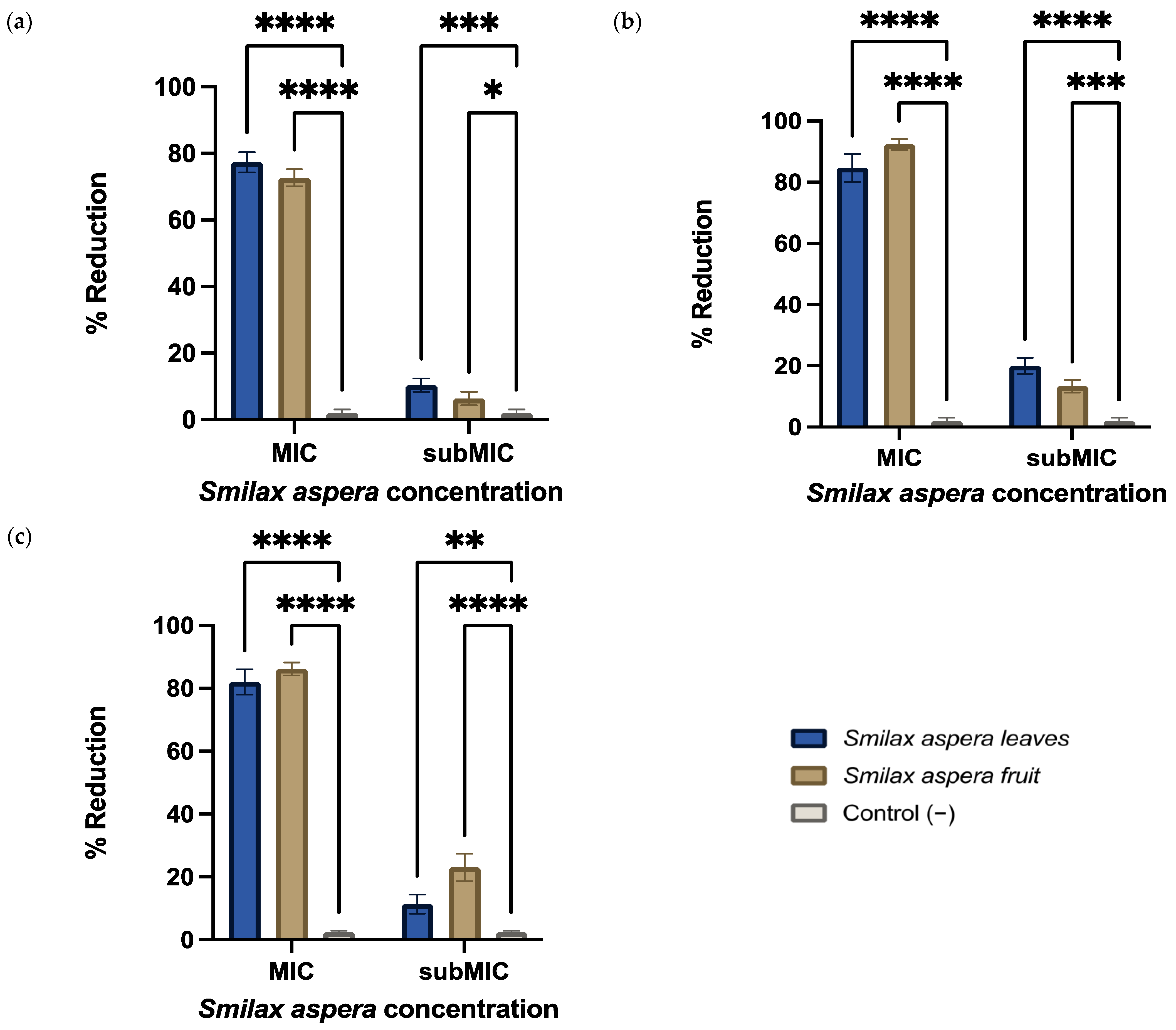
3.3.2. Anti-Biofilm Activity

Biofilm formation is a bacterial survival strategy wherein colonies enhance intercellular communication mechanisms under stressful conditions to create a more resistant and cohesive community. In the context of fire blight, black rot, and Psa canker pathogenesis, evaluating anti-biofilm properties is crucial. Smilax aspera leaf and fruit extracts were added at concentrations both at and below their MICs to bacterial suspensions. Subsequently, biofilm formation was quantified using spectrophotometer readings. Figure 6 depicts a significant decrease in biofilm formation when compared to the control. The leaf extracts reduced biofilm formation by 85%, 78%, and 82% when used at their MIC concentrations against Xcc, EA, and Psa, respectively. This effect was slightly stronger with the fruit extract, reaching a reduction in biofilm formation of 92.5% against Xcc, 73% against EA, and 86.5% against Psa. This observation can primarily be attributed to the phenolic compounds present in the leaves and fruits, which are believed to act as anti-biofilm agents.

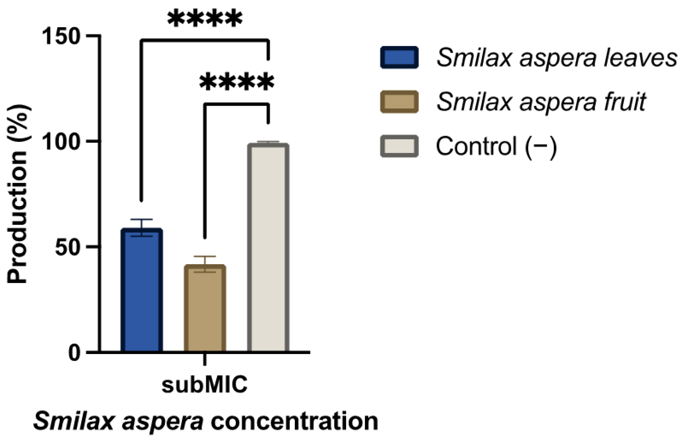
3.3.3. Amylovoran Production

In the pursuit of a more profound understanding of how exposure to the two S. aspera extracts affects virulence, this study investigated their impact on amylovoran production. The extracts from leaves and fruits used at a subMIC concentration, 750 µg·mL−1, resulted in a reduction in amylovoran synthesis by 41% and 58%, respectively, as depicted in Figure 7. The virulence of EA relies on amylovoran production, and these results suggest that the phytoconstituents may target this crucial virulence factor, aligning with the previously observed effects.

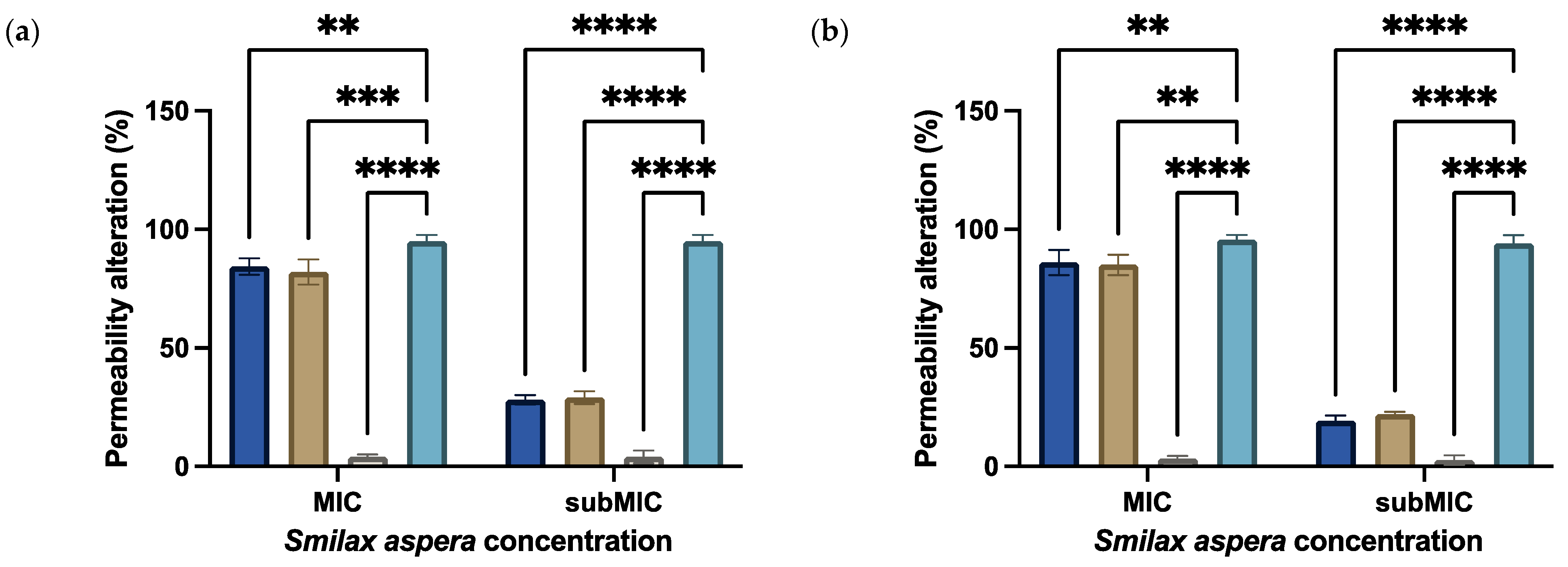
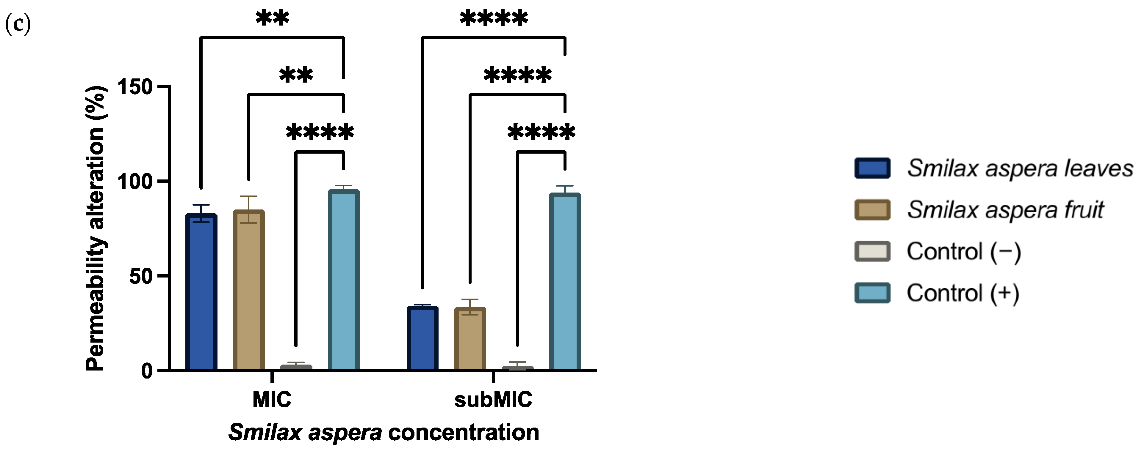
3.3.4. Permeability Alteration

This assay was designed to determine whether extract concentrations known for their antibacterial properties could impact bacterial membrane permeability. Diverse concentrations of each extract were assessed on bacterial suspensions at various time points. To assess membrane changes, PI—a fluorescent intercalating agent—was introduced into bacterial suspensions of EA, Xcc, and Psa. PI cannot permeate intact membranes but becomes detectable when membrane integrity and permeability are compromised, as this agent enters the bacterial cell and intercalates with DNA bases.
As anticipated, upon reaching the MIC of the S. aspera extracts, noticeable alterations in membrane permeability were observed, akin to the positive control involving bacteria treated with a 10% bleach solution (Figure 8). However, even at reduced concentrations, such as MIC/2 (750 µg·mL−1), a discernible augmentation in membrane permeability to PI was evident. Specifically, the 1/2 MIC concentration resulted in 28%, 34%, and 21% increases in PI uptake for EA, Xcc, and Psa, respectively. This suggests that even at reduced extract concentrations, membrane integrity and permeability were compromised.

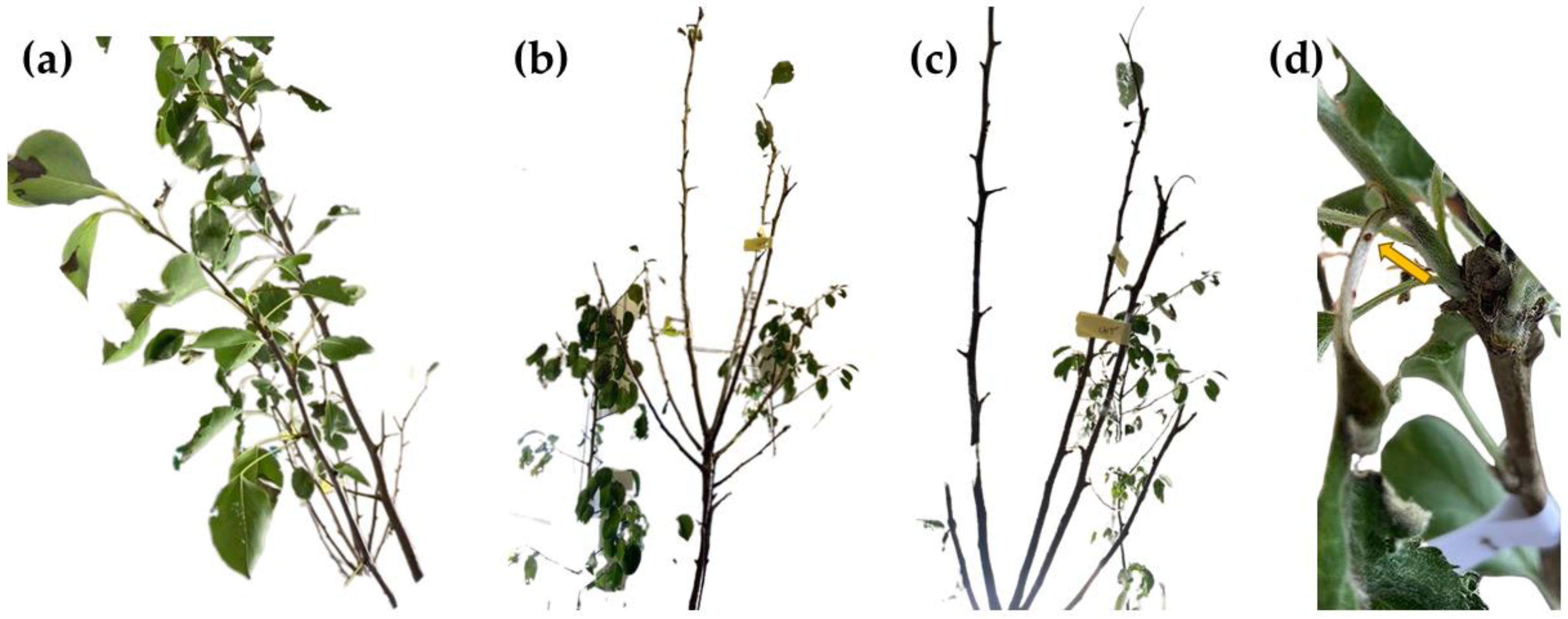
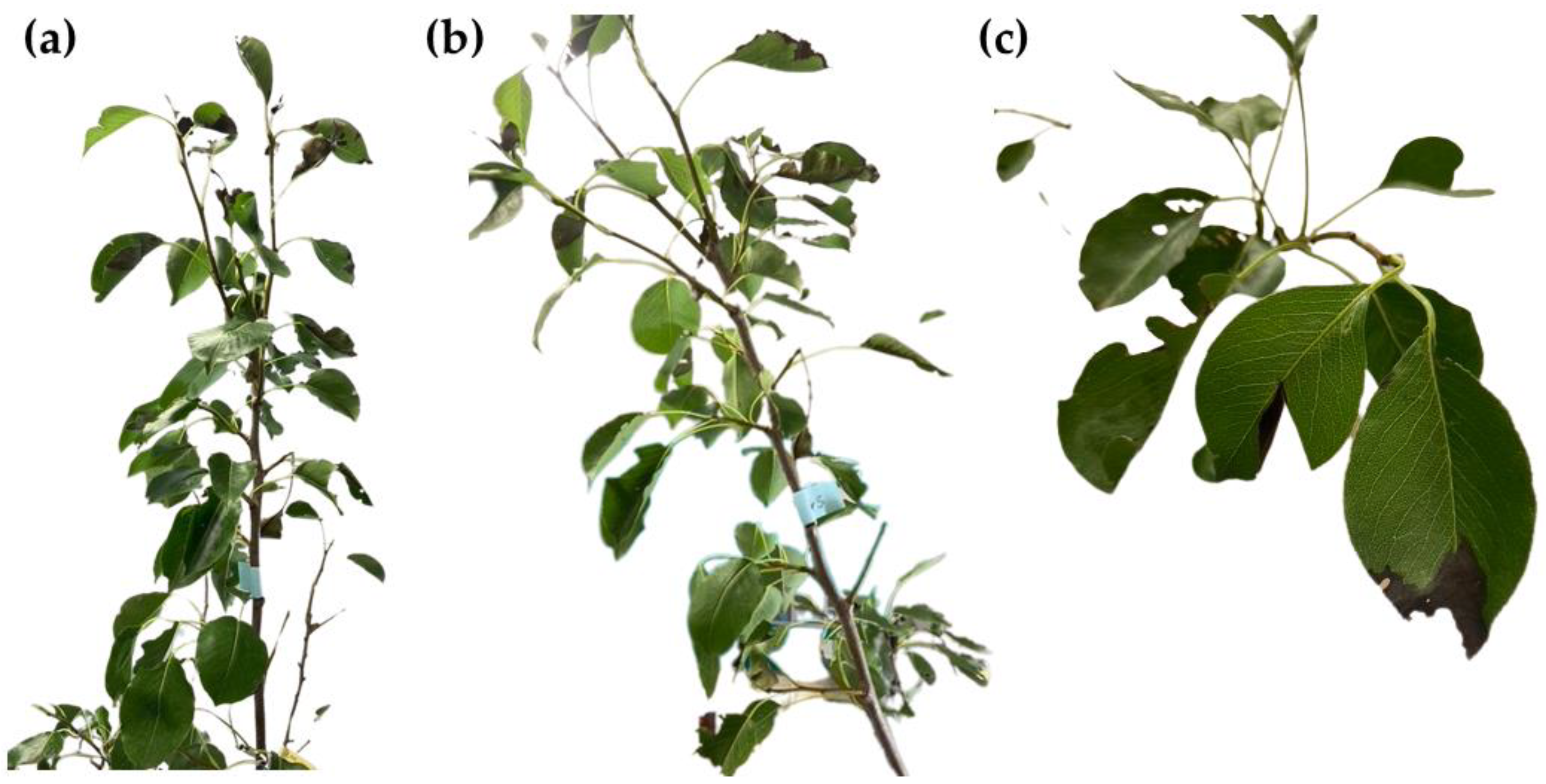
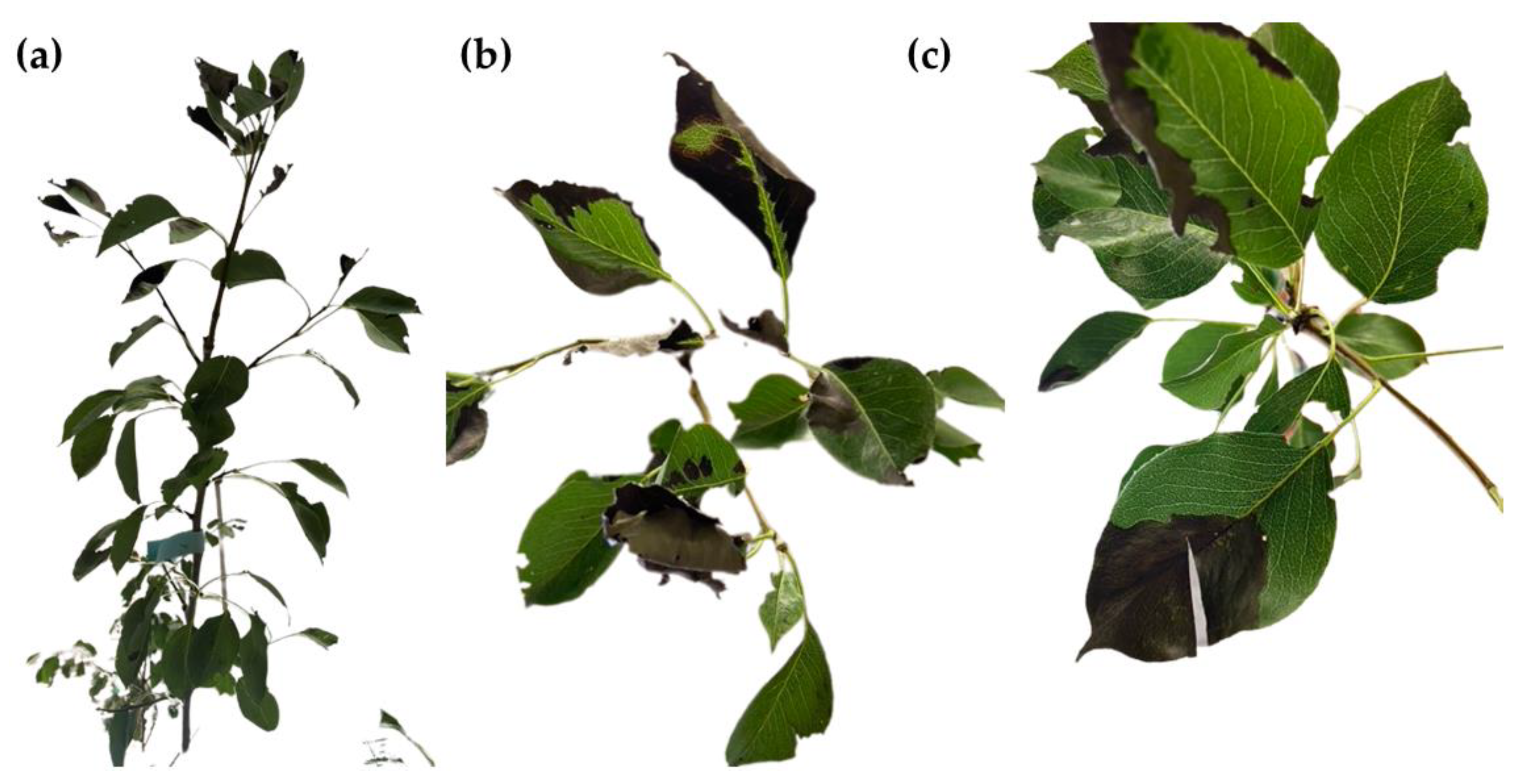
3.4. In Vivo Antibacterial Activity

To elucidate the efficacy of S. aspera extracts against EA, a preliminary investigation was conducted to assess the antibacterial impact within a plant system. This experimental approach provides insights into the dynamic interaction between the bacterium and plant cells, presenting a more realistic perspective.
Given that EA is the causative agent of fire blight in apple and pear trees, we assessed the antibacterial properties of S. aspera extracts on pear leaves previously inoculated with this bacterium. Shoot blight initiates at the tips of growing shoots and rapidly progresses down into older parts of the twig. Initially, blighted twigs appear water-soaked and subsequently turn dark brown or black. As affected shoots wilt, the twigs bend at the growth point, resembling a shepherd’s crook or an inverted “J”. Blighted leaves may persist on dead branches throughout the summer but fall during periods of high humidity. Under warm and humid conditions, infected shoots may exude droplets of creamy white bacteria.
The control pear tree branches, depicted in Figure 9a–c, exhibited progressive symptoms of fire blight over a span of 10 days post-inoculation. Initially, at 7 days post-inoculation (Figure 9b), the branches displayed the first signs of infection, with some areas devoid of leaves and exhibiting brown spots. By day 10 post-inoculation (Figure 9c), almost all the leaves had dropped, and characteristic wilting with dark-brown hooks was evident on some branches. Additionally, visible exudate production was observed on individual branches (Figure 9d), indicative of advanced disease progression.
In contrast, the branches treated with S. aspera leaf extract, as shown in Figure 10a–c, exhibited a significant reduction in disease severity. Two days post-inoculation (Figure 10a), no symptoms of fire blight were apparent. Seven days post-treatment with S. aspera leaf extract (Figure 10b), few necrotic areas were observed on some leaves, indicating partial efficacy of the treatment. However, by day 10 post-treatment (Figure 10c), the severity of necrotic areas was reduced by 80% compared to that of the control, with no signs of exudate production or hook wilting detected.
Similarly, treatment with S. aspera fruit extract, illustrated in Figure 11a–c, also led to a reduction in disease severity. While necrotic areas were observed on some leaves 7 days post-treatment (Figure 11b), indicative of partial efficacy, by day 10 post-treatment (Figure 11c), the severity of necrotic areas was reduced by 45% compared to the control, with no signs of exudate production or hook wilting observed. These findings highlight S. aspera extracts’ potential as a promising treatment option for managing fire blight in apple trees, particularly when derived from S. aspera leaves.

4. Discussion

4.1. Phytochemical Profile

Regarding the identified phytoconstituents, caution is warranted as the samples were collected from a single area. Discrepancies in the phytochemical profile and bioactivity may result from differences in the extraction process, individual genotype-dependent factors, location-specific intra-varietal variations, and seasonal fluctuations. Additionally, the potential presence of distinct chemotypes arising from minor genetic and epigenetic changes should be considered.
Regarding the phytochemicals exclusively identified in the leaf extract, methoxy-phenyl-oxime and 2,3-dihydro-3,5-dihydroxy-6-methyl-4H-pyran-4-one were also reported in the extracts of Rubia tinctorum L., known for its potent antimicrobial properties [16]. Among the chemical compounds found only in the fruit extract, dihydro-4-hydroxy-2(3H)-furanone was previously detected in Urtica dioica L., Equisetum arvense L. [17], and Ginkgo biloba L. [18] extracts. It is also a major constituent of Crocus sativus L. (corresponding to a content of 22%) and has demonstrated antifungal activity against various fungi (including Aspergillus fumigatus Fresen., Cryptococcus neoformans (Sanfelice) Vuill., Pyricularia oryzae Cavara, and Trichophyton rubrum (Castell.) Sabour.) [19]. As for 2-furanmethanol (or 2-hydroxymethyl-furan), a phytochemical related to 5-(hydroxymethyl)furan-3-carboxylic acid, it was previously found in Paederia foetida L. [20] and showed significant antifungal activity against fungi of the genus Aspergillus.
Concerning the chemical species shared by both extracts, 1-hydroxy-2-propanone, a primary alcohol substituent on acetone, was previously found in G. biloba [18]. 2,3-Butanediol, a short diol, is easily produced by different native bacteria from sugars or lignocellulosic biomasses. Catechol (or 1,2-dihydroxybenzene) is a ubiquitous phytochemical common to several plants including Allium sativum L. [21], R. tinctorum [16], Quercus ilex L. [22], Sambucus nigra L. [23], and Euphorbia serrata L. [24]. Both 2-hydroxy-γ-butyrolactone (position isomer of 3-hydroxy-γ-butyrolactone) and 2-hydroxy-2-cyclopenten-1-one were also identified in R. tinctorum [16]. N-(2-furoyl)-alanine, propyl ester may act as a precursor for most of the compounds identified in this analysis.
The examination of the phytochemicals identified in the S. aspera leaf and fruit extracts suggests the metabolism of parent compounds during the extraction procedure, with 1-(3,6,6-trimethyl-1,6,7,7a-tetrahydrocyclopenta[c]pyran-1-yl)ethenone being the most probable precursor for pyrans and 5-phenyl-2-furoyl-alanine, propyl ester fulfilling this role for furans (Figure 12).
Nonetheless, it is widely acknowledged that furans may also be formed directly through the dehydration of cellulose or fructose, resulting in anhydrosugar intermediates like levoglucosan and levoglucosenone. Subsequently, at 250 °C, these intermediates can be converted into furans, including furanone and dihydro-hydroxy-2(3H)-furanone, as well as acetaldehyde and glycolaldehyde [25,26]. Examination of their potential energy surfaces indicates that furans represent the minima in Gibbs free energy (ΔG) within the product mixture. Hence, it cannot be discounted that furans might be generated through coupling reactions of the oxygenated organic molecules present in the pyrolysis vapors, which are formed during the extraction and chromatography procedures (Figure 13).

4.2. On the Antibacterial Activity

4.2.1. Mechanism of Action

The specific mode of action of S. aspera extracts was not studied herein, but an additive or synergistic behavior is expected due to the presence of several compounds with well-established antimicrobial action, as discussed above.
As in the case of polyphenols from S. china [27], the most probable mechanism of action of polyphenols would involve the alteration of the bacterial cell wall and damage to the cell membrane, inducing the leakage of cell contents, ultimately resulting in bacterial decomposition and death.
Regarding non-phenolic constituents, monosaccharide derivatives are recognized for their extensive range of biological effects on both Gram-negative and Gram-positive organisms, demonstrating efficacy against bacteria such as Escherichia coli (Migula 1895) Castellani & Chalmers 1919; Bacillus subtilis G; Salmonella typhimurium (Loeffler 1892) Castellani & Chalmers 1919; and Staphylococcus aureus Rosenbach 1884 [28,29,30]. Specifically, the chromatographic analysis of S. aspera leaf extract identified the presence of α- and β-D-galactopyranoside. Recent research by Hosen et al. [31] investigated the antimicrobial activity of methyl β-D-galactopyranoside (MGP) and its analogs and derivatives. MGP and its esters were evaluated for their physicochemical and pharmacokinetic properties through a comprehensive computational study involving thermodynamics, molecular dynamics, and molecular docking, revealing efficient binding to key targets, such as CTX-M-15 extended-spectrum β-lactamase from E. coli (PDB:4HBT). Moreover, other derivatives [32] have demonstrated in vitro antibacterial potential against Bacillus cereus Frankland & Frankland 1887, B. subtilis, E. coli, S. typhimurium, and Pseudomonas aeruginosa (Schroeter 1872) Migula 1900.
However, within the context of prospective agricultural applications, further in-depth analyses are necessary to validate these hypotheses and explore other potential mechanisms, such as abnormal intracellular oxidative stress, integration into genomic DNA, or restriction of the production of extracellular polymers [33].

4.2.2. Activity of S. aspera and Other Smilax spp. Extracts

Regarding the Smilax species under investigation, S. aspera, there are few studies on its antimicrobial activity, with contradictory results. Gyawali et al. [34] prepared a methanolic extract of leaves and bark that exhibited high effectiveness against S. typhimurium and Pseudomonas spp. but only at concentrations as high as 4, 6, and 8%. Moreover, the extract showed low activity against E. coli, Klebsiella pneumoniae (Schroeter 1886) Trevisan 1887, and S. aureus. In turn, Mohammad Sawalha [35] found that ethanolic extracts of S. aspera fruits exhibited an inhibition zone of 20 mm against S. aureus and Candida albicans (C.P. Robin) Berkhout but showed no activity against E. coli. In comparison, the aqueous extraction only inhibited C. albicans, with an inhibition zone of 13 mm. Abbasołu and Türköz [36] prepared an extract from S. aspera fruits using chloroform and ethanol and tested it against bacteria such as E. coli, P. aeruginosa, Streptococcus faecalis Andrewes & Horder 1906, and S. aureus and fungi including C. albicans, Candida parapsilosis (Ashford) Langeron & Talice, and Candida pseudotropicalis (Castell.) Basgal (=Kluyveromyces marxianus (E.C. Hansen) Van der Walt), with inhibition values of 6300 μg·mL−1, higher than those reported herein. Higher antimicrobial activity was reported for steroidal saponins from S. aspera roots, which showed antifungal activity against C. albicans, Candida glabrata (H.W. Anderson) S.A. Mey. & Yarrow (=Nakaseomyces glabratus (H.W. Anderson) Sugita & Takashima), and Candida tropicalis (Castell.) Berkhout, with MIC values in the range of 25–50 μg·mL−1 [37], results similar to those obtained for spirostanol saponins from Smilax medica Bott. roots, with inhibition ranging between 12.5 and 50 μg·mL−1 [38], and in line with the biological properties of steroidal saponins sourced from other species of the Smilax genus reported by Tian et al. [39].
Concerning the antimicrobial efficacy of extracts from other species of the Smilax genus, a variety of solvents have been explored, but most studies have tested their activity against human pathogens, providing very few examples of activity tests against phytopathogens. As in the case of S. aspera, the results are not consistent from one study to another, but alcoholic extraction media are generally associated with the best results, as discussed below. It is worth noting that, given the diversity of plant parts chosen for the extraction (leaves, fruits, rhizomes, etc.) and the variety of extraction media, antimicrobial-activity-testing methods, assayed concentrations, and pathogens studied, direct efficacy comparisons are not applicable.
Concerning leaf extracts and fruit extracts like the ones studied herein, crude extracts derived from the aerial parts of Smilax larvata Griseb., including chloroform, ethyl acetate, hydroalcoholic, and n-hexane extracts, displayed no antimicrobial effects against S. aureus, Staphylococcus epidermidis (Winslow & Winslow 1908) Evans 1916, E. coli, P. aeruginosa, Agrobacterium tumefaciens H, and C. albicans. Only the ethanolic extract demonstrated a modest antifungal response, comparable to that of ketoconazole, against C. albicans [40].
Seo et al. [41] compared the antimicrobial activity of a leaf extract from Smilax china L. using DMSO, methanol, ethanol, acetone, and water as solvents. The DMSO extraction did not inhibit any of the studied pathogens: E. coli exhibited resistance to all five solvents; Listeria monocytogenes B showed greater susceptibility to the methanol extract (IZ = 11.7 mm), and S. aureus showed greater susceptibility to the ethanol extract (IZ = 10.7 mm), while S. typhimurium showed greater susceptibility to the aqueous extract (IZ = 11.8 mm). Additionally, polyphenols from S. china effectively inhibited bacteria such as B. subtilis, E. coli, L. monocytogenes, S. typhimurium, and S. aureus, with inhibition levels varying from 195.31 to 781.25 μg·mL−1 [27].
Ethanolic and methanolic extracts of Smilax glabra Roxb. and Smilax corbularia Kunth inhibited the growth of bacteria isolated from the mouth at 31,250–500,000 μg·mL−1; however, their aqueous extracts showed no activity [42]. The hexane leaf extract from Smilax macrophylla Griseb. displayed strong inhibitory effects against Alternaria alternata (Fr.) Keissl., Ganoderma lucidum (Curtis) P. Karst., Pasteurella multocida A, E. coli, B. subtilis, and S. aureus, with inhibition values ranging from 30,400 to 53,100 μg·mL−1. In contrast, its methanol extract demonstrated no activity against A. alternata, E. coli, and S. aureus [43]. The ethanolic leaf extract from Smilax perfoliata Lour. resulted in inhibition zones ranging from 10 to 13 mm against B. subtilis, Proteus mirabilis B, B. cereus, S. typhimurium, P. aeruginosa, S. epidermidis, and C. albicans; however, no inhibitory activity was observed against S. aureus and E. coli [44].
The ethanolic extract derived from the aerial parts of Smilax campestris Griseb. and its hexane and dichloromethane fractions exhibited high activity against C. albicans spp. and Cryptococcus gattii (Vanbreus. & Takashio) Kwon-Chung & Boekhout, with MIC values ≤ 2000 μg·mL−1. Conversely, the butanol and hydroalcoholic fractions demonstrated inactivity against all the studied yeasts [45].
Dhanya Shree et al. [46] explored the susceptibility of bacteria (B. cereus, S. typhimurium, E. coli, and Shigella flexneri Castellani & Chalmers 1919) and fungi (Aspergillus niger Tiegh. and Bipolaris sp.) to leaf and fruit methanolic extracts from Smilax zeylanica L. Unlike the results reported herein, in which both extracts showed similar effectiveness, in their study, the leaf extract demonstrated more prominent antibacterial activity when compared to the fruit extract, and both extracts proved effective against the tested fungi.
Rajbhandari and Paneru [47] evaluated the leaf extract of Smilax ovalifolia Roxb. ex D.Don in different solvents, namely, dichloromethane, ethyl acetate, or methanol, against E. coli and S. aureus, resulting in inhibition zones of approximately 16–19 mm for E. coli and 15–17 mm for S. aureus.
The in vitro assessment of ethylacetate, hexane, and methanol extracts from Smilax kraussiana Meisn. leaves was conducted by Hamid and Aiyelaagbe [48] against a panel of human-pathogenic microorganisms, encompassing six bacteria and six fungi. The findings revealed significant inhibitory effects on the growth of the twelve tested organisms. Particularly, the hexane and methanol extracts exhibited pronounced inhibition against B. subtilis and S. aureus (Gram-positive) within the concentration range of 25,000 to 200,000 μg·mL−1, surpassing the inhibitory potency of the ethylacetate extract. Conversely, all the extracts demonstrated comparatively lower antibacterial activity against Gram-negative bacteria, including E. coli, K. pneumoniae, P. aeruginosa, and Salmonella typhi (Schroeter 1886) Warren & Scott 1930. Notably, the three extracts displayed strong antifungal properties against A. niger, C. albicans, Epidermophyton floccosum (Harz) Langeron & Miloch, Penicillum notatum Westling (=Penicillium chrysogenum Thom), Rhizopus stolonifer (Ehrenb.) Vuill., and Tricophyton rubrum (Castell.) Sabour.
It should be noted that the previously mentioned significant variations in MIC values found in the literature are also applicable to root extracts: Joo et al. [49] reported that S. china root extracts exhibited strong antimicrobial activity (500 μg·mL−1) against Cutibacterium acnes (Gilchrist 1900) Scholz & Kilian 2016, a bacterium associated with acne. The ethanolic extract from Smilax glabra Roxb. rhizomes, along with the ethyl acetate and n-butanol fractions, demonstrated considerable activity against S. aureus (50 μg·mL−1), while water and ethyl acetate fractions exhibited activity against C. albicans and S. aureus, showcasing MIC values of 200 μg·mL−1 [50]. Nonetheless, according to McMurray et al. [51], the aqueous extract exhibited low activity, inhibiting L. monocytogenes, Salmonella enteritidis (Gaertner 1888) Castellani & Chalmers 1919, and E. coli within the range of 31,250–250,000 μg·mL−1. Cáceres et al. [52] investigated the antimicrobial, larvicidal, leishmanicidal, and schisonticidal activities of an ethanolic extract from Smilax domingensis Willd. rhizomes. The authors highlighted the results obtained for B. subtilis, Mycobacterium smegmatis (Trevisan 1889) Lehmann & Neumann 1899, P. aeruginosa, S. aureus, and Sporothrix schenckii Hektoen & C.F. Perkins, with MICs of 250 μg·mL−1, and reported a MIC value of 500 μg·mL−1 for S. typhimurium, C. neoformans, and Trichophyton mentagrophytes (C.P. Robin) R. Blanch.

4.2.3. Comparison with Synthetic Antimicrobials

In this study, bactericides from the aminoglycoside group (streptomycin) and polycyclic derivatives of naphthracenecarboxamides (tetracycline) were selected for comparison purposes, as they were initially the primary treatments employed to manage the bacterial infections under investigation.
The antibacterial activity of S. aspera fruit and leaf extracts was found to be lower than that of the two conventional antibiotics against all the bacterial strains under study. It is important to note, however, that the improper usage of the aforementioned conventional antibiotics has contributed to the emergence of resistant strains, necessitating the exploration of other broad-spectrum control techniques [33]. In fact, higher doses than those assayed in this study for the extracts (1500 μg·mL−1) have been reported for conventional bactericidal products. For example, streptomycin resistance was observed in some isolated E. amylovora strains from Ohio, with inhibition values spanning from 500 to 2500 μg·mL−1 [53]. Comparable findings were reported in California by Förster et al. [54], with MICs ranging from 0.5 to >2000 μg·mL−1.
Regarding streptomycin activity against P. syringae pv. actinidiae, Cameron and Sarojini [55] compiled results from 19 strains, showing inhibition values in the range of 3.5 to >2000 μg·mL−1. Lee et al. [56] studied 10 strains with inhibition between 10 and 500 μg·mL−1. For X. campestris pv. campestris, the authors observed susceptibility to ampicillin, penicillin, and tetracycline at concentrations ≤ 6.25 μg·mL−1; carbenicillin, cephalothin, gentamicin, and kanamycin at 25.80 μg·mL−1; and bacitracin, neomycin, and streptomycin at >400, 160, and 640 μg·mL−1, respectively [57]. These values are in line with those of other pathovars of X. campestris, which could be inhibited by amoxicillin, cephalexin, chloramphenicol, penicillin G, streptomycin sulfate, or tetracycline. Specifically, tetracycline inhibited X. campestris pv. musacearum at 20 μg·mL−1 [58] and X. campestris pv. vesicatoria at doses ranging from 0.24 μg·mL−1 [59] to 156 μg·mL−1 [60].
Despite their lower efficacy, the presence of multiple antimicrobial compounds with different and concurrent modes of action in the extracts could be an advantage over conventional bactericidal compounds, potentially preventing the development of resistant strains. Therefore, S. aspera extracts may play a role in combating drug-resistant bacteria or diminish antibiotic reliance, possibly via a synergistic combination with these antibiotics, as has also been suggested for S. china [61].

4.2.4. Comparison with Other Products for Pear Tree Protection

Regarding the comparison of in planta results, in [62], the application of hydroethanolic extracts of Thymus vulgaris L., Rhus coriaria L., and Eucalyptus globulus Labill. Reached efficacies of 61.72%, 46.52%, and 29.94%, respectively, against E. amylovora on three-year-old ‘Royal Gala’ apple trees at concentrations of 20% (200,000 μg·mL−1, i.e., two orders of magnitude higher than those evaluated in this study). In [63], three extracts of Moringa oleifera Lam., namely, methanolic extract (MIC = 1000 μg·mL−1), hydroalcoholic extract (MIC = 1000 μg·mL−1), and hydroalcoholic extract with maltodextrins (MIC = 1000 μg·mL−1), were applied to two-year-old apple trees of the ‘Gala’ cultivar. The maltodextrin extract demonstrated the highest effectiveness, resulting in an 80% reduction in wilting compared to the control. Likewise, the methanolic and hydroalcoholic extracts showed a reduction in the infected area by 65% and 71%, respectively (slightly lower than that obtained for S. aspera hydromethanolic leaf extract but at a lower application dose).

5. Conclusions

Gas chromatography–mass spectrometry analyses of S. aspera leaf and fruit hydromethanolic extracts revealed the shared presence of phytoconstituents such as 1-hydroxy-2-propanone, 2,3-butanediol, catechol, 2-hydroxy-γ-butyrolactone, and 2-hydroxy-2-cyclopenten-1-one. Both extracts demonstrated high antibacterial activity against three relevant phytopathogens under in vitro conditions, inhibiting P. syringae pv. actinidiae, X. campestris pv. campestris, and Erwinia amylovora at a dose of 1500 μg·mL−1. The permeability assays showed noticeable alterations in membrane permeability both at the MIC and MIC/2. At the MIC, the leaf extracts reduced biofilm formation by 85%, 78%, and 82% against Xcc, EA, and Psa, respectively, while the fruit extract achieved reductions of 92.5%, 73%, and 86.5%, respectively. Furthermore, when used at a dose of MIC/2, the leaf and fruit extracts resulted in a reduction in amylovoran synthesis by 41% and 58%, respectively. Subsequent in planta testing against EA demonstrated that the leaf extract (at the MIC) exhibited the highest effectiveness, resulting in an 80% reduction in wilting area, while the fruit extract led to a 45% reduction in wilting 10 days after inoculation. The reported results suggest that S. aspera extracts may hold promise with respect to combatting drug-resistant bacteria or lessening the need for antibiotics in agricultural settings when used in conjunction with antibiotics.

Supplementary Materials

The following supporting information can be downloaded at https://www.mdpi.com/article/10.3390/agronomy14020383/s1. Figure S1: ATR-FTIR spectra of S. aspera leaves and fruits prior to extraction; Table S1: Phytochemicals identified in S. aspera hydromethanolic leaf extract; Table S2: Phytochemicals identified in S. aspera hydromethanolic flower extract.

Author Contributions

Conceptualization, J.M.-G. and P.M.; methodology, P.M.-R., J.M.-G. and P.M.; validation, P.M.-R. and P.M.; formal analysis, R.F., E.S.-H. and P.M.-R.; investigation, R.F., E.S.-H., P.M.-R., J.M.-G. and P.M.; resources, J.M.-G. and P.M.; writing—original draft preparation, R.F., E.S.-H., P.M.-R. and J.M.-G.; writing—review and editing, R.F., E.S.-H. and P.M.-R.; visualization, R.F. and E.S.-H.; supervision, P.M.-R. and P.M.; project administration, P.M.-R. and J.M.-G.; funding acquisition, P.M.-R. and J.M.-G. All authors have read and agreed to the published version of the manuscript.

Funding

This research was funded by Junta de Castilla y León under project VA148P23, with FEDER co-funding.

Data Availability Statement

The data supporting the findings of this study are available within the article and its Supplementary Materials.

Acknowledgments

We acknowledge the Servizio Fitosanitario Regionale dell’Emilia-Romagna. The authors also gratefully acknowledge the support provided by Pilar Blasco and Pablo Candela at the Servicios Técnicos de Investigación, Universidad de Alicante, in conducting the GC–MS analyses.

Conflicts of Interest

The authors declare no conflicts of interest. The funders had no role in the design of the study; in the collection, analyses, or interpretation of data; in the writing of the manuscript; or in the decision to publish the results.

References

  1. Bhatta, S.; Joshi, L.R.; Kunwar, R.M.; Bussmann, R.W.; Paniagua-Zambrana, N.Y. Smilax aspera L., Smilax ovalifolia Roxb. Smilacaceae. In Ethnobotany of the Himalayas; Kunwar, R.M., Sher, H., Bussmann, R.W., Eds.; Springer International Publishing: Cham, Switzerland, 2021; pp. 1869–1879. [Google Scholar] [CrossRef]
  2. Quattrocchi, U. CRC World Dictionary of Medicinal and Poisonous Plants: Common Names, Scientific Names, Eponyms, Synonyms, and Etymology; CRC: Boca Raton, FL, USA, 2012; p. 3960. [Google Scholar]
  3. Kakouri, E.; Hatziagapiou, K.; Kanakis, C.; Nikola, O.; Lambrou, G.I.; Trigas, P.; Kanaka-Gantenbein, C.; Tarantilis, P.A. Cytotoxic and antioxidant activity of a chemically characterized extract of Smilax aspera leaves and stems. Appl. Sci. 2023, 13, 4784. [Google Scholar] [CrossRef]
  4. Salas-Coronado, R.; Hernández-Carlos, B.; Llaguno-Guilberto, J.; Santos-Sánchez Norma, F. Phenolic compounds in genus Smilax (Sarsaparilla). In Phenolic Compounds: Natural Sources, Importance, and Applications; Marcos, S.-H., Mariana, P.-T., Maria del Rosario, G.-M., Eds.; IntechOpen: Rijeka, Croatia, 2017; pp. 233–260. [Google Scholar] [CrossRef]
  5. Longo, L.; Vasapollo, G. Extraction and identification of anthocyanins from Smilax aspera L. berries. Food Chem. 2006, 94, 226–231. [Google Scholar] [CrossRef]
  6. Delgado-Pelayo, R.; Hornero-Méndez, D. Identification and quantitative analysis of carotenoids and their esters from sarsaparilla (Smilax aspera L.) berries. J. Agric. Food. Chem. 2012, 60, 8225–8232. [Google Scholar] [CrossRef] [PubMed]
  7. Santos, M.G.; Nunes da Silva, M.; Vasconcelos, M.W.; Carvalho, S.M.P. Scientific and technological advances in the development of sustainable disease management tools: A case study on kiwifruit bacterial canker. Front. Plant Sci. 2024, 14, 1306420. [Google Scholar] [CrossRef]
  8. Piqué, N.; Miñana-Galbis, D.; Merino, S.; Tomás, J. Virulence factors of Erwinia amylovora: A review. Int. J. Mol. Sci. 2015, 16, 12836–12854. [Google Scholar] [CrossRef]
  9. Donati, I.; Cellini, A.; Sangiorgio, D.; Vanneste, J.L.; Scortichini, M.; Balestra, G.M.; Spinelli, F. Pseudomonas syringae pv. actinidiae: Ecology, infection dynamics and disease epidemiology. Microb. Ecol. 2020, 80, 81–102. [Google Scholar] [CrossRef]
  10. Pedroncelli, A.; Puopolo, G. This tree is on fire: A review on the ecology of Erwinia amylovora, the causal agent of fire blight disease. J. Plant Pathol. 2023, in press. [Google Scholar] [CrossRef]
  11. Vicente, J.G.; Holub, E.B. Xanthomonas campestris pv. campestris (cause of black rot of crucifers) in the genomic era is still a worldwide threat to brassica crops. Mol. Plant Pathol. 2013, 14, 2–18. [Google Scholar] [CrossRef]
  12. Akhlaghi, M.; Tarighi, S.; Taheri, P. Effects of plant essential oils on growth and virulence factors of Erwinia amylovora. J. Plant Pathol. 2019, 102, 409–419. [Google Scholar] [CrossRef]
  13. Wilson, C.; Lukowicz, R.; Merchant, S.; Valquier-Flynn, H.; Caballero, J.; Sandoval, J.; Okuom, M.; Huber, C.; Brooks, T.D.; Wilson, E.; et al. Quantitative and qualitative assessment methods for biofilm growth: A mini-review. Res. Rev. J. Eng. Technol. 2017, 6, 1–25. [Google Scholar]
  14. Bellemann, P.; Bereswill, S.; Berger, S.; Geider, K. Visualization of capsule formation by Erwinia amylovora and assays to determine amylovoran synthesis. Int. J. Biol. Macromol. 1994, 16, 290–296. [Google Scholar] [CrossRef]
  15. Schneider, C.A.; Rasband, W.S.; Eliceiri, K.W. NIH Image to ImageJ: 25 years of image analysis. Nat. Methods 2012, 9, 671. [Google Scholar] [CrossRef]
  16. Langa-Lomba, N.; Sánchez-Hernández, E.; Buzón-Durán, L.; González-García, V.; Casanova-Gascón, J.; Martín-Gil, J.; Martín-Ramos, P. Activity of anthracenediones and flavoring phenols in hydromethanolic extracts of Rubia tinctorum against grapevine phytopathogenic fungi. Plants 2021, 10, 1527. [Google Scholar] [CrossRef]
  17. Langa-Lomba, N.; Buzón-Durán, L.; Martín-Ramos, P.; Casanova-Gascón, J.; Martín-Gil, J.; Sánchez-Hernández, E.; González-García, V. Assessment of conjugate complexes of chitosan and Urtica dioica or Equisetum arvense extracts for the control of grapevine trunk pathogens. Agronomy 2021, 11, 976. [Google Scholar] [CrossRef]
  18. Sánchez-Hernández, E.; González-García, V.; Palacio-Bielsa, A.; Lorenzo-Vidal, B.; Buzón-Durán, L.; Martín-Gil, J.; Martín-Ramos, P. Antibacterial activity of Ginkgo biloba extracts against Clavibacter michiganensis subsp. michiganensis, Pseudomonas spp., and Xanthomonas vesicatoria. Horticulturae 2023, 9, 461. [Google Scholar] [CrossRef]
  19. Zheng, C.-J.; Li, L.; Ma, W.-H.; Han, T.; Qin, L.-P. Chemical constituents and bioactivities of the liposoluble fraction from different medicinal parts of Crocus sativus. Pharm. Biol. 2011, 49, 756–763. [Google Scholar] [CrossRef] [PubMed]
  20. Wang, L.; Jiang, Y.; Han, T.; Zheng, C.; Qin, L. A phytochemical, pharmacological and clinical profile of Paederia foetida and P. scandens. Nat. Prod. Commun. 2014, 9, 879–886. [Google Scholar] [CrossRef] [PubMed]
  21. Teixeira, A.; Sánchez-Hernández, E.; Noversa, J.; Cunha, A.; Cortez, I.; Marques, G.; Martín-Ramos, P.; Oliveira, R. Antifungal activity of plant waste extracts against phytopathogenic fungi: Allium sativum peels extract as a promising product targeting the fungal plasma membrane and cell wall. Horticulturae 2023, 9, 136. [Google Scholar] [CrossRef]
  22. Sánchez-Hernández, E.; Balduque-Gil, J.; Barriuso-Vargas, J.J.; Casanova-Gascón, J.; González-García, V.; Cuchí-Oterino, J.A.; Lorenzo-Vidal, B.; Martín-Gil, J.; Martín-Ramos, P. Holm oak (Quercus ilex subsp. ballota (Desf.) Samp.) bark aqueous ammonia extract for the control of invasive forest pathogens. Int. J. Mol. Sci. 2022, 23, 11882. [Google Scholar] [CrossRef]
  23. Sánchez-Hernández, E.; Balduque-Gil, J.; González-García, V.; Barriuso-Vargas, J.J.; Casanova-Gascón, J.; Martín-Gil, J.; Martín-Ramos, P. Phytochemical profiling of Sambucus nigra L. flower and leaf extracts and their antimicrobial potential against almond tree pathogens. Int. J. Mol. Sci. 2023, 24, 1154. [Google Scholar] [CrossRef] [PubMed]
  24. Sánchez-Hernández, E.; González-García, V.; Palacio-Bielsa, A.; Casanova-Gascón, J.; Navas-Gracia, L.M.; Martín-Gil, J.; Martín-Ramos, P. Phytochemical constituents and antimicrobial activity of Euphorbia serrata L. extracts for Borago officinalis L. crop protection. Horticulturae 2023, 9, 652. [Google Scholar] [CrossRef]
  25. Kundu, C.; Biswas, S.; Thomas, B.S.; Appadoo, D.; Duan, A.; Bhattacharya, S. Evolution of functional group of lignocellulosic biomass and its delignified form during thermal conversion using synchrotron-based THz and laboratory-based in-situ DRIFT spectroscopy. Fuel 2023, 348, 128579. [Google Scholar] [CrossRef]
  26. Kim, S.; Evans, T.J.; Mukarakate, C.; Bu, L.; Beckham, G.T.; Nimlos, M.R.; Paton, R.S.; Robichaud, D.J. Furan production from glycoaldehyde over HZSM-5. ACS Sustain. Chem. Eng. 2016, 4, 2615–2623. [Google Scholar] [CrossRef]
  27. Xu, M.; Xue, H.; Li, X.; Zhao, Y.; Lin, L.; Yang, L.; Zheng, G. Chemical composition, antibacterial properties, and mechanism of Smilax china L. polyphenols. Appl. Microbiol. Biotechnol. 2019, 103, 9013–9022. [Google Scholar] [CrossRef]
  28. Islam, M.; ArİFuzzaman, A.; Rahman, M.; Rahman, M.A.; Kawsar, S.M.A.K. Novel methyl 4,6-O-benzylidene-a-D-glucopyranoside derivatives: Synthesis, structural characterization and evaluation of antibacterial activities. Hacettepe J. Biol. Chem. 2019, 47, 153–164. [Google Scholar] [CrossRef]
  29. Kawsar, S.M.A. Regioselective synthesis, characterization, and antimicrobial activities of some new monosaccharide derivatives. Sci. Pharm. 2014, 82, 1–20. [Google Scholar] [CrossRef]
  30. Misbah, M.M.; Ferdous, J.; Bulbul, M.Z.; Chowdhury, T.S.; Dey, S.; Hasan, I.; Kawsar, S.M. Evaluation of MIC, MBC, MFC and anticancer activities of acylated methyl β-D-galactopyranoside esters. Int. J. Biosci. 2020, 16, 299–309. [Google Scholar]
  31. Hosen, M.A.; El Bakri, Y.; Rehman, H.M.; Hashem, H.E.; Saki, M.; Kawsar, S.M.A. A computational investigation of galactopyranoside esters as antimicrobial agents through antiviral, molecular docking, molecular dynamics, pharmacokinetics, and bioactivity prediction. J. Biomol. Struct. Dyn. 2023, 42, 1015–1030. [Google Scholar] [CrossRef] [PubMed]
  32. Ahmmed, F.; Islam, A.U.; Mukhrish, Y.E.; Bakri, Y.E.; Ahmad, S.; Ozeki, Y.; Kawsar, S.M.A. Efficient antibacterial/antifungal activities: Synthesis, molecular docking, molecular dynamics, pharmacokinetic, and binding free energy of galactopyranoside derivatives. Molecules 2022, 28, 219. [Google Scholar] [CrossRef] [PubMed]
  33. Han, Q.; Feng, L.; Zhang, Y.; Zhang, R.; Wang, G.; Zhang, Y. Effect of juglone against Pseudomonas syringae pv actinidiae planktonic growth and biofilm formation. Molecules 2021, 26, 7580. [Google Scholar] [CrossRef] [PubMed]
  34. Gyawali, R.; Dahal, B.; Gautam, R.; Shrestha, S.; Joshi, S.; Luitel, A.; Khanal, D. Phytochemical studies traditional medicinal plants of Nepal and their formulations. Int. J. Biol. Pharm. Allied Sci. 2014, 3, 189–203. [Google Scholar]
  35. Mohammad Sawalha, M.K. Phytochemical screening of wild Ephedra foeminea and Smilax aspera fruits and their in-vitro pharmacological activities. M.Sc. Thesis, Al-Quds University, Jerusalem, Israel, 2018. [Google Scholar]
  36. Abbasołu, U.; Türköz, S. Antimicrobial activities of saponin extracts from some indigenous plants of Turkey. Int. J. Pharmacogn. 2008, 33, 293–296. [Google Scholar] [CrossRef]
  37. Belhouchet, Z.; Sautour, M.; Miyamoto, T.; Lacaille-Dubois, M.-A. Steroidal saponins from the roots of Smilax aspera subsp. mauritanica. Chem. Pharm. Bull. 2008, 56, 1324–1327. [Google Scholar] [CrossRef] [PubMed]
  38. Sautour, M.; Miyamoto, T.; Lacaille-Dubois, M.-A. Steroidal saponins from Smilax medica and their antifungal activity. J. Nat. Prod. 2005, 68, 1489–1493. [Google Scholar] [CrossRef] [PubMed]
  39. Tian, L.-W.; Zhang, Z.; Long, H.-L.; Zhang, Y.-J. Steroidal saponins from the genus Smilax and their biological activities. Nat. Prod. Bioprospect. 2017, 7, 283–298. [Google Scholar] [CrossRef] [PubMed]
  40. Konopatzki Hirota, B.C.; da Silva Paula de Oliveira, C.; Merino, F.; dos Santos Verdam, M.; Da Silva, C.; Murakami, F.; Miguel, O.; Miguel, M. Larvicide and antifungal activities of sarsaparilla (Smilax larvata) extracts. Int. J. Pharm. Pharm. Sci. 2015, 7, 308–311. [Google Scholar]
  41. Seo, H.-K.; Lee, J.-H.; Kim, H.-S.; Lee, C.-K.; Lee, S.-C. Antioxidant and antimicrobial activities of Smilax china L. leaf extracts. Food Sci. Biotechnol. 2012, 21, 1723–1727. [Google Scholar] [CrossRef]
  42. Jeeno, P.; Tongban, S.; Yana, P.; Wongta, A.; Sutan, K.; Yadoung, S.; Hongsibsong, S. Tentative identification of phytochemicals from Smilax glabra and Smilax corbularia extracts by LC-QTOF/MS and their bioactive potential. Plants 2022, 11, 2089. [Google Scholar] [CrossRef] [PubMed]
  43. Zubair, M.; Rizwan, K.; Rashid, U.; Saeed, R.; Saeed, A.A.; Rasool, N.; Riaz, M. GC/MS profiling, in vitro antioxidant, antimicrobial and haemolytic activities of Smilax macrophylla leaves. Arab. J. Chem. 2017, 10, S1460–S1468. [Google Scholar] [CrossRef]
  44. Borkataky, M. Antimicrobial and antioxidant activity of Smilax perfoliata Lour. Der. Pharm. Lett. 2014, 6, 246–250. [Google Scholar]
  45. Morais, M.I.; Pinto, M.E.A.; Araújo, S.G.; Castro, A.H.F.; Duarte-Almeida, J.M.; Rosa, L.H.; Rosa, C.A.; Johann, S.; Lima, L.A.R.d.S. Antioxidant and antifungal activities of Smilax campestris Griseb. (Smilacaceae). Nat. Prod. Res. 2014, 28, 1275–1279. [Google Scholar] [CrossRef]
  46. Dhanya Shree, V.S.; Arbin, A.; Saema Noorain, G.K.; Sahana, B.K.; Prashith Kekuda, T.R. Preliminary phytochemical analysis, antimicrobial and antioxidant activity of Smilax zeylanica L. (Smilacaceae). J. Drug Deliv. Ther. 2018, 8, 237–243. [Google Scholar] [CrossRef]
  47. Rajbhandari, M.; Paneru, D.P. Phytochemical analysis and antimicrobial activity of Smilax ovalifolia Roxb. Ex D. Don. Nepal J. Sci. Technol. 2020, 19, 89–96. [Google Scholar] [CrossRef]
  48. Hamid, A.; Aiyelaagbe, O. The screening of phytoconstituents, antibacterial and antifungal properties of Smilax kraussiana leaves. Pharm. Sin. 2011, 2, 267–273. [Google Scholar]
  49. Joo, J.-H.; Han, M.-H.; Kim, J.-I.; Kim, J.-E.; Jung, K.-H.; Oh, H.S.; Chung, Y.S.; An, H.J.; Lee, J.D.; Moon, G.-S.; et al. Antimicrobial activity of Smilax china L. root extracts against the acne-causing bacterium, Cutibacterium acnes, and its active compounds. Molecules 2022, 27, 8331. [Google Scholar] [CrossRef]
  50. Xu, S.; Shang, M.-Y.; Liu, G.-X.; Xu, F.; Wang, X.; Shou, C.-C.; Cai, S.-Q. Chemical constituents from the rhizomes of Smilax glabra and their antimicrobial activity. Molecules 2013, 18, 5265–5287. [Google Scholar] [CrossRef] [PubMed]
  51. McMurray, R.L.; Ball, M.E.E.; Tunney, M.M.; Corcionivoschi, N.; Situ, C. Antibacterial activity of four plant extracts extracted from traditional chinese medicinal plants against Listeria monocytogenes, Escherichia coli, and Salmonella enterica subsp. enterica serovar Enteritidis. Microorganisms 2020, 8, 962. [Google Scholar] [CrossRef]
  52. Cáceres, A.; Cruz, S.M.; Martínez, V.; Gaitán, I.; Santizo, A.; Gattuso, S.; Gattuso, M. Ethnobotanical, pharmacognostical, pharmacological and phytochemical studies on Smilax domingensis in Guatemala. Rev. Bras. Farmacogn. 2012, 22, 239–248. [Google Scholar] [CrossRef]
  53. Jimenez Madrid, A.M.; Ivey, M.L.L. An overview of streptomycin resistance in Erwinia amylovora from Ohio apple orchards. Plant Health Prog. 2023, 24, 56–61. [Google Scholar] [CrossRef]
  54. Förster, H.; McGhee, G.C.; Sundin, G.W.; Adaskaveg, J.E. Characterization of streptomycin resistance in isolates of Erwinia amylovora in California. Phytopathology 2015, 105, 1302–1310. [Google Scholar] [CrossRef]
  55. Cameron, A.; Sarojini, V. Pseudomonas syringae pv. actinidiae: Chemical control, resistance mechanisms and possible alternatives. Plant Pathol. 2013, 63, 1–11. [Google Scholar] [CrossRef]
  56. Lee, J.-H.; Kim, J.-H.; Kim, G.-H.; Jung, J.-S.; Hur, J.-S.; Koh, Y.-J. Comparative analysis of Korean and Japanese strains of Pseudomonas syringae pv. actinidiae causing bacterial canker of kiwifruit. Plant Pathol. J. 2005, 21, 119–126. [Google Scholar] [CrossRef]
  57. Chang, C.; Donaldson, R.; Crowley, M.; Pinnow, D. A new semiselective medium for the isolation of Xanthomonas campestris pv. campestris from crucifer seeds. Phytopathology 1991, 81, 449–453. [Google Scholar] [CrossRef]
  58. Yemata, G.; Fetene, M. In vitro antibacterial activity of some antibiotics against Xanthomonas campestris pv. musacearum: Ensete ventricosum (Welw.) Cheesman pathogen. J. Phytopathol. Pest Manag. 2017, 4, 48–57. [Google Scholar]
  59. Kim, J.Y.; Kim, J.Y.; Park, J.Y.; Kim, J.-S.; Seo, M.-K.; Shin, M.-K.; Kim, J.-H. Synergistic bactericidal effects of carvone and β-lactams against Xanthomonas campestris pv. vesicatoria. Appl. Biol. Chem. 2023, 66, 44. [Google Scholar] [CrossRef]
  60. Kavitha, K.S.; Satish, S. Evaluation of antimicrobial and antioxidant activities from Toona ciliata Roemer. J. Anal. Sci. Technol. 2013, 4, 23. [Google Scholar] [CrossRef]
  61. Wang, M.; Bai, Q.-X.; Zheng, X.-X.; Hu, W.-J.; Wang, S.; Tang, H.-P.; Yu, A.-Q.; Yang, B.-Y.; Kuang, H.-X. Smilax china L.: A review of its botany, ethnopharmacology, phytochemistry, pharmacological activities, actual and potential applications. J. Ethnopharmacol. 2024, 318, 116992. [Google Scholar] [CrossRef]
  62. Bastas, K.K. Effects of some plant extracts against Erwinia amylovora by the induced resistance on apple in vitro and in vivo conditions. In Proceedings of the 6th International Conference on Sustainable Agriculture and Environment (ICSAE-6), Konya, Turkey, 3–5 October 2019; pp. 504–509. [Google Scholar]
  63. Fontana, R.; Macchi, G.; Caproni, A.; Sicurella, M.; Buratto, M.; Salvatori, F.; Pappadà, M.; Manfredini, S.; Baldisserotto, A.; Marconi, P. Control of Erwinia amylovora growth by Moringa oleifera leaf extracts: In vitro and in planta effects. Plants 2022, 11, 957. [Google Scholar] [CrossRef]
Figure 1. GC–MS chromatograms of S. aspera (a) leaf and (b) fruit hydromethanolic extracts.
Agronomy 14 00383 g001
Figure 2. Structures of some of the chemical species found in S. aspera leaf extract.
Agronomy 14 00383 g002
Figure 3. Structures of some of the chemical species found in S. aspera fruit extract.
Agronomy 14 00383 g003
Figure 4. Chemical structures of some of the chemical species identified in both S. aspera leaf and fruit extracts.
Agronomy 14 00383 g004
Figure 5. Antimicrobial activity of S. aspera extracts against (a) Erwinia amylovora; (b) Pseudomonas syringae pv. actinidiae; and (c) Xanthomonas campestris pv. campestris. The presented data reflect the mean ± SD of three independent experiments, each conducted in triplicate. The levels of significance are indicated as follows: ns p ≥ 0.1; ** p < 0.01; **** p ≤ 0.0001.
Agronomy 14 00383 g005
Figure 6. Percentage of in vitro biofilm reduction assay on (a) E. amylovora; (b) P. syringae pv. actinidiae; and (c) X. campestris pv. campestris compared to the untreated control. The presented data reflect the mean ± SD of three independent experiments, each conducted in triplicate, with values expressed as percentages. The levels of significance are indicated as follows: * p < 0.1; ** p < 0.01; *** p < 0.001; **** p ≤ 0.0001.
Agronomy 14 00383 g006
Figure 7. Percentage of amylovoran production in E. amylovora in the presence of S. aspera leaf and fruit extracts compared to the negative control. The presented data reflect the mean ± SD of three independent experiments, each conducted in triplicate, with values expressed as percentages. The level of significance is indicated as follows: **** p < 0.001.
Agronomy 14 00383 g007
Figure 8. In vitro membrane permeability assay on (a) E. amylovora; (b) P. syringae pv. actinidiae; and (c) X. campestris pv. campestris, assessed according to PI intake compared to the untreated control. The presented data reflect the mean ± SD of three independent experiments, with three replicates each and values expressed as percentages. The levels of significance are indicated as follows: ** p < 0.01, *** p < 0.001, and **** p < 0.0001.
Agronomy 14 00383 g008aAgronomy 14 00383 g008b
Figure 9. In planta assay with E. amylovora inoculation without subsequent treatment (positive control): (a) a control pear tree branch pre-inoculation, (b) a control pear tree branch 7 days post-inoculation, (c) a control pear tree branch 10 days after inoculation, and (d) zoomed-in view of exudate production.
Agronomy 14 00383 g009
Figure 10. Results of an in planta assay depicting the effect of the S. aspera leaf extract against E. amylovora: (a) a pear tree branch 2 days post-inoculation, (b) a pear tree branch 7 days after treatment, and (c) a pear tree branch 10 days after treatment with a focus on individual leaves.
Agronomy 14 00383 g010
Figure 11. In planta assay illustrating the effect of the S. aspera fruit extract against E. amylovora: (a) a pear tree branch 2 days post-inoculation, (b) a pear tree branch 7 days after treatment, and (c) a pear tree branch 10 days after treatment with a specific focus on individual leaves.
Agronomy 14 00383 g011
Figure 12. Suggested pyran and furan precursors.
Agronomy 14 00383 g012
Figure 13. Proposed pathways for the transformation of carbohydrate-derived products into furans.
Agronomy 14 00383 g013
Table 1. Band assignments for ATR-FTIR spectra of S. aspera leaf and fruit samples. Wavenumbers are expressed in cm−1.
LeafFruitAssignment
33543378O–H stretching of alcohols (e.g., 2,3-butanediol) and phenols
29162915C–H stretching of alkanes, methyl, and methylene groups
28482849C–H stretching of aldehydes
17321716C=O stretching of ketones (e.g., 1-hydroxy-2-propanone and 2-hydroxy-2-cyclopenten-1-one) and aldehydes
16401640C=C stretching of alkenes and aromatic compounds
15161521C=C stretching and C=C–H bending vibrations of the aromatic ring (e.g., in catechol)
14721465C–H bending of alkanes, methyl and methylene groups, and aldehydes
13771376C–H bending of alkanes and methyl groups
12421246C–O stretching vibration in esters
11611164C–O–C stretching mode
10661066N–O stretching frequencies in oximes
890 C–H bending in alkenes; C–H out-of-plane deformation of substituted benzene rings
Table 2. Antimicrobial susceptibility (OD600) of the three phytopathogens to conventional antibiotics.
PathogenStreptomycinTetracyclineSmilax aspera
Leaf Extract
Smilax aspera
Fruit Extract
Erwinia amylovora0.000 ± 0.0000.000 ± 0.0000.014 ± 0.0020.018 ± 0.003
Pseudomonas syringae pv. actinidiae0.000 ± 0.0000.000 ± 0.0000.001 ± 0.0000.003 ± 0.001
Xanthomonas campestris pv. campestris0.000 ± 0.0000.000 ± 0.0000.001 ± 0.0010.001 ± 0.001
Disclaimer/Publisher’s Note: The statements, opinions and data contained in all publications are solely those of the individual author(s) and contributor(s) and not of MDPI and/or the editor(s). MDPI and/or the editor(s) disclaim responsibility for any injury to people or property resulting from any ideas, methods, instructions or products referred to in the content.

