Communication

New Insights on Alternative Hosts of Xanthomonas vasicola pv. vasculorum, the Causal Agent of Bacterial Leaf Streak of Maize

by
Talita Vigo Longhi
1,2,*,
Renata Rodrigues Robaina
1,
Deived Uilian de Carvalho
1,2,
Admilton Gonçalves de Oliveira
2,
Rui Pereira Leite Junior
1 and
Maria Isabel Balbi-Peña
2
1
Instituto de Desenvolvimento Rural do Paraná-IAPAR/Emater (IDR-Paraná), Km 375 Celso Garcia Cid Road, Londrina 86047-902, Brazil
2
Departamento de Agronomia, Universidade Estadual de Londrina (UEL), Km 380 Celso Garcia Cid Road, Londrina 86057-970, Brazil
*
Author to whom correspondence should be addressed.
Agronomy 2023, 13(4), 1073; https://doi.org/10.3390/agronomy13041073
Submission received: 30 November 2022 / Revised: 6 March 2023 / Accepted: 9 March 2023 / Published: 7 April 2023
(This article belongs to the Special Issue Epidemiology of Plant Pathogenic Bacteria)

Abstract

:
Bacterial leaf streak (BLS) of maize (Zea mays), caused by the bacterium Xanthomonas vasicola pv. vasculorum (Xvv), was first reported in Brazil in 2018. In this study, we evaluated 52 species of cultivated plants, cover crops, forage, and grasses that are used in succession or crop rotation with maize, and weeds with natural occurrence in maize-producing regions, to determine their potentials as alternative hosts for Xvv. We investigated (i) the pathogenicity of Xvv based on symptom development, (ii) epiphytic colonization of the bacterium in asymptomatic plants, and (iii) bacterial colonization in plant tissues using scanning electron microscopy (SEM) in symptomatic and asymptomatic species. Ten species, all belonging to the Poaceae family, presented symptoms after Xvv infection, including Avena sativa (cvs. IPR Afrodite and IPR Esmeralda), A. strigosa (cv. IPR 161), Hordeum vulgare (cv. BRS Cauê), Oryza sativa (cv. IPR 117), Brachiaria brizantha (Brizantha and cv. Marandu), Digitaria horizontalis, D. insularis, Echinochloa colonum, Eleusine indica, and Sorghum arundinaceum. Furthermore, epiphytic colonization by Xvv was observed in 23 asymptomatic species. Scanning micrographs revealed that Xvv cells and their aggregates were distributed throughout the leaf surface. In addition, bacterial colonization in the intercellular tissues of the substomatal chambers of white oat, black oat, and maize was observed across the tissue fractures. Despite showing typical symptoms of Xvv infection, SEM examination revealed evidence of Xvv colonization only on the leaf surface of rice. In asymptomatic species, such as rye, sorghum, and millet, a low number of bacterial cells were found on the leaf surface. However, no evidence of internal tissue colonization was observed in millet fractures, suggesting that Xvv survives only epiphytically in this species.

1. Introduction

Bacterial leaf streak (BLS) of maize (Zea mays), caused by the bacterium Xanthomonas vasicola pv. vasculorum (Xvv), was first reported in Brazil in 2018 [1]. However, this bacterial disease has previously been reported in maize-producing areas in the U.S. and Argentina [2,3,4,5,6,7]. In Brazil, BLS is present in several municipalities of the northern and western regions of the state of Paraná and has challenged agricultural researchers and Brazilian growers [7].
BLS symptoms in maize are initially characterized by the presence of small translucent, water-soaked spots and anasarca on leaves that expand along the interveinal tissues, forming elongated streaks with irregular and wavy edges [2,7]. The lesions range from an intense yellow to brown color, and afterwards become necrotic. Other plant species have also been reported as hosts for Xvv. In 1935, the occurrence of sugarcane (Saccharum officinarum) systemically infected with Xvv was reported in New South Wales, Australia [8]. This bacterium has also been reported in South Africa since the 1940s, occurring naturally in maize [9]. Currently, Xvv is present in several important crop-producing areas worldwide [10,11,12]. Several plant species have been reported as hosts for Xvv, including Areca catechu, Dictyosperma album, Roystonea regia, Eucalyptus grandis, Thysanolaena maxima, Avena sativa, Oryza sativa, and Sorghum bicolor [5,10,11,13,14].
According to Schuster and Coyne [15,16], several plant-pathogenic bacteria can survive between crop cycles in annual and perennial plant hosts, which often do not show any symptoms. Bacterial survival in alternative host plants is an important source of inoculum for posterior crops. New information on the survival of Xvv in cultivated or spontaneous plant species is of paramount importance for appropriate management of this disease in maize-producing areas. The pathogenicity of Xvv in oats, rice, sugarcane, and sorghum has previously been experimentally demonstrated in the U.S. using artificial inoculation [5,11], but the potential of these cultivated plants and non-cultivated ones as important alternative hosts for Xvv has not yet been investigated in Brazil.
In this context, a wide range of annual mono-and dicotyledonous plants, including cultivated plants, cover crops, grasses, and forage that undergo succession and crop rotation with maize, and weeds with occurrence in maize-producing areas, were evaluated in this study in order to identify potential alternative hosts for Xvv. These plant species were inoculated with Xvv under greenhouse conditions. Accordingly, we investigated (i) the pathogenicity of Xvv for different plant species through visual evaluation of symptoms and re-isolation of the bacterium, (ii) epiphytic colonization of Xvv in asymptomatic plants, and (iii) bacterial colonization in plant tissues by scanning electron microscopy (SEM) in species that were symptomatic and asymptomatic.

2. Materials and Methods

2.1. Origin and Plant Cultivation

We included 52 species of plants in this study: 28 mono- and 24 dicotyledons (Table 1, Table 2 and Table 3). More than 1cultivar was tested for Avena sativa, Brachiaria brizantha, B. decumbens, and Panicum maximum, comprising 58 genotypes. The common maize hybrid IPR 164 and white maize hybrid IPR 127 were included as susceptible hosts to BLS [17]. Seeds of annually cultivated plants, cover crops, forage, and weeds were provided by the Instituto de Desenvolvimento Rural do Paraná—IAPAR/Emater (IDR-Paraná), Londrina, state of Paraná, Brazil. Grass seeds were provided by the Empresa Brasileira de Pesquisa Agropecuária (Embrapa—Gado de Corte), Campo Grande, state of Mato Grosso do Sul, Brazil.
Seeds of each plant species were sown in 8L plastic pots containing a soil/sand/organic matter mixture (3:1:1) free of BLS bacterium. The number of seeds per pot varied according to species and seed size. After seedling emergence, thinning was performed, leaving at least 10 seedlings per pot for grasses, forage, cover crops, andweeds, and 3–10 plants for cultivated plants based on size. For each genotype, 4 potted plants were used: 1 for the negative control and 3 containing plants inoculated with Xvv suspension. Potted plants were maintained in a semi-climatized greenhouse at the IDR-Paraná in Londrina, state of Paraná, Brazil, under partially controlled temperature and relative humidity (Figure 1).

2.2. Isolates of Xvv and Plant Inoculation

The RL1 strain of Xvv [1] from the bacterial culture collection of the Bacteriology Laboratoryof the IDR-Paraná, Londrina, state of Paraná, Brazil, was used in this study for plant inoculation. The identity of the bacterial culture was confirmed by polymerase chain reaction (PCR), using the specific primers Xvv3_F (5′-CAAGCAGAGCATGGCAAAC-3′) and Xvv3_R (5′-CACGTAGAACCGGTCTTTTGG-3′) specific for Xvv as reported by Lang et al. [11]. This primer set amplifies a 207-bp fragment of the bacterial genome [11]. The pathogenicity of the bacterial isolates was confirmed by inoculating the hybrid maize cv. IPR 164 maintained under controlled conditions.
The RL1 strain suspensions used for plant inoculations were prepared from bacterial cultures grown on nutrient-agar (NA) medium at 28 °C for 24–72 h. The aerial parts of the plants were artificially inoculated with a bacterial suspension at a concentration of 1 × 10⁸ CFU·mL−1 of the RL1 strain of Xvv (OD600 = 0.1, Genesys™ 150, ThermoFisher Scientific, Madison, WI, USA). Plants were inoculated by spraying until complete runoff (~20–30 mL per plant). Control plants were sprayed with sterilized distilled water. All plants were maintained in a humid chamber for 24 h before and after inoculation. Maize plants were inoculated at the V3–V4 phenological stage, while rice, oat, millet, wheat, triticale, buckwheat, and radish plants were inoculated 17 days after sowing when plants had 5 or more expanded leaves. Crotalaria spectabilis, pigeon pea, white lupin, and other plants were inoculated 30 days after sowing. The experiments were repeated twice.

2.3. Pathogenicity and Epiphytic Colonization of Xvv in Multiple Plant Species

The pathogenicity of Xvv in different plants was determined by the development of symptoms and bacterial re-isolation according to Lang et al. [11]. Fifteen days after inoculation (DAI), plants were visually evaluated for the presence of symptoms on leaves. Once symptoms were observed, symptomatic leaves were collected, taken to the laboratory, and subjected to re-isolation of Xvv. Leaf tissues between healthy and diseased areas were cut in small sections (2–4 mm), placed in 70% alcohol for 1 min, transferred to 1.0% sodium hypochlorite (NaOCl) for 1 min, and washed 3 times in sterilized distilled water. After surface disinfection, the leaf tissues were macerated in sterilized water. After 5 min, aliquots were deposited in Petri dishes containing NA medium (Kasvi®, São José dos Pinhais, PR, Brazil) and scattered using a Drigalski spreader. After 24–72 h, plates were examined for the presence of Xvv colonies. The identity of the bacterial colonies was confirmed by PCR, as described in Section 2.4. Plants were considered symptomatic when showing typical symptoms of the bacterial infection, and confirmed by PCR assay. Plants that did not show any visual symptoms of Xvv infection were subjected to isolation of the epiphytic bacterial population. The evaluation of the epiphytic bacterial population inthe leaf samples was performed 21 DAI in the first experiment. In the second experiment, this evaluation was carried out at 15, 21, and 30 DAI.
To evaluate the epiphytic population, 1.0 g of leaves was placed in 50 mL phosphate-buffered saline (PBS: 6.25 g KH2PO4, 8.75 g K2HPO4, 1.0 g bacteriological peptone, 1.0 L distilled water, and 1 aliquot of Tween 20) [18], in a 125 mL Erlenmeyer flask and stirred at 150 rpm and 28 °C for 1 h using an orbital shaker. After incubation, leaf washes were diluted to 10−2 in PBS buffer and placed in Petri dishes containing NA medium supplemented with cyclohexamide (50 mg·mL−1). Plates were maintained at 28 °C and evaluated after 24–72 h. The epiphytic bacterial population was determined by counting the colonies that grew on the medium, which were expressed as log CFU·g−1. Five to ten colonies per plate, suspected to be Xvv, were selected to confirm Xvv identity by PCR, according to the methodology described in the following section.

2.4. PCR Assessment for Xvv

The total genomic DNA of each bacterial culture was obtained from a suspension of 1 × 10⁸ CFU·mL−1 (1.0 mL) subjected to a 100 °C water bath for 5 min, followed by rapid cooling in ice [19]. The PCR reaction was performed using specific oligonucleotide primers for Xvv (Xvv3_F and Xvv3_R) [11]. The final volume of the reaction was 25 μL, which included a mixture of 1.0 μL of each specific primer (0.5 mM), 1.0 μL of dNTP (5 mM), 0.8 μL of MgCl2 (50 mM), 2.5 μL of buffer (1×), 0.2 μL of Recombinant Taq DNA Polymerase (5 U·μL−1), 17.5 μL of ultrapure water, and 1.0 μL of total DNA. Thirty cycles of amplification were performed, comprising denaturing at 94 °C for 30 s, primer annealing at 55 °C for 30 s, and final extension at 72 °C for 1 min in a Verit 96-well thermal cycler (Applied Biosystems™, Marsiling, Woodlands, Singapore).
The PCR products were electrophoresed on a 1.0% (w/v) agarose gel in 1× TAE buffer. Samples of Xvv-inoculated maize plants were used as positive controls. Agarose gel was pre-stained with ethidium bromide (0.1 μg·mL−1). The PCR products were visualized and photographed (L-PIX EX, Loccus do Brasil Ltd., Cotia, SP, Brazil) under UV light.

2.5. Scanning Electron Microscopy (SEM) Analysis of Plants Colonized with Xvv

Symptomatic leaf tissues of oat cvs. IPR Afrodite and IPR Esmeralda, black oat cv. IPR 161, maize cv. IPR 164, rice cv. IPR 117, millet cv. IPR Santa Teresa do Oeste, sorghum cv. BRS 658, and rye cv. IPR 89 were cut with a 0.5 mm diameter awl. Leaf samples were prepared for SEM and examined at the Electron Microscopy and Microanalysis Laboratory (LMEM) of the Universidade Estadual de Londrina (UEL), Londrina, PR, Brazil. Plant material was fixed in a 2.5% glutaraldehyde + 3.0% paraformaldehyde + 0.2 M sodium cacodylate buffer (pH 7.2) for 4 h, followed by washing in 0.1 M sodium cacodylate buffer (3 × 10 min each) and post-fixing with 1.0% osmium tetroxide (OsO4) for 1 h. Samples were washed again in 0.1 M sodium cacodylate buffer (3 × 10 min each) and dehydrated in increasing concentrations of ethanol (50%, 60%, 70%, 80%, 90%, and 100%) for 10 min each, except for the 100% concentration that was subjected to two washes of 10 min each. Afterwards, the specimens were subjected to critical point drying with liquid CO2 (Critical Point Dryer CPD 030, Bal-Tec, Balzers, Liechtenstein). They were then placed on stubs containing double-sided carbon adhesive tape, placed on a metalizer (Sputter Coater—SCD 050, Bal-Tec, Balzers, Liechtenstein) to cover the surface with a thin layer of gold, and examined under a scanning electron microscope (Quanta 200, Philips FEI, Eindhoven, Netherlands) [20]. Some samples were subjected to brittle fracture by cutting with a scalpel before SEM analysis.

3. Results

3.1. Pathogenicity and Epiphytic Colonization of Different Plant Species by Xvv

Among the 58 plant species and cultivars evaluated in this study, 12 genotypes were symptomatic when inoculated with the RL1 strain of Xvv (Table 1, Table 2 and Table 3). The symptomatic species were Avena sativa (cv. IPR Afrodite and cv. IPR Esmeralda), A. strigosa (cv. IPR 161), Hordeum vulgare (cv. BRS Cauê), Oryza sativa (cv. IPR 117), Brachiaria brizantha (Brizantha and cv. Marandu), Digitaria horizontalis, D. insularis, Echinochloa colonum, Eleusine indica, and Sorghum arundinaceum. All species belonged to the same botanical family, Poaceae, and can be potential hosts of Xvv (Table 1, Table 2 and Table 3). None of the other species showed any typical symptoms when inoculated with the RL1 strain of Xvv (Table 1, Table 2 and Table 3). However, Xvv was detected epiphytically colonizing the phyllosphere of most cultivated plants, cover crops, forage, grasses, and weeds included in this study (Table 1, Table 2 and Table 3). Among the weeds, only three species, Chloris polydactyla (Poaceae), Galinsoga paviflora (Asteraceae), and Richardia brasiliensis (Rubiaceae), showed no epiphytic colonization by Xvv (Table 3).
The epiphytic survival period of the RL1 strain varied among the plant species (Table 4). For the cultivated plants and cover crop groups, Xvv showed epiphytic survival for up to 30 days on millet, rye, wheat, and bean, while sugarcane, triticale, sunflower, white and blue lupin, velvet bean, cotton, and buckwheat had survival periods of 21 days (Table 4). Regarding forage and grass species, Xvv survival was up to 30 days in B. decumbens (cv. Basilisk), and Crotalaria spectabilis, and up to 21 days in B. brizantha (cv. Piatã and MG-5 Vitória), B. decumbens, B. humidicola, B. ruzisiensis, Panicum maximum cv. MG-12 Paredão, and Cajanus cajan (Table 4). Xvv survived for only 15 days on P. maximum cv. Tanzânia (Table 4).
Xvv survived epiphytically for up to 30 days on most of the weeds tested (Table 4). In Lolium multiflorum (Poaceae) and Bidens pilosa (Asteraceae), bacterial survival was 21 days, whereas in Conyza spp. (Asteraceae) and Raphanus sativus (Brassicaceae), the epiphytic survival period was only 15 days (Table 4). Regarding the epiphytic colonization of Xvv, differences were observed among the species, ranging from 2.70 log10 CFU·g−1 of leaf tissue in B. decumbens cv. Basilisk at 30 DAI, to 7.00 log10 CFU·g−1 in L. multiflorum and L. albus at 21 DAI (Table 4). Results also indicated variation in the epiphytic populations of Xvv in different leaves of the same plant.
The results obtained in this study also indicate that several non-host plant species can favor epiphytic colonization by Xvv. These plants could be a source of Xvv inoculum for BLS outbreaks in maize-producing areas. Therefore, knowledge of potential symptomatic and asymptomatic host plants that can serve as Xvv sources is of paramount importance for epidemiological studies and the establishment of measures to better manage this disease.
In symptomatic species, foliar symptoms were observed from seven up to nine days after inoculation, including small circular or irregular lesions of soaked tissue, typical of anasarca, which were well-defined on both sides of the leaves. After this period, differences were observed in symptoms among the species (Figure 2 and Figure 3). In maize, lesions developed, but they remained restricted to the interveinal regions of the leaf and had a translucent appearance when visualized against light. Lesions on the other species of cultivated plants, cover crops, forage, and grasses, had variations in shape and color (Figure 2 and Figure 3).
In oat, the lesions, initially brownish-yellow in color, were numerous and coalescent, covering a substantial part of the leaf surface. Some areas also became chlorotic (Figure 2A–C). Several lesions were evident on oat, but the severity of the symptoms was not evaluated. The oat cv. IPR Afrodite had the highest number of lesions. In barley, the symptoms presented as multiple straw-yellow to cream-colored punctuations, with the occurrence of lesion coalescence (Figure 2D). Common maize, used as Xvv-positive controls, showed typical lesions of BLS in the form of yellow to brown streaks along the interveinal regions, progressing to necrosis (Figure 2E). Bacterial exudates were observed on the adaxial surface of the leaf, indicating extensive multiplication of Xvv in the tissues.
Lesions on leaves of B. brizantha (Brizantha) were characterized by very narrow streaks (1–2 mm wide) with well-defined margins, which were brownish-yellow in color and parallel to the veins (Figure 3A). Considering all tested weeds, D. insularis had the most severe symptoms, consisting of brownish-yellow streaks surrounded by a yellow leaf area, covering a large surface of the leaf blade (Figure 3B). In E. colonum, the lesions consisted of small, round, reddish spots that were irregularly distributed along the leaf (Figure 3C). In D. horizontalis, reddish lesions were distributed throughout the leaf (Figure 3D). In S. arundinaceum, the lesions were bright, wine-red in color, and coalesced, forming large irregular spots (Figure 3E). Regarding the severity of symptoms in this group of symptomatic forage plants and weeds, it was possible to observe evident differences in the severity of the disease, with D. insularis showing the most severe symptoms, although this was not quantitatively evaluated. Bacteria were isolated from symptomatic leaves to obtain pure cultures of Xvv. The identity of the cultures was confirmed by PCR.

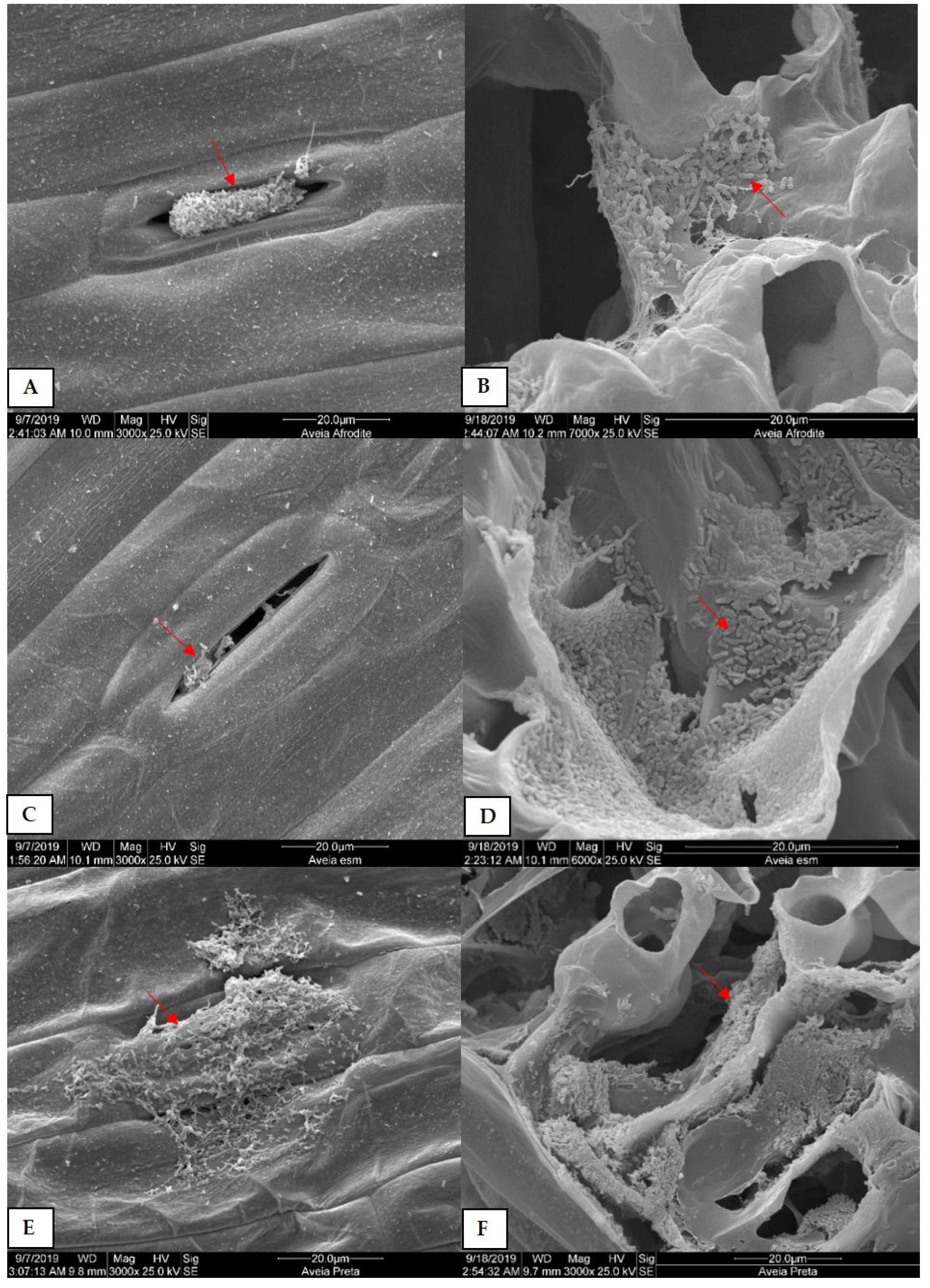
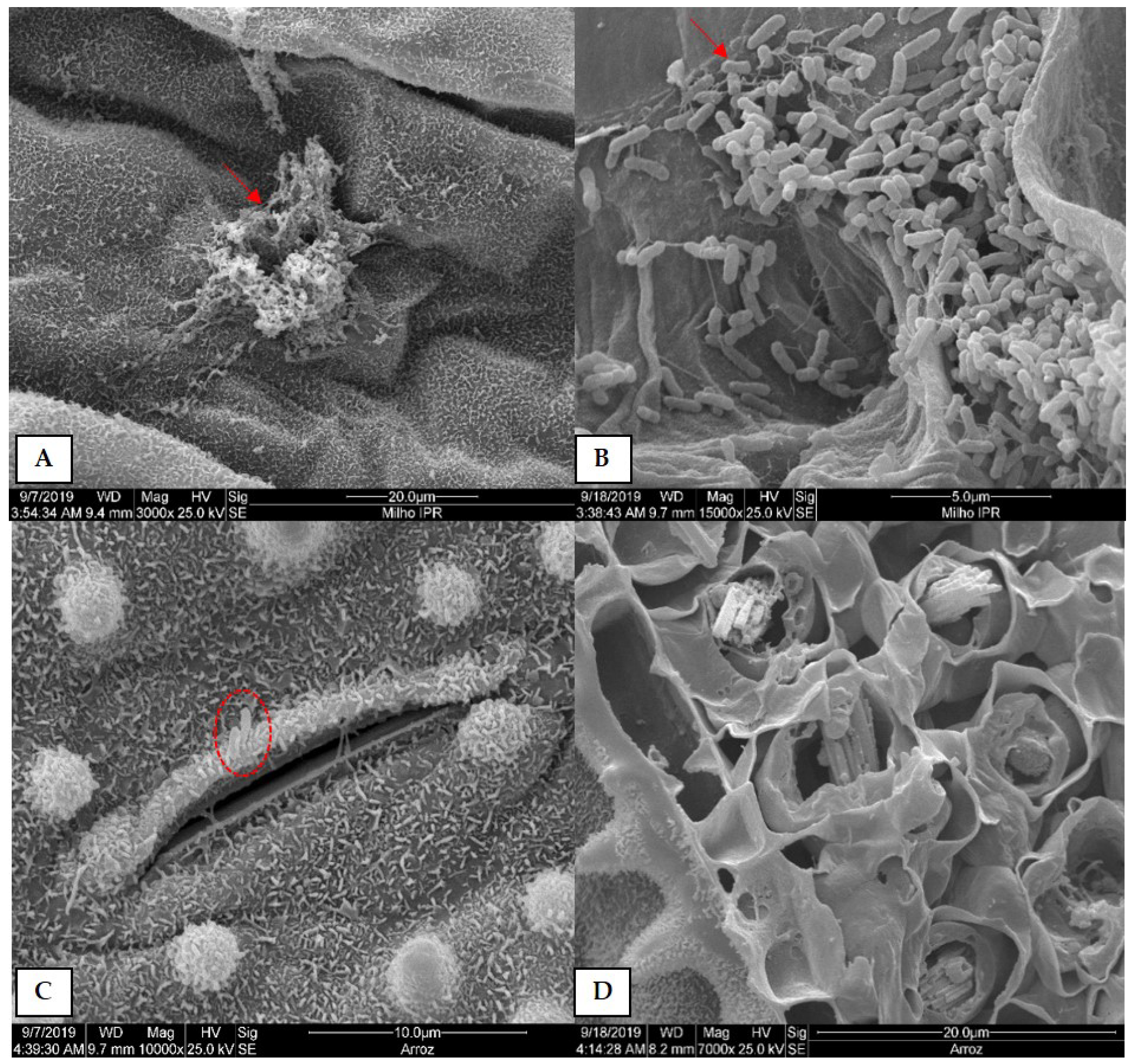
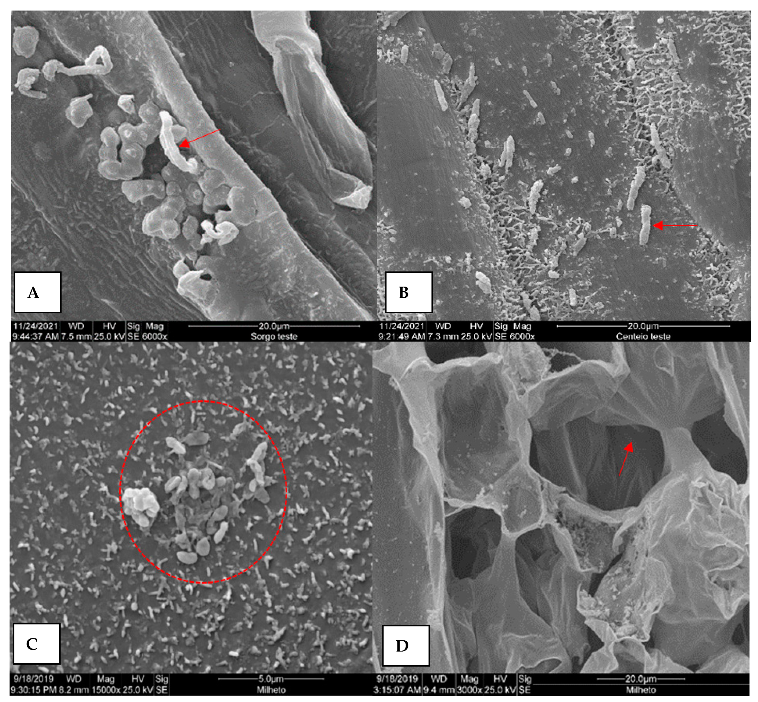
3.2. Scanning Electron Microscopy (SEM) Analysis of Plants Colonized by Xvv

Scanning electron microscopy (SEM) was performed on plant samples from symptomatic species (oat cvs. IPR Afrodite and IPR Esmeralda, black oat cv. IPR 161, maize cv. IPR 164, and rice cv. IPR 117) and on three other species of the Poaceae family, which did not show any typical disease symptoms (sorghum cv. BRS 658, rye cv. IPR 89, and millet cv. IPR St. Tereza do Oeste) when inoculated with the RL1 strain of Xvv. Samples were collected at 15 DAI, when symptomatic plants showed typical symptoms due to infection by the bacterium.
Xvv cells and aggregates were frequently observed on the adaxial surface of the leaves of oat cvs. IPR Afrodite and IPR Esmeralda (Figure 4A,C), black oat (Figure 4E), maize (Figure 5A), rice (Figure 5C), sorghum (Figure 6A), and rye (Figure 6B). Fracture images revealed bacterial colonization in the intercellular spaces and substomatal chambers in the leaf tissues of oat cvs. IPR Afrodite (Figure 4B) and IPR Esmeralda (Figure 4D), black oat cv. IPR 161 (Figure 4F), and maize IPR 164 (Figure 5B). Exopolysaccharides (EPS) and mucus were also observed around the bacterial cells (Figure 4B,D,F and Figure 5B).
In rice, the presence of bacterial cells around the stomata was detected (Figure 5C); however, it was not possible to visualize the colonization of Xvv in the intercellular spaces in the fractures (Figure 5D). In sorghum (Figure 6A) and rye (Figure 6B), even when asymptomatic, it was possible to observe slight colonization of Xvv on leaf surfaces, in agreement with the results of the epiphytic survival of the bacteria verified for these species (Table 1). In millet, the morphology of bacterial cells was altered (Figure 6C), indicating possible cell death. Bacterial colonization of the mesophyll in these tissues was not observed during fracture examinations (Figure 6D). This suggests that millet is the only evaluated species on which Xvv can survive epiphytically (Table 1).

4. Discussion

Xvv pathogenicity was evaluated for 52 mono- and dicotyledonous plant species of different botanical groups. Ten species were identified as potentially symptomatic host plants for Xvv. These species were restricted to the Poaceae family, except Cyperus esculentus, which is a member of the Cyperaceae family. These results corroborate those of previous studies on alternative hosts for Xvv [5,11,14].
The results obtained in this study expand the range of potential host plants for Xvv, which previously included only a few species such as maize, sorghum, sugarcane, broom bamboo, palm species, and eucalyptus [8,9,10,11,13]. Although our findings demonstrate the pathogenicity of Xvv through artificial inoculation under controlled conditions for several plant species, including some not yet reported in the literature as hosts for this bacterium, the results may not represent the natural pathogen–host interaction under field conditions. Artificial inoculations may favor pathogen infection, as the high concentration of the inoculum can act more aggressively than under natural conditions.
In a previous study [14], eight species identified as symptomatic hosts for Xvv under greenhouse conditions were transplanted and evaluated under natural field infestation of Xvv. However, only two of the eight species were infected and confirmed to be susceptible to Xvv under natural conditions of the disease occurrence. Moreover, it was observed that the symptoms in maize were more severe than those in the potential alternative hosts. Similar findings were reported by Lang et al. [11] and Hartman et al. [14], indicating that Xvv may have shown higher aggressiveness or adaptation to infect maize.
Sugarcane and sorghum have previously been reported as symptomatic hosts of Xvv in Nebraska and Kansas, in theU.S. [5,11]. Sorghum readily showed symptoms when artificially inoculated with Xvv under greenhouse conditions [11]. However, no symptoms were evident under field conditions, even in sorghum that was adjacent to BLS-affected maize. Interestingly, sugarcane and sorghum showed no typical symptoms when inoculated with the RL1 strain of Xvv in our study. However, it was possible to recover the bacterium from the phyllosphere of these plants up to 21 days after inoculation, indicating epiphytic colonization of these plants by Xvv.
The differences in the host range among studies can be partially explained by the pathogenic variability or genetic diversity of the Xvv strain, which was extensively studied in previous work [21,22]. These studies demonstrated that the aggressiveness level of Xvv varied among strains, even though all tested strains showed some level of pathogenicity to different sugarcane cultivars [23]. To date, comparative studies of Xvv strains from Brazil with strains from other countries have not been reported. However, genomic sequencing studies of different Xvv strains obtained in the U.S., Argentina, and South Africa revealed that these isolates belong to the same taxonomic clade [24]. However, it is still unclear whether the U.S. and Argentine Xvv strains differ from those from South Africa in terms of host plants [11]. Initial studies with one U.S. and one South African strain of Xvv and four strains of X. vasicola pv. holcicola showed no differences in symptoms in maize [11], although these studies were limited to few strains of Xvv and only one maize cultivar.
Similar to our findings, A. sativa and O. sativa have been reported as hosts of Xvv by Lang et al. [11] and Hartman et al. [14]. Three Avena spp. cultivars were evaluated in our study: two of A. sativa and one of A. strigosa, and different levels of Xvv susceptibility were observed. A. sativa cv. IPR Afrodite showed the most severe disease symptoms compared with the other cultivars. These results reinforce the importance of evaluating the reaction of different genotypes of each species to precisely and safely determine the susceptibility to Xvv infection. H. vulgare plants were symptomatic when inoculated with the RL1 strain of Xvv. This species was reported as a non-host for Xvv in an earlier study [14], as no symptoms or endophytic colonization were found at that time.
Furthermore, this is the first report of Xvv pathogenicity in Brazil for some speciesthat include B. brizantha, B. brizantha cv. Marandu, D. horizontalis, D. insularis, E. colonum, E. indica, and S. arundinaceum. Therefore, these results are important, as forage plants such as the Brachiaria group are perennial and are used in intercropping with maize. Simultaneous cultivation of the grains and straw of maize and the roughage of forage such as Brachiaria spp. are common for cattle grazing [25].
All evaluated weed species are common in Brazil, and inhabit major crop-producing areas, pastures, and gardens. These plant species have high reproductive capability [26]. Among them, S. arundinaceum and D. insularis are perennial species and are difficult to control.
The tropical and subtropical climate of Brazil favors some regions to have two or more crop seasons during the year in the same area, mainly soybean and maize off-season succession [27]. Based on recent crop seasons, three-quarters of the Brazilian maize production was from off-season maize, cultivated during the autumn and winter [28]. However, the investment in weed control for off-season maize is generally low, which results in increased weed infestation in the main production areas [27]. D. insularis is considered one of the most problematic weeds for this continuous intensive system of crop production, particularly with the occurrence of herbicide resistance [29]. The severity of symptoms and epiphytic colonization of Xvv found in D. insularis indicates that this species is a potential host for bacterial survival in maize-producing areas. In this context, it is of utmost importance to mitigate strategies to control the BLS disease in areas of natural occurrence of the pathogen, considering all plant species that are present in the agricultural system of the region. Perennial hosts such as B. decumbens may represent a potential risk for maize production, as Xvv can persist in these hosts season by season [14]. Furthermore, the pathogen can survive in aboveground debris of the alternative hosts and might serve as natural reservoirs of inoculum for new infections. Therefore, further studies are required to fully understand the interactions between Xvv and potential host weed plants.
Bacterial survival on the surface and/or inside of cells of cultivated plants, forage, or weeds is one of the main sources of inoculum for the development of new bacterial disease outbreaks in the main production areas [15]. In the present study, Xvv was detected epiphytically colonizing several cultivated plants and cover crops, including mono- and dicotyledonous species. Thus, this bacterium can epiphytically colonize plants of different botanical groups, such as Amaranthaceae, Asteraceae, Brassicaceae, Commelinaceae, Euphorbiaceae, Fabaceae, Malvaceae, Poaceae, and Polygonaceae.
Our findings demonstrated a variation in the epiphytic populations sampled from different leaves of the same plant, which is commonly observed in epiphytic bacteria [30,31]. The population size of epiphytic bacteria can vary by more than 1000 times from one leaf to another, even when grown under similar environmental conditions [31,32,33,34].
When analyzing the epiphytic colonization of Xvv in forage, we found that all tested species enabled bacterial survival. Regarding weeds, 86% of the tested species showed epiphytic survival of the pathogen. In contrast, C. polydactyla, G. paviflora, and R. brasiliensis showed no epiphytic colonization by Xvv. This could be related to the low bacterial titer present in the phyllosphere of weeds during the evaluation period. Other factors, such as bacterial composition in the phyllosphere, species, leaf type, phenological stage, and climatic conditions, may also have influenced the epiphytic colonization [35,36,37]. Leaf type is one of the factors that can influence the dynamics of epiphytic populations [38,39]. Leaves with trichomes tend to host larger bacterial populations than those with waxy cuticles [38]. Another factor to be considered during bacterial hosting is the availability of nutrient sources in the leaves, such as sugars, which can regulate the size of the epiphytic population [30].
Scanning electron microscopy (SEM) analyses enabled visualization of the epiphytic colonization of Xvv in several potential hosts. SEM showed extensive colonization of Xvv in the presence of exopolysaccharides (EPS) in maize cv. IPR 164, A. sativa, and A. strigosa. Studies have suggested that EPS have several functions, including bacterial protection against plant bacteriostatic substances and reduced direct contact with plant cells to minimize host defense responses [40]. Moreover, EPS promote water retention in plant tissue, which stimulates tissue rupture, allowing movement, invasion, or colonization by phytopathogens from distant sites [40,41,42]. In the genus Xanthomonas, xanthan is an EPS secreted by Xvv [43]. This EPS is involved in several functions, such as suppression of the plant’s local defense and inhibition of callose deposition promoting pathogen dissemination [44,45,46]. In previous studies of X. citri subsp. citri (Xcc), xanthan was observed acting during the later stages of the infection process, probably increasing bacterial adherence to the host tissue and epiphytic survival, thus facilitating colonization in long-distance tissues [47].
Although we evaluated a broad range of genotypes in this study, 58 different plants, additional investigations are required to determine whether other mono- and dicotyledonous cultivated plants or those that occur in maize-growing areas can potentially host Xvv. Our findings revealed that several cultivated plants, cover crops, forage, and weeds can host Xvv pathogenically and/or epiphytically. For this reason, rational use of these plants within an integrated disease-management program is needed to minimize possible BLS outbreaks in maize-producing areas. Practices using green manure and crop rotation should be conducted carefully, whenever possible, with non-host species for Xvv in areas with BLS incidence.

5. Conclusions

This is the first study conducted in Brazil to report alternative hosts for Xanthomonas vasicola pv. vasculorum (Xvv). Based on our findings, in addition to maize, several other symptomatic host plants of Xvv were found, including Avena sativa, A. strigosa, Brachiariabrizantha (Brizantha and cv. Marandu), Digitaria horizontalis, D. insularis, Echinochloacolonum, Eleusine indica, Hordeum vulgare, and Sorghum arundinaceum. Moreover, the phyllospheres of several species of cultivated plants, cover crops, forages, grasses, and weeds are suitable niches for the epiphytic survival of Xvv, which must be considered during management programs for bacterial leaf streak (BLS) control.

Author Contributions

Conceptualization and methodology, R.P.L.J., M.I.B.-P. and T.V.L.; formal analysis and investigation, T.V.L., R.R.R. and D.U.d.C.; scanning electron microscopy (SEM) analysis, T.V.L. and A.G.d.O.; resources and funding acquisition, R.P.L.J. and M.I.B.-P.; data curation and writing—original draft preparation, T.V.L. and D.U.d.C.; writing—review and editing, R.P.L.J. and M.I.B.-P. All authors have read and agreed to the published version of the manuscript.

Funding

This research was funded by Coordenação de Aperfeiçoamento de Pessoal de Nível Superior (Capes, Grant No. 88882.448321/2019-01), Instituto de Desenvolvimento Rural do Paraná, IAPAR/Emater (IDR-Paraná), Superintendência de Ciência, Tecnologia e Ensino Superior (SETI) do Paraná, Fundação Araucária (FA, Chamada 03/2021) and Universidade Estadual de Londrina (UEL).

Data Availability Statement

All data generated and analyzed during this study are presented in the published version of this article.

Acknowledgments

The authors would like to thank the staff and faculty of UEL and IDR-Paraná for their technical support. T.V.L. thanks the Capes for scholarship, IDR, SETI, FA and UEL for partial support for publication costs.

Conflicts of Interest

The authors declare no conflict of interest.

References

  1. Leite, R.P., Jr.; Custódio, A.A.P.; Madalosso, T.; Robaina, R.R.; Duin, I.M.; Sugahara, V.H. First report of the occurrence of bacterial leaf streak of corn caused by Xanthomonas vasicola pv. vasculorum in Brazil. Plant Dis. 2019, 103, 145. [Google Scholar] [CrossRef]
  2. Broders, K. Status of bacterial leaf streak of corn in the United States. In Proceedings of the Integrated Crop Management Conference, Ames, IA, USA, 29–30 November 2017; pp. 111–115. [Google Scholar]
  3. Damicone, J.; Cevallos, F.; Olson, J. First report of bacterial leaf streak of corn caused by Xanthomonas vasicola pv. vasculorum in Oklahoma. Plant Dis. 2018, 102, 437. [Google Scholar] [CrossRef]
  4. Jamann, T.M.; Plewa, D.; Mideros, S.X.; Bissonnette, S. First report of bacterial leaf streak of corn caused by Xanthomonas vasicola pv. vasculorum in Illinois. Plant Dis. 2019, 103, 1018. [Google Scholar] [CrossRef]
  5. Korus, K.A.; Lang, J.M.; Adesemoye, A.O.; Block, C.C.; Pal, N.; Leach, J.E.; Jackson-Ziems, T.A. First report of Xanthomonas vasicola causing bacterial leaf streak on corn in the United States. Plant Dis. 2017, 101, 1030. [Google Scholar] [CrossRef]
  6. Plazas, M.C.; de Rossi, R.L.; Brücher, E.; Guerra, F.A.; Vilaro, M.L.; Guerra, G.D.; Wu, G.; Ortiz-Castro, M.C.; Broders, K. First report of Xanthomonas vasicola pv. vasculorum causing bacteria leaf streak of maize (Zea mays) in Argentina. Plant Dis. 2018, 102, 1026. [Google Scholar] [CrossRef]
  7. Leite, R.P., Jr.; Custódio, A.A.P.; Madalosso, T.; Robaina, R.R.; Duin, I.M.; Sugahara, V.H. Estria Bacteriana do Milho no Paraná; IAPAR: Londrina, Brazil, 2018. [Google Scholar]
  8. North, D.S. The Gumming Disease of the Sugar-Cane, its Dissemination and Control; The Colonial Sugar Refining Co. Ltd.: Sydney, Australia, 1935; p. 149. [Google Scholar]
  9. Karamura, G.; Smith, J.; Studholme, D.; Kubiriba, J.; Karamura, E. Comparative pathogenicity studies of the Xanthomonas vasicola species on maize, sugarcane and banana. Afr. J. Plant Sci. 2015, 9, 385–400. [Google Scholar]
  10. Coutinho, T.A.; Van der Westhuizen, L.; Roux, J.; McFarlane, S.A.; Venter, S.N. Significant host jump of Xanthomonas vasicola from sugarcane to a Eucalyptus grandis clone in South Africa. Plant Pathol. 2015, 64, 576–581. [Google Scholar] [CrossRef]
  11. Lang, J.M.; Du Charme, E.; Ibarra Caballero, J.; Luna, E.; Hartman, T.; Ortiz-Castro, M.; Korus, K.J.; Rascoe, J.; Jackson-Ziems, T.A.; Broders, K.; et al. Detection and characterization of Xanthomonas vasicola pv. vasculorum (Cobb 1894) comb. nov. causing bacterial leaf streak of corn in the United States. Phytopathology 2017, 107, 1312–1321. [Google Scholar] [CrossRef] [Green Version]
  12. Rott, P.; Girard, J.C. Sugarcane producing countries/locations and their diseases. In A Guide to Sugarcane Diseases; Rott, P., Bailey, R.A., Comstock, J.C., Croft, B.J., Saumtally, A.S., Eds.; La Librairie du Cirad: Montpellier, France, 2000; pp. 323–336. [Google Scholar]
  13. Ricaud, C.; Autrey, L.J.C. Gumming disease. In Diseases of Sugarcane: Major Diseases; Elsevier: Amsterdam, The Netherlands, 1989; pp. 21–38. [Google Scholar]
  14. Hartman, T.; Tharnish, B.; Harbour, J.; Yuen, G.Y.; Jackson-Ziems, T.A. Alternative hosts in the families Poaceae and Cyperaceae for Xanthomonas vasicola pv. vasculorum, causal agent of bacterial leaf streak of corn. Phytopathology 2020, 110, 1147–1152. [Google Scholar] [CrossRef] [PubMed]
  15. Schuster, M.L.; Coyne, D.P. Survival mechanisms of phytopathogenic bacteria. Annu. Rev. Phytopathol. 1974, 12, 199–221. [Google Scholar] [CrossRef]
  16. Schuster, M.L.; Coyne, D.P. Survival of plant parasitic bacteria of plants grown in tropics with emphasis on beans (Phaseolus vulgaris). Fitopatol. Bras. 1977, 2, 117–130. [Google Scholar]
  17. Robaina, R.R.; Longhi, T.V.; Zeffa, D.M.; Gonçalves, L.S.A.; Leite, R.P., Jr. Development of a protocol and a diagrammatic scale for quantification of bacterial leaf streak disease on young plants of maize. Plant Dis. 2020, 104, 2921–2927. [Google Scholar] [CrossRef] [PubMed]
  18. Leite, R.P., Jr. The Relationship between Pathogenic and Nonpathogenic Strains of Pseudomonas syringae and Bacterial Brown Spot Disease in Beans. Master’s Thesis, University of Wisconsin, Madison, WI, USA, 1986; p. 107. [Google Scholar]
  19. Gonçalves, R.M.; Schipanski, C.A.; Koguishi, L.; Soman, J.M.; Sakate, R.K.; Júnior, T.A.; Maringoni, A.C. Alternative hosts of Curtobacterium flaccumfaciens pv. flaccumfaciens, causal agent of bean bacterial wilt. Eur.J. Plant Pathol. 2017, 148, 357–365. [Google Scholar] [CrossRef] [Green Version]
  20. Kitajima, E.W.; Leite, B. Curso Introdutório de Microscopia Eletrônica de Varredura; NAP/MEPA ESALQ: Piracicaba, Brazil, 1999; p. 48. [Google Scholar]
  21. Wasukira, A.; Coulter, M.; Al-Sowayeh, N.; Thwaites, R.; Paszkiewicz, K.; Kubiriba, J.; Smith, J.; Grant, M.; Studholme, D.J. Genome sequencing of Xanthomonas vasicola pathovar vasculorum reveals variation in plasmids and genes encoding lipopolysaccharide synthesis, type-IV pilus and type-III secretion effectors. Pathogens 2014, 3, 211–237. [Google Scholar] [CrossRef]
  22. Studholme, D.J.; Wicker, E.; Abrare, S.M.; Aspin, A.; Bogdanove, A.; Broders, K.; Dubrow, Z.; Grant, M.; Jones, J.B.; Karamura, G.; et al. Transfer of Xanthomonas campestris pv. arecae and X. campestris pv. musacearum to X. vasicola (Vauterin) as X. vasicola pv. arecae comb. nov. and X. vasicola pv. musacearum comb. nov. and description of X. vasicola pv. vasculorum pv. nov. Phytopathology 2020, 110, 1153–1160. [Google Scholar] [CrossRef] [Green Version]
  23. Peros, J.P. Variability in colony type and pathogenicity of the causal agent of sugarcane gumming Xanthomonas campestris pv. vasculorum (Cobb) Dye. J. Plant Dis. Prot. 1988, 95, 541–545. Available online: https://www.jstor.org/stable/43386650 (accessed on 3 November 2022).
  24. Perez-Quintero, A.L.; Ortiz-Castro, M.; Lang, J.M.; Rieux, A.; Wu, G.; Liu, S.; Chapman, T.A.; Chang, C.; Ziegle, J.; Peng, Z.; et al. Genomic acquisitions in emerging populations of Xanthomonas vasicola pv. vasculorum infecting corn in the United States and Argentina. Phytopathology 2020, 110, 1161–1173. [Google Scholar] [CrossRef]
  25. Ceccon, G. Consórcio Milho-Braquiária; Embrapa Agropecuária Oeste: Dourados, Brazil, 2013; Available online: https://www.infoteca.cnptia.embrapa.br/bitstream/doc/982597/1/LVCONSORCIOMB.pdf (accessed on 11 September 2022).
  26. Lorenzi, H. Plantas Daninhas do Brasil: Terrestres, Aquáticas, Parasitas e Tóxicas; Plantarum: Nova Odessa, Brazil, 2008; p. 640. [Google Scholar]
  27. Adegas, F.S.; Voll, E.R.; Gazziero, D.L.P. Manejo de plantas daninhas em milho safrinha em cultivo solteiro ou consorciado à braquiária ruziziensis. Pesqui. Agropecu. Bras. 2011, 46, 1226–1233. [Google Scholar] [CrossRef] [Green Version]
  28. Conab. National Supply Company: Agricultural Information System. 2023. Available online: https://www.conab.gov.br/info-agro/safras/graos (accessed on 5 March 2023).
  29. Gonçalves Netto, A.; Cordeiro, E.M.; Nicolai, M.; de Carvalho, S.J.; Ovejero, R.F.L.; Brunharo, C.A.; Zucchi, M.I.; Christoffoleti, P.J. Population genomics of Digitaria insularis from soybean areas in Brazil. Pest Manag. Sci. 2021, 77, 5375–5381. [Google Scholar] [CrossRef]
  30. Mercier, J.; Lindow, S.E. Role of leaf surface sugars in colonization of plants by bacterial epiphytes. Appl. Environ. Microbiol. 2000, 66, 369–374. [Google Scholar] [CrossRef] [Green Version]
  31. Kinkel, L.L.; Wilson, M.; Lindow, S.E. Utility of microcosm studies for predicting phylloplane bacterium population sizes in the field. Appl. Environ. Microbiol. 1996, 62, 3413–3423. [Google Scholar] [CrossRef] [PubMed] [Green Version]
  32. Hirano, S.S.; Upper, C.D. Population biology and epidemiology of Pseudomonas syringae. Annu. Rev. Phytopathol. 1990, 28, 155–177. [Google Scholar] [CrossRef]
  33. Kinkel, L.L.; Lindow, S.E. Invasion and exclusion among coexisting Pseudomonas syringae strains on leaves. Appl. Environ. Microbiol. 1993, 59, 3447–3454. [Google Scholar] [CrossRef] [Green Version]
  34. Kinkel, L.L.; Wilson, M.; Lindow, S.E. Effect of sampling scale on the assessment of epiphytic bacterial populations. Microb. Ecol. 1995, 29, 283–297. [Google Scholar] [CrossRef] [PubMed]
  35. Kinkel, L.L. Microbial population dynamics on leaves. Annu. Rev. Phytopathol. 1997, 35, 327–347. [Google Scholar] [CrossRef] [PubMed]
  36. Hirano, S.S.; Upper, C.D. Bacteria in the leaf ecosystem with emphasis on Pseudomonas syringae a pathogen, ice nucleus, and epiphyte. Microbiol. Mol. Biol. Rev. 2000, 64, 624–653. [Google Scholar] [CrossRef] [PubMed] [Green Version]
  37. Leveau, J.H. A brief from the leaf: Latest research to inform our understanding of the phyllosphere microbiome. Curr. Opin. Microbiol. 2019, 49, 41–49. [Google Scholar] [CrossRef]
  38. O’brien, R.D.; Lindow, S.E. Effect of plant species and environmental conditions on epiphytic population sizes of Pseudomonas syringae and other bacteria. Phytopathology 1989, 79, 619–627. [Google Scholar] [CrossRef]
  39. Toussaint, V.; Benoit, D.L.; Carisse, O. Potential of weed species to serve as a reservoir for Xanthomonas campestris pv. vitians, the causal agent of bacterial leaf spot of lettuce. Crop Prot. 2012, 41, 64–70. [Google Scholar] [CrossRef]
  40. Denny, T.P. Autoregulator-dependent control of extracellular polysaccharide production in phytopathogenic bacteria. Eur. J. Plant Pathol. 1999, 105, 417–430. [Google Scholar] [CrossRef]
  41. Stoodley, P.; Sauer, K.; Davies, D.G.; Costerton, J.W. Biofilms as complex differentiated communities. Annu. Rev. Microbiol. 2002, 56, 187–209. [Google Scholar] [CrossRef] [Green Version]
  42. Dow, J.M.; Crossman, L.; Findlay, K.; He, Y.Q.; Feng, J.X.; Tang, J.L. Biofilm dispersal in Xanthomonas campestris is controlled by cell–cell signaling and is required for full virulence to plants. Proc. Natl. Acad. Sci. USA 2003, 100, 10995–11000. [Google Scholar] [CrossRef] [PubMed] [Green Version]
  43. Harding, N.E.; Cleary, J.M.; Ielpi, L. Genetics and biochemistry of xanthan gum production by Xanthomonas campestris. In Food Biotechnology Microorganism; VCH Publishers Inc.: New York, NY, USA, 1995; pp. 495–514. [Google Scholar]
  44. Braun, E.J. Colonization of resistant and susceptible maize plants by Erwinia stewartii strains differing in exopolysaccharide production. Physiol. Mol. Plant Pathol. 1990, 36, 363–379. [Google Scholar] [CrossRef]
  45. Saile, E.; McGarvey, J.A.; Schell, M.A.; Denny, T.P. Role of extracellular polysaccharide and endoglucanase in root invasion and colonization of tomato plants by Ralstonia solanacearum. Phytopathology 1997, 87, 1264–1271. [Google Scholar] [CrossRef] [PubMed] [Green Version]
  46. Yun, M.H.; Torres, P.S.; Oirdi, M.E.; Rigano, L.A.; Gonzalez-Lamothe, R.; Marano, M.R.; Castagnaro, A.P.; Dankert, M.A.; Bouarab, K.; Vojnov, A.A. Xanthan induces plant susceptibility by suppressing callose deposition. Plant Physiol. 2006, 141, 178–187. [Google Scholar] [CrossRef] [Green Version]
  47. Dunger, G.; Relling, V.M.; Tondo, M.L.; Barreras, M.; Ielpi, L.; Orellano, E.G.; Ottado, J. Xanthan is not essential for pathogenicity in citrus canker but contributes to Xanthomonas epiphytic survival. Arch. Microbiol. 2007, 188, 127–135. [Google Scholar] [CrossRef]
Figure 1. Maximum (Tmax), minimum (Tmin), and average (Tmed) air temperature (°C) and relative humidity (RH, %) inside a semi-climatized greenhouse at the Instituto de Desenvolvimento Rural do Paraná-IAPAR/Emater, Londrina, PR, Brazil, from February to November 2019 (A) and February to October 2021 (B).
Agronomy 13 01073 g001
Figure 2. Leaves of cultivated plants showing symptoms of bacterial leaf streak (BLS) 15 days after inoculation (DAI) with the RL1 strain of Xanthomonas vasicola pv. vasculorum (Xvv): oat cvs. IPR Afrodite (A) and IPR Esmeralda (B) with yellowed spots on leaves progressing to chlorosis; (C) black oat cv. IPR 161 with yellowed spots on leaves progressing to chlorosis; (D) barley cv. BRS Cauê with straw-yellow spots along the veins; and (E) maize cv. IPR 164 with streaks and wavy margins on leaves.
Agronomy 13 01073 g002
Figure 3. Leaves of forages and weeds showing symptoms of bacterial leaf streak (BLS) 15 days after inoculation (DAI) with the RL1 strain of Xanthomonas vasicola pv. vasculorum (Xvv): (A) Brachiaria brizantha (Brizantha) with symptoms of BLS; (B) Digitaria insularis with symptoms of chlorosis; (C) Echinochloa colonum with red spots; (D) Digitaria horizontalis with reddish punctuations; and (E) Sorghum arundinaceum with red spots on the leaf surface and necrosis in the leaf margin.
Agronomy 13 01073 g003
Figure 4. Scanning electron microscopy (SEM) of the leaf surface and leaf fractures of oat 15 days after inoculation (DAI) with Xanthomonas vasicola pv.vasculorum (Xvv). (A) Oat cv. IPR Afrodite with cluster of Xvv cells in the stomata (red arrow); (B) oat cv. IPR Afrodite with Xvv colonization in the leaf mesophyll with bacterial aggregates and production of amorphous material suggesting exopolysaccharides (EPS, red arrow); (C) oat cv. IPR Esmeralda with cluster of Xvv cells in the stomata (red arrow); (D) oat cv. IPR Esmeralda with Xvv colonization in the leaf mesophyll with bacterial aggregates and production of amorphous material suggesting EPS (red arrow); (E) black oat cv. IPR161 with cluster of Xvv cells in the stomata (red arrow); (F) black oat cv. IPR161 with Xvv colonization in leaf mesophyll with bacterial aggregates and production of amorphous material suggesting EPS (red arrow).
Agronomy 13 01073 g004
Figure 5. Scanning electron microscopy (SEM) of the adaxial surface of leaves and leaf fractures of maize cv. IPR 164 and rice cv. IPR 117 at 15 days after inoculation (DAI) with Xanthomonas vasicola pv. vasculorum (Xvv). (A) Maize cv. IPR 164 with cluster of Xvv cells in the stomatal chamber (red arrow); (B) maize IPR 164 with cluster of Xvv cells in the leaf mesophyll covered by amorphous material suggesting exopolysaccharides (EPS, red arrow); (C) rice cv. IPR 117 with Xvv cells colonizing the region close to the stomata (dashed ellipse); (D) rice cv. IPR 117 with internal structure of the leaf mesophyll, without evidence of bacterial cells.
Agronomy 13 01073 g005
Figure 6. Scanning electron microscopy (SEM) of the surface of asymptomatic plants of sorghum cv. BRS 658, rye cv. IPR 89, and millet cv. IPR St. Tereza do Oeste at 15 days after inoculation (DAI) with Xanthomonas vasicola pv. vasculorum (Xvv). (A) Adaxial leaf surface of sorghum cv. BRS 658 with scattered Xvv cells in the epidermis (red arrow); (B) adaxial leaf surface of rye cv. IPR 89 with scattered Xvv cells in the epidermis (red arrow); (C) Xvv cell aggregates demonstrating altered morphology, indicating cell death on the adaxial surface of millet cv. IPR St. Tereza do Oeste (dashed circle); (D) fractures showing the internal structure of the leaf mesophyll, without evidence of bacterial colonization in millet cv. IPR St. Tereza do Oeste (red arrow).
Agronomy 13 01073 g006
Table 1. Pathogenicity and epiphytic colonization of Xanthomonas vasicola pv. vasculorum (Xvv), the causal agent of bacterial leaf streak (BLS) of maize, to cultivated plants and cover crops.
Botanical GroupScientific NameCommon NameCultivarS1EC2
Monocotyledonous
PoaceaeAvena sativaOatIPR Afrodite+N/A
A. sativaOatIPR Esmeralda+N/A
A. strigosaBlack oatIPR 161+N/A
Hordeum vulgareBarleyBRS Cauê+N/A
Oryza sativaRiceIPR 117+N/A
Pennisetum glaucumMilletIPR St. Tereza do Oeste+
Saccharum officinarumSugar caneNot determined +
Secale cerealeRyeIPR 89+
Sorghum bicolorSorghumBRS 658+
×TriticosecaleTriticaleIPR Caiapó+
Triticum aestivumWheatIPR Potyporã+
Zea mays3White maizeIPR 127+N/A
Z. mays3Common maizeIPR 164+N/A
Dicotyledonous
AsteraceaeHelianthus annuusSunflowerBRS 417+
Glycine maxSoybeanPotência+
Lupinus albusWhite lupinNot determined+
L. angustifoliusBlue lupinIPR 124+
FabaceaeMucuna aterrima cv. Mucuna pretaBlack velvet-beanNot determined+
M. pruriensBengal velvet-beanNot determined+
Phaseolus vulgarisBeanCarioca+
MalvaceaeGossypium hirsutumCottonFMT 701+
PolygonaceaeFagopyrum esculentum MoenchBuckwheatIPR 91+
S1, symptoms were assessed at 15 DAI and Xvv association confirmed by PCR. EC2, epiphytic colonization: Xvv isolation in asymptomatic plants was performed at 21 DAI in the first experiment and at 15, 21, and 30 DAI in the second experiment. The isolated Xvv was confirmed by PCR. +, presence of symptoms or Xvv; −, absence of symptoms or Xvv; N/A, not assessed. Zea mays3, IPR 127 and IPR 164 maize hybrids were included in the experiment as Xvv-positive controls.
Table 2. Pathogenicity and epiphytic colonization of Xanthomonas vasicola pv. vasculorum (Xvv), the causal agent of bacterial leaf streak (BLS) of maize, in forage and grasses.
Botanical GroupScientific NameCommon NameCultivarS1EC2
Monocotyledonous
PoaceaeBrachiariabrizanthaBrizantha bread grassNot determined+N/A
B. brizanthaMarandu bread grassMarandu+N/A
B. brizanthaPiatã bread grassBRS Piatã+
B. brizanthaMG-5 Vitória bread grassMG-5 Vitória+
B. decumbensBasilisk bread grassBasilisk+
B. decumbensDecumbens bread grassNot determined+
B. humidicolaHumidicola bread grassNot determined+
B. ruzisiensisRuzisiensis bread grassNot determined+
Panicum maximumMG-12 Paredão Guinea grassMG12 Paredão+
P. maximumTanzania Guinea grassTanzânia-1+
Zea mays3White maizeIPR 127+N/A
Z. mays3Common maizeIPR 164+N/A
Dicotyledonous
FabaceaeCajanus cajanPigeon peaIAPAR 43+
Crotalaria spectabilisShowy rattlepodNot determined+
S1, symptoms were assessed at 15 DAI and Xvv association confirmed by PCR. EC2, epiphytic colonization: Xvv isolation in asymptomatic plants was performed at 21 DAI in the first experiment and at 15, 21, and 30 DAI in the second experiment. The isolated Xvv was confirmed by PCR. +, presence of symptoms or Xvv; −, absence of symptoms or Xvv; N/A, not assessed. Zea mays3, IPR 127 and IPR 164 maize hybrids were included in the experiment as Xvv-positive controls.
Table 3. Pathogenicity and epiphytic colonization of Xanthomonas vasicola pv. vasculorum (Xvv), the causal agent of bacterial leaf streak (BLS) of maize, in weeds.
Botanical GroupScientific NameCommon NameS1EC2
Monocotyledonous
PoaceaeBrachiaria plantagineaPlantain signalgrass+
Cenchrus echinatusSouthern sandbur+
Chloris gayanaRhodes grass+
C. polydactylaFinger grass
Digitaria horizontalisJamaican crabgrass+N/A
D. insularisSourgrass+N/A
Echinochloa colonumJungle rice+N/A
Eleusine indicaCrowfootgrass+N/A
Lolium multiflorumAnnual ryegrass+
Panicum maximumGuinea grass+
Pennisetum purpureumNapier grass+
Rhynchelytrum repensNatal grass+
Sorghum arundinaceumCommon wild sorghum+N/A
Zea mays3White maize+N/A
Z. mays3Common maize+N/A
Dicotyledonous
AmaranthaceaeAmaranthus hybridusSmooth pigweed+
A. viridisSlender amaranth+
AsteraceaeAcanthospermum hispidumBristly starbur+
Bidens pilosaBeggar’s tick+
Conyza spp.Hairy fleabane+
Galinsoga parvifloraGallant soldier
Tridax procumbensCoatbuttons+
BrassicaceaeRaphanus raphanistrumWild radish+
R. sativusRadish+
CommelinaceaeCommelina benghalensisWandering jew+
EuphorbiaceaeEuphorbia heterophyllaWild poinsettia+
MalvaceaeSidarhom bifoliaArrow-leaf sida+
RubiaceaeRichardia brasiliensisTropical Mexican clover
S1, symptoms were assessed at 15 DAI and Xvv association confirmed by PCR. EC2, epiphytic colonization: Xvv isolation in asymptomatic plants was performed at 21 DAI in the first experiment and at 15, 21, and 30 DAI in the second experiment. The isolated Xvv was confirmed by PCR. +, presence of symptoms or Xvv; −, absence of symptoms or Xvv; N/A, not assessed. Zea mays3, IPR 127 and IPR 164 maize hybrids were included in the experiment as Xvv-positive controls.
Table 4. Maximum recovery period in days after inoculation (DAI) and colony-forming units (Log10CFU·g−1) of Xanthomonas vasicola pv. vasculorum (Xvv), the causal agent of bacterial leaf streak (BLS) of maize, in cultivated plants, cover crops, forage, grasses and weeds.
Botanical GroupScientific NameCommon NameXvv
Maximum Recovery Period (DAI) 1CFU
(Log10·g−1) 2
Cultivated plants and cover crops
PoaceaePennisetum glaucumMillet305.46
Saccharum officinarumSugarcane214.40
Secale cerealeRye306.36
Sorghum bicolorSorghum215.18
×TriticosecaleTriticale215.40
Triticum aestivumWheat306.71
AsteraceaeGlycine maxSoybean213.30
Helianthus annuusSunflower216.02
Lupinus albusWhite lupin217.00
L. angustifoliusBlue lupin214.00
FabaceaeMucuna pruriensBengal velvet-bean214.30
M. aterrima cv. Mucuna pretaBlack velvet-bean216.01
Phaseolus vulgarisBean305.00
MalvaceaeGossypium hirsutumCotton215.04
PolygonaceaeFagopyrum esculentum MoenchBuckwheat215.93
Forages and grasses
PoaceaeBrachiaria brizanthaPiatã bread grass213.48
B. brizanthaMG-5 Vitória bread grass213.65
B. decumbensDecumbensbread grass214.74
B. decumbensBasilisk bread grass302.70
B. humidicolaHumidicola bread grass214.00
B. ruzisiensisRuzisiensis bread grass215.00
Panicum maximumTanzânia Guinea grass154.00
P. maximumMG-12 Paredã Guinea grass 214.81
FabaceaeCajanus cajanPigeon pea214.18
Crotalaria spectabilisShowy rattlepod305.23
Weeds
PoaceaeB. plantagineaPlantain signalgrass215.95
Cenchrus echinatusSouthern sandbur305.00
Chloris gayanaRhodes grass306.27
Lolium multiflorumAnnual ryegrass217.00
P. maximumGuinea grass305.18
Pennisetum purpureumNapier grass305.92
Rhynchelytrum repensNatal grass306.40
AmaranthaceaeAmaranthus hybridusSmooth pigweed303.85
A. viridisSlender amaranth303.98
Asteraceae Acanthospermum hispidumBristly starbur305.18
Bidens pilosaBeggar’s tick215.00
Conyza spp.Hairy fleabane154.00
Tridax procumbensCoatbuttons303.70
BrassicaceaeRaphanus raphanistrumWild radish305.48
Raphanus sativusRadish154.28
CommelinaceaeCommelina benghalensisWandering jew305.18
EuphorbiaceaeEuphorbia heterophyllaWild poinsettia303.93
MalvaceaeSidarhombifoliaArrow-leaf sida305.00
1, DAI, days after inoculation. 2, Maximum value of epiphytic population recovered from the phyllosphere of plants in the two experiments, considering all the evaluations.
Disclaimer/Publisher’s Note: The statements, opinions and data contained in all publications are solely those of the individual author(s) and contributor(s) and not of MDPI and/or the editor(s). MDPI and/or the editor(s) disclaim responsibility for any injury to people or property resulting from any ideas, methods, instructions or products referred to in the content.

