Article

An Endophytic Fungal Isolate Paecilomyces lilacinus Produces Bioactive Secondary Metabolites and Promotes Growth of Solanum lycopersicum under Heavy Metal Stress

1
Department of Botany, Abdul Wali Khan University Mardan, Mardan 23200, KP, Pakistan
2
Center of Biotechnology and Microbiology, University of Peshawar, Peshawar 25120, KP, Pakistan
3
Department of Zoology, Abdul Wali Khan University Mardan, Mardan 23200, KP, Pakistan
4
Department of Horticulture and Life Science, Yeungnam University, Gyeongsan 38541, Republic of Korea
5
Department of Applied Biosciences, School of Life Sciences, Kyungpook National University, Daegu 41566, Republic of Korea
*
Authors to whom correspondence should be addressed.
Agronomy 2023, 13(3), 883; https://doi.org/10.3390/agronomy13030883
Submission received: 8 February 2023 / Revised: 13 March 2023 / Accepted: 13 March 2023 / Published: 16 March 2023

Abstract

:
Heavy metal (HM) contamination in the soil and accumulation in plants poses a serious threat to crops and human health. HMs such as cadmium, mercury, lead and arsenic are widely acknowledged for their adverse effects on plants such as chlorosis, necrosis, low food quality and crop yields. Endophytic fungi secrete secondary metabolites and enhance the plant’s ability to tolerate stressful conditions. However, the role of most fungal endophytes in their host plant growth or production of metabolites under HM stress conditions needs further understanding. In the present study, we studied the HM stress alleviation capability of the endophytic fungus, Paecilomyces lilacinus (MRF), isolated from the roots of Justicia adathoda. We studied two heavy metals, namely lead and cobalt. The culture filtrate (CF) of P. lilacinus revealed IAA (68.17 µg/mL), phenols (43.31 µg/mL), flavonoids (40.59 µg/mL), sugar content (97.83 µg/mL) and proline (17 µg/mL). Additionally, DPPH-free radical scavenging activity and the antibacterial potential against Salmonella typhi and Shigella sonnei of the CF demonstrated positive results. The gas chromatography mass spectrometry analysis of the CF manifested different constituents, including (1) Trichloromethane, (2) 3,7,11,15-Tetramethyl-2-hexadecen-1-ol, (3) phytol, (4) 1,2-Benzenedicarboxylic acid, (5) bis(2-ethylhexyl) ester, (6) squalene and (7) Cyclotrisiloxane Hexamethyl. Moreover, the plant growth-promoting activity of the P. lilacinus (MRF) strain revealed a robust increase in root and shoot growth, and the fresh and dry weight of S. lycoprsicum. Further, the IAA, phenols, flavonoids, sugar, proline, relative water content and protein contents also increased in the S. lycoprsicum inoculated with P. lilacinus as compared to the control plants. The present study revealed that the inoculation of P. lilacinus alleviates the damages of HM stress and improves the physicochemical characteristics of S. lycoprsicum.

1. Introduction

Contamination of agronomic soil by heavy metals (HMs) is a serious problem in many areas all over the world [1]. The remediation of soils contaminated with HMs is a public concern and is gaining considerable attention in the present scenario. The accumulation of fertilizers, pesticides, industrial leftovers, metal plating, sewage irrigation, tanning, atmospheric deposition and other chemicals on the earth’s crust paved the way for the HM stress factor that adversely affects both plants and humans [2,3]. A limited number of HMs are nondetrimental or rather important [4]; however, the increased concentration poses a serious threat to the fine balance of our natural ecosystem [5].
Crops grown under a high concentration of HMs have negative effects on crop quality and yield [6,7]. This is because HM stress leads to reduced plant growth, leaf chlorosis, necrosis, turgor loss, a decrease in seed germination rate and the damaging of photosynthetic machinery [8]. To overcome this, HM contamination from the environment, physical, chemical and biological treatments have been employed; however, some of them are expensive, time consuming and revealed mechanical complexity [9]. Plants as hyperaccumulators of heavy metals are also used for the decontamination of soil; however, most plants are not suitable because of their slow growth in HM-contaminated soils [10]. Several microorganisms have been identified as effective heavy metal detoxifiers [11]. Recently, endophytes were reported in plant growth promotion under HM-contaminated soils, while the diversity and ecological role of fungal endophytes in the HM contaminated soil are the least discovered [12,13,14,15].
The microbe’s resistance to HMs is greatly influenced by the concentration and availability of HMs in the environment. The decontamination of HMs is governed by parameters such as metal type, medium, nature and microbial species [16]. Several microorganisms have been identified with the potential of HM decontamination [17], and microbial remediation has a number of advantages, including a low cost and preservation of the soil structure. HMs can be removed by bacteria and fungi, and various studies have demonstrated the tremendous potential of endophytic fungi in HM-contaminated soils [18]. Endophytic fungi can be utilized as biofertilizers, increasing the availability of nutrients to crops and so enriching the soil’s nutrients [19]. Due to the beneficial roles of fungal endophytes and, particularly, the phytostimulation potential under environmental stress to develop appropriate biofertilizers, this research has gained attention [20]. Under stressful conditions, fungal endophytes release several secondary metabolites that facilitate plant growth and development [21]. These secondary metabolites are produced for a variety of reasons, including signaling, defense and forming interactions with the host plant [22,23,24]. Phytohormones such as auxin, gibberellins (GAs) and cytokinins are produced by fungal endophytes and are reported in various studies.
Fungal endophytes are involved in plant growth and development, and have a number of beneficial impacts on shoot and root development [20,24,25]. Similarly, fungal endophytes produce a variety of important enzymes which perform a pivotal function in the growth and development of the plant [8]. The present study aimed to mitigate the adverse effects of heavy metal stress on S. lycoprsicum by using the endophytic fungus P. lilacinus (MRF) isolated from Justicia adathoda and to evaluate the physicochemical characteristics of S. lycopersicum under HM stress conditions.

2. Materials and Methods

2.1. Isolation and Screening of Fungal Endophyte

For the isolation of fungal endophytes, Justicia adathoda samples were collected from the mountainous area of district Nowshera (KP). The plant samples were shifted in sterilized polythene bags to the Plant Microbes Interaction (PMI) laboratory in the Department of Botany at Abdul Wali Khan University Mardan (AWKUM). Healthy leaves and stem segments (15 each) were washed (three times with tap water) and then the surface was sterilized with 1% sodium hypochlorite for 3–4 min, followed by ethanol 70% for 1 min and finally washed again with sterile distilled water 3 times [26]. The separated parts were cut into small pieces of 0.5 cm in laminar hood, placed on petri plates containing Hagum media and incubated at 25 °C until the appearance of fungal colonies from the internal tissues of the plant samples. The further purification of fungi was performed on PDA media supplemented with 100 µg/mL of streptomycin to stop the bacterial growth and incubated at 30 °C for 7 days [27]. The purified strains were grown in 50 mL Czapek broth media (1% glucose, 0.05% KCl, 1% peptone, 0.05% MgSO4·7H2O, 0.001% FeSO4·7H2O; pH = 7.3 ± 0.2) for 7 days in a shaking incubator at 30 °C and 120 rpm [28]. The fungal mycelia were separated from the culture filtrate using filter paper.

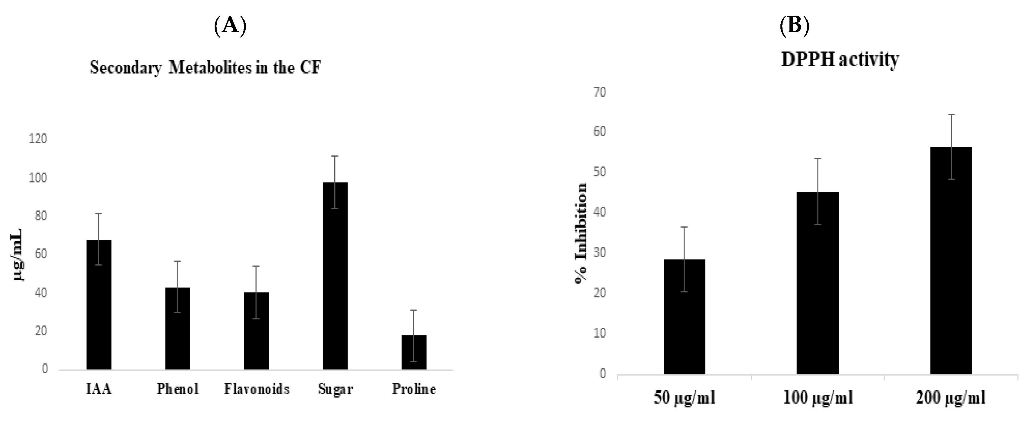
2.2. Screening of Culture Filtrate for Secondary Metabolites

2.2.1. Determination of IAA and Total Phenolics in the Culture Filtrate

Ahmad et al. [29] was followed for the IAA determination in CF. In an eppendorf tube, 1.5 mL of culture filtrate was placed and centrifuged at 10,000 rpm for 2 min. In a test tube, 1 mL of the supernatant and 2 mL of the Salkowski reagent were mixed thoroughly and kept in a dark condition for 30 min at 25 °C, and the absorbance was checked at 530 nm. The colorimetric method previously used by Jayanthi et al., [30] was used to quantify the total phenolic content with slight modifications. To 0.5 mL of the CF, 0.5 mL ethanol was added and centrifuged at 10,000 rpm for 20 min. Folin Ciocalteau reagent 0.5 mL and 4 mL Distilled water added, and after 3 min, 2 ml of Na2CO3 was also added. The mixture was heated until a dark blue color appeared and cooled. The UV–vis spectrophotometer was used for absorbance at 650 nm.

2.2.2. Determination of Total Flavonoids, Proline and Soluble Sugars

The concentration of flavonoids’ content in fungal CF was determined using the spectrophotometric technique [31]. In a test tube, 0.5 mL of CF was mixed with 4.3 mL of methanol (80%). Then, 0.1 mL of aluminum chloride (10%) and 0.1 mL of potassium acetate (10%) was added. The samples were incubated at room temperature for 30 min, and the absorbance was measured at 510 nm. Similarly, the technique of Bates et al. [32] was used for the determination of proline, and the absorbance was measured at 520 nm with toluene as a blank. For soluble sugars, the method of Lakshmi and Narasimha [33] was followed using 2 mL aliquots of CF and was added to 3 mL of the DNS (3, 5-Dinitrosalicylic acid) reagent. The mixture was boiled in a heated water bath for five minutes; after cooling, the absorbance was measured at 540 nm using a spectrophotometer.

2.2.3. DPPH-Free Radical Scavenging Activity (2,2-Diphenylpicrylhydrazyl)

The culture filtrate was passed through two layers of cheesecloth to separate the mycelia from the CF and extracted with 2× ethyl acetate. A separating funnel was used to separate the organic phase, which was then evaporated to dryness at 45 °C using a vacuum rotary evaporator. The residue was weighed and dissolved in methanol and used for antioxidant activity [34]. The antioxidant activity (DPPH-free radical scavenging) of the prepared samples was checked, through test samples. The variation in the optical density of radicals was noted. The endophytic fungal filtrate was employed at concentrations of 50, 100 and 200 g/mL. The DPPH solution (0.5 mmol/L) was prepared using 95% methanol. In the test sample, 2 mL of the DPPH solution was added and incubated for 30 min at room temperature in the dark. The OD was measured at 517 nm after 30 min [35].

2.3. Antibacterial Activity of Fungal CF by Well Diffusion Method

Fresh CF was added to the separating funnel, with a similar volume of ethyl acetate, and left to homogenize for an hour. On the basis of polarity, 2 clear phases were formed. The upper phase comprising secondary metabolites dissolved in ethyl acetate and the lower phase contained media. The upper phase was collected and evaporated in a rotary evaporator. The crude endophytic fungal filtrate was used for antibacterial activity by a well diffusion procedure [36]. By using a sterile spreader, freshly prepared bacterial inoculums (100 L) were evenly distributed over petri dishes containing the nutrient agar medium. A total of 4 wells of 6 mm were made. A stock solution of endophytic fungal extracts was made. A volume of 40 µL of each extract was added to each well for all bacterial strains. The positive control (Streptomycin) was used. DMSO (1/10th dilution) was used as a negative control. A standard clear scale was used to measure the zone of the inhibition of fungal extracts around each well.

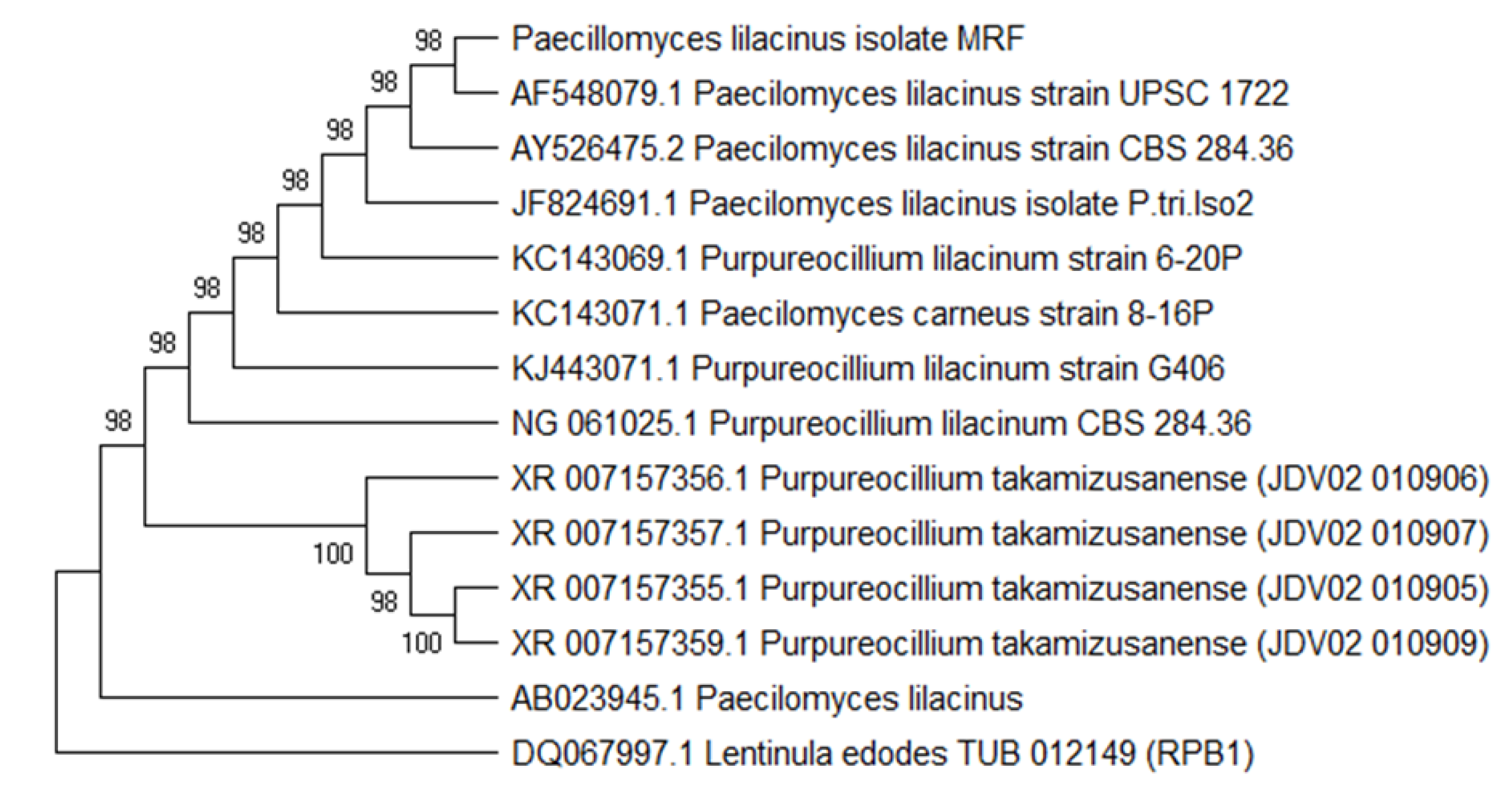
2.4. Extraction of Genomic DNA and Molecular Identification

The genomic DNA extraction and PCR of the fungal isolate were carried out according to the method of Khan et al., [28]. Using NS1 and NS24 sequences, the effective fungal isolate was identified by sequencing the internal transcribed region (ITS) of 18S rDNA. For this purpose, a Big Dye terminator cycle sequencing kit v.3.1 was employed. At the Macrogen Inc., Seoul, Republic of Korea, an automated DNA sequencing machine (Applied Biosystems, Foster City, CA, USA) was used to examine both the PCR sequencing and amplification. The PCR product was sequenced first, and then a homology search was performed using the online tool, BLAST.

2.5. Gas Chromatography Mass Spectrometry Analysis of the CF and Identification of Phytocompounds

The GC-MS analysis for compounds’ identification in fungal CF was performed using the GC-MS equipment (Thermo Scientific Co., Waltham, MA, USA). The National Institute of Standards and Technology’s 2008 (NIST-2008) database, which contains over 62,000 patterns, was used to interpret GC-MS mass spectra. The spectrums of unknown components were compared to the spectrums of known components in the NIST library.

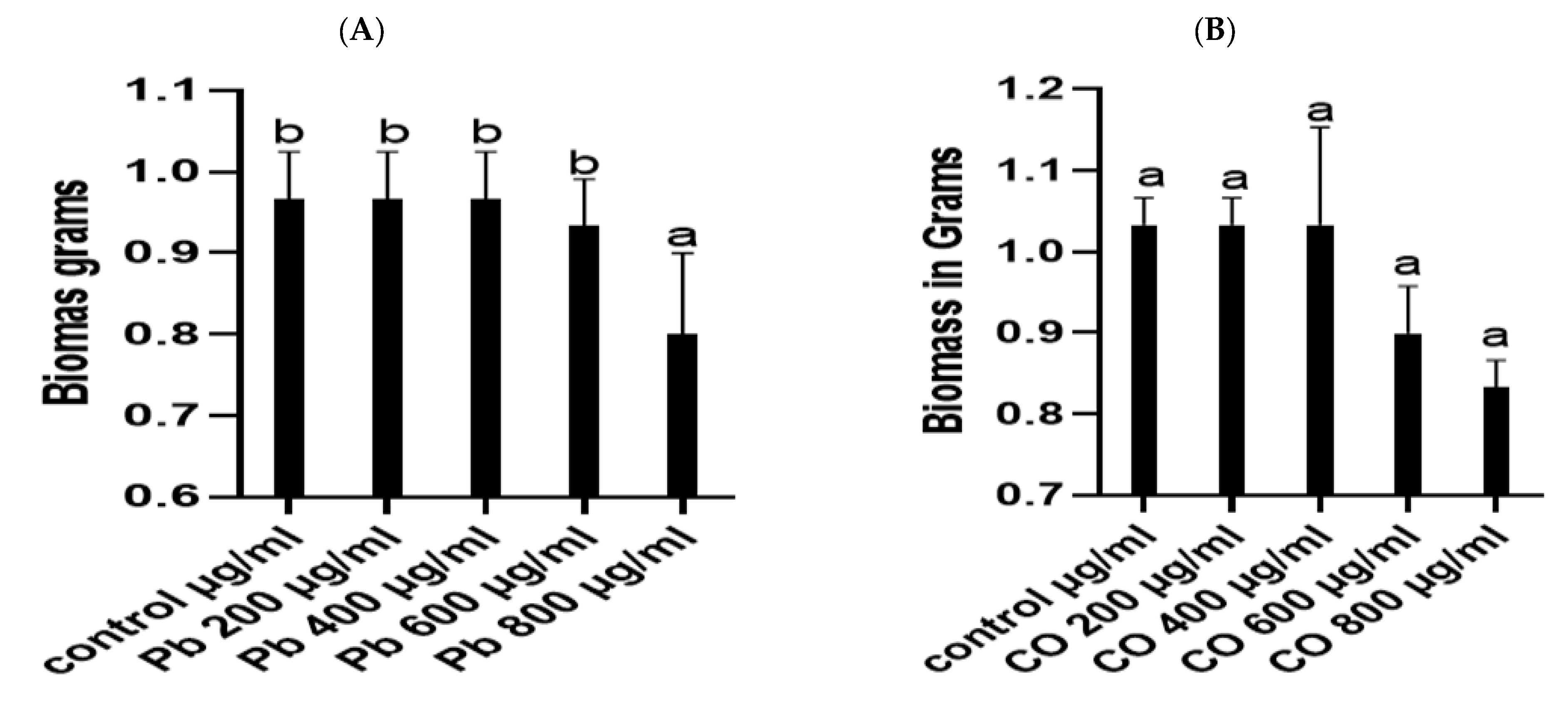
2.6. Heavy Metal Tolerance of Fungal Strains

The HM tolerance of fungal isolates was determined using the method of Khoshdil et al., [21]. In 500 mL conical flasks containing varied concentrations of lead and cobalt, such as 200 µg/mL, 400 µg/mL, 600 µg/mL and 800 µg/mL, 50 mL of czapek broth was added. After that, the inoculum of fungal isolates was introduced to each flask. Each flask was shifted to a shaking incubator for seven days and set at 120 rpm and 27 °C. By using the filter paper, culture filtrate and mycelia were separated. The fresh and dry weights were measured as well as the culture filtrate that was prepared in different concentrations, and the production of IAA, flavonoids and the phenolic content of the cultures was determined.

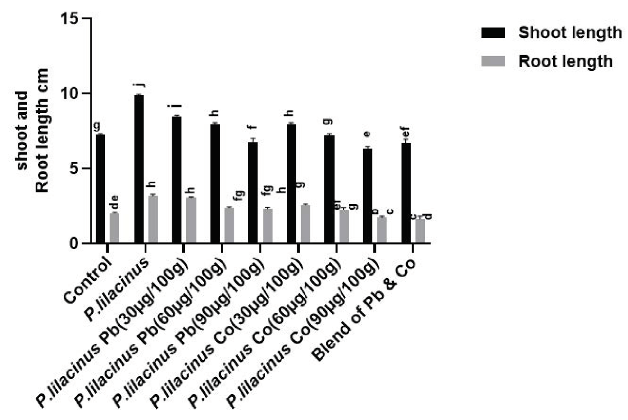
2.7. Determination of Fungal Growth and Screening of Rice Seedlings

The growth of strains was checked in czapek media containing 200 to 800 µg/mL of the lead cobalt and negative control for one week in a shaker at 30 °C and 120 rpm. The filtration with filter paper separated the CF from the fungus mycelium. Finally, the mycelium was weighed. The rice seedlings at the second leaf stage were used for the screening of culture filtrate for growth promoting or inhibiting activities. Mutant rice seeds were collected, surface-sterilized and incubated at 37 °C until radical and plumule formation. Three seedlings per pot were picked and placed in a pot containing 30 mL of water–agar medium (0.8%). The pots were shifted to the growth chamber (day/night cycle: 14 h, 28 °C ± 0.3; 10 h, 25 °C ± 0.3; relative humidity: 70%) for 14 days. A total of 100 mL of fungal CF was applied to the tip of the seedling. After 7 days of incubation, the growth-promoting and inhibitory effects of fungal extracts were determined by measuring growth parameters.

2.8. Pot Experiment for Growth Promotion and Stress Alleviation Activity

The seeds of Solanum lycopersicum were surface sterilized by soaking in 0.1 percent HgCl2 for 5 min and then washed for 5 min in sterilized and distilled water. Seedlings were allowed to grow for one week after germination. The isolated strain, P. lilacinus, was applied to each seedling for the alleviation of lead and cobalt stresses. The pots containing germinated plants were kept in a randomized block design with four replicates per treatment: HMs (lead and cobalt) at concentrations of 0, 30, 60 and 90 mg/100 g inoculated. The blend of lead and cobalt also applied 30 mg/100 g of each, with a final blend of 60 mg/100 g. The air-dried soil was spiked with HMs and thoroughly homogenized. The metal-spiked soil was then mixed with P. lilacinus at the rate of 0.67 g per 100 g of soil. The pots were kept in a greenhouse for 5 days, with daily watering to allow the fungus to grow and absorb metal. The plants were harvested after 45 days when seedlings were at the 7–6 leaves stage, and different physiochemical attributes were estimated.

2.8.1. Estimation of Root Shoot Length and Fresh Dry Weight

A transparent scale was used to measure the length of the roots and shoots. The plant fresh weight was recorded immediately after harvesting, and dry weight was measured later, after 48 h of drying in a 70 °C oven.

2.8.2. Determination of Proline Content and RWC

The procedure of Bates et al., [32] was followed with slight modifications for the determination of proline in the test sample. The absorbance was measured at 520 nm, and toluene was used as a blank. The leaves were collected and their fresh weight (FW) was instantly weighed. For the determination of the turgid weight, the leaves were soaked in distilled water for 6 h at 24 °C (TW). Finally, the dry weight of the samples was weighed after 48 h of drying at 78 °C [29]. For the calculation of the RWC value, the following formula was followed:
RWC = [(FW − DW)∕(TW − DW)] × 100

2.8.3. Estimation of Total Sugar and Protein in Plant Samples

For determination of the total sugar, the procedure of Dubois et al., [37] was followed. The absorbance was measured at 620 nm using a spectrophotometer. The amount of protein was calculated using the method of Bradford [38]. In 1 mL of phosphate buffer, fresh leaves (0.5 g) were homogenized (pH 7.0). The crude homogenate was centrifuged at 5000 rmp for 10 min. A half milliliter of freshly made trichloroacetic acid (TCA) was added, and the mixture was centrifuged for 15 min at 8000× g. A total of 1 mL of 0.1 N NaOH was used to dissolve the material, and 5 mL of the Bradford reagent was added. A spectrophotometer was used to measure the absorbance at 595 nm (SHIMADZU UV-2450, Kyoto, Japan).

2.8.4. Estimation of Total Phenolics, Flavonoids and IAA Estimation

Gurupavithra and Jayachitra [39] employed the Folin–Ciocalteau reagent method to estimate the total phenolics concentration. Using 80 percent ethanol as a blank, the optical density of the supernatant was measured at 650 nm. The flavonoid concentration was determined using the aluminium chloride colorimetric method described by [40]. The optical density was measured at 415 nm. The Salkowski reagent was used to determine the quantitative quantification of IAA in leaves, as stated by Zhishen et al., [40]. A UV spectrophotometer was used to measure the UV absorption at 540 nm.

2.9. Statistical Analysis

Data were recorded in the form of a triplicate and expressed as mean ± Standard error of mean (SEM). The data were then quantified for normality and homogeneity, and the statistical investigations were carried out by means of a one-way analysis of variance (ANOVA), followed by the multiple Duncan range test by using the statistical software SPSS, V 20.0 (SPSS, Chicago, IL, USA).

3. Results

3.1. Isolation, Screening and Identification of Fungal Endophyte

A total of 11 fungal endophytes were isolated from Justicia adathoda. The fungal endophytes were grown on PDA plates and were grouped on the basis of their morphological traits. On the basis of morphological traits and initial screening results, the isolate MRF was selected for further studies, and its culture filtrate (CF) was subjected to the evaluation of different secondary metabolites. The isolate MRF showed the highest number of secondary metabolites, i.e., IAA (68.17 µg/mL), phenols (43.31 µg/mL), flavonoids (40.59 µg/mL), total soluble sugar (97.83 µg/mL and proline (17 µg/mL) (Figure 1A). DPPH-free radical scavenging activity was performed, and the highest percentage (%) inhibition was demonstrated by the concentration of 200 µg/mL (56.55%), while 50 and 100 µg/mL demonstrated 28.55% and 45.35%, respectively (Figure 1B).
The fungal DNA of MRF was extracted for molecular identification. Internal Transcribed Spacer (ITS) regions of the fungal endophyte was sequenced and the phylogenetic analysis was carried out by using MEGA 6.0. The neighbor joining approach was used for the phylogenetic analysis of the fungal isolate MRF, and the tree was made from 15 taxa and aligned ITS sequences with 100 bootstrap replications. The strain was chosen using a BLAST search that demonstrated the highest sequence homology similarity. The results of the BLAST search demonstrated the highest sequence similarity (73%) between the fungal isolate MRF and Paecilomyces. The tree indicated that the isolated strain MRF belongs to Paecilomyces lilacinus (Figure 2). Our isolate (P. lilacinus isolate MRF) is making a 98% homology clad with the P. lilacinus strain, UPSC1722. The sequence was downloaded using BLAST, and the phylogenetic tree was constructed using the Maximum Parsimony method.

3.2. Heavy Metal Stress Tolerance of P. lilacinus

The growth of P. lilacinus was checked in the Czapek broth containing 200, 400, 600 and 800 µg/mL of Pb. The weight of the fresh biomass under cobalt stress was unaffected at 200 and 400 µg/mL, i.e., 0.966 g; meanwhile, when increasing the concentrations up to 600 µg/mL, a slight change was observed, i.e., at 0.933 g, while at 800 µg/mL (0.8 g), a significant decrease in the weight of the biomass was noted (Figure 3A). The growth of P. lilacinus was also checked for CO stress. The lower concentrations were unable to decrease the weight of the biomass, while higher concentrations of Cobalt (600 and 800 µg/mL) decreased the weight of the biomass (Figure 3B).
The heavy metals’ resistance of P. lilacinus was checked in the Czapek broth containing 200 µg/mL, 400 µg/mL, 600 µg/mL and 800 µg/mL of CO, Pb. The tolerance was estimated on the basis of the secretion of IAA, phenols and flavonoids in the CF. The secretion of flavonoids in the CF containing cobalt and lead at a concentration of 200 µg/mL and 400 µg/mL was unaffected; meanwhile, when increasing the concentrations up to 600 µg/mL, a slight change was observed, while at 800 µg/mL, a significant decrease in the secretion of flavonoids was observed (Table 1). Similarly, the phenols were also checked in the Czapek broth containing 200 µg/mL, 400 µg/mL, 600 µg/mL and 800 µg/mL of CO, Pb. The secretion of phenols was unaffected in the 200, 400 and 600 µg/mL concentrations, while a clear decrease was detected when the concentration was increased to 800 µg/mL, as shown in the Table 1. IAA was also analyzed by following the same concentration, and IAA was unaffected but a slight decrease was observed at 800 µg/mL (Table 2).

3.3. Antibacterial Activity and Chemical Analysis of the CF

The antibacterial activity of the CF of P. lilacinus was carried out against Salmonella typhi and Shigella sonnei. Endophytic P. lilacinus inhibited the growth of both bacterial strains. The Salmonella typhi gave an inhibition zone of 11.7 mm, while the inhibition zone of P. lilacinus against Shigella sonnei was 10.35 mm (Table 3). Additionally, the chemical constituents from the gas chromatography mass spectrometry analysis of the CF revealed different compounds, as listed in Table 4.

3.4. Effect of P. lilacinus on the Growth of Solanum lycopersicum

The root shoot length and fresh dry weight of S. lycopersicum L. inoculated with P. lilacinus (MRF) and without the fungal endophyte was recorded. The plants inoculated with the cobalt and lead stress demonstrated a significant decrease in the shoot and root lengths, and the fresh dry weights of S. lycopersicum at Pb (90 µg/100 g) significantly decreased the shoot length of 4.26 ± 0.14 cm and root length of 1.4 ± 0.15 cm and CO (90 µg/100 g) 4.36 ± 0.8, as compared to control plants with lengths of 7.23 ± 0.08 cm and 2.03 ± 0.03 cm. The plants inoculated with only MRF increased the length of the plants’ shoot, i.e., 9.86 ± 0.88 cm with a root length of 3.16 ± 0.12 cm. Cobalt and lead-stressed plants with MRF alleviated the HM stress (Figure 4). The fresh and dry weights of S. lycopersicum plants was also noted. Fresh and dry weights of lead- and cobalt-stressed plants at different concentrations was decreased; the highest decrease was noted in lead- and cobalt-stressed plants at 90 µg/100 g, i.e., 1.23 ± 0.03, 0.99 ± 0.11 and dry weights of 0.026 ± 0.003 g and 0.02 ± 0.01 g, as compared to control weights of 1.54 ± 0.04. Fresh and dry weights were improved in MRF-inoculated plants of 1.4 ± 0.008 g and 0.05 ± 0.003 g. Meanwhile, stressed plants inoculated with MRF significantly alleviated the HM-stressed plants, as shown in Figure 4.

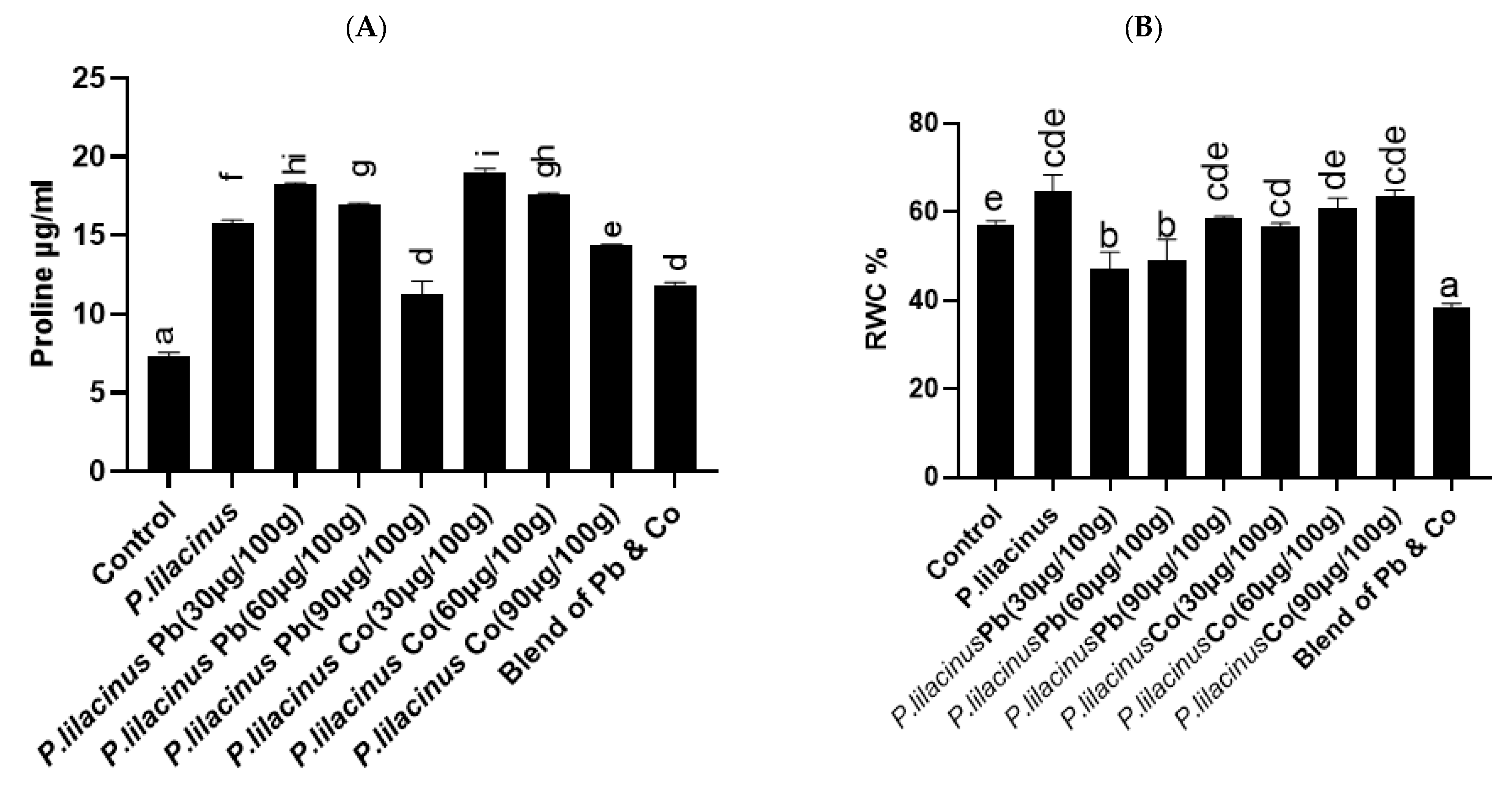
3.5. P. lilacinus Enhanced Proline Content and RWC of S. lycopersicum

The proline content was increased in the P. lilacinus (MRF) associated with S. lycopersicum in comparison to non-associated and control plants. Our results revealed that P. lilacinus (MRF) associated with S. lycopersicum produced 15.80 ± 0.16 µg/mL proline content, which is high as compared to control plants of 7.31 ± 0.25 µg/mL. Similarly, the proline content was increased in P. lilacinus (MRF)-associated stressed plants. The result indicated that 18.22 ± 0.11 µg/mL of proline was produced at a concentration of Pb 30 µg/100 g in P. lilacinus-associated lead stressed plants, which is clearly higher, as compared to lead-stressed plants of 10.74 ± 0.092 µg/mL. When the lead stress concentration was increased up to 30 and 60 µg/100 g, the proline content was slightly decreased, as shown in Figure 5A. The proline content was also enhanced in the P. lilacinus-associated cobalt-stressed plants of 19.03 ± 0.22 µg/mL, as compared to the stressed plants of 6.47 ± 0.22 µg/mL. A slight decrease was observed when the concentration was increased up to 30 and 60 µg/100 g. The RWC decreased in the stressed plants when compared with the control. However, the co-inoculation of P. lilacinus has recovered the losses of relative water content. The application of P. lilacinus increased the percentage of RWC, as shown in Figure 5B.

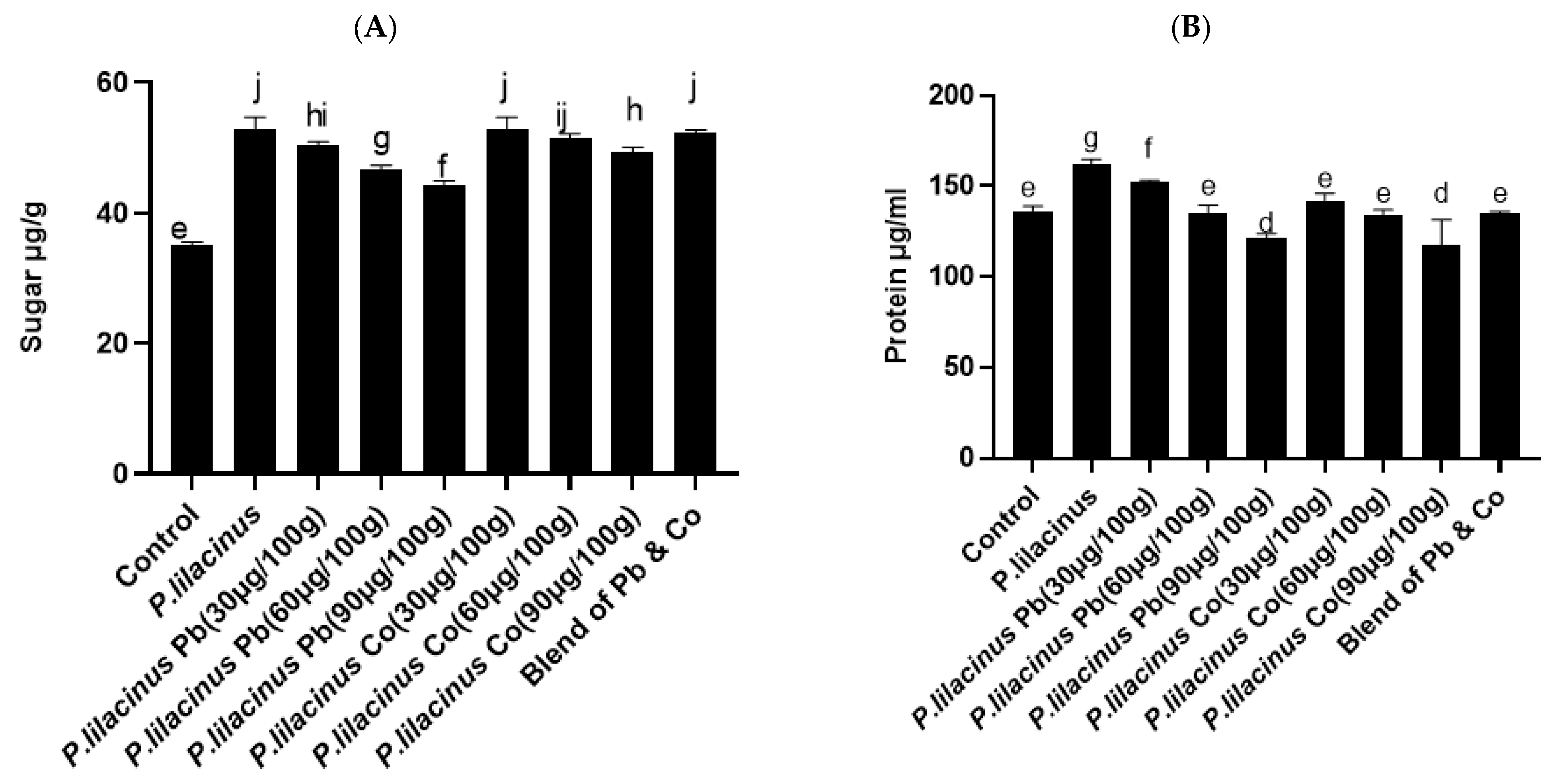
3.6. P. lilacinus Effect on Sugar and Protein Contents in S. lycopersicum

Higher amounts of soluble sugar were recorded in the S. lycopersicum plant under lead and cobalt stresses. The result revealed that soluble sugar was increased by 52 ± 1.05 µg/mL in plants inoculated with P. lilacinus. P. lilacinus-associated cobalt-stressed (30 µg/100 g) plants produced 50.47 ± 0.25 µg/mL of sugar, which is higher than the plants inoculated with the cobalt-stressed plant of 30.95 ± 0.058 µg/mL. Similarly, lead-stressed plants inoculated with P. lilacinus also produced the highest amount of soluble sugar (50.47 ± 0.25 µg/mL) in Figure 6A. Additionally, P. lilacinus improved the protein content in S. lycopersicum under the HM stress. The obtained results revealed that P. lilacinus increased the amount of protein. A higher protein content was observed in P. lilacinus inoculated plants (162.43 ± 1.41 µg/mL), while in the control plants, the amount of protein was decreased (136.43 ± 1.41 µg/mL). The amount of protein in fungal endophyte-inoculated cobalt and lead-stressed plants was higher than in cobalt- and lead-stressed plants’ different concentrations, so the highest amount of protein was recorded at concentrations of 30 µg/100 g of cobalt and lead stress, i.e., 151.90 ± 0.75 µg/mL, 141.73 ± 2.54 µg/mL, respectively, as shown in Figure 6B.

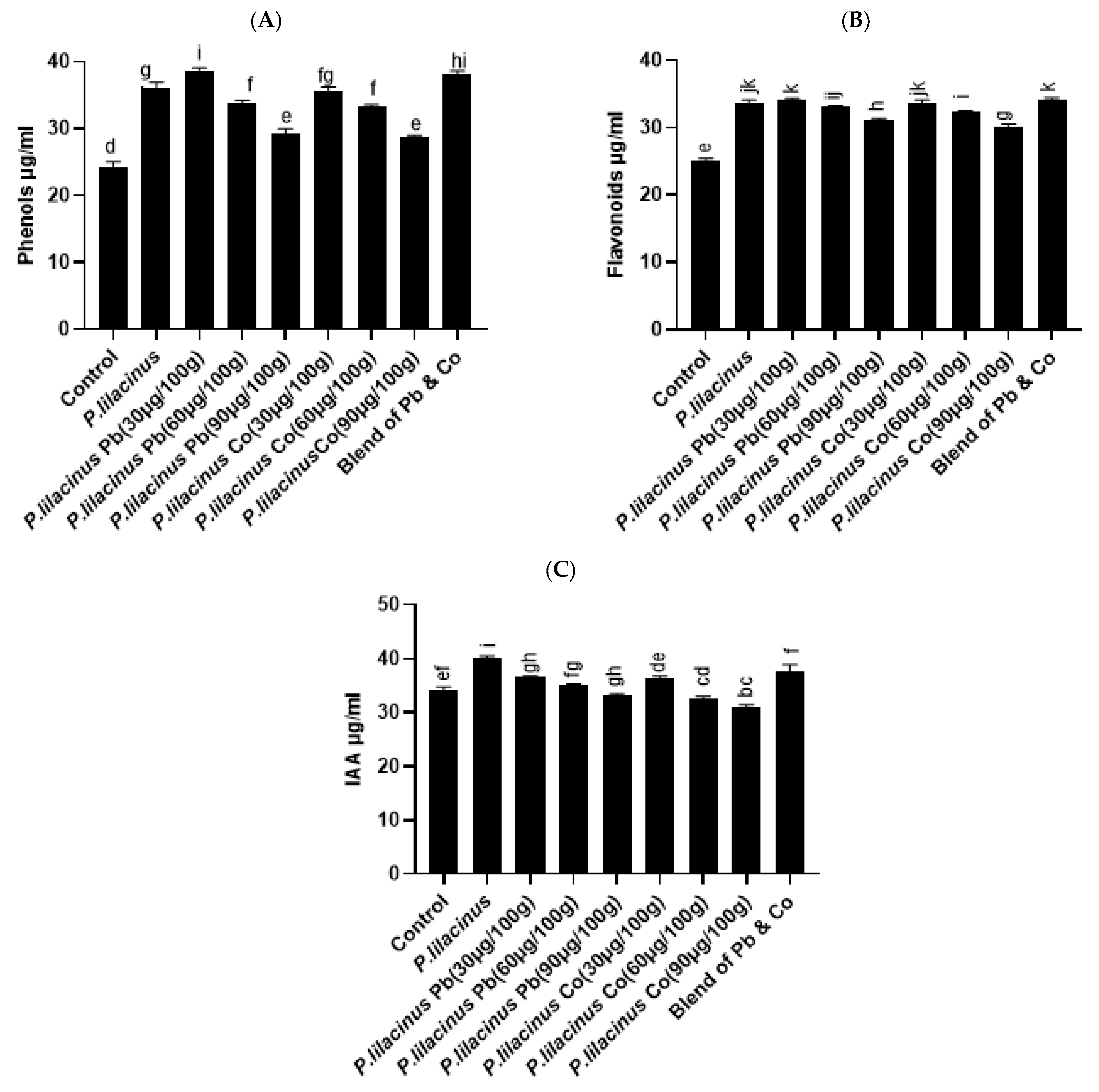
3.7. P. lilacinus Improved Effect on Phenols, Flavonoids and IAA Contents in S. lycopersicum

Higher amounts of phenols and flavonoids were produced in plants inoculated with P. lilacinus-associated plants. The results revealed that P. lilacinus-inoculated plants increased the amount of total phenol content (36.09 ± 0.48 µg/mL), while in control plants the amount of phenols was significantly decreased (24.22 ± 0.45 µg/mL). The phenols in S. lycopersicum plants inoculated with the fungal endophyte and the lead and cobalt stress with different concentrations was also increased, while in plants inoculated with lead and cobalt stress, the amount of phenols was significantly decreased (Figure 7A). The flavonoids’ content was also increased in plants associated with P. lilacinus as well as P. lilacinus plants inoculated with cobalt and lead stress (Figure 7B). S. lycopersicum inoculated with P. lilacinus indicated an increase in the IAA content. The IAA content was highly increased in the plants inoculated with P. lilacinus (40.20 ± 0.31 µg/mL). The amount of IAA was significantly decreased in the plants inoculated with stressed plants of 30, 60, 90 µg/100 g, as compared to plants inoculated with the fungal endophyte, as shown in Figure 7C.

4. Discussion

HM stress is a serious environmental problem and greatly affects crop productivity in different areas all over the world. Crops growing in such polluted soils demonstrate a decrease in growth and yields and are harmful to human health [41,42]. Recently, different studies have reported the effects of different HMs on plants and found a wide variety of harmful effects on plants. The entrance of these heavy metals to the plant is still unclear [43]. The HM stress increases the ionic imbalance in plant tissues and leads to a water scarcity in most plants, particularly crops [5], and the accumulation of HMs may interfere with the uptake and transportation of essential elements, such as xylem and phloem, and damage the natural metabolism, causing chloroplast degradation [42,44]. On the other hand, HMs cause diseases in various parts of the human body, including the liver, kidneys, bones, lungs and other parts of the body [45]; the most common diseases are nephrotoxicity, neurotoxicity, hepatotoxicity, skin toxicity and cardiovascular toxicity [46]. The bioremediation strategy is one of the most basic and effective approaches, especially in agricultural soils, and different microorganisms are used for the bioremediation of HM. Endophytic fungi and plants have long-standing symbiotic relationships, which clearly explain the importance of endophytes in the host plant tolerance during stress. The use of endophytes to reduce stress and increase the nutritional availability is an attractive and pragmatic approach [47,48]. However, the role of fungal endophytes in the bioremediation of HMs and their role in abiotic stress mitigation in crop plants needs to be investigated.
In the current study, a total of 11 endophytic fungi were isolated from Justicia adathoda, where P. lilacinus was selected on the basis of stress alleviation and different secondary metabolite production. Similarly, the fungal endophyte revealed a higher tolerance index and was growing effectively in the broth media containing 800 µg/mL of the metal. Initially, secondary metabolites were determined in the CF of P. lilacinus, where P. lilacinus produces greater amounts of IAA that contribute to plant growth promotion and abiotic stress tolerance [49]. Phenols, flavonoids, sugar and proline were also determined in the CF of the fungal endophyte. The gas chromatography mass spectrometry analysis of the CF of P. lilacinus revealed a total of seven compounds. Each compound possesses different biological and pharmacological potentials, i.e., a 3,7,11,15-Tetramethyl-2-hexadecen-1-ol antimicrobial [50], antifungal [51], phytol antimicrobial [50], antioxidant [52], squalene antibacterial and antioxidant potentials, with anti-tumor, cancer preventive, and chemopreventative qualities [53]. Furthermore, 1,2 Benzenedicarboxylic acid, diisooctyl ester antifungal [54], antimicrobial, antifouling [55], cyclotrisiloxane, hexamethyl antimicrobial and antioxidant [56] are potentials also included. In each gas chromatography, the mass spectrometry analysis of Trichloromethane has also been identified. According to the available literature, Trichloromethane did not possess any biological and pharmacological potential to the best of our knowledge, whereas the present study reports the antioxidant and antibacterial potential of Trichloromethane for the very first time.
P. lilacinus (MRF) has significantly increased the shoot length of S. lycopersicum, 9.86 ± 0.08 cm, and root length, 3.16 ± 0.12 cm, as compared to control plants of 7.23 ± 0.22 cm and a root of 2.03 ± 0.03 cm. These results are supported by the findings of Khan et al., [57], in which plant growth parameters such as shoot length, root length, dry biomass, leaf area and chlorophyll content were found to be higher in the inoculated RSF-6L plants than in non-inoculated or Cd-free plants [58]. Similarly, Khan and Lee [58] also revealed that the endophyte P. funiculosum symbiosis reduced the Cu stress and improved the soybean plant growth; these positive effects are attributed to their ability to release bioactive gibberellins. Proline plays a vital role in plants. It protects the plants from abiotic stresses as well as recovering plants from stress more quickly and is also known for enhancing enzymatic activities. Proline scavenges the ROS produced in plants under numerous biotic and abiotic stresses [59]. Sometimes, the ROS is produced in the absence of stress and becomes scavenged by naturally occurring antioxidants, such as proline; the stress conditions cause an imbalance of the ROS and antioxidants produced in cross-protection [60]. In our study, the significant increase in the proline content was noticed in the lead- and cobalt-stressed S. lycopersicum, as compared to the control untreated plants. The application of P. lilacinus has resulted in 15.80 µg/mL of proline, which is double the control plants (7.31 µg/mL). Similar results have been reported by Saradhi [61]. Our study is also in accordance with that of Ikram et al., [5]; in their study, an increase in the proline content in wheat plants inoculated with the endophytic fungi, Penicillium ruqueforti, occurred more often compared to non-inoculated plants.
Sugar plays an important role in a plant’s life cycle, as it is a primary source of energy and food reserve material, as well as a transitional metabolite in a variety of biochemical processes [60]. In the current study, the sugar content was improved up to 52.89 µg/mL in the P. lilacinus (MRF)-inoculated plants, which was higher than in the control plants. Meanwhile, in P. lilacinus-inoculated plants with different concentrations of lead- and cobalt-stressed plants, the sugar content was slightly reduced. Our current study is supported by Husna et al., [42]; their HM-tolerant endophytic fungi Aspergillus welwitschiae enhanced the sugar content in the Glycine max L. Proteins are biological macromolecules that perform a wide range of enzymatic, structural and functional roles in plants [62]. Proteins are also known for their pivotal role in plant stress responses as well as in HM stress adaptations [63]. In this study, we found that P. lilacinus enhanced the protein content up to (162.43 µg/mL) in S. lycopersicum, and, as compared to the control plants, it is significantly higher. The protein content was also higher in the P. lilacinus-inoculated lead- and cobalt-stressed plants. The protein’s content was also enhanced when the mixture of these HMs was also applied to the S. lycopersicum L. Our current results are co-related with the findings of Ban et al., [64].
Non-enzymatic antioxidants such as phenolics and flavonoids scavenge harmful radicals. Plants also need these secondary metabolites to defend themselves and scavenge harmful radicals under HM stress [65]. In our study, P. lilacinus enhanced the phenols and flavonoids in the S. lycopersicum. IAA is one of the most versatile phytohormones and is responsible for plant development under normal conditions, as well as for growth in stress conditions. IAA is important for plant adaptation and has been demonstrated to rise in response to HM stress. In the current study, the IAA level was increased in P. lilacinus-inoculated S. lycopersicum plants up to 40.20 µg/mL. These findings are supported by Khan et al., [49]. Our results suggest that the endophytic fungi, P. lilacinus, can be used to mitigate the adverse effects of HM stress on S. lycopersicum to grow normally under HM-stressed environmental conditions. Conclusively, Paecilomyces lilacinus (MRF) has a great potential to improve the growth of Solanum lycopersicum under heavy metal stress conditions. The inoculation of the fungal endophyte P. lilacinus (MRF) under metal stress conditions enhanced the production of phytohormone (IAA) and secondary metabolites. Our results revealed that P. lilacinus (MRF) can be used as a bioremediating agent and as a phytostimulant for crops’ growth promotion in heavy metal-affected soils. Thus, the fungal endophyte, P. lilacinus, seems to be a promising candidate, which can be used to alleviate the adverse effects of metal stress on the cultivation of S. lycopersicum in metal-polluted areas.

Author Contributions

M.H., F.G.J. and G.J. conceived and designed the research. M.M. and F.G.J. conducted the experiments. S.A.K., I.-J.L., G.R. and M.H. contributed new reagents or analytical tools. G.J., G.R. and M.M. analyzed the data. M.H., S.A.K., S.A. and F.G.J. wrote the manuscript. All authors have read and agreed to the published version of the manuscript.

Funding

This work was supported by the National Research Foundation of Korea (NRF) grant funded by the Korea government (MSIT) (No. 2022R1A2c1008993). The APC was funded by I.-J.L.

Informed Consent Statement

This article does not contain any studies with human participants or animals by any of the authors.

Data Availability Statement

The datasets generated during and/or analyzed during the current study are available from the corresponding author on reasonable request.

Conflicts of Interest

All authors declare that they have no conflict of interest.

References

  1. Wang, Y.; Liu, Y.; Zhan, W.; Zheng, K.; Wang, J.; Zhang, C.; Chen, R. Stabilization of heavy metal-contaminated soils by biochar: Challenges and recommendations. Sci. Total Environ. 2020, 729, 139060. [Google Scholar] [CrossRef] [PubMed]
  2. Tang, J.; Zhang, J.; Ren, L.; Zhou, Y.; Gao, J.; Luo, L.; Yang, Y.; Peng, Q.; Huang, H.; Chen, A. Diagnosis of soil contamination using microbiological indices: A review on heavy metal pollution. J. Environ. Manag. 2019, 242, 121–130. [Google Scholar] [CrossRef] [PubMed]
  3. Pogrzeba, M.; Rusinowski, S.; Krzyżak, J. Macroelements and heavy metals content in energy crops cultivated on contaminated soil under different fertilization—Case studies on autumn harvest. Environ. Sci. Pollut. Res. 2018, 25, 12096–12106. [Google Scholar] [CrossRef] [PubMed] [Green Version]
  4. Kowalska, J.B.; Mazurek, R.; Gąsiorek, M.; Zaleski, T. Pollution indices as useful tools for the comprehensive evaluation of the degree of soil contamination–A review. Environ. Geochem. Health 2018, 40, 2395–2420. [Google Scholar] [CrossRef] [PubMed] [Green Version]
  5. Ikram, M.; Ali, N.; Jan, G.; Jan, F.G.; Rahman, I.U.; Iqbal, A.; Hamayun, M. IAA producing fungal endophyte Penicillium roqueforti Thom., enhances stress tolerance and nutrients uptake in wheat plants grown on heavy metal contaminated soils. PLoS ONE 2018, 13, e0208150. [Google Scholar] [CrossRef] [PubMed] [Green Version]
  6. Ali, S.; Moon, Y.-S.; Hamayun, M.; Khan, M.A.; Bibi, K.; Lee, I.-J. Pragmatic role of microbial plant biostimulants in abiotic stress relief in crop plants. J. Plant Interact. 2022, 17, 705–718. [Google Scholar] [CrossRef]
  7. Yan, A.; Wang, Y.; Tan, S.N.; Mohd Yusof, M.L.; Ghosh, S.; Chen, Z. Phytoremediation: A promising approach for revegetation of heavy metal-polluted land. Front. Plant Sci. 2020, 11, 359. [Google Scholar] [CrossRef]
  8. Hossain, M.A.; Piyatida, P.; da Silva, J.A.T.; Fujita, M. Molecular mechanism of heavy metal toxicity and tolerance in plants: Central role of glutathione in detoxification of reactive oxygen species and methylglyoxal and in heavy metal chelation. J. Bot. 2012, 2012, 872875. [Google Scholar] [CrossRef] [Green Version]
  9. Dhaliwal, S.S.; Singh, J.; Taneja, P.K.; Mandal, A. Remediation techniques for removal of heavy metals from the soil contaminated through different sources: A review. Environ. Sci. Pollut. Res. 2020, 27, 1319–1333. [Google Scholar] [CrossRef]
  10. Prasad, M.N.V.; Freitas, H.; Fraenzle, S.; Wuenschmann, S.; Markert, B. Knowledge explosion in phytotechnologies for environmental solutions. Environ. Pollut. 2010, 158, 18–23. [Google Scholar] [CrossRef]
  11. Hussain, A.; Shah, M.; Hamayun, M.; Qadir, M.; Iqbal, A. Heavy metal tolerant endophytic fungi Aspergillus welwitschiae improves growth, ceasing metal uptake and strengthening antioxidant system in Glycine max L. Environ. Sci. Pollut. Res. 2021, 29, 15501–15515. [Google Scholar] [CrossRef]
  12. Chang, J.S.; Kim, Y.H.; Kim, K.W. The ars genotype characterization of arsenic-resistant bacteria from arsenic-contaminated gold–silver mines in the Republic of Korea. Appl. Microbiol. Biotechnol. 2008, 80, 155–165. [Google Scholar] [CrossRef] [PubMed]
  13. Zhang, Y.; Zhang, Y.; Liu, M.; Shi, X.; Zhao, Z. Dark septate endophyte (DSE) fungi isolated from metal polluted soils: Their taxonomic position, tolerance, and accumulation of heavy metals in vitro. J. Microbiol. 2008, 46, 624–632. [Google Scholar] [CrossRef] [PubMed]
  14. Xiao, X.; Luo, S.; Zeng, G.; Wei, W.; Wan, Y.; Chen, L.; Xi, Q. Biosorption of cadmium by endophytic fungus (EF) Microsphaeropsis sp. LSE10 isolated from cadmium hyperaccumulator Solanum nigrum L. Bioresour. Technol. 2010, 101, 1668–1674. [Google Scholar] [CrossRef] [PubMed]
  15. Jung, H.; Ali, S.; Kim, J.Y.; Kim, W.-C. Transgenic Arabidopsis expressing acdS gene of Pseudomonas veronii-KJ alleviate the adverse effects of salt and water-logging stress. Plant Breed. Biotechnol. 2018, 6, 221–232. [Google Scholar] [CrossRef]
  16. Hassen, A.; Saidi, N.; Cherif, M.; Boudabous, A. Resistance of environmental bacteria to heavy metals. Biores. Technol. 1998, 64, 7–15. [Google Scholar] [CrossRef]
  17. Njoku, K.L.; Akinyede, O.R.; Obidi, O.F. Microbial Remediation of Heavy Metals Contaminated Media by Bacillus megaterium and Rhizopus stolonifer. Sci. Afr. 2020, 10, e00545. [Google Scholar] [CrossRef]
  18. Yan, L.; Zhu, J.; Zhao, X.; Shi, J.; Jiang, C.; Shao, D. Beneficial effects of endophytic fungi colonization on plants. Appl. Microbiol. Biotechnol. 2019, 103, 3327–3340. [Google Scholar] [CrossRef]
  19. Yadav, A.N. Biodiversity and biotechnological applications of host-specific endophytic fungi for sustainable agriculture and allied sectors. Acta Sci. Microbiol. 2018, 1, 1–5. [Google Scholar]
  20. Jan, F.G.; Hamayun, M.; Hussain, A.; Jan, G.; Ali, S.; Khan, S.A.; Lee, I.J. Endophytic Candida membranifaciens from Euphorbia milii L. Alleviate Salt Stress Damages in Maize. Agronomy 2022, 12, 2263. [Google Scholar] [CrossRef]
  21. Khushdil, F.; Jan, F.G.; Jan, G.; Hamayun, M.; Iqbal, A.; Hussain, A.; Bibi, N. Salt stress alleviation in Pennisetum glaucum through secondary metabolites modulation by Aspergillus terreus. Plant Physiol. Biochem. 2019, 144, 127–134. [Google Scholar] [CrossRef] [PubMed]
  22. Lugtenberg, B.J.J.; Caradus, J.R.; Johnson, L.J. Fungal endophytes for sustainable crop production. FEMS Microbiol. Ecol. 2016, 92. [Google Scholar] [CrossRef] [PubMed]
  23. Ali, S.; Khan, A.L.; Ali, L.; Rizvi, T.S.; Khan, S.A.; Hussain, J.; Al-Harrasi, A. Enzyme inhibitory metabolites from endophytic Penicillium citrinum isolated from Boswellia sacra. Arch. Microbiol. 2017, 199, 691–700. [Google Scholar] [CrossRef] [PubMed]
  24. Ali, S.; Khan, S.A.; Hamayun, M.; Iqbal, A.; Khan, A.L.; Hussain, A.; Shah, M. Endophytic fungi from Caralluma acutangula can secrete plant growth promoting enzymes. Fresenius Environ. Bull. 2019, 28, 2688–2696. [Google Scholar]
  25. Rigobelo, E.C.; Baron, N.C. Endophytic fungi: A tool for plant growth promotion and sustainable agriculture. Mycology 2021, 13, 39–55. [Google Scholar] [CrossRef]
  26. Deepthi, V.C.; Sumathi, S.; Faisal, M.; Elyas, K.K. Isolation and identification of endophytic fungi with antimicrobial activities from the leaves of Elaeocarpus sphaericus (gaertn.) K. Schum and Myristicafragranshoutt. Intern. J. Pharmac. Sci. Res. 2018, 9, 2783–2791. [Google Scholar]
  27. Khan, S.A.; Hamayun, M.; Khan, A.L.; Lee, I.J.; Shinwari, Z.K.; Kim, J.G. Isolation of plant growth promoting endophytic fungi from dicots inhabiting coastal sand dunes of Korea. Pak. J. Bot. 2012, 44, 1453–1460. [Google Scholar]
  28. Khan, S.A.; Hamayun, M.; Yoon, H.; Kim, H.Y.; Suh, S.J.; Hwang, S.K.; Kim, J.M.; Lee, I.J.; Choo, Y.S.; Yoon, U.H.; et al. Plant growth promotion and Penicillium citrinum. BMC Microbiol. 2008, 8, 231. [Google Scholar] [CrossRef] [Green Version]
  29. Ahmad, F.; Ahmad, I.; Khan, M.S. Indole acetic acid production by the indigenous isolates of Azotobacter and fluorescent Pseudomonas in the presence and absence of tryptophan. Turk. J. Biol. 2005, 29, 29–34. [Google Scholar]
  30. Jayanthi, G.; Kamalraj, S.; Karthikeyan, K.; Muthumary, J. Antimicrobial and antioxidant activity of the endophytic fungus Phomopsis sp. GJJM07 isolated from Mesua ferrea. Int. J. Curr. Sci. 2011, 1, 85–90. [Google Scholar]
  31. Saravanan, S.; Parimelazhagan, T. In vitro antioxidant, antimicrobial and anti-diabetic properties of polyphenols of Passiflora ligularis Juss. Fruit Pulp. Food Sci. Hum. Wellness 2014, 3, 56–64. [Google Scholar] [CrossRef] [Green Version]
  32. Bates, L.S.; Waldren, R.P.; Teare, I.D. Rapid determination of free proline for water-stress studies. Plant Soil. 1973, 39, 205–207. [Google Scholar] [CrossRef]
  33. Lakshmi, A.S.; Narasimha, G. Production of cellulases by fungal cultures isolated from forest litter soil. Ann. For. Res. 2012, 55, 85–92. [Google Scholar]
  34. Dhayanithy, G.; Subban, K.; Chelliah, J. Diversity and biological activities of endophytic fungi associated with Catharanthus roseus. BMC Microbiol. 2019, 19, 22. [Google Scholar] [CrossRef] [PubMed]
  35. Yadav, M.; Yadav, A.; Yadav, J.P. In vitro antioxidant activity and total phenolic content of endophytic fungi isolated from Eugenia jambolana Lam. Asian Pac. J. Trop. Med. 2014, 7, S256–S261. [Google Scholar] [CrossRef] [PubMed] [Green Version]
  36. Taye, B.; Giday, M.; Animut, A.; Seid, J. Antibacterial activities of selected medicinal plants in traditional treatment of human wounds in Ethiopia. Asian Pac. J. Trop. Biomed. 2011, 1, 370–375. [Google Scholar] [CrossRef] [PubMed] [Green Version]
  37. Dubois, M.; Gilles, K.A.; Hamilton, J.K.; Rebers, P.T.; Smith, F. Colorimetric method for determination of sugars and related substances. Anal. Chem. 1956, 28, 350–356. [Google Scholar] [CrossRef]
  38. Bradford, M.M. A rapid and sensitive method for the quantitation of microgram quantities of protein utilizating the principle of protein dyes binding. Anal. Biochem. 1976, 72, 248–254. [Google Scholar] [CrossRef]
  39. Gurupavithra, S.; Jayachitra, A. Isolation and identification of endophytic fungi from Ocimum sanctum and analyse its antioxidant properties. Int. J. Res. Pharmaceut. Biomed. Sci. 2013, 4, 1120–1125. [Google Scholar]
  40. Zhishen, J.; Mengcheng, T.; Jianming, W. The determination of flavonoid contents in mulberry and their scavenging effects on superoxide radicals. Food Chem. 1999, 64, 555–559. [Google Scholar] [CrossRef]
  41. Chibuike, G.U.; Obiora, S.C. Heavy metal polluted soils: Effect on plants and bioremediation methods. Appl. Environ. Soil Sci. 2014, 2014, 1–12. [Google Scholar] [CrossRef] [Green Version]
  42. Husna, H.; Hussain, A.; Shah, M.; Hamayun, M.; Iqbal, A.; Qadir, M.; Asim, S.; Lee, I.-J. Stemphylium lycopersici and Stemphylium solani improved antioxidant system of soybean under chromate stress. Front. Microbiol. 2022, 4314. [Google Scholar] [CrossRef] [PubMed]
  43. Shakoor, N.; Adeel, M.; Azeem, I.; Ahmad, M.A.; Zain, M.; Abbas, A.; Hussain, M.; Jiang, Y.; Zhou, P.; Li, Y.; et al. Interplay of higher plants with lithium pollution: Global trends, meta-analysis, and perspectives. Chemosphere 2022, 310, 136663. [Google Scholar] [CrossRef]
  44. Natasha; Shahid, M.; Saleem, M.; Anwar, H.; Khalid, S.; Tariq, T.Z.; Murtaza, B.; Amjad, M.; Naeem, M.A. A multivariate analysis of comparative effects of heavy metals on cellular biomarkers of phytoremediation using Brassica oleracea. Int. J. Phytoremediation 2020, 22, 617–627. [Google Scholar] [CrossRef] [PubMed]
  45. Pirhadi, M.; Shariatifar, N.; Bahmani, M.; Manouchehri, A. Heavy metals in wheat grain and its impact on human health: A mini-review. J. Chem. Health Risks 2022, 12, 421–426. [Google Scholar]
  46. Mitra, S.; Chakraborty, A.J.; Tareq, A.M.; Emran, T.B.; Nainu, F.; Khusro, A.; Idris, A.M.; Khandaker, M.U.; Osman, H.; Alhumaydhi, F.A.; et al. Impact of heavy metals on the environment and human health: Novel therapeutic insights to counter the toxicity. J. King Saud Univ.-Sci. 2022, 101865. [Google Scholar] [CrossRef]
  47. Cheplick, G.P. Symbiotic fungi and clonal plant physiology. New Phytol. 2004, 413–415. [Google Scholar] [CrossRef]
  48. Ali, S.; Park, S.-K.; Kim, W.-C. The pragmatic introduction and expression of microbial transgenes in plants. J. Microbiol. Biotechnol. 2018. [Google Scholar] [CrossRef] [Green Version]
  49. Khan, A.R.; Ullah, I.; Waqas, M.; Shahzad, R.; Hong, S.J.; Park, G.S.; Jung, B.K.; Lee, I.J.; Shin, J.H. Plant growth-promoting potential of endophytic fungi isolated from Solanum nigrum leaves. World J. Microbiol. Biotechnol. 2015, 31, 1461–1466. [Google Scholar] [CrossRef]
  50. Farhat, H.; Urooj, F.; Tariq, A.; Sultana, V.; Ansari, M.; Ahmad, V.U.; Ehteshamul-Haque, S. Evaluation of antimicrobial potential of endophytic fungi associated with healthy plants and characterization of compounds produced by endophytic Cephalosporium and Fusarium solani. Biocatal. Agric. Biotechnol. 2019, 18, 101043. [Google Scholar] [CrossRef]
  51. Karimzadeh, K.; Zahmatkesh, A. Phytochemical screening, antioxidant potential, and cytotoxic effects of different extracts of red algae (Laurencia snyderiae) on HT29 cells. Res. Pharm. Sci. 2021, 16, 400. [Google Scholar] [CrossRef]
  52. Ibrahim, M.; Oyebanji, E.; Fowora, M.; Aiyeolemi, A.; Orabuchi, C.; Akinnawo, B.; Adekunle, A.A. Extracts of endophytic fungi from leaves of selected Nigerian ethnomedicinal plants exhibited antioxidant activity. BMC Compl. Med. Ther. 2021, 21, 98. [Google Scholar] [CrossRef] [PubMed]
  53. Zayed, M.Z.; Wu, A.; Sallam, S.M. Comparative phytochemical constituents of Leucaena leucocephala (Lam.) leaves, fruits, stem barks, and wood branches grown in Egypt using GC-MS method coupled with multivariate approaches. BioResources 2019, 14, 996–1013. [Google Scholar] [CrossRef]
  54. Banaras, S.; Javaid, A.; Khan, I.H. Potential antifungal constituents of Sonchus oleraceous against Macrophomina phaseolina. Intern. J. Agric. Biol. 2020, 24, 1376–1382. [Google Scholar]
  55. Saravanan, S.; ParimelazhaganKalaivani, C.S.; Sathish, S.S.; Janakiraman, N.; Johnson, M. GC-MS studies on Andrographis paniculata (Burm. f.) Wall. Ex Nees—A medicinally important plant. Int. J. Med. Arom. Plants 2012, 2, 69–74. [Google Scholar]
  56. Pamila, U.A.; Karpagam, S. GC-MS Analysis of Ethanolic Extract of Alternanthera philoxeroides and Alternanthera bettzickiana from India. World Res. J. Biol. Biol. Sci. 2017, 2, 2–4. [Google Scholar]
  57. Khan, A.R.; Ullah, I.; Waqas, M.; Park, G.S.; Khan, A.L.; Hong, S.J.; Ullah, R.; Jung, B.K.; Park, C.E.; Ur-Rehman, S.; et al. Host plant growth promotion and cadmium detoxification in Solanum nigrum, mediated by endophytic fungi. Ecotoxicol. Environ. Saf. 2017, 136, 180–188. [Google Scholar] [CrossRef] [PubMed]
  58. Khan, A.L.; Lee, I.J. Endophytic Penicillium funiculosum LHL06 secretes gibberellin that reprograms Glycine max L. growth during copper stress. BMC Plant Biol. 2013, 13, 86. [Google Scholar] [CrossRef] [Green Version]
  59. Hayat, S.; Hayat, Q.; Alyemeni, M.N.; Wani, A.S.; Pichtel, J.; Ahmad, A. Role of proline under changing environments: A review. Plant Signal. Behav. 2012, 7, 1456–1466. [Google Scholar] [CrossRef] [Green Version]
  60. Ahmad, M.A.; Javed, R.; Adeel, M.; Rizwan, M.; Yang, Y. PEG 6000-stimulated drought stress improves the attributes of in vitro growth, steviol glycosides production, and antioxidant activities in Stevia rebaudiana Bertoni. Plants 2020, 9, 1552. [Google Scholar] [CrossRef]
  61. Saradhi, P.P. Proline accumulation under heavy metal stress. J. Plant Physiol. 1991, 138, 554–558. [Google Scholar] [CrossRef]
  62. Rasheed, F.; Markgren, J.; Hedenqvist, M.; Johansson, E. Modeling to understand plant protein structure-function relationships—Implications for seed storage proteins. Molecules 2020, 25, 873. [Google Scholar] [CrossRef] [PubMed] [Green Version]
  63. Singh, S.; Parihar, P.; Singh, R.; Singh, V.P.; Prasad, S.M. Heavy metal tolerance in plants: Role of transcriptomics, proteomics, metabolomics, and ionomics. Front. Plant Sci. 2016, 6, 1143. [Google Scholar] [CrossRef] [Green Version]
  64. Ban, Y.; Tang, M.; Chen, H.; Xu, Z.; Zhang, H.; Yang, Y. The response of dark septate endophytes (DSE) to heavy metals in pure culture. PLoS ONE 2012, 7, e47968. [Google Scholar] [CrossRef] [PubMed]
  65. Bartwal, A.; Mall, R.; Lohani, P.; Guru, S.K.; Arora, S. Role of secondary metabolites and brassinosteroids in plant defense against environmental stresses. J. Plant Growth Reg. 2013, 32, 216–232. [Google Scholar] [CrossRef]
Figure 1. (A) Secondary metabolites in the CF of P. lilacinus. IAA = Indole-3-acetic acid, Phe = phenolic and Flav = flavonoids, proline and sugar contents. (B) Represents DPPH free radical scavenging activity of the CF of P. lilacinus.
Agronomy 13 00883 g001
Figure 2. Phylogenetic tree of P. lilacinus (MRF). The evolutionary history was inferred using the Maximum Parsimony method. Tree #1 out of 10 of most parsimonious trees (length = 576) is shown. The consistency index is 1.000000, the retention index is 1.000000, and the composite index is 1.000000 for all sites and parsimony-informative sites (in parentheses). The percentage of replicate trees in which the associated taxa clustered together in the bootstrap test (100 replicates) are shown next to the branches. The MRF tree was obtained using the Subtree–Pruning–Regrafting (SPR) algorithm with search level 1, in which the initial trees were obtained by the random addition of sequences (10 replicates). This analysis involved 14 nucleotide sequences. There were a total of 1065 positions in the final dataset. Evolutionary analyses were conducted in MEGA X.
Agronomy 13 00883 g002
Figure 3. (A) Represent weight of fresh biomass of P. lilacinus under lead stress. Bars represents mean of triplicates with ±SE. Alphabets above the bars are different significantly from one another at p ≤ 0.05. (B) Represents weight of fresh biomass of P. lilacinus under cobalt stress. Bars represents mean of triplicates with ±SE. Alphabets above the bars are different significantly from one another at p ≤ 0.05.
Agronomy 13 00883 g003
Figure 4. Effect of lead and cobalt stresses on S. lycopersicum inoculated with P. lilacinus and without P. lilacinus. Bars represent mean of triplicates with ±SE. Alphabets above the bars are significantly different from one another at p ≤ 0.05.
Agronomy 13 00883 g004
Figure 5. (A) Effect of lead and cobalt stresses on proline of S. lycopersicum inoculated with P. lilacinus and without P. lilacinus. Bars represents mean of triplicates with ±SE. Alphabets above the bars are significantly different from one another at p ≤ 0.05. (B) Effect of lead and cobalt stresses on RWC of S. lycopersicum inoculated with P. lilacinus and without P. lilacinus. Bars represents mean of triplicates with ±SE. Alphabets above the bars are significantly different from one another at p ≤ 0.05.
Agronomy 13 00883 g005
Figure 6. (A) Effect of lead and cobalt stresses on sugar of S. lycopersicum inoculated with P. lilacinus and without P. lilacinus. Bars represents mean of triplicates with ±SE. Alphabets above the bars are different significantly from one another at p ≤ 0.05. (B) Effect of lead and cobalt stresses on protein of S. lycopersicum inoculated with P. lilacinus and without P. lilacinus. Bars represents mean of triplicates with ±SE. Alphabets above the bars are different significantly from one another at p ≤ 0.05.
Agronomy 13 00883 g006
Figure 7. (A) Effect of lead and cobalt stresses on phenols of S. lycopersicum inoculated with P. lilacinus and without P. lilacinus. Bars represents mean of triplicates with ±SE. Alphabets above the bars are different significantly from one another at p ≤ 0.05. (B) Effect of lead and cobalt stresses on flavonoids of S. lycopersicum inoculated with P. lilacinus and without P. lilacinus. Bars represents mean of triplicates with ±SE. Alphabets above the bars are significantly different from one another at p ≤ 0.05. (C) Effect of lead and cobalt stresses on IAA of S. lycopersicum inoculated with P. lilacinus and without P. lilacinus. Bars represent mean of triplicates with ±SE. Alphabets above the bars are significantly different from one another at p ≤ 0.05.
Agronomy 13 00883 g007
Table 1. Effect of lead stress on the secretion of IAA, phenols and flavonoids.
TreatmentsIAAPhenolsFlavonoids
Control62.45 ± 1.6 b54.34 ± 0.96 c40.31 ± 0.29 bc
Lead 200 µg/mL62.16 ± 3.3 b55.47 ± 1.13 c41.69 ± 1.24 c
Lead 400 µg/mL66.07 ± 1.8 b54.19 ± 1.64 c40.06 ± 0.42 bc
Lead 600 µg/mL58.87 ± 2.5 ab48.93 ± 0.89 b37.95 ± 0.58 b
Lead 800 µg/mL53.04 ± 0.5 a40.12 ± 0.74 b31.71 ± 0.69 a
Each datum represent the mean of triplicates with SE. Different alphabets are significantly different. Duncun test: p ≤ 0.05.
Table 2. Effect of different concentrations of cobalt stress on the secretion of IAA, phenols and flavonoids.
TreatmentsIAA, µg/mLPhenols, µg/mLFlavonoids, µg/mL
Control62.45 ± 1.3 b55.75 ± 1.5 b41.55 ± 0.60 c
Cobalt 200 µg/mL63.39 ± 2.4 b56.36 ± 1.2 b42.76 ± 0.87 c
Cobalt 400 µg/mL63.02 ± 5.5 b57.1 ± 1.3 b43.48 ± 0.38 c
Cobalt 600 µg/mL59.44 ± 3.0 ab52.16 ± 3.0 b37.95 ± 1 b
Cobalt 800 µg/mL51.21 ± 0.75 a36.94 ± 1.0 a32.54 ± 1.01 a
Each datum represent the mean of triplicates with SE. Different alphabets are significantly different. Duncun test: p ≤ 0.05.
Table 3. Antibacterial activity of the CF of P. lilacinus.
Bacterial StrainMean Zone of Inhibition (mm)
Salmonella typhi9.55
Shigella sonnei10.36
Table 4. Chemical constituents from the gas chromatography mass spectrometry analysis of CF of P. lilacinus.
CompoundRTProbabilityChemical Formula
Trichloromethane1.3381.31CHCl3
Phytol79.9720.64C20H40O
3,7,11,15-Tetramethyl-2-hexadecen-1-ol21.2736.23C20H40O
1,2-Benzenedicarboxylic acid, diisooctyl ester23.1260.35C38H56O8
Squalene24.4559.22C₃₀H₅₀
Cyclotrisiloxane, hexamethyl-25.6757.87C6H18O5
bis (2-ethylhexyl) ester23.4568.49(CH2)8(COOC8H17)2
Disclaimer/Publisher’s Note: The statements, opinions and data contained in all publications are solely those of the individual author(s) and contributor(s) and not of MDPI and/or the editor(s). MDPI and/or the editor(s) disclaim responsibility for any injury to people or property resulting from any ideas, methods, instructions or products referred to in the content.

