Article

Microwaves Induce Histological Alteration of Ovaries and Testis in Rhynchophorus ferrugineus Oliv. (Coleoptera: Curculionidae)

1
Department of Veterinary Medicine and Animal Production, University of Naples Federico II, Via Delpino 1, 80137 Naples, Italy
2
Department of Physics “Ettore Pancini”, University of Naples Federico II, Via Cintia, Complesso Monte S. Angelo, 80126 Naples, Italy
3
Department of Agriculture, University of Naples Federico II, Via Reggia 100, Portici, 80146 Naples, Italy
4
Plant Protection Service of Campania Region, V. G. Porzio Centro Direzionale is 6, 80143 Naples, Italy
*
Author to whom correspondence should be addressed.
Agronomy 2023, 13(2), 420; https://doi.org/10.3390/agronomy13020420
Submission received: 22 December 2022 / Revised: 23 January 2023 / Accepted: 28 January 2023 / Published: 31 January 2023
(This article belongs to the Special Issue Ecological Management of Pests)

Abstract

:
The Red Palm Weevil (RPW) is one of the major pests of palms, frequently leading to the plants death. Action plans and the development of bio/physical strategies to contrast RPW diffusions are strongly recommended due to the serious concerns related to environmental pollution and insects’ resistance to chemicals. In the present study, we investigated morphological alterations of the ovaries and testes in adult RPW exposed to 2.45 GHz for 5, 15, and 30 s. During these treatments, the relative increase in temperature and the days of survival after irradiation were monitored. Then, RPWs were processed for macroscopical and microscopical analysis. Histological lesions of the ovaries and testes were characterized by the degeneration and necrosis of germinal cells, which increased with the increase in the time of irradiation and the temperature. By the same token, an increase in the temperature of irradiated insects was associated with a decrease in their survival time. These observations lead us to conclude that MWs could represent a useful tool for reducing or eliminating the reproductive capacity of this dreaded insect.

1. Introduction

The Red Palm Weevil (RPW), Rhynchophorus ferrugineus (Oliver) (Coleoptera: Curculionidae), is a large polyphagous insect native to southern Asia and Malaysia and is one of the most important pests of numerous species of the Palmae plant family, which have considerable economic and environmental interest. RPW damages more than 41 different palm species in the world [1], but particularly date palms, coconut palms, and oil palms.
Its infestation was recorded in 50% of date-producing countries [2], causing yield losses up to 0.7–10 tons/ha [3]. This invasive alien species (IAS) has spread rapidly in the countries of the Mediterranean basin, where it has seriously compromised the survival of Phoenix canariensis (Chabaud), leading to profound changes in the urban landscape. Its infestation was recorded in 50% of date producing countries [2] causing yield losses up to 0.7–10 tons/ha [3]. Today, RPW is widely distributed in Africa, Oceania, the USA, the Middle East, and Europe (including Italy) [4]. Its invasive potential is a consequence of elevated female fecundity, of the ability to complete several generations in a year even in the same tree, and of the ability to fly for long distances, along with the tolerance to a wide range of climates due to the habit of hiding inside the host palm tree. Females are usually attracted by palm volatiles and lay several eggs in dying or damaged parts of the palm, although undamaged palms could also be attacked [3]. After a few days, eggs hatch into larvae, which develop within the trunks of palm trees, frequently leading to the plants death. Considering that external symptoms of the infestations are rarely visible at very early stages and that weevil presence is often not detectable until fatal damage has occurred [5]. Several control methods have been applied within an integrated pest management (IPM) strategy [6]; these include the use of insecticides [7,8,9,10,11], pheromone traps for adult monitoring [12,13,14,15], plant extracts [5,16], as well as biological control [1,17,18]. Since some of these treatments have caused substantial environmental pollution, and no effective and economic method has been adopted for the total eradication of the RPW pest, the use of a Sterile Insect Technique (SIT) adopting ionizing radiation is being developed [19,20,21,22,23]. Radiofrequency and microwaves (MW) are non-ionizing radiations, and their use has been proposed in the management of a wide range of agricultural pests, exploiting the thermal death induced in the insects. Recently, the heating of P. canariensis palms with MW has been suggested to reduce and control the RPW population [24,25,26,27] inducing a thermal challenge capable of killing the insects without damaging the palm tissues, considering that the insects have a lower thermal tolerance than the host matrices. We focused our attention at 2.45 GHz, one of the most used frequencies for industrial applications, and, consequently, at this frequency, cheaper sources are available. A simple numerical model able to simulate the temperature distribution inside a palm with reference to microwave heating for the disinfestation of living plants attacked by RPW has been proposed [28]. As matter of fact, in semi-field tests we observed a high percentage of dead insects (100% dead pupae in 3 out of 4 palms, and 80% total) when they were in the pupal stage, i.e., blocked inside cocoons that are typically located near the surface. Moreover, it was observed that it is difficult to reach a lethal temperature for RPW in the inner part of the trunk [28,29]. Considering that when exposed to MW, an adults first reaction is an attempt to escape, we investigated the influence of short-term microwave exposure. Preliminary results on the effects of MW on RPWs’ tissues were pointed out in a previous study [25]. Therefore, the objective of our work has been to deepen our investigations to evaluate the correlation between the effects induced by MW on the reproductive system and the dose in terms of increase of temperature with respect to the initial temperature (ΔT) [°C], time exposures, and survival days after irradiation in strictly controlled exposure conditions.

2. Materials and Methods

2.1. Rearing of the Insect

The rearing of the adult and larvae RPW was carried out entirely in the laboratories of the Department of Agriculture (University of Naples Federico II), in climatic chambers at 25 ± 2 °C of temperature, 60–70% RH and with a photoperiod of L12:D12. Adults obtained by field captures with Tripheron ® traps were cleaned and put into plastic containers of a rectangular shape (20 × 20 × 40 cm3) closed with a grid lid to allow for ventilation of the indoor environment (5 males and 5 females per box). A layer of coconut fibers was placed on the bottom of the container to absorb excess moisture. The coconut fibers also serve as shelter for the beetles. Some apples were provided in the cages for the adult feedings and as a substrate for oviposition. After about 72 h, the apples with eggs were moved to other plastic containers, allowing the development of the larvae until the building of the cocoon. Cocoons were placed in plastic containers until adult emergence. 29 adults were sexed and used for the experimental tests 72 h after emergence. After MW irradiation, adults RPW were separated for each group (5 s, 15 s, and 30 s treatments), and the number of eggs laid was calculated during all the rearing periods.

2.2. MW Irradiation

Twenty-five adult RPW (12 females and 13 males) were exposed at 2.45 GHz for different durations (5 s, 15 s, 30 s); four adult RPW (2 females and 2 males) were untreated and used as controls. The insects, positioned in a plastic cylindrical plastic container (35 mm diameter, 60 mm height) without the cover, were introduced in a customized microwave applicator fed by a magnetron (Alter TMA20) and were exposed to the minimum incident power density of 5.4 W/cm2 available by the magnetron source (200 W). The applicator is a short-circuited rectangular waveguide (WR340, 8.6 cm × 4.3 cm) connected to a cylindrical waveguide (diameter of 35 mm, 100-mm long, centered with respect to the narrow side of the rectangular WG and located 140 mm from the short), which allows temperature measurements through an infrared thermocamera (Flir A40) with negligeable electromagnetic field perturbation [29,30]. During exposure, the temperature distribution on the surface of the insects, free of movements, was acquired. Moreover, the possibility of movement of the insect into the container reduced stress and gave a more general scenario without a fixed orientation respecting the incident electric field; on the other hand, temperature increases could be different even for the same treatment.

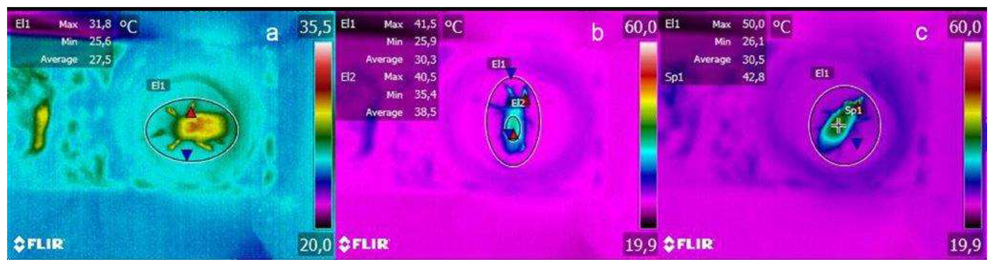
2.3. Thermogram Analysis

The infrared thermocamera was 30 cm from the sample holder and frame, and measurements were acquired for about 30 s, 60 s, and 80 s, depending on the duration of the exposures, by using the Flir ThermaCAM software. Thermogram frames were then blindly analyzed by means of FlirTool software to determine the maximum temperature reached by the insect as a result of the exposure and to assess the increase in temperature (ΔT) [°C] compared to that measured before the source was turned on.

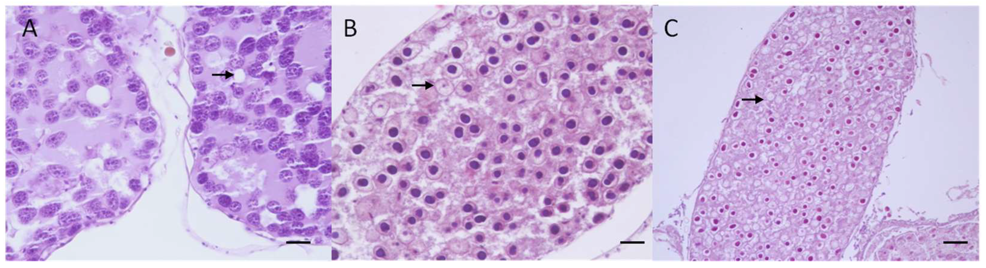
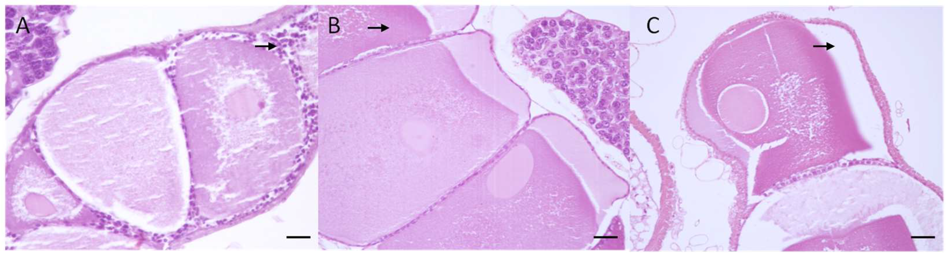
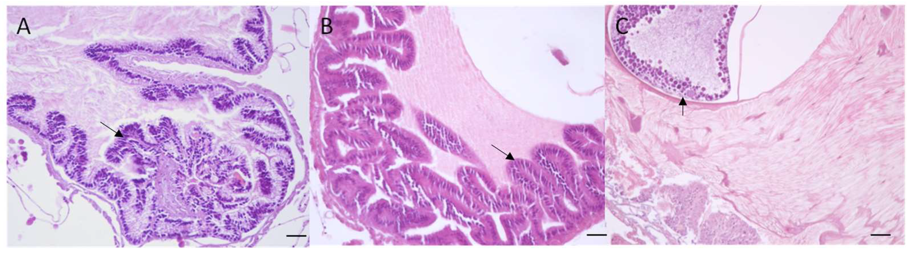
2.4. Macroscopical and Microscopical Analysis

Dissected organs were processed for histological analysis, fixed in 10% formalin, dehydrated in a graded series of alcohol solutions (Carlo Erba srl, Cornaredo, Italy), embedded in paraffin wax (Bioptica srl, Milan, Italy) using an automatic embedding processor (VTP300, Bio-Optica, Milan, Italy), sectioned into 5 μm thick sections with a microtome (PFM 3007 Rotary Microtome, Bioptica srl, Milan, Italy), and stained with haematoxylin and eosin (H & E, Bioptica srl, Milan, Italy) using an automatic tissue slide stainer (ST5010 Autostainer XL, Leica, Germany). Tissue preparations were observed using light microscopy (E-400; Nikon Eclipse, Tokyo, Japan), and histological images were captured by a video camera (TKC1380E; JVC, Tokyo, Japan) coupled to the microscope by the ACT1 program.
Control samples (4) were processed as already stated above. Histological sections of ovaries and testicles were subjectively analyzed by pathologists, and for each sample, a score from 0 to 3 was assigned, considering the severity of the following alterations: vacuolar degeneration, nuclear necrosis (pyknosis, karyorrhexis), desquamation and loss of germinal cells. The score assigned for each sample was the following: 0—no pathological alterations; 1—mild pathological alterations; 2—moderate pathological alterations; 3—severe pathological alterations.

2.5. Statistical Analysis

Statistical analysis was performed using GraphPad Prism version 7 software (GraphPad Software Inc., La Jolla, CA, USA). All data were tested for normality using the Shapiro-Wilk normality test. Since the data were normally distributed, the one-way ANOVA with Turkey’s post-test was used to evaluate the significance of differences in terms of mean value of ΔT (°C) between groups exposed to different irradiation times (5 s, 15 s, and 30 s) and survival times between irradiated and control samples.
Linear regression analyses were used to evaluate the correlation between survival days after irradiation and ΔT (°C) reached by the samples, whereas nominal logistic regression analysis was used to evaluate the relationship between ΔT (°C) and the severity of histologic damage detected; JMP Pro version 15.0.0 (SAS Institute Inc., Campus Drive, Cary, NC, USA) was used for this purpose.

3. Results

3.1. Thermogram Results

The one-way ANOVA results showed statistical significance between ΔT (°C) at 5 s (9.5 ± 2.4), 15 s (13.9 ± 6.2), and 30 s (20.41 ± 6.5) of exposure (p < 0.0019) (Figure 1, Table 1 and Table 2). Tukey’s post test showed a statistical significance only between 5 s and 30 s irradiated samples (p < 0.0014; q = 5.769; degrees of freedom DF = 22).

3.2. Survival and Fecundity Data

Survival days after irradiation decreased rapidly, proceeding from untreated insects (mean group value = 41.75 ± 21.18) to 5 s (mean group value = 21.6 ± 17.39), 15 s (mean group value = 17.75 ± 15.52), and 30 s irradiated samples (mean group value = 16.3 ± 7.24) (Table 1 and Table 2). However, the ANOVA showed statistical significance exclusively between the control group and the 30 s group (p = 0.043; q = 3.988; DF = 25).
Fecundity data (total number of laid eggs) showed a remarkable decrease from untreated samples (207) to 5 s (111) and 15 s samples (117), with a drastic reduction in the 30 s irradiated samples (28).

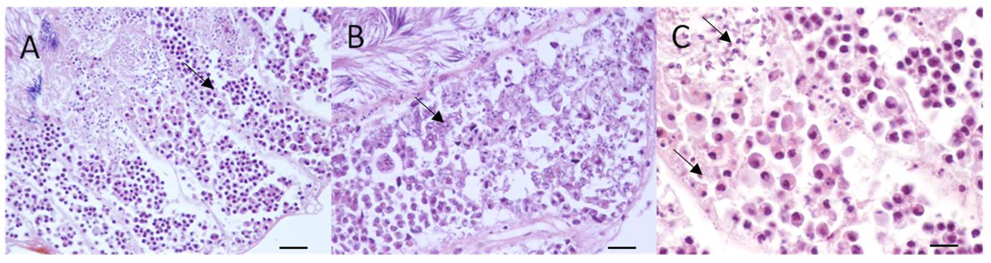
3.3. Macroscopical and Microscopical Results

As reported in other studies [20,31], the morphology of RPW female genital systems consists of two ovaries with their lateral oviducts, a common oviduct, vagina, and spermatheca. Each ovary consists of a terminal filament, germarium, and vitellarium, which open into the calyx of the later oviduct. The male reproductive system consists of two pairs of testicular lobes, the deferent duct, accessory glands, and the prostate [21]. No evident changes were observed in our controls, while evident alterations are shown in the 30 s irradiated RPWs, which appeared smaller in size and softer than normal. On the contrary, in all irradiated samples, microscopical changes were observed, consisting of lesions with a variable degree of severity, ranging from degeneration to necrosis, which increased with the increase in irradiation time.
Female RPWs showed mild (3/4 samples; 75%) to moderate (1/4 sample; 25%) lesions at 5 s, moderate (2/4 samples; 50%) to severe lesions (2/4; 50%) at 15 s, and severe lesions (4/4; 100%) at 30 s. Germarium—In 5 s irradiated samples (4/24), the trophocytes and oogonia were swollen and vacuoles of variable size appeared in the cytoplasm (vacuolar degeneration) (Figure 2A). As the process progressed (15 s: 4/24 and 30 s: 4/24), the cells showed more severe swelling, increased eosinophilia of the cytoplasm, and nuclei underwent shrinkage and became densely basophilic (pyknosis) or fragmentated (karyorrhexis) (Figure 2B,C). Vitellarium—In 5 s irradiated samples (4/24), the oocytes appeared to have changesin shape, the cytoplasm of the follicular epithelial cells appeared vacuolated, and the nuclei became pyknotic (Figure 3A). Yolk granules in the cytoplasm were not homogenous and were abnormally distributed, and the germinal vesicle was severely degenerated and vacuolated. Increasing the irradiation time (15 s: 4/24 and 30 s: 4/24) (Figure 3B), caused the follicular epithelium cells to become hyperplastic and separate from the oocytes; homogenization of the cytoplasm, a decrease in the amount of yolk granules, and the disappearance of the germinal vesicle were also detected (Figure 3C). Calyx—in all irradiated samples (12/24), the epithelial cells that line the calyx appeared hypertrophic and hyperplastic and often underwent degeneration, desquamation, and, in severe cases, pyknosis and karyorrhexis (Figure 4A,B). In the 30 s irradiated samples, the follicles also underwent degeneration and then atrophy. The substance “of unknown nature” present in the lumen appeared homogenous, with evident basophilic focal mineralized deposits, which largely consisted of calcium salts (Figure 4C). Histological lesions in male irradiated RPWs (12/24) consisted of a variable degree of degeneration, which increased with the increase of the irradiation time (5 s, 15 s, and 30 s) (Table 1 and Table 2). Moderate (2/2; 50%) to severe (2/2; 50%) lesions were observed at 5 s and 15 s (16/24), consisting of disorganization and exfoliation of germinal cells (Figure 5A,B). Early changes in the germinal cells included failure of maturation of spermatozoa and degeneration of spermatids. In 30 s irradiated samples (4/24) the degenerative changes became more evident (moderate 1/4; 25%; severe 3/4; 75%) and also appeared in the precursors of spermatids and the affected areas were more extensive. In most samples, the seminal epithelium becomes necrotic and desquamates, resulting in loss of germinal cells. A total lack of germ cells arrested spermatogenesis (Figure 5C).

3.4. Statistical Results

A statistically significant positive correlation was observed between the levels of ΔT (°C) detected and survival time (r2 = 0.035, p < 0.001) in all irradiated samples. The nominal logistic regression analysis also revealed a statistically significant relationship between the ΔT (°C) and the severity of histological damage detected in the irradiated specimens (R2 (U) = 0.45; p < 0.001).

4. Discussion

The use of high temperatures for insect pest control is based on the knowledge that insects have limited physiological capacity to regulate their body temperature, resulting in a diverse number of adverse biochemical changes [32,33]. MW, electromagnetic fields oscillating at high frequencies (300 MHz 300 GHz), can induce thermal effects when interacting with biological tissues. Interest in the possibility of controlling insects with MW dates back nearly 70 years and was focused particularly on stored-grain insect control [26]. Massa et al. [34] evaluated the thermal effects on RPW (larvae and adults) with the application of hot air by inserting the specimens in an electrically heated incubator. Lethal temperatures can be achieved with high probability when the host is at least 50 °C for 30 min. They showed that this temperature is easily reached on the surface of the palm during microwave treatments, while the internal region was at a lower temperature due to the low thermal conductivity of the palm tissues [25]. As a matter of fact, penetration depths in both the palm tissues (healthy and damaged) and in the weevil in different stages (adult, puparia chamber, larva) indicate that, due to the different water content in the medium [24], in palm tissues only the external section is heated directly by the MW, while in the insects the absorption was higher in the pupa chamber and the larva with respect to the adults. Therefore, microwave treatment will directly affect only the external part of the palm, while the inner zones will be subject to heating via thermal conduction. The treatment can be optimized by fixing the goal temperature (i.e., 55 °C) and turning on and off the MW source with a proper temporal cycle in order to increase the temperature inside the palm without reaching a too high temperature on the surface [25]. The insects that are reached directly by the MW on the external part of the palm will rapidly increase their temperature, due to both palm and insect heating (higher in the larvae than adults), whereas the ones inside the tree can be subjected to a temperature increase by the thermal conduction from the surrounding environment. A transient region will occur where insects will be affected by both mechanisms. In semi-field tests, the survival of microwave-treated palm insects was monitored for 24 h after the treatments (30–45 min) with a commercially available ring applicator (12,000 W nominal power source). An extremely high mortality was recorded in the case of insects blocked in cocoons. Adults were more affected than larvae (which were probably more inside the plant). The total effectiveness was between 60 and 90% [25]. Thus, MW seem to be able to destroy insects in the external area (eggs, small larvae, and insects in cocoons) without damaging the palm; of course, larvae inside the plant and adults that escape have a higher survival probability. Data reported in this work show that exposure to a 200 W power source (incident power density 5.4 W/cm2) eve for a few seconds is sufficient to affect the overall survival of adult RPWs over the entire course of their lives. Experiments showed that the rice weevil (Sitophilus oryzae) and confused flour beetle (Tribolium confusum) exposed at 39 MHz in wheat and wheat shorts were capable of reproduction [35]. However, the more severe sublethal treatments greatly reduced the number of progeny [36]. Differences in susceptibility were found between developmental stages within species. In general, the adult stages were more susceptible to control by radiofrequency treatment than the immature stages [28]. Studies on reproductive tissues of adult insects exposed to MW treatments indicated that lowered reproductive capacity resulted from probable heat damage to sperm cells and ovarian tissues [25,36,37] and that temperature effects on gonads and gamete maturation are stronger when temperatures become stressfully high [38]. For these reasons, adult female and male RPWs were exposed at MWs for different durations and the increase in temperature (ΔT °C), the survival days, and the histological alterations of the ovaries and testes were evaluated. In our irradiated samples, ΔT (°C) increased with the increase in the irradiation time, with significant differences only between groups exposed for 5 s and 30 s. It is worthwhile to note that only surface temperatures were monitored by an IR thermal camera, and higher temperatures in internal tissues are expected due to their higher water content. Moreover, the histological lesions were characterized by degeneration and necrosis of male germinal cells and degeneration, hyperplasia, and necrosis of female follicular cells, as reported also in RPWs treated with insecticidal agents [39], gamma rays [20,21], and zinc sulfate [40]. The severity of histological lesions also increased with the increase in irradiation time, showing severe alterations (necrosis) only in almost all male samples (75%), and in all female samples irradiated at 30 s. Therefore, in these exposure conditions, 30 s may be considered the advisable irradiation time for reducing the reproductive activity of RPWs, also regarding fecundity data (total number of eggs laid), which showed a remarkable decrease from untreated samples (207) to 5 s (111) and 15 s samples (117), with a drastic reduction in the 30 s irradiated samples (28).
Our data also showed that, even if the survival days decreased with irradiation time, significant differences only occurred between untreated and irradiated samples. Therefore, according to the present results, there is a direct relationship between the induced ΔT °C, severity of histological alterations, and irradiation time, suggesting that the increase in temperature obtained in our study could really induce histological alterations to ovaries and testes and could progressively induce an alteration of fecundity and fertility, but could not compromise the general health of the insect and not cause death.
These data are consistent with those reported by Peng et al. [41]. They observed that, in general, the longevity of adult RPW decreased with increasing temperature: both female and male longevity declined at 36 °C; in addition, this temperature suppresses mating frequency and sperm transfer, as well as fecundity. The present results allow us to conclude that the impact of MWs on the ovary could lead to an incomplete mating due to non-production of eggs or sterility of eggs, while MWs on the testis could induce loss of germinal cells, which could progressively induce a total lack of germ cells with the consequent arrest of spermatogenesis. Further investigations are needed to establish if MWs are able to fully sterilize insects, affecting sperm motility and/or reducing eggs numbers.

5. Conclusions

RPW can only be effectively fought by resorting to an IPM strategy, which has an ecological base and focuses on the use of a wide range of pest control options instead of relying only on the use of pesticides. Developing or implementing an IPM program for a crop involves a systematic application of knowledge about the crop and the pests involved [42]. The results of this study confirm that MWs could be an eco-compatible way to fight the action of the RPW not only by reaching lethal temperatures but also by reducing both the survival and the reproductive ability of this dreaded insect too. Considering that our evaluation has been performed on relatively small groups of weevils, treated under strictly controlled dosimetric experimental conditions, and only for a relatively short time of exposure, we believe that further studies that could reproduce the complexity of the wild environment are necessary. In this contest, the influence of different host palms (i.e., the Phoenix canariensis palm vs. the Phoenix dactylifera (date palm)), ambient temperature, and that of the palm [43] should be considered.

Author Contributions

M.M., R.M. and P.M. conceived and designed the research; R.G. and E.C. performed RPW collecting and rearing; R.M. conducted MW applications and thermal analysis; K.P., P.M. and M.M. conducted histological analysis; B.R. conducted macroscopical analysis and helped analyze data and write the manuscript. All authors have read and agreed to the published version of the manuscript.

Funding

The work has been supported by the Campania Region in the framework of PSR Campania 2007–2013 Misura 124 MIPALM project CUP N.B95C12000040004, and partially carried out within the Agritech National Research Center and received funding from the European Union Next-Generation EU (piano nazionale di ripresa e resilienza (PNRR)—missione 4 componente 2, investimento 1.4—D.D. 1032 17 June 2022, CN00000022). This manuscript reflects only the authors’ views and opinions, neither the European Union nor the European Commission can be considered responsible for them.

Data Availability Statement

The datasets and materials used and analyzed during the current study are available from the bank of our Pathological Anatomy Laboratory (Department of Veterinary Medicine and Animal Productions, University of Naples, Italy), with an identification code.

Acknowledgments

The authors thank Dott. Serena Montagnaro (Department of Veterinary Medicine and Animal Production, University of Naples) for statistical assistance.

Conflicts of Interest

The authors declare no conflict of interest.

References

  1. Griffo, R.; Martino, V.; Pesapane, G. Rhynchophorus ferrugineus detected on other species of palms. Il Floric. 2008, 1–2, 37–39. Available online: https://gd.eppo.int/taxon/RHYCFE/hosts (accessed on 14 January 2023). (In Italian).
  2. Qayyum, M.A.; Miao, Y.; Hou, Y. Integration of entomopathogenic fungi and eco-friendly insecticides for management of red palm weevil, Rhynchophorus ferrugineus (Olivier). Saudi J. Biol. Sci. 2019, 27, 1811–1817. [Google Scholar] [PubMed]
  3. Singh, S.P.; Rethinam, P. Trapping—A major tactic of BIPM strategy of palm weevils. Coconut Res. Dev. J. 2005, 21, 34. [Google Scholar] [CrossRef]
  4. Yuezhong, L.; Zeng-Rong, Z.; Ruiting, J.; Lian-Sheng, W. The red palm weevil, Rhynchophorus ferrugineus (Coleoptera: Curculionidae), newly reported from Zhejiang, China and update of geographical distribution. Fla. Entomol. 2009, 92, 386–387. [Google Scholar] [CrossRef] [Green Version]
  5. Shaarawi, F.A.; Kamel, K.E.; Al Qahtani, A.M. Toxicity and histopathologic effects of two plant extracts, as compared to an insecticide, on the midgut of the fourth instar larvae of the red palm weevil, Rhynchophorus ferrugineus (Oliver) (Coleoptera: Curculionidae). J. Union Arab. Biol. 2000, 13, 491–503. [Google Scholar]
  6. Giblin-Davis, R.M.; Faleiro, J.R.; Jacas, J.A.; Peña, J.E.; Vidyasagar, P.S.P.V. Biology and Management of the Red Palm Weevil, Rhynchophorus ferrugineus. In Potential Invasive Pests of Agricultural Crops, 1st ed.; CABI: Wallingford, UK, 2013; p. 33. [Google Scholar]
  7. Darrag, H.M.; Almuhanna, H.T.; Hakami, E.H. Secondary Metabolites in Basil, Bio-Insecticide, Inhibition Effect, and In Silico Molecular Docking against Proteolytic Enzymes of the Red Palm Weevil (Rhynchophorus ferrugineus). Plants 2022, 11, 1087. [Google Scholar] [CrossRef]
  8. Shar, M.U.; Rustamani, M.A.; Nizamani, S.M.; Bhutto, L.A. Red palm weevil (Rhynchophorus ferrugineus Olivier) infestation and its chemical control in Sindh province of Pakistan. Afr. J. Agric. Res. 2012, 11, 1666–1673. [Google Scholar]
  9. Llácer, E.; Jacas, J.A. Efficacy of phosphine as a fumigant against Rhynchophorus ferrugineus (Coleoptera: Curculionidae) in palms. Span. J. Agric. Res. 2010, 8, 775–779. [Google Scholar] [CrossRef] [Green Version]
  10. Vidyasagar, P.; Bhat, S.K. Pest management in coconut gardens. J. Plant. Crops 1991, 19, 163–182. [Google Scholar]
  11. Aldawood, A. Guidelines on curative pesticide treatments (chemical trunk injection). In Red Palm Weevil: Guidelines on Management Practices; Elkakhy, M., Faleiro, J.R., Eds.; FAO: Rome, Italy, 2020. [Google Scholar]
  12. Hallett, R.H.; Gries, G.; Gries, R.; Borden, J.H.; Czyzewska, E.; Oehlschlager, A.C.; Pierce, H.D.; Angerilli, N.D.P.; Rauf, A. Aggregation pheromones of two Asian palm weevils, Rhynchophorus ferrugineus and R. vulneratus. Naturwissenschaften 1993, 80, 328–331. [Google Scholar] [CrossRef]
  13. Guarino, S.; Bue, P.L.; Peri, E.; Colazza, S. Responses of Rhynchophorus ferrugineus adults to selected synthetic palm esters: Electroantennographic studies and trap catches in an urban environment. Pest Manag. Sci. 2010, 67, 77–81. [Google Scholar] [CrossRef] [PubMed]
  14. Abraham, V.A.; Al Shuaibi, M.A.; Faleiro, J.R.; Abuzuhairah, R.A.; Vidyasagar, P.S.P.V. An integrated management approach for the red palm weevil, Rhynchophorus ferrugineus Oliv., a key pest of date palm in the Middle East. Int. J. Sci. Res. Agric. Sci. 1998, 3, 77–84. [Google Scholar]
  15. Oehlschlager, A.C.; Chinchilla, C.M.; Gonzalez, L.M.; Jiron, L.F.; Mexzon, R.; Morgan, B. Development of a Pheromone-based trap for the American Palm Weevil, Rhynchophorus palmarum (L.). J. Econ. Entomol. 1993, 86, 1381–1392. [Google Scholar] [CrossRef]
  16. Nassar, M.M.I.; Abdullah, M.A. Evaluation of azadirachtin for control of the red palm weevil, Rhynchophorus ferrugineus (Oliver) (Coleoptera: Curculionidae). J. Egypt. Ger. Soc. Zool. 2001, 36, 163–173. [Google Scholar]
  17. Nurashikin-Khairuddin, W.; Abdul-Hamid, S.N.A.; Mansor, M.S.; Bharudin, I.; Othman, Z.; Jalinas, J. A Review of Entomopathogenic Nematodes as a Biological Control Agent for Red Palm Weevil, Rhynchophorus ferrugineus (Coleoptera: Curculionidae). Insects 2022, 13, 245. [Google Scholar] [PubMed]
  18. Faleiro, J.R. A review of the issues and management of the red palm weevil. Rhynchophorus ferrugineus (Coleoptera: Rhynchophoridae) in coconut and date palm during the last one hundred years. Int. J. Trop. Insect Sci. 2006, 26, 135–154. [Google Scholar]
  19. Mazza, G.; Inghilesi, A.F.; Stasolla, G.; Cini, A.; Cervo, R.; Benvenuti, C.; Francardi, V.; Cristofaro, M.; Arnone, S.; Roversi, P.F. Sterile Rhynchophorus ferrugineus males efficiently impair reproduction while maintaining their sexual competitiveness in a social context. J. Pest Sci. 2016, 89, 459–468. [Google Scholar]
  20. El Naggar, S.E.M.; Mohamed, H.F.; Mahmoud, E.A. Studies on the morphology and histology of the ovary of red palm weevil female irradiated with gamma rays. J. Asia Pac. Entomol. 2010, 13, 9–16. [Google Scholar]
  21. Paoli, F.; Dallai, R.; Cristofaro, M.; Arnone, C.S.; Francardi, V.; Roversi, P.F. Morphology of the male reproductive system, sperm ultrastructure and γirradiation of the red palm weevil Rhynchophorus ferrugineus Oliv. (Coleoptera: Dryophthoridae). Tissue Cell 2014, 46, 274–285. [Google Scholar] [CrossRef]
  22. Al-Ayedh, H.Y.; Rasool, K.G. Sex ratio and the role of mild relative humidity in mating behaviour of red date palm weevil Rhynchophorus ferrugineus Oliv. (Coleoptera: Curculionidae) gamma-irradiated adults. J. Appl. Entomol. 2010, 134, 157–162. [Google Scholar] [CrossRef]
  23. Al-Ayedh, H.Y.; Rasool, K.G. Determination of the optimum sterilizing radiation dose for control of the red date palm weevil Rhynchophorus ferrugineus Oliv. (Coleoptera: Curculionidae). Crop Prot. 2010, 29, 1377–1380. [Google Scholar] [CrossRef]
  24. Massa, R.; Migliore, M.D.; Panariello, G.; Pinchera, D.; Schettino, F.; Caprio, E.; Griffo, R. Wide Band Permittivity Measurements of Palm (Phoenix canariensis) and Rhynchophorus ferrugineus (Coleoptera: Curculionidae) for RF Pest Control. J. Microw. Power Electromagn. Energy 2014, 3, 158–169. [Google Scholar] [CrossRef]
  25. Massa, R.; Panariello, G.; Migliore, M.D.; Pinchera, D.; Schettino, F.; Griffo, R.; Martano, M.; Power, K.; Maiolino, P.; Caprio, E. Microwave heating: A promising and eco-compatible solution to fight the spread of red palm weevil. Arab. J. Plant Prot. 2019, 37, 143–148. [Google Scholar] [CrossRef]
  26. Mady, H.Y.; Ahmed, M.M.; El Namaky, A.H. Electromagnetic wave and Microwave heating: An eco-compatible solution to control Rhynchophorus ferrugineus (Olivier) (Coleoptera: Curculionidae). J. Biopestic. 2021, 14, 132–140. [Google Scholar]
  27. Rmili, H.; Alkhalifeh, K.; Zarouan, M.; Zouch, W.; Islam, M.T. Numerical analysis of the microwave treatment of palm trees infested with the red palm weevil pest by using a circular array of vivaldi antennas. IEEE Access 2020, 8, 152342–152350. [Google Scholar] [CrossRef]
  28. Massa, R.; Panariello, G.; Pinchera, D.; Schettino, F.; Caprio, E.; Griffo, R.; Migliore, M.D. Experimental and numerical evaluations on palm microwave heating for Red Palm Weevil pest control. Sci. Rep. 2017, 7, 45299. [Google Scholar] [CrossRef] [PubMed] [Green Version]
  29. Massa, R.; Greco, A.; Caprio, E.; Panariello, G.; Migliore, M.D.; Pinchera, D.; Schettino, F.; Griffo, R. Experimental Results on the Effectiveness of Microwave Treatment of Phoenix Canariensis Palm Infested by Rhynchophorus ferrugineus. In Proceedings of the 2015 IEEE 15th Mediterranean Microwave Symposium (MMS), Lecce, Italy, 30 November–2 December 2015. [Google Scholar]
  30. Romeo, S.; D’Avino, C.; Pinchera, D.; Zeni, O.; Scarfì, M.R.; Massa, R. A waveguide applicator for in vitro exposures to single or multiple ICT frequencies. IEEE Trans. Microw. Theory Tech. 2013, 61, 1994–2004. [Google Scholar] [CrossRef]
  31. Kamel, K.E.; Mohamed, M.I.; Shaarawi, F.A. Histology and ultrastructure of the ovary of the red palm weevil, Rhynchophorus ferrugineus (Olivier) (Coleoptera: Curculionidae). J. Egypt. Ger. Soc. Zool. 2005, 47, 87–105. [Google Scholar]
  32. Beckett, S.J.; Fields, P.G.; Subramanyam, B.H. Disinfestation of stored products and associated structures using heat. In Heat Treatments for Post-Harvest Pest Control: Theory and Practice; Tang, J., Mitcham, E., Wang, S., Lurie, S., Eds.; CABI: Wallingford, UK, 2007; pp. 182–237. [Google Scholar]
  33. Hansen, J.D.; Johnson, J.A.; Winter, D.A. History and use of heat in pest control: A review. Int. J. Pest Manag. 2011, 57, 267–289. [Google Scholar]
  34. Massa, R.; Caprio, E.; De Santis, M.; Griffo, R.; Migliore, M.D.; Panariello, G.; Pinchera, D.; Spigno, P. Microwave treatment for pest control: The case of Rhynchophorus ferrugineus in Phoenix canariensis. EPPO Bull. 2011, 41, 128–135. [Google Scholar]
  35. Nelson, S.O. Review and assessment of radio-frequency and microwave energy for stored-grain insect control. Trans. ASAE 1996, 39, 1475–1484. [Google Scholar] [CrossRef] [Green Version]
  36. Nelson, S.O.; Stetson, L.E.; Rhine, J.J. Factors influencing effectiveness of radiofrequency electric fields for stored-grain insect control. Trans. Am. Soc. Agric. Eng. 1966, 9, 809–0815. [Google Scholar] [CrossRef]
  37. Rai, R.S.; Ball, H.J.; Nelson, S.O.; Stetson, L.E. Cytopathological effects of radiofrequency electric fields on reproductive tissue of adult Teneberio molitor (Coleoptera: Tenebrionidae). Ann. Entomol. Soc. Am. 1974, 67, 687–690. [Google Scholar] [CrossRef]
  38. Blanckenhorn, W.U.; Henseler, C. Temperature-dependent ovariole and testis maturation in the yellow dung fly. Entomol. Exp. Et Appl. 2005, 116, 159–165. [Google Scholar] [CrossRef]
  39. El-Bokl, M.M.; Baker, R.F.A.; El-Gammal, H.L.; Mahmoud, M.Z. Biological and histopathological effects of some insecticidal agents against red palm weevil Rhynchophorus ferrugineus. Egypt. Acad. J. Biol. Sci. 2010, 1, 7–22. [Google Scholar] [CrossRef]
  40. MAl-Dhafar, Z.M.; Sharaby, A. Effect of Zinc Sulfate against the Red Palm Weevil Rhynchophorus ferrugineus with Reference to Their Histological Changes on the Larval Midgut and Adult Reproductive System. J. Agric. Sci. Technol. 2012, 2, 888–900. [Google Scholar]
  41. Peng, L.; Miao, Y.; Hou, Y. Demographic comparison and population projection of Rhynchophorus ferrugineus (Coleoptera: Curculionidae) reared on sugarcane at different temperatures. Sci. Rep. 2016, 6, 31659. [Google Scholar] [CrossRef]
  42. Mustapha, E.B.; Jose Romeno Hilario, F.D.S. Date Palm Pests and Diseases: Integrated Management Guide; International Center for Agricultural Research in the Dry Areas (ICARDA): Beirut, Lebanon, 2018. [Google Scholar]
  43. Massa, R.; Schettino, F.; Panariello, G.; Migliore, M.D.; Pinchera, D.; Chirico, G.; D’Silva, C.J.; Griffo, R.; Yaseen, T. Using Microwave Technology against Red Palm Weevil: An Innovative Sustainable Strategy to Contrast a Lethal Pest of Date Palms. Acta Hortic. 2023, in press.
Figure 1. Thermograms of an RPW adult male exposed for 5 s (a), 15 s (b), and 30 s (c). The minimum/maximum and average temperatures monitored into the ROI (Region of Interest, ellipses) are reported on the left.
Agronomy 13 00420 g001
Figure 2. Female RPW. Ovary (germarium). Increase of vacuolar degeneration and nuclear necrosis (arrows) progressing from 5 s ((A); 40×) to 15 s ((B); 40×) and 30 s ((C); 20×) (Hematoxylin and Eosin, scale bar = 10 µm).
Agronomy 13 00420 g002
Figure 3. Female RPW. Ovary (vitellarium). 5 s irradiated RPW showed vacuolar degeneration and necrosis of follicular epithelial cells (thin arrow) ((A); 20×); progressing from 15 s irradiated samples ((B); 40×) to 30 s irradiated samples ((C); 20×), there was an increase of homogenization of cytoplasm associated (thick arrow) with a decrease in the amount of yolk granules (long arrow) (Hematoxylin and Eosin, scale bar = 10 µm).
Agronomy 13 00420 g003
Figure 4. Female RPW. Ovary (calyx). Epithelial cells that line the calyx appeared hypertrophic and hyperplastic, progressing from 5 s (arrow) ((A); 40×) to 15 s (arrow) ((B); 40×). Atrophy of mature follicles and basophilic focal mineralized deposits (arrow) were observed in 30 s irradiated samples ((C); 20×) (Hematoxylin and Eosin, scale bar = 10 µm).
Agronomy 13 00420 g004
Figure 5. Male RPW. Testis. Disorganization and exfoliation of germinal cells and degeneration of spermatids were observed in 5 s and 15 s irradiated samples (arrows) ((A), 20×; (B), 40×). In 30 s irradiated samples degenerative changes appeared in the precursors of spermatids and seminal epithelium, which became necrotic and desquamate (arrows) ((C); 40×) (Hematoxylin and Eosin, scale bar 10 µm).
Agronomy 13 00420 g005
Table 1. Number of samples, irradiation time, the relative increase respecting the initial temperature (ΔT) [°C], survival days, and score of histological lesions.
SampleSexIrradiation TimeΔT [°C]Survival Days Score of Histological Lesions
1FUNTREATED 23 0
2FUNTREATED 30 0
3MUNTREATED 43 0
4MUNTREATED 71 0
5F5 s.8.940 1
6F5 s.9.528 ½
7F5 s.9.97 1
8F5 s.7.611 1
9M5 s.14.47 2
10M5 s.5.712 1
11M5 s.10 14 1
12M5 s.1054 2
13F15 s.14.53 2
14F15 s.26.23 3
15F15 s.7.912 3
16F15 s.9.332 2
17M15 s.13.6322
18M15 s.16.97 2
19M15 s.6.843 1
20M15 s.16.610 3
21F30 s.2216 3
22F30 s.26.628 3
23F30 s.22.412 3
24F30 s.2613 3
25M30 s.24.621 3
26M30 s.16.721 2/3
27M30 s.24.8213
28M30 s.13.214 3
29M30 s.7.41 Not assessed
Table 2. ΔT [°C] and survival days as mean and standard deviation.
Number of Samples (Sex)Irradiation Time ΔT [°C]
(Mean ± SD)
Survival Days
(Mean ± SD)
Score of
Histological Lesions
4 (2 females and 2 males) Untreated 41.75 ± 21.180
8 (4 females and 4 males)5 s.9.5 ± 2.421.62 ± 17.391
8 (4 females and 4 males)15 s.13.9 ± 6.217.75 ± 15.522
8 (4 females and 4 males)30 s.20.4 1 ± 6.516.03 ± 7.63
Disclaimer/Publisher’s Note: The statements, opinions and data contained in all publications are solely those of the individual author(s) and contributor(s) and not of MDPI and/or the editor(s). MDPI and/or the editor(s) disclaim responsibility for any injury to people or property resulting from any ideas, methods, instructions or products referred to in the content.

