Article

Thymol Induces Cell Death of Fusarium oxysporum f. sp. niveum via Triggering Superoxide Radical Accumulation and Oxidative Injury In Vitro

1
Hexian New Countryside Development Research Institute, College of Horticulture, Nanjing Agricultural University, Nanjing 210095, China
2
College of Food and Biological Engineering, Shaanxi University of Science and Technology, Xi’an 710021, China
3
Laboratory for Food Quality and Safety-State Key Laboratory Cultivation Base of Ministry of Science and Technology, Institute of Food Safety and Nutrition, Jiangsu Academy of Agricultural Sciences, Nanjing 210014, China
4
College of Plant Protection, Shandong Agricultural University, Taian 271018, China
5
Nanjing Institute of Environmental Sciences, Ministry of Ecology and Environment, Nanjing 210042, China
*
Authors to whom correspondence should be addressed.
These authors contributed equally to this work.
Agronomy 2023, 13(1), 189; https://doi.org/10.3390/agronomy13010189
Submission received: 29 November 2022 / Revised: 24 December 2022 / Accepted: 31 December 2022 / Published: 6 January 2023

Abstract

:
Fusarium oxysporum f. sp. niveum (FON) causes watermelon wilt that is one of the major disease-causing yield losses of watermelon. Sustainable development of agriculture requires controlling watermelon wilt disease with good environmental performance. One important approach is to identify environmental-friendly compounds with inhibitory activity against FON. Thymol is a plant-derived compound that is safe for ecology. Little is known about the application of thymol in agriculture. In this study, we studied the inhibitory activity of thymol against FON by using morphological, physiological, and histochemical approaches. Thymol significantly inhibited colony diameter of FON in a dose-dependent manner, with EC50 at 21 µg/mL. Thymol at 10, 21, and 35 µg/mL decreased the fresh weight of FON mycelia by 29.0%, 50.6%, and 69.5%, respectively. Microscopic observation revealed irregular damage and loss of shape of mycelia upon thymol exposure. Thymol induced the accumulation of superoxide radical in mycelial cells and accompanied increased activity of antioxidative enzymes (SOD, superoxide dismutase; CAT, catalase). Thymol induced membrane permeability was indicated by lipid peroxidation and electrolyte leakage (increased by 29–58%) in mycelial cells. These results suggested that thymol induced oxidative damage in mycelia, which may be one of the possible reasons for thymol-induced mycelial cell death observed with fluorescent detection. Thymol decreased the production of conidia and inhibited the germination of conidia. Thymol induced superoxide radical accumulation, lipid peroxidation, and cell death in conidia as well. All of these results revealed the inhibitory activity of thymol against FON, which may have resulted from the superoxide radical-induced oxidative injury in both conidia and mycelia of FON.

1. Introduction

Fusarium oxysporum is a kind of soil-borne pathogenic fungi comprising over one hundred host-specific species with global distributions. F. oxysporum produces conidia to survive in even extreme soil environments. Germinated conidia infect plant roots, developing mycelia in vascular tissues [1]. Different subspecies of F. oxysporum cause Fusarium wilt on various plants and crops, such as tomato (Fusarium oxysporum f. sp. lycopersici) [2], eggplant (Fusarium oxysporum f. sp. melongenae) [3], potato (Fusarium oxysporum f. sp. Batatas) [4], watermelon (Fusarium oxysporum f. sp. niveum) [5], and banana (Fusarium oxysporum f. sp. cubense) [6], etc., [7].
Fusarium oxysporum f. sp. niveum (FON) is a specific Fusarium species infecting watermelon, causing major yield losses in watermelon. FON mycelia can penetrate to the root xylem followed by spreading to the shoots rapidly, leading to wilting and necrosis of the whole plant of watermelon [8]. FON outbreaks occurred at early stages also causing damping off in watermelon seedlings, restricting the growth of seedlings [9]. Infection of watermelon plants by FON even leads to seed infestation [10]. Generally, FON has four races (race 0, 1, 2, and 3) [11]. The occurrence of Fusarium wilt is dependent on both the subdivisions of FON races and different commercial varieties of watermelons [12]. FON is found and recorded in six continents including 44 countries with watermelon growing regions [13]. Effective control of FON is important to rescue watermelon from yield losses worldwide.
There are several approaches to control Fusarium wilt disease. Breeding resistant crop cultivars can help control Fusarium wilt. Resistant cultivars often fail to resist the infection of multiple F. oxysporum species [14]. The evolution of effector proteins in FON can easily overcome resistance [15]. Grafting watermelon is effective in suppressing Fusarium wilt caused by FON [16]. Fusarium wilt may occur again on grafter watermelon with the loss of resistance of rootstocks [17]. FON belongs to soilborne phytopathogen. Modulating rhizosphere soil microbial community can affect the infection of FON on watermelon [18]. This can be achieved by soil amendment with crop straw and organic substances. This approach may fail to combat multiple FON subspecies [19]. Wheat intercropping can enhance the resistance of watermelon to FON, which may be due to the effect of organic acid secreted by wheat roots on both watermelon resistance and FON pathogenicity [20]. However, this may not be applicable for the areas where wheat cannot grow. Using fungicides is also an effective approach to control Fusarium wilt in the field [21]. Fusarium wilt can be controlled by variable chemical fungicides, such as prothioconazole, thiophanate-methyl, and phenamacril etc., [22]. However, long-term use of chemical fungicides easily causes the drug resistance of FON [23]. In addition, the residues of chemical fungicides pose a potential risk to the environment and human health. Biocontrol of Fusarium wilt has been drawing increasing attention [24]. Besides antagonistic strains (e.g., Bacillus amyloliquefaciens, Bacillus velezensis, Paenibacillus polymyxa, and Acinetobacter calcoaceticus) [25,26,27,28], identifying environmental-friendly chemicals against F. oxysporum is also important to provide potential candidates for the development of fungicides with good environment performance.
Metabolites from plants are important natural resources for discovering antimicrobial chemicals. Thymol (2-isopropyl-5-methylphenol) is the main monoterpene phenol in essential oils isolated from Lamiaceae plants. Thymol is an environmental-friendly compound with antimicrobial and antioxidant properties, which have been widely used in pharmaceutics and food preservatives [29]. Thymol even has the potential to deal with multi-drug-resistant microorganisms [30,31]. The antimicrobial activity of thymol has been associated with the induction of oxidative injury and ionic imbalance [32]. Until now, little information is known about the effect of thymol against crop pathogens. Thymol shows an antifungal effect against Fusarium solani and Fusarium graminearum [32,33], but whether and how thymol inhibits FON in vitro remains unclear.
In this work, the inhibitory effect of thymol against FON in vitro was investigated. Then, we studied thymol-induced oxidative injury and cell death in both mycelia and conidia in FON. Finally, the possible mechanism and their significance are discussed.

2. Materials and Methods

2.1. Culturing Strains

FON (race 2) was obtained from the Institute for Plant Protection in Jiangsu Academy of Agricultural Sciences. The strain of FON was allowed to grow in potato dextrose agar (PDA) medium at 28 °C in an incubator. For culturing mycelia, an agar plug (5 mm in diameter) taken from PDA-cultured FON (5 days old) was transferred into potato dextrose broth (PDB) medium, culturing in an incubator (175 rpm) at 28 °C for 24 h. For culturing conidia, we cultured FON in Bilay’s medium in an incubator (175 rpm) at 28 °C for 24 h. The conidia were washed and collected with sterilized water [33].

2.2. Testing the Sensitivity of FON to Thymol In Vitro

Thymol was obtained from Sigma-Aldrich (Merck, Beijing, China) at an analytical grade (>98.5%). Thymol was dissolved in ethanol to prepare a stocking solution at 10 mg/mL. Then, it was diluted to different concentrations (10–50 µg/mL) with sterilized water or culturing medium. FON was cultured on PDA plate containing thymol at different concentrations (0, 10, 20, 30, 40, and 50 µg/mL) for 120 h. Colony diameter were measured after culturing at 28 °C for 5 days. The liner regression of colony diameter was prepared to calculate the median effective concentration of thymol (EC50) [33,34]. For the assessment of mycelial fresh weight, FON was cultured in PDB medium containing thymol at different concentrations (0, 10, 21, and 35 µg/mL) for 120 h. Then, the culture mixture was filtered with a filter paper, followed by measuring the weight of mycelia.
For the evaluation of conidial production, thymol at different concentrations (0, 10, 21, and 35 µg/mL) were added to Bilay’s medium culturing FON for 24 h. Then, we counted the number of conidia for each treatment, respectively. For the assessment of conidial germination, collected conidia (1 × 10⁶/mL) were cultured on agar medium containing thymol at different concentrations (0, 10, 21, and 35 µg/mL) for germination at 28 °C. The emergence of germ tube from the conidium was considered as the beginning of conidial germination, which was observed under light microscope (Eclipse, TE2000-S, Nikon, Melville, NY, USA). The germinated conidia were counted at 2, 4, 6, 8, 10, and 24 h, respectively, after thymol treatment.

2.3. Microscopic Observation of Mycelia

Mycelia after thymol treatment were harvested and washed with distilled water, followed by observation under a light microscope (ECLIPSE Series, TE2000-S, Nikon, Tokyo, Japan). The mycelia were also fixed with glutaraldehyde and graded ethanol for the visualization under SEM (scanning electron microscope) (EVO-LS10, ZEISS, Germany), according to our previously published approach [33].

2.4. Fluorescent Detection of Superoxide Radical, Lipid Peroxidation, and Cell Death in Mycelia and Conidia

Fluorescent probe DHE (dihydroethidium) was used to detect superoxide radical specifically in mycelia and conidia. The mycelia or conidia after thymol treatment at different concentrations (0, 10, 21, and 35 µg/mL) for 24 h were harvested and washed with distilled water. Then, the mycelia or conidia were incubated in DHE solution (10 µM) at 25 °C for 30 min to allow the absorption of DHE into cells to react with intracellular superoxide radical, followed by observation and photographing under a fluorescent microscope (ECLIPSE, TE2000-S, Nikon, Japan). Fluorescent probes C11 BODIPY (10 µM) and PI (propidium iodide) (5 µM) were used to detect lipid peroxidation and cell death, respectively, based on similar procedures [35,36,37].

2.5. Determining Electrical Conductivity of Myclia

About 0.5 g of mycelia were resuspended in 20 mL of distilled water. Then, we used a conductivity meter (DDSJ-308F, LEICI, Shanghai, China) to measure the conductivity at 5, 10, 20, 40, 60, 80, 100, 120, 140, 160 and 180 min, respectively [38]. Finally, the mycelia were incubated in boiling water for 5 min for the measurement of the final conductivity. Three replicates for each treatment were performed. The relative conductivity was calculated as follows:
Relative   conductivity   % = ( Conductivity / Final   conductivity ) × 100

2.6. Determining TBARS (Thiobarbituric Acid Reactive Substances) Content in Mycelia

An detection kit (A003; Nanjing Jiancheng Bioengineering Institute, Nanjing, China) was selected to determine the TBARS level [39,40]. About 0.2 g of mycelia were ground and homogenized in 1.6 mL 10% TCA (trichloroacetic acid), followed by centrifuging at 4000 rpm for 10 min. The supernatant was collected for determining TBARS content. The supernatant (0.5 mL) was mixed with 0.5 mL 0.67% TBA (1,3-diethyl-2-thiobarbituric acid), followed by incubation in boiling water for 15 min. Then the absorbance at 532 nm was recorded to determine the content of TBARS (with 10% TCA as blank control).

2.7. Determining Glycerol Content in Mycelia

The glycerol content in mycelia was determined by cupric glycerinate colorimetry method [38]. Mycelia after treatment were ground with liquid nitrogen. About 0.1 g ground mycelia were resuspended in 4 mL sterilized water, incubated at 80 °C for 15 min. Then the mixture was centrifuged at 8500 rpm for 10 min. The supernatant was collected for the determination of glycerol content. The supernatant (2 mL) was mixed with 0.2 mL CuSO4 solution (0.05 g/L) and 0.7 mL NaOH solution (0.05 g/mL). Then the mixture was shaken at 100 rpm for 12 min, followed by filtration for measurement of the absorbance at 630 nm. A standard curve of glycerol (2.5–10 mg/mL) was prepared simultaneously to quantify the content of glycerol in mycelial samples.

2.8. Assays of Antioxidative Enzyme Activities in Mycelia

About 0.1 g fresh mycelia were ground with pre-cold PBS (phosphate buffer solution, 50 mM, pH 7.0), followed by centrifuging (10,000 rpm) at 4 °C for 15 min. The supernatant was collected for determining the activities of antioxidative enzymes. The activity of SOD (superoxide dismutase) and CAT (catalase) was determined by using commercial kits A001-1-1 and A007-1-1, respectively (Nanjing Jiancheng Bioengineering Institute, China) [39,41]. The total protein content in mycelial samples was determined by using the Bradford method with bovine serum albumin as standard [42].

2.9. Statistical Analysis

Each result was presented as the mean of three replicates with SD standard deviation. Statistical analysis was performed by using SPSS 14.0. ANOVA (one-way analysis of variance) was calculated to evaluate the significant difference between two designated treatments at p < 0.05. For multiple comparison, we used LSD (least significant difference test) to evaluate the significant difference among different treatments at p < 0.05.

3. Results

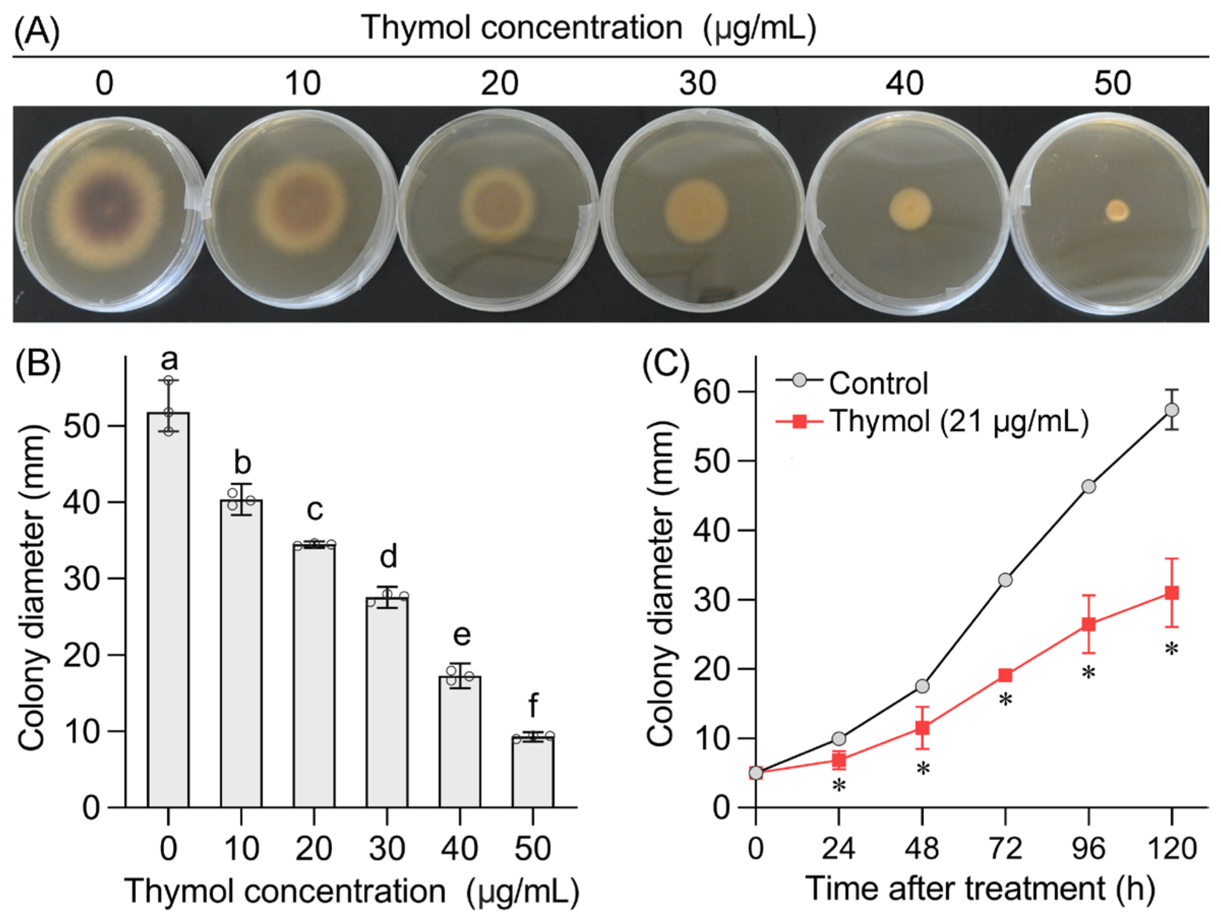
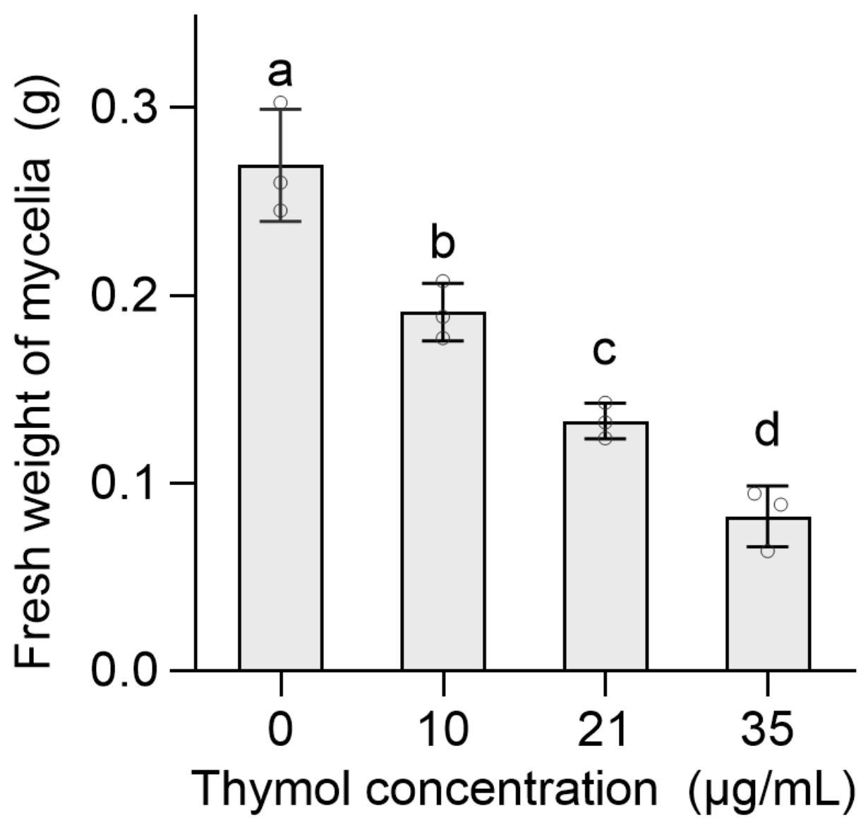
3.1. Thymol Inhibited Mycelial Growth of FON

FON grew on the PDA plate containing thymol at different concentrations for 120 h for the evaluation of mycelial growth. Thymol at 10–50 µg/mL inhibited mycelial growth in a dose-dependent manner (Figure 1A,B). Thymol at 50 µg/mL almost completely inhibited the growth of FON. The EC50 of thymol against mycelial growth was 21 µg/mL based on calculated linear regression equation (Y = 3.181X + 0.778). Then, thymol at 21 µg/mL was used to study the growth of FON in a time-course experiment. Thymol at 21 µg/mL began to significantly inhibit mycelial growth after treatment for 12 h, leading to a decreased growth speed with a prolonged treatment time (Figure 1C).
Thymol at 10, 21, and 35 µg/mL was selected as low, medium, and high concentration, respectively, to perform the following experiments. Compared to the control group, the fresh weight of FON mycelia remarkably decreased by 29.0%, 50.6%, and 69.5% upon the exposure of thymol at 10, 21, and 35 µg/mL, respectively (Figure 2).

3.2. Thymol Damaged Mycelial Structure of FON

Light microscopic observation indicated that thymol-treated mycelia became thinner than that of the control. More vacuole-like structures appeared in mycelial cells upon thymol treatment. Thymol at high concentrations (35 µg/mL) even broke the mycelia (Figure 3A, Figure S1). Then, SEM was used to further observe morphological changes of mycelia in detail. Compared to smooth mycelia in the control group, thymol induced wrinkles and collapse in mycelia. A high dose of thymol resulted in irregular damage and loss of shape of mycelia (Figure 3B, Figure S2).

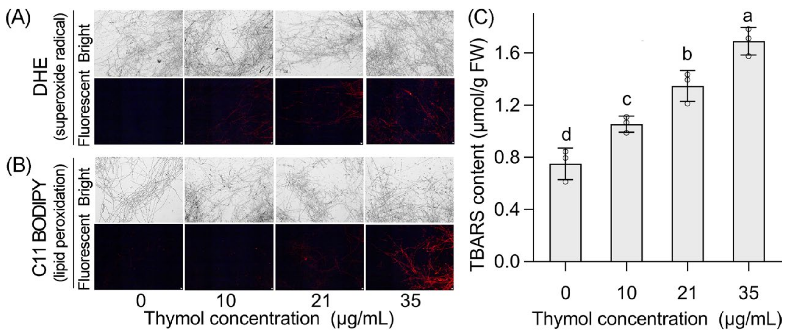
3.3. Thymol Induced Cell Death and Oxidative Injury in the Mycelia of FON

A specific probe PI was used to label dead cells in the mycelia of FON. PI can only cross a damaged cell membrane, entering into dead cells to emit red fluorescence. The mycelia showed extensive red fluorescence with the increase in thymol concentration (Figure 4), suggesting that thymol induced mycelial cell death.
Then, a specific fluorescent probe DHE was used to label mycelial superoxide radical, one the important ROS (reactive oxygen species). The mycelia accumulated more superoxide radicals with the increase in thymol concentration (Figure 5A). Accumulated ROS can cause an oxidative injury by attacking the membrane lipid, leading to lipid peroxidation. We used a specific fluorescent probe C11 BODIPY to detect mycelial lipid peroxidation in vivo. Thymol treatment induced extensive fluorescence of C11 BODIPY in a dose-dependent manner (Figure 5B). The content of TBARS is a typic indicator of cell membrane lipid peroxidation. Mycelial TBARS content significantly increased by 40.5%, 79.6%, and 125.4% upon thymol treatment at 10, 21, and 35 µg/mL, respectively (Figure 5C).
Accumulated ROS can activate antioxidative enzymes in cells. The activities of two typical antioxidative enzymes, SOD (superoxide dismutase), and CAT (catalase) were measured. SOD and CAT activity in mycelia increased significantly upon thymol at 10 and 21 µg/mL. A high concentration of thymol resulted in the decrease in the activity of these two enzymes (Figure 6A). These results suggested that thymol-triggered ROS accumulation activated antioxidative enzymes in the mycelia of FON.

3.4. Thymol Induced Electrolyte Leakage and Osmotic Stress in the Mycelia of FON

Thymol induced memebrane lipid peroxidation, suggesting possible membrane damage of mycelial cells. This may lead to the electrolyte leakage that can be indicated by the increase in conductivity. As expected, thymol treatment led to a significant increase in relative conductivity in mycelial in a dose-dependent manner (Figure 7A). In addition, thymol treatment led to the increase in glycerol content in mycelia (Figure 7B), suggesting osmotic stress occurred in thymol-treated mycelia.

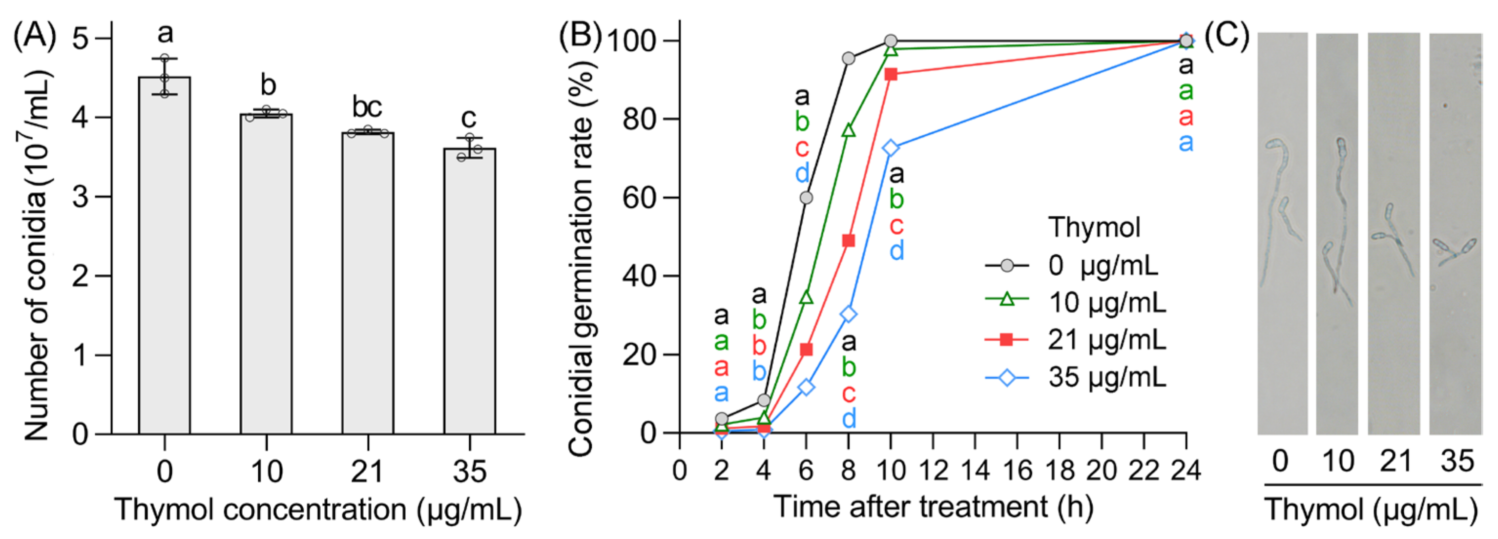
3.5. Thymol Inhibited Conidial Production and Conidial Germination of FON

Thymol inhibited conidial production in a dose-dependent manner. The number of conidia significantly decreased by 10.3%, 15.5%, and 19.9% upon thymol at 10, 21, and 35 µg/mL, respectively (Figure 8A). The effect of thymol on conidial germination was determined as well. In the control group without thymol, all the tested conidia germinated completely at 10 h. Thymol treatment resulted in a significant decrease in the conidial germination rate at 2–10 h as compared to control (Figure 8B). The conidia in all treatments finally completed germination at 24 h, but thymol delayed this process (Figure 8B). Thymol-treated conidia showed much shorter mycelia than that of the control after treatment for 6 h (Figure 8B).

3.6. Thymol Induced Oxidative Injury and Cell Death in the Conidia of FON

DHE, C11 BODIPY, and PI were used to indicate superoxide radical, lipid peroxidation, and cell death in conidia, respectively. All the conidia treated with thymol showed extensive fluorescence as compared to the control group, suggesting that thymol induced oxidative injury and cell death in the conidia of FON (Figure 9).

4. Discussion

Seeking environmental-friendly chemicals against FON is one of the probable approaches controlling watermelon Fusarium wilt diseases [43,44]. In the present study, we found that thymol showed effective inhibitory activity against FON in vitro. Thymol treatment resulted in the loss of integrity of mycelial structure, leading to the inhibition of FON growth. Thymol was able to damage both mycelia and conidia of FON by inducing oxidative injury and cell death. Using ROS as an antimicrobial mechanism is an efficient approach for the development of novel fungicide [45]. This is also applicable for identifying the activity of essential oils against phytopathogenic fungi [46]. Thymol induced ROS accumulation in the mycelia of FON. This may lead to membrane disruption supported by the observation of the increase in electrolyte leakage and lipid peroxidation in mycelia.
The intracellular ROS in fungi includes superoxide radicals, hydrogen sulfide, singlet oxygen, hydroxyl radical, etc. Excessive ROS can react readily with intracellular macromolecules, leading to cell dysfunction and significant damage to cell structure [47]. In Aspergillus fumigatus and Aspergillus flavus, thymol was able to induce the accumulation of total ROS detected with fluorescent probe DCFH-DA [48,49]. In this study, we found that thymol induced remarkable accumulation of superoxide radical (detected specifically with probe DHE) in FON. Superoxide radical is frequently the first ROS to be generated in cells upon stimuli because it needs only one electron transferred to oxygen [50]. These data suggest that the inhibitory effect of thymol on FON growth is closely related to the accumulation of ROS, especially for superoxide radical generated.
Two endogenous sources of superoxide radical production exist in fungal cells. In mitochondrion, ETC (electron transport chain) complex I and III are the major sites for the production of superoxide radical during the reduction of oxygen. Stress conditions can disturb the ETC, thereby intensifying the production of superoxide radicals [51]. Another site is NADPH (nicotinamide adenine dinucleotide phosphate) oxidase (NOX) located in the plasma membrane. NOX is a flavoenzyme complex with the ability of catalyzing the reduction of oxygen to produce superoxide radical by transferring electrons from NADPH across the membrane [52]. Stress conditions can induce cell oxidative stress by activating NOX-dependent superoxide radicals diffusing into cytosol [53,54]. It has been reported that thymol results in mitochondrial dysfunction and ROS production in non-small lung cancer cells [55]. Further studies are needed to elucidate whether thymol induces the production of superoxide radicals in FON through the above mechanisms.
In fungal cells, accumulated ROS can activate antioxidative enzymes. SOD can catalyze superoxide radicals to form hydrogen peroxide that can be further transformed to water by CAT. Therefore, ROS accumulation always accompanies the increase in the activity of antioxidative enzymes, which can be considered as typically oxidative responses [51]. Thymol induced ROS accumulation and the increase in the activity of SOD and CAT in mycelia, accompanied the occurrence of lipid peroxidation. These results suggested that thymol induced an oxidative stress response in FON. We observed the increase in the activity of SOD and CAT in response to thymol at a low to medium concentration (10–21 µg/mL). Thymol at a high concentration (35 µg/mL) repressed their activity. The possible reason was that thymol at high doses induced severe toxicity, resulting in protein degradation in collapsed mycelial cells.
The stimuli frequently induced fungal osmotic stress that is indicated by the HOG (high osmolarity glycerol) pathway-dependent glycerol generation [56]. The regulation of the HOG pathway has been associated with the antifungal activity of thymol against human pathogenic fungus Cryptococcus neoformans [57]. In this study, we found that thymol treatment resulted in the accumulation of glycerol in FON mycelia, suggesting the probable occurrence of osmotic stress. In fungi, oxidative stress can regulate osmotic stress through the modification of HOG signaling components (e.g., Hog1) by ROS [51]. The HOG pathway can also modulate oxidative stress in fungi. The key components of the HOG pathway (Sln1-Pbs2-Hog1) are involved in the regulation of the expression of SOD2 in Candida albicans upon fungicide (berberine hydrochloride) exposure [58]. Thymol induced both oxidative stress and osmotic responses in FON. Further identifying the interaction between them would help understand thymol-inhibited growth of FON.
Conidium, an asexual type of spore in fungi, facilitates the rapid asexual development of fungi [59]. Phenotyping of spore germination is a useful approach to screen and discover specific fungicides [60]. Thymol effectively repressed the conidial activity and germination of FON, suggesting the potential of using thymol as fungicide against FON. The development of fungal spores needs endogenous ROS at a moderate level [61], but excessive ROS-induced oxidative injury is frequently associated with fungicide-inhibited spore germination [62,63]. The inactivation of the conidia of FON may result from the ROS accumulation and oxidative damage. ROS can induce secondary necrosis in the conidia of Aspergillus fumigatus [64]. Thymol treatment resulted in the accumulation of superoxide radical and cell death in the conidia of FON, suggesting that thymol-induced superoxide radicals may trigger cell death. It is important to control fungal pathogens by targeting intrinsic cell death pathways [65]. It has been reported that thymol is able to induce conidial apoptosis in Aspergillus flavus [66]. Further studies could focus on different cell death pathways induced by thymol, which may help illustrate the mechanism for the inhibitory activity of thymol against FON in vitro.
In conclusion, thymol (a natural monoterpene phenol) showed effective inhibitory activity against phytopathogen FON in vitro. Thymol inhibited mycelial growth, conidial production, and conidial germination in FON. All of these processes involved the accumulation of endogenous superoxide radicals and intracellular oxidative injury. The detailed mechanism needs to be investigated further, but our current results reveal the action of thymol against FON. This would be applicable for helping potential candidates to develop environmental-friendly pesticide for the control of Fusarium wilt disease on watermelon.

Supplementary Materials

The following supporting information can be downloaded at: https://www.mdpi.com/article/10.3390/agronomy13010189/s1, Figure S1: Light microscopic observation of FON mycelia upon thymol treatment; Figure S2: Scanning electron microscopic (SEM) observation of FON mycelia upon thymol treatment.

Author Contributions

Conceptualization, L.Y. and S.H.; methodology, Y.H. and J.C.; investigation, Y.H., J.Z., C.S., X.C., Y.W., H.L., J.C. and L.Z.; writing—original draft preparation, Y.H. and J.Z.; writing—review and editing, L.Y., S.H., Z.S. and J.C. All authors have read and agreed to the published version of the manuscript.

Funding

This work was supported by “Jiangsu Agricultural Science and Technology Innovation Fund, grant number CX(20)1011” and “China Agriculture Research System, grant number CARS-23-B16”.

Data Availability Statement

No applicable.

Acknowledgments

We would like to thank Huimin Yang for helping histochemical analysis.

Conflicts of Interest

The authors declare no conflict of interest.

References

  1. Gordon, T.R. Fusarium oxysporum and the Fusarium wilt syndrome. Annu. Rev. Phytopathol. 2017, 55, 23–29. [Google Scholar] [CrossRef] [PubMed]
  2. Srinivas, C.; Nirmala, D.D.; Narasimha, M.K.; Mohan, C.D.; Lakshmeesha, T.R.; Singh, B.; Kalagatur, N.K.; Niranjana, S.R.; Hashem, A.; Alqarawi, A.A.; et al. Fusarium oxysporum f. sp. lycopersici causal agent of vascular wilt disease of tomato: Biology to diversity—A review. Saudi J. Biol. Sci. 2019, 26, 1315–1324. [Google Scholar] [CrossRef] [PubMed]
  3. Dong, Z.; Hsiang, T.; Luo, M.; Xiang, M. Draft genome sequence of an isolate of Fusarium oxysporum f. sp. melongenae, the causal agent of Fusarium Wilt of eggplant. Genome Announc. 2017, 5, e01597-16. [Google Scholar] [CrossRef] [PubMed] [Green Version]
  4. Lin, Y.; Zou, W.; Lin, S.; Onofua, D.; Yang, Z.; Chen, H.; Wang, S.; Chen, X. Transcriptome profiling and digital gene expression analysis of sweet potato for the identification of putative genes involved in the defense response against Fusarium oxysporum f. sp. batatas. PLoS ONE 2017, 12, e0187838. [Google Scholar] [CrossRef] [PubMed] [Green Version]
  5. Fulton, J.C.; Amaradasa, B.S.; Ertek, T.S.; Iriarte, F.B.; Sanchez, T.; Ji, P.; Paret, M.L.; Hudson, O.; Ali, M.E.; Dufault, N.S. Phylogenetic and phenotypic characterization of Fusarium oxysporum f. sp. niveum isolates from Florida-grown watermelon. PLoS ONE 2021, 16, e0248364. [Google Scholar] [CrossRef]
  6. Rebouças, T.A.; de Jesus Rocha, A.; Cerqueira, T.S.; Adorno, P.R.; Barreto, R.Q.; dos Santos Ferreira, M.; Lino, L.S.M.; de Oliveira Amorim, V.B.; dos Santos-Serejo, J.A.; Haddad, F.; et al. Pre-selection of banana somaclones resistant to Fusarium oxysporum f. sp. cubense, subtropical race 4. Crop Prot. 2021, 147, 105692. [Google Scholar] [CrossRef]
  7. Joshi, R. A review of Fusarium oxysporum on its plant interaction and industrial use. Crit. Rev. Microbiol. 2018, 6, 112–115. [Google Scholar] [CrossRef]
  8. Martyn, R.D. Fusarium wilt of watermelon: 120 years of research. Hortic. Rev. 2014, 42, 349–442. [Google Scholar]
  9. Kleczewski, N.M.; Egel, D.S. A diagnostic guide for Fusarium wilt of watermelon. Plant Health Prog. 2011, 12, 27. [Google Scholar] [CrossRef] [Green Version]
  10. Petkar, A.; Ji, P. Infection courts in watermelon plants leading to seed infestation by Fusarium oxysporum f. sp. niveum. Phytopathology 2017, 107, 828–833. [Google Scholar] [CrossRef] [Green Version]
  11. Hudson, O.; Hudson, D.; Ji, P.; Ali, M.E. Draft genome sequences of three Fusarium oxysporum f. sp. niveum isolates used in designing markers for race differentiation. Microbiol. Resour. Announc. 2020, 9, e01004-20. [Google Scholar] [CrossRef]
  12. Costa, A.E.D.S.; Cunha, F.S.D.; Araújo, K.M.G.; Lima Neto, I.S.; Capucho, A.S.; Borel, J.C.; Ishikawa, F.H. Morph-agronomic characterization of watermelon accessions with resistance to Fusarium Wilt. An. Acad. Bras. Cienc. 2021, 93, e20191359. [Google Scholar] [CrossRef]
  13. Hudson, O.; Fulton, J.C.; Dong, A.K.; Dufault, N.S.; Ali, M.E. Fusarium oxysporum f. sp. niveum molecular diagnostics past, present and future. Int. J. Mol. Sci. 2021, 22, 9735. [Google Scholar] [CrossRef]
  14. Bubici, G.; Kaushal, M.; Prigigallo, M.I.; Gómez-Lama Cabanás, C.; Mercado-Blanco, J. Biological control agents against Fusarium Wilt of banana. Front. Microbiol. 2019, 10, 616. [Google Scholar] [CrossRef] [Green Version]
  15. Niu, X.; Zhao, X.; Ling, K.S.; Levi, A.; Sun, Y.; Fan, M. The FonSIX6 gene acts as an avirulence effector in the Fusarium oxysporum f. sp. niveum-watermelon pathosystem. Sci. Rep. 2016, 6, 28146. [Google Scholar] [CrossRef] [Green Version]
  16. Keinath, A.P.; Hassell, R.L. Suppression of Fusarium Wilt caused by Fusarium oxysporum f. sp. niveum Race 2 on grafted triploid watermelon. Plant Dis. 2014, 98, 1326–1332. [Google Scholar] [CrossRef] [Green Version]
  17. Cháves-Gómez, J.L.; Becerra-Mutis, L.M.; Chávez-Arias, C.C.; Restrepo-Díaz, H.; Gómez-Caro, S. Screening of different Physalis genotypes as potential rootstocks or parents against vascular wilt using physiological markers. Front. Plant Sci. 2020, 11, 806. [Google Scholar] [CrossRef]
  18. An, M.; Zhou, X.; Wu, F.; Ma, Y.; Yang, P. Rhizosphere soil microorganism populations and community structures of different watermelon cultivars with differing resistance to Fusarium oxysporum f. sp. niveum. Can. J. Microbiol. 2011, 57, 355–365. [Google Scholar] [CrossRef]
  19. Zhou, X.; Li, C.; Liu, L.; Zhao, J.; Zhang, J.; Cai, Z.; Huang, X. Control of Fusarium wilt of lisianthus by reassembling the microbial community in infested soil through reductive soil disinfestation. Microbiol. Res. 2019, 220, 1–11. [Google Scholar] [CrossRef]
  20. Lv, H.; Cao, H.; Nawaz, M.A.; Sohail, H.; Huang, Y.; Cheng, F.; Kong, Q.; Bie, Z. Wheat intercropping enhances the resistance of watermelon to Fusarium Wilt. Front. Plant Sci. 2018, 9, 696. [Google Scholar] [CrossRef] [Green Version]
  21. Elmer, W.H.; Zuverza-Mena, N.; Triplett, L.R.; Roberts, E.L.; Silady, R.A.; White, J.C. Foliar application of copper oxide nanoparticles suppresses Fusarium wilt development on chrysanthemum. Environ. Sci. Technol. 2021, 55, 10805–10810. [Google Scholar] [CrossRef] [PubMed]
  22. Acosta-González, U.; Silva-Rojas, H.V.; Fuentes-Aragón, D.; Hernández-Castrejón, J.; Romero-Bautista, A.; Rebollar-Alviter, A. Comparative performance of fungicides and biocontrol products in the management of Fusarium Wilt of blackberry. Plant Dis. 2022, 106, 1419–1427. [Google Scholar] [CrossRef] [PubMed]
  23. Hudson, O.; Waliullah, S.; Ji, P.; Ali, M.E. Molecular characterization of laboratory mutants of Fusarium oxysporum f. sp. niveum resistant to prothioconazole, a demethylation inhibitor (DMI) fungicide. J. Fungi 2021, 7, 704. [Google Scholar] [CrossRef] [PubMed]
  24. Raza, W.; Ling, N.; Zhang, R.; Huang, Q.; Xu, Y.; Shen, Q. Success evaluation of the biological control of Fusarium wilts of cucumber, banana, and tomato since 2000 and future research strategies. Crit. Rev. Biotechnol. 2017, 37, 202–212. [Google Scholar] [CrossRef] [PubMed]
  25. Wang, H.; Cai, X.Y.; Xu, M.; Tian, F. Enhanced biocontrol of cucumber Fusarium wilt by combined application of new antagonistic Bacteria Bacillus amyloliquefaciens B2 and phenolic acid-degrading fungus Pleurotus ostreatus P5. Front. Microbiol. 2021, 12, 700142. [Google Scholar] [CrossRef]
  26. Luo, W.; Liu, L.; Qi, G.; Yang, F.; Shi, X.; Zhao, X. Embedding Bacillus velezensis NH-1 in microcapsules for biocontrol of cucumber Fusarium Wilt. Appl. Environ. Microbiol. 2019, 85, e03128-18. [Google Scholar] [CrossRef] [Green Version]
  27. Zhai, Y.; Zhu, J.X.; Tan, T.M.; Xu, J.P.; Shen, A.R.; Yang, X.B.; Li, J.L.; Zeng, L.B.; Wei, L. Isolation and characterization of antagonistic Paenibacillus polymyxa HX-140 and its biocontrol potential against Fusarium wilt of cucumber seedlings. BMC Microbiol. 2021, 21, 75. [Google Scholar] [CrossRef]
  28. Khalil, M.M.R.; Fierro-Coronado, R.A.; Peñuelas-Rubio, O.; Villa-Lerma, A.G.; Plascencia-Jatomea, R.; Félix-Gastélum, R.; Maldonado-Mendoza, I.E. Rhizospheric bacteria as potential biocontrol agents against Fusarium wilt and crown and root rot diseases in tomato. Saudi J. Biol. Sci. 2021, 28, 7460–7471. [Google Scholar] [CrossRef]
  29. Marchese, A.; Orhan, I.E.; Daglia, M.; Barbieri, R.; Lorenzo, A.D.; Nabavi, S.F.; Gortzi, O.; Izadi, M.; Nabavi, S.M. Antibacterial and antifungal activities of thymol: A brief review of the literature. Food Chem. 2016, 210, 402–414. [Google Scholar] [CrossRef]
  30. Ahmad, A.; Vuuren, S.V.; Viljoen, A. Unravelling the complex antimicrobial interactions of essential oils—The case of Thymus vulgaris (thyme). Molecules 2014, 19, 2896–2910. [Google Scholar] [CrossRef] [Green Version]
  31. Palaniappan, K.; Holley, R.A. Use of natural antimicrobials to increase antibiotic susceptibility of drug resistant bacteria. Int. J. Food Microbiol. 2010, 140, 164–168. [Google Scholar] [CrossRef]
  32. Eloy, M.A.; Ribeiro, R.; Meireles, L.M.; Cutrim, T.A.S.; Francisco, C.S.; Javarini, C.L.; Borges, W.S.; Costa, A.V.; Queiroz, V.T.; Scherer, R.; et al. Thymol as an interesting building block for promising fungicides against Fusarium solani. J. Agric. Food Chem. 2021, 69, 6958–6967. [Google Scholar] [CrossRef]
  33. Gao, T.; Zhou, H.; Zhou, W.; Hu, L.B.; Chen, J.; Shi, Z.Q. The fungicidal activity of thymol against Fusarium graminearum via inducing lipid peroxidation and disrupting ergosterol biosynthesis. Molecules 2016, 21, 770. [Google Scholar] [CrossRef] [Green Version]
  34. Thomulka, K.W.; Abbas, C.G.; Young, D.A.; Lange, J.H. Evaluating median effective concentrations of chemicals with bioluminescent bacteria. Bull. Environ. Contam. Toxicol. 1996, 56, 446–452. [Google Scholar] [CrossRef]
  35. Yu, J.; Wang, W.N.; Matei, N.; Li, X.; Pang, J.W.; Mo, J.; Chen, S.P.; Tang, J.P.; Yan, M.; Zhang, J.H. Ezetimibe attenuates oxidative stress and neuroinflammation via the AMPK/Nrf2/TXNIP pathway after MCAO in rats. Oxid. Med. Cell. Longev. 2020, 2020, 4717258. [Google Scholar] [CrossRef]
  36. Domínguez-Rebolledo, A.E.; Martínez-Pastor, F.; Fernández-Santos, M.R.; del Olmo, E.; Bisbal, A.; Ros-Santaella, J.L.; Garde, J.J. Comparison of the TBARS assay and BODIPY C11 probes for assessing lipid peroxidation in red deer spermatozoa. Reprod. Domest. Anim. 2010, 45, e362. [Google Scholar]
  37. Kellermeier, F.; Chardon, F.; Amtmann, A. Natural variation of Arabidopsis root architecture reveals complementing adaptive strategies to potassium starvation. Plant. Physiol. 2013, 161, 1421–1432. [Google Scholar] [CrossRef] [Green Version]
  38. Duan, Y.; Ge, C.; Liu, S.; Chen, C.; Zhou, M. Effect of phenylpyrrole fungicide fludioxonil on morphological and physiological characteristics of Sclerotinia sclerotiorum. Pestic. Biochem. Physiol. 2013, 106, 61–67. [Google Scholar] [CrossRef]
  39. Yang, Y.; Fan, F.; Zhuo, R.; Ma, F.; Gong, Y.; Wan, X.; Jiang, M.; Zhang, X. Expression of the laccase gene from a white rot fungus in Pichia pastoris can enhance the resistance of this yeast to H2O2-mediated oxidative stress by stimulating the glutathione-based antioxidative system. Appl. Environ. Microbiol. 2012, 78, 5845–5854. [Google Scholar] [CrossRef] [Green Version]
  40. McCord, J.M.; Fridovich, I. Superoxide dismutase an enzymic function for erythrocuprein(hemocuprein). J. Biol. Chem. 1969, 244, 6049–6055. [Google Scholar] [CrossRef]
  41. Gao, C.; Fei, X.; Wang, M.; Chen, Q.; Zhao, N. Cardamomin protects from diabetes-induced kidney damage through modulating PI3K/AKT and JAK/STAT signaling pathways in rats. Int. Immunopharmacol. 2022, 107, 108610. [Google Scholar] [CrossRef] [PubMed]
  42. Bradford, M.M. A rapid and sensitive method for the quantitation of microgram quantities of protein utilizing the principle of protein-dye binding. Anal. Biochem. 1976, 72, 248–254. [Google Scholar] [CrossRef] [PubMed]
  43. Huang, J.; Wang, D.; Li, H.; Tang, Y.Q.; Ma, X.; Tang, H.Q.; Lin, M.; Liu, Z. Antifungal activity of an artificial peptide aptamer SNP-D4 against Fusarium oxysporum. Peer J. 2022, 10, e12756. [Google Scholar] [CrossRef] [PubMed]
  44. Du, R.; Liu, J.; Sun, P.; Li, H.; Wang, J. Inhibitory effect and mechanism of Tagetes erecta L. fungicide on Fusarium oxysporum f. sp. niveum. Sci. Rep. 2017, 7, 14442. [Google Scholar] [CrossRef] [PubMed] [Green Version]
  45. Dryden, M. Reactive oxygen species: A novel antimicrobial. Int. J. Antimicrob. Agents 2018, 51, 299–303. [Google Scholar] [CrossRef]
  46. Lee, J.E.; Seo, S.M.; Huh, M.J.; Lee, S.C.; Park, I.K. Reactive oxygen species mediated-antifungal activity of cinnamon bark (Cinnamomum verum) and lemongrass (Cymbopogon citratus) essential oils and their constituents against two phytopathogenic fungi. Pestic. Biochem. Physiol. 2020, 168, 104644. [Google Scholar] [CrossRef]
  47. Segal, L.M.; Wilson, R.A. Reactive oxygen species metabolism and plant-fungal interactions. Fungal Genet. Biol. 2018, 110, 1–9. [Google Scholar] [CrossRef] [Green Version]
  48. Oiki, S.; Nasuno, R.; Urayama, S.I.; Takagi, H.; Hagiwara, D. Intracellular production of reactive oxygen species and a DAF-FM-related compound in Aspergillus fumigatus in response to antifungal agent exposure. Sci. Rep. 2022, 12, 13516. [Google Scholar] [CrossRef]
  49. Shen, Q.; Zhou, W.; Li, H.; Hu, L.; Mo, H. ROS involves the fungicidal actions of thymol against spores of Aspergillus flavus via the induction of nitric oxide. PLoS ONE 2016, 11, e0155647. [Google Scholar] [CrossRef] [Green Version]
  50. Larosa, V.; Remacle, C. Insights into the respiratory chain and oxidative stress. Biosci. Rep. 2018, 38, BSR20171492. [Google Scholar] [CrossRef] [Green Version]
  51. Yaakoub, H.; Mina, S.; Calenda, A.; Bouchara, J.P.; Papon, N. Oxidative stress response pathways in fungi. Cell Mol. Life Sci. 2022, 79, 333. [Google Scholar] [CrossRef]
  52. Dirschnabel, D.E.; Nowrousian, M.; Cano-Domínguez, N.; Aguirre, J.; Teichert, I.; Kück, U. New insights into the roles of NADPH oxidases in sexual development and ascospore germination in Sordaria macrospora. Genetics 2014, 196, 729–744. [Google Scholar] [CrossRef]
  53. Derr, A.M.; Faustoferri, R.C.; Betzenhauser, M.J.; Gonzalez, K.; Marquis, R.E.; Quivey, R.G., Jr. Mutation of the NADH oxidase gene (nox) reveals an overlap of the oxygen- and acid-mediated stress responses in Streptococcus mutans. Appl. Environ. Microbiol. 2012, 78, 1215–1227. [Google Scholar] [CrossRef] [Green Version]
  54. Liu, R.; Zhang, X.; Ren, A.; Shi, D.K.; Shi, L.; Zhu, J.; Yu, H.S.; Zhao, M.W. Heat stress-induced reactive oxygen species participate in the regulation of HSP expression, hyphal branching and ganoderic acid biosynthesis in Ganoderma lucidum. Microbiol. Res. 2018, 209, 43–54. [Google Scholar] [CrossRef]
  55. Balan, D.J.; Rajavel, T.; Das, M.; Sathya, S.; Jeyakumar, M.; Devi, K.P. Thymol induces mitochondrial pathway-mediated apoptosis via ROS generation, macromolecular damage and SOD diminution in A549 cells. Pharmacol. Rep. 2021, 73, 240–254. [Google Scholar] [CrossRef]
  56. Yaakoub, H.; Sanchez, N.S.; Ongay-Larios, L.; Courdavault, V.; Calenda, A.; Bouchara, J.P.; Coria, R.; Papon, N. The high osmolarity glycerol (HOG) pathway in fungi†. Crit. Rev. Microbiol. 2022, 8, 657–695. [Google Scholar] [CrossRef]
  57. Jung, K.W.; Chung, M.S.; Bai, H.W.; Chung, B.Y.; Lee, S. Investigation of antifungal mechanisms of thymol in the human fungal pathogen, Cryptococcus neoformans. Molecules 2021, 26, 3476. [Google Scholar] [CrossRef]
  58. Huang, X.; Yi, Y.; Yong, J.; Sun, J.; Song, Z.; Li, D.; Li, Y. Inhibitory effect of berberine hydrochloride against Candida albicans and the role of the HOG-MAPK pathway. J. Antibiot. 2021, 74, 807–816. [Google Scholar] [CrossRef]
  59. Sephton-Clark, P.C.S.; Voelz, K. Spore germination of pathogenic filamentous fungi. Adv. Appl. Microbiol. 2018, 102, 117–157. [Google Scholar]
  60. Ortiz, S.C.; Huang, M.; Hull, C.M. Discovery of fungus-specific targets and inhibitors using chemical phenotyping of pathogenic spore germination. mBio. 2021, 12, e0167221. [Google Scholar] [CrossRef]
  61. Tudzynski, P.; Heller, J.; Siegmund, U. Reactive oxygen species generation in fungal development and pathogenesis. Curr. Opin. Microbiol. 2012, 15, 653–659. [Google Scholar] [CrossRef] [PubMed]
  62. Dou, S.; Liu, S.; Xu, X.; OuYang, Q.; Tao, N. Octanal inhibits spore germination of Penicillium digitatum involving membrane peroxidation. Protoplasma 2017, 254, 1539–1545. [Google Scholar] [CrossRef] [PubMed]
  63. Lai, T.; Li, B.; Qin, G.; Tian, S. Oxidative damage involves in the inhibitory effect of nitric oxide on spore germination of Penicillium expansum. Curr. Microbiol. 2011, 62, 229–234. [Google Scholar] [CrossRef] [PubMed]
  64. Oliveira, M.; Pereira, C.; Bessa, C.; Araujo, R.; Saraiva, L. Hydrogen peroxide-induced secondary necrosis in conidia of Aspergillus fumigatus. Can. J. Microbiol. 2016, 62, 95–101. [Google Scholar] [CrossRef] [Green Version]
  65. Kulkarni, M.; Stolp, Z.D.; Hardwick, J.M. Targeting intrinsic cell death pathways to control fungal pathogens. Biochem. Pharmacol. 2019, 162, 71–78. [Google Scholar] [CrossRef]
  66. Hu, L.B.; Ban, F.F.; Li, H.B.; Qian, P.P.; Shen, Q.S.; Zhao, Y.Y.; Mo, H.Z.; Zhou, X. Thymol induces conidial apoptosis in Aspergillus flavus via stimulating K+ eruption. J. Agric. Food Chem. 2018, 66, 8530–8536. [Google Scholar] [CrossRef]
Figure 1. Thymol inhibited radial growth of FON. (A) Phenotype of mycelial growth on PDA plate under thymol treatment for 120 h. (B) Colony diameter of FON under thymol treatment for 120 h. (C) Time-course measurement of FON colony diameter under thymol (21 µg/mL). Bars indicated SD of three replicates. Different lowercase letters in (B) indicated significant difference among different treatments (LSD, n = 3, p < 0.05). Asterisk in (C) indicated significant difference between control and thymol treatment at each time point (ANOVA, n = 3, p < 0.05).
Agronomy 13 00189 g001
Figure 2. Effect of thymol on the fresh weight of mycelia of FON. The FON was cultured in PDB medium containing thymol (0, 10, 21, and 35 µg/mL) for 120 h, followed by the measurement of mycelial fresh weight. Bars indicated SD of three replicates. Different lowercase letters indicated significant difference among different treatments (LSD, n = 3, p < 0.05).
Agronomy 13 00189 g002
Figure 3. Thymol induced morphological changes of FON mycelia. (A) Light microscopic observation of mycelia upon thymol treatment for 24 h. (B) SEM observation of mycelia upon thymol treatment for 24 h. Left panel and right penal indicated the magnification of 500× and 2000×, respectively.
Agronomy 13 00189 g003
Figure 4. PI-indicated cell death in the mycelia of FON upon thymol treatment. The mycelia were treatment with thymol (0, 10, 21, and 35 µg/mL) for 24 h, followed by incubation with PI and fluorescent observation.
Agronomy 13 00189 g004
Figure 5. Thymol induced oxidative injury in the mycelia of FON. The mycelia were treated with thymol (0, 10, 21, and 35 µg/mL) for 24 h. Then the mycelia were collected for (A) DHE-based detection of superoxide radical, (B) C11 BODIPY-based detection of lipid peroxidation, and (C) measurement of TRARS content. Bars in (C) indicated SD of three replicates. Different lowercase letters in (C) indicated significant difference among different treatments (LSD, n = 3, p < 0.05).
Agronomy 13 00189 g005
Figure 6. Effect of thymol on the activity of antioxidative enzymes in the mycelia of FON. The mycelia were treated with thymol (0, 10, 21, and 35 µg/mL) for 24 h. Then the mycelia were collected for the assay of (A) SOD activity and (B) CAT activity. Bars indicated SD of three replicates. Different lowercase letters indicated significant difference among different treatments (LSD, n = 3, p < 0.05).
Agronomy 13 00189 g006
Figure 7. Thymol induced increase in relative conductivity and glycerol content in the mycelia of FON. (A) The relative conductivity of mycelia under thymol treatment (0, 10, 21, and 35 µg/mL) was measured in a time-course process. (B) The mycelia were treated with thymol (0, 10, 21, and 35 µg/mL) for 24 h, followed by the determination of glycerol content. Bars indicated SD of three replicates. Different lowercase letters in (B) indicated significant difference among different treatments (LSD, n = 3, p < 0.05).
Agronomy 13 00189 g007
Figure 8. The effect of thymol on conidia production and conidial germination of FON. (A) The mycelia were cultured in Bilay’s medium containing thymol (0, 10, 21, and 35 µg/mL) to produce conidia for 24 h, followed by counting the number of produced conidia. Different lowercase letters indicated significant difference among different treatments (ANOVA, n = 3, p < 0.05). (B) The conidia were allowed to be germinated under thymol (0, 10, 21, and 35 µg/mL) treatment. The germinated conidia were counted at specific time points, respectively. Different lowercase letters indicated significant difference among different treatments at each time point (ANOVA, n = 3, p < 0.05). For each time point, the colors of different lowercase letters correspond to the colors of different legends indicating different treatments. (C) Microscopic observation of conidial germination after thymol treatment (0, 10, 21, and 35 µg/mL) for 6 h. Bars indicated SD of three replicates.
Agronomy 13 00189 g008
Figure 9. The effect of thymol on superoxide radical, lipid peroxidation, and cell death in the conidia of FON. The conidia were treated with thymol (0, 10, 21, and 35 µg/mL) for 24 h. Then the conidia were collected for (A) DHE-based detection of superoxide radical, (B) C11 BODIPY-based detection of lipid peroxidation, and (C) PI-based detection of cell death.
Agronomy 13 00189 g009
Disclaimer/Publisher’s Note: The statements, opinions and data contained in all publications are solely those of the individual author(s) and contributor(s) and not of MDPI and/or the editor(s). MDPI and/or the editor(s) disclaim responsibility for any injury to people or property resulting from any ideas, methods, instructions or products referred to in the content.

